jpet #208603 pharmacological inhibition of...
TRANSCRIPT
JPET #208603
1
PHARMACOLOGICAL INHIBITION OF THE RENAL OUTER MEDULLARY
POTASSIUM CHANNEL CAUSES DIURESIS AND NATRIURESIS
IN THE ABSENCE OF KALIURESIS
Maria L. Garcia, Birgit T. Priest, Magdalena Alonso-Galicia, Xiaoyan Zhou,
John P. Felix, Richard M. Brochu, Timothy Bailey, Brande Thomas-Fowlkes,
Jessica Liu, Andrew Swensen, Lee-Yuh Pai, Jianying Xiao, Melba Hernandez,
Kimberly Hoagland, Karen Owens, Haifeng Tang, Reynalda K. de Jesus,
Sophie Roy, Gregory J. Kaczorowski, and Alexander Pasternak
Departments of Ion Channels (M.L.G., B.T.P., J.P.F., R.M.B., T.B., B.T-F., J.L., A.S.,
G.J.K.), Hypertension (M.A.-G., X.Z., L.-Y.P., J.X., M.H., S.R.), Drug Metabolism
(K.O.), and Medicinal Chemistry (H.T., R. K.J., A.P.)
Merck Research Laboratories, PO Box 2000, Rahway, New Jersey
Safety and Exploratory Pharmacology (K.H.), Merck Research Laboratories
West Point, Pennsylvania
JPET Fast Forward. Published on October 18, 2013 as DOI:10.1124/jpet.113.208603
Copyright 2013 by the American Society for Pharmacology and Experimental Therapeutics.
This article has not been copyedited and formatted. The final version may differ from this version.JPET Fast Forward. Published on October 18, 2013 as DOI: 10.1124/jpet.113.208603
at ASPE
T Journals on June 27, 2018
jpet.aspetjournals.orgD
ownloaded from
JPET #208603
2
Running Title Page
ROMK inhibitors cause diuresis and natriuresis in the absence of kaliuresis
Corresponding author:
Alexander Pasternak PhD
Director, Chemistry
Kenilworth Discovery Chemistry
2000 Galloping Hill Road, K15-ME2
Kenilworth, NJ 07033
(908)-740-6847
Fax: (908)-740-2207
E-mail: [email protected]
Text pages:
Number of tables: 5
Number of figures: 7
Number of references: 26
Abstract words: 242
Introduction words: 735
Discussion words: 887
Abbreviations: ROMK, renal outer medullary potassium channel; TALH, thick
ascending loop of Henle; DCT, distal convoluted tubule; CCD, cortical collecting duct;
This article has not been copyedited and formatted. The final version may differ from this version.JPET Fast Forward. Published on October 18, 2013 as DOI: 10.1124/jpet.113.208603
at ASPE
T Journals on June 27, 2018
jpet.aspetjournals.orgD
ownloaded from

JPET #208603
3
Kir, inwardly rectifying potassium channel; DMSO, dimethyl sulfoxide; HCTZ,
hydrochlorothiazide; MEM, minimum essential medium; FBS, fetal bovine serum;
HBSS, Hank’s buffered saline solution; SD, Sprague-Dawley; PE, polyethylene; BW,
body weight; PEG, polyethylene glycol; PAH, para-aminohippurate, LC-MS/MS, liquid
chromatography-tandem mass spectrometry.
Recommended Section: Cardiovascular
This article has not been copyedited and formatted. The final version may differ from this version.JPET Fast Forward. Published on October 18, 2013 as DOI: 10.1124/jpet.113.208603
at ASPE
T Journals on June 27, 2018
jpet.aspetjournals.orgD
ownloaded from

JPET #208603
4
Abstract
The renal outer medullary potassium (ROMK) channel is located at the apical membrane
of epithelial cells lining the thick ascending loop of Henle and cortical collecting duct,
and plays an important role in kidney physiology by regulating salt re-absorption. Loss-
of-function mutations in the human ROMK channel are associated with antenatal type II
Bartter’s syndrome, an autosomal recessive life-threatening salt wasting disorder with
mild hypokalemia. Similar observations have been reported from studies with ROMK
knock out mice and rats. Importantly, heterozygous carriers of Kir1.1 mutations
associated with antenatal Bartter's syndrome have reduced blood pressure, and a
decreased risk of developing hypertension by age 60. Although selective ROMK
inhibitors would be expected to represent a new class of diuretics, this hypothesis has not
been pharmacologically tested. Compound A (5-(2-(4-(2-(4-(1H-tetrazol-1-
yl)phenyl)acetyl)piperazin-1-yl)ethyl)isobenzofuran-1(3H)-one), a potent ROMK
inhibitor with appropriate selectivity and characteristics for in vivo testing, has been
identified. Compound A accesses the channel through the cytoplasmic side and binds to
residues lining the pore within the transmembrane region below the selectivity filter. In
normotensive rats and dogs, acute oral administration of A caused concentration-
dependent diuresis and natriuresis that were comparable to hydrochlorothiazide. Unlike
hydrochlorothiazide, however, compound A did not cause any significant urinary
potassium losses or changes in plasma electrolyte levels. These data indicate that
pharmacological inhibition of ROMK has the potential for affording diuretic/natriuretic
efficacy similar to that of clinically used diuretics, but without the dose-limiting
hypokalemia associated with the use of loop and thiazide-like diuretics.
This article has not been copyedited and formatted. The final version may differ from this version.JPET Fast Forward. Published on October 18, 2013 as DOI: 10.1124/jpet.113.208603
at ASPE
T Journals on June 27, 2018
jpet.aspetjournals.orgD
ownloaded from

JPET #208603
5
Introduction
The kidneys play a critical role in the long-term regulation of blood pressure. In
humans, all identified mutations in genes that cause Mendelian forms of hypertension or
hypotension act in the kidney to alter net renal salt and water reabsorption (Lifton et al.,
2001). Several mechanisms involving ion channels, exchangers, and transporters, act in
an integrated manner along the nephron to regulate salt and water reabsorption. At the
thick ascending limb of Henle (TALH), ~30% of salt reabsorption occurs through the
luminal Na+/K+/2Cl- co-transporter, which is the target of furosemide, a loop diuretic
used clinically in the treatment of congestive heart failure. In the distal convoluted tubule
(DCT), the Na+/Cl- co-transporter is responsible for ~7% of salt reabsorption. This co-
transporter represents the clinical target of the thiazide-class of diuretics, used in the
treatment of hypertension. The final step in the regulation of salt reabsorption takes place
in the cortical collecting duct (CCD) through the amiloride-sensitive epithelial sodium
channel. Because of the tight coupling between sodium reabsorption and potassium
secretion at the CCD, the use of loop or thiazide diuretics is clinically associated with
hypokalemia, while amiloride intervention causes hyperkalemia.
Bartter’s syndrome, characterized by renal salt wasting and polyuria-associated
low blood pressure and hypokalemic alkalosis, is caused by recessive loss-of-function
mutations in any one of the four genes involved in salt reabsorption at the TALH: the
apical Na+/K+/2Cl- co-transporter, the basolateral Cl- (α or β subunits) channel, and the
renal outer medullary potassium (ROMK) channel, the product of the KCNJ1 gene,
present at the apical membrane (Palmer et al., 1997; Xu et al., 1997; Lu et al., 2002). The
This article has not been copyedited and formatted. The final version may differ from this version.JPET Fast Forward. Published on October 18, 2013 as DOI: 10.1124/jpet.113.208603
at ASPE
T Journals on June 27, 2018
jpet.aspetjournals.orgD
ownloaded from

JPET #208603
6
ROMK channel has attracted significant interest because of the finding that heterozygous
carriers of channel mutations associated with type II Bartter’s syndrome have reduced
blood pressure and a decreased risk of developing hypertension by age 60 (Ji et al.,
2008). Moreover, it is important to note that ROMK knock out mice and rats
recapitulate the phenotype of type II Bartter’s syndrome (Lu et al., 2002; Lorenz et al.,
2002; Zhou et al., 2013). These data suggest that ROMK represents a target for the
development of novel diuretics for the treatment of hypertension and/or heart failure.
ROMK is a member of the inwardly rectifying family of potassium (Kir) channels
(Nichols and Lopatin, 1997). The expression of ROMK (Kir1.1) appears to be almost
exclusively restricted to the apical membrane of the epithelial cells lining the TALH and
the CCD (Xu et al., 1997; Palmer et al., 1997; Lu et al., 2002). At the TALH, ROMK
participates in potassium recycling across the apical membrane that is critical for the
proper function of the furosemide-sensitive Na+/K+/2Cl- co-transporter because the K+
concentration in the luminal fluid is much lower than that of Na+ and Cl-. At the CCD,
ROMK provides a pathway for potassium secretion that is tightly coupled to sodium
reabsorption through the amiloride-sensitive epithelial sodium channel. Because of the
presence of ROMK channels at both TALH and CCD, selective inhibitors of this channel
would be predicted to provide equivalent or superior diuretic/natriuretic efficacy to loop
diuretics, such as furosemide, with the added potential benefit of attenuating the
hypokalemia associated with the use of loop diuretics or thiazides. Despite all of the
evidence supporting ROMK as a novel therapeutic target, the development of selective
channel inhibitors has only recently been attempted. In addition to the peptide tertiapin,
which blocks with high affinity the rat but not the human channel (Jin and Lu, 1998;
This article has not been copyedited and formatted. The final version may differ from this version.JPET Fast Forward. Published on October 18, 2013 as DOI: 10.1124/jpet.113.208603
at ASPE
T Journals on June 27, 2018
jpet.aspetjournals.orgD
ownloaded from

JPET #208603
7
Felix et al., 2006), two independent groups have reported the identification of small
molecule ROMK inhibitors (Lewis et al., 2009; Bhave et al., 2011; Tang et al., 2012).
In the present study, compound A, the discovery of which is described in a
separate study (Tang et al., 2013), 5-(2-(4-(2-(4-(1H-tetrazol-1-
yl)phenyl)acetyl)piperazin-1-yl)ethyl)isobenzofuran-1(3H)-one, was characterized. This
agent inhibits the rat and human Kir1.1 channels with high affinity, and displays good
selectivity across other ion channel super-families, as well as pharmacokinetic properties
suitable for in vivo testing. Acute oral dosing of A was shown to produce dose-
dependent diuresis and natriuresis in normotensive rats and dogs of similar magnitude to
that of hydrochlorothiazide, but with no significant kaliuresis, and no changes in plasma
electrolyte levels. Taken together, these data indicate that pharmacological inhibition of
ROMK provides diuretic/natriuretic efficacy similar to that of clinically used diuretics,
but with the potential benefit of reducing the hypokalemia associated with the use of loop
and thiazide class diuretics.
This article has not been copyedited and formatted. The final version may differ from this version.JPET Fast Forward. Published on October 18, 2013 as DOI: 10.1124/jpet.113.208603
at ASPE
T Journals on June 27, 2018
jpet.aspetjournals.orgD
ownloaded from
JPET #208603
8
Materials and Methods
Materials-. Compound A, 5-(2-(4-(2-(4-(1H-tetrazol-1-yl)phenyl)acetyl)piperazin-1-
yl)ethyl)isobenzofuran-1(3H)-one, was synthesized at Merck Research Laboratories,
Rahway, NJ, as described (Tang et al., 2013). FluxORTM thallium detection kit was
obtained from Life Technologies, Carlsbad, CA. Probenecid, anhydrous dimethyl
sulfoxide (DMSO), and ouabain were obtained from Sigma-Aldrich, St. Louis, MO.
86RbCl was obtained from Perkin Elmer, Inc., Waltham, MA. QuickChange II site-
directed mutagenesis kit was from Stratagene, La Jolla, CA. The pCI-neo vector and the
transfection reagents FUGENE® 6 and FUGENE® HD were from Promega, Madison,
WI. Hydrochlorothiazide (HCTZ) was purchased from MP Biomedicals, LLC, Solon,
OH. All other reagents were obtained from commercial sources and were of the highest
purity commercially available.
Cells- All tissue culture reagents were obtained from Life Technologies, Carlsbad, CA.
All Kir1.1 constructs represent the ROMK1 splice form of Kir1.1. HEK293 cell lines
stably transfected with human Kir1.1 (hKir1.1) or rat Kir1.1 (rKir1.1), CHO cell line
stably expressing hKir1.1 (CHO-hKir1.1), and MDCKII-Flp cell line stably expressing
rKir1.1 (MDCK-rKir1.1) were obtained as previously described (Felix et al., 2006; Felix
et al., 2012). A CHO cell line stably expressing hKir2.3 (CHO-hKir2.3) was constructed
by transfecting cells with hKir2.3-pCIneo expression plasmid using FUGENE6. Stable
pools of CHO cells expressing hKir2.3 were prepared by geneticin selection (1000
μg/ml), and analyzed for functional expression of hKir2.3 using a membrane potential,
fluorescence resonance energy transfer-based assay as described (Solly et al., 2008).
This article has not been copyedited and formatted. The final version may differ from this version.JPET Fast Forward. Published on October 18, 2013 as DOI: 10.1124/jpet.113.208603
at ASPE
T Journals on June 27, 2018
jpet.aspetjournals.orgD
ownloaded from
JPET #208603
9
Individual stable cell lines were generated by limiting dilution under continuous selection
with geneticin. HEK293 cells stably expressing either human Kir4.1 (HEK-hKir4.1) or
human Kir7.1 (HEK-hKir7.1) were prepared by transfecting cells with hKir4.1-phCMV1
or hKir7.1-phCMV1 expression plasmids using FUGENE HD. Stables pools of cells
were selected using 1000 μg/ml geneticin. Individual stable cell lines were generated by
limiting dilution under continuous selection with geneticin, and analyzed for functional
expression of Kir4.1 or Kir7.1 using a fluorescence-based thallium flux assay. HEK293
cells stably transfected with human Kir2.1 were obtained from EMD Millipore
Corporation, Billerica, MA. HEK293 cells were grown in minimum essential media
(MEM) Alpha medium, 10% fetal bovine serum (FBS), 500 µg/ml geneticin, 1x
penicillin/streptomycin/glutamine and 1x MEM non-essential amino acids. CHO cells
were grown in Iscove's Modified Dulbecco's Medium supplemented with HT Supplement
Solution with 10% heat inactivated FBS, 500 mg/mL geneticin, and 1%
penicillin/streptomycin. The MDCK-rKir1.1 cell line was grown in DMEM+Glutamax
supplemented with penicillin/streptomycin, 200 μg/ml hygromicin B, and 10% FBS. All
lines were maintained at 37ºC in a 10% CO2 atmosphere.
Kir1.1 Thallium Flux Assay- Permeation of thallium through open hKir1.1 channels was
determined as previously described (Felix et al., 2012). Briefly, HEK293 cells stably
transfected with hKir1.1 were plated using a Thermo Scientific Matrix WellMate®
(Thermo Scientific Inc., Waltham, MA) at approximately 20,000 cells/well on black-
wall, clear bottom, 384-well poly-D-lysine-coated plates (Becton Dickinson, Franklin
Lakes, NJ) in 50 µl growth medium, and incubated overnight (16 – 20 hours) at 37°C in a
This article has not been copyedited and formatted. The final version may differ from this version.JPET Fast Forward. Published on October 18, 2013 as DOI: 10.1124/jpet.113.208603
at ASPE
T Journals on June 27, 2018
jpet.aspetjournals.orgD
ownloaded from

JPET #208603
10
10% CO2 atmosphere. All liquid handling was done on a Thermo Scientific Matrix
PlateMate® 2X3. Cell growth medium was removed and cells were then incubated with
0.025 ml of a solution containing FluxOR dye loading reagent, prepared according to the
manufacturer instructions in Hank's Buffered Saline Solution (HBSS) containing 1.26
mM CaCl2 and 0.49 mM MgCl2 (Life Technologies), pH adjusted to 7.4 by addition of
NaOH. After incubation in the dark for 90 min at ambient temperature (22-24ºC), cells
were washed once with 0.04 ml of HBSS buffer solution, and incubated in the dark for 30
min at ambient temperature (22-24ºC) with 0.025 ml of FluxOR assay solution containing
2.5 mM probenecid, and 300 μM ouabain, in the absence or presence of test compound.
At the end of the 30 min incubation period, the plate was placed in a FLIPRTETRA
instrument (Molecular Devices, Sunnyvale, CA), illuminated at 490 nm, and fluorescence
emission was recorded at 525 nm. After an 80 s baseline reading, 0.00625 ml of a 5X
solution containing 7.5 mM thallium sulfate, 0.75 mM K2SO4, prepared in the FluxOR
chloride-free buffer was added, and fluorescence emission was recorded for an additional
8-9 minutes, with an exposure time of 0.4 s and a read interval of 10 s. The change in
fluorescence emission (F/F0) was calculated by averaging the three readings just prior to
the signal reaching a plateau level, usually from 330-360 s (F), and the baseline
calculated by averaging the initial four readings usually from 1-40 s.
Kir1.1 86Rb+ Flux Assays- The ability of 86Rb+ to permeate through Kir1.1 channels was
evaluated as previously described (Felix et al., 2012). Briefly, CHO cells stably
expressing hKir1.1 or HEK293 cells stably expressing rKir1.1 were seeded at 120,000
cells/well in either 96-well white, opaque bottom tissue culture plates (PerkinElmer, Inc.,
This article has not been copyedited and formatted. The final version may differ from this version.JPET Fast Forward. Published on October 18, 2013 as DOI: 10.1124/jpet.113.208603
at ASPE
T Journals on June 27, 2018
jpet.aspetjournals.orgD
ownloaded from

JPET #208603
11
Waltham, MA) or clear bottom poly-D-lysine coated plates (BioCoat™, Becton
Dickinson, Franklin Lakes, NJ) in complete growth medium containing 1.5 μCi/ml 86Rb+,
and incubated in 10% CO2 at 37°C overnight. On the day of the assay, the 86Rb+-
containing medium was removed, and the cells were washed once with Low K assay
buffer containing (in mM): 126.9 NaCl, 4.6 KCl, 2 CaCl2, 1 MgCl2, 10 Hepes/NaOH, pH
7.4. High K assay buffer (100 μl) containing (in mM) 121.5 NaCl, 10 KCl, 2 CaCl2, 1
MgCl2, 10 Hepes/NaOH, pH 7.4, with or without test compound was added and cells
were incubated at ambient temperature (22-24 °C) for 30 min. An aliquot (30 μl) of the
assay buffer was removed and added to 170 μl of MicroScint 20 scintillation cocktail
(Perkin Elmer, Inc., Waltham, MA) in 96-well plates (Packard OptiPlate-96, EMD
Millipore, Billerica, MA), and the remaining assay buffer was discarded. Cells were
solubilized in the presence of 1% sodium dodecyl sulfate, and 170 μl MicroScint 20 was
added to each well. Radioactivity associated with the assay buffer and cells was
determined on a TopCount counter (Packard, GMI, Ramsey, MN). The amount of
radioactivity in the assay buffer (% efflux) was normalized to the total radioactivity
content of the assay buffer and cells. For experiments in which serum was included,
compounds were prepared in high K assay buffer, in the absence or presence of 10, 30, or
100% human (CHO-Kir1.1) or rat (HEK-rKir1.1) serum. All other steps were carried out
as described above. For Transwell assays, 80,000 MDCK-rKir1.1 cells in 0.5 ml were
seeded in BD Falcon™ cell culture inserts, 0.4 μm pore, transparent PET membrane
(Becton Dickinson, Franklin Lakes, NJ), and 2 ml of growth media was added to the
lower chamber (Felix et al., 2012). Cells were allowed to adhere and grow at 37°C in
10% CO2 for 3 days. The Transwell media (apical compartment) was replaced with 0.5
This article has not been copyedited and formatted. The final version may differ from this version.JPET Fast Forward. Published on October 18, 2013 as DOI: 10.1124/jpet.113.208603
at ASPE
T Journals on June 27, 2018
jpet.aspetjournals.orgD
ownloaded from
JPET #208603
12
ml of fresh media, and the growth media was removed from the lower chamber
(basolateral compartment), and replaced with 2 ml of growth media containing 1.5
μCi/ml of 86Rb+, followed by overnight incubation at 37°C in 10% CO2. The Transwell
medium was replaced with 0.4 ml of Low K assay buffer, and the Transwell was
transferred to a chamber containing 2 ml of Low K assay buffer to remove excess of
86Rb+. Transwell medium was replaced with 0.4 ml of High K assay buffer, with or
without 10 μM compound A, and the Transwell was transferred to a chamber containing
2 ml of High K assay buffer, with or without 10 μM compound A. After 30 minutes
incubation at ambient temperature, the Transwell filter was removed and radioactivity
associated with cells and the apical and basolateral media was determined on a TopCount
counter after addition of MicroScint 20.
Electrophysiological Assay- Block of wild-type and mutant hKir1.1 channels by
compound A was examined by whole cell voltage clamp as previously described (Felix et
al., 2006). Briefly, experiments were performed at room temperature, using an EPC-9
amplifier and Pulse software (HEKA Electronics, Lamprecht, Germany). Data were
acquired at 10 kHz and filtered at 2.9 kHz. The internal (pipet) solution contained 130
mM KCl, 5 mM NaCl, 2 mM MgCl2, 5 mM EGTA, 0.2 mM MgATP, 5 mM Na-HEPES,
pH 7.4. Bath solutions containing two different concentrations of K+, each composed of
(140-x) mM NaCl, x mM KCl, 2.7 mM CaCl2, 0.5 mM MgCl2, 5 mM Na-HEPES, pH
7.4, were used in every experiment to assess the quality of the recording. Only cells with
a shift in the reversal potential within 5 mV of the calculated shift for a K+ selective
current were used. Kir1.1 currents were recorded using voltage ramps over at least 100
This article has not been copyedited and formatted. The final version may differ from this version.JPET Fast Forward. Published on October 18, 2013 as DOI: 10.1124/jpet.113.208603
at ASPE
T Journals on June 27, 2018
jpet.aspetjournals.orgD
ownloaded from
JPET #208603
13
mV and, unless noted otherwise, the current corresponding to a membrane potential of
-100 mV was used to determine fractional inhibition by compound A.
Site-directed mutagenesis – Site-directed mutagenesis of Kir1.1, cloned into the pCIneo
expression plasmid, was performed using the QuickChange II kit according to the
manufacturer’s protocol. For all mutants, the entire open reading frame was sequenced to
exclude secondary amino acid changes. For characterization by whole-cell voltage
clamp, mutant constructs were transiently transfected into TsA-201 cells using
FuGENE® 6, as previously described (Felix et al., 2006).
Other Ion Channel Assays- The functional activities of Kir2.1 (HEK-hKir2.1), Kir4.1
(HEK-hKir4.1), and Kir7.1 (HEK-Kir7.1) channels were determined in thallium flux
assays using identical conditions as those described for hKir1.1 (Felix et al., 2012). The
activity of Kir2.3 (CHO-hKir2.3) was evaluated by 86Rb+ flux using identical conditions
as those described for Kir1.1 (Felix et al., 2012). All procedures for evaluation of human
ether-a go-go related gene (hERG) (CHO-hERG) by either QPatch™ automated
electrophysiology or by 35S-MK-499 binding (Schmalhofer et al., 2010), the human
voltage-gated sodium channel Nav1.5 (HEK-hNav1.5) using a FRET-based membrane
potential assay (Felix et al., 2004), and the L-type calcium channel Cav1.2 (HEK-
hCav1.2) in a fluorescence calcium influx assay (Abbadie et al., 2010) have been
previously described.
This article has not been copyedited and formatted. The final version may differ from this version.JPET Fast Forward. Published on October 18, 2013 as DOI: 10.1124/jpet.113.208603
at ASPE
T Journals on June 27, 2018
jpet.aspetjournals.orgD
ownloaded from

JPET #208603
14
Ancillary Target Binding Assays- In vitro binding assay screening for a panel of 166
ancillary targets was performed by MDS Pharma Services (King of Prussia, PA).
Animal Studies- All protocols for animal experiments were approved by the Institutional
Animal Care and Use Committee of Merck Research Laboratories (Rahway, NJ and West
Point, PA) and adhere to the guidelines of the Committee for Research and Ethical Issues.
Renal Excretory Function Studies in Anesthetized Rats- Twelve-week old male Sprague
Dawley (SD) rats (body weight 300~350g) were anesthetized with thiobutabarbital
sodium (Inactin, 100-110 mg/kg ip; Sigma-Aldrich, St. Louis, MO) and then placed on a
heating pad to maintain rectal temperature at 37°C throughout the study. A tracheostomy
was performed and a polyethylene (PE) tube (PE-250) was inserted to facilitate
spontaneous breathing. A PE-50 catheter was inserted into the left femoral artery to
allow for intermittent blood sampling and continuous monitoring of arterial blood
pressure using a digital data acquisition system (EMKA Technologies Inc., Falls Church,
VA). The left femoral vein was cannulated with a PE-50 catheter for infusion of a
solution of 6% albumin at a rate of 0.4 ml/100 g body weight per hour (BW/h) initially,
followed by infusion of a maintenance solution of 1% albumin at a rate of 0.35 ml/100 g
BW/h. The right jugular vein was also cannulated with a PE-50 catheter for infusion of
vehicle (10% ethanol/40% polyethylene glycol (PEG) 400 /50% water) and either
compound A or HCTZ (both compounds were dissolved in the above mentioned vehicle)
at a rate of 0.05 ml/100 g BW/h. This vehicle, when tested in a separate study, was
shown to have no effect on renal excretory function (data not shown). The bladder was
This article has not been copyedited and formatted. The final version may differ from this version.JPET Fast Forward. Published on October 18, 2013 as DOI: 10.1124/jpet.113.208603
at ASPE
T Journals on June 27, 2018
jpet.aspetjournals.orgD
ownloaded from
JPET #208603
15
catheterized for urine collection with PE-100 tubing. After 60-min stabilization, urine
was collected over two consecutive 30-min periods, with blood samples being withdrawn
at their midpoint in order to assess control values of renal excretory function and blood
electrolytes during vehicle administration. Subsequently, compound A at 1.55 mg/kg/h
or HCTZ at 5.0 mg/kg/h were administered by constant intravenous infusion for 1 h, and
two successive 30-min sample collections were carried out, as described for the control
vehicle period. Blood and urine electrolytes (Na+, Cl-, and K+) were measured with an i-
STAT Portable Clinical Analyzer (HESKA Corporation, Loveland, CO) and a Roche
Modular Chemistry System (Roche Diagnostics, Indianapolis, IN), respectively.
Rat Diuresis Assay- Adult male SD rats (275-350 g body weight) were acclimated to
single housing in metabolism cages with free access to food and water for at least three
days before the experiments. On the day of the study, animals were transferred from
metabolism cages to shoebox cages, and access to food and water was restricted for the
entire duration of the study. Vehicle (Imwitor 742:Tween 80 (1:1, v:v)) or compound
was administered at a dose volume of 1 ml/kg by oral gavage. After 30 min, voiding was
induced by giving each animal a saline load (18 ml/kg by oral gavage). Animals were
then transferred back to metabolism cages for urine collection over the next four hours at
room temperature. Volume of urine voided was recorded for each rat; urine samples
were then centrifuged, aliquoted, and frozen at -20°C until analyzed. HCTZ was used as
a positive control. When needed, blood samples were obtained by jugular vein puncture
to determine compound plasma exposure levels.
This article has not been copyedited and formatted. The final version may differ from this version.JPET Fast Forward. Published on October 18, 2013 as DOI: 10.1124/jpet.113.208603
at ASPE
T Journals on June 27, 2018
jpet.aspetjournals.orgD
ownloaded from

JPET #208603
16
Dog Diuresis Assay- Female mongrel dogs were trained to lie quietly on their back. A
sterile Foley catheter with lubricant on its tip was inserted into the urinary bladder after a
local topical anesthetic, such as cetacaine spray or lidocaine gel, was applied to the
urethra and surrounding tissue for the comfort of the dog. Once inserted into the bladder
a balloon was gently inflated to retain the catheter within the bladder, which remained in
place for the duration of the urine collection period (total 3 hours). The animals were
then placed in a padded sling (size appropriate, manufactured by Alice King Chatham)
during the experiment. Sterile percutaneous catheters were inserted into saphenous and
cephalic veins for blood collection for chemistry, hematology and compound level
analysis. Immediately following collection of control blood and urine samples (two 30
minutes collection periods), vehicle or compound was administered orally by gavage
(feeding tube). Six additional blood and urine collections (30 min for each period) were
obtained. Urine volume was recorded for each collection period. Urine and blood
samples were then centrifuged, aliquoted, and frozen at -20°C until analyzed. Dogs were
continually observed while in sling restraint. Upon completion of the study, the Foley
catheter was gently removed and dogs were returned to their home cages.
Plasma Urine, and Kidney Level Analysis- Plasma, urine, and homogenized kidney tissue
concentrations of compound A were determined by liquid chromatography-tandem mass
spectrometry (LC-MS/MS) using an Applied Biosystems/MDS Sciex API 5000
LC/MS/MS mass spectrometer (Applied Biosystems/MDS Sciex, Foster City, CA)
operated in positive ion atmospheric pressure chemical ionization mode with multiple-
reaction monitoring. Plasma was prepared for analysis by addition of 300 μl of
This article has not been copyedited and formatted. The final version may differ from this version.JPET Fast Forward. Published on October 18, 2013 as DOI: 10.1124/jpet.113.208603
at ASPE
T Journals on June 27, 2018
jpet.aspetjournals.orgD
ownloaded from

JPET #208603
17
acetonitrile to a 50 μl sample of plasma. The mixture was then vortexed and centrifuged.
The clear liquid at the top was pipetted away from the pellet that was formed at the
bottom of the tube, and injected directly on to the LC/MS/MS. The kidney homogenate
was prepared by adding 3 parts of water for every 1 part tissue (v:w), and then shaking
vigorously with grinding beads. At the end of the process, the homogenate becomes an
opaque liquid that can be readily pipetted. To an aliquot of the homogenate a six-fold
volume of acetonitrile was added, and the sample was vortexed and centrifuged. The
liquid at the top (supernatant) was injected directly on to the LC/MS/MS. Extracts were
chromatographed using a Phenomenex Kinetex 1.7 μm PFP 50 x 2.1 mm column
(Phenomenex, Torrance, CA), and eluted at 0.75 ml/min using a linear gradient of
acetonitrile.
Statistics- IC50 values for inhibition were determined according to the Hill equation from
concentration-response curves by a non-linear regression analysis, where all parameters
were left unconstrained. Data are presented as either mean ±SD or mean ± SEM of n
experiments. Statistical analysis was conducted using either ANOVA followed by
Dunnett's post hoc test using Prism (Version 4.0.3, Graphpad, La Jolla, CA) or Student’s
t-test, as appropriate. Statistical significance was defined as two-tailed P < 0.05.
This article has not been copyedited and formatted. The final version may differ from this version.JPET Fast Forward. Published on October 18, 2013 as DOI: 10.1124/jpet.113.208603
at ASPE
T Journals on June 27, 2018
jpet.aspetjournals.orgD
ownloaded from

JPET #208603
18
Results
Compound A Inhibits Kir1.1 Channels The search for potent and selective Kir1.1
inhibitors has led to the identification of compound A (Figure 1). In HEK cells stably
expressing hKir1.1 channels, A inhibits thallium flux through these channels with an IC50
value (mean ± SD) of 24 ± 7 nM (n = 3) (Figure 2A). In functional cell-based assays that
measure the ability of 86Rb+ to permeate through human or rat Kir1.1 channels, A inhibits
these channels with IC50 values (mean ± SD) of 89 ± 6 (n = 6) (Figure 2B) and 135 ± 15
(n = 2) nM (Figure 2C), respectively, in the absence of serum, and 506 ± 23 (n = 2)
(Figure 2B) and 576 ± 164 (n =2) nM (Figure 2C), respectively, in the presence of 100%
human or rat serum.
The selectivity of A was assessed in functional assays of other related Kir
channels, such as cardiac Kir2.1, and renal Kir2.3, Kir4.1, and Kir7.1. In the absence of
serum and at concentrations of up to 100 μM, A had no significant effect on either
thallium flux through Kir2.1 (Figure 2A) or 86Rb+ flux through Kir2.3 channels (Figure
2B). Thallium flux through either Kir4.1 or Kir7.1 channels were not significantly
inhibited by A at concentrations of up to 100 μM, in the absence of serum (Figure 1,
Supplemental Data). In electrophysiological recordings of hERG channels, A inhibits
with an IC50 (average ± SEM) of 5.6 ± 1.3 μM (n=8), whereas in a binding assay that
measures the interaction of 35S-MK-499 with membranes derived from HEK cells
expressing the hERG channel, A displays an IC50 value of 5.9 ± 0.4 μM (n= 5), a value
that is similar to that determined by electrophysiology. In functional assays, A inhibited
the human voltage-gated sodium channel Nav1.5 by 42% at 30 μM, and the human
voltage-gated calcium channel Cav1.2 by 19% at 100 μM. In a panel of 166 enzyme and
This article has not been copyedited and formatted. The final version may differ from this version.JPET Fast Forward. Published on October 18, 2013 as DOI: 10.1124/jpet.113.208603
at ASPE
T Journals on June 27, 2018
jpet.aspetjournals.orgD
ownloaded from

JPET #208603
19
radioligand binding assays run by MSD Pharma Services, A, when tested at 10 μM, only
inhibited the serotonin transporter with an IC50 of 9.1 μM.
The above data suggest that A is a potent and selective Kir1.1 inhibitor. The
pharmacokinetic properties of A in rats and dogs (Table 1) indicate that the compound
has moderate clearance rates (rat 40 ml/min/kg, dog 36 ml/min/kg) and good oral
bioavailability (rat 33%, dog 80%), making it suitable for in vivo evaluation.
Mechanism of Inhibition of Kir1.1 Channels by Compound A Inhibition of hKir1.1
channels by compound A was examined in more detail using standard whole cell voltage
clamp protocols and HEK cells stably expressing hKir1.1 channels. Voltage ramps from
-80 mV to +20 mV were applied at regular intervals to monitor hKir1.1 currents under
control conditions and in the presence of increasing concentrations of compound A
(Figure 3). The amplitude of the inward current at -80 mV was inhibited by compound A
with an IC50 (mean ± SD) of 92 ± 51 nM (n=3). Similarly, the outward current at +20
mV was inhibited with an IC50 (mean ± SD) of 42 ± 20 nM (n=3) (Figure 3C). The
potency observed in the electrophysiology study was consistent with data generated in the
thallium and 86Rb+ flux assays (29 nM and 106 nM, respectively, see above). The data
further suggests that inhibition of hKir1.1 by compound A is not dependent on the
direction of current flow (P = 0.16 for comparing IC50 values at -80 mV and +20 mV).
To gain information about the binding site of compound A in the hKir1.1 channel,
several site-directed mutants were generated. Mutagenesis focused mainly on amino
acids that differed between hKir1.1 and hKir2.1, and that were predicted to face the ion
conduction path, based on comparisons with the chicken Kir2.2 channel (Tao et al., 2009)
This article has not been copyedited and formatted. The final version may differ from this version.JPET Fast Forward. Published on October 18, 2013 as DOI: 10.1124/jpet.113.208603
at ASPE
T Journals on June 27, 2018
jpet.aspetjournals.orgD
ownloaded from

JPET #208603
20
Amino acids in hKir1.1 were mutated individually to the corresponding amino acids
found in hKir2.1. All mutant channels were transiently expressed in TsA-201 cells, and
examined by whole-cell voltage clamp. Results from these mutagenesis studies are
summarized in Table 2, and representative current traces are shown in Figure 4. Two of
the mutant constructs (S130A and L166V) did not generate measurable currents. With
the exception of mutations at position 171, all mutant channels were inhibited potently by
compound A. N171 in hKir1.1 was initially mutated to aspartate, found in the
homologous position in hKir2.1. As expected, based on the work by MacKinnon and
others (Lu and MacKinnon, 1994), currents generated by hKir1.1-N171D were strongly
inwardly rectifying. Bath application of 100 nM compound A had no discernible effect
on these currents, whereas 10 μM compound A inhibited the current at -100 mV by 59%
(Figure 4A). Based on the prominent outcome of introducing a negative charge at
position 171, a charge-neutral substitution to glutamine was examined. Like wild-type,
hKir1.1-N171Q currents were weakly inwardly rectifying; however block by compound
A, while more potent than for N171D, was still shifted to weaker potency by almost an
order of magnitude (Figure 4B, Table 2).
MDCK-rKir1.1 cells grown on permeable Transwell supports provide a polarized
system where the apical and basolateral membranes are physically separated by an
impermeable barrier due to the formation of tight junctions (Simons and Virta, 2006).
Addition of 86Rb+ to the basolateral compartment allows the accumulation of the isotope
inside the cell through activity of the ouabain-sensitive Na+/K+-ATPase pump.
Following removal of 86Rb+ from the basolateral compartment, efflux of the isotope into
the apical side can be inhibited by addition of the peptide blocker tertiapin-K12/Q13 to
This article has not been copyedited and formatted. The final version may differ from this version.JPET Fast Forward. Published on October 18, 2013 as DOI: 10.1124/jpet.113.208603
at ASPE
T Journals on June 27, 2018
jpet.aspetjournals.orgD
ownloaded from

JPET #208603
21
the apical, but not the basolateral compartment, suggesting that Kir1.1 is exclusively
expressed at the apical surface of the MDCK-rKir1.1 cells, and that the two membrane
compartments are indeed separated by tight junctions formed by the monolayer of cells.
When the same experiment is carried out with 10 μM compound A, inhibition of 86Rb+
flux into the apical compartment occurs regardless of whether A was added to the apical
or basolateral compartments (Figure 5). These data are consistent with the idea that
channel inhibition results from A accessing the channel from the cytoplasmic side.
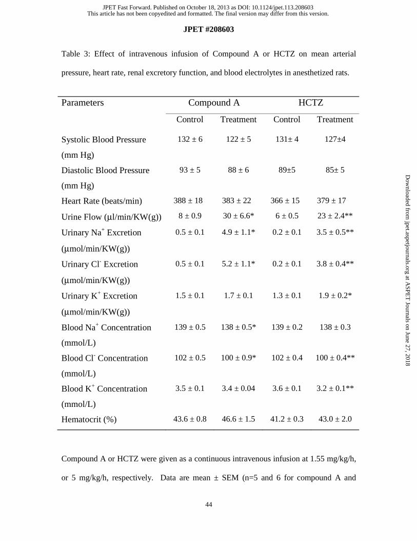
Renal Excretory Function Studies in Anesthetized SD Rats. The diuretic activity of
compound A was assessed in anesthetized euvolemic rats. Following intravenous
infusion of either compound A or HCTZ for 1 h, significant and comparable increases in
urine flow and urinary Na+ and Cl- excretion rates were observed with both agents (Table
3). Urinary K+ excretion, however, was only significantly increased by HCTZ, but not by
compound A. HCTZ also caused a significant decrease in blood K+ levels. Plasma levels
for compound A during the infusion period averaged 1.36 ± 0.13 μM, approximately
three times above the IC50 for inhibition of rat Kir1.1 channels in the presence of 100%
serum. Neither compound A nor HCTZ caused significant changes in blood pressure or
heart rate during the study (Table 3). Vehicle had no effect on renal excretory function
(data not shown). These data indicate that even at a greater diuretic and natriuretic dose,
compound A induces less urinary K+ losses compared to HCTZ. Despite significant
diuresis observed with both agents, blood K+ levels did not significantly change with
compound A, but significantly decreased with HCTZ.
This article has not been copyedited and formatted. The final version may differ from this version.JPET Fast Forward. Published on October 18, 2013 as DOI: 10.1124/jpet.113.208603
at ASPE
T Journals on June 27, 2018
jpet.aspetjournals.orgD
ownloaded from
JPET #208603
22
Compound A Elicits Diuresis and Natriuresis in Conscious, Volume-loaded Rats.
Oral administration of compound A evoked dose-dependent increases in urine output in
conscious, volume-loaded, SD rats (Figure 6, panel A). Four hours after oral dosing,
compound A significantly increased urine flow starting at a dose of 3 mg/kg (2.6-fold vs.
vehicle) while maximal increases were observed at 50 mg/kg (4.3-fold vs. vehicle).
Diuretic efficacy of compound A appeared to reach a plateau at doses between 10 and 50
mg/kg under these experimental conditions. In similar experiments, HCTZ significantly
and dose-dependently increased urine flow starting at 10 mg/kg (2.5-fold vs. vehicle)
while maximal increases were observed at 100 mg/kg (4.0-fold vs. vehicle). Thus, the
diuretic efficacy of compound A and HCTZ appear to be similar at the highest doses
tested. A similar trend was observed when comparing the natriuretic efficacy of
compound A and HCTZ (Figure 6, panel B). Four hours post-dosing, compound A and
HCTZ significantly, and dose-dependently, increased urinary Na+ excretion starting at
doses of 10 mg/kg. Maximal natriuretic effects were observed at the highest doses tested,
with compound A increasing urinary Na+ excretion by 3.8-fold and HCTZ by 3.4-fold
compared to vehicle-treated rats. Importantly, despite robust diuresis and natriuresis
associated with compound A administration, urinary K+ excretion did not significantly
change with any of the doses tested, but was significantly increased by the highest dose
of HCTZ (100 mg/kg, 1.4-fold vs. vehicle) (Figure 6, panel C). These data indicate that
compound A, at maximally efficacious diuretic and natriuretic doses, does not lead to
significant urinary K+ losses, an effect that is different from the well-known, and
documented here, HCTZ-induced kaliuresis. After 4.5 h following oral dosing,
concentrations of compound A in plasma were below 0.2 μM in rats given the 1 and 3
This article has not been copyedited and formatted. The final version may differ from this version.JPET Fast Forward. Published on October 18, 2013 as DOI: 10.1124/jpet.113.208603
at ASPE
T Journals on June 27, 2018
jpet.aspetjournals.orgD
ownloaded from

JPET #208603
23
mg/kg doses, but 0.39 and 0.92 μM, respectively, in rats dosed with 10 and 50 mg/kg.
Compound A levels were higher in urine samples, ranging from 0.74 μM in rats dosed
with 3 mg/kg up to 4.8 μM in rats dosed with the 50 mg/kg dose. In a parallel PK study,
concentrations of compound A were determined in plasma and kidney samples from rats
dosed with 10 mg/kg compound A (Table 4). Kidney levels of compound A were
approximately 3.9 to 6.7-fold larger than those found in plasma.
Compound A causes Diuresis and Natriuresis in Conscious Dogs. Oral administration
of compound A increased urine output in conscious, euvolemic dogs (Figure 7, panel A).
Compound A dosed at either 10 or 50 mg/kg led to significant dose- and time-dependent
increases in urine output. Maximal diuretic effect of compound A was obtained with the
50 mg/kg dose, three hours post-dosing, with urine output increasing from baseline
values of 0.43 ml/min up to 2.68 ml/min (8-fold change vs. baseline), but values appeared
to plateau after 1.5 h post-dosing. Oral HCTZ dosed at 10 mg/kg also increased urine
flow in a time-dependent fashion, and maximal diuretic effect of HCTZ was seen at 1 h
post-dosing. Urine flow values increased from 0.35 ml/min at baseline up to a maximum
of 1.62 ml/min at 1h, but values remained constant for the remainder of the study (~3.5 to
4.8-fold change vs. baseline). Similarly, compound A led to dose- and time-dependent
increases in urinary Na+ excretion in dogs (Figure 7, panel B). Maximal natriuretic effect
was seen with 50 mg/kg compound A, three hours post-dosing. Urinary Na+ excretion
increased from baseline values of 55 μEq/min up to 435 μEq/min (16-fold change vs.
baseline), and values appeared to plateau after 1.5 h post-dosing. Oral HCTZ also
increased urinary Na+ excretion in a time-dependent fashion; and maximal natriuresis
This article has not been copyedited and formatted. The final version may differ from this version.JPET Fast Forward. Published on October 18, 2013 as DOI: 10.1124/jpet.113.208603
at ASPE
T Journals on June 27, 2018
jpet.aspetjournals.orgD
ownloaded from
JPET #208603
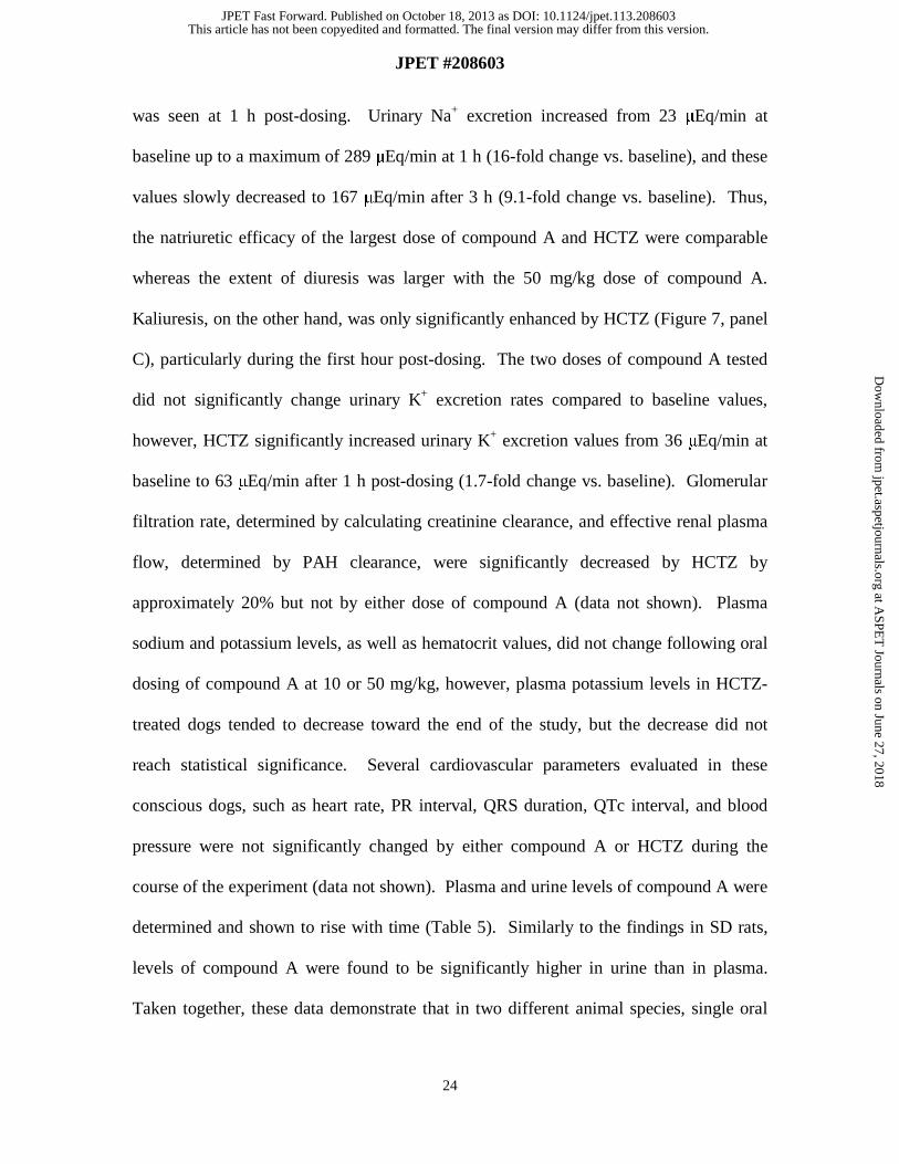
24
was seen at 1 h post-dosing. Urinary Na+ excretion increased from 23 μEq/min at
baseline up to a maximum of 289 μEq/min at 1 h (16-fold change vs. baseline), and these
values slowly decreased to 167 μEq/min after 3 h (9.1-fold change vs. baseline). Thus,
the natriuretic efficacy of the largest dose of compound A and HCTZ were comparable
whereas the extent of diuresis was larger with the 50 mg/kg dose of compound A.
Kaliuresis, on the other hand, was only significantly enhanced by HCTZ (Figure 7, panel
C), particularly during the first hour post-dosing. The two doses of compound A tested
did not significantly change urinary K+ excretion rates compared to baseline values,
however, HCTZ significantly increased urinary K+ excretion values from 36 μEq/min at
baseline to 63 μEq/min after 1 h post-dosing (1.7-fold change vs. baseline). Glomerular
filtration rate, determined by calculating creatinine clearance, and effective renal plasma
flow, determined by PAH clearance, were significantly decreased by HCTZ by
approximately 20% but not by either dose of compound A (data not shown). Plasma
sodium and potassium levels, as well as hematocrit values, did not change following oral
dosing of compound A at 10 or 50 mg/kg, however, plasma potassium levels in HCTZ-
treated dogs tended to decrease toward the end of the study, but the decrease did not
reach statistical significance. Several cardiovascular parameters evaluated in these
conscious dogs, such as heart rate, PR interval, QRS duration, QTc interval, and blood
pressure were not significantly changed by either compound A or HCTZ during the
course of the experiment (data not shown). Plasma and urine levels of compound A were
determined and shown to rise with time (Table 5). Similarly to the findings in SD rats,
levels of compound A were found to be significantly higher in urine than in plasma.
Taken together, these data demonstrate that in two different animal species, single oral
This article has not been copyedited and formatted. The final version may differ from this version.JPET Fast Forward. Published on October 18, 2013 as DOI: 10.1124/jpet.113.208603
at ASPE
T Journals on June 27, 2018
jpet.aspetjournals.orgD
ownloaded from

JPET #208603
25
doses of compound A evokes a diuretic and natriuretic response that is comparable to
HCTZ, but with minimal changes in urinary K+ excretion rate and without any significant
cardiovascular liabilities.
This article has not been copyedited and formatted. The final version may differ from this version.JPET Fast Forward. Published on October 18, 2013 as DOI: 10.1124/jpet.113.208603
at ASPE
T Journals on June 27, 2018
jpet.aspetjournals.orgD
ownloaded from

JPET #208603
26
Discussion
The results of this study illustrate the characterization of the first small molecule
Kir1.1 inhibitor with appropriate selectivity profile and pharmacokinetic properties for in
vivo evaluation. Compound A is a potent inhibitor of Kir1.1 channels stably expressed in
heterologous systems, and accesses the channel through the cytoplasmic side. Residues
lining the pore within the transmembrane region of the channel below the selectivity filter
appear to contribute to high affinity inhibition of Kir1.1 by A. Pharmacological
inhibition of ROMK upon oral dosing of A leads to diuresis and natriuresis in two
different animal species, SD rats and dogs. The magnitude of these events is similar to
those of the clinically used diuretic HCTZ, but A appears to have a more favorable
urinary potassium/sodium ratio than relevant doses of HCTZ in these acute studies.
Taken together, these data suggest that selective inhibitors of ROMK represent a novel
mechanism for developing diuretic/natriuretic agents with the potential for enhanced
efficacy, and assuming that acute effects predict chronic pharmacology, a more favorable
potassium balance than the diuretics that are currently used in the treatment of
hypertension and/or congestive heart failure.
Within the diuretic class, thiazides, such as HCTZ, are the most widely used as
first line therapy to treat uncomplicated hypertension, or as add-on therapy to other
mechanism of action drugs, such as angiotensin-converting enzyme inhibitors, and
angiotensin II receptor blockers (Sood et al., 2010). Even when HCTZ is used as part of
combination therapy with renin-angiotensin-aldosterone system blockade, patients with
resistant hypertension often fail to achieve adequate blood pressure control; ROMK
This article has not been copyedited and formatted. The final version may differ from this version.JPET Fast Forward. Published on October 18, 2013 as DOI: 10.1124/jpet.113.208603
at ASPE
T Journals on June 27, 2018
jpet.aspetjournals.orgD
ownloaded from
JPET #208603
27
inhibitors may offer potential added efficacy benefit in these populations. Hypokalemia
(serum potassium concentration < 3.5 mEq/L) and elevations in fasting blood glucose are
the major liabilities associated with thiazides (Palmer and Naderi, 2007). Loop diuretics,
such as furosemide, are mostly used to treat acute episodes of pulmonary and peripheral
edema in congestive heart failure patients, but their chronic use in the disease is not
recommended because the drugs lose efficacy with time and cause hypokalemia at higher
doses. No new diuretics have been developed within the past four decades, but an ideal
novel diuretic should be potassium neutral, provide equal or greater efficacy to clinically
used diuretics, and be suitable for combination therapy. In this sense, ROMK has
emerged as an attractive novel target for the development of such agents (Ji et al., 2008).
ROMK is present in two different regions of the nephron (Xu et al., 1997).
Inhibition of ROMK at the TALH should mimic the effect of furosemide in providing
natriuresis/diuresis. In addition, inhibition of ROMK at the CCD, where it participates in
potassium secretion, may ameliorate the hypokalemia caused by loop and thiazide
diuretics. The results presented in this study with the selective, small molecule A seem to
support these expectations of a ROMK inhibitor. Thus, A provides diuresis/natriuresis
effects comparable to those of clinically used diuretics but with a more favourable
urinary K/Na ratio in two different species, and under different experimental paradigms.
It is interesting to note that inhibition of ROMK at the CCD does not appear to decrease
urinary potassium excretion, which could lead to hyperkalemia, despite the fact that
ROMK contributes to potassium secretion in that part of the nephron. However, we can
speculate that other mechanism(s) present at the CCD, such as high-conductance,
calcium-activated potassium channels, are likely to contribute to potassium secretion, in
This article has not been copyedited and formatted. The final version may differ from this version.JPET Fast Forward. Published on October 18, 2013 as DOI: 10.1124/jpet.113.208603
at ASPE
T Journals on June 27, 2018
jpet.aspetjournals.orgD
ownloaded from

JPET #208603
28
particular under conditions of high luminal flow rates that result from inhibition of salt
reuptake at the TALH. Indeed, compensatory mechanisms between ROMK and high-
conductance, calcium-activated potassium channels in the distal part of the nephron have
been observed in studies with mice lacking either channel (Rieg et al., 2007). Like
furosemide and thiazide diuretics, ROMK inhibitors are expected to activate the renin-
angiotensin system, which could attenuate the extent of natriuresis/diuresis upon chronic
dosing. Chronic treatment with A will be needed to determine if diuretic/natriuretic
resistance develops with time which may limit the utility of ROMK inhibitors as mono-
therapy agents. If resistance develops, it would be important to evaluate whether ROMK
inhibitors could be administered in combination with an angiotensin converting enzyme
or an angiotensin II receptor blocker to enhance their efficacy. In addition, chronic
treatment will also provide insight into the effects of ROMK inhibitors on plasma
potassium levels over time.
The search for Kir1.1 inhibitors has provided a limited number of compounds
with appropriate potency, selectivity, physico-chemical, and pharmacokinetic properties
to be used in proof of concept studies (Lewis et al., 2009; Bhave et al., 2011; Tang et al.,
2012). Compound A represents the first Kir1.1 inhibitor that fulfils the above criteria.
The selectivity of A for Kir1.1 versus other Kir channels, such as Kir2.1 and Kir2.3, is
especially noteworthy. Part of this selectivity appears to arise from the nature of specific
residues that line the channel’s pore within the transmembrane domain below the
selectivity filter, although other region(s) of the channel may also contribute to the high
affinity interaction of A with Kir1.1 channels. Although more studies need to be done to
determine the in vivo efficacy of A after chronic dosing, and the possibility of
This article has not been copyedited and formatted. The final version may differ from this version.JPET Fast Forward. Published on October 18, 2013 as DOI: 10.1124/jpet.113.208603
at ASPE
T Journals on June 27, 2018
jpet.aspetjournals.orgD
ownloaded from

JPET #208603
29
combination with other mechanism of action drugs, the results of the present study
support the idea that ROMK represents a target of interest for the development of novel
diuretics with the potential of having an improved plasma potassium profile.
This article has not been copyedited and formatted. The final version may differ from this version.JPET Fast Forward. Published on October 18, 2013 as DOI: 10.1124/jpet.113.208603
at ASPE
T Journals on June 27, 2018
jpet.aspetjournals.orgD
ownloaded from
JPET #208603
30
Acknowledgments
We thank Randal Bugianesi, Rodolfo Haedo, Michael Margulis, and Kashmira
Shah for expert technical assistances, and Drs. Euan MacIntyre, Sandy G Mills, Adam
Weinglass, and Lihu Yang for important discussions during the course of this work.
This article has not been copyedited and formatted. The final version may differ from this version.JPET Fast Forward. Published on October 18, 2013 as DOI: 10.1124/jpet.113.208603
at ASPE
T Journals on June 27, 2018
jpet.aspetjournals.orgD
ownloaded from

JPET #208603
31
Authorship Contributions
Participated in research design: Garcia, Priest, Alonso-Galicia, Zhou, Felix, Owens, Roy,
Kaczorowski, Pasternak
Conducted experiments: Priest, Alonso-Galicia, Zhou, Felix, Brochu, Bailey, Thomas-
Fowlkes, Liu, Swensen, Hernandez, Pai, Xiao, Hoagland, Owens
Contributed new reagents or analytic tools: Tang, de Jesus, Pasternak
Performed data analysis: Garcia, Priest, Alonso-Galicia, Zhou, Felix, Thomas-Fowlkes,
Liu, Swensen, Hernandez, Pai, Hoagland, Owens
Wrote or contributed to the writing of the manuscript: Garcia, Priest, Alonso-Galicia,
Zhou, Kaczorowski, Pasternak
This article has not been copyedited and formatted. The final version may differ from this version.JPET Fast Forward. Published on October 18, 2013 as DOI: 10.1124/jpet.113.208603
at ASPE
T Journals on June 27, 2018
jpet.aspetjournals.orgD
ownloaded from
JPET #208603
32
References
Abbadie C, McManus OB, Sun SY, Bugianesi RM, Dai G, Haedo RJ, Herrington JB,
Kaczorowski GJ, Smith MM, Swensen AM, Warren VA, Williams B, Arneric SP,
Eduljee C, Snutch TP, Tringham EW, Jochnowitz N, Liang A, Euan MacIntyre D,
McGowan E, Mistry S, White VV, Hoyt SB, London C, Lyons KA, Bunting PB,
Volksdorf S and Duffy JL (2010) Analgesic effects of a substituted N-triazole
oxindole (TROX-1), a state-dependent, voltage-gated calcium channel 2 blocker.
J Pharmacol Exp Ther 334:545-555.
Bhave G, Chauder BA, Liu W, Dawson ES, Kadakia R, Nguyen TT, Lewis LM, Meiler J,
Weaver CD, Satlin LM, Lindsley CW, and Denton JS (2011) Development of a
selective small-molecule inhibitor of Kir1.1, the renal outer medullary potassium
channel. Mol Pharmacol 79:42-50.
Felix JP, Liu J, Schmalhofer WA, Bailey T, Bednarek MA, Kinkel S, Weinglass AB,
Kohler M, Kaczorowski GJ, Priest BT and Garcia ML (2006) Characterization of
Kir1.1 channels with the use of a radiolabeled derivative of tertiapin.
Biochemistry 45:10129-10139.
Felix JP, Priest BT, Solly K, Bailey T, Brochu RM, Liu CJ, Kohler MG, Kiss L, Alonso-
Galicia M, Tang H, Pasternak A, Kaczorowski GJ and Garcia ML (2012) The
inwardly rectifying potassium channel Kir1.1: development of functional assays
to identify and characterize channel inhibitors. Assay Drug Dev Technol 10:417-
431.
Felix JP, Williams BS, Priest BT, Brochu RM, Dick IE, Warren VA, Yan L, Slaughter
RS, Kaczorowski GJ, Smith MM and Garcia ML (2004) Functional assay of
This article has not been copyedited and formatted. The final version may differ from this version.JPET Fast Forward. Published on October 18, 2013 as DOI: 10.1124/jpet.113.208603
at ASPE
T Journals on June 27, 2018
jpet.aspetjournals.orgD
ownloaded from

JPET #208603
33
voltage-gated sodium channels using membrane potential-sensitive dyes. Assay
Drug Dev Technol 2:260-268.
Ji W, Foo JN, O'Roak BJ, Zhao H, Larson MG, Simon DB, Newton-Cheh C, State MW,
Levy D and Lifton RP (2008) Rare independent mutations in renal salt handling
genes contribute to blood pressure variation. Nat Genet 40:592-599.
Jin W and Lu Z (1998) A novel high-affinity inhibitor for inward-rectifier K+ channels
Biochemistry 37:13291-13299.
Lewis LM, Bhave G, Chauder BA, Banerjee S, Lornsen KA, Redha R, Fallen K,
Lindsley CW, Weaver CD and Denton JS (2009) High-throughput screening
reveals a small-molecule inhibitor of the renal outer medullary potassium channel
and Kir7.1. Mol Pharmacol 76:1094-1103.
Lifton RP, Gharavi AG and Geller DS (2001) Molecular mechanisms of human
hypertension. Cell 104:545-556.
Lorenz JN, Baird NR, Judd LM, Noonan WT, Andringa A, Doetschman T, Manning PA,
Liu LH, Miller ML and Shull GE (2002) Impaired renal NaCl absorption in mice
lacking the ROMK potassium channel, a model for type II Bartter's syndrome. J
Biol Chem 277:37871-37880.
Lu M, Wang T, Yan Q, Yang X, Dong K, Knepper MA, Wang W, Giebisch G, Shull GE
and Hebert SC (2002) Absence of small conductance K+ channel (SK) activity in
apical membranes of thick ascending limb and cortical collecting duct in ROMK
(Bartter's) knockout mice. J Biol Chem 277:37881-37887.
Lu Z and MacKinnon R (1994) Electrostatic tuning of Mg2+ affinity in an inward-
rectifier K+ channel. Nature 371:243-246.
This article has not been copyedited and formatted. The final version may differ from this version.JPET Fast Forward. Published on October 18, 2013 as DOI: 10.1124/jpet.113.208603
at ASPE
T Journals on June 27, 2018
jpet.aspetjournals.orgD
ownloaded from
JPET #208603
34
Nichols CG and Lopatin AN (1997) Inward rectifier potassium channels. Annu Rev
Physiol 59:171-191.
Palmer BF and Naderi AS (2007) Metabolic complications associated with use of
thiazide diuretics. J Am Soc Hypertens 1:381-392.
Palmer LG, Choe H and Frindt G (1997) Is the secretory K channel in the rat CCT
ROMK? Am J Physiol 273:F404-410.
Rieg T, Vallon V, Sausbier M, Sausbier U, Kaissling B, Ruth P and Osswald H (2007)
The role of the BK channel in potassium homeostasis and flow-induced renal
potassium excretion. Kidney Int 72:566-573.
Schmalhofer WA, Swensen AM, Thomas BS, Felix JP, Haedo RJ, Solly K, Kiss L,
Kaczorowski GJ and Garcia ML (2010) A pharmacologically validated, high-
capacity, functional thallium flux assay for the human Ether-a-go-go related gene
potassium channel. Assay Drug Dev Technol 8:714-726.
Simon DB, Karet FE, Rodriguez-Soriano J, Hamdan JH, DiPietro A, Trachtman H,
Sanjad SA and Lifton RP (1996) Genetic heterogeneity of Bartter's syndrome
revealed by mutations in the K+ channel, ROMK. Nat Genet 14:152-156.
Simons K and Virta H (2006) Growing Madin-Darby canine kidney cells for studying
epithelial cell biology, in Cell Biology (Celis JE ed) pp 127-131, Elsevier Inc.,
USA
Solly K, Cassaday J, Felix JP, Garcia ML, Ferrer M, Strulovici B and Kiss L (2008)
Miniaturization and HTS of a FRET-based membrane potential assay for Kir
channel inhibitors. Assay Drug Dev Technol 6:225-234.
This article has not been copyedited and formatted. The final version may differ from this version.JPET Fast Forward. Published on October 18, 2013 as DOI: 10.1124/jpet.113.208603
at ASPE
T Journals on June 27, 2018
jpet.aspetjournals.orgD
ownloaded from
JPET #208603
35
Sood N, Reinhart KM and Baker WL (2010) Combination therapy for the management of
hypertension: A review of the evidence. Am J Health Syst Pharm 67:885-894.
Tang H, Walsh SP, Yan Y, de Jesus RK, Shahripour A, Teumelsan N, Zhu Y, Ha S,
Owens KA, Thomas-Fowlkes B, Felix JP, Liu J, Kohler M, Priest BT, Bailey T,
Brochu R, Alonso-Galicia M, Kaczorowski GJ, Roy S, Yang L, Mills SG, Garcia
ML and Pasternak A (2012) Discovery of selective small molecule ROMK
inhibitor as potential new mechanism diuretics. ACS Medicinal Chemistry Letters
3(5):367-372.
Tang H, de Jesus RK, Walsh SP, Zhu Y, Yan Y, Priest BT, Swensen AM, Alonso-Galicia
M, Felix JP, Brochu RM, Bailey T, Thomas-Fowlkes B, Zhou X, Pai L-Y,
Hampton C, Hernandez M, Owens K, Roy S, Kaczorowski GJ, Yang L, Garcia
ML and Pasternak A (2013) Discovery of a novel sub-class of ROMK channel
inhibitors typified by 5-(2-(4-(2-(4-(1H-tetrazol-1-yl)phenyl)acetyl)piperazin-1-
yl)ethyl)isobenzofuran-1(3H)-one. Bioorg. & Med. Chem. Lett. 23(21):5829-
5832.
Tao X, Avalos JL, Chen J and MacKinnon R (2009) Crystal structure of the eukaryotic
strong inward-rectifier K+ channel Kir2.1 at 3.1 Å resolution. Science 326:1668-
1674.
Xu JZ, Hall AE, Peterson LN, Bienkowski MJ, Eessalu TE and Hebert SC (1997)
Localization of the ROMK protein on apical membranes of rat kidney nephron
segments. Am J Physiol 273:F739-748.
Zhou X, Zhang Z, Shin MK, Horwitz SB, Levorse JM, Zhu L, Sharif-Rodriguez W,
Streltsov DY, Dajee M, Hernandez M, Pan Y, Urosevic-Price O, Wang L, Forrest
This article has not been copyedited and formatted. The final version may differ from this version.JPET Fast Forward. Published on October 18, 2013 as DOI: 10.1124/jpet.113.208603
at ASPE
T Journals on June 27, 2018
jpet.aspetjournals.orgD
ownloaded from
JPET #208603
36
G, Szeto D, Zhu Y, Cui Y, Michael B, Balogh LA, Welling PA, Wade JB, Roy S,
Sullivan KA. (2013) Heterozygous Disruption of Renal Outer Medullary
Potassium Channel in Rats Is Associated With Reduced Blood Pressure.
Hypertension 62:288-294.
This article has not been copyedited and formatted. The final version may differ from this version.JPET Fast Forward. Published on October 18, 2013 as DOI: 10.1124/jpet.113.208603
at ASPE
T Journals on June 27, 2018
jpet.aspetjournals.orgD
ownloaded from
JPET #208603
37
Footnotes
Reprint requests:
Alexander Pasternak PhD
Director, Chemistry
Kenilworth Discovery Chemistry
2015 Galloping Hill Road, K15-ME2
Kenilworth, NJ 07033
(908)-740-6847
Fax: (908)-740-2207
E-mail: [email protected]
M.L.G., B.T. P., and M.A.-G. contributed equally to this work
Current affiliation:
MLG and GJK: Kanalis Consulting, L.L.C., Edison, New Jersey
BTP: Lilly Research Laboratories, Eli Lilly & Co., Indianapolis, Indiana
MAG: Forest Research institute, Inc., Jersey City, New Jersey
JPF: Purdue Pharma LP, Cranbury, New Jersey
RMB: Novartis, Cambridge, Massachusetts
TB: ImClone Systems, New York, New York
AS: AbbVie, North Chicago, Illinois
This article has not been copyedited and formatted. The final version may differ from this version.JPET Fast Forward. Published on October 18, 2013 as DOI: 10.1124/jpet.113.208603
at ASPE
T Journals on June 27, 2018
jpet.aspetjournals.orgD
ownloaded from

JPET #208603
38
Figure Legends
Figure 1. Structure of compound A, 5-(2-(4-(2-(4-(1H-tetrazol-1-
yl)phenyl)acetyl)piperazin-1-yl)ethyl)isobenzofuran-1(3H)-one.
Figure 2. Compound A inhibits Kir1.1 channels. (A) HEK293 cells stably expressing
hKir1.1 (●) or hKir2.1 (▲) channels were preloaded with the FluxOR reagent, and
incubated in the absence or presence of increasing concentrations of A, as described
under Materials and Methods. Upon recording the emission of the dye for 90 sec, a
thallium sulfate/potassium sulfate solution was added, and fluorescence was monitored
for an additional 510 sec. Data were analyzed by the Hill equation where all parameters
were left unconstrained. Compound A inhibits hKir1.1 with an IC50 value of 30 nM and
nH of 1. In marked contrast, A does not have any significant effect on hKir2.1 at
concentrations of up to 100 μM. Data shown are mean ± SD (n= 4). (B and C) CHO-
hKir1.1 (●, �, �, �) (B), CHO-hKir2.3 (▲) (B) or HEK-rKir1.1 (C) cells were
incubated with 86Rb+ overnight as indicated under Materials and Methods. On the day of
the experiment the medium was removed and cells were placed into assay medium
containing 0 ( ● ) ( ▲ ), 10 ( � ), 30 ( � ), or 100% ( � ) human (B) or rat (C) serum, in
the absence or presence of increasing concentrations of compound A, and incubated at
room temperature for 30 min. The amount of 86Rb+ efflux was calculated as indicated
under Materials and Methods. Data were analyzed by the Hill equation where all
parameters were left unconstrained. Data shown are mean ± SD (n=4). IC50 values
This article has not been copyedited and formatted. The final version may differ from this version.JPET Fast Forward. Published on October 18, 2013 as DOI: 10.1124/jpet.113.208603
at ASPE
T Journals on June 27, 2018
jpet.aspetjournals.orgD
ownloaded from

JPET #208603
39
(nM): (B) 93 ( ● ), 161 ( � ), 312 ( � ), 522 ( � ); (C) 124 ( ● ), 159 ( � ), 246 ( � ), 469
( � ).
Figure 3. Inhibition of hKir1.1 channels by compound A. Human Kir1.1 channels stably
expressed in HEK293 cells were examined by whole cell voltage-clamp as described
under Materials and Methods. (A) Kir1.1 currents were recorded during 200 ms voltage
ramps from -80 mV to +20 mV in control (black) and in the presence of 100 nM
compound A (grey). External K+ concentration was 40 mM. (B) Currents measured at a
membrane potential of -80 mV were plotted as a function of time for a representative cell.
Bath application of rising concentrations of compound A is indicated by the arrows. (C)
Currents corresponding to membrane potentials of -80 mV and +20 mV were measured,
and the observed inhibition was plotted as a function of compound A concentration for a
representative cell. The solid lines represent the results of fitting the Hill equation to the
data points, yielding an IC50 of 53 nM and Hill coefficient of 1.4 for currents recorded at
-80 mV, and an IC50 of 47 nM and Hill coefficient of 1.5 for currents recorded at +20
mV.
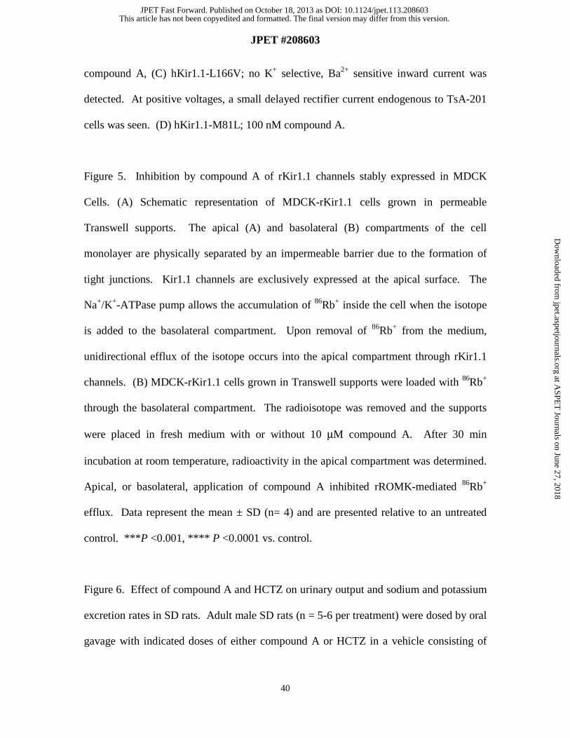
Figure 4. Inhibition of hKir1.1 mutant channels by compound A. Mutated hKir1.1
channels were transiently expressed in TsA-201 cells as described under Materials and
Methods. Currents were recorded in response to voltage ramps, using 140 mM external
K+, and are shown in the absence (black) and in the presence (grey) of the indicated
concentration of compound A. Channel constructs and compound A concentrations were
as follows: (A) hKir1.1-N171D; 10 μM compound A, (B) hKir1.1-N171Q; 2 μM
This article has not been copyedited and formatted. The final version may differ from this version.JPET Fast Forward. Published on October 18, 2013 as DOI: 10.1124/jpet.113.208603
at ASPE
T Journals on June 27, 2018
jpet.aspetjournals.orgD
ownloaded from
JPET #208603
40
compound A, (C) hKir1.1-L166V; no K+ selective, Ba2+ sensitive inward current was
detected. At positive voltages, a small delayed rectifier current endogenous to TsA-201
cells was seen. (D) hKir1.1-M81L; 100 nM compound A.
Figure 5. Inhibition by compound A of rKir1.1 channels stably expressed in MDCK
Cells. (A) Schematic representation of MDCK-rKir1.1 cells grown in permeable
Transwell supports. The apical (A) and basolateral (B) compartments of the cell
monolayer are physically separated by an impermeable barrier due to the formation of
tight junctions. Kir1.1 channels are exclusively expressed at the apical surface. The
Na+/K+-ATPase pump allows the accumulation of 86Rb+ inside the cell when the isotope
is added to the basolateral compartment. Upon removal of 86Rb+ from the medium,
unidirectional efflux of the isotope occurs into the apical compartment through rKir1.1
channels. (B) MDCK-rKir1.1 cells grown in Transwell supports were loaded with 86Rb+
through the basolateral compartment. The radioisotope was removed and the supports
were placed in fresh medium with or without 10 μM compound A. After 30 min
incubation at room temperature, radioactivity in the apical compartment was determined.
Apical, or basolateral, application of compound A inhibited rROMK-mediated 86Rb+
efflux. Data represent the mean ± SD (n= 4) and are presented relative to an untreated
control. ***P <0.001, **** P <0.0001 vs. control.
Figure 6. Effect of compound A and HCTZ on urinary output and sodium and potassium
excretion rates in SD rats. Adult male SD rats (n = 5-6 per treatment) were dosed by oral
gavage with indicated doses of either compound A or HCTZ in a vehicle consisting of
This article has not been copyedited and formatted. The final version may differ from this version.JPET Fast Forward. Published on October 18, 2013 as DOI: 10.1124/jpet.113.208603
at ASPE
T Journals on June 27, 2018
jpet.aspetjournals.orgD
ownloaded from

JPET #208603
41
Imwitor 742:Tween 80 (1:1, v:v). After 30 min, voiding was induced by giving each
animal a saline load as indicated under Materials and Methods. Urine collection took
place over the next four hours at room temperature, and analyzed for volume (A), sodium
(B) and potassium (C) content. Data are presented as fold change relative to values
obtained in vehicle-treated animals. Baseline values for vehicle group were: 3.29 ± 0.42
mL/4 h (urinary output), 0.33 ± 0.06 mmol/4 h (urinary sodium excretion), and 0.72 ±
0.13 mmol/4 h (urinary potassium excretion). *P < 0.05 vs. vehicle.
Figure 7. Effect of compound A and HCTZ on urinary output and sodium and potassium
excretory rates in conscious dogs. Female dogs (n = 3 per treatment) were dosed by oral
gavage with either vehicle (Imwitor 742:Tween 80 (1:1, v:v)), compound A or HCTZ.
Urine samples were collected before and after oral dosing at 30-min intervals up to 3
hours post-dosing, and analyzed for volume (A), sodium (B) and potassium (C) content.
The zero time point (BL) represents the average of two 30-min baseline clearance periods
collected prior to administration of vehicle or test compound. Data (mean ± SEM) are
presented as fold change relative to baseline values obtained prior to test article dosing to
each animal.
This article has not been copyedited and formatted. The final version may differ from this version.JPET Fast Forward. Published on October 18, 2013 as DOI: 10.1124/jpet.113.208603
at ASPE
T Journals on June 27, 2018
jpet.aspetjournals.orgD
ownloaded from

JPET #208603
42
Tables
Table 1: Pharmacokinetic Properties of Compound A in Rats and Dogs
Rat Dog
Cl (ml/min/kg) 40 36
T1/2 (h) 0.62 1.4
Vdss (L/kg) 0.66 1.9
nAUC (μM•hr/(mg/kg)) 0.34 0.88
Tmax (h) 0.67
F (%) 33 80
Pharmacokinetic parameters were obtained following intravenous and oral
administration of compound A to SD rats or dogs. Compound A was dissolved
in a PEG 200:water (70:30, v:v) solution and dosed to rats or dogs either
intravenously at 1 mg/kg or orally at 2 mg/kg. Plasma concentrations were
determined by LC-MS/MS following protein precipitation with acetonitrile. Cl,
plasma clearance; T1/2, terminal half-life; Vdss, volume of distribution at the
steady state; F, oral bioavailability; nAUC, dose-normalized area under the
plasma concentration vs. time curve following oral dosing.
This article has not been copyedited and formatted. The final version may differ from this version.JPET Fast Forward. Published on October 18, 2013 as DOI: 10.1124/jpet.113.208603
at ASPE
T Journals on June 27, 2018
jpet.aspetjournals.orgD
ownloaded from

JPET #208603
43
Table 2: Block of Kir1.1 site-directed mutants by compound A.
hKir1.1 construct n IC50 (nM)
WT 81
M81L 1 <100
L128F 4 <100
S130A 4 ND
Q152E 3 <100
L166V 5 ND
V168C 3 <100
N171D 4 ~7100
N171Q 2 ~720
Mutant constructs were expressed in TsA-201 cells, and inhibition by compound A was
examined by whole-cell voltage clamp as indicated under Experimental Procedures. ND
denotes constructs that did not generate detectable currents.
This article has not been copyedited and formatted. The final version may differ from this version.JPET Fast Forward. Published on October 18, 2013 as DOI: 10.1124/jpet.113.208603
at ASPE
T Journals on June 27, 2018
jpet.aspetjournals.orgD
ownloaded from
JPET #208603
44
Table 3: Effect of intravenous infusion of Compound A or HCTZ on mean arterial
pressure, heart rate, renal excretory function, and blood electrolytes in anesthetized rats.
Parameters Compound A HCTZ
Control Treatment Control Treatment
Systolic Blood Pressure
(mm Hg)
132 ± 6 122 ± 5 131± 4 127±4
Diastolic Blood Pressure
(mm Hg)
93 ± 5 88 ± 6 89±5 85± 5
Heart Rate (beats/min) 388 ± 18 383 ± 22 366 ± 15 379 ± 17
Urine Flow (μl/min/KW(g)) 8 ± 0.9 30 ± 6.6* 6 ± 0.5 23 ± 2.4**
Urinary Na+ Excretion
(μmol/min/KW(g))
0.5 ± 0.1 4.9 ± 1.1* 0.2 ± 0.1 3.5 ± 0.5**
Urinary Cl- Excretion
(μmol/min/KW(g))
0.5 ± 0.1 5.2 ± 1.1* 0.2 ± 0.1 3.8 ± 0.4**
Urinary K+ Excretion
(μmol/min/KW(g))
1.5 ± 0.1 1.7 ± 0.1 1.3 ± 0.1 1.9 ± 0.2*
Blood Na+ Concentration
(mmol/L)
139 ± 0.5 138 ± 0.5* 139 ± 0.2 138 ± 0.3
Blood Cl- Concentration
(mmol/L)
102 ± 0.5 100 ± 0.9* 102 ± 0.4 100 ± 0.4**
Blood K+ Concentration
(mmol/L)
3.5 ± 0.1 3.4 ± 0.04 3.6 ± 0.1 3.2 ± 0.1**
Hematocrit (%) 43.6 ± 0.8 46.6 ± 1.5 41.2 ± 0.3 43.0 ± 2.0
Compound A or HCTZ were given as a continuous intravenous infusion at 1.55 mg/kg/h,
or 5 mg/kg/h, respectively. Data are mean ± SEM (n=5 and 6 for compound A and
This article has not been copyedited and formatted. The final version may differ from this version.JPET Fast Forward. Published on October 18, 2013 as DOI: 10.1124/jpet.113.208603
at ASPE
T Journals on June 27, 2018
jpet.aspetjournals.orgD
ownloaded from
JPET #208603
45
HCTZ studies, respectively). * P<0.05, ** P <0.01 vs. control. Urine flow and urinary
electrolyte (Na+, Cl-, and K+) excretion rates were corrected for kidney weight (KW).
This article has not been copyedited and formatted. The final version may differ from this version.JPET Fast Forward. Published on October 18, 2013 as DOI: 10.1124/jpet.113.208603
at ASPE
T Journals on June 27, 2018
jpet.aspetjournals.orgD
ownloaded from

JPET #208603
46
Table 4: Plasma and kidney concentrations of compound A after oral administration to
SD rats.
Time (h) Plasma (μM) Kidney (μM) Kidney/Plasma
0.25 0.37 1.4 3.9
1 0.61 4.1 6.7
4.5 0.65 3.3 5.1
SD rats (n =3 per each time point) were orally dosed with 10 mg/kg compound A
formulated as a 10 mg/ml suspension in Imwitor 742: Tween 80 (1:1, v:v). Plasma and
kidney concentrations were determined by LC-MS/MS.
This article has not been copyedited and formatted. The final version may differ from this version.JPET Fast Forward. Published on October 18, 2013 as DOI: 10.1124/jpet.113.208603
at ASPE
T Journals on June 27, 2018
jpet.aspetjournals.orgD
ownloaded from

JPET #208603
47
Table 5. Plasma and kidney concentrations of compound A after oral administration to
dogs.
Collection Time Plasma Concentration (μM) Urine Concentration (μM)
(min) 10 mg/kg 50 mg/kg 10 mg/kg 50 mg/kg
Control 0 0 0 0
0-30 0.1 1.5 4.0 18.7
30-60 0.1 3.2 7.3 32.2
60-90 0.1 2.9 5.5 29.4
90-120 0.2 3.9 5.7 50.0
120-150 0.2 8.5 7.9 74.2
150-180 0.2 9.9 7.7 85.2
Female dogs (n=3) were orally dosed with either 10 or 50 mg/kg compound A at a dose
volume of 0.5 ml/kg in Imwitor 742: Tween 80 (1:1, v:v). Plasma and urine samples
were collected before and after oral administration of compound A at 30-min interval
periods for up to 3 hours post-dosing. Concentrations of compound A were determined
by LC-MS/MS.
This article has not been copyedited and formatted. The final version may differ from this version.JPET Fast Forward. Published on October 18, 2013 as DOI: 10.1124/jpet.113.208603
at ASPE
T Journals on June 27, 2018
jpet.aspetjournals.orgD
ownloaded from

This article has not been copyedited and formatted. The final version may differ from this version.JPET Fast Forward. Published on October 18, 2013 as DOI: 10.1124/jpet.113.208603
at ASPE
T Journals on June 27, 2018
jpet.aspetjournals.orgD
ownloaded from

This article has not been copyedited and formatted. The final version may differ from this version.JPET Fast Forward. Published on October 18, 2013 as DOI: 10.1124/jpet.113.208603
at ASPE
T Journals on June 27, 2018
jpet.aspetjournals.orgD
ownloaded from

This article has not been copyedited and formatted. The final version may differ from this version.JPET Fast Forward. Published on October 18, 2013 as DOI: 10.1124/jpet.113.208603
at ASPE
T Journals on June 27, 2018
jpet.aspetjournals.orgD
ownloaded from

This article has not been copyedited and formatted. The final version may differ from this version.JPET Fast Forward. Published on October 18, 2013 as DOI: 10.1124/jpet.113.208603
at ASPE
T Journals on June 27, 2018
jpet.aspetjournals.orgD
ownloaded from

This article has not been copyedited and formatted. The final version may differ from this version.JPET Fast Forward. Published on October 18, 2013 as DOI: 10.1124/jpet.113.208603
at ASPE
T Journals on June 27, 2018
jpet.aspetjournals.orgD
ownloaded from

This article has not been copyedited and formatted. The final version may differ from this version.JPET Fast Forward. Published on October 18, 2013 as DOI: 10.1124/jpet.113.208603
at ASPE
T Journals on June 27, 2018
jpet.aspetjournals.orgD
ownloaded from

This article has not been copyedited and formatted. The final version may differ from this version.JPET Fast Forward. Published on October 18, 2013 as DOI: 10.1124/jpet.113.208603
at ASPE
T Journals on June 27, 2018
jpet.aspetjournals.orgD
ownloaded from