jpet fast forward. published on march 17, 2010 as doi:10...
TRANSCRIPT
JPET #166173
1. TITLE PAGE
A Benzothiophene Inhibitor of MAPK-Activated Protein Kinase 2 (MK2) Inhibits TNFα
Production and has Oral Anti-Inflammatory Efficacy in Acute and Chronic Models of
Inflammation.
Robert J. Mourey, Barry L. Burnette, Sarah J. Brustkern, J. Scott Daniels, Jeffrey L.
Hirsch, William F. Hood, Marvin J. Meyers, Stephen J. Mnich, Betsy S. Pierce, Matthew
J. Saabye, John F. Schindler, Sarah A. South, Elizabeth G. Webb, Jian Zhang, and David
R. Anderson.
Discovery Biology (R.J.M., B.L.B., S.J.B., J.L.H., W.F.H., S.J.M., M.J.S., J.F.S., E.G.W.,
J.Z.), Medicinal Chemistry (M.J.M., B.S.P., D.R.A.), Pharmacokinetics, Dynamics and
Metabolism (J.S.D., S.A.S), Inflammation Research Unit, Pfizer Global Research and
Development, Chesterfield, Missouri
JPET Fast Forward. Published on March 17, 2010 as DOI:10.1124/jpet.110.166173
Copyright 2010 by the American Society for Pharmacology and Experimental Therapeutics.
This article has not been copyedited and formatted. The final version may differ from this version.JPET Fast Forward. Published on March 17, 2010 as DOI: 10.1124/jpet.110.166173
at ASPE
T Journals on June 4, 2018
jpet.aspetjournals.orgD
ownloaded from
JPET #166173
2
RUNNING TITLE PAGE
Running Title: Benzothiophene MK2 Kinase Inhibitors are Anti-Inflammatory
Corresponding Author:
David R. Anderson
Chemistry Discovery
Pfizer Global Research & Development
Eastern Point Road
Groton, CT 06340
Telephone: 860-686-9254
FAX: 860-686-7842
Email: [email protected]
Number of text pages: 48
Number of tables: 5
Number of figures: 7
Number of references: 40
Number of words in the:
Abstract: 247
Introduction: 746
Discussion: 1440
This article has not been copyedited and formatted. The final version may differ from this version.JPET Fast Forward. Published on March 17, 2010 as DOI: 10.1124/jpet.110.166173
at ASPE
T Journals on June 4, 2018
jpet.aspetjournals.orgD
ownloaded from
JPET #166173
3
Abbreviations: MK2, MAPKAP kinase 2; PF-3644022, (10R)-10-methyl-3-(6-
methylpyridin-3-yl)-9,10,11,12-tetrahydro-8H-[1,4]diazepino[5',6':4,5]thieno[3,2-
f]quinolin-8-one; RA, rheumatoid arthritis; TNF, tumor necrosis factor; IL-1; interleukin-
1; MAPKAP, MAPK-activated protein; PRAK, p38-regulated and activated kinase; LPS,
lipopolysaccharide; SCW, streptococcal cell wall; MNK, MAPK-interacting kinase; GST,
glutathione-S-transferase; MSK, mitogen- and stress-activated protein kinase; FITC,
fluorescein isothiocyanate; HSP27, heat shock protein 27; SPR, surface plasmon
resonance; hPBMCs, human peripheral blood mononuclear cells; MAPK, mitogen-
activated protein kinase; HWB, human whole blood; HPLC, high-performance liquid
chromatography; LC-MS/MS, liquid chromatography-tandem mass spectrometry; ELISA,
enzyme-linked immunosorbent assay; PK-PD, pharmacokinetic-pharmacodynamic.
Recommended section assignment: Inflammation, Immunopharmacology, and Asthma
This article has not been copyedited and formatted. The final version may differ from this version.JPET Fast Forward. Published on March 17, 2010 as DOI: 10.1124/jpet.110.166173
at ASPE
T Journals on June 4, 2018
jpet.aspetjournals.orgD
ownloaded from
JPET #166173
4
3. ABSTRACT
Activation of the p38 kinase pathway in immune cells leads to the transcriptional and
translational regulation of pro-inflammatory cytokines. MAPKAP kinase 2 (MK2), a
direct downstream substrate of p38 kinase, regulates LPS-stimulated TNFα and IL-6
production through modulating the stability and translation of these mRNAs. Developing
small molecule inhibitors of MK2 may yield anti-inflammatory efficacy with a different
safety profile relative to p38 kinase inhibitors. This report describes the pharmacologic
properties of a benzothiophene MK2 inhibitor [(10R)-10-methyl-3-(6-methylpyridin-3-
yl)-9,10,11,12-tetrahydro-8H-[1,4]diazepino[5',6':4,5]thieno[3,2-f]quinolin-8-one; PF-
3644022]. PF-3644022 is a potent freely reversible ATP-competitive compound that
inhibits MK2 kinase activity (Ki = 3 nM) with good selectivity when profiled against 200
human kinases. In the human U937 monocytic cell line or PBMCs, PF-3644022 potently
inhibits TNFα production with similar activity (IC50 = 160 nM). PF-3644022 blocks
TNFα and IL-6 production in LPS-stimulated human whole blood with IC50 values of 1.6
μM and 10.3 μM, respectively. Inhibition of TNFα in U937 cells and blood correlates
closely with inhibition of phospho-HSP27, a target biomarker of MK2 activity. PF-
3644022 displays good pharmacokinetic parameters in rats and is orally efficacious in
both the rat acute LPS-induced TNFα model and the chronic streptococcal cell wall-
induced arthritis model. Dose-dependent inhibition of TNFα production in the acute
model, and inhibition of paw swelling in the chronic model is observed with ED50 values
of 6.9 and 20 mg/kg, respectively. PF-3644022 efficacy in the chronic inflammation
model is strongly correlated with maintaining a Cmin greater than the EC50 measured in
the rat LPS-induced TNFα model.
This article has not been copyedited and formatted. The final version may differ from this version.JPET Fast Forward. Published on March 17, 2010 as DOI: 10.1124/jpet.110.166173
at ASPE
T Journals on June 4, 2018
jpet.aspetjournals.orgD
ownloaded from
JPET #166173
5
4. INTRODUCTION
Rheumatoid arthritis (RA) is a chronic inflammatory disease characterized by an
imbalance of pro- and anti-inflammatory cytokines, autoimmunity, joint inflammation
and eventual joint destruction (McInnes and Schett, 2007). Evidence supporting the role
of the pro-inflammatory cytokines TNFα, IL-1β and IL-6 has been demonstrated in both
animal models and human clinical trials (Dayer et al., 2001; Scott and Kingsley, 2006;
Hennigan and Kavanaugh, 2008). Use of biologic therapeutics that neutralize these
cytokines have shown some clinical success in reducing joint pain and inflammation
while retarding joint destruction (Smolen and Steiner, 2003). Limits to the use of
biologics in RA include high cost of protein pharmaceuticals, parenteral administration,
loss of efficacy over time, risk of infection, and a significant portion of patients who
show partial or no response to these agents. Therefore, development of orally-active
small molecule inhibitors that target signaling pathways regulating inflammatory
cytokine production could add significant value to unmet medical need.
In 1994, p38α kinase was identified as a target that regulates inflammatory
cytokine biosynthesis (Lee et al., 1994). Since then, kinases have been hotly pursued as
drugable targets that regulate inflammation signaling pathways (Gaestel et al., 2007;
Gaestel et al., 2009). Activation of the p38 kinase pathway in immune cells leads to the
transcriptional and translational regulation of pro-inflammatory cytokine synthesis
(Winzen et al., 1999). Many p38 kinase inhibitors have subsequently been developed
that demonstrate inhibition of TNFα, IL-1β and IL-6 production and display anti-
inflammatory efficacy in animal models (Pettus and Wurz, 2008). In clinical trials,
many p38 kinase inhibitors were discontinued due to unacceptable safety profiles, namely
This article has not been copyedited and formatted. The final version may differ from this version.JPET Fast Forward. Published on March 17, 2010 as DOI: 10.1124/jpet.110.166173
at ASPE
T Journals on June 4, 2018
jpet.aspetjournals.orgD
ownloaded from
JPET #166173
6
elevated liver enzymes with significant incidence of skin rash (Dominguez et al., 2005).
Of the compounds that advanced to testing in RA patients, initial short-term efficacy was
observed, but was subsequently lost with further treatment presumably due to feedback
control of the p38 kinase network (Hammaker and Firestein, 2010). It is possible,
therefore, that other targets upstream or downstream in the p38 kinase pathway may
avoid this feedback loop while exhibiting enhanced safety.
Activated p38 kinase directly phosphorylates and activates the mitogen-activated
protein kinase-activated protein (MAPKAP) kinases MK2, MK3 and MK5 (also known
as PRAK) (Gaestel, 2006). Prior to 1999, it was unclear which downstream p38 kinase
pathway components regulated TNFα production. Using MK2-/- knockout mice, TNFα
levels were reduced approximately 90% through a post-transcriptional mechanism,
demonstrating that MK2 is essential for lipopolysachharide (LPS)-induced TNFα
biosynthesis (Kotlyarov et al., 1999). IL-6 and interferon-γ were also significantly
reduced, while IL-1 showed only modest reduction (Kotlyarov et al., 1999). MK2
regulates LPS-stimulated TNFα and IL-6 production through modulating the stability and
translation of TNF and IL-6 mRNAs via AU-rich elements in the 3’-untranslated region
(Neininger et al., 2002). Interestingly, MK3-/- mice showed little reduction in LPS-
stimulated TNFα levels while the MK2-/-MK3-/- double knockout exhibited complete
inhibition, indicating that MK2 is the major MAPKAP kinase regulating TNFα
production (Ronkina et al., 2007). No effect on TNFα synthesis or other cytokines was
observed in PRAK-/- mice (Shi et al., 2003) . MK2-/- mice were also resistant to collagen-
induced arthritis in a murine model of RA (Hegen et al., 2006). MK2, therefore, is an
attractive target for development of anti-inflammatory kinase inhibitors.
This article has not been copyedited and formatted. The final version may differ from this version.JPET Fast Forward. Published on March 17, 2010 as DOI: 10.1124/jpet.110.166173
at ASPE
T Journals on June 4, 2018
jpet.aspetjournals.orgD
ownloaded from
JPET #166173
7
The MK2 knockout mouse has been useful in defining MK2’s role in
inflammation. Having a potent and selective MK2 kinase inhibitor as an investigative
tool, however, would be advantageous to further explore the biology of MK2 and the p38
kinase pathway. Potent MK2 inhibitors have been recently described (Anderson et al.,
2005; Anderson et al., 2007; Trujillo et al., 2007; Wu et al., 2007; Goldberg et al., 2008;
Schlapbach et al., 2008; Xiong et al., 2008; Anderson et al., 2009a; Anderson et al.,
2009b; Keminer et al., 2009), but few show nanomolar potency in cells (Schlapbach et al.,
2008; Anderson et al., 2009b). Developing potent, selective MK2 inhibitors that have
optimized pharmacologic properties for activity in blood or in vivo has been extremely
difficult, with just one compound from the pyrrolopyridine series reported to have oral
efficacy in blocking TNFα production in LPS-challenged rats (Anderson et al., 2007).
PF-3644022 represents a potent and selective benzothiophene MK2 inhibitor, the
first MK2 inhibitor described with oral efficacy in both acute and chronic models of
inflammation. This ATP-competitive compound potently inhibits MK2 enzyme activity
with good selectivity across 200 human kinases. PF-3644022 potently inhibits LPS-
stimulated TNFα production in cells and blood, and when dosed orally in LPS-
challenged rats. PF-3644022 exhibits good pharmacokinetic properties demonstrating
efficacy in the streptococcal cell wall (SCW)-induced arthritis model.
This article has not been copyedited and formatted. The final version may differ from this version.JPET Fast Forward. Published on March 17, 2010 as DOI: 10.1124/jpet.110.166173
at ASPE
T Journals on June 4, 2018
jpet.aspetjournals.orgD
ownloaded from
JPET #166173
8
5. METHODS
Preparation of PF-3644022. PF-3644022, [(10R)-10-methyl-3-(6-
methylpyridin-3-yl)-9,10,11,12-tetrahydro-8H-[1,4]diazepino[5',6':4,5]thieno[3,2-
f]quinolin-8-one, was prepared by the Pfizer Discovery Medicinal Chemistry
Department (Chesterfield, MO) as described (Anderson et al., 2009b). The molecular
weight and formula of the parent compound is 374.47 g/mol and C21H18N4OS. The free
base form was used for all dosing studies and to calculate solution concentration. Fresh
10 mM PF-3644022 stock concentrations were made in 100% DMSO to support enzyme
and cell studies and kept at room temperature for no more than 2 weeks.
Generation of Recombinant Protein Kinases. MAPKAP kinase family
recombinant proteins were generated in-house. N-terminally truncated MK2 (amino
acids 45-400) was expressed and purified as described (Schindler et al., 2002). To
support compound binding studies to MK2, N-terminally-biotinylated MK2 (amino acids
45-371) was expressed in E. coli using the BirA expression system (Smith et al., 1998).
MK3 (accession #U43784), PRAK (accession #AF032437) and MNK1 (accession
#AB000409) were expressed in E. coli as glutathione-S-transferase (GST)-fusion proteins
and affinity purified over glutathione-sepharose (Amersham Pharmacia Biotech,
Piscataway, NJ). The GST-purification tag was removed by thrombin-cleavage and the
proteins further purified to homogeneity over Mono Q-sepharaose (Amersham Pharmacia
Biotech). The C-terminal kinase domain of MSK1 (amino acids 367-802) (accession
#AF074393) and MSK2 (amino acids 351-772) (accession #AJ010119) were expressed
as N-terminal 6xHis-tagged fusion proteins in baculovirus-infected Sf9 insect cells.
This article has not been copyedited and formatted. The final version may differ from this version.JPET Fast Forward. Published on March 17, 2010 as DOI: 10.1124/jpet.110.166173
at ASPE
T Journals on June 4, 2018
jpet.aspetjournals.orgD
ownloaded from
JPET #166173
9
MSK1 and MSK2 were purified to near homogeneity over Ni-NTA-agarose (Qiagen,
Germantown, MD).
In Vitro Kinase Assays and Inhibition Kinetics. Activated p38α prepared
according to (Hope et al., 2009) was used to activate recombinant MAPKAP kinases by
incubation at a 1:50 molar ratio with 250 μM ATP for 1 hour at 30°C. The kinase
activity of MK2 was followed using fluorescently-labeled heat shock protein 27 (HSP27)
peptide (FITC-KKKALSRQLSVAA) and a Caliper LabChip 3000 (Caliper Life Sciences,
Hopkinton, MA). Phosphorylated peptide was separated from substrate peptide
electrophoretically and quantified. All kinase reactions were performed at room
temperature in 20 mM HEPES, containing 10 mM MgCl2, 1 mM DTT, 0.01% BSA, and
0.0005% Tween-20, pH 7.5. Unless specified otherwise, the reactions were initiated by
the addition of enzyme. For endpoint experiments, reactions were terminated during the
linear phase by the addition of 30 mM EDTA. The kinase selectivity experiments were
performed with the MgATP concentration fixed at the Km(app) determined for each
enzyme.
To determine the mechanism of action of PF-3644022 binding, the initial
velocities in the presence and absence of PF-3644022 with ATP as the varied substrate
while the HSP27 peptide concentration was held constant. The data were fit to the
competitive inhibition model (equation 1), noncompetitive inhibition model (equation 2)
or an uncompetitive inhibition model (equation 3). In these equations, Vmax is the
maximum velocity, Km is the Michaelis –Menton constant for the varied substrate, S is
the concentration of the varied substrate, I is the concentration of the inhibitor, and Kis
and Kii are the slope and intercept inhibition constants, respectively. The best fit was
This article has not been copyedited and formatted. The final version may differ from this version.JPET Fast Forward. Published on March 17, 2010 as DOI: 10.1124/jpet.110.166173
at ASPE
T Journals on June 4, 2018
jpet.aspetjournals.orgD
ownloaded from
JPET #166173
10
based on an F-test and resulted in the lowest standard errors for the inhibition constants.
The apparent inhibition constants (Ki) were determined using GraFit 5.0 (Leatherbarrow,
2001).
equation 1: v = Vmax * S / (Km * (1+I/Kis) + S)
equation 2 v = Vmax * S / (Km * (1+I/Kis) + S * (1+I/Kii))
equation 3 v = Vmax * S / (Km + S * (1+I/Kii))
Other MAPKAP enzymes were assayed for activity using an ion exchange separation
method for the detection of 33P-labeled product peptide as described (Anderson et al.,
2007). PF-3644022 was evaluated for inhibition of 200 human kinases using an in-
house 30 kinase selectivity panel (Card et al., 2009) and 170 kinases from the Upstate
Kinase Profiler service (Millipore, Bedford, MA). The kinase selectivity experiments
were performed with the MgATP concentration fixed at the Km(app) determined for each
enzyme.
MK2 Inhibitor Binding Studies. Surface plasmon resonance (SPR)
spectroscopy employing standard Biacore methodology on a Biacore 3000 instrument
(GE Healthcare, Piscataway, NJ) was used to follow real-time binding kinetics of PF-
3644022 to immobilized biotinylated MK2. The binding studies were performed at 25°C
using a running buffer of 10 mM HEPES, 150 mM NaCl, 0.005% P20 detergent with a
flow rate of 60 μl/min. PF-3644022 (1-100 nM) was injected over immobilized MK2 for
4 min and then dissociation followed for 15 min. For biotin-MK2 capture, a CM5 chip
(Biacore, Piscataway, NJ) was first pre-conditioned and then streptavidin immobilized
using amine coupling with N-hydroxysuccinimide and N-ethyl-N’-(3-
dimethylaminopropyl) carbodiimide according to Biacore methods. For kinetic analyses,
This article has not been copyedited and formatted. The final version may differ from this version.JPET Fast Forward. Published on March 17, 2010 as DOI: 10.1124/jpet.110.166173
at ASPE
T Journals on June 4, 2018
jpet.aspetjournals.orgD
ownloaded from
JPET #166173
11
sensorgrams were double reference subtracted and BiaEvaluation (Biacore) used to
determine the association and dissociation constants using a Langmuir 1:1 model.
Cell-Based Assays. The U937 human premonocytic cell line was obtained from
the American Type Culture Collection (Rockvile, MD) and differentiated to a
monocyte/macrophage phenotype with phorbol myristate acetate (Sigma Chemical, St.
Louis, MO) as described (Burnette et al., 2009). Human peripheral blood mononuclear
cells (hPBMCs) were prepared from venous blood of donors collected anonymously with
informed consent at an on-site clinic. Venous blood was collected into sodium heparin
tubes and hPBMCs isolated by density gradient centrifugation using Histopaque 1077
(Sigma Chemical) as per manufacturer’s directions. U937 cells and hPBMCs were
cultured as described (Burnette et al., 2009; Hope et al., 2009).
The ability of PF-3644022 to inhibit LPS-stimulated cytokine production in U937
cells and hPBMCs was evaluated following a 1 hr pre-treatment of compound in cell
culture media containing less than 1% DMSO final concentration. All cell incubations
were done at 37°C. Culture media TNFα levels were measured 4 hr following LPS-
stimulation at 100 ng/ml using an electrochemoluminescence MesoScale Discovery
TNFα kit (MesoScale Discovery, Gaithersburg, MD). In hPBMCs, TNFα, IL-1, IL-6
and IL-8 were measured at 16 hr following LPS-stimulation using a 4-plex human
cytokine MSD plate (MesoScale Discovery). For mitogen-activated protein kinase
(MAPK) signaling studies in U937 cells, cells were pretreated with PF-3644022 at
varying concentrations for 1 hr prior to LPS stimulation at 100 ng/ml for 30 min. Cell
lysates were prepared and analyzed for phospho-Ser78-HSP27, phospho-p38 and
phospho-JNK levels by Western blot analysis as described (Anderson et al., 2007).
This article has not been copyedited and formatted. The final version may differ from this version.JPET Fast Forward. Published on March 17, 2010 as DOI: 10.1124/jpet.110.166173
at ASPE
T Journals on June 4, 2018
jpet.aspetjournals.orgD
ownloaded from
JPET #166173
12
Quantitation of Western blots was performed using Alexa-conjugated secondary
antibodies (Invitrogen, Madison, WI) and fluorescent LICOR (LICOR Biosciences,
Lincoln, NE) scanning. MK2 activity in U937 cells, measured by monitoring the
phosphorylation of the MK2 substrate HSP27, was quantitated in cell lysates using a
phospho-Ser82 HSP27 and total HSP27 MSD kit. Phospho-HSP27 levels were
normalized in cell lysates to total HSP27 protein levels.
LPS-Stimulated Human Whole Blood. Venous blood from human donors was
collected in sodium heparin tubes (Baxter Healthcare, Deerfield, IL) and assayed for
LPS-stimulated cytokine production as described (Burnette et al., 2009). In brief, PF-
3644022 was added to human whole blood (HWB) ex vivo 1 hr prior to stimulation with
100 ng/ml LPS at 37°C. TNFα was measured 4 hr post-stimulation by assaying plasma
with a human TNFα MSD kit. TNFα, IL-1, IL-6 and IL-8 in plasma was also measured
after 16 hr LPS stimulation of HWB using a 4-plex human cytokine MSD plate
(MesoScale Discovery). MK2 activity in HWB was measured by monitoring phospho-
Ser82 HSP27 and total HSP27 levels following a 30 min stimulation of 100 ng/ml LPS
using a Dissociation-Enhanced Lanthanide Fluorescent Immunoassay (DELFIA; Perkin
Elmer, Waltham, MA) as described (Burnette et al., 2009).
Measurement of PF-3644022 in Plasma. Plasma was analyzed for total PF-
3644022 by a high-performance liquid chromatography (HPLC) method. In brief,
calibration standards ranging from 0.27 nM to 13 μM were prepared by fortifying
appropriate amounts of PF-3644022 to blank control plasma by a series of dilutions.
Samples and calibration standards were briefly vortex-mixed and 0.025 ml aliquots were
transferred from the vials into corresponding 96-well plates. Internal standard working
This article has not been copyedited and formatted. The final version may differ from this version.JPET Fast Forward. Published on March 17, 2010 as DOI: 10.1124/jpet.110.166173
at ASPE
T Journals on June 4, 2018
jpet.aspetjournals.orgD
ownloaded from
JPET #166173
13
solution (0.25 μM tolbutamide in 97.5% methanol/ 2.5% acetonitrile containing 1%
formic acid) was then added as a 0.225 ml aliquot to all samples. The plates were
centrifuged at approximately 3800 rpm for 5 min. A total of 90 μl of supernatant were
transferred to a new 96-well TomTec Quadra 96-320 automatic sample handling system
(Hamden, CT, USA). The samples (5 μl) were injected onto the liquid chromatography-
tandem mass spectrometry (LC-MS/MS) system for analysis. Samples were
chromatographed with a Rheos pump (Thermo Fisher Scientific, USA) and an Extend
C18 (20 mm x 2.1 mm, 5 μm particle size) column (Agilent, Santa Clara, CA) connected
to a HTS-PAL autosampler from Leap Technologies (Carrboro, NC). The mobile phases
were 10 mM ammonium acetate in 95% water / 5% methanol (A) and 10 mM ammonium
acetate in methanol (B). The running condition was 70% A for 0.5 min isocratically,
ramped to 100% B in 1 min, holding for 0.9 min followed by dropping to 30% B in 0.1
min and holding at 30% B another 0.5 min before the next injection. The total run time
was 3 min. The HPLC flow rate was maintained at 0.4 ml/min for the entire analysis. An
API 4000 triple quadruple mass spectrometer (AB/MDS-Sciex, Concord, Ont., Canada)
with a turbo-ionspray interface operated in positive ionization mode was used for the
multiple reaction monitoring LC-MS/MS analyses. The mass spectrometric conditions
were optimized for detection of PF-3644022 and tolbutamide. The following precursor
product ion transitions were used for multiple reaction monitoring: PF-3644022: m/z 375
→ 291 and tolbutamide, m/z 271 → 155, respectively.
LPS-Induced TNFα Production in Rats. All rat in vivo studies were reviewed
and approved by the Pfizer Institutional Animal Care and Use Committee according to
guidelines sanctioned by the Association for Assessment and Accreditation of Laboratory
This article has not been copyedited and formatted. The final version may differ from this version.JPET Fast Forward. Published on March 17, 2010 as DOI: 10.1124/jpet.110.166173
at ASPE
T Journals on June 4, 2018
jpet.aspetjournals.orgD
ownloaded from
JPET #166173
14
Animal Care, International. In the LPS-induced acute endotoxemia inflammation model,
adult male Lewis rats (225-250 g; Harlan, Indianapolis, IN) were fasted 18 hr prior to
oral dosing and allowed free access to water. PF-3644022 was prepared as a suspension
in a vehicle consisting of 0.5% methylcellulose (Sigma Chemical), 0.025% Tween 20
(Sigma Chemical) in water. PF-3644022 or vehicle was orally administered in a volume
of 1 ml using an 18-gauge gavage needle 4 hr prior to LPS challenge. LPS was
administered by injection into the penile vein at 1 mg/kg in 0.5 ml sterile saline and blood
was collected 90 min later by cardiac puncture. Serum TNFα levels were measured
using a rat TNFα enzyme-linked immunosorbent assay (ELISA) (Burnette et al., 2009)
and PF-3644022 levels determined by LC-MS/MS.
Streptococcal Cell Wall-Induced Arthritis in Rats. Arthritis was induced in
125-140 g female Lewis rats (Harlan) by a single intraperitoneal injection of
peptidoglycan-polysaccharide complexes from group A streptococcal cell wall (SCW)
purchased from Lee Laboratories (Grayson, GA) as described (Hope et al., 2009). The
disease course is biphasic with an acute inflammatory non-T cell dependent phase on
days 1-3 followed by a chronic T cell-dependent inflammatory-erosive arthritis
developing over days 14-28. Animals developing the acute inflammatory phase were
pooled into groups of 7-8 animals per group and dosed with PF-3644022 or
methylcellulose-Tween-20 vehicle by oral gavage (1 mL) twice a day for days 10-21.
Hind paw swelling volumes were measured on day 21 using a displacement
plethysmometer. After the last PF-3644022 dose on day 21, plasma was collected at
various times up to 11 hr to determine compound exposure parameters.
This article has not been copyedited and formatted. The final version may differ from this version.JPET Fast Forward. Published on March 17, 2010 as DOI: 10.1124/jpet.110.166173
at ASPE
T Journals on June 4, 2018
jpet.aspetjournals.orgD
ownloaded from
JPET #166173
15
Determination of In Vivo Rat Pharmacokinetic Parameters of PF-3644022.
Male Sprague-Dawley rats weighing 275 to 300 grams were purchased from Charles
River Laboratories (Wilmington, DE) and acclimated to their surroundings for
approximately one week with food and water provided ad libitum. A minimum of one
day prior to study, animals were anesthetized with Isoflurane (to effect) and then
implanted with Culex (BASi, West Lafayette, IN) vascular catheters in the carotid artery.
Animals were acclimated in Culex cages overnight prior to dosing. Patency of the
carotid artery catheter was maintained using the “tend” function of Culex ABS. PF-
3644022 was administered dissolved in 70% normal saline/20% polyethylene glycol-
400/10% ethanol (i.v.) or suspended in 0.5% hydroxypropylmethylcellulose/0.1% Tween
80 in distilled water (p.o.). Blood collections were obtained from the carotid artery and
performed by the Culex at 2 (i.v. only), 5, 15, 30 minutes and 1, 2, 4, 6, 8, 12, 18, and 24
hours. Plasma was separated and frozen for analysis.
Concentrations below the limit of quantitation (BLQ) were reported as zero (0)
and were used in the evaluation of mean concentrations and the estimation of AUC. The
peak plasma concentration (Cmax) and the time to reach peak concentration (Tmax) were
recorded directly from individual plasma concentration-time profiles. The terminal log-
linear phase of the plasma concentration-time curve was identified by linear regression of
data points, which yielded an R-squared value. The terminal half-life (t½) was calculated
as ln(2) divided by absolute value of the slope of the terminal log-linear phase. The area
under the plasma concentration-time curve from time zero to time of the last quantifiable
concentration (t) (AUC0-t) was determined using the linear trapezoidal method. The area
under the plasma concentration-time curve from time zero to infinity (AUC0-∞) was
This article has not been copyedited and formatted. The final version may differ from this version.JPET Fast Forward. Published on March 17, 2010 as DOI: 10.1124/jpet.110.166173
at ASPE
T Journals on June 4, 2018
jpet.aspetjournals.orgD
ownloaded from
JPET #166173
16
determined as AUC0-t plus the extrapolated area. The extrapolated area was determined
by dividing the last observed plasma concentration by the slope of the terminal log-linear
phase. The initial plasma concentrations (C0) following IV dosing were extrapolated
from the apparent distribution phase for individual animals following IV administration.
Systemic plasma clearance (CLp) was calculated as dose/ AUC0-∞ while the volume of
distribution at steady state (Vdss) was calculated as CL × MRT, where MRT (mean
residence time) was defined as AUMC0-∞/AUC0-∞. The absolute oral bioavailability (F)
was then calculated as a ratio of the mean dose-normalized AUC (0-t or 0-∞) following
PO administration to the mean dose-normalized AUC (0-t or 0-∞) following IV
administration. The extent of binding of PF-3644022 to rat and human plasma was
determined in vitro using an ultracentrifugation method as described (Burnette et al.,
2009).
This article has not been copyedited and formatted. The final version may differ from this version.JPET Fast Forward. Published on March 17, 2010 as DOI: 10.1124/jpet.110.166173
at ASPE
T Journals on June 4, 2018
jpet.aspetjournals.orgD
ownloaded from
JPET #166173
17
6. RESULTS
The MK2 Inhibitor PF-3644022 is Competitive with ATP and Displays Good
Selectivity Across the Human Kinome. Compounds from the benzothiophene series
were identified in a Pfizer compound library screen showing both potent MK2 inhibitory
activity and good cellular potency at blocking LPS-stimulated TNFα production
(Anderson et al., 2009a). Modifying the hinge binding element of the scaffold as well as
adding selectivity-promoting substituents led to further increases in MK2 enzyme
potency and cellular activity, while enhancing kinase selectivity and oral bioavailability
(Anderson et al., 2009b). The structure of our lead compound, PF-3644022, is shown in
Fig.1. PF-3644022 in DMSO stock solutions was shown to lose activity over time.
Analysis by LC-MS/MS showed that the compound contained significant oxidation of the
thiophene ring by 4 weeks in DMSO at room temperature (data not shown). PF-3644022,
however, was stable in aqueous solutions with undetectable levels of oxidation after 4-8
weeks. Therefore, PF-3644022 was solubilized in DMSO and used immediately to
support analytical, enzyme and cell studies.
PF-3644022 inhibits recombinant MK2 kinase activity with an IC50 value of 5.2
nM (Table 1). Additional characterization of PF-3644022 showed that it is competitive
versus MgATP with a competitive inhibition constant of 3.0 nM (Fig. 2A and Table 1).
The competitive inhibition versus MgATP is apparent in the plot (Fig. 2B) which shows a
family of lines intersecting on the 1/v axis, indicative of competitive inhibition. Given
that a number of studies have demonstrated that the structure of the ATP binding sites of
protein kinases can be effected by the phosphorylation state, we also determined the
binding kinetics of PF-3644022 against the nonphosphorylated form of MK2 using SPR.
This article has not been copyedited and formatted. The final version may differ from this version.JPET Fast Forward. Published on March 17, 2010 as DOI: 10.1124/jpet.110.166173
at ASPE
T Journals on June 4, 2018
jpet.aspetjournals.orgD
ownloaded from
JPET #166173
18
Rapid association and dissociation kinetics were observed for PF-3644022 binding to
nonphoposphorlyted MK2 (Table 1). Taken together these rate constants provide a Kd
value of 5.9 nM, which is similar the competitive inhibition constant determined with the
phosphorylated form of MK2. Enzyme kinetic studies and crystallographic analyses
performed with MK2 supports that PF-3644022 binds in the MK2 ATP pocket (Anderson
et al., 2009a; Anderson et al., 2009b).
The inhibitory activity of PF-3644022 against other MAPKAP kinase family
members was evaluated as described under Methods. At Km levels of ATP for each
kinase, PRAK is inhibited with equivalent potency as MK2, while close family member
MK3 has an IC50 of 53 nM, about 10-fold weaker (Table 1). Other than MNK2 with an
IC50 of 148 nM, other family members were largely not inhibited showing at least several
hundred-fold selectivity versus MK2. Although PF-3644022 is an ATP-competitive
kinase inhibitor, it displays good selectivity against a diverse panel of 200 human kinases
tested at Km levels of ATP (Supplementary Table 1). Of the 200 kinases tested for
percent inhibition with 1 μM PF-3644022, 16 kinases showed greater than 50% inhibition.
These kinases were further profiled to generate IC50 values (Table 2). Of the 16 kinases
examined for IC50 values, 13 kinases were less than 100-fold selective and nine kinases
showed less than 25-fold selectivity versus MK2. Ten of the 16 kinases that PF-3644022
inhibited were not surprisingly, members of the CAMK group where MK2 resides. One
or two other kinases representing the CMGC, STE, TK and TKL groups were also
inhibited by PF-3644022 (Table 2). More importantly, excluding MK3 and MNK2
which may contribute a minor component to TNFα production, other potential TNFα-
This article has not been copyedited and formatted. The final version may differ from this version.JPET Fast Forward. Published on March 17, 2010 as DOI: 10.1124/jpet.110.166173
at ASPE
T Journals on June 4, 2018
jpet.aspetjournals.orgD
ownloaded from
JPET #166173
19
regulating kinase targets such as ERKs, IKKs, JNKs, MEKs, MKKs and p38α/β were not
significantly inhibited by PF-3644022 (Supplementary Table 1).
PF-3644022 Blocks LPS-Stimulated TNFα Production in Cells and Whole
Blood. TNFα production induced by LPS-stimulation in the U937 monocytic cell line
was measured four hr after LPS addition which was previously shown to coincide with
peak TNFα levels in cell culture. PF-3644022 blocked LPS-induced TNFα production
with an IC50 of 159 nM (Table 3, Figure 3). The effect of PF-3644022 on
phosphorylation of MAPK pathway members as well as MK2 activity measured by
phosphorylation of HSP27 was quantitated in U937 cell lysates generated after
stimulating cells with LPS for 30 min. PF-3644022 had little effect on blocking p38α or
JNK1/2 phosphorylation while inhibiting phospho-Ser78-HSP27 levels with a similar
concentration-response as TNFα production (Fig. 3A). ERK and c-jun phosphorylation
levels were also unchanged by PF-3644022 treatment (data not shown). Inhibition of
MK2 activity as measured by inhibition of phospho-Ser82 HSP27 levels indicates an IC50
of 201 nM (Table 3). These data show that TNFα inhibition in LPS-stimulated U937
cells correlates with inhibition of MK2 activity and not inhibition of other MAPK
pathway targets implicated in TNFα production.
In addition to the U937 cell line, PF-3644022 inhibits TNFα production in
hPBMCs and HWB. Inhibition by PF-3644022 in hPBMCs is similar to U937 cells
(Figure 3B), with an IC50 of 160 nM (Table 3). Interestingly, the cellular inhibition of
TNFα production and MK2 activity is approximately 30-fold weaker than in the MK2
enzyme assay, perhaps due to competition by much higher cellular ATP levels. TNFα
inhibition in LPS-stimulated HWB is further right-shifted with an IC50 of 2 μM, which
This article has not been copyedited and formatted. The final version may differ from this version.JPET Fast Forward. Published on March 17, 2010 as DOI: 10.1124/jpet.110.166173
at ASPE
T Journals on June 4, 2018
jpet.aspetjournals.orgD
ownloaded from
JPET #166173
20
correlates nicely with inhibition of MK2 activity measured in HWB lysates (Table 3).
The lower potency of PF-3644022 in HWB can be rationalized by factoring in the
measured human plasma protein binding of 93.6% (data not shown), resulting in an
unbound concentration or free fraction IC50 of 126 nM, similar to the PF-3644022
potency in U937 and hPBMCs. Furthermore, the potency of PF-3644022 in cell culture
can be right shifted with increasing amounts of serum (data not shown). The effect of
PF-3644022 on other pro-inflammatory cytokines IL-1β and IL-6, as well as the
chemokine IL-8, was explored in LPS-stimulated hPBMCs and HWB (Fig. 4). Inhibition
of IL-1β, IL-6 and IL-8 levels by PF-3644022 in hPBMCs is observed (Fig 4A), albeit
with approximately 10-fold weaker IC50 values of 1-2 μM relative to TNFα inhibition
(Table 3). Although the concentration-response curves have steep slopes between 1-5
μM, no cell toxicity is detected up to 20 μM PF-3644022 (data not shown). The
inhibition of LPS-stimulated IL-6 production by PF-3644022 in HWB is similarly 10-
fold right shifted as in hPBMCs, with an IC50 of 10 μM (Figure 4B, Table 3).
Interestingly, in HWB, IL-8 and IL-1β are weakly inhibited by PF-3644022 (Figure 4B).
PF-3644022 Inhibits TNFα Production in the Acute LPS-Challenged Rat
Model. Although many benzothiophene inhibitors exhibit submicromolar potency in
cells, very few have sufficient pharmacokinetic properties to support in vivo evaluation
or activity. The pharmacokinetic parameters measured for PF-3644022 in rats are shown
in Table 4. PF-3644022 shows good oral bioavailability and is rapidly absorbed in
suspension dosing with a Tmax of 0.17 hr, a good volume of distribution and terminal
half-life of 9.1 hr. Clearance of PF-3644022 from circulating rat blood is also reasonably
low. The exposure and pharmacokinetic parameters of PF-3644022 at a 3 mg/kg
This article has not been copyedited and formatted. The final version may differ from this version.JPET Fast Forward. Published on March 17, 2010 as DOI: 10.1124/jpet.110.166173
at ASPE
T Journals on June 4, 2018
jpet.aspetjournals.orgD
ownloaded from
JPET #166173
21
suspension dose suggests that PF-3644022 may have sufficient properties to support in
vivo evaluation of anti-inflammatory efficacy in rat models.
The oral efficacy of PF-3644022 was evaluated in the acute LPS-challenged rat
model. Single doses of PF-3644022 ranging from 0.2 mg/kg to 60 mg/kg were given to
Lewis rats as an oral suspension in methacellulose-Tween vehicle 4 hr prior to LPS-
challenge. Intravenous administration of LPS to Lewis rats produces a rapid and
transient elevation of TNFα levels in plasma that peaks 1-2 hr after LPS injection. TNFα
and compound levels in the rat plasma were measured 90 min after LPS injection. Oral
dosing of PF-3644022 yields a nice dose-dependent response (Fig. 5A) for TNFα
inhibition with an ED50 and ED80 of 6.9 and 19 mg/kg, respectively (Table 5). This
corresponds to an EC50 and EC80 of 1.4 and 4.2 μM total concentration of PF-3644022
5.5 hr after an initial oral dose (Fig. 5B, Table 5). The EC50 in the rat acute LPS-
challenge model is similar to PF-3644022 activity in HWB (Table 3) and when corrected
for rat plasma protein binding (92.1%), the unbound free fraction EC50 is 110 nM (Table
5), again correlating well with TNFα inhibition in U937 cells and hPBMCs (Table 3).
The pharmacokinetic and pharmacodynamic (PK-PD) response of TNFα inhibition in the
endotoxin-stimulated rat model was examined up to 36 hr following a single ED80 oral
dose of PF-3644022 (Fig. 5C). For each timepoint where plasma PF-3644022 levels are
measured, that group of rats is injected with LPS 90 min prior to blood collection. A nice
mirrored response is seen between PF-3644022 and TNFα levels over time, with minimal
TNFα production observed at maximal PF-3644022 plasma levels. Maximal TNFα
response returns as compound is cleared from the blood, demonstrating an immediate and
reversible pharmacodynamic response to MK2 inhibition in this model.
This article has not been copyedited and formatted. The final version may differ from this version.JPET Fast Forward. Published on March 17, 2010 as DOI: 10.1124/jpet.110.166173
at ASPE
T Journals on June 4, 2018
jpet.aspetjournals.orgD
ownloaded from
JPET #166173
22
PF-3644022 Suppresses Chronic Inflammation in the Streptococcal Cell
Wall-Induced Arthritis Rat Model. The rat SCW model is characterized by a biphasic
inflammation response with an acute phase on days 1-5 followed by a more severe and
chronic inflammation phase from days 10-21. In the acute phase, hemorrhage and fibrin
deposition in the joint synovial space occurs with the accumulation of activated
macrophages in the soft tissue. In the more severe chronic phase, intense cell infiltration,
joint inflammation and bone destruction in the rat paw is observed. TNFα and IL-
1β play a role in the disease process as neutralizing antibodies to these cytokines show
the ability to attenuate the disease (Kuiper et al., 1998). PF-3644022 shows dose-
dependent inhibition of chronic paw swelling measured on day 21 following 12 days of
oral dosing b.i.d. (Fig 6A). The observed ED50 for PF-3644022 is 20 mg/kg, while the
ED80 could only be estimated to be 50-100 mg/kg because maximal efficacy for MK2
inhibition appears to plateau at approximately 80% inhibition in this model (Table 5). At
calculated EC50 and EC80 PF-3644022 concentrations in rat plasma, the mean Cmax and
Cmin values are 11 and 0.91 μM, respectively (Table 5). Interestingly, the Cmin at EC50 in
the chronic rat SCW model is similar to the EC50 for TNFα inhibition in the acute rat
LPS-model, suggesting that efficacy may be driven by Cmin. This finding is also
reinforced when evaluating the exposure-time response generated at various doses in the
rat SCW experiment #3 on day 21 (Fig. 6B). At efficacious doses generating greater than
50% inhibition of paw swelling, the Cmin must exceed the rat LPS EC50 for TNFα
inhibition for nearly the entire dosing period. In other words, at least 50% of MK2
activity must be inhibited at all times for an MK2 inhibitor to be efficacious in the rat
SCW chronic arthritis inflammation model.
This article has not been copyedited and formatted. The final version may differ from this version.JPET Fast Forward. Published on March 17, 2010 as DOI: 10.1124/jpet.110.166173
at ASPE
T Journals on June 4, 2018
jpet.aspetjournals.orgD
ownloaded from
JPET #166173
23
The Pharmacologic Profile of the MK2 Inhibitor PF-3644022 Strongly Links
TNFα Inhibition with Efficacy in Acute and Chronic Models of Inflammation. The
potency and efficacy of PF-3644022 was compared across in vitro and in vivo assays,
including recombinant human MK2 activity, LPS-induced TNFα production in U937
cells, hPBMCs, HWB, and efficacy in rat acute and chronic models of inflammation.
When inhibition is plotted against PF-3644022 concentrations, using unbound free
fraction plasma levels in the case of ex vivo or in vivo studies, there is very good
correlation between compound concentration and efficacy (Fig. 7). Inhibition of TNFα
production in cells through direct inhibition of MK2 enzyme activity leads to efficacy in
rat acute and chronic models of inflammation. The composite EC50 value for these
studies is 139 nM (Fig. 7). This is the minimum unbound plasma concentration needed
to inhibit greater than 50% cellular activity for TNFα production, and may represent a
Cmin needed for efficacy in chronic inflammatory disease. The close correlation observed
in these assays allows an initial prediction of the exposures that may be required for
efficacy in human TNFα-mediated inflammatory diseases.
This article has not been copyedited and formatted. The final version may differ from this version.JPET Fast Forward. Published on March 17, 2010 as DOI: 10.1124/jpet.110.166173
at ASPE
T Journals on June 4, 2018
jpet.aspetjournals.orgD
ownloaded from
JPET #166173
24
7. DISCUSSION
When it was reported that MK2 is essential for LPS-stimulated TNFα production
(Kotlyarov et al., 1999), many pharmaceutical companies who had ongoing p38 kinase
programs initiated MK2 projects as alternate approaches to modulating the p38 kinase
pathway. Although numerous screening campaigns were run, initial leads were sparse,
highly promiscuous, and lacked cell activity. Although several different crystal forms of
MK2 were generated by us and other groups, it is extremely difficult to obtain high
diffracting data sets, thus limiting the application of structure-based drug design. Of the
MK2 structures that were solved, MK2 was shown to have a narrow and deep ATP-
binding cleft (Anderson et al., 2007; Hillig et al., 2007; Anderson et al., 2009a; Anderson
et al., 2009b). Because of this narrow cleft, MK2 inhibitors need to be mostly planar to
bind to the ATP pocket. Typical approaches of optimizing selectivity by appending
substituents out of the binding plane was not readily applicable to MK2 inhibitors. Most
kinase inhibitors bind to the ATP pocket through a nitrogen-containing ring or element
that binds to the kinase hinge region and another interaction gained through hydrogen
bonding with the conserved catalytic Lys or Asp of the activation loop. The nitrogen
atom of the polyaromatic core forms a hydrogen bond to the hinge and the 4-methyl-3-
pyridinyl group of PF-3644022 imparts selectivity against other kinases (Anderson et al.,
2009b). The configuration of the stereogenic center on the lactam ring is important for
potency and the lactam carbonyl is required for interactions with the conserved catalytic
Lys and activation loop Asp.
In this report we refer to PF-3644022 as a selective MK2 inhibitor. In actuality,
PF-3644022 is a MK2/MK3/PRAK inhibitor. PRAK is inhibited with equal potency to
This article has not been copyedited and formatted. The final version may differ from this version.JPET Fast Forward. Published on March 17, 2010 as DOI: 10.1124/jpet.110.166173
at ASPE
T Journals on June 4, 2018
jpet.aspetjournals.orgD
ownloaded from
JPET #166173
25
MK2 while MK3 is approximately 10-fold weaker (Table 1). While no structure of
PRAK has been reported, the structure of MK3 recently has (Cheng et al., 2009). The
structures of MK2 and MK3 superimpose well, but there are slight differences between
MK2 and MK3 noted in the ATP binding pocket. MK2 Leu141 is replaced with slightly
larger Met121 in MK3, whose side chain protrudes at the bottom of the adenine pocket.
Perhaps this modification or others are responsible for the 10-fold weaker inhibition of
MK3 activity by PF-3644022. Based on MK2-/-, MK3-/- and MK2-/-MK3-/- mouse studies,
inhibition of LPS-stimulated TNFα synthesis is predominantly through MK2. Even with
a MK2/MK3/PRAK inhibitor, PF-3644022 will inhibit a subset of substrates in the p38
kinase pathway and may offer advantages to the more global effects of a p38 kinase
inhibitor. Additionally, PF-3644022 inhibits seven other CAMK superfamily members
with less than 100-fold selectivity, as well as a scattering of four other kinases across the
human kinome (Table 2). None of these other kinases are implicated in TNFα
production. PF-3644022 is the most selective MK2 inhibitor described to date.
Of the MK2 inhibitor chemotypes reported, few have submicromolar potency at
inhibiting TNFα production in cells, perhaps due to poor physiochemical properties, poor
cell permeability, poor biochemical efficiency (BE) (ratio of binding affinity to target
versus cellular activity), or inadequate enzyme potency. Of several MK2 chemotypes
investigated within Pfizer, only the benzothiophenes have cellular IC50 values less than
500 nM. PF-3644022 is a highly permeable and potent MK2 inhibitor (Ki = 3 nM), yet
exhibits poor BE with at least 30-fold weaker activity at inhibiting TNFα production in
cells (Table 3). Given that PF-3644022 is an ATP competitive inhibitor, the shift in
cellular potency may be due to competition with high cellular concentrations of ATP (~5
This article has not been copyedited and formatted. The final version may differ from this version.JPET Fast Forward. Published on March 17, 2010 as DOI: 10.1124/jpet.110.166173
at ASPE
T Journals on June 4, 2018
jpet.aspetjournals.orgD
ownloaded from
JPET #166173
26
mM). We have determined that the binding constant of MgATP for non-active MK2 is
30 μM (data not shown). The high affinity of non-active MK2 for ATP is in contrast to
the very low affinity of ATP for non-active p38 (>10 mM). The observation that most
ATP competitive p38 MAPK inhibitors bind with similar affinity to both the activated
and non-activated kinase while MgATP strongly prefers the phosphorylated, active form
of p38 kinase suggests that p38 kinase inhibitors will maintain enzyme potency in
cellular systems (Schindler et al., 2007).
Additional increases in MK2 inhibitor BE may be achieved through further
increases in Ki, tight binding slow off-rate kinetics, non-competitive, uncompetitive or
irreversible mechanisms. We believe that the best Ki values achievable with MK2 are
low nM, as we were unable to achieve further potency even after gaining additional
interactions in the ATP pocket. We also developed several irreversible MK2 inhibitors as
tool compounds that did in fact exhibit BEs near 1, but had insufficient selectivity to
explore as drug leads. In several MK2 screening campaigns, non-competitive or
“allosteric” leads were never identified, and to date, none have been reported by others.
In a BE analysis of 50 marketed drugs (Swinney, 2004), 76% had BEs greater than 0.4.
For PF-3644022, the calculated BE is 0.03, similar to statin drugs. Swinney concluded
that the lower the BE, the more drug would be required for efficacy and the lower the
therapeutic index, with higher incidence of toxicity. Although the MK2 knockout mouse
validated MK2 as a very attractive target for TNFα inhibition, the very low BE suggests
low probability of success developing MK2 inhibitors as drugs.
Achieving activity in whole blood with MK2 inhibitors has been very challenging,
possibly due to high plasma protein binding (e.g. >98%). The measured plasma protein
This article has not been copyedited and formatted. The final version may differ from this version.JPET Fast Forward. Published on March 17, 2010 as DOI: 10.1124/jpet.110.166173
at ASPE
T Journals on June 4, 2018
jpet.aspetjournals.orgD
ownloaded from
JPET #166173
27
binding for PF-3644022 was 93.6% and 92.1% in human and rat blood, respectively,
providing sufficient unbound concentrations to display activity in blood. In LPS-
stimulated human monocytes or HWB, p38 kinase inhibitors show nearly equivalent
potency in blocking TNFα, IL-1β and IL-6 through regulating cytokine production at
both a transcriptional and post-transcriptional level (Burnette et al., 2009; Hope et al.,
2009). In LPS-stimulated hPBMCs, PF-3644022 blocks these cytokines as well,
although approximately 10-fold more active at inhibiting TNFα (Table 3). In LPS-
stimulated HWB, however, PF-3644022 predominantly inhibits TNFα while IL-6
inhibition is 10-fold weaker (Table 3). Little to no inhibition of IL-1β or IL-8 is seen up
to 25 μM. These results are consistent with MK2 regulating TNFα and IL-6 through a
post-transcriptional mechanism, primarily through modulating the stability and
translation of TNFα and IL-6 mRNA (Neininger et al., 2002). The ability of MK2
inhibitors to preferentially block TNFα, IL-6 to a lesser extent and IL-1 weakly was
suggested from LPS-stimulated MK2-/- spleenocytes where TNFα, IL-6, and IL-1β were
inhibited 92%, 72% and 40%, respectively (Kotlyarov et al., 1999).
PF-3644022 is orally efficacious at inhibiting TNFα production in LPS-
challenged rats and blocking paw swelling in the chronic SCW-arthritis model. Whereas
p38 kinase inhibitors maximally inhibit TNFα production 100% in the rLPS model and
90-95% paw swelling in the rSCW model (Burnette et al., 2009; Hope et al., 2009), MK2
inhibitors are somewhat less efficacious. PF-3644022 inhibits up to 90% TNFα levels in
rLPS and only 80% paw swelling in rSCW model. In the rSCW model, a p38 kinase
inhibitor was shown to protect against inflammation-mediated joint and bone destruction
(Burnette et al., 2009). Unfortunately, bone and cartilage histology were not measured in
This article has not been copyedited and formatted. The final version may differ from this version.JPET Fast Forward. Published on March 17, 2010 as DOI: 10.1124/jpet.110.166173
at ASPE
T Journals on June 4, 2018
jpet.aspetjournals.orgD
ownloaded from
JPET #166173
28
our rSCW studies with PF-3644022. It would have been interesting to see if joint
preservation was sufficient with MK2 inhibitor-mediated reductions in TNFα and IL-6
alone, especially since IL-1β has been implicated in bone and cartilage destruction in this
model (Wilder et al., 1989).
In this report, we show excellent correlation that MK2 inhibition in cells is closely
linked to TNFα inhibition in human cells and in rats, and to reduction of paw swelling in
a chronic model of arthritis (Fig. 7). To achieve maximal efficacy in the rSCW model,
we show that a Cmin equivalent to at least the rLPS-TNFα EC50 must be maintained
throughout dosing of PF-3644022 (Fig. 6). At a half-maximal response level, PF-
3644022 total exposure in blood would be 1-11 μM (Table 5). However, if an EC80
exposure is needed in humans for RA efficacy, then PF-3644022 total blood levels would
be 5-50 μM. Projected human doses based on the pharmacology described in this paper
would be large, more than 300 mg twice a day (unpublished data). Given the
biochemical inefficiency of MK2 as an anti-inflammatory target and the constant
micromolar blood levels required for MK2 inhibition, sufficient kinase selectivity to
establish an acceptable therapeutic index is challenging. That said, we continued
developing PF-3644022 and evaluated its safety profile in rats, dogs and monkeys.
While PF-3644022 is well tolerated in rats, acute hepatotoxicity is observed in dogs and
monkeys at insufficient margins to continue developing PF-3644022. Similar toxicity is
also observed with other molecules in the benzothiophene series suggesting that liver
toxicity is likely scaffold- related (Daniels, J.S., personal communication).
This article has not been copyedited and formatted. The final version may differ from this version.JPET Fast Forward. Published on March 17, 2010 as DOI: 10.1124/jpet.110.166173
at ASPE
T Journals on June 4, 2018
jpet.aspetjournals.orgD
ownloaded from
JPET #166173
29
8. ACKNOWLEDGMENTS
We thank Thomas L. Fevig, David L. Brown and Daniel R. Dukesherer for assistance in
preparative scale-up of PF-3644022 and Po-Chang Chiang for compound milling to
support in vivo animal model testing.
This article has not been copyedited and formatted. The final version may differ from this version.JPET Fast Forward. Published on March 17, 2010 as DOI: 10.1124/jpet.110.166173
at ASPE
T Journals on June 4, 2018
jpet.aspetjournals.orgD
ownloaded from
JPET #166173
30
References
Anderson DR, Hegde S, Reinhard E, Gomez L, Vernier WF, Lee L, Liu S, Sambandam A,
Snider PA and Masih L (2005) Aminocyanopyridine inhibitors of mitogen
activated protein kinase-activated protein kinase 2 (MK-2). Bioorg Med Chem
Lett 15:1587-1590.
Anderson DR, Meyers MJ, Kurumbail RG, Caspers N, Poda GI, Long SA, Pierce BS,
Mahoney MW and Mourey RJ (2009a) Benzothiophene inhibitors of MK2. Part
1: structure-activity relationships, assessments of selectivity and cellular potency.
Bioorg Med Chem Lett 19:4878-4881.
Anderson DR, Meyers MJ, Kurumbail RG, Caspers N, Poda GI, Long SA, Pierce BS,
Mahoney MW, Mourey RJ and Parikh MD (2009b) Benzothiophene inhibitors of
MK2. Part 2: improvements in kinase selectivity and cell potency. Bioorg Med
Chem Lett 19:4882-4884.
Anderson DR, Meyers MJ, Vernier WF, Mahoney MW, Kurumbail RG, Caspers N, Poda
GI, Schindler JF, Reitz DB and Mourey RJ (2007) Pyrrolopyridine inhibitors of
mitogen-activated protein kinase-activated protein kinase 2 (MK-2). J Med Chem
50:2647-2654.
Burnette BL, Selness S, Devraj R, Jungbluth G, Kurumbail R, Stillwell L, Anderson G,
Mnich S, Hirsch J, Compton R, De Ciechi P, Hope H, Hepperle M, Keith RH,
Naing W, Shieh H, Portanova J, Zhang Y, Zhang J, Leimgruber RM and
Monahan J (2009) SD0006: a potent, selective and orally available inhibitor of
p38 kinase. Pharmacology 84:42-60.
This article has not been copyedited and formatted. The final version may differ from this version.JPET Fast Forward. Published on March 17, 2010 as DOI: 10.1124/jpet.110.166173
at ASPE
T Journals on June 4, 2018
jpet.aspetjournals.orgD
ownloaded from
JPET #166173
31
Card A, Caldwell C, Min H, Lokchander B, Hualin X, Sciabola S, Kamath AV, Clugston
SL, Tschantz WR, Leyu W and Moshinsky DJ (2009) High-throughput
biochemical kinase selectivity assays: panel development and screening
applications. J Biomol Screen 14:31-42.
Cheng R, Felicetti B, Palan S, Toogood-Johnson I, Scheich C, Barker J, Whittaker M and
Hesterkamp T (2009) High-resolution crystal structure of human mapkap kinase 3
in complex with a high affinity ligand. Protein Sci 19:168-173.
Dayer JM, Feige U, Edwards CK, 3rd and Burger D (2001) Anti-interleukin-1 therapy in
rheumatic diseases. Curr Opin Rheumatol 13:170-176.
Dominguez C, Powers DA and Tamayo N (2005) p38 MAP kinase inhibitors: many are
made, but few are chosen. Curr Opin Drug Discov Devel 8:421-430.
Gaestel M (2006) MAPKAP kinases - MKs - two's company, three's a crowd. Nat Rev
Mol Cell Biol 7:120-130.
Gaestel M, Kotlyarov A and Kracht M (2009) Targeting innate immunity protein kinase
signalling in inflammation. Nat Rev Drug Discov 8:480-499.
Gaestel M, Mengel A, Bothe U and Asadullah K (2007) Protein kinases as small
molecule inhibitor targets in inflammation. Curr Med Chem 14:2214-2234.
Goldberg DR, Choi Y, Cogan D, Corson M, DeLeon R, Gao A, Gruenbaum L, Hao MH,
Joseph D, Kashem MA, Miller C, Moss N, Netherton MR, Pargellis CP, Pelletier
J, Sellati R, Skow D, Torcellini C, Tseng YC, Wang J, Wasti R, Werneburg B,
Wu JP and Xiong Z (2008) Pyrazinoindolone inhibitors of MAPKAP-K2. Bioorg
Med Chem Lett 18:938-941.
This article has not been copyedited and formatted. The final version may differ from this version.JPET Fast Forward. Published on March 17, 2010 as DOI: 10.1124/jpet.110.166173
at ASPE
T Journals on June 4, 2018
jpet.aspetjournals.orgD
ownloaded from
JPET #166173
32
Hammaker D and Firestein GS (2010) "Go upstream, young man": lessons learned from
the p38 saga. Ann Rheum Dis 69 Suppl 1:i77-82.
Hegen M, Gaestel M, Nickerson-Nutter CL, Lin LL and Telliez JB (2006) MAPKAP
kinase 2-deficient mice are resistant to collagen-induced arthritis. J Immunol
177:1913-1917.
Hennigan S and Kavanaugh A (2008) Interleukin-6 inhibitors in the treatment of
rheumatoid arthritis. Ther Clin Risk Manag 4:767-775.
Hillig RC, Eberspaecher U, Monteclaro F, Huber M, Nguyen D, Mengel A, Muller-
Tiemann B and Egner U (2007) Structural basis for a high affinity inhibitor bound
to protein kinase MK2. J Mol Biol 369:735-745.
Hope HR, Anderson GD, Burnette BL, Compton RP, Devraj RV, Hirsch JL, Keith RH,
Li X, Mbalaviele G, Messing DM, Saabye MJ, Schindler JF, Selness SR, Stillwell
LI, Webb EG, Zhang J and Monahan JB (2009) Anti-inflammatory properties of a
novel N-phenyl pyridinone inhibitor of p38 mitogen-activated protein kinase:
preclinical-to-clinical translation. J Pharmacol Exp Ther 331:882-895.
Keminer O, Kraemer J, Kahmann J, Sternberger I, Scheich C, Jungmann J, Schaert S,
Winkler D, Ichihara O, Whittaker M, Ullmann D and Hesterkamp T (2009) Novel
MK2 inhibitors by fragment screening. Comb Chem High Throughput Screen
12:697-703.
Kotlyarov A, Neininger A, Schubert C, Eckert R, Birchmeier C, Volk HD and Gaestel M
(1999) MAPKAP kinase 2 is essential for LPS-induced TNF-alpha biosynthesis.
Nat Cell Biol 1:94-97.
This article has not been copyedited and formatted. The final version may differ from this version.JPET Fast Forward. Published on March 17, 2010 as DOI: 10.1124/jpet.110.166173
at ASPE
T Journals on June 4, 2018
jpet.aspetjournals.orgD
ownloaded from
JPET #166173
33
Kuiper S, Joosten LA, Bendele AM, Edwards CK, 3rd, Arntz OJ, Helsen MM, Van de
Loo FA and Van den Berg WB (1998) Different roles of tumour necrosis factor
alpha and interleukin 1 in murine streptococcal cell wall arthritis. Cytokine
10:690-702.
Leatherbarrow RJ (2001) GraFit Version 5. Erithacus Software Ltd., Horley, United
Kingdom.
Lee JC, Laydon JT, McDonnell PC, Gallagher TF, Kumar S, Green D, McNulty D,
Blumenthal MJ, Heys JR, Landvatter SW and et al. (1994) A protein kinase
involved in the regulation of inflammatory cytokine biosynthesis. Nature
372:739-746.
McInnes IB and Schett G (2007) Cytokines in the pathogenesis of rheumatoid arthritis.
Nat Rev Immunol 7:429-442.
Neininger A, Kontoyiannis D, Kotlyarov A, Winzen R, Eckert R, Volk HD, Holtmann H,
Kollias G and Gaestel M (2002) MK2 targets AU-rich elements and regulates
biosynthesis of tumor necrosis factor and interleukin-6 independently at different
post-transcriptional levels. J Biol Chem 277:3065-3068.
Pettus LH and Wurz RP (2008) Small molecule p38 MAP kinase inhibitors for the
treatment of inflammatory diseases: novel structures and developments during
2006-2008. Curr Top Med Chem 8:1452-1467.
Ronkina N, Kotlyarov A, Dittrich-Breiholz O, Kracht M, Hitti E, Milarski K, Askew R,
Marusic S, Lin LL, Gaestel M and Telliez JB (2007) The mitogen-activated
protein kinase (MAPK)-activated protein kinases MK2 and MK3 cooperate in
This article has not been copyedited and formatted. The final version may differ from this version.JPET Fast Forward. Published on March 17, 2010 as DOI: 10.1124/jpet.110.166173
at ASPE
T Journals on June 4, 2018
jpet.aspetjournals.orgD
ownloaded from
JPET #166173
34
stimulation of tumor necrosis factor biosynthesis and stabilization of p38 MAPK.
Mol Cell Biol 27:170-181.
Schindler JF, Godbey A, Hood WF, Bolten SL, Broadus RM, Kasten TP, Cassely AJ,
Hirsch JL, Merwood MA, Nagy MA, Fok KF, Saabye MJ, Morgan HM, Compton
RP, Mourey RJ, Wittwer AJ and Monahan JB (2002) Examination of the kinetic
mechanism of mitogen-activated protein kinase activated protein kinase-2.
Biochim Biophys Acta 1598:88-97.
Schindler JF, Monahan JB and Smith WG (2007) p38 pathway kinases as anti-
inflammatory drug targets. J Dent Res 86:800-811.
Schlapbach A, Feifel R, Hawtin S, Heng R, Koch G, Moebitz H, Revesz L, Scheufler C,
Velcicky J, Waelchli R and Huppertz C (2008) Pyrrolo-pyrimidones: a novel class
of MK2 inhibitors with potent cellular activity. Bioorg Med Chem Lett 18:6142-
6146.
Scott DL and Kingsley GH (2006) Tumor necrosis factor inhibitors for rheumatoid
arthritis. N Engl J Med 355:704-712.
Shi Y, Kotlyarov A, Laabeta K, Gruber AD, Butt E, Marcus K, Meyer HE, Friedrich A,
Volk HD and Gaestel M (2003) Elimination of protein kinase MK5/PRAK
activity by targeted homologous recombination. Mol Cell Biol 23:7732-7741.
Smith PA, Tripp BC, DiBlasio-Smith EA, Lu Z, LaVallie ER and McCoy JM (1998) A
plasmid expression system for quantitative in vivo biotinylation of thioredoxin
fusion proteins in Escherichia coli. Nucleic Acids Res 26:1414-1420.
Smolen JS and Steiner G (2003) Therapeutic strategies for rheumatoid arthritis. Nat Rev
Drug Discov 2:473-488.
This article has not been copyedited and formatted. The final version may differ from this version.JPET Fast Forward. Published on March 17, 2010 as DOI: 10.1124/jpet.110.166173
at ASPE
T Journals on June 4, 2018
jpet.aspetjournals.orgD
ownloaded from
JPET #166173
35
Swinney DC (2004) Biochemical mechanisms of drug action: what does it take for
success? Nat Rev Drug Discov 3:801-808.
Trujillo JI, Meyers MJ, Anderson DR, Hegde S, Mahoney MW, Vernier WF, Buchler IP,
Wu KK, Yang S, Hartmann SJ and Reitz DB (2007) Novel tetrahydro-beta-
carboline-1-carboxylic acids as inhibitors of mitogen activated protein kinase-
activated protein kinase 2 (MK-2). Bioorg Med Chem Lett 17:4657-4663.
Wilder RL, Lafyatis R, Yocum DE, Case JP, Kumkumian GK and Remmers EF (1989)
Mechanisms of bone and cartilage destruction in rheumatoid arthritis: lessons
from the streptococcal cell wall arthritis model in LEW/N rats. Clin Exp
Rheumatol 7 Suppl 3:S123-127.
Winzen R, Kracht M, Ritter B, Wilhelm A, Chen CY, Shyu AB, Muller M, Gaestel M,
Resch K and Holtmann H (1999) The p38 MAP kinase pathway signals for
cytokine-induced mRNA stabilization via MAP kinase-activated protein kinase 2
and an AU-rich region-targeted mechanism. EMBO J 18:4969-4980.
Wu JP, Wang J, Abeywardane A, Andersen D, Emmanuel M, Gautschi E, Goldberg DR,
Kashem MA, Lukas S, Mao W, Martin L, Morwick T, Moss N, Pargellis C, Patel
UR, Patnaude L, Peet GW, Skow D, Snow RJ, Ward Y, Werneburg B and White
A (2007) The discovery of carboline analogs as potent MAPKAP-K2 inhibitors.
Bioorg Med Chem Lett 17:4664-4669.
Xiong Z, Gao DA, Cogan DA, Goldberg DR, Hao MH, Moss N, Pack E, Pargellis C,
Skow D, Trieselmann T, Werneburg B and White A (2008) Synthesis and SAR
studies of indole-based MK2 inhibitors. Bioorg Med Chem Lett 18:1994-1999.
This article has not been copyedited and formatted. The final version may differ from this version.JPET Fast Forward. Published on March 17, 2010 as DOI: 10.1124/jpet.110.166173
at ASPE
T Journals on June 4, 2018
jpet.aspetjournals.orgD
ownloaded from
JPET #166173
36
10. Footnotes
This study was sponsored by Pfizer Inc.
Portions of this work were presented at the following conference: Daniels, J.S., Lai,
Y., Davis, J.W., South, S.A., Stevens, J.C., Mourey, R.J., and Anderson, D.R. (2008)
Inhibition of hepatobiliary transporters by a novel kinase inhibitor contributes to liver
toxicity in nonclinical species. Great Lakes Drug Metabolism Discussion Group; 2008
May 1-2; Indianapolis, Indiana.
The person to who reprint requests should be addressed. Dr. David R. Anderson
Pfizer Global Research & Development
Eastern Point Road
Groton, CT 06340
Email: [email protected]
This article has not been copyedited and formatted. The final version may differ from this version.JPET Fast Forward. Published on March 17, 2010 as DOI: 10.1124/jpet.110.166173
at ASPE
T Journals on June 4, 2018
jpet.aspetjournals.orgD
ownloaded from
JPET #166173
37
11. Legends for Figures
Fig. 1. Structure of PF-3644022
Fig. 2. Competitive inhibition pattern for PF-3644022 versus recombinant MK2 enzyme.
Initial velocities were obtained with ATP as the varied substrate (0.625, 1.25, 2.5,
5, 10, 20 μM). The FITC-HSP27 peptide concentration was held constant at 500
nM. The concentrations of PF-3644022 used were 0 (inverted open triangles), 1.6
(filled triangles), 3.1 (open triangles), 6.2 (filled squares), 12.5 (open squares), 25
(filled circles) and 50 (open circles) nM. A, nonlinear fit of data (duplicate data
points) to a competitive inhibition model using GraFit 5.0. B, Lineweaver-Burk
double reciprocal plot of the data shown in Fig. 1A. The enzymatic competitive
inhibition constant for PF-364022 in this representative experiment is 2.72 +/-
0.11 nM (S.E.).
Fig. 3. PF-3644022 inhibition of MK2 activity and TNFα inhibition in LPS-stimulated
U937 cells, human PBMCs and HWB. A, inhibition of MK2 activity (p-Ser78-
HSP27), phospho-JNK, phospho-p38 MAPK kinases and TNFα production in
U937 cells at increasing concentrations of PF-3644022. U937 cells were
stimulated with 100 ng/ml LPS for 30 min and then cell lysates prepared as
described in Methods. Lysate proteins were resolved by SDS-PAGE and HSP27,
p38 and JNK total and phospho-specific protein levels detected by Western
blotting. Immunoreactivity levels were quantitated using fluorescently-tagged
This article has not been copyedited and formatted. The final version may differ from this version.JPET Fast Forward. Published on March 17, 2010 as DOI: 10.1124/jpet.110.166173
at ASPE
T Journals on June 4, 2018
jpet.aspetjournals.orgD
ownloaded from
JPET #166173
38
secondary antibodies and LiCor scanning. The ratio of phospho- to total target
protein was plotted versus PF-3644022 concentration with 100% control defined
as LPS-stimulated signal without compound. TNFα levels in the culture
supernatant were measured 4 h after LPS-stimulation as described in Methods.
Data points shown are representative of a single experiment repeated three times.
Non-linear regression of the data using a 4-parameter fit generated TNFα and p-
HSP27 IC50 values of 0.19 +/- 0.04 nM and 0.27 +/- 0.08 nM (S.E.), respectively.
B, PF-3644022 inhibition of LPS-stimulated TNFα production in U937 cells,
human PBMCs and HWB relative to MK2 enzyme inhibition. Concentration
response for PF-3644022 inhibition of TNFα levels in culture media or in plasma
from U937 cells, hPBMCs or HWB stimulated with 100 ng/ml LPS for 4 h is
shown. Inhibition of MK2 enzyme by PF-3644022 was determined in the MK2
Caliper assay as described in Methods. Curves were generated using 4-parameter
non-linear regression of data points (mean +/- S.E.M.) from three separate
experiments (in duplicate). Calculated IC50 values for MK2 and TNFα inhibition
by PF-3644022 are: MK2 (9.0 +/- 0.4 nM), U937 (142 +/- 19 nM), hPBMC (214
+/- 49 nM), HWB (1376 +/- 434 nM).
Fig. 4. Dose-dependent inhibition of LPS-induced inflammatory cytokines in human
PBMCs (A) and HWB (B) treated with PF-3644022. Following stimulation of
isolated hPBMCs in culture or HWB ex vivo with 100 ng/ml LPS, TNFα (closed
circles) or IL-1β (open circles), IL-6 (closed squares), and IL-8 (open squares)
levels were measured at 4 or 16 h, respectively, and quantitated from culture
This article has not been copyedited and formatted. The final version may differ from this version.JPET Fast Forward. Published on March 17, 2010 as DOI: 10.1124/jpet.110.166173
at ASPE
T Journals on June 4, 2018
jpet.aspetjournals.orgD
ownloaded from
JPET #166173
39
media or human plasma using a MSD multiplex human cytokine assay kit. Data
points shown are mean values (+/- S.E.M.) from two independent experiments (in
duplicate) using three separate blood donors for both (A) and (B). Curves were
generated using 4-parameter non-linear regression of the data points and IC50
values for cytokine inhibition in (A) are TNFα (0.214 +/- 0. 049 μM), IL-1β
(2.71 +/- 0.64 μM), IL-6 (1.40 +/- 0.18 μM), IL-8 (2.59 +/- 0.70 μM); and in (B)
are TNFα (1.38 +/- 0.43 μM), IL-1β (>20 μM), IL-6 (12.7 +/- 1.0 μM), IL-8
(>20 μM).
Fig. 5. Effect of PF-3644022 on LPS-induced TNFα production in rats as a function of
dose (A), total plasma concentration (B) and pharmacokinetic-pharmacodynamic
(PK-PD) response (C). Adult male Lewis rats (~225-250 g, 5 rats/group) were
orally dosed with PF-3644022 suspension or vehicle (0.5% methylcellulose,
0.025% Tween 20) 4 h prior to i.v. administration of 1 mg/kg LPS. Blood was
collected 90 min after LPS challenge and serum TNFα levels determined by
ELISA. Compound blood levels were quantitated by LC-MS/MS. Three
independent experiments were run dosing 0.2, 0.6, 2, 6, 20 and 60 mg/kg PF-
3644022. Mean (+/- S.E.M.) data from all three experiments are plotted for dose-
response (A) and concentration-response (B). Curves were generated using 4-
parameter non-linear regression of the data points and ED50, ED80, EC50 and EC80
values are shown in Table 5. In a PK-PD study of PF-3644022 in LPS-challenged
rats, three independent experiments were run (5 animals/group) using an ~ED80
oral dose (20 mg/kg) of PF-3644022 for various time points (0, 0.5, 1, 2, 4, 8, 12,
This article has not been copyedited and formatted. The final version may differ from this version.JPET Fast Forward. Published on March 17, 2010 as DOI: 10.1124/jpet.110.166173
at ASPE
T Journals on June 4, 2018
jpet.aspetjournals.orgD
ownloaded from
JPET #166173
40
24, 36 h) prior to i.v. challenge by LPS (1 mg/kg). Blood was collected 90 min
later and analyzed for TNFα (closed circles) and PF-3644022 (open circles) levels
as described above. Mean values (+/- S.E.M.) from three experiments for TNFα
levels and PF-3644022 concentration in blood over time are shown in (C).
Fig. 6. PF-3644022 dose-dependently reduces paw inflammation in the rat SCW-induced
arthritis model. PF-3644022 was administered orally twice daily from days 10 to
21, then hind paw volumes measured and blood collected at various times on day
21 after the last dose for determination of compound levels in plasma as described
in Methods. Mean values of animal groups (4-8 female Lewis rats/group) from
three separate experiments are plotted as percent inhibition of paw swelling
versus dose (A). The concentration of PF-3644022 in plasma over time was
determined using 3 rats per dose after the last dose of PF-3644022 in experiment
#3 (B). ED50, EC50, Cmax, Cmin and AUC values calculated from this study are
reported in Table 5.
Fig. 7. PF-3644022 pharmacologic profile links MK2 inhibition with inhibition of TNFα
production in human cells and blood, and efficacy in rat acute and chronic models
of inflammation. A plot of response versus PF-3644022 concentration
summarizes data from several studies in this report: concentration to inhibit LPS-
stimulated p-HSP27 and TNFα production in U937 cells (Fig. 3A) and hPBMCs
(Fig. 3B), unbound plasma concentration or free fraction (ff) of PF-3644022 in
HWB for TNFα inhibition (Fig 4B), concentration (ff at 5.5 hr) in the acute LPS-
This article has not been copyedited and formatted. The final version may differ from this version.JPET Fast Forward. Published on March 17, 2010 as DOI: 10.1124/jpet.110.166173
at ASPE
T Journals on June 4, 2018
jpet.aspetjournals.orgD
ownloaded from

JPET #166173
41
challenged rat TNFα model (Fig. 5B), and inhibition of paw swelling (Cmin ff)
from the chronic SCW-induced rat arthritis model (Fig. 6B). A 4-parameter non-
linear regression fit of the data indicates a collective EC50 = 0.139, r2 = 0.81.
This article has not been copyedited and formatted. The final version may differ from this version.JPET Fast Forward. Published on March 17, 2010 as DOI: 10.1124/jpet.110.166173
at ASPE
T Journals on June 4, 2018
jpet.aspetjournals.orgD
ownloaded from
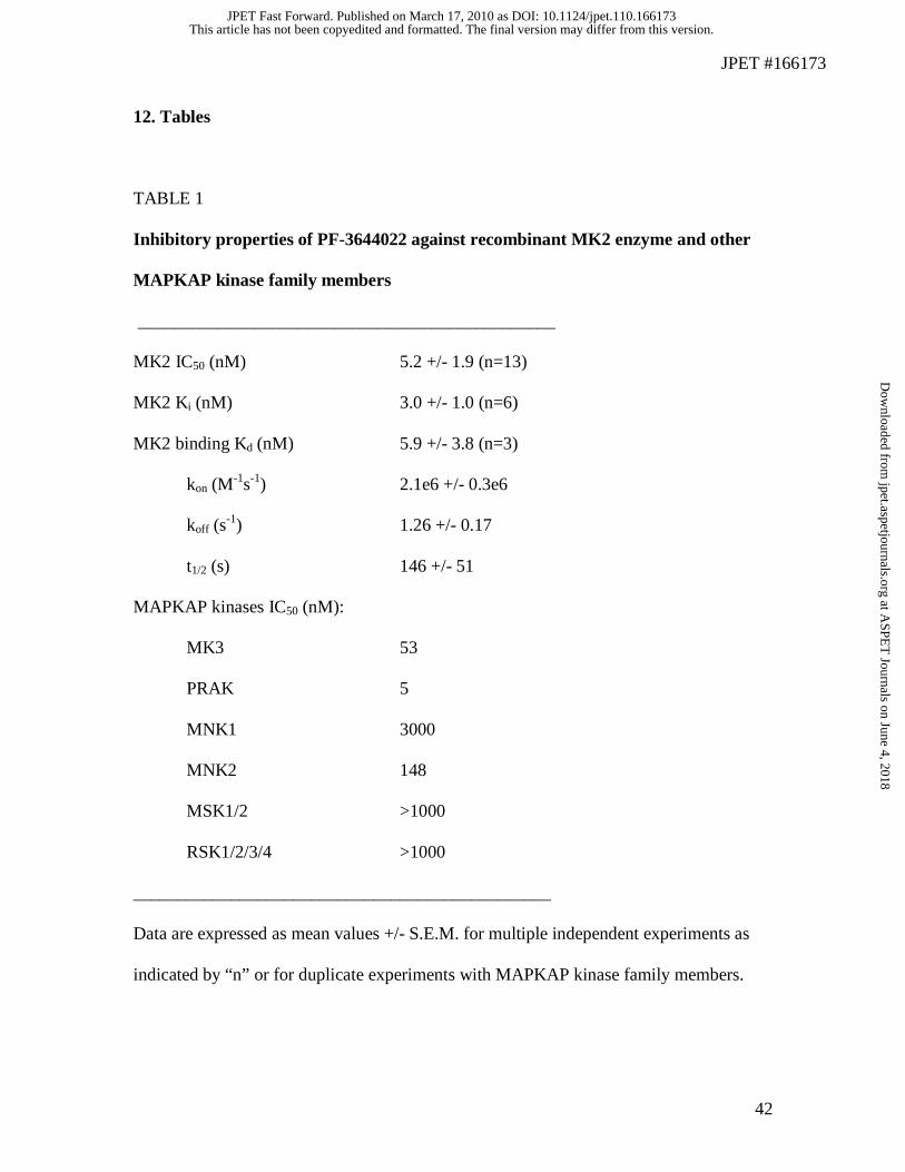
JPET #166173
42
12. Tables
TABLE 1
Inhibitory properties of PF-3644022 against recombinant MK2 enzyme and other
MAPKAP kinase family members
_______________________________________________
MK2 IC50 (nM) 5.2 +/- 1.9 (n=13)
MK2 Ki (nM) 3.0 +/- 1.0 (n=6)
MK2 binding Kd (nM) 5.9 +/- 3.8 (n=3)
kon (M-1s-1) 2.1e6 +/- 0.3e6
koff (s-1) 1.26 +/- 0.17
t1/2 (s) 146 +/- 51
MAPKAP kinases IC50 (nM):
MK3 53
PRAK 5
MNK1 3000
MNK2 148
MSK1/2 >1000
RSK1/2/3/4 >1000
_______________________________________________
Data are expressed as mean values +/- S.E.M. for multiple independent experiments as
indicated by “n” or for duplicate experiments with MAPKAP kinase family members.
This article has not been copyedited and formatted. The final version may differ from this version.JPET Fast Forward. Published on March 17, 2010 as DOI: 10.1124/jpet.110.166173
at ASPE
T Journals on June 4, 2018
jpet.aspetjournals.orgD
ownloaded from
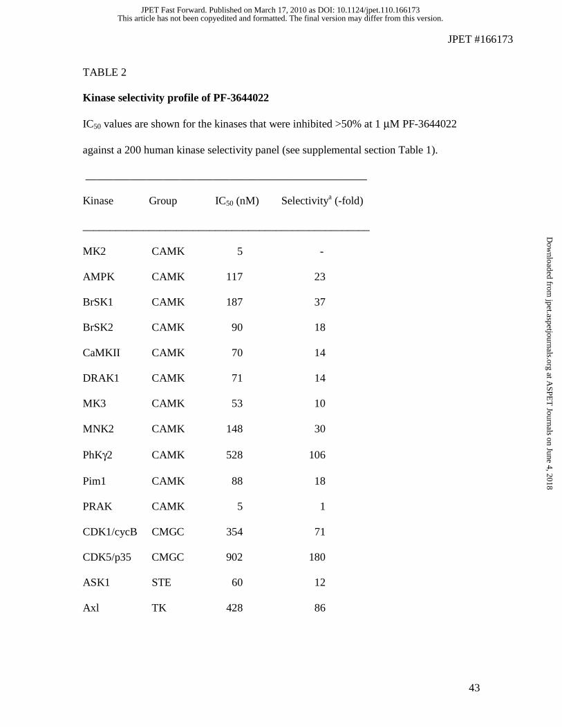
JPET #166173
43
TABLE 2 Kinase selectivity profile of PF-3644022
IC50 values are shown for the kinases that were inhibited >50% at 1 μM PF-3644022
against a 200 human kinase selectivity panel (see supplemental section Table 1).
___________________________________________________
Kinase Group IC50 (nM) Selectivitya (-fold)
____________________________________________________
MK2 CAMK 5 -
AMPK CAMK 117 23
BrSK1 CAMK 187 37
BrSK2 CAMK 90 18
CaMKII CAMK 70 14
DRAK1 CAMK 71 14
MK3 CAMK 53 10
MNK2 CAMK 148 30
PhKγ2 CAMK 528 106
Pim1 CAMK 88 18
PRAK CAMK 5 1
CDK1/cycB CMGC 354 71
CDK5/p35 CMGC 902 180
ASK1 STE 60 12
Axl TK 428 86
This article has not been copyedited and formatted. The final version may differ from this version.JPET Fast Forward. Published on March 17, 2010 as DOI: 10.1124/jpet.110.166173
at ASPE
T Journals on June 4, 2018
jpet.aspetjournals.orgD
ownloaded from
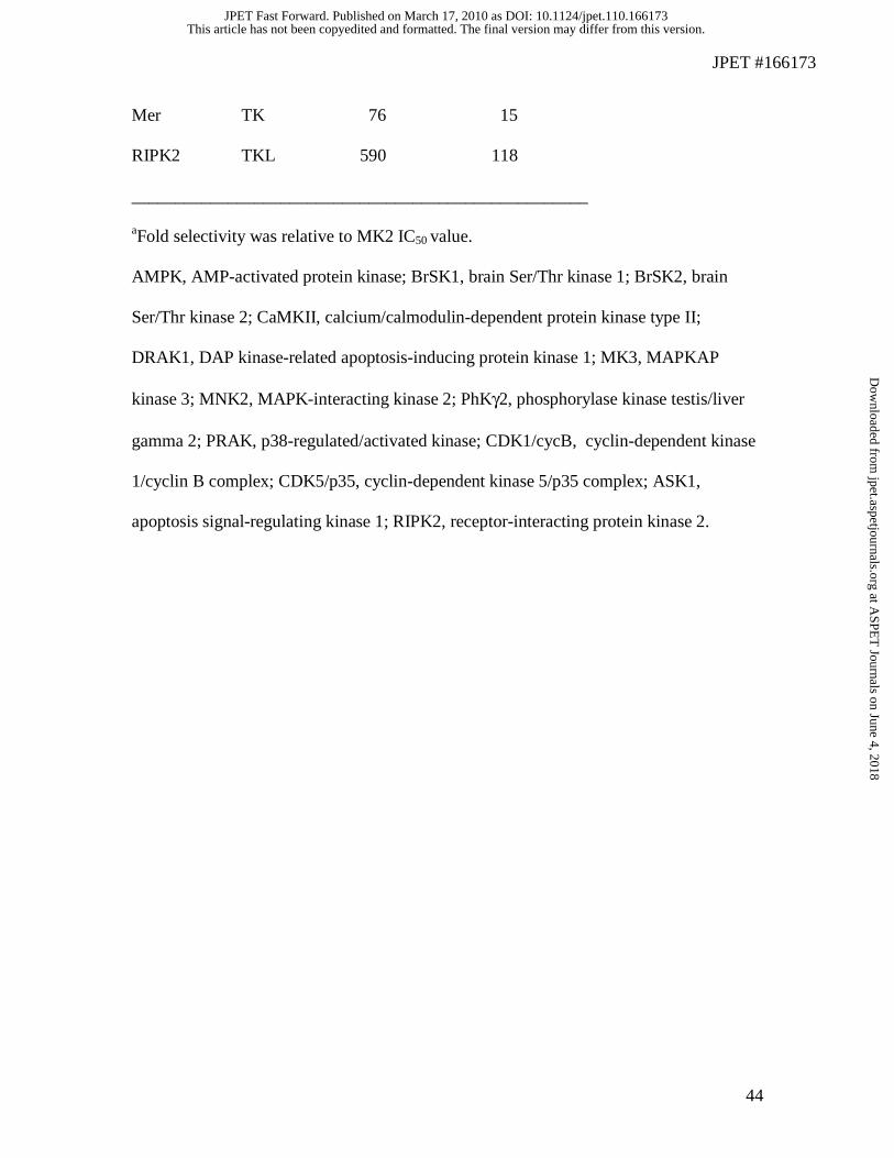
JPET #166173
44
Mer TK 76 15
RIPK2 TKL 590 118
____________________________________________________ aFold selectivity was relative to MK2 IC50 value. AMPK, AMP-activated protein kinase; BrSK1, brain Ser/Thr kinase 1; BrSK2, brain
Ser/Thr kinase 2; CaMKII, calcium/calmodulin-dependent protein kinase type II;
DRAK1, DAP kinase-related apoptosis-inducing protein kinase 1; MK3, MAPKAP
kinase 3; MNK2, MAPK-interacting kinase 2; PhKγ2, phosphorylase kinase testis/liver
gamma 2; PRAK, p38-regulated/activated kinase; CDK1/cycB, cyclin-dependent kinase
1/cyclin B complex; CDK5/p35, cyclin-dependent kinase 5/p35 complex; ASK1,
apoptosis signal-regulating kinase 1; RIPK2, receptor-interacting protein kinase 2.
This article has not been copyedited and formatted. The final version may differ from this version.JPET Fast Forward. Published on March 17, 2010 as DOI: 10.1124/jpet.110.166173
at ASPE
T Journals on June 4, 2018
jpet.aspetjournals.orgD
ownloaded from
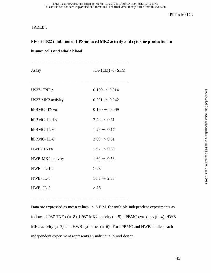
JPET #166173
45
TABLE 3
PF-3644022 inhibition of LPS-induced MK2 activity and cytokine production in
human cells and whole blood.
______________________________________________
Assay IC50 (μM) +/- SEM
_______________________________________________
U937- TNFα 0.159 +/- 0.014
U937 MK2 activity 0.201 +/- 0.042
hPBMC- TNFα 0.160 +/- 0.069
hPBMC- IL-1β 2.78 +/- 0.51
hPBMC- IL-6 1.26 +/- 0.17
hPBMC- IL-8 2.09 +/- 0.51
HWB- TNFα 1.97 +/- 0.80
HWB MK2 activity 1.60 +/- 0.53
HWB- IL-1β > 25
HWB- IL-6 10.3 +/- 2.33
HWB- IL-8 > 25
_______________________________________________
Data are expressed as mean values +/- S.E.M. for multiple independent experiments as
follows: U937 TNFα (n=8), U937 MK2 activity (n=5), hPBMC cytokines (n=4), HWB
MK2 activity (n=3), and HWB cytokines (n=6). For hPBMC and HWB studies, each
independent experiment represents an individual blood donor.
This article has not been copyedited and formatted. The final version may differ from this version.JPET Fast Forward. Published on March 17, 2010 as DOI: 10.1124/jpet.110.166173
at ASPE
T Journals on June 4, 2018
jpet.aspetjournals.orgD
ownloaded from
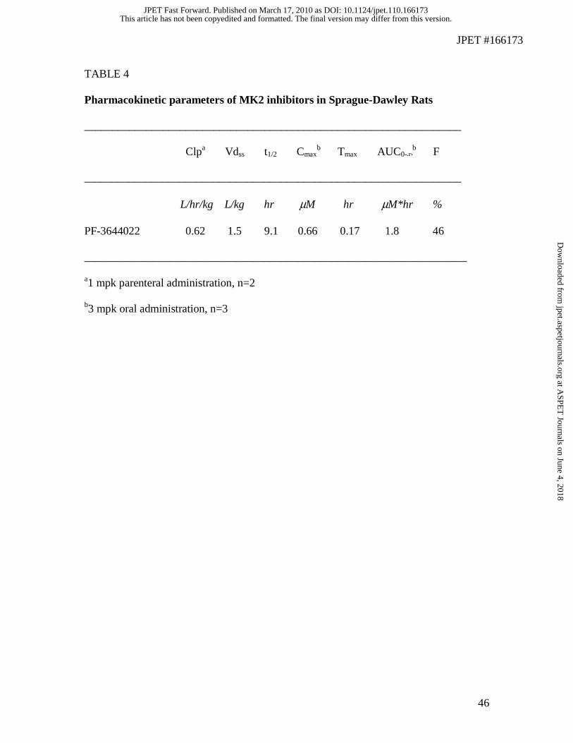
JPET #166173
46
TABLE 4
Pharmacokinetic parameters of MK2 inhibitors in Sprague-Dawley Rats
___________________________________________________________________
Clpa Vdss t1/2 Cmaxb Tmax AUC0-∞
b F
___________________________________________________________________
L/hr/kg L/kg hr μM hr μM*hr %
PF-3644022 0.62 1.5 9.1 0.66 0.17 1.8 46
____________________________________________________________________
a1 mpk parenteral administration, n=2
b3 mpk oral administration, n=3
This article has not been copyedited and formatted. The final version may differ from this version.JPET Fast Forward. Published on March 17, 2010 as DOI: 10.1124/jpet.110.166173
at ASPE
T Journals on June 4, 2018
jpet.aspetjournals.orgD
ownloaded from
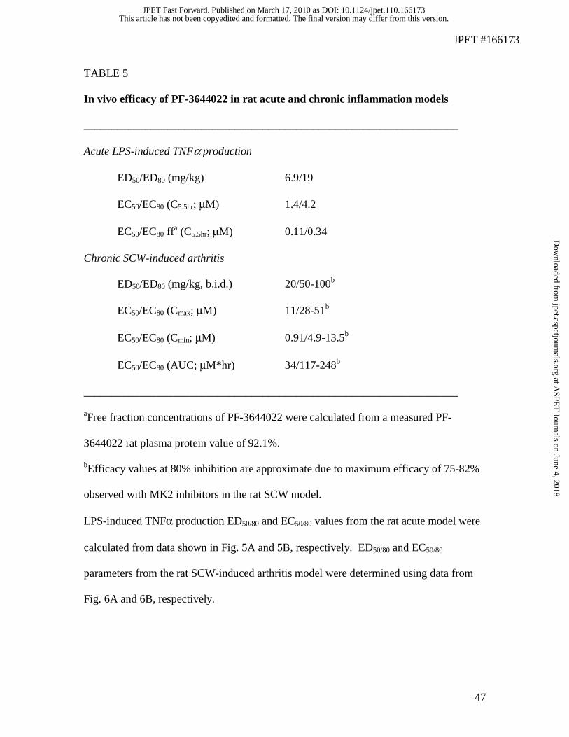
JPET #166173
47
TABLE 5
In vivo efficacy of PF-3644022 in rat acute and chronic inflammation models
___________________________________________________________________
Acute LPS-induced TNFα production
ED50/ED80 (mg/kg) 6.9/19
EC50/EC80 (C5.5hr; μM) 1.4/4.2
EC50/EC80 ffa (C5.5hr; μM) 0.11/0.34
Chronic SCW-induced arthritis
ED50/ED80 (mg/kg, b.i.d.) 20/50-100b
EC50/EC80 (Cmax; μM) 11/28-51b
EC50/EC80 (Cmin; μM) 0.91/4.9-13.5b
EC50/EC80 (AUC; μM*hr) 34/117-248b
___________________________________________________________________
aFree fraction concentrations of PF-3644022 were calculated from a measured PF-
3644022 rat plasma protein value of 92.1%.
bEfficacy values at 80% inhibition are approximate due to maximum efficacy of 75-82%
observed with MK2 inhibitors in the rat SCW model.
LPS-induced TNFα production ED50/80 and EC50/80 values from the rat acute model were
calculated from data shown in Fig. 5A and 5B, respectively. ED50/80 and EC50/80
parameters from the rat SCW-induced arthritis model were determined using data from
Fig. 6A and 6B, respectively.
This article has not been copyedited and formatted. The final version may differ from this version.JPET Fast Forward. Published on March 17, 2010 as DOI: 10.1124/jpet.110.166173
at ASPE
T Journals on June 4, 2018
jpet.aspetjournals.orgD
ownloaded from
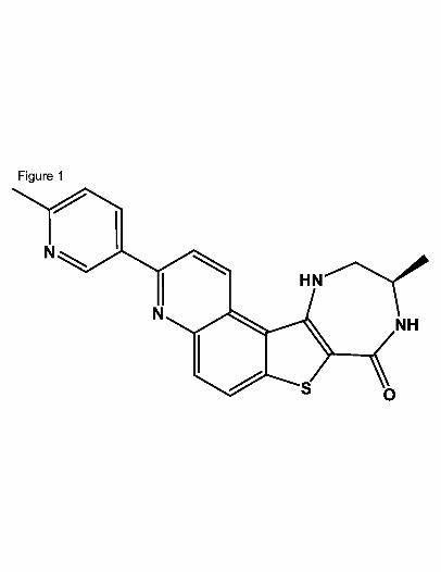
This article has not been copyedited and formatted. The final version may differ from this version.JPET Fast Forward. Published on March 17, 2010 as DOI: 10.1124/jpet.110.166173
at ASPE
T Journals on June 4, 2018
jpet.aspetjournals.orgD
ownloaded from
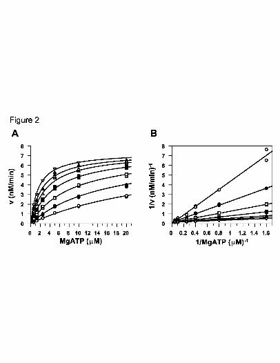
This article has not been copyedited and formatted. The final version may differ from this version.JPET Fast Forward. Published on March 17, 2010 as DOI: 10.1124/jpet.110.166173
at ASPE
T Journals on June 4, 2018
jpet.aspetjournals.orgD
ownloaded from
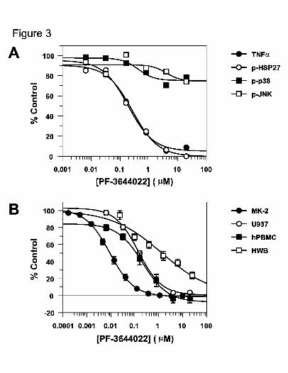
This article has not been copyedited and formatted. The final version may differ from this version.JPET Fast Forward. Published on March 17, 2010 as DOI: 10.1124/jpet.110.166173
at ASPE
T Journals on June 4, 2018
jpet.aspetjournals.orgD
ownloaded from
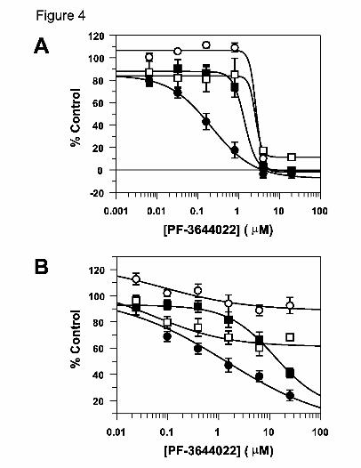
This article has not been copyedited and formatted. The final version may differ from this version.JPET Fast Forward. Published on March 17, 2010 as DOI: 10.1124/jpet.110.166173
at ASPE
T Journals on June 4, 2018
jpet.aspetjournals.orgD
ownloaded from
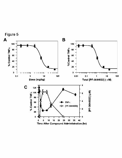
This article has not been copyedited and formatted. The final version may differ from this version.JPET Fast Forward. Published on March 17, 2010 as DOI: 10.1124/jpet.110.166173
at ASPE
T Journals on June 4, 2018
jpet.aspetjournals.orgD
ownloaded from
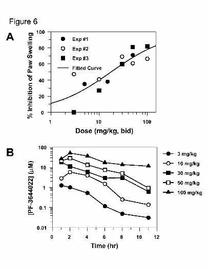
This article has not been copyedited and formatted. The final version may differ from this version.JPET Fast Forward. Published on March 17, 2010 as DOI: 10.1124/jpet.110.166173
at ASPE
T Journals on June 4, 2018
jpet.aspetjournals.orgD
ownloaded from
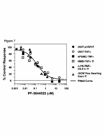
This article has not been copyedited and formatted. The final version may differ from this version.JPET Fast Forward. Published on March 17, 2010 as DOI: 10.1124/jpet.110.166173
at ASPE
T Journals on June 4, 2018
jpet.aspetjournals.orgD
ownloaded from