jpet fast forward. published on may 22, 2007 as...
TRANSCRIPT
JPET #122218
Page 1 of 32
Title Page
PDE4B5, a novel, super-short, brain-specific cAMP phosphodiesterase-4 variant whose
isoform-specifying N-terminal region is identical to that of cAMP phosphodiesterase-
4D6 (PDE4D6)
York-Fong Cheung, Zhengyan Kan, Philip Garrett-Engele, Irene Gall, Hannah Murdoch,
George S. Baillie, Luiz Miguel Camargo, Jason M. Johnson, Miles D. Houslay and John
C. Castle
Molecular Pharmacology Group, Wolfson Building, Division of Biochemistry and
Molecular Biology, Institute of Biomedical & Life Sciences, University of Glasgow,
Glasgow, G12 8QQ, Scotland, United Kingdom (YFC, IG, HM, GSB, MDH)
Rosetta Inpharmatics LLC, a wholly owned subsidiary of Merck & Co., Inc., Seattle,
Washington, 98109, USA (ZK, PGE, JMJ, JCC)
Merck Research Laboratories, BMB-8-118, 33 Avenue Louis Pasteur, Boston, MA
02115, USA (LMC)
JPET Fast Forward. Published on May 22, 2007 as DOI:10.1124/jpet.107.122218
Copyright 2007 by the American Society for Pharmacology and Experimental Therapeutics.
This article has not been copyedited and formatted. The final version may differ from this version.JPET Fast Forward. Published on May 22, 2007 as DOI: 10.1124/jpet.107.122218
at ASPE
T Journals on A
ugust 31, 2018jpet.aspetjournals.org
Dow
nloaded from
JPET #122218
Page 2 of 32
Running Title Page
Running title: PDE4B5, a novel super-short cAMP phosphodiesterase-4 variant
Corresponding author:
John C. Castle
Rosetta Inpharmatics
401 Terry Ave North, Seattle, Washington 98109
USA
Tel. 1-206-802-6337
Fax 1-206-802-7303
E-Mail: [email protected]
Section Cellular and Molecular
Number of text pages 27
Number of tables 3
Number of figures 8
Number of references 59
Number of words in the abstract 219
Number of words in the introduction 655
Number of words in the discussion 940
Non-standard abbreviations:
UCR, upstream conserved regions; DMSO, dimethyl sulfoxide; ORF, open reading
frame; DISC1, gene Disrupted In SChizophrenia 1; PCR, polymerase chain reaction; RT,
reverse transcription; DOTAP, Dithiothreitol, N--{1-(2,3-Dioleoyloxy)propyl}-N,N,N,-
trimethylammonium methylsulfate; DTT, dithiothreitol
This article has not been copyedited and formatted. The final version may differ from this version.JPET Fast Forward. Published on May 22, 2007 as DOI: 10.1124/jpet.107.122218
at ASPE
T Journals on A
ugust 31, 2018jpet.aspetjournals.org
Dow
nloaded from
JPET #122218
Page 3 of 32
Abstract
The cAMP-specific phosphodiesterase-4 (PDE4) gene family is the target of several
potential selective therapeutic inhibitors. The four PDE4 genes generate several distinct
protein-coding isoforms through the use of alternative promoters and 5' coding exons.
Using mouse transcripts, we identified a novel, super-short isoform of human PDE4B
encoding a novel 5' terminus, which we label PDE4B5. The protein coding region of the
novel 5' exon is conserved across vertebrates, chicken, zebrafish, and fugu. RT-PCR and
qPCR measurements show this isoform is brain-specific. The novel protein is 58 ± 2
kDa; has cAMP hydrolyzing enzymatic activity and is inhibited by PDE4 selective
inhibitors rolipram and ariflo (cilomilast). Confocal and sub-cellular fractionation
analyses show that it is distributed predominantly and unevenly within the cytosol. The
16 novel N-terminal residues of PDE4B5 are identical to the 16 N-terminal residues of
the super-short isoform of PDE4D (PDE4D6), which is also brain-specific. PDE4B5 is
able to bind the scaffold protein DISC1, whose gene has been linked to schizophrenia.
Microarray expression profiling of the PDE4 gene family shows that specific PDE4 genes
are enriched in muscle and blood fractions; however, only by monitoring the individual
isoforms is the brain-specificity of the super-short PDE4D and PDE4B isoforms revealed.
Understanding the distinct tissue specificity of PDE4 isoforms will be important for
understanding phosphodiesterase biology and opportunities for therapeutic intervention.
This article has not been copyedited and formatted. The final version may differ from this version.JPET Fast Forward. Published on May 22, 2007 as DOI: 10.1124/jpet.107.122218
at ASPE
T Journals on A
ugust 31, 2018jpet.aspetjournals.org
Dow
nloaded from
JPET #122218
Page 4 of 32
Introduction
Signaling systems coordinate most cellular functions and thus provide key targets
for drug discovery. Identifying appropriate targets and generating selective modulators
of these targets present major challenges in the initial stages of drug discovery. Different
isoenzymes are often found at key control points in signaling networks, allowing cells to
tailor signaling pathways through changes in activity, regulation, spatial distribution, and
compartmentalization (Wong and Scott, 2004).
The cAMP signaling pathway plays a pivotal role in many key cellular processes
(Tasken and Aandahl, 2004; Smith et al., 2006). Indeed, gradients of cAMP have been
identified in various cell types (Zhang et al., 2001; Zaccolo and Pozzan, 2002;
Willoughby et al., 2006), leading to compartmentalized responses (Tasken and Aandahl,
2004; Wong and Scott, 2004; Smith et al., 2006). Underpinning the formation of such
gradients is the degradation of cAMP by phosphodiesterases (PDEs) (Jurevicius et al.,
2003; Mongillo et al., 2004; Lynch et al., 2005; McCahill et al., 2005; Willoughby et al.,
2006) targeted to specific intracellular sites and signaling complexes (Houslay and
Adams, 2003; Baillie and Houslay, 2005). There are many PDE gene families, eight of
which code for proteins able to hydrolyze cAMP (Manganiello and Degerman, 1999;
Francis et al., 2001; Beavo and Brunton, 2002; Lugnier, 2006). To date, members of the
PDE3 and PDE4 families have been shown to play an important role in determining
compartmentalized cAMP signaling (Jurevicius et al., 2003; Mongillo et al., 2004; Lynch
et al., 2005; McCahill et al., 2005; Willoughby et al., 2006), making it important to
appreciate the range of isoforms that form these families as a prelude to determining their
functional roles.
The cAMP-specific phosphodiesterase 4 (PDE4) family is the target of several
selective inhibitors having therapeutic potential as anti-inflammatory agents, anti-
depressants and cognitive enhancers (Huang et al., 2001; O'Donnell and Zhang, 2004;
Renau, 2004; Spina, 2004; Houslay et al., 2005). Four genes (PDE4A, PDE4B, PDE4C,
PDE4D) generate a large set of PDE4 isoforms through the use of distinct promoters and
alternative pre-mRNA splicing (Conti et al., 2003; Houslay and Adams, 2003; Houslay et
al., 2005). Their unique N-terminal regions, encoded by specific 5’ exons, define
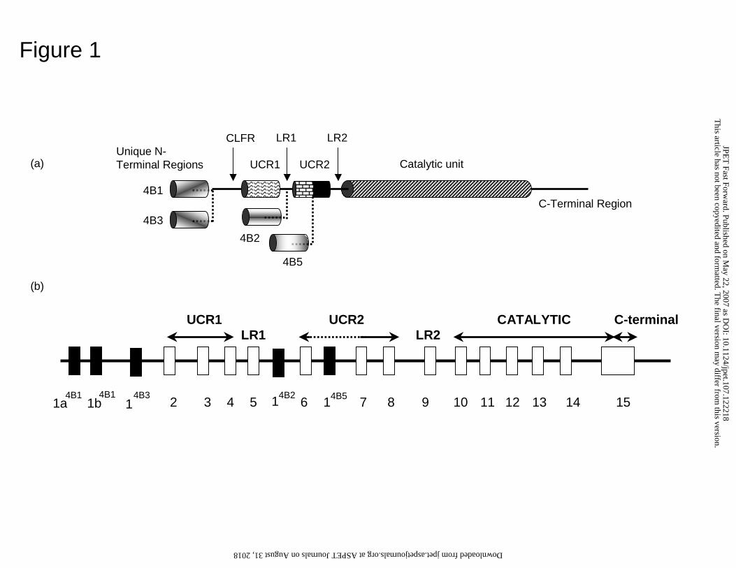
individual PDE4 isoforms (Fig. 1). Accordingly, PDE4 isoforms are subcategorized into
This article has not been copyedited and formatted. The final version may differ from this version.JPET Fast Forward. Published on May 22, 2007 as DOI: 10.1124/jpet.107.122218
at ASPE
T Journals on A
ugust 31, 2018jpet.aspetjournals.org
Dow
nloaded from
JPET #122218
Page 5 of 32
long forms, which possess the regulatory upstream conserved regions, UCR1 and UCR2;
short isoforms, which lack UCR1; or super-short isoforms, which lack UCR1 and have a
truncated UCR2.
The PDE4B gene has been linked to schizophrenia in humans (Millar et al., 2005;
Pickard et al., 2007) and knockout of the PDE4B gene in mice both generates an anti-
depressant-like profile (O'Donnell and Zhang, 2004) and compromises the generation of
airway hyper-reactivity and inflammatory actions mediated by macrophages (Jin et al.,
2005). Also, chronic nicotine treatment, which has an anti-depressant action, causes
down-regulation of PDE4B transcripts in the nucleus accumbens, prefrontal cortex and
hippocampus of rats (Polesskaya et al., 2007).
The human PDE4B gene has been shown to encode a number of distinct isoforms,
namely the long PDE4B1 (Bolger et al., 1993) and PDE4B3 (Huston et al., 1997)
isoforms and the short PDE4B2 isoform (Bolger et al., 1993; Obernolte et al., 1993).
While an additional long isoform, called PDE4B4, has been identified in rodents, this
isoform is not encoded by the human genome (Shepherd et al., 2003). Indeed, particular
PDE4 isoforms appear to have specific functional roles as evidenced from siRNA-
mediated knockdown studies in cells (Lynch et al., 2005) and from physiological studies
showing that nocturnal increases in PDE4B2 provide a negative feedback role in
adrenergic/cAMP signalling in the pineal gland (Kim et al., 2007).
Here we identify and characterize the first super-short isoform (PDE4B5) encoded
by the PDE4B gene. This isoform is highly conserved across species, is active, and
responds to drug inhibition. We also show, for the first time, conservation between
PDE4 sub-families of an isoform-specific N-terminal region, with the PDE4B5 N-
terminal isoform of PDE4B5 being identical to the N-terminus of the super-short isoform
encoded by the PDE4D gene, PDE4D6 (Wang et al., 2003).
This article has not been copyedited and formatted. The final version may differ from this version.JPET Fast Forward. Published on May 22, 2007 as DOI: 10.1124/jpet.107.122218
at ASPE
T Journals on A
ugust 31, 2018jpet.aspetjournals.org
Dow
nloaded from
JPET #122218
Page 6 of 32
Methods
Reagents
[3H]-cyclic AMP and ECL were purchased from Amersham International
(Amersham, UK). DOTAP and protease inhibitor tablets were obtained from Boehringer
Mannheim (Mannheim, Germany). Bradford was purchased from Bio-Rad (Herts, UK).
All other materials were from Sigma (Poole, UK). Protein G-sepharose beads was
purchased from Amersham Biosciences. Anti-FLAG M2 was supplied by Sigma-Aldrich.
PDE4B antisera was as previously described (Huston et al., 1997).
Computational prediction and analysis of PDE4B novel splice variant
Mouse transcripts were aligned to the mouse genome and the orthologous human
genomic loci (Kan et al., 2005) and the resulting splice patterns were compared with
those of human transcripts to identify novel patterns. Inferred novel splice variant
transcript sequences were extracted from the human genome sequence. The nucleotide
and translated protein sequences of the variant were searched against NCBI human
transcript and protein databases using the on-line BLAST server
(http://www.ncbi.nlm.nih.gov/BLAST/). Genomic annotations and conservation in the
PDE4B gene locus were inspected using the UCSC genome browser, hg18 human
assembly (http://genome.ucsc.edu/). Conserved TFBS predictions were taken from the
browser tracks named “HMR Conserved Transcription Factor Binding Sites” by Matt
Weirauch and Brian Raney at the University of California at Santa Cruz.
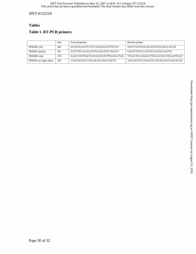
RT-PCR
We used the Qiagen OneStep RT-PCR kit (Qiagen Cat. # 210212). The PCR
component involved 35 cycles of 94°C – 30 seconds, 63.5°C – 40 seconds and 72°C – 50
to 120 seconds. Products were resolved on a 2% agarose gel run at 100 volts in TAE
buffer. Primer sequences and expected band sizes for PDE4B5, the PDE4B C-terminus,
PDE4D6, and the PDE4D N-terminus (excluding PDE4D6) are shown in Table 1. RT-
PCR primers were designed to be specific to their target.
TaqMan measurements
This article has not been copyedited and formatted. The final version may differ from this version.JPET Fast Forward. Published on May 22, 2007 as DOI: 10.1124/jpet.107.122218
at ASPE
T Journals on A
ugust 31, 2018jpet.aspetjournals.org
Dow
nloaded from
JPET #122218
Page 7 of 32
TaqMan primer-probe reagents were obtained through the Applied Biosystems
Assays-by-Design custom assay service (Foster City, CA). Probe sequences were
designed to straddle the unique splice junction characteristic of each alternative splice
form. TaqMan assays were performed on an ABI 7900 real time PCR instrument in 10 µl
assays that were run in triplicate in a 384-well format optical PCR plate. The assays were
calibrated with isoform-specific RT-PCR clones using the standard curve method
(http://www.appliedbiosystems.com/support/tutorials/pdf/essentials_of_real_time_pcr.pd
f). Standard curves generated from plasmid clones were linear across at least six orders of
magnitude, and all reported values derived for total tissue RNA fell within the range of
these standard curves. RNA was converted to cDNA for TaqMan measurements using a
commercially available kit from Applied Biosystems. All assays were normalized on a
tissue-to-tissue basis by adding a constant amount of input total RNA into the RT
reaction. Taqman primer locations are labeled as 'C' in Fig. 2a.
Microarray data
Custom oligonucleotide microarrays were purchased from Agilent Technologies
(Palo Alto, California). We designed these arrays to monitor the expression of 18,000
genes and associated alternate splicing events (Johnson et al., 2003). After alignment of
107,551 full-length human mRNA transcripts to the human genome, probes were
designed to target every exon (60mers) and every exon-exon junction (36mers on 10
nucleotide T stilts). Poly[A]+ mRNA was amplified with a full-length amplification
method using random-priming sequences to reproduce the entire transcript (Castle et al.,
2003). Fluorescent dye-labeling, hybridization conditions, and scanning were performed
as previously (Hughes et al., 2001). Each amplified sample was hybridized twice against
a common reference pool in a dye-swap experiment. The reference pool included 20
disease-free adult tissues, including peripheral leukocytes but excluding other blood
fractions. Ratios shown are the mean of three probes located in regions common to all
isoforms.
Tissues for RT-PCR, Taqman, and microarray measurements
This article has not been copyedited and formatted. The final version may differ from this version.JPET Fast Forward. Published on May 22, 2007 as DOI: 10.1124/jpet.107.122218
at ASPE
T Journals on A
ugust 31, 2018jpet.aspetjournals.org
Dow
nloaded from
JPET #122218
Page 8 of 32
Cell lines and human tissues were purchased as mRNA or total RNA from
Clontech (Mountain View, California). Each tissue sample was pooled from multiple
donors, typically 12, by the vendor.
SDS/PAGE and Western Blotting
Acrylamide gels (4-12%) were used and the samples boiled for 5 min after being
resuspended in SDS sample buffer. Gels were run at 100 V/gel for 1-2h with cooling.
For detection of transfected PDE by western blotting, 2-50 µg protein samples were
separated by SDS-PAGE and then transferred to nitrocellulose before being
immunoblotted using the indicated specific antisera. Labeled bands were identified using
peroxidase linked to anti-rabbit IgG and the Amersham ECL western blotting kit was
used as a visualization protocol. We used polyclonal antisera able to detect all active
human PDE4B isoforms as described previously (Huston et al., 1997). This polyclonal
was raised against the extreme C-terminal region that is unique to the PDE4B sub-family
and is found in all known active PDE4B isoforms.
Constructs
The ORF encoding human PDE4B5 (EF595686) was engineered for expression in
pcDNA3. PDE4B1 (Genbank accession L20966) was used as a PCR template to
incorporate the common super-short form region of PDE4B into the new construct. The
sequence of the novel 15 N-terminal amino acids of PDE4B5 plus 6 amino acids (249-
254) of PDE4B1, a start codon, and a Not1 restriction enzyme site were incorporated into
the 5’ primer. The 3’ primer incorporated amino acids from C-terminal region of
PDE4B1, a stop codon and a Kpn1 enzyme restriction site. We also generated a FLAG
tagged version where the FLAG tag (N-Asp-Tyr-Lys-Asp-Asp-Asp-Asp-Lys-C) was
incorporated into the 3’ primer before the stop codon. A PCR reaction using Taq DNA
polymerase, PDE4B1 DNA template, and the above primers generated a fragment of
approximately 1558 bp, which was purified using PCR Purification Kit (Qiagen,
Crawley, UK). This fragment was digested with Not1 and Kpn1 prior to ligation (Rapid
DNA ligation Kit, Roche Diagnostic GmbH, Mannheim, Germany) into the multiple
This article has not been copyedited and formatted. The final version may differ from this version.JPET Fast Forward. Published on May 22, 2007 as DOI: 10.1124/jpet.107.122218
at ASPE
T Journals on A
ugust 31, 2018jpet.aspetjournals.org
Dow
nloaded from
JPET #122218
Page 9 of 32
cloning site (MCS) of pcDNA3.1 (Invitrogen, Paisley, UK) to generate either PDE4B4-
pcDNA3 or PDE4B5-FLAG-pcDNA3. Generation of a plasmid encoding N-terminally
FLAG epitope-tagged version of the 100kFDa full length DISC1 has been described
previously (Millar et al., 2005).
Transient expression of PDE4B isoforms in COS7 cells.
Transfection was done using the COS7 SV40-transformed monkey kidney cell
line maintained at 37˚C in an atmosphere of 5% CO2 / 95% air in complete growth
medium containing DMEM supplemented with 0.1% penicillin/streptomycin (10000
units ml-1), glutamine (2 mM) and 10 % FCS. As described previously (Huston et al.,
1997; Rena et al., 2001; Wallace et al., 2005), COS7 cells were transfected using DEAE
Dextran. The DNA to be transfected (10 µg) was mixed, and incubated for 15 min with
200 µl of 10 mg ml-1 DEAE-dextran in PBS to give a ‘DNA-dextran’ mix. When cells
reached 70% confluency confluence, in 100 mm dishes, the medium was removed and
the cells were given 10 ml of fresh DMEM containing 0.1 mM chloroquine and the
DNA-dextran mix (450 µl). The cells were then incubated for 4 h at 37˚C. After this
period the medium was removed and the cells shocked with 10% DMSO in PBS. After
PBS washing, the cells were returned to normal growth medium and left for a further two
days before use. For determination of PDE activity the cells were homogenized in KHEM
buffer (50 mM KCl, 10 mM EGTA, 1.92 mM MgCl2, 1 mM dithiothreitol, 50 mM
Hepes, final pH7.2,) containing ‘complete’ protease inhibitors (Boehringer Mannheim) of
final concentrations 40 µg ml-1 PMSF, 156 µg ml-1 benzamine, 1 µg ml-1 aprotonin, 1 µg
ml-1 leupeptin, 1 µg ml-1 pepstatin A and 1 µg ml-1 antipain. In such transfected cells,
>98% of the total PDE activity is due to the recombinant PDE4 isoform (Huston et al.,
1997). In some instances the transfected COS7 cells were plated onto 6 well plates for
use in experiments and then serum-starved over night before being treated with the
indicated ligands for the stated lengths of time.
Sub-cellular fractions.
Disruption of COS7 cells was done as described (McPhee et al., 1995; Bolger et
al., 1996; Huston et al., 1997). Cell homogenization was performed in KHEM buffer (50
This article has not been copyedited and formatted. The final version may differ from this version.JPET Fast Forward. Published on May 22, 2007 as DOI: 10.1124/jpet.107.122218
at ASPE
T Journals on A
ugust 31, 2018jpet.aspetjournals.org
Dow
nloaded from
JPET #122218
Page 10 of 32
mM KCl, 50 mM Hepes, KOH (pH 7.2), 10 mM EGTA, 1.92 mM MgCl2) containing 1
mM dithiothreitol and a mixture of protease inhibitors at final concentrations of 40 µg/ml
PMSF, 156 µg/ml benzamidine, 1 µg/ml aprotinin, 1 µg/ml leupeptin, 1 µg/ml pepstatin
A and 1 µg/ml antipain. Pellet fractions were also resuspended in this mixture. We then
generated a low-speed, P1 pellet (1000 gav for 10 min) and a high-speed, P2 pellet (60
min at 100000 gav), which left a high-speed supernatant (S2) fraction. The
homogenization procedure was complete in that no detectable latent lactate
dehydrogenase activity was present in the P1 pellet, indicating an absence of cytosolic
proteins. Equal volumes of samples were applied such that detection indicates relative
distribution across these three cellular sub-fractions.
Confocal analyses
Confocal imaging for analyzing PDE4 isoforms was performed as described
(Rena et al., 2001; Shepherd et al., 2003; Wallace et al., 2005). Here, PDE4B5 was
transiently over-expressed and visualized in COS cells using PDE4B specific antisera
(Huston et al., 1997). Cells were transfected using DOTAP (Roche, GmbH, Mannheim,
Germany), with the PDE4A11- pcDNA3 plasmid. Protein was expressed for 48h and
cells were fixed in 4% paraformaldehyde containing 5% sucrose. After permeabilisation
in 0.2% triton, proteins were blocked using 10% goat serum and 2% BSA before
PDE4A11 was detected using an antibody raised against the C-terminus of human
PDE4A and stained using Alexa 594 (Molecular Probes, Invitrogen, Paisley, UK). Cells
were observed using a Zeiss Pascal laser scanning microscope.
Immunoprecipitation
HEK293 cells expressing PDE4B5 and/or FlagDISC1 were washed with ice-cold
PBS and lysed in PBS containing 1% Triton X-100, 1 mM DTT, 10 mM NaF and 5 mM
NaPPi with a protease inhibitor cocktail added (Roche, West Sussex, UK). Lysates were
solubilized by rotation on a rotary wheel for 30 min at 4 °C. Insoluble material was
removed by a 15 min centrifugation at 14,000 gav at 4 °C followed by pre-clearing of
lysed supernatants by incubation with protein G-sepharose beads for 30 min at 4 °C.
Equalized amounts of pre-cleared lysates were incubated with the PDE4B antibody for a
This article has not been copyedited and formatted. The final version may differ from this version.JPET Fast Forward. Published on May 22, 2007 as DOI: 10.1124/jpet.107.122218
at ASPE
T Journals on A
ugust 31, 2018jpet.aspetjournals.org
Dow
nloaded from
JPET #122218
Page 11 of 32
minimum of 3 hours at 4 °C and immunocomplexes were captured following incubation
with protein G-sepharose beads for a further 1-2 hours. The immunoprecipitates were
washed three times in lysis buffer and eluted from the beads by the addition of Laemmli
buffer (Laemmli, 1970).
Assay of cAMP PDE activity
PDE activity using 1 µM cAMP as substrate was assayed by a modification of the
procedure of Thompson and Appleman (1971) and Rutten et al. (1973), as described
previously (Marchmont and Houslay, 1980; Sullivan et al., 1998; Rena et al., 2001). All
assays were conducted at 30 °C, and, in all experiments, a freshly prepared slurry of
Dowex:H2O:ethanol (1:1:1) was used. In all experiments, initial rates were taken from
linear time courses of activity. Dose-dependent inhibition by rolipram was determined in
the presence of 1 µM cAMP concentrations of cAMP as substrate over the indicated
range of rolipram concentrations. The IC50 was then determined from these values.
Rolipram was dissolved in 100% DMSO as a 1 mM stock and diluted in 20 mM TrisCl,
pH 7.4, 10 mM MgCl2 to provide a range of concentrations in the assay. The residual
levels of DMSO were shown not to affect PDE activity over the ranges used in this study.
Protein analysis.
Protein concentration was determined using BSA as standard (Bradford, 1976).
This article has not been copyedited and formatted. The final version may differ from this version.JPET Fast Forward. Published on May 22, 2007 as DOI: 10.1124/jpet.107.122218
at ASPE
T Journals on A
ugust 31, 2018jpet.aspetjournals.org
Dow
nloaded from
JPET #122218
Page 12 of 32
Results
Genomic identification of PDE4B5
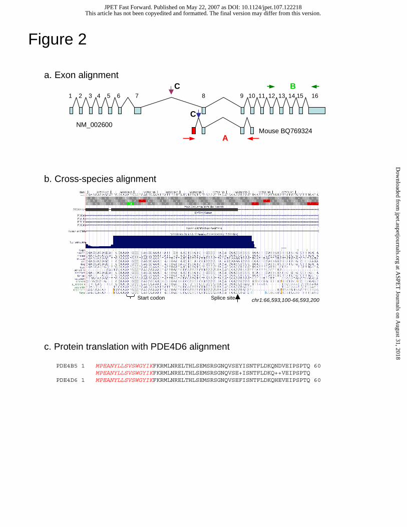
Mouse ESTs (e.g., BQ769324) aligned to the human genome indicate a
previously unknown 5’ exon of the PDE4B gene (Methods, Fig. 2a). From the novel
first exon splice-site, the 54 nucleotides upstream (in the 5' direction) and the six
nucleotides downstream (in the 3' direction) show very high conservation across fifteen
species. At the 5' end of this highly conserved sequence exists an in-frame ATG putative
start codon (Fig. 2b). The sequence beyond 6 nt upstream of the ATG contains many
insertions and deletions across species, as are also present beyond 6 nt downstream of the
splice site. However, between the putative start codon and the splice site, no insertions,
deletions, or stop codons exist in any of the species examined, including vertebrates,
chicken, zebrafish, and fugu. This high rate of protein reading frame evolutionary
conservation suggests the existence of a functional protein coding region.
If translated, the alternate first exon would produce a protein of 484 amino acids
with a unique N-terminal region of 16 amino acids (Fig. 2c). This protein would preserve
the phosphodiesterase catalytic domain but eliminate the 250 N-terminal residues found
in long isoforms, replacing them with the novel 16 amino acids. Thus the putative novel
PDE4B isoform, which we label PDE4B5, would fall into the category of a super-short
isoform (Houslay, 2001), lacking UCR1 and having a truncated UCR2 (Fig. 1).
Intriguingly, BLAST search against the human protein database found that the
PDE4B novel variant is homologous to PDE4D6, a recently discovered PDE4D splice
variant. The putative protein coding PDE4B5 and PDE4D6 nucleotide sequences show
81% identity (40 of 48 identical nucleotides). Furthermore, the residues of the two
unique N-terminus proteins perfectly match over the entire 16 amino acid sequence (Fig.
2c). This, to our knowledge, is the first description of conservation of amino acid
sequence over a PDE4 isoform-specific N-terminal region.
Validation and expression of PDE4B5
To validate human transcription of PDE4B5 isoform identified by mouse
transcripts, we designed RT-PCR primers targeting the prediction. The forward primer
was placed in the unique, novel exon of PDE4B5 and the reverse primer in the PDE4B
This article has not been copyedited and formatted. The final version may differ from this version.JPET Fast Forward. Published on May 22, 2007 as DOI: 10.1124/jpet.107.122218
at ASPE
T Journals on A
ugust 31, 2018jpet.aspetjournals.org
Dow
nloaded from
JPET #122218
Page 13 of 32
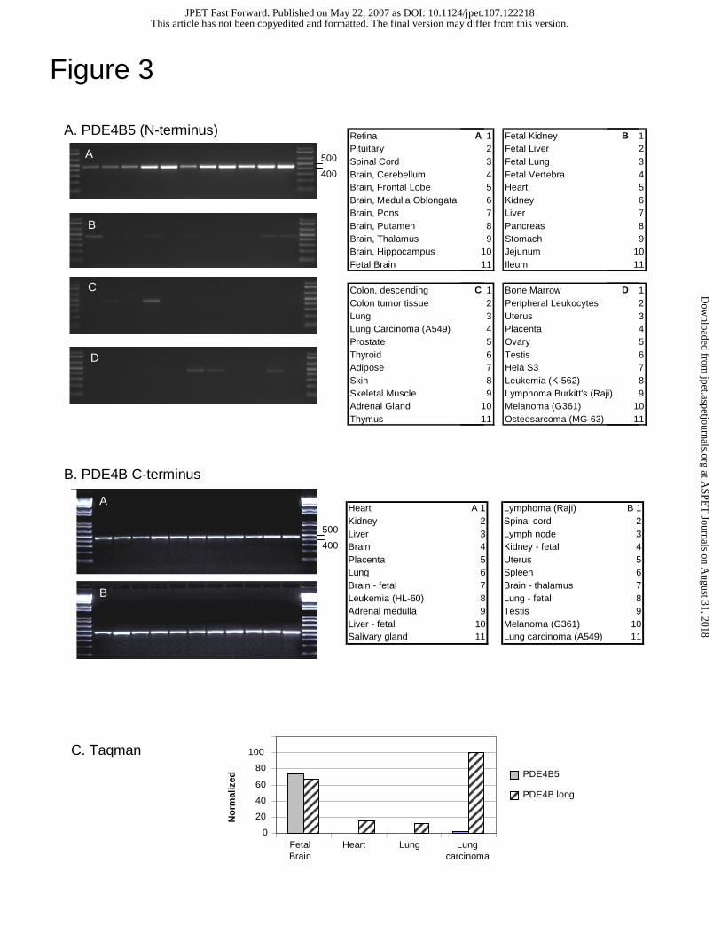
catalytic region (Fig 2a, A; Methods) and both primers were designed to be specific to
PDE4B. The gel image shows a bright band at the predicted size (442 nt) in brain
sections (fetal brain, cerebellum, frontal lobe, pons, putamen, thalamus and
hippocampus) (Fig. 3a). Weaker signals were observed in retina, spinal cord, pituitary,
fetal kidney, jejunum, ileum, lung carcinoma A549 cells, testis, HELA cells and G361
melanoma cells, and no band of other size was observed. The RT-PCR gel for total
PDE4B expression, using PDE4B-specifc probes targeting the shared catalytic unit,
shows fairly ubiquitous expression (Fig. 3b). We then quantitatively measured PDE4B5
and PDE4B N-terminus (short and long, but not super-short) transcript levels using
TaqMan in four tissues (Fig. 3c.). These measurements show expression of the PDE4B
long and short forms in all four tissues while PDE4B5 (super-short) expression is largely
constrained to fetal brain, with low levels in cell line A549, in agreement with the RT-
PCR gel images.
Subsequent analysis shows only limited homology matches between PDE4B
primers and PDE4D. The PDE4B5 forward primer, which lies outside of the coding
region, and the PDE4B5 reverse primer, in the PDE4B common catalytic domain, have a
maximum continuous homology to the PDE4D locus at a level of four and six
nucleotides, respectively. However, even if these matches were to produce products,
their size would be significantly different (> 50nt) than the expected 442 nt band.
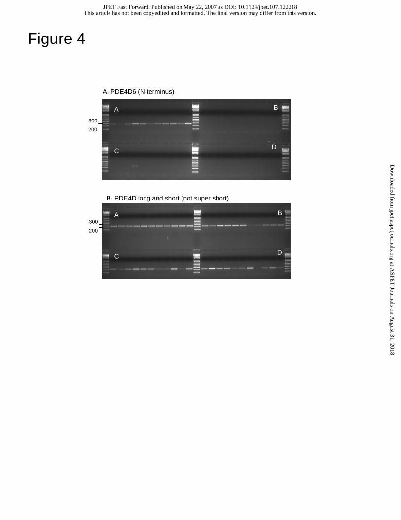
Given the homology to PDE4D6, we measured PDE4D6 expression using RT-
PCR, as per PDE4B5. Again, we designed the forward primer specific to the first exon
of PDE4D6, unique to the super-short isoform, and placed the reverse primer in the
common catalytic region. We also designed primers to amplify the longer isoforms of
PDE4D but not the super-short form, placing the forward primer in the region common to
the long and short isoforms but not present in the super-short isoform. The PDE4D6 gel
image (Fig.4a) shows expression primarily in brain tissues. The PDE4D long and short
gel image (Fig. 4b) shows expression in many tissues, but very low expression in liver,
kidney, and K-562.
Given the high expression similarity of PDE4B5 and PDE4D6 isoforms at the
sequence and expression level, we examined whether they are similarly regulated.
Conservation of the 2000 nucleotides upstream of the common PDE4B5/PDE4D6 ATG
This article has not been copyedited and formatted. The final version may differ from this version.JPET Fast Forward. Published on May 22, 2007 as DOI: 10.1124/jpet.107.122218
at ASPE
T Journals on A
ugust 31, 2018jpet.aspetjournals.org
Dow
nloaded from
JPET #122218
Page 14 of 32
codon is low but several cross-species conserved transcription factor binding sites are
predicted for each isoform. Upstream of PDE4B5, conserved POU1F1, RFX1, RFX,
BRN2, and OCT1 binding sites are predicted and upstream of PDE4D6 only SRF is
predicted. Thus, these predictions find no common transcription factor binding sites
between these two super-short isoforms, suggesting that the expression of these two
super-short species is distinctly controlled.
Expression of PDE4 genes in human tissues
We measured the expression of PDE4A, PDE4B, PDE4C, and PDE4D using
microarray experiments and show relative expression to a pool of tissues (Fig. 5). The
microarray probes were designed to monitor constitutively transcribed exons and
junctions, and thus monitor overall gene expression. PDE4A shows slightly higher
expression in monocytes, skeletal muscle, testis, and pons. PDE4B shows high
enrichment in blood fractions and nervous system tissues. PDE4C shows ubiquitous
expression. PDE4D is enriched in blood fractions and skeletal muscle, in agreement with
the PDE4D gel image.
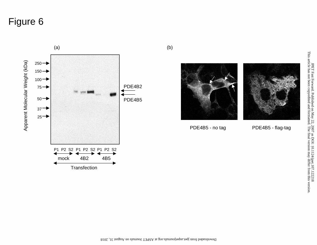
Size of PDE4B5 on SDS-PAGE
The ORF of PDE4B5 was engineered for expression in mammalian cells by
cloning into pcDNA3. Transfection of COS1 cells with PDE4B5-pcDNA3 allowed for
the detection of a single immunoreactive species of 58 ± 2 kDa, detected using a PDE4B
specific antiserum (Fig. 6a). Untransfected cells express PDE4B2 at levels not evident at
the exposure level used here, which detects only the recombinant species, which is
overexpressed so as to provide >98% of total PDE activity in these cells. This size agreed
well with a predicted size of 57.7 kDa, as derived from primary amino acid sequence.
The short PDE4B2 isoform migrated as a 68 kDa species, as shown previously by us
(Huston et al., 1997; Shepherd et al., 2003; Lynch et al., 2005).
Activity of PDE4B5
We transfected cells to express PDE4B5 and treated them with the archetypal
PDE4-selective inhibitor, rolipram. Over 97% of the total cAMP PDE activity was
This article has not been copyedited and formatted. The final version may differ from this version.JPET Fast Forward. Published on May 22, 2007 as DOI: 10.1124/jpet.107.122218
at ASPE
T Journals on A
ugust 31, 2018jpet.aspetjournals.org
Dow
nloaded from
JPET #122218
Page 15 of 32
inhibited by 1 µM cAMP rolipram as a substrate. Assayed with 1 µM cAMP, such
transfected cells had a cAMP PDE activity of 2-4 nmol cAMP hydrolyzed/min/mg cell
protein while empty vector transfected cells had an activity of 4-6 pmol cAMP
hydrolyzed/min/mg cell protein (n=3). Thus in PDE4B5-transfected cells, PDE4B5
accounts for > 98% of the total cAMP activity.
Analysis of PDE4B5 activity showed that it had a Km for cAMP of 5.8 ± 0.4 µM
(n=3). By analyzing equal immunoreactive amounts of PDE4B5 and PDE4B2, expressed
in COS1 cells lysates, we were able to determine the Vmax of PDE4B5 as 18 ± 3% of
PDE4B2 in these cells.
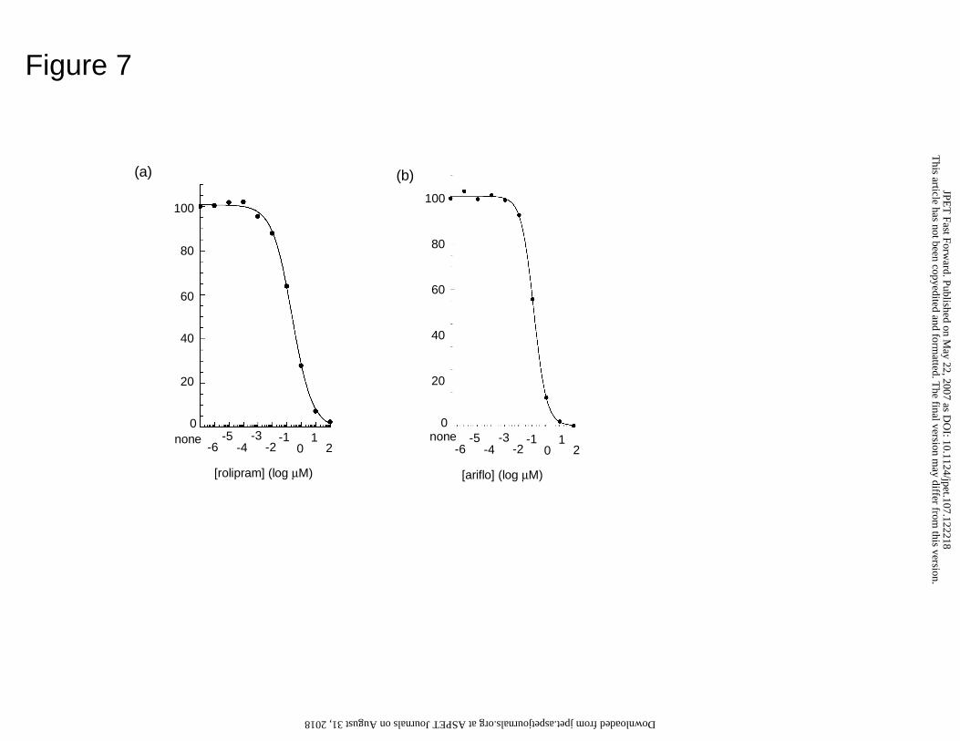
We then determined the sensitivity of PDE4B5 to inhibition by rolipram and
ariflo (cilomilast), a compound that has been in phase 3 clinical trials for COPD (Fig. 7).
This gave IC50 values of 380 ± 63 nM (n = 4) and 114 ± 17 nM (n = 3), for rolipram and
ariflo, respectively. Rolipram binds to the catalytic site of PDE4B, thus providing
competitive inhibition. Using the Cheng-Prussof equation (KI = IC50/(1+(S/Km))), Ki
values for inhibition of PDE4B5 by rolipram and ariflo are 324 and 97 nM, respectively.
Intracellular distribution of PDE4B5
COS1 cells transfected to express PDE4B5 were disrupted and separated out in
low speed membrane (P1), high speed membrane (P2) and high speed supernatant (S2)
fractions (Fig. 6a). These were then analyzed on a volume for volume basis for both
PDE4 activity and for PDE4B5 immunoreactivity to determine the relative distribution of
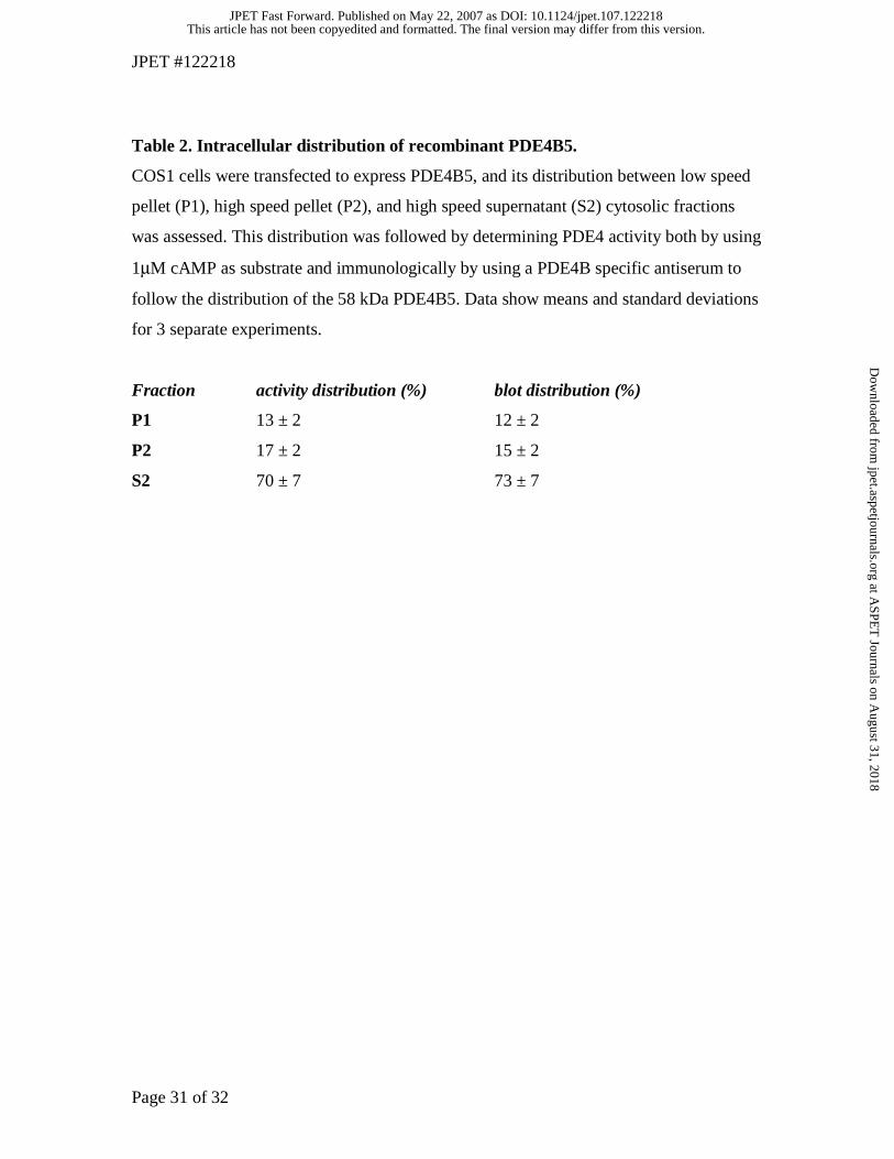
PDE4B5 among these three fractions (Table 2). This analysis showed that PDE4B5 was
found predominantly in the high speed supernatant, cytosolic fraction, but was also
evidently associated with membrane fractions, which accounted for around 30% of the
total PDE4B5 (Table 2).
We also analyzed the distribution of PDE4B5 in transfected COS1 cells (Fig. 6b).
As can be seen, while PDE4B5 is excluded from the nucleus, it is distributed throughout
the cytosol with small amounts associated with the plasma membrane. Distribution
through the cell interior is uneven, which may indicate its association with cytosolic
vesicles/complexes in addition to soluble forms. This would be consistent with
This article has not been copyedited and formatted. The final version may differ from this version.JPET Fast Forward. Published on May 22, 2007 as DOI: 10.1124/jpet.107.122218
at ASPE
T Journals on A
ugust 31, 2018jpet.aspetjournals.org
Dow
nloaded from
JPET #122218
Page 16 of 32
biochemical fractionation. A similar distribution was seen using a FLAG epitope-tagged
form of PDE4B5 (Fig. 6b).
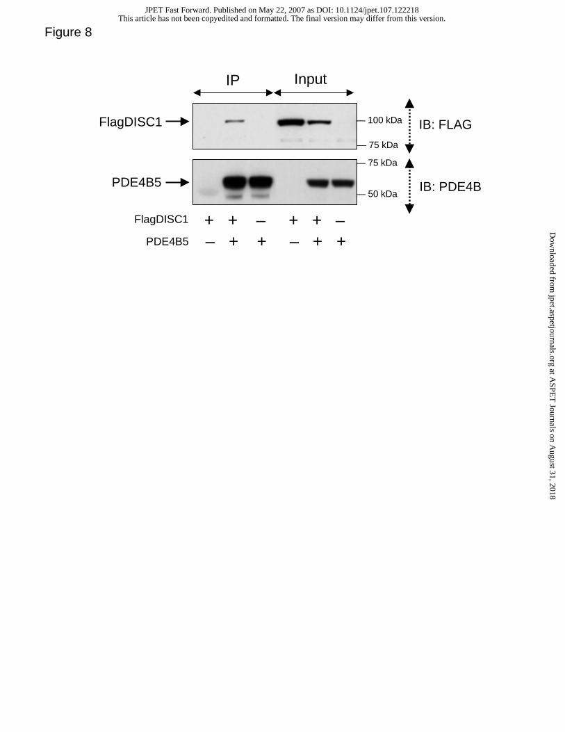
PDE4B5 interacts with DISC1.
DISC1 has previously been shown to interact with the long PDE4B1 and PDE4B3
isoforms as well as the short PDE4B2 isoform (Millar et al, 2005). Here we show that the
novel, super-short PDE4B5 isoform is able to interact with the full-length 100kDa DISC1
(fl-DISC1) isoform. This is evident from their co-immunoprecipitation as a complex from
HEK293 cells that were co-transfected to express both PDE4B5 and fl-DISC1 (Fig. 8).
This article has not been copyedited and formatted. The final version may differ from this version.JPET Fast Forward. Published on May 22, 2007 as DOI: 10.1124/jpet.107.122218
at ASPE
T Journals on A
ugust 31, 2018jpet.aspetjournals.org
Dow
nloaded from
JPET #122218
Page 17 of 32
Discussion
In determining the human transcriptome, the set of human mRNA genes has been
largely established and the current need is to establish alternative isoforms, their
expression, and their function. To this end, the availability of genomic sequence and
RNA transcripts from non-human species is a powerful, complementary resource to
human RNA transcripts (Kan et al., 2005).
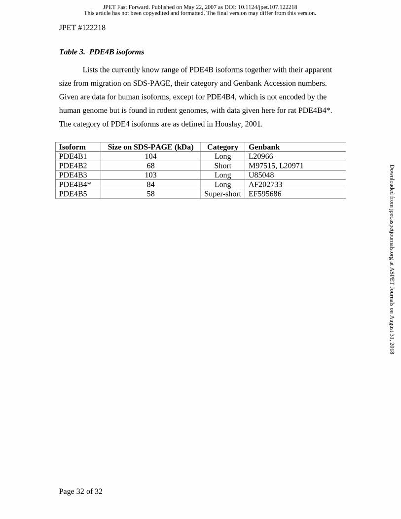
The human PDE4B gene has been shown to encode the long PDE4B1 and PDE4B3
isoforms and the short PDE4B2 isoform (Fig. 1; Table 3). These three isoforms are also
seen in rodents, which additionally express the short PDE4B4 isoform, which is not
found in humans (Shepherd et al., 2003) (Table 3). Here, we show that mouse EST
transcripts predict a novel PDE4B isoform, which we label PDE4B5. This transcript
replaces the seven 5' exons of transcript NM_002600 with a single novel 5' exon. Using
RT-PCR, we confirmed human transcription of the variant and established that it is
expressed specifically in brain sections. The 3' region of this novel exon contains a
putative in-frame ATG start codon, followed by a novel 16 residue ORF. That this ORF
is conserved in vertebrae, chicken, frog, zebrafish, and fugu strongly suggests a
functional role. The protein encoded by this isoform would be 484 residues, would
contain the PDE catalytic domain and PDE4B C-terminus, but would lack the UCR1
domain and encode a naked, truncated UCR2 domain. Because the protein would be
shorter than the previously identified long and short PDE4B forms, we classify it as a
'super-short' isoform of PDE4B (Houslay, 2001) (Table 3).
By transfecting a PDE4B5 cDNA construct into COS1 cells, we were able to
demonstrate that the cDNA generates a protein of 58 ± 2 kDa. Immunoreactivity
measurements show that while this protein is found in membranes, it is found dominantly
in the cytosol (high speed supernatant fraction). Confocal microscopy
immunolocalisation observations of recombinant PDE4B5 using an anti-PDE4B
antiserum similarly show a distribution throughout the cytosol with small amounts
associated with the plasma membrane. PDE4B5 shows an uneven distribution throughout
the cytosol, which may indicate association with cytosolic vesicles/complexes in addition
to soluble forms.
This article has not been copyedited and formatted. The final version may differ from this version.JPET Fast Forward. Published on May 22, 2007 as DOI: 10.1124/jpet.107.122218
at ASPE
T Journals on A
ugust 31, 2018jpet.aspetjournals.org
Dow
nloaded from
JPET #122218
Page 18 of 32
The transfected PDE4B5 cDNA generates an active protein with a Km for cAMP of
5.8 ± 0.4 µM. Rolipram binds in the PDE4 catalytic domain and indeed inhibits activity
of this super-short isoform (containing the catalytic domain). Measuring inhibition by
rolipram and ariflo, we find IC50 values of 380 ± 63 nM (n = 4) and 114 ± 17 nM (n = 3),
respectively.
Comparing PDE4B5 to other PDE4 genes, we found that the novel 16 residues at the
N-terminus of PDE4B5 perfectly match the N-terminus of the recently discovered super-
short isoform of PDE4D, called PDE4D6 (Wang et al., 2003). Expanding on previous
assessments (Wang et al., 2003), we found that the PDE4D6 transcript, like PDE4B5, is
brain specific. However, we identified no common transcription factor binding sites
upstream of each isoform, suggesting that these isoforms are independently regulated.
Microarray gene expression profiling shows PDE4B and PDE4D enriched in white
blood cell fractions. PDE4D is also over-expressed in skeletal muscle while PDE4B is
high in several nervous tissues, including dorsal raphe and hypothalamus, and, to a lesser
degree, in muscle. However, what is invisible to these microarray "gene monitoring”
experiments is the high specificity of individual isoforms to specific tissues, such as the
high specificity of PDE4B5 and PDE4D6 isoforms to brain sections.
Pharmaceutical manipulation of PDE4 genes may aid in treatment of diseases, such as
stroke and inflammation, including COPD and asthma. Indeed, PDE4B and PDE4D gene
expression is enriched in white-blood cell fractions, possibly suggesting an inflammatory
role. PDE4B activity has also been implicated in psychiatric disorders via its interaction
with DISC1 (Millar et al., 2005; Pickard et al., 2007), one of the most validated genetic
risk factors for schizophrenia (Porteous and Millar, 2006). Indeed, very recently it has
been shown that missense mutations in mouse DISC1 that confer depression-like and
schizophrenia-like phenotypes interfere with PDE4B binding to DISC1 (Steven et al.,
2007). Importantly, knockout of the PDE4B gene in mice yields an anti-depressant-like
phenotype (O'Donnell and Zhang, 2004) as does chemical ablation of PDE4 activity
using the selective inhibitor rolipram (Wachtel, 1983; Zhang et al., 2002; Zhang et al.,
2006; Kanes et al., 2007). Indeed, the anti-depressant phenotype observed upon chronic
nicotinic treatment of rats leads to a specific down-regulation of PDE4B transcripts in
brain (Polesskaya et al., 2007), consistent with a key role of PDE4B in regulating
This article has not been copyedited and formatted. The final version may differ from this version.JPET Fast Forward. Published on May 22, 2007 as DOI: 10.1124/jpet.107.122218
at ASPE
T Journals on A
ugust 31, 2018jpet.aspetjournals.org
Dow
nloaded from
JPET #122218
Page 19 of 32
depression and psychosis. Both long (PDE4B1, PDE4B3) and short (PDE4B2) short
isoforms have been shown to interact with DISC1 and UCR2 identified as a binding site
(Millar et al., 2005). Intriguingly, here we show that the super-short PDE4B5 isoform can
also interact with DISC1 (Fig. 8). This indicates that either the interaction site in UCR2
lies within the residual portion of the truncated UCR2 found in PDE4B2 or there is an
additional site for interaction with DISC1 that lies within the PDE4B catalytic unit.
Indeed, recent analyses of various scaffold proteins that serve to sequester PDE4 have
identified multiple binding sites (Bolger et al., 2006; Baillie et al., 2007; Sachs et al.,
2007; Stefan et al., 2007) and our recent observations (H Murdoch and MD Houslay,
unpublished) indicate that the PDE4B catalytic unit does indeed contain a further binding
site for fl-DISC1.
Despite the pharmaceutical significance of PDE4, its repertoire of isoforms available
to the cell, how these isoforms are regulated, their distinct biological function, and their
relationship to disease state are still unclear. Here, we have used mouse transcripts and
isoform-specific expression monitoring to discover an intriguing, novel, active, brain-
specific PDE4B isoform.
Acknowledgements
JCC thanks Rosetta’s Gene Expression Laboratory for microarray data, Sherri
Bloomer for project management support, and Chris Raymond and Chris Armour for
consultations.
This article has not been copyedited and formatted. The final version may differ from this version.JPET Fast Forward. Published on May 22, 2007 as DOI: 10.1124/jpet.107.122218
at ASPE
T Journals on A
ugust 31, 2018jpet.aspetjournals.org
Dow
nloaded from
JPET #122218
Page 20 of 32
References
Baillie GS, Adams DR, Bhari N, Houslay TM, Vadrevu S, Meng D, Li X, Dunlop A,
Milligan G, Bolger GB, Klussmann E and Houslay MD (2007) Mapping binding
sites for the PDE4D5 cAMP-specific phosphodiesterase to the N- and C-domains
of beta-arrestin using spot-immobilized peptide arrays. Biochem J 404:71-80.
Baillie GS and Houslay MD (2005) Arrestin times for compartmentalised cAMP
signalling and phosphodiesterase-4 enzymes. Curr Opin Cell Biol 17:129-134.
Beavo JA and Brunton LL (2002) Cyclic nucleotide research -- still expanding after half
a century. Nat Rev Mol Cell Biol 3:710-718.
Bolger G, Michaeli T, Martins T, St John T, Steiner B, Rodgers L, Riggs M, Wigler M
and Ferguson K (1993) A family of human phosphodiesterases homologous to the
dunce learning and memory gene product of Drosophila melanogaster are
potential targets for antidepressant drugs. Mol Cell Biol 13:6558-6571.
Bolger GB, Baillie GS, Li X, Lynch MJ, Herzyk P, Mohamed A, Mitchell LH, McCahill
A, Hundsrucker C, Klussmann E, Adams DR and Houslay MD (2006) Scanning
peptide array analyses identify overlapping binding sites for the signalling
scaffold proteins, beta-arrestin and RACK1, in cAMP-specific phosphodiesterase
PDE4D5. Biochem J 398:23-36.
Bolger GB, McPhee I and Houslay MD (1996) Alternative splicing of cAMP-specific
phosphodiesterase mRNA transcripts. Characterization of a novel tissue-specific
isoform, RNPDE4A8. J Biol Chem 271:1065-1071.
Bradford MM (1976) A rapid and sensitive method for the quantitation of microgram
quantities of protein utilizing the principle of protein-dye binding. Anal Biochem
72:248-254.
Castle J, Garrett-Engele P, Armour CD, Duenwald SJ, Loerch PM, Meyer MR, Schadt
EE, Stoughton R, Parrish ML, Shoemaker DD and Johnson JM (2003)
Optimization of oligonucleotide arrays and RNA amplification protocols for
analysis of transcript structure and alternative splicing. Genome Biol 4:R66.
Conti M, Richter W, Mehats C, Livera G, Park JY and Jin C (2003) Cyclic AMP-specific
PDE4 Phosphodiesterases as Critical Components of Cyclic AMP Signaling. J
Biol Chem 278:5493-5496.
This article has not been copyedited and formatted. The final version may differ from this version.JPET Fast Forward. Published on May 22, 2007 as DOI: 10.1124/jpet.107.122218
at ASPE
T Journals on A
ugust 31, 2018jpet.aspetjournals.org
Dow
nloaded from
JPET #122218
Page 21 of 32
Francis SH, Turko IV and Corbin JD (2001) Cyclic nucleotide phosphodiesterases:
relating structure and function. Prog Nucleic Acid Res Mol Biol 65:1-52.
Houslay MD (2001) PDE4 cAMP-specific phosphodiesterases. Prog Nucleic Acid Res
Mol Biol 69:249-315.
Houslay MD and Adams DR (2003) PDE4 cAMP phosphodiesterases: modular enzymes
that orchestrate signalling cross-talk, desensitization and compartmentalization.
Biochem J 370:1-18.
Houslay MD, Schafer P and Zhang KY (2005) Keynote review: phosphodiesterase-4 as a
therapeutic target. Drug Discov Today 10:1503-1519.
Huang Z, Ducharme Y, Macdonald D and Robichaud A (2001) The next generation of
PDE4 inhibitors. Curr Opin Chem Biol 5:432-438.
Hughes TR, Mao M, Jones AR, Burchard J, Marton MJ, Shannon KW, Lefkowitz SM,
Ziman M, Schelter JM, Meyer MR, Kobayashi S, Davis C, Dai H, He YD,
Stephaniants SB, Cavet G, Walker WL, West A, Coffey E, Shoemaker DD,
Stoughton R, Blanchard AP, Friend SH and Linsley PS (2001) Expression
profiling using microarrays fabricated by an ink-jet oligonucleotide synthesizer.
Nat Biotechnol 19:342-347.
Huston E, Lumb S, Russell A, Catterall C, Ross AH, Steele MR, Bolger GB, Perry MJ,
Owens RJ and Houslay MD (1997) Molecular cloning and transient expression in
COS7 cells of a novel human PDE4B cAMP-specific phosphodiesterase,
HSPDE4B3. Biochem J 328 ( Pt 2):549-558.
Jin SL, Lan L, Zoudilova M and Conti M (2005) Specific role of phosphodiesterase 4B in
lipopolysaccharide-induced signaling in mouse macrophages. J Immunol
175:1523-1531.
Johnson JM, Castle J, Garrett-Engele P, Kan Z, Loerch PM, Armour CD, Santos R,
Schadt EE, Stoughton R and Shoemaker DD (2003) Genome-wide survey of
human alternative pre-mRNA splicing with exon junction microarrays. Science
302:2141-2144.
Jurevicius J, Skeberdis VA and Fischmeister R (2003) Role of cyclic nucleotide
phosphodiesterase isoforms in cAMP compartmentation following beta2-
This article has not been copyedited and formatted. The final version may differ from this version.JPET Fast Forward. Published on May 22, 2007 as DOI: 10.1124/jpet.107.122218
at ASPE
T Journals on A
ugust 31, 2018jpet.aspetjournals.org
Dow
nloaded from
JPET #122218
Page 22 of 32
adrenergic stimulation of ICa,L in frog ventricular myocytes. J Physiol 551:239-
252.
Kan Z, Garrett-Engele PW, Johnson JM and Castle JC (2005) Evolutionarily conserved
and diverged alternative splicing events show different expression and functional
profiles. Nucleic Acids Res 33:5659-5666.
Kanes SJ, Tokarczyk J, Siegel SJ, Bilker W, Abel T and Kelly MP (2007) Rolipram: a
specific phosphodiesterase 4 inhibitor with potential antipsychotic activity.
Neuroscience 144:239-246.
Kim JS, Bailey MJ, Ho AK, Moller M, Gaildrat P and Klein DC (2007) Daily rhythm in
pineal phosphodiesterase (PDE) activity reflects adrenergic/3',5'-cyclic adenosine
5'-monophosphate induction of the PDE4B2 variant. Endocrinology 148:1475-
1485.
Laemmli UK (1970) Cleavage of structural proteins during the assembly of the head of
bacteriophage T4. Nature 227:680-685.
Lugnier C (2006) Cyclic nucleotide phosphodiesterase (PDE) superfamily: a new target
for the development of specific therapeutic agents. Pharmacol Ther 109:366-398.
Lynch MJ, Baillie GS, Mohamed A, Li X, Maisonneuve C, Klussmann E, van Heeke G
and Houslay MD (2005) RNA silencing identifies PDE4D5 as the functionally
relevant cAMP phosphodiesterase interacting with beta arrestin to control the
protein kinase A/AKAP79-mediated switching of the beta2-adrenergic receptor to
activation of ERK in HEK293B2 cells. J Biol Chem 280:33178-33189.
Manganiello VC and Degerman E (1999) Cyclic nucleotide phosphodiesterases (PDEs):
diverse regulators of cyclic nucleotide signals and inviting molecular targets for
novel therapeutic agents. Thromb Haemost 82:407-411.
Marchmont RJ and Houslay MD (1980) A peripheral and an intrinsic enzyme constitute
the cyclic AMP phosphodiesterase activity of rat liver plasma membranes.
Biochem J 187:381-392.
McCahill A, McSorley T, Huston E, Hill EV, Lynch MJ, Gall I, Keryer G, Lygren B,
Tasken K, van Heeke G and Houslay MD (2005) In resting COS1 cells a
dominant negative approach shows that specific, anchored PDE4 cAMP
phosphodiesterase isoforms gate the activation, by basal cyclic AMP production,
This article has not been copyedited and formatted. The final version may differ from this version.JPET Fast Forward. Published on May 22, 2007 as DOI: 10.1124/jpet.107.122218
at ASPE
T Journals on A
ugust 31, 2018jpet.aspetjournals.org
Dow
nloaded from
JPET #122218
Page 23 of 32
of AKAP-tethered protein kinase A type II located in the centrosomal region. Cell
Signal 17:1158-1173.
McPhee I, Pooley L, Lobban M, Bolger G and Houslay MD (1995) Identification,
characterization and regional distribution in brain of RPDE-6 (RNPDE4A5), a
novel splice variant of the PDE4A cyclic AMP phosphodiesterase family.
Biochem J 310 ( Pt 3):965-974.
Millar JK, Pickard BS, Mackie S, James R, Christie S, Buchanan SR, Malloy MP, Chubb
JE, Huston E, Baillie GS, Thomson PA, Hill EV, Brandon NJ, Rain JC, Camargo
LM, Whiting PJ, Houslay MD, Blackwood DH, Muir WJ and Porteous DJ (2005)
DISC1 and PDE4B are interacting genetic factors in schizophrenia that regulate
cAMP signaling. Science 310:1187-1191.
Mongillo M, McSorley T, Evellin S, Sood A, Lissandron V, Terrin A, Huston E,
Hannawacker A, Lohse MJ, Pozzan T, Houslay MD and Zaccolo M (2004)
Fluorescence resonance energy transfer-based analysis of cAMP dynamics in live
neonatal rat cardiac myocytes reveals distinct functions of compartmentalized
phosphodiesterases. Circ Res 95:67-75.
O'Donnell JM and Zhang HT (2004) Antidepressant effects of inhibitors of cAMP
phosphodiesterase (PDE4). Trends Pharmacol Sci 25:158-163.
Obernolte R, Bhakta S, Alvarez R, Bach C, Zuppan P, Mulkins M, Jarnagin K and
Shelton ER (1993) The cDNA of a human lymphocyte cyclic-AMP
phosphodiesterase (PDE IV) reveals a multigene family. Gene 129:239-247.
Pickard BS, Thomson PA, Christoforou A, Evans KL, Morris SW, Porteous DJ,
Blackwood DH and Muir WJ (2007) The PDE4B gene confers sex-specific
protection against schizophrenia. Psychiatr Genet 17:129-133.
Polesskaya OO, Smith RF and Fryxell KJ (2007) Chronic nicotine doses down-regulate
PDE4 isoforms that are targets of antidepressants in adolescent female rats. Biol
Psychiatry 61:56-64.
Porteous DJ and Millar JK (2006) Disrupted in schizophrenia 1: building brains and
memories. Trends Mol Med 12:255-261.
This article has not been copyedited and formatted. The final version may differ from this version.JPET Fast Forward. Published on May 22, 2007 as DOI: 10.1124/jpet.107.122218
at ASPE
T Journals on A
ugust 31, 2018jpet.aspetjournals.org
Dow
nloaded from
JPET #122218
Page 24 of 32
Rena G, Begg F, Ross A, MacKenzie C, McPhee I, Campbell L, Huston E, Sullivan M
and Houslay MD (2001) Molecular cloning and characterization of the novel
cAMP specific phosphodiesterase, PDE4A10. Mol. Pharmacol. 59:996-1011.
Renau TE (2004) The potential of phosphodiesterase 4 inhibitors for the treatment of
depression: opportunities and challenges. Curr Opin Investig Drugs 5:34-39.
Rutten WJ, Schoot BM and De Pont JJHHM (1973) Adenosine 3',5'-monophosphate
phosphodiesterase assay in tissue homogenates. Biochim. Biophys. Acta 315:378-
383.
Sachs BD, Baillie GS, McCall JR, Passino MA, Schachtrup C, Wallace DA, Dunlop A,
J., MacKenzie KF, Klussmann E, Lynch MJ, Sikorski SL, Nuriel T, Tsigelny I,
Zhang J, Houslay MD, Chao MV and Akassoglou K (2007) p75 Neurotrophin
receptor regulates tissue fibrosis through inhibition of plasminogen activation via
a PDE4/cAMP/PKA pathway. J. Cell Biol. in press.
Shepherd M, McSorley T, Olsen AE, Johnston LA, Thomson NC, Baillie GS, Houslay
MD and Bolger GB (2003) Molecular cloning and subcellular distribution of the
novel PDE4B4 cAMP-specific phosphodiesterase isoform. Biochem J 370:429-
438.
Siepel A, Bejerano G, Pedersen JS, Hinrichs AS, Hou M, Rosenbloom K, Clawson H,
Spieth J, Hillier LW, Richards S, Weinstock GM, Wilson RK, Gibbs RA, Kent
WJ, Miller W and Haussler D (2005) Evolutionarily conserved elements in
vertebrate, insect, worm, and yeast genomes. Genome Res 15:1034-1050.
Smith FD, Langeberg LK and Scott JD (2006) The where's and when's of kinase
anchoring. Trends Biochem Sci 31:316-323.
Spina D (2004) The potential of PDE4 inhibitors in respiratory disease. Curr Drug
Targets Inflamm Allergy 3:231-236.
Stefan E, Wiesner B, Baillie GS, Mollajew R, Henn V, Lorenz D, Furkert J, Santamaria
K, Nedvetsky P, Hundsrucker C, Beyermann M, Krause E, Pohl P, Gall I,
MacIntyre AN, Bachmann S, Houslay MD, Rosenthal W and Klussmann E
(2007) Compartmentalization of cAMP-dependent signaling by
phosphodiesterase-4D is involved in the regulation of vasopressin-mediated water
reabsorption in renal principal cells. J Am Soc Nephrol 18:199-212.
This article has not been copyedited and formatted. The final version may differ from this version.JPET Fast Forward. Published on May 22, 2007 as DOI: 10.1124/jpet.107.122218
at ASPE
T Journals on A
ugust 31, 2018jpet.aspetjournals.org
Dow
nloaded from
JPET #122218
Page 25 of 32
Steven J, Clapcote SJ, Lipina TV, Millar JK, Mackie S, Christie S, Ogawa F, Lerch JP,
Trimble K, Uchiyama M, Sakuraba Y, Kaneda H, Shiroishi T, Houslay MD,
Henkelman RM, Sled JG, Gondo Y, Porteous DJ and Roder JC (2007)
Behavioural phenotypes of Disc1 missense mutations in mice. Neuron in press.
Sullivan M, Rena G, Begg F, Gordon L, Olsen AS and Houslay MD (1998) Identification
and characterization of the human homologue of the short PDE4A cAMP-specific
phosphodiesterase RD1 (PDE4A1) by analysis of the human HSPDE4A gene
locus located at chromosome 19p13.2. Biochem J 333:693-703.
Tasken K and Aandahl EM (2004) Localized effects of cAMP mediated by distinct routes
of protein kinase A. Physiol Rev 84:137-167.
Thompson WJ and Appleman MM (1971) Multiple cyclic nucleotide phosphodiesterase
activities from rat brain. Biochemistry 10:311-316.
Wachtel H (1983) Potential antidepressant activity of rolipram and other selective cyclic
adenosine 3',5'-monophosphate phosphodiesterase inhibitors. Neuropharmacology
22:267-272.
Wallace DA, Johnston LA, Huston E, MacMaster D, Houslay TM, Cheung YF, Campbell
L, Millen JE, Smith RA, Gall I, Knowles RG, Sullivan M and Houslay MD
(2005) Identification and characterization of PDE4A11, a novel, widely expressed
long isoform encoded by the human PDE4A cAMP phosphodiesterase gene. Mol
Pharmacol 67:1920-1934.
Wang D, Deng C, Bugaj-Gaweda B, Kwan M, Gunwaldsen C, Leonard C, Xin X, Hu Y,
Unterbeck A and De Vivo M (2003) Cloning and characterization of novel
PDE4D isoforms PDE4D6 and PDE4D7. Cell Signal 15:883-891.
Willoughby D, Wong W, Schaack J, Scott JD and Cooper DM (2006) An anchored PKA
and PDE4 complex regulates subplasmalemmal cAMP dynamics. Embo J
25:2051-2061.
Wong W and Scott JD (2004) AKAP signalling complexes: focal points in space and
time. Nat Rev Mol Cell Biol 5:959-970.
Zaccolo M and Pozzan T (2002) Discrete microdomains with high concentration of
cAMP in stimulated rat neonatal cardiac myocytes. Science 295:1711-1715.
This article has not been copyedited and formatted. The final version may differ from this version.JPET Fast Forward. Published on May 22, 2007 as DOI: 10.1124/jpet.107.122218
at ASPE
T Journals on A
ugust 31, 2018jpet.aspetjournals.org
Dow
nloaded from
JPET #122218
Page 26 of 32
Zhang HT, Huang Y, Jin SL, Frith SA, Suvarna N, Conti M and O'Donnell JM (2002)
Antidepressant-like profile and reduced sensitivity to rolipram in mice deficient in
the PDE4D phosphodiesterase enzyme. Neuropsychopharmacology 27:587-595.
Zhang HT, Zhao Y, Huang Y, Deng C, Hopper AT, De Vivo M, Rose GM and O'Donnell
JM (2006) Antidepressant-like effects of PDE4 inhibitors mediated by the high-
affinity rolipram binding state (HARBS) of the phosphodiesterase-4 enzyme
(PDE4) in rats. Psychopharmacology (Berl) 186:209-217.
Zhang J, Ma Y, Taylor SS and Tsien RY (2001) Genetically encoded reporters of protein
kinase A activity reveal impact of substrate tethering. Proc Natl Acad Sci U S A
98:14997-15002.
This article has not been copyedited and formatted. The final version may differ from this version.JPET Fast Forward. Published on May 22, 2007 as DOI: 10.1124/jpet.107.122218
at ASPE
T Journals on A
ugust 31, 2018jpet.aspetjournals.org
Dow
nloaded from
JPET #122218
Page 27 of 32
Footnotes
MDH thanks the Medical Research Council (UK) (G8604010) for financial support.
ZK is currently at Genentech, Inc., 1 DNA Way, South San Francisco, CA 94080, USA.
This article has not been copyedited and formatted. The final version may differ from this version.JPET Fast Forward. Published on May 22, 2007 as DOI: 10.1124/jpet.107.122218
at ASPE
T Journals on A
ugust 31, 2018jpet.aspetjournals.org
Dow
nloaded from
JPET #122218
Page 28 of 32
Legends for Figures
Figure 1. PDE4B isoforms.
PDE4 isoforms encoded by the human PDE4B gene. Indicated are the N-terminal
regions unique to each isoform, the regulatory Upstream Conserved Regions, UCR1 and
UCR2, the Linker Regions, LR1 and LR2, together with the catalytic unit and the
PDE4B-specific C-terminal region. Isoforms are grouped as long forms that have both
UCR1 and UCR2, short forms that lack UCR1, and super-short forms that both lack
UCR1 and have a truncated UCR2. Isoforms are PDE4B1 (human, M25350; rat,
AF202732), PDE4B2 (human, M28413; rat, L27058), PDE4B3 (human, L27058; rat,
U95748) and PDE4B5 (human submission pending). PDE4B4 (rat, AF202732) isoform is
found in rodents and not shown here.
Figure 2. Bioinformatics identification of PDE4B5.
(a) Human genomic alignment of the human transcript NM_002600 and mouse EST
BQ769324, including the novel 5' exon (red). A and B show RT-PCR primer locations
and C shows Taqman probe sites. (b) Genomic sequence underlying the novel first
exon, showing high conservation in 15 species in the putative protein coding region
(Siepel et al., 2005). (c) Protein translation of the novel PDE4B5 variant, with novel
sequence shown in red italics, aligned to PDE4D6 (super-short isoform).
Figure 3. PDE4B5 expression
(a) Expression of PDE4B5 using RT-PCR. (b) Expression of the PDE4B catalytic unit,
found in all PDE4B isoforms. (c) Taqman measurements of the PDE4B5 and the PDE4B
long-isoform N-terminus. The expression level is normalized to the highest observed
expression. Primer locations for all experiments are indicated in Figure 2a.
Figure 4. PDE4D6 expression
(a) Expression of PDE4D6 using RT-PCR. Tissues are arranged as per Figure 3a. (b)
Expression of the PDE4D catalytic unit, found in all PDE4D isoforms.
This article has not been copyedited and formatted. The final version may differ from this version.JPET Fast Forward. Published on May 22, 2007 as DOI: 10.1124/jpet.107.122218
at ASPE
T Journals on A
ugust 31, 2018jpet.aspetjournals.org
Dow
nloaded from
JPET #122218
Page 29 of 32
Figure 5. Microarray expression of PDE4 genes
Figure 6. Immunodetection of recombinant PDE4B5 expressed in mammalian cells.
(a) Immunoblot detection of recombinant PDE4B2 and PDE4B5 in lysates from
transfected COS1 cells identifying PDE4B immmunoreactivity in the P1, P2 and S2
fractions with analysis of mock transfected cells also shown. Loading of P1, P2 and S2
fractions were done on an equal volume basis with identical amounts of protein loading
for the untransfected and transfected cells in the S2 fraction and with equal exposure to
allow comparison. (b) Immunolocalisation by confocal microscopy of recombinant
PDE4B5 expressed in COS1 cells as either untagged form, detected with anti-PDE4B
antiserum, or FLA epitope-tagged from, with anti-FLAG antibody.
Figure 7. Inhibition of PDE4B5 by rolipram and ariflo
Dose response curves for inhibition of recombinant PDE4B5 activity expressed in COS1
cells lysates and assayed with 1µM cAMP substrate for (a) rolipram and (b) ariflo. The
examples shown are typical of plots done on 3 separate occasions.
Figure 8. Co-immunoprecipitation of the full-length DISC1 with PDE4B5
PDE4B5 and N-terminally FLAG-tagged full length 100kDa DISC1 were
expressed in various combinations in HEK293 cells. Anti-PDE4B immunoprecipitates
were resolved by SDS-PAGE and immunoblotted to detect the FLAG tag (upper panel).
Similar levels of immuno-capture of PDE4B5 were determined by immunoblotting with
the PDE4B antibody (lower panel). The relative expression levels of Flag-tagged fl-
DISC1 and PDE4B5 in ~5% of total cell lysate input used for co-immunoprecipitation
assays were determined by direct immunoblotting with the PDE4B antisera (lower panel)
and the anti-FLAG M2 antibody (upper panel). IP – immunoprecipitate; IB – imunoblot.
This article has not been copyedited and formatted. The final version may differ from this version.JPET Fast Forward. Published on May 22, 2007 as DOI: 10.1124/jpet.107.122218
at ASPE
T Journals on A
ugust 31, 2018jpet.aspetjournals.org
Dow
nloaded from
JPET #122218
Page 30 of 32
Tables
Table 1. RT-PCR primers
Size Forward primer Reverse primer
PDE4B5 only 442 ACTGTGAATTCTTTCAAAGGGATTTGTG GGTCTATTGTGAGAATATCCAGCCACAT
PDE4B catalytic 431 TGTCTTCACAGATTTGGAGATCCTGGCT CGGTCTGTCCATTGCCGATACAATTC
PDE4D6 only 276 AAACTATTTACTGTCAGTGTCTTGGGGCTAC TTAACTCCAAACCTTGGGATACTTGAATTAGT
PDE4D not super-short 242 CTAGAGACCCTACAGACCAGGCACTC ATCAATTTCTTGACTCCACTGATCTGAGACAT
This article has not been copyedited and formatted. The final version may differ from this version.JPET Fast Forward. Published on May 22, 2007 as DOI: 10.1124/jpet.107.122218
at ASPE
T Journals on A
ugust 31, 2018jpet.aspetjournals.org
Dow
nloaded from
JPET #122218
Page 31 of 32
Table 2. Intracellular distribution of recombinant PDE4B5.
COS1 cells were transfected to express PDE4B5, and its distribution between low speed
pellet (P1), high speed pellet (P2), and high speed supernatant (S2) cytosolic fractions
was assessed. This distribution was followed by determining PDE4 activity both by using
1µM cAMP as substrate and immunologically by using a PDE4B specific antiserum to
follow the distribution of the 58 kDa PDE4B5. Data show means and standard deviations
for 3 separate experiments.
Fraction activity distribution (%) blot distribution (%)
P1 13 ± 2 12 ± 2
P2 17 ± 2 15 ± 2
S2 70 ± 7 73 ± 7
This article has not been copyedited and formatted. The final version may differ from this version.JPET Fast Forward. Published on May 22, 2007 as DOI: 10.1124/jpet.107.122218
at ASPE
T Journals on A
ugust 31, 2018jpet.aspetjournals.org
Dow
nloaded from
JPET #122218
Page 32 of 32
Table 3. PDE4B isoforms
Lists the currently know range of PDE4B isoforms together with their apparent
size from migration on SDS-PAGE, their category and Genbank Accession numbers.
Given are data for human isoforms, except for PDE4B4, which is not encoded by the
human genome but is found in rodent genomes, with data given here for rat PDE4B4*.
The category of PDE4 isoforms are as defined in Houslay, 2001.
Isoform Size on SDS-PAGE (kDa) Category Genbank PDE4B1 104 Long L20966 PDE4B2 68 Short M97515, L20971 PDE4B3 103 Long U85048 PDE4B4* 84 Long AF202733 PDE4B5 58 Super-short EF595686
This article has not been copyedited and formatted. The final version may differ from this version.JPET Fast Forward. Published on May 22, 2007 as DOI: 10.1124/jpet.107.122218
at ASPE
T Journals on A
ugust 31, 2018jpet.aspetjournals.org
Dow
nloaded from
LR2LR1
C-Terminal Region
Unique N-Terminal Regions
CLFR
4B5
1a4B1
14B3
2 3 4 5 6 1 7 8 9 10 11 12 13 14 154B5
UCR1 UCR2 CATALYTICLR2LR1
Catalytic unitUCR1 UCR2
4B1
4B3
4B2
14B21b
4B1
(a)
(b)
C-terminal
Figure 1
This article has not been copyedited and form
atted. The final version m
ay differ from this version.
JPET
Fast Forward. Published on M
ay 22, 2007 as DO
I: 10.1124/jpet.107.122218 at ASPET Journals on August 31, 2018 jpet.aspetjournals.org Downloaded from
NM_002600
1 2 3 4 5 6 7 8 9 10 11 12 13 14 15 16
Mouse BQ769324
BC
A
C
Start codon Splice site
a. Exon alignment
b. Cross-species alignment
c. Protein translation with PDE4D6 alignment
PDE4B5 1 MPEANYLLSVSWGYIKFKRMLNRELTHLSEMSRSGNQVSEYISNTFLDKQNDVEIPSPTQ 60MPEANYLLSVSWGYIKFKRMLNRELTHLSEMSRSGNQVSE+ISNTFLDKQ++VEIPSPTQ
PDE4D6 1 MPEANYLLSVSWGYIKFKRMLNRELTHLSEMSRSGNQVSEFISNTFLDKQHEVEIPSPTQ 60
chr1:66,593,100-66,593,200
Figure 2
This article has not been copyedited and formatted. The final version may differ from this version.JPET Fast Forward. Published on May 22, 2007 as DOI: 10.1124/jpet.107.122218
at ASPE
T Journals on A
ugust 31, 2018jpet.aspetjournals.org
Dow
nloaded from
0
20
40
60
80
100
Fetal Brain
Heart Lung Lung carcinoma
No
rmal
ized PDE4B5
PDE4B long
C. Taqman
A. PDE4B5 (N-terminus)
B. PDE4B C-terminus
C
D
A
B
Retina A 1 Fetal Kidney B 1Pituitary 2 Fetal Liver 2Spinal Cord 3 Fetal Lung 3Brain, Cerebellum 4 Fetal Vertebra 4Brain, Frontal Lobe 5 Heart 5Brain, Medulla Oblongata 6 Kidney 6Brain, Pons 7 Liver 7Brain, Putamen 8 Pancreas 8Brain, Thalamus 9 Stomach 9Brain, Hippocampus 10 Jejunum 10Fetal Brain 11 Ileum 11
Colon, descending C 1 Bone Marrow D 1Colon tumor tissue 2 Peripheral Leukocytes 2Lung 3 Uterus 3Lung Carcinoma (A549) 4 Placenta 4Prostate 5 Ovary 5Thyroid 6 Testis 6Adipose 7 Hela S3 7Skin 8 Leukemia (K-562) 8Skeletal Muscle 9 Lymphoma Burkitt's (Raji) 9Adrenal Gland 10 Melanoma (G361) 10Thymus 11 Osteosarcoma (MG-63) 11
Heart A 1Kidney 2Liver 3Brain 4Placenta 5Lung 6Brain - fetal 7Leukemia (HL-60) 8Adrenal medulla 9Liver - fetal 10Salivary gland 11
A
B
Lymphoma (Raji) B 1Spinal cord 2Lymph node 3Kidney - fetal 4Uterus 5Spleen 6Brain - thalamus 7Lung - fetal 8Testis 9Melanoma (G361) 10Lung carcinoma (A549) 11
500
400
500
400
Figure 3
This article has not been copyedited and formatted. The final version may differ from this version.JPET Fast Forward. Published on May 22, 2007 as DOI: 10.1124/jpet.107.122218
at ASPE
T Journals on A
ugust 31, 2018jpet.aspetjournals.org
Dow
nloaded from
B. PDE4D long and short (not super short)
A. PDE4D6 (N-terminus)
CD
A B
CD
A B
300
200
300
200
Figure 4
This article has not been copyedited and formatted. The final version may differ from this version.JPET Fast Forward. Published on May 22, 2007 as DOI: 10.1124/jpet.107.122218
at ASPE
T Journals on A
ugust 31, 2018jpet.aspetjournals.org
Dow
nloaded from
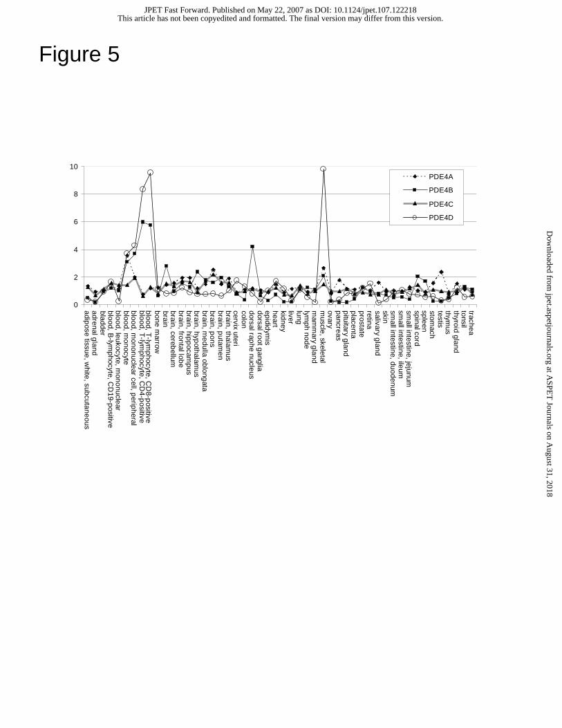
Figure 5
0
2
4
6
8
10
adipose tissue, w
hite, subcutaneousadrenal glandbladderblood, B
-lymphocyte, C
D19-positive
blood, leukocyte, mononuclear
blood, monocyte
blood, mononuclear cell, p
eripheral
blood, T-lym
phocyte, CD
4-positive
blood, T-lym
phocyte, CD
8-positive
bone marrow
brainbrain, cereb
ellumbrain, frontal lob
ebrain, hipp
ocampus
brain, hypothalam
usbrain, m
edulla oblongata
brain, pons
brain, putam
enbrain, thalam
uscervix utericolondorsal rap
henucleus
dorsal root ganglia
epididymis
heartkidneyliverlunglym
ph nodem
amm
ary glandm
uscle, skeletalovarypancreaspituitary glandplacentaprostateretinasalivary glandskinsm
all intestine, duodenumsm
all intestine, ileumsm
all intestine, jejunumspinal cordspleenstom
achtestisthym
usthyroid glandtonsiltrachea
PDE4A
PDE4B
PDE4C
PDE4D
This article has not been copyedited and formatted. The final version may differ from this version.JPET Fast Forward. Published on May 22, 2007 as DOI: 10.1124/jpet.107.122218
at ASPE
T Journals on A
ugust 31, 2018jpet.aspetjournals.org
Dow
nloaded from
(a)
PDE4B5
PDE4B2
Transfection
4B54B2mock
PDE4B5 - flag-tagPDE4B5 - no tag
(b)
P1 P2 S2
Figure 6
P1 P2 S2P1 P2 S2
50
75
100
150
250
37
25
App
aren
t Mol
ecul
ar W
eigh
t (kD
a)
This article has not been copyedited and form
atted. The final version m
ay differ from this version.
JPET
Fast Forward. Published on M
ay 22, 2007 as DO
I: 10.1124/jpet.107.122218 at ASPET Journals on August 31, 2018 jpet.aspetjournals.org Downloaded from
none
(a) (b)
none0
100
80
60
40
20
0
100
80
60
40
20
[rolipram] (log µM)
-6-5
-4-3
-2-1
01
2
[ariflo] (log µM)
-6-5
-4-3
-2-1
01
2
Figure 7
This article has not been copyedited and form
atted. The final version m
ay differ from this version.
JPET
Fast Forward. Published on M
ay 22, 2007 as DO
I: 10.1124/jpet.107.122218 at ASPET Journals on August 31, 2018 jpet.aspetjournals.org Downloaded from
IP Input
FlagDISC1 IB: FLAG
IB: PDE4BPDE4B5
— 100 kDa
— 50 kDa
— 75 kDa
— 75 kDa
FlagDISC1
PDE4B5
+ + – + + –– + + – + +
Figure 8This article has not been copyedited and formatted. The final version may differ from this version.
JPET Fast Forward. Published on May 22, 2007 as DOI: 10.1124/jpet.107.122218 at A
SPET
Journals on August 31, 2018
jpet.aspetjournals.orgD
ownloaded from