kaice arminda lafavers - scholarworks.iupui.edu
TRANSCRIPT

A FORWARD GENETIC APPROACH TO IDENTIFYING NOVEL
CALCIUM REGULATORS IN TOXOPLASMA GONDII
Kaice Arminda LaFavers
Submitted to the faculty of the University Graduate School
in partial fulfillment of the requirements
for the degree
Doctor of Philosophy
in the Department of Pharmacology and Toxicology,
Indiana University
November 2017
ii
Accepted by the Graduate Faculty of Indiana University, in partial
fulfillment of the requirements for the degree of Doctor of Philosophy.
Doctoral Committee
____________________________
Gustavo Arrizabalaga, Ph.D., Chair
____________________________
Nickolay Brustovetsky, Ph.D.
___________________________
Theodore Cummins, Ph.D.
____________________________
Stacey Gilk, Ph.D.
July 25, 2017
____________________________
William Sullivan, Ph.D.
iii
Acknowledgements
I would like to thank Dr. Vern Carruthers for sharing the Rh∆Ku80 strain
used for endogenous tagging and generation of the gra41 knockout as well as the
gra2 knockout strains along with its parental and complemented strains and the
MIC2 antibody, Dr. Peter Bradley for the GRA7 and ROP1 antibodies, Dr. John
Boyle for the Pru∆Ku80 strain used for endogenous tagging and the generation of
the gra41 Type II knockout strain, Dr. Barry Stein for valuable training and advice
in obtaining the transmission electron microscopy of the gra41 parental, knockout
and complemented strains and Dr. Sanofar Abdeen for training and advice in
recombinant protein purification. I would like to thank Karla Marquez Noguera, in
the laboratory of Dr. Silvia Moreno at University of Georgia for conducting the
calcium measurement experiments, Dr. Isabelle Coppens at Johns Hopkins for
conducting the immunoelectron microscopy imaging, and Dr. Gustavo
Arrizabalaga and Erin Garrison for the sequencing of the forward genetic mutant.
I would also like to thank all members of Arrizabalaga and Sullivan labs for
scientific and personal support. Finally, I would like to thank all of the members of
my research committee, Drs. Brustovetsky, Cummins, Gilk and Sullivan, along with
my mentor, Dr. Arrizabalga, for their continued mentorship and support throughout
my graduate career. This research has been funded by NIH grants R21AI119516,
R01AI123457, and RO3AI101624 to Gustavo Arrizabalaga as well as an NIH
training grant AI060519 and a fellowship from the American Heart Association
16PRE27260042 to Kaice LaFavers.
iv
Kaice Arminda LaFavers
A FORWARD GENETIC APPROACH TO IDENTIFYING NOVEL
CALCIUM REGULATORS IN TOXOPLASMA GONDII
Toxoplasma gondii is an obligate intracellular eukaryotic pathogen that causes
severe neurologic disease in immunocompromised adults and congenitally
infected neonates. Events critical to the propagation of T. gondii, such as invasion
and egress, are regulated by calcium-dependent signaling. In order to identify
unique components of the parasite’s calcium signaling networks, members of the
Arrizabalaga laboratory have used a forward genetics approach to isolate mutants
with altered sensitivity to the calcium ionophore A23187. Exposing extracellular
parasites to A23187 induces protein secretion, motility and cytoskeletal
rearrangements and prolonged treatment causes exhaustion of factors required
for invasion, which results in what is referred to as ionophore induced death
(iiDeath). Mutants capable of surviving this treatment were isolated from a
chemically mutagenized population. Whole genome sequencing of one such
mutant, MBD2.1, identified a nonsense mutation in a protein of unknown function
(TGGT1_069070, ToxoDBv7.2) Complementation of MBD 2.1 with a wild-type
copy of TGGT1_069070 restored sensitivity to iiDeath treatment. Endogenous
tagging of this locus revealed that the encoded protein is secreted from a unique
parasite secretory organelle known as the dense granule into the parasitophorous
vacuole, leading to its designation as TgGRA41. Complete knockout of TgGRA41
recapitulates the resistance to iiDeath observed in MBD2.1 but also exhibits a

v
dramatic decrease in propagation in tissue culture not seen in the original mutant.
The knockout shows defects in multiple steps of the lytic including compromised
invasion efficiency and premature egress of parasites from host cells. Cytosolic
calcium measurements of extracellular parasites show enhanced uptake of
calcium in the knockout strain as compared to parental and complemented,
suggesting that the loss of TgGra41 results in calcium dysregulation. Together,
these results provide a novel insight into the role that the parasitophorous vacuole
of T. gondii plays in calcium homeostasis and calcium-dependent signaling
processes.
Gustavo Arrizabalaga, Ph.D., Chair

vi
Table of Contents
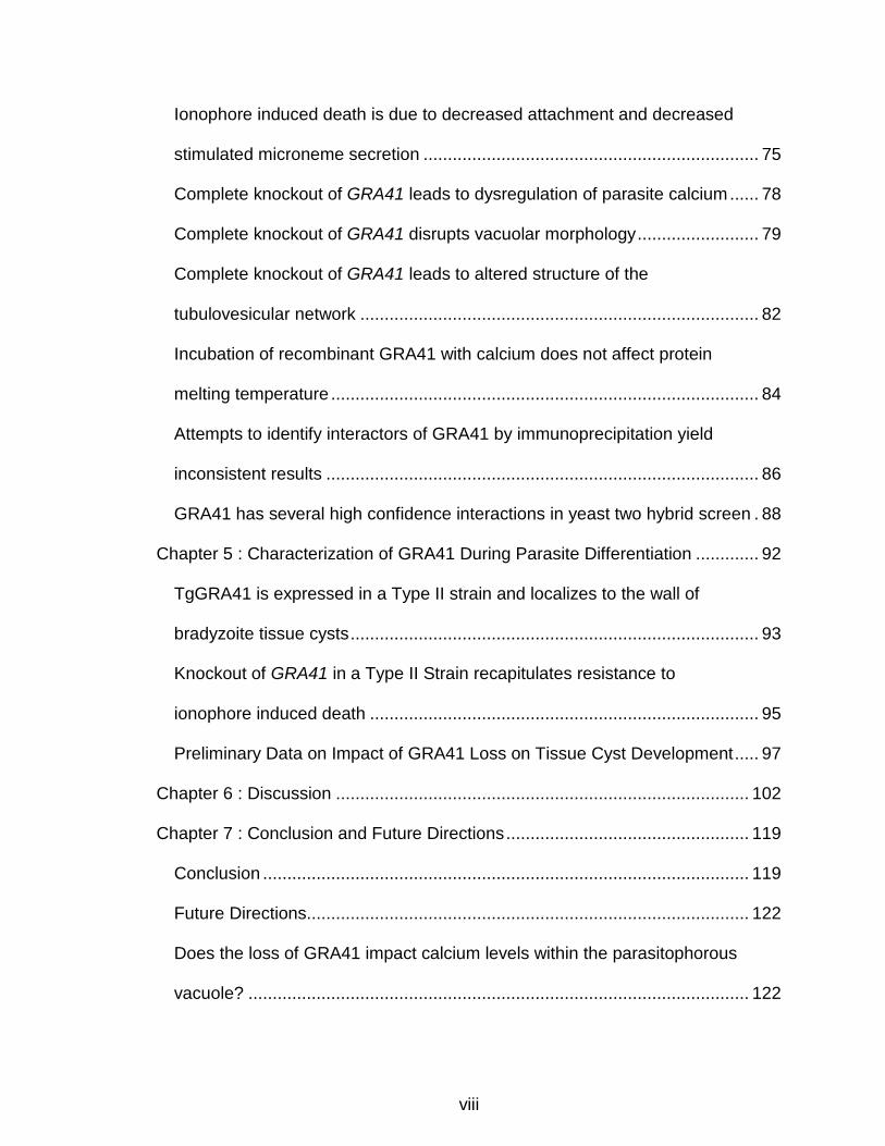
List of Tables ........................................................................................................ x
List of Figures .......................................................................................................xi
List of Abbreviations ........................................................................................... xiii
Chapter 1 : Introduction ........................................................................................ 1
Specific Aims .................................................................................................... 1
Clinical Significance .......................................................................................... 3
T. gondii Life Cycle ........................................................................................... 6
T. gondii Lytic Cycle .......................................................................................... 9
Cell Biology of T. gondii .................................................................................. 12
Calcium Signaling in T. gondii ......................................................................... 22
Chapter 2 : Methods ........................................................................................... 29
Parasite propagation ....................................................................................... 29
Genome sequencing and complementation .................................................... 29
iiDeath survival assay ..................................................................................... 30
Micronemal secretion assay ........................................................................... 31
Generation of Type I endogenously tagged GRA41 line ................................. 32
Immunofluorescence assays .......................................................................... 32
Western blot analysis ...................................................................................... 33
Triton-X 114 membrane partitioning ................................................................ 34
Electron microscopy ........................................................................................ 35
Generation of Type I GRA41 knockout strain ................................................. 36
Generation of GRA41 knockout complemented clones .................................. 39
vii
Parasite growth assays ................................................................................... 40
Parasite egress assays ................................................................................... 40
Parasite invasion assays ................................................................................ 40
Calcium measurements .................................................................................. 41
Recombinant protein production ..................................................................... 42
Calcium thermal shift assays .......................................................................... 43
Immunoprecipitation ........................................................................................ 44
Yeast two hybrid screen .................................................................................. 46
Generation of Type II endogenously tagged GRA41 line ................................ 47
Generation of Type II GRA41 knockout strain ................................................ 48
Bradyzoite differentiation assays .................................................................... 49
Mouse studies of chronic toxoplasmosis ......................................................... 50
Chapter 3 : Identification and Characterization of GRA41 .................................. 54
Nonsense mutation in novel gene is responsible for iiDeath- phenotype of
MBD2.1 ........................................................................................................... 54
TGGT1_069070 encodes a novel dense granule protein, GRA41 .................. 58
Complete knockout of GRA41 recapitulates iiDeath- phenotype ..................... 62
Complete knockout of GRA41 results in decreased plaquing efficiency ......... 65
Complementation of GRA41 knockout with the parental gene rescues both
iiDeath sensitivity and lytic cycle defects ........................................................ 66
Complete knockout of GRA41 affects timing of natural egress ....................... 70
Chapter 4 : Functional Analysis of GRA41 ......................................................... 75
viii
Ionophore induced death is due to decreased attachment and decreased
stimulated microneme secretion ..................................................................... 75
Complete knockout of GRA41 leads to dysregulation of parasite calcium ...... 78
Complete knockout of GRA41 disrupts vacuolar morphology ......................... 79
Complete knockout of GRA41 leads to altered structure of the
tubulovesicular network .................................................................................. 82
Incubation of recombinant GRA41 with calcium does not affect protein
melting temperature ........................................................................................ 84
Attempts to identify interactors of GRA41 by immunoprecipitation yield
inconsistent results ......................................................................................... 86
GRA41 has several high confidence interactions in yeast two hybrid screen . 88
Chapter 5 : Characterization of GRA41 During Parasite Differentiation ............. 92
TgGRA41 is expressed in a Type II strain and localizes to the wall of
bradyzoite tissue cysts .................................................................................... 93
Knockout of GRA41 in a Type II Strain recapitulates resistance to
ionophore induced death ................................................................................ 95
Preliminary Data on Impact of GRA41 Loss on Tissue Cyst Development ..... 97
Chapter 6 : Discussion ..................................................................................... 102
Chapter 7 : Conclusion and Future Directions .................................................. 119
Conclusion .................................................................................................... 119
Future Directions ........................................................................................... 122
Does the loss of GRA41 impact calcium levels within the parasitophorous
vacuole? ....................................................................................................... 122
ix
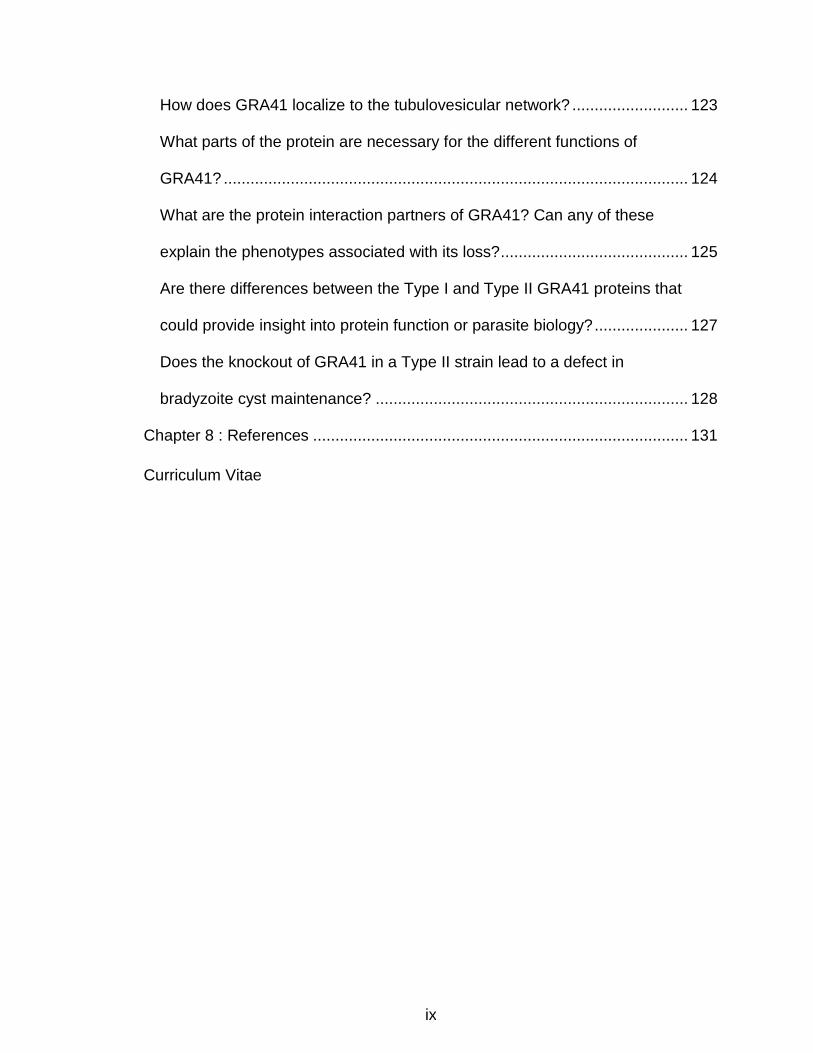
How does GRA41 localize to the tubulovesicular network? .......................... 123
What parts of the protein are necessary for the different functions of
GRA41? ........................................................................................................ 124
What are the protein interaction partners of GRA41? Can any of these
explain the phenotypes associated with its loss? .......................................... 125
Are there differences between the Type I and Type II GRA41 proteins that
could provide insight into protein function or parasite biology? ..................... 127
Does the knockout of GRA41 in a Type II strain lead to a defect in
bradyzoite cyst maintenance? ...................................................................... 128
Chapter 8 : References .................................................................................... 131
Curriculum Vitae
x
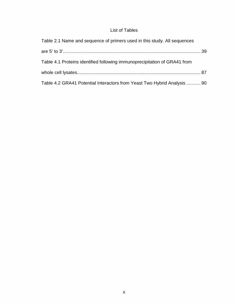
List of Tables
Table 2.1 Name and sequence of primers used in this study. All sequences
are 5’ to 3’. .......................................................................................................... 39
Table 4.1 Proteins identified following immunoprecipitation of GRA41 from
whole cell lysates ................................................................................................ 87
Table 4.2 GRA41 Potential Interactors from Yeast Two Hybrid Analysis ........... 90

xi
List of Figures
Figure 1.1 Life cycle of T. gondii ........................................................................... 8
Figure 1.2 Lytic Cycle of T. gondii ...................................................................... 10
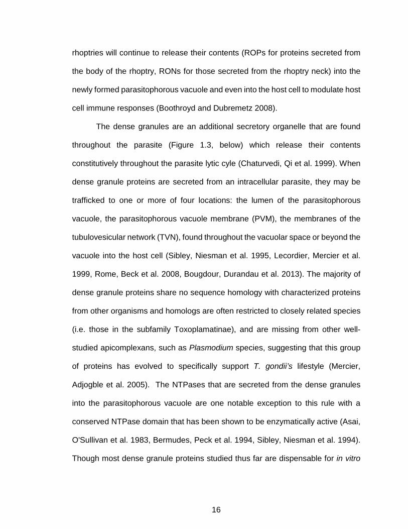
Figure 1.3 T. gondii tachyzoite with selected organelles drawn and identified. . 21
Figure 1.4 Key steps in the lytic cycle of T. gondii are accompanied by
fluctuations in calcium within the parasite and host cell ...................................... 23
Figure 1.5 Conservation of Calcium Signaling in T. gondii ................................. 24
Figure 3.1 Sequencing of TgGT1_069070 ......................................................... 55
Figure 3.2 The iiDeath- phenotype of MBD2.1 is due to the introduction of a
premature stop codon in TGGT1_069070. ......................................................... 57
Figure 3.3 TGGT1_069070 encodes a novel dense granule protein, GRA41 .... 60
Figure 3.4 Western Blot Analysis of Triton-X 114 partitioning of parasite
lysates from the GRA41 endogenously HA-tagged strain. ................................. 62
Figure 3.5 Complete knockout of GRA41 recapitulates the iiDeath- phenotype
seen in MBD2.1 mutant. ..................................................................................... 64
Figure 3.6 Complete knockout of GRA41 results in reduced plaquing
efficiency which is rescued by complementation. ............................................... 67
Figure 3.7 Complete knockout of GRA41 results in decreased invasion
efficiency ............................................................................................................ 70
Figure 3.8 Complete knockout of GRA41 leads to premature egress of
parasites. ............................................................................................................ 71
Figure 4.1 Ionophore treatment of extracellular parasites causes a decrease
in host cell attachment and microneme secretion. .............................................. 77

xii
Figure 4.2 GRA41 knockout parasites exhibit enhanced uptake of
extracellular calcium. .......................................................................................... 79
Figure 4.3 GRA41 knockout parasites have altered vacuole morphology and
division pattern. .................................................................................................. 81
Figure 4.4 GRA41 knockout parasites show altered morphology and size of
tubulovesicular network (TVN) as compared to the parental and
complemented strains ........................................................................................ 83
Figure 4.5 Ionophore induced death phenotype of Gra2 parental, knockout
and complemented strains.................................................................................. 84
Figure 4.6 Melt curve analysis of GRA41 recombinant protein incubated with
calcium ............................................................................................................... 85
Figure 5.1 GRA41 is expressed in a type II strain and localizes to the wall of
bradyzoite tissue cysts ....................................................................................... 94
Figure 5.2 Complete knockout of GRA41 in a Type II Strain recapitulates the
iiDeath- phenotype seen in Type I Knockout....................................................... 97
Figure 5.3 Differentiation of Type II Gra41 Knockout ........................................ 100
Figure 6.1 Proposed model for phenotypes associated with Loss of GRA41
in Type I Strain ................................................................................................. 113
xiii
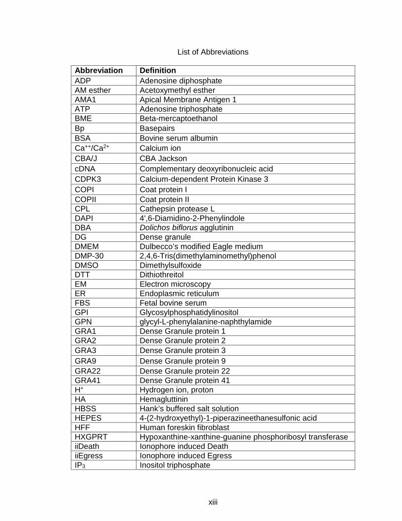
List of Abbreviations
Abbreviation Definition ADP Adenosine diphosphate AM esther Acetoxymethyl esther AMA1 Apical Membrane Antigen 1 ATP Adenosine triphosphate BME Beta-mercaptoethanol Bp Basepairs BSA Bovine serum albumin Ca++/Ca2+ Calcium ion CBA/J CBA Jackson cDNA Complementary deoxyribonucleic acid CDPK3 Calcium-dependent Protein Kinase 3 COPI Coat protein I COPII Coat protein II CPL Cathepsin protease L DAPI 4',6-Diamidino-2-Phenylindole DBA Dolichos biflorus agglutinin DG Dense granule DMEM Dulbecco’s modified Eagle medium DMP-30 2,4,6-Tris(dimethylaminomethyl)phenol DMSO Dimethylsulfoxide DTT Dithiothreitol EM Electron microscopy ER Endoplasmic reticulum FBS Fetal bovine serum GPI Glycosylphosphatidylinositol GPN glycyl-L-phenylalanine-naphthylamide GRA1 Dense Granule protein 1 GRA2 Dense Granule protein 2 GRA3 Dense Granule protein 3 GRA9 Dense Granule protein 9 GRA22 Dense Granule protein 22 GRA41 Dense Granule protein 41 H+ Hydrogen ion, proton HA Hemagluttinin HBSS Hank’s buffered salt solution HEPES 4-(2-hydroxyethyl)-1-piperazineethanesulfonic acid HFF Human foreskin fibroblast HXGPRT Hypoxanthine-xanthine-guanine phosphoribosyl transferase iiDeath Ionophore induced Death iiEgress Ionophore induced Egress IP3 Inositol triphosphate

xiv
IP3R Inositol triphosphate Receptor IPTG Isopropyl β-D-1-thiogalactopyranoside ISC6 IMC Sutures Component 6 K+ Potassium ion kD kiloDaltons LB Lysogeny broth Mbp Mega basepairs MIC2 Microneme Protein 2 MOI Multiplicity of infection MW Molecular Weight MYOA Myosin A PBS Phosphate buffered saline OR Predicted biological score PCR Polymerase chain reaction PLV Plant-like vacuole PM Plasma membrane PV Parasitophorous vacuole PVM Parasitophorous vacuole membrane qPCR Quantitative polymerase chain reaction RIPA Buffer Radioimmunoprecipitation assay buffer RON2 Rhoptry Neck Protein 2 RON4 Rhoptry Neck Protein 4 RON5 Rhoptry Neck Protein 5 RON8 Rhoptry Neck Protein 8 ROP1 Rhoptry Protein 1 ROP40 Rhoptry Protein 40 ROP5 Rhoptry Protein 5 ROP7 Rhoptry Protein 7 ROP8 Rhoptry Protein 8 RPMI Medium Roswell Park Memorial Institute Medium RyR Ryanodine receptor SDS-PAGE Sodium dodecyl sulfate – polyacrylamide gel electrophoresis SNV Single nucleotide variant SORTL Sortilin-like Receptor T. gondii Toxoplasma gondii TBS Tris-buffered saline TBST Tris-buffered saline with tween TVN Tubulovesicular network TX-100 Triton X-100 TX-114 Triton X-114 VAC Vacuole, refers to same organelle as plant-like vacuole YFP Yellow fluorescent protein

1
Chapter 1 : Introduction
Specific Aims
Toxoplasma gondii is an obligate intracellular eukaryotic pathogen
responsible for potentially fatal infections in immunocompromised individuals and
neonates. Current treatments for clinical Toxoplasmosis exhibit high rates of
toxicity and cannot clear the chronic form of the disease (Luft and Remington
1992). The identification and characterization of processes and proteins that are
both unique to the parasite and crucial for its survival is key for the discovery of
new drug targets. In this context, calcium-dependent signaling represents an ideal
target since it controls key aspects of the parasite’s life cycle, and depends on
many proteins that are unique to the parasite and absent in the mammalian host
(Arrizabalaga and Boothroyd 2004, Nagamune 2009, Lourido and Moreno 2015).
In order to identify novel calcium signaling factors, members of the Arrizabalaga
laboratory have used a forward genetic approach to isolate mutants able to resist
prolonged extracellular exposure to the calcium ionophore A23187. Ionophore
treatment of extracellular parasites induces excessive secretion of invasion-related
proteins, rendering parasites non-invasive, a phenomenon termed ionophore
induced death (iiDeath). Members of the Arrizabalaga and Boothroyd laboratories
were able to isolate a mutant, MBD 2.1, which remains invasive after this
treatment. This mutant is also hypersensitive to the chelation of intraparasitic
calcium by BAPTA-AM, suggesting that it may have disregulated calcium
homeostasis (Black, Arrizabalaga et al. 2000). Whole genome sequencing of the
mutant and its parental strain identified multiple single nucleotide variants, two of

2
which lead to changes in the primary sequence of encoded proteins. Given that
the common thread between the phenotype by which the MBD 2.1 mutant was
isolated (iiDeath resistance) and this sensitivity to the calcium chelator BAPTA-AM
is calcium, I hypothesize that identifying the causative mutation for iiDeath
resistance in MBD2.1 will allow us to identify a novel protein regulator of calcium
homeostasis within the parasite. I will test this hypothesis through the following
aims:
Specific Aim 1: Determine the causative mutation in mutant MBD2.1
and characterize the protein responsible for resistance to iiDeath. Chemical
mutagenesis and selection of the RH∆hxgprt strain led to the isolation of MBD2.1,
which exhibits resistance to iiDeath. Whole genome sequencing of the mutant and
parental strains identified candidate single nucleotide variants that could be
responsible for this phenotype. To identify the causative mutation, I will attempt to
complement the iiDeath- phenotype by transfecting in wild-type copies of the
mutated genes. Upon identification of the causative mutation, I will endogenously
tag the responsible gene to determine the localization of the encoded protein.
Additionally, I will generate a complete knockout of the responsible gene to verify
that this recapitulates the resistance to iiDeath and to further characterize any
phenotypes associated with the loss of this protein.
Specific Aim 2: Determine the mechanistic role of the iiDeath protein.
Based on the resistance to iiDeath and hypersensitivity to BAPTA-AM seen in
mutant MBD2.1, it is likely that the iiDeath protein impacts calcium homeostasis
within the parasite. With this in mind, I will utilize the iiDeath protein knockout

3
generated in Aim 1 to test calcium levels in the parasite cytosol under normal and
stimulated conditions. I will also generate recombinant iiDeath protein to
investigate whether this protein is capable of binding calcium. In parallel I will
explore the function of the iiDeath protein in an unbiased approach by identifying
interacting proteins. I will also examine the morphology of the iiDeath protein
knockout to look for any structural defects.
Specific Aim 3: Determine the impact of the iiDeath protein on the
establishment and progression of infection in vivo. The forward genetic
approach used to generate the MBD2.1 mutant was conducted in a highly virulent
strain that does not establish a chronic infection and cannot be used to determine
the full spectrum of the iiDeath protein’s role during infection. Accordingly, I will
generate a knockout of the iiDeath protein in a strain suitable for in vivo and
developmental studies. This will allow us to test the role of this novel protein in the
generation of chronic stage cysts in tissue culture and in vivo and development of
acute and chronic infection in vivo.
Clinical Significance
T. gondii is an obligate intracellular parasite of the phylum Apicomplexa that
causes widespread infection among many vertebrates, including humans (San
Miguel, Gutierrez-Exposito et al. 2016). It is estimated that approximately a third
of the world’s human population is infected with this opportunistic pathogen.
Humans become infected congenitally or by ingestion of either environmental
oocysts, which are shed in the feces of cats, or tissue cysts, found in the
undercooked meat of infected animals (Pappas, Roussos et al. 2009).
4
Though immunocompetent individuals will not generally experience
symptoms during infection, T. gondii can be particularly devastating in
immunocompromised individuals and those infected congenitally (Mazzillo,
Shapiro et al. 2013, Oray, Ozdal et al. 2015). During the acute stage of the
infection, T. gondii propagates through repeated lytic cycles of host cell invasion,
growth and egress as a rapidly dividing form known as the tachyzoite, resulting in
significant host cell death (Black and Boothroyd 2000). The tissue damage elicited
by parasite propagation is normally limited by an immune response that relies on
both CD4+ and CD8+ T cells, which induces conversion to the cyst-forming
bradyzoite stage and the establishment of a chronic infection (Landrith, Harris et
al. 2015).
In immunocompromised individuals and lymphoma patients, new infections
or rupture of pre-existing tissue cysts can lead to toxoplasmic encephalitis (Luft
and Remington 1992, Israelski and Remington 1993, Slavin, Meyers et al. 1994).
Toxoplasmic encephalitis can be a difficult diagnosis to make; the most common
associated symptoms are nonspecific headache, confusion and fever. However,
this can progress to coma and death if left untreated and in a small minority of
treated patients as well (Porter and Sande 1992). Neuropathological analysis of
toxoplasmic encephalitis by magnetic resonance imaging (MRI) shows visible
lesions that are the result of tissue damage caused by lysis of host cells by the
parasite (Neuen-Jacob, Figge et al. 1993). Current anti-retroviral treatments
directed at the human immunodeficiency virus (HIV) have reduced the overall
incidence and mortality of toxoplasmic encephalitis in the HIV-infected population,

5
one of the largest population groups at risk for developing this severe complication.
However, toxoplasmic encephalitis remains an important cause of morbidity and
mortality in the subset of this population whose HIV infection remains undetected
for prolonged periods of time, referred to as late presenters (Martin-Iguacel,
Ahlstrom et al. 2017). Toxoplasmic encephalitis continues to remain a concern for
other immunosuppressed patients, such as those who have received organ
transplants (Greenway, Sacco et al. 2017). Another important
immunocompromised population includes those individuals who are infected
congenitally, who may experience severe neurological problems or even death as
a result of T. gondii infection of the central nervous system, given the immature
nature of the fetal immune system (Wilson, Remington et al. 1980).
In all patient populations, T. gondii infection can result in the development
of eye infections to the retina (ocular toxoplasmosis) that can cause scarring,
blurred vision or even loss of sight. Though it was originally thought that the
majority of ocular toxoplasmosis cases were acquired congenitally, recent studies
show that many cases are acquired postnatally (Gilbert and Stanford 2000).
Current evidence suggests that both types of exposure lead to infection of the eye
tissue from parasites circulating in the blood rather than from the nervous system
(Park 2012). Despite this, they have very different clinical presentations if left
untreated, with postnatally acquired cases often resulting in a focal necrotizing
retinitis that may not dramatically impair vision (Cochereau-Massin, LeHoang et al.
1992), and congenitally acquired infections causing widespread lesions throughout

6
the retina, which often results in damage to the macula and vision loss (Mets,
Holfels et al. 1997).
T. gondii Life Cycle
The T. gondii life cycle is a complex one, utilizing both a definitive host in
which sexual reproduction may occur and intermediate hosts where the parasite
can only reproduce asexually (Figure 1.1, below). The definitive host of T. gondii
is the cat and the feline intestine is capable of supporting both asexual and sexual
reproduction of the parasite. Initial replication within the feline gut occurs asexually
before parasites switch to production of micro- and macrogametes in response to
as yet uncharacterized signals (Weiss and Kim 2014). Though these gametes are
capable of generating cross-fertilized parasites (Pfefferkorn and Pfefferkorn 1980),
the actual fertilization event has not been observed. The resulting oocysts
produced by the sexual stages are shed for 1-2 weeks in the feces of the cat and
are capable of initiating an acute infection in animals that consume them (Frenkel,
Dubey et al. 1970). Though domesticated cats play a key role in disease
transmission among humans, all tested felids are capable of producing oocysts. T.
gondii has been able to establish infections in a large variety of vertebrates
worldwide in any area where infected felines are present (Jewell, Frenkel et al.
1972).
The oocyst is the only stage in the life cycle of T. gondii capable of
undergoing development outside of a host cell (extracellular growth). After the
oocyst is shed in the feces of the cat its contents differentiate from a single
cytoplasmic mass known as the sporoblast to two sporocysts. Each sporocyst
7
contains four sporozoites capable of invading host cells and generating an acute
infection in the intermediate host (Birch-Andersen, Ferguson et al. 1976). The
oocysts are highly stable in the environment and can last from months to years,
depending on the moisture content and temperature of the soil; they are
particularly stable under cool, damp conditions (Lelu, Villena et al. 2012). Upon
ingestion, the sporozoites will excyst from the oocyst and are able to infect host
cells. Though the process of excystation has not been monitored in vivo, in vitro
studies show that a fluid containing trypsin and bile salts can stimulate this
process, suggesting that the trigger for excystation is the host digestive tract
(Speer, Clark et al. 1998). Within 1-2 lytic cycles of the sporozoite invading the
host, the sporozoite will convert to the rapidly replicating form of the parasite known
as the tachyzoite, which characterizes the acute infection in the intermediate host
(Speer, Tilley et al. 1995).
The tachyzoite life stage is responsible for the tissue damage, rapid
dissemination to distant tissues and symptomology seen in the acute stage of T.
gondii infection. Repeated cycles of host cell invasion, parasite replication and
egress by the tachyzoite result in the lysis and death of the host cell. In
immunocompetent individuals, the acute infection is rapidly cleared by the immune
system as described previously. The tachyzoite form of the parasite is not often
responsible for transmission of infections to a new host, except in the case of
congenital transmission or more rarely as a result of laboratory accidents or blood
transfusions (Parker and Holliman 1992, Karimi, Mardan et al. 2014). A mother
who is infected for the first time during pregnancy can transmit the disease to her

8
fetus as the tachyzoite can cross the placental barrier (Jones, Lopez et al. 2003).
The rate of fetal seroconversion and health outcomes vary over the course of
pregnancy and are inversely correlated with one another, with infection in the first
trimester relatively rare, but associated with severe birth defects and potential
miscarriage, and infection in the third trimester much more likely but with limited
impact on the fetus (Montoya and Liesenfeld 2004).
Figure 1.1 Life cycle of T. gondii The complete life cycle of T. gondii occurs in felids and their prey. Highly infectious oocysts are shed in the feces of the felid and are ingested by prey. Following acute infection, the parasite will form tissue cysts, which are highly infectious to felids. Humans can be infected by exposure to occysts and tissue cysts, congenitally, or via organ transplant/blood transfusion.
The tachyzoite converts to a slower growing, tissue cyst-forming stage
known as the bradyzoite in response to immune system pressure in the
intermediate host (Weiss and Kim 2000). The tissue cyst formed by the bradyzoite
is protected by a cyst wall, which is dependent on extensive glycosylation of its
components for tissue cyst persistence in a mouse model (Caffaro, Koshy et al.
2013). During the initial stages of tissue cyst formation and bradyzoite maturation,

9
the parasites continue to replicate slowly, but this replication becomes relatively
rare in mature cysts (Ferguson and Hutchison 1987). The bradyzoite is the final
life cycle stage observed in the immunocompetent intermediate host, but it is a
crucial one for propagation to a new host. In order to complete its life cycle, the
parasite must return to its definitive host, and evidence suggests that the most
infectious form of T. gondii for cats is the bradyzoite (Dubey 2006). Bradyzoite
cysts are also infectious to other intermediate hosts and cysts in raw or
undercooked meat are an important source of infection for humans (Tenter,
Heckeroth et al. 2000).
T. gondii Lytic Cycle
The lytic cycle of the T. gondii tachyzoite (Figure 1.2, below), which is at the
center of its propagation and pathogenesis, begins with the active attachment and
invasion of parasites into host cells, a process that is dependent on the secretion
of proteins from specialized secretory organelles known as the micronemes and
rhoptries (Carruthers and Sibley 1997). Invasion initiates the formation of a
parasitophorous vacuole (PV) through the invagination of the host cell membrane
(Suss-Toby, Zimmerberg et al. 1996). Following parasite growth within the PV, the
cycle is reinitiated after the parasites actively egress the host cell and invade
neighboring host cells. In recent years, many of the key molecular players in the
lytic cycle have been identified. Unique proteins with adhesive properties are first
released from the micronemes, where they interact with both the plasma
membrane of the parasite and the host cell surface (Carruthers and Sibley 1997,
Garcia-Reguet, Lebrun et al. 2000). Active invasion of the host cell is dependent

10
on a micronemal protein, Apical Membrane Antigen 1 (AMA1), along with proteins
secreted from the rhoptries, rhoptry neck proteins 2, 4, 5 and 8 (RONs 2,4,5,8).
These proteins assemble to form a moving junction, where RONs 2, 4,5, and 8 are
associated with the host cell plasma membrane and AMA1 is associated with the
parasite plasma membrane. A high affinity interaction between AMA1 and RON2
brings the parasite into close contact with the host cell plasma membrane and is
necessary for rapid invasion of the host cell as the parasite moves through the
moving junction into the host cytosol (Alexander, Mital et al. 2005, Lebrun, Michelin
et al. 2005, Alexander, Arastu-Kapur et al. 2006, Beck, Chen et al. 2014). Invading
parasites do not cross the host cell plasma membrane and instead cause it to
invaginate; the parasite then uses this membrane to form the membrane of the
Figure 1.2 Lytic Cycle of T. gondii The lytic cycle begins with the active attachment of a parasite (shown in black) to a new host cell (outlined in grey). The parasite then actively invades the host cell and invaginates the host cell membrane to create the parasitophorous vacuole. It then replicates within this vacuole before actively egressing from the host cell and moving to a nearby cell to begin the cycle again.
parasitophorous vacuole. During invasion, this membrane is stripped of the
majority of host cell transmembrane proteins, though GPI-anchored host proteins

11
have been found in the resulting vacuole membrane (Mordue, Desai et al. 1999).
Once the parasite is completely internalized, proteins secreted from the rhoptries
and a third secretory organelle, the dense granule, traffic to both the newly formed
vacuole and the host cell to create a niche for parasite replication (Carruthers and
Sibley 1997).
Once inside the host cell, tachyzoites replicate by a process known as
endodyogeny, in which a single mother parasite replicates its entire genome and
organelles before dividing into two daughter parasites. This process results in
ageometric expansion in parasite number, with doublings occurring every 5-9
hours in vitro, depending on the strain (Radke, Striepen et al. 2001). The number
of doublings that a parasite undergoes before leaving a single host cell depends
on a number of factors, including the size and type of host cell, the average number
of parasites per host cell (multiplicity of infection or MOI) and the presence of
signaling molecules (Nagamune, Hicks et al. 2008). When grown in human
foreskin fibroblasts (HFFs) in vitro, parasites typically complete 5-7 division cycles
over the course of 2-3 days before egressing (Black and Boothroyd 2000). In vivo,
tachyzoites are found replicating within immune cells shortly after the initiation of
an acute infection, where they typically only divide a few times before egressing
(Tomita, Yamada et al. 2009). Thus, it is possible that the kinetics of parasite
propagation in the mouse are quite different from what is seen in tissue culture.
In both natural and induced egress observed in vitro, the parasites actively
leave the cell in a coordinated manner that requires activation of the parasite
motility machinery. The parasite relies on an actin/myosin-based motility system
12
that is anchored just beneath the parasite membrane in a series of flattened sacs
known as the inner membrane complex (IMC). This complex, known as the
glideosome, consists of multiple IMC-bound proteins that act as an anchor for a
myosin motor protein which interacts with filamentous actin to generate a motive
force (Frenal, Polonais et al. 2010). Recent work from our lab demonstrates that
the activation of a calcium-dependent protein kinase (CDPK3) following an
increase in calcium levels within the parasite leads to the phosphorylation of the
glideosome’s myosin motor MYOA. This phosphorylation is required for maximal
activation of MYOA, which is required for efficient and rapid host cell egress (Gaji,
Johnson et al. 2015). Once this motility machinery is engaged, the parasites will
leave the cell and travel to a neighboring cell where they can reinitiate the process
of host cell attachment and invasion (Black and Boothroyd 2000).
Cell Biology of T. gondii
DNA-containing Organelles
T. gondii houses the majority of its genomic material in a large, centrally
located nucleus (Figure 1.3, below). The haploid genome (approximately 69 Mbp)
is spread among 14 chromosomes and encodes more than 8,000 genes (Lau, Lee
et al. 2016). Curated versions of the T. gondii genome are maintained online at
ToxoDB.org. T. gondii has two additional DNA-containing organelles, the
mitochondrion, found in all eukaryotes, and a plastid-like organelle known as the
apicoplast (Figure 1.3, below). As has been accepted to be the case for eukaryotic
mitochondria, the apicoplast is also thought to be the result of engulfment of an
endosymbiont (Gray 2017). Though the genome of the parasite’s single

13
mitochondrion remains unresolved, it has been shown that the parasite
mitochondrion is functional (i.e. generates a membrane potential through the
activity of the electron transport chain) in both intra and extracellular parasites
(Melo, Attias et al. 2000). Recent work has demonstrated that the parasite
mitochondrion imports proteins using components of the TOM complex, which has
been well characterized in model eukaryotes (Fukasawa, Oda et al. 2017).
Conditional knockdown of proteins in this complex block parasite replication in
vitro, suggesting that mitochondrial function is absolutely required for the
tachyzoite stage of the life cycle (van Dooren, Yeoh et al. 2016). The apicoplast is
a plastid-like organelle that is likely the result of a secondary endosymbiotic event
wherein the ancestor of the parasite engulfed a red algal photosynthetic eukaryote
to establish the chromalveolate lineage (Gould, Waller et al. 2008). Though many
chromalveolates retain photosynthetic capabilities within their plastids, members
of the apicomplexan phylum that have evolved as parasitic organisms have lost
this function (McFadden and Yeh 2017). In fact, it appears that the only reason
that the apicoplast is essential for T. gondii growth is due to the production of
precursors of the isoprenoid pathway that takes place inside this organelle (Nair,
Brooks et al. 2011).
Plasma Membrane and Associated Organelles
Just underneath the plasma membrane of T. gondii and all other alveolates
(apicomplexans, dinoflagelletas and ciliates) is a series of flattened sacs called
alveoli (Ruggiero, Gordon et al. 2015). In T. gondii, these sacs come together to
form the inner membrane complex (IMC), which covers the entirety of the plasma
14
membrane with the notable exception of the apical tip (Vivier and Petitprez 1969).
The sacs are restricted to the area beneath a structure called the apical polar ring,
leaving the apical tip of the parasite free, perhaps to facilitate secretion (Tran, de
Leon et al. 2010). In addition to acting as a starting point for the IMC, the apical
polar ring also acts as an anchor for 22 subpellicular microtubules that extend out
from the ring in a spiral (Russell and Burns 1984). The microtubules line up with
the IMC sacs to form a structure that helps the parasite to maintain its typical
banana shape (Nichols and Chiappino 1987).
In addition to its role in maintaining parasite morphology, the IMC also
houses the parasite motility machinery, called the glideosome (Opitz and Soldati
2002). Many of the proteins associated with the glideosome (Glideosome
Associated Proteins or GAPs) are anchored in the membranes of the IMC through
transmembrane domains (Bullen, Tonkin et al. 2009). They anchor the
actin/myosin motor complex that propels the parasite forward in its typical gliding
motion (Frenal, Polonais et al. 2010). Evidence also suggests that the IMC is
capable of storing calcium (Bonhomme, Pingret et al. 1993), which would make it
a convenient potential store for the calcium that is required for activation of
glideosome components by calcium-dependent protein kinases (Gaji, Johnson et
al. 2015).
The apical tip of the parasite contains a unique structure known as the
conoid, which is composed of microtubules arranged in a specific structure (Hu,
Roos et al. 2002). Normally the conoid is retracted within the apical polar ring that
demarcates the beginning of the IMC, but is extended during invasion (Del

15
Carmen, Mondragon et al. 2009) and in response to artificial induction of calcium
fluxes within the parasite (Mondragon and Frixione 1996). Though the exact
function of the conoid is unknown, it is plausible that this structure plays a key role
in parasite attachment and/or invasion. In fact, supporting this notion, non-
apicomplexan alveolates with an incomplete conoid and apical secretory
organelles use these structures to either partially or fully invade other free-living
protozoa to obtain nutrients required for growth (Beis, Andre et al. 2002,
Brugerolle, Bricheux et al. 2002, Leander, Kuvardina et al. 2003).
Secretory Organelles
The apical end of the parasite is also home to two unique secretory
organelles, the small oval micronemes and the long club-shaped rhoptries (Figure
1.3, below), whose contents are released at specific times during the lytic cycle
(Carruthers and Sibley 1997) and are essential for attachment, invasion, egress
and modulation of the host immune system (Huynh, Rabenau et al. 2003, Huynh
and Carruthers 2006, El Hajj, Lebrun et al. 2007, Fentress and Sibley 2011, Tyler
and Boothroyd 2011, Niedelman, Gold et al. 2012). The micronemes release
multiple adhesive proteins (MICs) responsible for forming the initial attachment of
the parasite to a new host cell prior to invasion (Carruthers, Giddings et al. 1999,
Cerede, Dubremetz et al. 2005). A complex known as the moving junction is then
formed from micronemal and rhoptry-derived proteins. It is through this structure
that the parasite will travel through to invade the host cell (Alexander, Mital et al.
2005, Straub, Peng et al. 2011, Beck, Chen et al. 2014). This complex and its role
in invasion are described in more detail in the T. gondii lytic cycle section. The
16
rhoptries will continue to release their contents (ROPs for proteins secreted from
the body of the rhoptry, RONs for those secreted from the rhoptry neck) into the
newly formed parasitophorous vacuole and even into the host cell to modulate host
cell immune responses (Boothroyd and Dubremetz 2008).
The dense granules are an additional secretory organelle that are found
throughout the parasite (Figure 1.3, below) which release their contents
constitutively throughout the parasite lytic cyle (Chaturvedi, Qi et al. 1999). When
dense granule proteins are secreted from an intracellular parasite, they may be
trafficked to one or more of four locations: the lumen of the parasitophorous
vacuole, the parasitophorous vacuole membrane (PVM), the membranes of the
tubulovesicular network (TVN), found throughout the vacuolar space or beyond the
vacuole into the host cell (Sibley, Niesman et al. 1995, Lecordier, Mercier et al.
1999, Rome, Beck et al. 2008, Bougdour, Durandau et al. 2013). The majority of
dense granule proteins share no sequence homology with characterized proteins
from other organisms and homologs are often restricted to closely related species
(i.e. those in the subfamily Toxoplamatinae), and are missing from other well-
studied apicomplexans, such as Plasmodium species, suggesting that this group
of proteins has evolved to specifically support T. gondii’s lifestyle (Mercier,
Adjogble et al. 2005). The NTPases that are secreted from the dense granules
into the parasitophorous vacuole are one notable exception to this rule with a
conserved NTPase domain that has been shown to be enzymatically active (Asai,
O'Sullivan et al. 1983, Bermudes, Peck et al. 1994, Sibley, Niesman et al. 1994).
Though most dense granule proteins studied thus far are dispensable for in vitro
17
growth, they have been shown to play key roles in in vivo virulence, vacuole
structure, host cell immune response and antigen presentation (Mercier, Cesbron-
Delauw et al. 1998, Mercier, Howe et al. 1998, Travier, Mondragon et al. 2008).
Other GRA proteins that are trafficked to the host cell have been shown to play
important roles in strain-specific activation of the NFkB pathway (Rosowski, Lu et
al. 2011), regulation of the p53 tumor suppressor pathway to enhance parasite
virulence (Bougdour, Durandau et al. 2013), and regulation of the p38a MAPK
pathway (Braun, Brenier-Pinchart et al. 2013).
Parasitophorous Vacuole
The parasite creates a parasitophorous vacuole during invasion by
invaginating the host cell plasma membrane and stripping it of the majority of its
proteins, rendering it non-fusogenic with the host cell endolysosomal system
(Mordue, Hakansson et al. 1999). Parasite secreted proteins, whose identities
were only recently identified, are trafficked into the vacuole membrane where they
form a pore which allows molecules less than 1300 daltons to pass from the host
cell cytosol to the parasitophorous vacuole lumen, allowing the parasite access to
host cell nutrients (Schwab, Beckers et al. 1994, Gold, Kaplan et al. 2015). Also
localized to the vacuole membrane are multiple rhoptry-derived kinases that are
key determinants of virulence in a mouse model due to their ability to
phosphorylate and inactivate the immunity-related GTPases used by mice to
control intracellular pathogens (Etheridge, Alaganan et al. 2014). Though the
export of multiple parasite proteins into the host cell suggests that the PVM must
also contain a complex capable of transporting proteins across this membrane, the

18
molecular components of such a complex have yet to be identified. An attempt to
identify T. gondii homologues to the components of the PV protein export complex
of its apicomplexan relative, Plasmodium, identified the nutrient pore described
above, suggesting that mechanisms for protein export across the PV membrane
within these two parasites evolved independently (Gold, Kaplan et al. 2015).
Within the vacuole is found a network of membranous tubules known as the
tubulovesicular network (TVN). The network is formed following the secretion of
multi-lamellar vesicles from the basal end of the parasite shortly after host cell
infection. These vesicles then fuse together to form the long membranous tubules
of the TVN, which are 40-60 nm in diameter and may extend up to 1 μM long.
Electron microscopy analysis of the TVN shows that some, but not all of the tubules
are continuous with the PVM (Sibley, Niesman et al. 1995). Multiple dense granule
proteins are required for the formation of the TVN, and genetic disruption of these
proteins leads to the accumulation of membranous material or partially formed
tubules within the vacuole. Though vacuoles without a TVN are capable of growth
in vitro, they show dramatically decreased virulence in a mouse model of infection
(Mercier, Howe et al. 1998).
Endomembrane Trafficking Organelles
Proteins are trafficked to their final destinations through an endomembrane
system that consists of a single perinuclear endoplasmic reticulum (ER) and a
single Golgi stack (Figure 1.3, below). Secretory proteins are targeted to this
system by a signal recognition particle-dependent translocation pathway, which
has been shown to be partially conserved between T. gondii and eukaryotic model

19
organisms (Sheiner and Soldati-Favre 2008). It is likely that transport between the
ER and Golgi is mediated by COPI/COPII coated vesicles as in model eukaryotes,
based on the functional conservation of sequences for targeting to these two
compartments (Hoppe and Joiner 2000), though these proteins have not been
characterized within the parasite. In contrast to higher eukaryotes, which can have
hundreds of Golgi stacks, T. gondii maintains a single stack of 3-5 Golgi cisternae
that is located directly adjacent to the ER exit site (Pelletier, Stern et al. 2002).
From the Golgi, the default secretory pathway will take proteins either to the dense
granules, discussed above, or the parasite plasma membrane (Karsten, Qi et al.
1998, Striepen, He et al. 1998). Proteins targeted to the apicomplexan specific
secretory organelles, the micronemes and rhoptries (discussed below) are
recognized by a transmembrane Golgi protein, the sortilin-like receptor TgSORTL,
which prevents them from following the default secretory pathway. This receptor
recruits components of the protein transport machinery that instead allows these
proteins to reach their final destination in the micronemes or rhoptries (Sloves,
Delhaye et al. 2012). While transport into the mitochondrion occurs post-
translationally through translocons located in the mitochondrial membrane,
transport to the other endosymbiont-derived organelle, the apicoplast, begins in
the ER/golgi. Apicoplast proteins are targeted via a bipartite signal sequence that
contains both a signal peptide to direct them to the ER and a transit peptide that
directs them to the apicoplast (Yung and Lang-Unnasch 1999). Preliminary
characterization of translocons localized to membranes of the apicoplast suggest
that this organelle imports proteins using translocons similar to those found in

20
chloroplast membranes, as would be expected (van Dooren, Tomova et al. 2008,
Sheiner, Fellows et al. 2015). For remaining organelles, such as the
acidocalcisomes or the plant-like vacuole (PLV), it remains unclear how proteins
are targeted to these locations, though at least one PLV-localized protein contains
a predicted signal peptide, suggesting proteins targeted to this organelle also pass
through the ER/golgi secretory system (LaFavers, Alveal, and Arrizabalaga,
unpublished data).
Ion Storage Organelles
T. gondii contains multiple organelles that play important roles in ion
homeostasis (Figure 1.3, below). The first of these, the acidocalcisome, is found
in many other species including prokaryotes and eukaryotes and contains large
amounts of oxygen, sodium, chlorine, potassium, zinc, phosphate, and
magnesium in addition to the calcium that gives it its name (Luo, Vieira et al. 2001,
Docampo, de Souza et al. 2005). The calcium stores within this organelle have
been shown to be released in response to alkalinizing agents such as NH4Cl or
proton ionophores such as nigericin or monensin (Moreno and Zhong 1996).
Though the exact function of the acidocalcisome is unclear, the genetic ablation of
a Ca2+-ATPase localized to the acidocalcisome results in elevated cytosolic
calcium and decreased invasion of host cells, leading to decreased virulence in
vivo, suggesting this organelle plays an important role in sequestering parasite
calcium from the cytosol (Luo, Ruiz et al. 2005).
The plant-like vacuole (PLV or VAC) is another organelle important for ion
homeostasis. Proteins localized to this large vacuole include multiple proteases
21
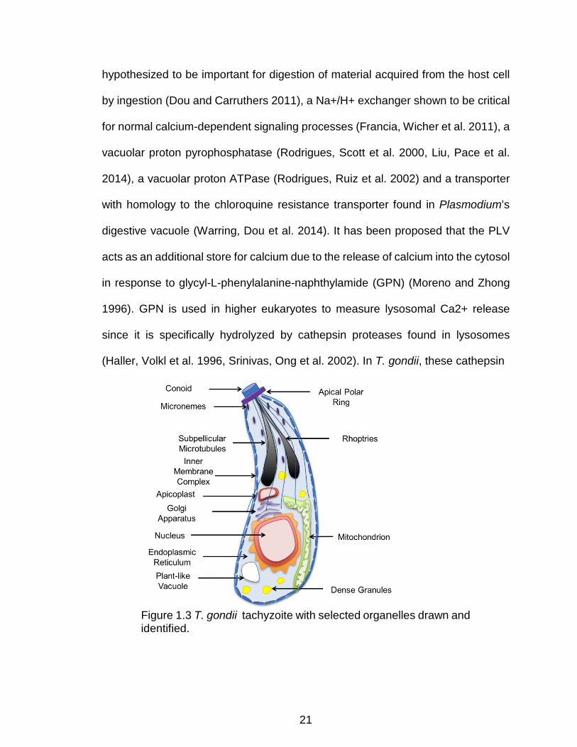
hypothesized to be important for digestion of material acquired from the host cell
by ingestion (Dou and Carruthers 2011), a Na+/H+ exchanger shown to be critical
for normal calcium-dependent signaling processes (Francia, Wicher et al. 2011), a
vacuolar proton pyrophosphatase (Rodrigues, Scott et al. 2000, Liu, Pace et al.
2014), a vacuolar proton ATPase (Rodrigues, Ruiz et al. 2002) and a transporter
with homology to the chloroquine resistance transporter found in Plasmodium’s
digestive vacuole (Warring, Dou et al. 2014). It has been proposed that the PLV
acts as an additional store for calcium due to the release of calcium into the cytosol
in response to glycyl-L-phenylalanine-naphthylamide (GPN) (Moreno and Zhong
1996). GPN is used in higher eukaryotes to measure lysosomal Ca2+ release
since it is specifically hydrolyzed by cathepsin proteases found in lysosomes
(Haller, Volkl et al. 1996, Srinivas, Ong et al. 2002). In T. gondii, these cathepsin
Figure 1.3 T. gondii tachyzoite with selected organelles drawn and identified.

22
proteases are found in the PLV, making it a likely source of Ca2+ release in
response to GPN (Parussini, Coppens et al. 2010, Dou and Carruthers 2011).
Phenotypes associated with disruption of PLV localized proteins can vary widely
with genetic disruption of the Na+/H+ exchanger of the PLV resulting in
increased sensitivity to osmotic stress (Francia, Wicher et al. 2011), while
disruption of the L- type cathepsin protease (TgCPL) of the PLV results in
accumulation of undigested host cell protein material (Dou, McGovern et al.
2014). Together these disparate phenotypes suggest that the PLV is a
multifunctional organelle that not only plays an important role in ion homeostasis
but also in digesting proteins ingested from the host cell cytosol.
Calcium Signaling in T. gondii
Many of the events in T. gondii’s lytic cycle such as egress, motility, invasion
and micronemal protein secretion are accompanied by and dependent on calcium
fluxes within both the parasite and the host cell (Arrizabalaga and Boothroyd
2004). Calcium levels increase in the host cell, the PV and the parasite cytoplasm
just prior to the initiation of parasite egress from its host cell (Borges-Pereira, Budu
et al. 2015). Oscillations in parasite calcium, which are enhanced in the presence
of extracellular calcium, have been observed during periods of parasite motility
using both chemical and genetically encoded calcium indicators (Lovett and Sibley
2003, Borges-Pereira, Budu et al. 2015). Secretion of proteins from the
micronemes, which are required for parasite attachment to a host cell, can be
stimulated by artificially inducing calcium fluxes (Carruthers and Sibley 1999).

23
The parasite can access the calcium required for these signaling events
from both intraparasitic calcium stores and the extracellular milieu once it has
egressed from its host cell (Moreno, Ayong et al. 2011). T. gondii has been shown
by transmission electron microscopy of precipitated calcium to store intracellular
calcium within the perinuclear endoplasmic reticulum (ER) as well large
Figure 1.4 Key steps in the lytic cycle of T. gondii are accompanied by fluctuations in calcium within the parasite and host cell 1. Parasite cytosolic calcium increases stimulate the release of micronemal proteins required for efficient host cell attachment. 2. Invasion of the parasite is accompanied by a dramatic drop in parasite cytosolic calcium. 3. Host cell cytosolic calcium and 4. parasite cytosolic calcium increase just prior to egress of parasites to host cells. 5. The active motility of the parasite is accompanied by oscillations in parasite cytosolic calcium.
cytoplasmic vacuoles, which likely are what has been referred to as plant like
vacuoles (PLV), (Miranda, Pace et al. 2010), and within the flattened sacs of the
inner membrane complex that lie just beneath the parasite plasma membrane
(Bonhomme, Pingret et al. 1993). The localization of calcium stores within the ER
is supported by pharmacological studies using triggers of ER calcium release, such
as thapsigargin, which induce invasion related events including protein secretion
and cytoskeletal rearrangement of the apical end of the parasite (Moreno and
24
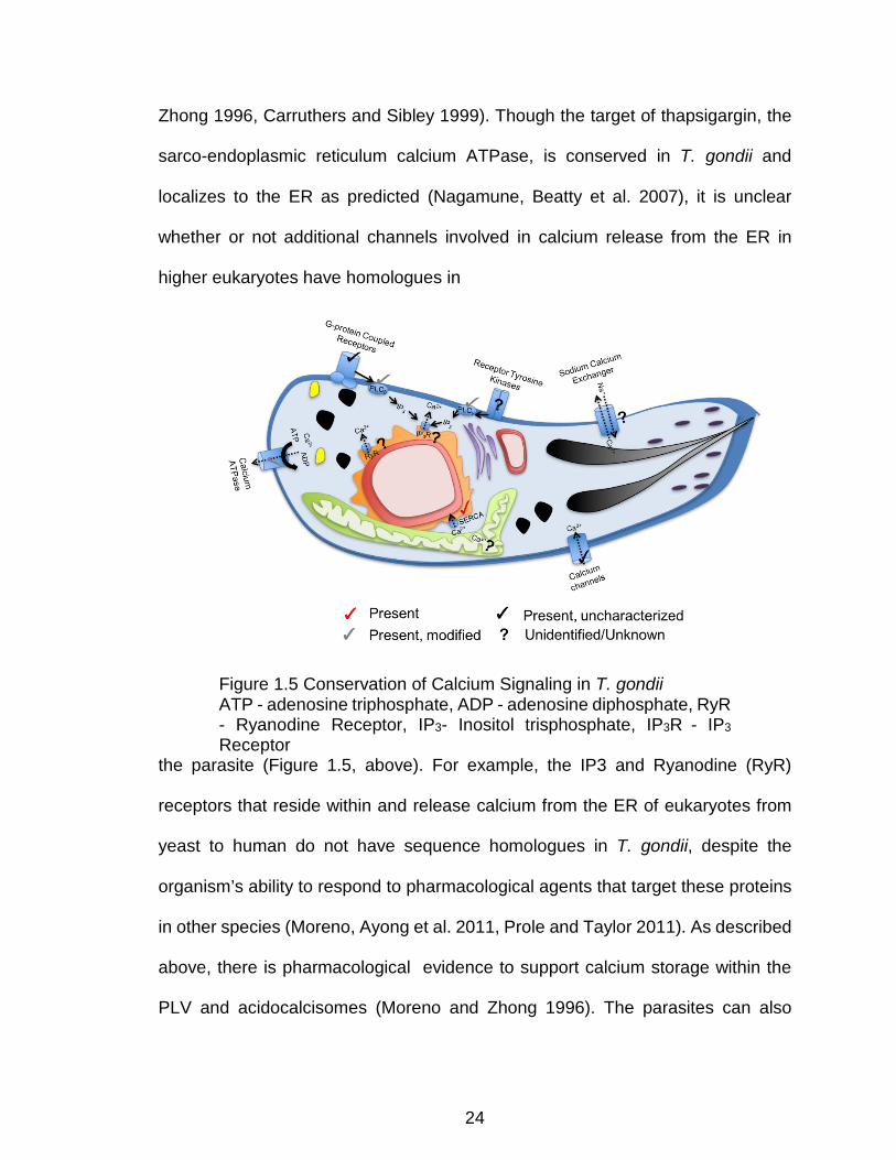
Zhong 1996, Carruthers and Sibley 1999). Though the target of thapsigargin, the
sarco-endoplasmic reticulum calcium ATPase, is conserved in T. gondii and
localizes to the ER as predicted (Nagamune, Beatty et al. 2007), it is unclear
whether or not additional channels involved in calcium release from the ER in
higher eukaryotes have homologues in
Figure 1.5 Conservation of Calcium Signaling in T. gondii ATP - adenosine triphosphate, ADP - adenosine diphosphate, RyR - Ryanodine Receptor, IP3- Inositol trisphosphate, IP3R - IP3 Receptor
the parasite (Figure 1.5, above). For example, the IP3 and Ryanodine (RyR)
receptors that reside within and release calcium from the ER of eukaryotes from
yeast to human do not have sequence homologues in T. gondii, despite the
organism’s ability to respond to pharmacological agents that target these proteins
in other species (Moreno, Ayong et al. 2011, Prole and Taylor 2011). As described
above, there is pharmacological evidence to support calcium storage within the
PLV and acidocalcisomes (Moreno and Zhong 1996). The parasites can also

25
access calcium when extracellular; this calcium is likely taken up by a nifedipine-
sensitive calcium channel in the parasite plasma membrane, though the channel
responsible for this uptake has not been identified (Pace, McKnight et al. 2014).
Once inside a cell, the parasite divides within the PV, where it is presumed to have
access to the host cell calcium through the presence of a nonselective pore in the
PVM, though the exact molecular mechanism for calcium import from the PV
lumen across the parasite plasma membrane has yet to be elucidated (Gold,
Kaplan et al. 2015). Interestingly, electron microscopy analysis of intracellular
tachyzoites suggests that calcium is concentrated within the TVN (Bonhomme,
Pingret et al. 1993). Whether this accumulation of calcium within the TVN is an
active process or if it plays a role in the parasite’s biology is not known. Calcium
levels within the parasite and the PV are thought to remain relatively constant
during the replicative phase of the parasite life cycle, with levels within the host
cell, PV and parasite spiking just prior to egress to activate protein secretion and
parasite motility and begin the lytic cycle over again (Borges-Pereira, Budu et al.
2015). Interestingly, some PV-localized proteins seem to play a role in calcium-
dependent signaling processes, such as egress. GRA1 was initially identified as a
highly abundant, calcium-binding protein found in the lumen of the PV, though its
exact function is unclear since genetic disruption of this gene has not been
successful (Cesbron-Delauw, Guy et al. 1989). An additional GRA protein, GRA22,
that has not been shown to bind calcium directly, has nevertheless been shown to
play a role in the timing of natural egress, as the loss of this protein results in early
egress of parasites from host cells (Okada, Marmansari et al. 2013).

26
Though many of the key molecular mechanisms and factors that respond
to calcium during the invasion and egress of T. gondii have been identified, how
the parasite detects and regulates the calcium fluxes is not completely understood.
In this context, ionophores such as ionomycin, A23187, and nigericin have been
instrumental in studying calcium signaling in T. gondii (Mondragon and Frixione
1996, Pingret, Millot et al. 1996, Stommel, Ely et al. 1997, Black, Arrizabalaga et
al. 2000, Arrizabalaga, Ruiz et al. 2004, Fruth and Arrizabalaga 2007, Caldas, de
Souza et al. 2010). These ionophores are carrier ionophores which can exchange
Ca2+ (ionomycin, A23187) or K+ (nigericin) ions for H+ ions across membranes until
the ions reach electrochemical equilbirum (Pressman 1976). Brief (<2 minute)
treatment of intracellular parasites with the calcium ionophores ionomycin or
A23187 results in rapid exit from the host cell, a process known as ionophore
induced egress (iiEgress). In contrast, treatment of intracellular parasites with
nigericin requires a much longer treatment of 30 minutes before 100% of vacuoles
have left their host cells, but it is likely that nigericin causes the activation of
signaling pathways that lead to the eventual release of calcium to trigger egress,
since mutants delayed in responding to calcium fluxes do not undergo egress in
response to nigericin (Fruth and Arrizabalaga 2007). Brief treatment (2-5 min) of
extracellular parasites with calcium ionophore A23187 induces micronemal
secretion and parasite motility (Carruthers and Sibley 1999). When treatment with
ionophore A23187 is prolonged (45-60 min for Type I strains), the parasites lose
their ability to attach and invade host cells, resulting in parasite death (ionophore
induced death, iiDeath) (Mondragon and Frixione 1996, Black, Arrizabalaga et al.
27
2000). In an effort to identify the proteins that allow T. gondii to respond to calcium,
former members of the Arrizabalaga and Boothroyd laboratories have exploited
these calcium ionophore induced phenomena to isolate mutants with altered
sensitivity to A23187. From a series of selections and screens six independent
mutants have been isolated that fall into three phenotypic categories: delay in
iiEgress and resistance to iiDeath, delay in iiEgress but normal sensitivity to
iiDeath, and resistance to iiDeath but normal iiEgress (Black, Arrizabalaga et al.
2000, Lavine, Knoll et al. 2007). Members of the Arrizabalaga laboratory have
previously reported that all mutants that are both delayed in iiEgress and resistant
to extracellular ionophore death have causative mutations in a calcium dependent
protein kinase, TgCDPK3, which regulates egress by phosphorylating the major
motor driving T. gondii motility (Garrison, Treeck et al. 2012, Gaji, Johnson et al.
2015).
To understand how T. gondii responds to calcium fluxes I focused our
attention to the mutant strain MBD2.1, which is able to survive prolonged exposure
to the ionophore while extracellular, but has no delay in iiEgress. Besides
ionophore resistance, this mutant strain also exhibited hypersensitivity to treatment
of extracellular parasites with the intracellular calcium chelator BAPTA-AM,
suggesting that it has altered calcium homeostasis or sensitivity (Black,
Arrizabalaga et al. 2000). Here I describe how these phenotypes are due to a
nonsense mutation in a previously uncharacterized protein, GRA41, which
localizes to the parasites’ secretory organelles known as dense granules and is
secreted into the PV, where it associates with the TVN. Importantly, I also show

28
that GRA41 is critical for calcium homeostasis and the timing of natural non-
induced egress. In conjunction, our findings suggest a connection between the
TVN and ion homeostasis within parasite, and thus a novel role for the vacuole of
this important pathogen.

29
Chapter 2 : Methods
Parasite propagation
T. gondii tachyzoites were propagated by passaging in human foreskin
fibroblasts (HFFs, purchased from the American Tissue Culture Collection, ATCC)
in a humidified incubator maintained at a temperature of 37°C and 5% CO2
concentration. Normal growth medium used was Dulbecco’s Modified Eagle
Medium (DMEM) with 10% fetal bovine serum (FBS), 2 mM L-glutamine and 50
μg/mL penicillin-streptomycin.
Genome sequencing and complementation
Preparation of genomic DNA was done by previous members of the
Arrizabalaga laboratory. Extracellular parasites from strains MBD2.1 and
RH∆hxgprt (the parental strain) were purified through a 3 μm filter to eliminate
human cell contamination. Genomic DNA from both strains was isolated using the
DNeasy Blood and Tissue Kit (Qiagen). Sample preparation, sequencing, genome
assembly, and annotation was performed at the University of Idaho IBEST
Genomics Resources Core facility. Genomic DNA libraries were constructed using
the Illumina TruSeq library kit and quantified with rtPCR using the Kapa Illumina
library quantification kit. 100bp paired-end Illumina sequencing was used to an
estimated > 100x coverage per genome. Mapping of Illumina sequence was
performed using GMAP to the TGGT1 reference sequence from ToxoDB (Gajria,
Bahl et al. 2008) with output to SAM format files for further processing. Genomic
variants in the mutant strains in comparison to the reference sequence were
detected and extracted from the mapped data using the Broad institutes GATK
30
toolkit. Data was exported as a variant call format (VCF) file, which listed each
genomic variant, its position in the genome, and the quality of sequence data for
that particular region. In total 14 SNVs were detected between the mutant and
parental strain. The two SNVs that resulted in missense or nonsense mutations in
MBD2.1 were confirmed by sequencing fragments of genomic DNA amplified by
PCR from both the parental and mutant strains and that spanned regions with
putative mutations.
To determine which of the identified variants in the mutant was responsible
for the phenotype, a cosmid-based complementation approach was used.
Cosmids generated from the RH (Type I strain) containing the genomic regions of
interest were identified on ToxoDB.org and were graciously provided by Dr. David
Sibley at Washington University, St. Louis. For TGGT1_069070 cosmid TOXO119
was linearized by digestion with NotI (New England Biolabs), purified and
electroporated into T. gondii tachyzoites of the MBD 2.1 mutant according to
established protocols (Kim, Soldati et al. 1993, Soldati and Boothroyd 1993).
Transfected parasites were maintained in the presence of 1 μM pyrimethamine
prior to cloning by limiting dilution to select for stable transformants.
iiDeath survival assay
The iiDeath survival assay was performed as described previously (Black,
Arrizabalaga et al. 2000). In brief, intracellular parasites were harvested by
passage through a 27 gauge needle three times before dilution and treatment with
either 1 μM A23187 or DMSO solvent control for 45 or 60 minutes (10 or 30
minutes for Type II strains) in a humidified incubator at 37°C and 5% CO2

31
concentration. At each time point, 500 parasites were removed from the treatment
and allowed to infect a confluent monolayer of HFFs in a twelve well plate format
for two hours before changing the media to remove uninvaded parasites. Parasites
were allowed to grow and form plaques for 6 days before the cultures were fixed
and scored. Each combination of treatment and time point was the average of a
minimum of three technical replicates per experiment and the experiments were
performed a minimum of three times for statistical analysis. The percent survival
for each strain and time point was calculated as a ratio of the number of plaques
scored in the wells infected with treated parasites as compared to the wells
infected with untreated (DMSO solvent control) parasites.
Micronemal secretion assay
Micronemal protein secretion was assessed by suspending extracellular
parasites at a concentration of 1x10⁹/mL in complete DMEM. Calcium ionophore
A23187 or DMSO control was added to each sample to a final concentration of 1
μM and incubated at 37°C for one hour. Parasites were separated from secreted
proteins in the media by centrifugation at 3,000 x g’s for 3 minutes at 4°C. The
resulting supernatant was subjected to a second spin under the same conditions
to ensure all of the parasite pellet was removed from the sample. The pellets were
resuspended in 2X Laemmli Sample Buffer (Bio-Rad) with 5% freshly added
betamercaptoethanol (BME) to a final concentration of 1x10⁹/mL and treated as
described in Western Blot analysis (below). Supernatants were combined with 4X
Laemmli Sample Buffer (Bio-Rad) with 10% freshly added BME and treated as
described in Western blot analysis. The resulting Western blots were probed with
32
mouse anti-MIC2 antibody to detect endogenous MIC2 protein (Wan, Carruthers
et al. 1997) and visualized as described in Western Blot analysis.
Generation of Type I endogenously tagged GRA41 line
For the expression of GRA41 tagged with hemagglutinin at the carboxyl
terminal of the encoded protein, an 800 base pair fragment of parasite genomic
DNA was amplified by PCR with specific primers GRA41 Tag.FOR and GRA41
Tag.REV (see Table 2.1, for sequence of all primers used in this study) and
directionally cloned into the PacI site of the 3xHA.Lic.DHFR-TS plasmid using In-
Fusion Cloning (Clontech). The 3xHA.LIC.DHFR-TS plasmid is a derivative of the
YFP.LIC.DHFR-TS plasmid (Huynh and Carruthers 2009) with the YFP coding
sequence replaced by a triple hemagglutinin tag. The resulting construct was
verified by restriction digestion and sequencing. The plasmid construct was
linearized with the restriction enzyme XcmI, which cuts within the region containing
the insert and allows for integration of the construct by single homologous
recombination when transfected into the RH∆ku80 strain (Huynh and Carruthers
2009). T. gondii tachyzoites were transfected with the linearized vector by
electroporation according to established protocols (Soldati and Boothroyd 1993).
Transfected parasites were maintained in the presence of 1 μM pyrimethamine
prior to cloning by limiting dilution to select for stable transformants.
Immunofluorescence assays
For immunofluorescence assays, HFFs grown on glass coverslips infected
18-24 hours prior were fixed with 3.5% methanol-free formaldehyde in PBS,
blocked in PBS/3% BSA and permeabilized in PBS/3% BSA/0.2% TX-100.
33
Samples were then incubated in primary antibodies (Rabbit anti-HA, Cell Signaling
Technology, mouse anti-Gra1, Biotem) diluted in PBS/3% BSA/0.2% TX-100,
washed and then incubated in secondary antibodies (goat anti-mouse/rabbit
Alexafluor 488/594 conjugated, Invitrogen) diluted in PBS/3% BSA. Coverslips
were mounted onto microscope slides with Vectashield mounting media containing
DAPI (Vector Laboratories). IFAs were inspected using a Nikon Eclipse E100080i
microscope and images captured with a Hamamatsu C4742-95 charge-coupled
device camera using NIS elements software.
Western blot analysis
To examine protein expression by Western blot, parasites were lysed in 150
mM NaCl, 50 mM Tris-Cl, pH 7.5, 0.1% NP-40 for a minimum of twenty minutes.
Samples were centrifuged at 14,000 x g’s for 20 minutes to remove insoluble
proteins before combining with an equal amount of 2X Laemmli Sample Buffer
(Bio-Rad) with freshly added 5% BME (Thermo Scientific) and heating at ~95°C
for 5 minutes. Samples were separated on a 4-20% gradient SDS-PAGE gel (Bio-
Rad) before transferring to a nitrocellulose membrane using a Trans-Blot semi-dry
transfer cell (Bio-Rad). Nitrocellulose membranes were blocked for a minimum of
30 minutes in Tris-buffered saline/tween (TBST) with 5% non-fat dry milk (NFDM)
before being probed with Rabbit anti-HA at a dilution of 1:5000 (Cell Signaling
Technologies), mouse anti-ROP1 at a dilution of 1:5000 (Schwartzman and Krug
1989) or mouse anti-SAG1 at a dilution of 1:5000 (Genway) for a minimum of one
hour. They were then washed a minimum of three times for ten minutes with TBST
before incubating with the appropriate horse radish peroxidase conjugated
34
secondary antibody (Sigma) for one hour. Membranes were washed again before
incubating with SuperSignal West Femto Maximum Sensitivity Substrate (Thermo
Scientific) for five minutes. Blots were imaged using the FluorChem E system
(Protein Simple) for analysis of chemiluminescent Western blots.
Triton-X 114 membrane partitioning
Membrane partitioning of lysates with Triton-X 114 (Sigma-Aldrich) was
performed as described previously (Rome, Beck et al. 2008). Briefly, parasites
were lysed in 10% Triton-X 114, 10 mM Tris, pH 7.4, 5 mM NaCl on ice for 30
minutes, before centrifuging the samples at 2500 x g’s for five minutes before
removing the supernatant to a new tube for partitioning. Lysates were incubated
at 30°C for five minutes, followed by centrifugation at 3,000 x g’s for 5 minutes to
separate samples into the aqueous and detergent phases. The aqueous phase
was re-extracted by incubating with 10% Triton-X 114, 10 mM Tris, pH 7.4, 5 mM
NaCl on ice for 5 minutes, then at 30°C for five minutes, followed by centrifugation
as described above. The detergent phase was re-extracted by incubating with PBS
on ice for 5 minutes, then at 30°C for five minutes, followed by centrifugation as
described above. Proteins were precipitated from the final aqueous and detergent
phases by adding two volumes of ice cold acetone to each sample and incubating
at -20°C for a minimum of two hours, followed by centrifugation at 15,000 x g’s for
ten minutes. The resulting pellets were washed once more with ice cold acetone
before they were resuspended in 2X Laemlli Sample Buffer (Bio-Rad) with freshly
added 5% BME (Thermo Scientific) and processed for Western blot analysis as
described above.
35
Electron microscopy
For transmission electron microscopy, confluent monolayers of HFFs were
infected with parasites of the strain of interest 24 hours prior to sample preparation.
Infected cells were washed four times with PBS before fixing for one hour in freshly
prepared 2.5% glutaraldehyde in 100 mM sodium cacodylate, pH 7.4 buffer in the
dark. Fixative was then removed and samples were washed three times with PBS
before being harvested by gentle scraping followed by centrifugation at 3,000xg’s
for 10 minutes. The pellets were then incubated in 1% osmium tetroxide in 100
mM sodium cacodylate pH 7.4 buffer that had been reduced by adding solid
potassium ferrocyanide to a final concentration of 1.5% (w/v) and incubated in the
dark at 4 °C for one hour. The pellets were washed three times with 100 mM
sodium cacodylate, pH 7.4 buffer before dehydration in a series of ethanol washes.
Pellets were dehydrated with five minute incubations in 30%, 50%, 70%, 90% and
95% ethanol followed by four five minute washes of 100% ethanol. The samples
were infiltrated with Embed-812/Araldite 502 resin (Electron Microscopy
Sciences), at a dilution of 1:1 resin with 100% ethanol and in 3 changes of 100%
resin. The formulation of resin used was 12.5 ml Embed-812, 7.5 ml Araldite-502,
27.5 ml Dodecenyl Succinic Anhydride, and 0.95 ml DMP-30. All resin incubations
were for at least 2 hours and were done at room temperature in a rotator at low
speed. After the final change the inserts were polymerized at 65 degrees Celsius.
Remaining steps were done with the assistance of the Indiana University-
Blomington Electron Miroscopy Center (http://iubemcenter.indiana.edu). Thin
sections were obtained using a diamond knife, mounted on copper grids and

36
stained with uranyl acetate and lead citrate before imaging on a JEOL 1010
transmission electron microscope. Images were captured with a 1k x 1k Gatan
CCD camera (MegaScan model 794) and a tungsten filament was used as an
electron source.
For immunoelectron microscopy, HFFs infected with parasites for 24-36
hours were washed three times with PBS before fixing for one hour in freshly
prepared 4% paraformaldehyde (EM grade, VWR) in 250 mM HEPES pH 7.4.
Fixative was then removed and replaced with a solution of 8% paraformaldehyde
in 250 mM HEPES pH 7.4 for overnight incubation at 4°C. Samples were harvested
by gentle scraping to detach sheets of cells followed by centrifugation at 2,000 rpm
for 10 minutes. Additional steps were carried out by Dr. Isabelle Coppins of Johns
Hopkins University. Infected cells were pelleted in 10% fish skin gelatin and the
gelatin-embedded pellets were infiltrated overnight with 2.3 M sucrose at 4°C and
frozen in liquid nitrogen. Ultrathin cryosections were incubated in PBS and 1% fish
skin gelatin containing mouse anti-HA antibody at 1/250 dilution, and then exposed
to the secondary antibody that were revealed with 10 nm protein A-gold
conjugates. Sections were observed and images were recorded with a Philips
CM120 Electron Microscope (Eindhoven, the Netherlands) under 80 kV.
Generation of Type I GRA41 knockout strain
The GRA41 knockout strain, RH∆ku80∆gra41, was generated by replacing
the entire GRA41 coding sequence with the HXGPRT selectable marker by double
homologous recombination in the RH∆ku80 parental strain (Huynh and Carruthers
2009). The pGRA41KO vector was generated by cloning fragments approximately
37
1.5 kbp in length directly upstream (primers GRA41 KO 5’ Flank.FOR and GRA41
KO 5’ Flank.REV, Table 2.1) and downstream (primers GRA41 KO 3’ Flank.FOR
and GRA41 KO 3’ Flank.REV, Table 2.1) of the GRA41 coding sequence into the
HindIII and NotI sites of the pMini vector (Donald, Carter et al. 1996) by In-Fusion
cloning (Clontech). The resulting construct was verified by restriction digestion and
sequencing. T. gondii tachyzoites were transfected with purified linearized vector
by electroporation according to established protocols (Soldati and Boothroyd
1993). Transfected parasites were maintained in the presence of 50 μg/ml
mycophenolic acid and 50 μg/ml xanthine prior to cloning by limiting dilution to
select for stable transformants. Individual clones were screened by PCR to verify
correct replacement of the GRA41 locus with the HXGPRT selectable marker. To
confirm the correct insertion, primers complementary to the 3’ end of the HXGPRT
selectable marker and downstream of the insertion site were used to perform a
PCR analysis of genomic DNA isolated from GRA41 knockout clones. A primer
within the 3’ homology region of the pGRA41KO vector (p1, Figure 3.5, GRA41
3’UTR Mid.FOR, Table 2.1) and one downstream of the insertion site (p3, Figure
3.5, GRA41 Downstream.REV, Table 2.1) were used as a positive control, while a
primer within the selectable marker of the pGRA41KO vector (p2, Figure 3.5,
DHFR 3’UTR End.FOR, Table 2.1) was combined with the same downstream
primer (p3) to identify clones with the desired incorporation of the pGRA41KO
vector. Loss of GRA41 mRNA transcript was verified by qPCR analysis (primers
GRA41 qPCR.FOR and GRA41 qPCR.REV, Table 2.1) using the SYBR Green

38
Primer Name Sequence Gra41 Tag.FOR TACTTCCAATCCAATTTAATGCAATAGAGACGC
AGCACCCTTG Gra41 Tag.REV TCCTCCACTTCCAATTTTAGCTTCGGGTAGGTG
AAGTTTAG Gra41 qPCR.FOR ACGGCAGCTTGAGATACCAC Gra41 qPCR.REV GCAGCCCTGGTATTGTATCG Gra41 KO 5'Flank.FOR CGGTATCGATAAGCTTTTTGGTGTGGGCCTGT
CGAAT Gra41 KO 5'Flank.REV CGTGCTGATCAAGCTTCCACGCGTGGATTGCA
GAAAA Gra41 KO 3' Flank.FOR AGTTCTAGAGCGGCCGCTTTTGGTACCTCCAT
GGGCAAGG Gra41 KO 3' Flank.REV ACCGCGGTGGCGGCCGCATACAAAGGCGAGG
TCATTGCTGTG GraX 41 3'UTR Mid.FOR GCGGATTTGTGGCTACAATACTGC Gra41 Downstream.REV CTGCTCTATGTCCTCTGTTCGATGT DHFR 3'UTR End.FOR GGCCATTCATGCCAGTCAGT TgGT1_236870 qPCR.FOR
TTGATGACGACGAGGATGAA
TgGT1_236870 qPCR.REV
GTATGCGGGTAACGACGAGT
TgGT1_236860 qPCR.FOR
TCTCGCGACAACATTTCAAG
TgGT1_236860 qPCR.REV
GCTTTGTCTTCGCTTCCATC
Gra41 qPCR.FOR ACGGCAGCTTGAGATACCAC Gra41 qPCR.REV GCAGCCCTGGTATTGTATCG RPL29 qPCR.FOR GGCGAAATCAAAGAACCACAC RPL29 qPCR.REV TCGGACTTCATCATGCCTCTC GRA41 pet28a.FOR CGCGCGGCAGCCATATGGACTGTATTCCTTCG
CATCAA GRA41 pet28a.REV GTCATGCTAGCCATATGTTATTCGGGTAGGTG
AAGTTTAGA Type II Gra41 Tag.FOR TTCCAATCCAATTTAATTAATTGAAATACACCCT
AGACGGCACAC Type II Gra41 Tag.REV CCACTTCCAATTTTAATTAATTTGGGTAGGTGA
AGTTCAGAAAGC Type II Gra41 KO 5’ Flank.FOR
CGGTATCGATAAGCTTTTTGGTGTGGGCCTGTCGAAT
Type II Gra41 KO 5’ Flank.REV
CGTGCTGATCAAGCTTCCACGCGTGGATTGCAGAAAA
Type II Gra41 KO 3’ Flank. FOR
AGTTCTAGAGCGGCCGCTTTTGGTACCTCCATGGGCAAGG
39
detection reagent (Fast Syber Green Master Mix, Applied Biosystems). Relative
gene expression was calculated by normalizing to levels of the housekeeping gene
ribosomal protein 29 (TGGT1_2345500, Primers RPL29 qPCR.FOR and RPL29
qPCR.REV, Table 2.1) Relative transcript abundance of the genes directly flanking
GRA41 (TGGT1_236860 using primers TGGT1_236860 qPCR.FOR and
TGGT1_236860 qPCR.REV and TGGT1_236870 using primers TGGT1_236870
qPCR.FOR and TGGT1_236870 qPCR.REV, Table 2.1) was also measured by
qPCR analysis to ensure that their levels did not differ significantly from the
parental strain.
Generation of GRA41 knockout complemented clones
The GRA41 knockout strain, RH∆ku80∆gra41 was complemented by
transfecting with the cosmid TOXO119 previously utilized for the complementation
of mutant MBD2.1 described above. Complemented clones were selected for
integration of the gene back into its original locus by selecting against the presence
of the HXGPRT selectable marker using 80 μg/ml 6-thioxanthine. Following limited
dilution cloning, individual clones were subjected to treatment with 50 μg/ml
mycophenolic acid and 50 μg/ml xanthine and qPCR analysis of GRA41 transcript
levels to identify clones which were either HXGPRT+GRA41- or HXGPRT-GRA41+.
Transcript levels of GRA41 were normalized to the levels of ribosomal protein
RPL29 as described above.
Type II Gra41 KO 3’ Flank. REV
ACCGCGGTGGCGGCCGCATACAAAGGCGAGGTCATTGCTGTG
Table 2.1 Name and sequence of primers used in this study. All sequences are 5’ to 3’.
40
Parasite growth assays
Growth of all parasite strains was assessed by determining plaquing
efficiency and growth rate as follows. Intracellular parasites were harvested from
a confluent monolayer of HFFs by passage through a 27-gauge needle 2-3 times.
Parasites were then diluted in normal growth medium and allowed to infect a
confluent monolayer of HFFs and returned to a humidified incubator for two hours
before changing the media to remove any uninvaded parasites. For plaque assays,
parasites were allowed to form plaques for six days before the monolayer was
fixed and stained with Crystal Violet (ACROS Organics) to visualize plaques. For
doubling assays, parasites were allowed to grow for twelve, twenty-four and or
thirty hours before they were fixed and stained with Hema3 Manual Staining
System (Fisher) to count the number of parasites per vacuole for a minimum of 50
vacuoles per sample.
Parasite egress assays
Egress in response to DTT treatment was assessed as follows. Parasites
were allowed to invade host cells for 24 hours prior to the start of the assay.
Infected monolayers were washed twice with 1X PBS and once with 1X HBSS
(Invitrogen) before incubating in either 5 mM DTT in 1X HBSS or buffer only control
for the required time. Monolayers were then fixed with methanol and stained with
Hema3 Manual Staining System (Fisher) to assess parasite egress.
Parasite invasion assays
The efficiency of attachment and invasion for various parasite strains was
measured as follows. Intracellular parasites were harvested as for plaque assays.

41
Parasites were then diluted in normal growth medium and allowed to infect a
confluent monolayer of HFFs for two hours. Unattached parasites were removed
by washing with PBS three times. Cells were then fixed in 3.5% methanol-free
formaldehyde before blocking in PBS/3% BSA. Prior to permeabilization,
extracellular parasites were labeled with mouse anti-P30 antibody (Genway) for
one hour in PBS/3% BSA. Cells were then washed to eliminate unbound antibody
and permeabilized in PBS/3% BSA/0.2% TX-100 before incubation with rabbit anti-
T. gondii (MyBioSource) for one hour. Cells were washed and then incubated with
goat anti-mouse Alexa fluor 488 and goat anti-rabbit Alexa flour 594 (Invitrogen)
for one hour before washing and mounting in Vectashield mounting media with
DAPI (Vector Labs). Intracellular and extracellular parasites were distinguished
between by the presence of dual-color staining (extracellular parasites) or single-
color staining (intracellular parasites, which only stain after permeabilization). The
efficiency of attachment was calculated by counting the total number of parasites
for ten random fields of view and normalizing to 100% attachment for the parental
strain. The efficiency of invasion was calculated as the percentage of invaded
(intracellular) parasites out of the total number of parasites.
Calcium measurements
Calcium measurements of free cytosolic calcium were done by Karla
Marquez-Nogueras of the Moreno laboratory at the University of Georgia. Loading
of parasites with Fura-2 was performed as previously described (Moreno and
Zhong, 1996). Briefly, freshly egressed parasites were washed and then
centrifuged twice at 500 x g’s for 10 minutes at room temperature in buffer A (116

42
mM NaCl, 5.4 mM KCl, 0.8 mM MgSO4, 5.5 mM d-glucose and 50 mM HEPES,
pH 7.4). Then, parasites were resuspended to a final density of 1x10⁹ parasites/ml
in Ringer buffer (155 mM NaCl, 3 mM KCl, 1 mM MgCl2, 3 mM NaH2PO4H2O, 10
mM HEPES, pH 7.3, and 5 mM glucose; plus 1.5% sucrose and 5 μM of Fura-2-
AM). The suspension was incubated for 26 minutes in a 26°C water bath with mild
agitation. Subsequently, the parasites were washed twice with Ringer’s buffer to
remove extracellular dye. Parasites were resuspended to a final density of 1x10⁹
parasites/ml in Ringer’s buffer and kept on ice. Parasites are known to be viable
for a few hours under these conditions (Pace, McKnight et al. 2014). For
fluorescence measurements, 2x10⁷ parasites/mL were added to a cuvette with 2.5
mL of Ringer’s buffer. Cuvette was placed in a thermostatically controlled Hitachi
F-7000 fluorescence spectrophotometer, using the conditions for Fura-2 excitation
(340 and 380nm) and emission (510nm). The Fura-2 fluorescence response to
intracellular Ca2+ concentration ([Ca2+]i) was calibrated from the ratio of 340/380
nm fluorescence values after subtraction of the background fluorescence of the
cells at 340 and 380 nm as previously described (Grynkiewicz et al., 1985).
Changes in [Ca2+]i (ΔF [Ca2+]) were measured by subtracting the highest peak of
calcium in the first 50 seconds after addition of calcium minus the baseline.
Recombinant protein production
The GRA41 recombinant expression vector was generated by cloning the
GRA41 coding sequence without the signal peptide into the pet28a (+) vector
(EMD Biosciences) using In-fusion Cloning (Clontech). The GRA41 coding
sequence was amplified using primers GRA41 pet28a.FOR and GRA41

43
pet28a.REV (Table 1) and inserted into the NdeI sites of the pet28a (+) vector. The
resulting construct was verified by restriction digestion and sequencing. Rosetta
(DE3) pLysS E. coli BL21 competent cells (Novagen) were transformed with
purified vector according to the manufacturer’s protocol. Transformed cells were
then grown at 37°C in LB media to an O.D. of 0.6 before inducing protein
expression with 1 mM IPTG for 3 hours. After induction, cells were harvested by
centrifugation at 10,000 x g’s for 20 minutes at 4°C. Pellets were stored at -80°C.
Additional steps were done with the assistance of Dr. Sanofar Abdeen of the
Johnson laboratory at Indiana University School of Medicine. Pellets were
resuspended in ~100 mL of lysis buffer (50 mM Tris-HCl, pH 7.4, 10 mM MgSO4,
1 mM beta-mercaptoethanol, 0.1% Triton-X 100, 5% glycerol) before adding
lysozyme to a final concentration of 50 μg/mL and incubating for ten minutes on
ice. Samples were then sonicated on ice for six minutes (10 seconds on, twenty
seconds rest) before removing the insoluble portion by centrifugation at 14,000 x
g’s for 30 minutes at room temperature. The supernatant was filtered through a
0.45 µm filter before purifying on a 5 ml HisTrap HP Nickel column (GE) on a NGC
Quest FPLC (Bio-Rad). Fractions were collected analyzed by SDS-PAGE and
Western Blot to determine purity. Fractions positive for the His-tagged protein were
combined and further purified using a HiLoad 26/600 Superdex 200 size exclusion
column (GE).
Calcium thermal shift assays
Differential scanning fluorimetry was utilized to measure the ability of
calcium ions to bind to and stabilize the secondary structures of recombinant

44
proteins as described in (Niesen, Berglund et al. 2007) using recombinant CDPK3
(Gaji, Johnson et al. 2015) as a positive control. N-terminally His tagged GRA41
(final concentration of 8 μM) or CDPK3 (final concentration of 3.5 μM) in 100 mM
HEPES, pH 7.0, 150 mM NaCl was incubated with 4X Sypro Orange (Sigma
Aldrich) and CaCl2 in a two-fold dilution series ranging from 0-250 μM. Melt curve
analysis was performed from 25 to 99°C with 1°C steps on a StepOne Plus Real-
Time PCR System (Applied Biosystems). Data were analyzed in Prism (GraphPad)
to determine melting temperatures for each condition.
Immunoprecipitation
Human foreskin fibroblasts were infected with the strain containing an
endogenous HA tag at the C terminus of GRA41 for twenty-four hours before
harvesting. Samples were harvested by scraping the infected monolayer and
solubilizing in RIPA buffer (10 mM Tris-Cl, pH 8.0, 1 mM EDTA, 0.5 mM EGTA,
1% Triton X-100, 0.1% sodium deoxycholate, 0.1% SDS, 140 mM NaCl) with
added protease inhibitors (cOmplete Mini Protease Inhibitor Cocktail, Roche) for a
minimum of twenty minutes with rotation. Samples were sonicated for ten second
pulses 2-3 x, with a minimum rest on ice of twenty seconds between pulses.
Insoluble material was removed by centrifuging for 20 minutes at 13,000 x g’s and
4°C. The supernatant was removed and incubated with high affinity rat anti-HA
antibody (Roche) overnight at 4°C with rotation. The following morning, the
antibody/antigen complex was bound to Protein G-conjugated Dynabeads
(Invitrogen) by incubating the supernatant/antibody mixture with freshly washed
Dynabeads for ten minutes at room temperature with rotation. The precipitated

45
beads were isolated using a magnet and washed three times with RIPA buffer plus
protease inhibitors. Proteins were eluted from the beads by adding 2X Laemmli
sample buffer with 5% betamercaptoethanol to the beads and incubating at 95°C
for ten minutes. The beads were isolated using a magnet and the sample buffer
containing eluted antigen was removed. The sample was run on a 4-20% Mini
Protean TGX Precast Gel (Bio-rad) and the gel was stained using the Pierce Silver
Stain kit (ThermoFisher Scientific). Regions containing visible bands were excised
with a clean razor blade and stored in PBS before mass spectrometry analysis.
Additional steps were performed by the Indiana University Bloomington Laboratory
for Biological Mass Spectrometry (http://www.chem.indiana.edu/bms/). The
samples were reduced with 10 mM DTT in 10 mM ammonium bicarbonate and
then alkylated with 55 mM iodoacetamide (prepared in 10 mM ammonium
bicarbonate). Alkylated samples were digested by trypsin (Promega) overnight at
37°C. Digested peptides were injected onto the C18 column. Peptides were eluted
with a linear gradient from 3 to 40% acetonitrile (in water with 0.1% FA) developed
over 70 min at room temperature at a flow rate of 300 nL/min, and effluent was
electro-sprayed into the mass spectrometer. A blank was run prior to and between
the sample runs to make sure there was no significant signal from solvents, column
or carryovers. Database search was carried out using SequestTM algorithms
against the UniProt database for T. gondii. The results were further refined by
removing hits with an expect value>0.0025. Common contaminants found in other
unrelated datasets were removed and the list was restricted to proteins with a
predicted signal peptide.
46
Yeast two hybrid screen
Yeast two-hybrid screening was performed by Hybrigenics Services,
S.A.S., Paris, France (http://www.hybrigenics-services.com). The coding
sequence for T. gondii GRA41 (amino acids 25-179) was PCR-amplified and
cloned into pB66 as a C-terminal fusion to the Gal4 DNA-binding domain (Gal4-
Gra41). The construct was checked by sequencing the entire insert and used as a
bait to screen a random-primed T. gondii wild-type RH strain cDNA library
constructed into pP6. pB66 derives from the original pAS2ΔΔ vector (Fromont-
Racine, Rain et al. 1997) and pP6 is based on the pGADGH plasmid (Bartel, Chien
et al. 1993).
62.4 million clones (6-fold the complexity of the library) were screened using
a mating approach with YHGX13 (Y187 ade2-101::loxP-kanMX-loxP, mat-alpha)
and CG1945 (mata) yeast strains as previously described (Fromont-Racine, Rain
et al. 1997). 337 His+ colonies were selected on a medium lacking tryptophan,
leucine and histidine and supplemented with 0.5 mM 3-aminotriazole to handle bait
autoactivation. The prey fragments of the positive clones were amplified by PCR
and sequenced at their 5’ and 3’ junctions. The resulting sequences were used to
identify the corresponding interacting proteins in the GenBank database (NCBI)
using a fully automated procedure. A confidence score (PBS, for Predicted
Biological Score) was attributed to each interaction as previously described
(Formstecher, Aresta et al. 2005). The PBS relies on two different levels of
analysis. Firstly, a local score takes into account the redundancy and
independency of prey fragments, as well as the distribution of reading frames and
47
stop codons in overlapping fragments. Secondly, a global score takes into account
the interactions found in all the screens performed at Hybrigenics using the same
library. This global score represents the probability of an interaction being
nonspecific. For practical use, the scores were divided into four categories, from A
(highest confidence) to D (lowest confidence). A fifth category (E) specifically flags
interactions involving highly connected prey domains previously found several
times in screens performed on libraries derived from the same organism. Finally,
several of these highly connected domains have been confirmed as false-positives
of the technique and are now tagged as F. The PBS scores have been shown to
positively correlate with the biological significance of interactions (Rain, Selig et al.
2001, Wojcik, Boneca et al. 2002).
Generation of Type II endogenously tagged GRA41 line
For the expression of GRA41 tagged with hemagglutinin at the carboxyl
terminal of the encoded protein, an 1700 base pair fragment of Pru genomic DNA
was amplified by PCR with specific primers Type II GRA41 Tag.FOR and Type II
GRA41 Tag.REV (see Table 2.1, for sequence of primers used in this study) and
directionally cloned into the PacI site of the 3xHA.Lic.DHFR-TS plasmid using In-
Fusion Cloning (Clontech). The 3xHA.LIC.DHFR-TS plasmid is a derivative of the
YFP.LIC.DHFR-TS plasmid (Huynh and Carruthers 2009) with the YFP coding
sequence replaced by a triple HA tag. The resulting construct was verified by
restriction digestion and sequencing. The plasmid construct was linearized with
the restriction enzyme XcmI, which cuts within the region containing the insert and
allows for integration of the construct by single homologous recombination when

48
transfected into the Pru∆ku80 strain (Fox, Falla et al. 2011) stably expressing
luciferase, which can be used to visualize parasite propagation in mice using a
bioluminescence-based imaging system (Saeij, Boyle et al. 2005). T. gondii
tachyzoites were transfected with the linearized vector by electroporation
according to established protocols (Soldati and Boothroyd 1993). Transfected
parasites were maintained in the presence of 1 μM pyrimethamine prior to cloning
by limiting dilution to select for stable transformants.
Generation of Type II GRA41 knockout strain
The GRA41 knockout strain, Pru∆ku80∆gra41, was generated by replacing
the entire GRA41 coding sequence with the HXGPRT selectable marker by double
homologous recombination in the Pru∆ku80∆hxgprt parental strain(Fox, Falla et
al. 2011). The pGRA41KO-GFP vector was generated by cloning fragments
approximately 1.5 kbp in length directly upstream (primers Type II GRA41 KO 5’
Flank.FOR and Type II GRA41 KO 5’ Flank.REV, Table 2.1) and downstream
(primers Type II GRA41 KO 3’ Flank.FOR and Type II GRA41 KO 3’ Flank.REV,
Table 2.1) of the GRA41 coding sequence into the HindIII and NotI sites of the
pmini-GFP.ht vector (Donald, Carter et al. 1996, Arrizabalaga, Ruiz et al. 2004) by
In-Fusion cloning (Clontech). The resulting construct was verified by restriction
digestion and sequencing. T. gondii tachyzoites were transfected with purified
linearized vector by electroporation according to established protocols (Soldati and
Boothroyd 1993). Transfected parasites were maintained in the presence of 50
μg/ml mycophenolic acid and 50 μg/ml xanthine prior to cloning by limiting dilution
to select for stable transformants. Individual clones were screened first for the
49
presence of GFP by live imaging (constructs integrated by double homologous
recombination should be GFP negative) and then by PCR to verify correct
replacement of the GRA41 locus with the HXGPRT selectable marker.
To confirm the correct insertion, primers complementary to the 3’ end of
HXGPRT selectable marker and downstream of the insertion site were used to
perform a PCR analysis of genomic DNA isolated from GRA41 knockout clones.
A primer within the 3’ homology region of the pGRA41KO vector (p1, Figure 5.2,
GRA41 3’UTR Mid.FOR, Table 2.1) and one downstream of the insertion site (p3,
Figure 5.2, GRA41 Downstream.REV, Table 2.1) were used as a positive control,
while a primer within the selectable marker of the pGRA41KO vector (p2, Fig. 3,
DHFR 3’UTR End.FOR, Table 2.1) was combined with the same downstream
primer (p3) to identify clones with the desired incorporation of the pGRA41KO
vector.
Bradyzoite differentiation assays
The ability of parasite strains to differentiate from tachyzoites to bradyzoites
was assessed as follows. Intracellular parasites were harvested from a confluent
monolayer of HFFs by passage through a 27-gauge needle 2-3 times. Parasites
were then diluted in normal growth medium and allowed to infect a confluent
monolayer of HFFs grown on glass coverslips and returned to a humidified
incubator for two hours before replacing the media with RPMI switching media
(RPMI 1640, Invitrogen, pH 8.1, 5% FBS, 50 mM HEPES), placing inside an
airtight container and placing in a CO2 deprivation incubator at 37°C. At 4 or 7
days, the cells were fixed, blocked, permeabilized and probed as described in the
50
immunofluorescence assay methods above. Rhodamine-labeled Dolichos biflorus
agglutinin (DBA, Vector Laboratories) was added at a dilution of 1:250 during the
secondary fluorophore-conjugated antibody step to stain the bradyzoite tissue cyst
wall. Coverslips were mounted onto microscope slides with Vectashield mounting
media that included DAPI (Vector Laboratories). IFAs were inspected using a
Nikon Eclipse E100080i microscope and a minimum of 100 vacuoles for each
strain and timepoint were scored as either cyst-wall positive or cyst-wall negative.
The percentage of cyst wall positive vacuoles was expressed as a percentage of
the total.
Mouse studies of chronic toxoplasmosis
The ability of parasites to disseminate and form tissue cysts in vivo was
tested as follows. Intracellular parasites were washed 2-3 times with sterile PBS
before being harvested from a confluent monolayer of HFFs by passage through
a 27-gauge needle 2-3 times. Host cell debris was removed by passage through a
3 μm filter. Parasites were counted and diluted in sterile PBS and transported at
room temperature to the animal facility. Insulin syringes (28 gauge, Becton
Dickinson) were used to deliver 100 μl of diluted parasite intraperitoneally to female
CBA/J mice. Mice were monitored daily during the acute phase of the disease (up
to 14 days) and 2-3x a week thereafter for signs of sickness. Two mice were lost
during the course of the study (one each in the highest dose for the parental and
knockout strains), but due to injuries sustained by the mice and the lack of recently
observed acute symptoms of toxoplasmosis (ruffled coats, swollen abdomen,
lethargy or difficulty walking), it was deemed that these mice did not perish as a
51
direct result of infection and they were removed from further analysis. At 28 days
post-infection, mice were anesthetized in a CO2 chamber followed by cervical
dislocation. Blood was collected for serum analysis and the brains were harvested
to measure cyst burden. Brains were placed in Eppendorf tubes on ice for the
remainder of the procedure. Each brain was ground in 1500 μl of ice-cold PBS
using a mortar and pestle before removing 250 μl for immunofluorescence
analysis. The remainder was immediately placed at -80°C for long-term storage.
Samples were centrifuged at 3,000 x g’s for 5 minutes at room temperature and
the supernatants removed. Brain tissue was resuspended in 150 μl of freshly
prepared 3% formaldehyde in PBS and let stand for 20 minutes at room
temperature prior to centrifugation at 3,000 x g’s for 5 minutes at room
temperature. The supernatant was removed and brains were resuspended in 150
μl of 100 mM glycine in PBS and let stand for 5 minutes at room temperature prior
to centrifugation at 3,000 x g’s for 5 minutes at room temperature. The supernatant
was removed and brains were resuspended in 150 μl of 3% BSA/0.2% TX-100 in
PBS and let stand overnight at 4°C prior to centrifugation at 3,000 x g’s for 5
minutes at room temperature. The supernatant was removed and brains were
resuspended in 125 μl of 3% BSA/0.2% TX-100 in PBS containing a 1:250 dilution
of Rhodamine-labeled DBA (Vector Laboratories) and let stand for one hour at
room temperature prior to centrifugation at 3,000 x g’s for 5 minutes at room
temperature. The brains were washed three times by resuspending in 150 μl of
wash buffer (0.2% TX-100 in PBS) followed by centrifugation at 3,000 x g’s for 5
minutes at room temperature. After the final centrifugation, the supernatant was
52
removed and 5 μl of brain material was pipetted onto three microscope slides per
brain using a large bore pipette. Brains were gently flattened by placing a coverslip
over the top and pressing down gently. Coverslips were secured with clear nail
polish. The number of cysts per slide was enumerated using a Nikon Eclipse
E100080i microscope. The total number of cysts for the three slides for each
mouse was combined and multiplied by 26 to obtain an estimate of the total
number of cysts per mouse brain as described previously (Lavine, Knoll et al.
2007).
Serum was collected by allowing the mouse blood to coagulate at room
temperature for a minimum of thirty minutes in an Eppendorf tube. Serum was
separated from coagulated blood cells by centrifuging twice at 4°C for 10 minutes
at 5,000 x g’s. The resulting serum was used in a dot blot assay to verify that mice
had been successfully infected. The dot blot assay was conducted as described
previously (Benmerzouga, Checkley et al. 2015). Briefly, approximately 5 x 10⁶
freshly egressed parasites of the parental strain, Pru∆ku80, were centrifuged at
room temperature for 10 minutes at 1,000 x g’s. All but 100 μl of the supernatant
was removed and the parasites were resuspended in the remaining volume.
Approximately 8-10 μl of resuspended parasites were pipetted onto a nitrocellulose
membrane that had been soaked in PBS for 5 minutes. The resuspended parasites
were allowed to dry onto the membrane before blocking the membrane with
TBST/5% NFDM for 45 minutes. Individual blots were incubated with serum from
a single mouse overnight at 4°C at a dilution of 1:50 in TBST/5% NFDM. The
following morning, blots were washed three times with TBST for ten minutes each

53
before incubating with goat anti-mouse conjugated to horse radish peroxidase at
a dilution of 1:5000 for a minimum of one hour at room temperature. Blots were
again washed three times with TBST for ten minutes each before incubating for
five minutes with SuperSignal West Femto Maximum Sensitivity Substrate
(Thermo Scientific) for five minutes. Blots were imaged using the FluorChem E
system (Protein Simple) for analysis of chemiluminescent Western blots.
Seropositivity of individual mice was determined by comparing dot blot results with
positive and negative control sera from previous experiments and seronegative
mice were removed from statistical analysis of the data.

54
Chapter 3 : Identification and Characterization of GRA41
In this chapter, my aim is to identify and characterize the protein responsible
for resistance to iiDeath in mutant MBD2.1 by sequencing the mutant to identify
variations in the mutant genome and complementing these mutations with wild-
type copies of the genes impacted. I then endogenously tag the protein of interest
to determine its localization and generate a complete knockout to study its impact
on parasite function.
Nonsense mutation in novel gene is responsible for iiDeath- phenotype of
MBD2.1
To identify the causative mutation in mutant strain MBD2.1, whole-genome
sequencing was undertaken of both the mutant and its parental strain, RH∆hxgprt.
Single nucleotide variants (SNVs) between the two were mapped against the T.
gondii genomic database (ToxoDB). In total 18 SNVs occurred between mutant
and parental; 5 were in intergenic regions, 11 within introns and 2 in exons. Of the
two mutations within exons, which were both confirmed by PCR and sequencing,
one results in a missense mutation in the hypothetical protein TGGT1_306020 and
the other in a nonsense mutation in the hypothetical protein TGGT1_069070
(ToxoDB v7.2). Although both transcriptomic and proteomic evidence could be
found for TGGT1_069070 in version 7.2 of the T. gondii genomic database, this
predicted gene has not been annotated as a gene in subsequent genome versions.
Analysis of a cDNA library using 5’ and 3’ RACE confirmed the gene model of
TGGT1_069070 shown in ToxoDBv7.2 (Figure 3.1, below). TGGT1_069070
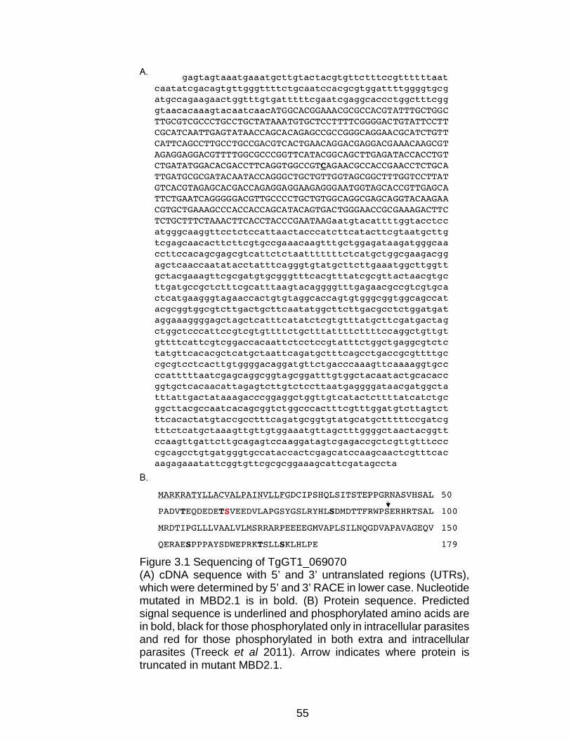
55
Figure 3.1 Sequencing of TgGT1_069070 (A) cDNA sequence with 5’ and 3’ untranslated regions (UTRs), which were determined by 5’ and 3’ RACE in lower case. Nucleotide mutated in MBD2.1 is in bold. (B) Protein sequence. Predicted signal sequence is underlined and phosphorylated amino acids are in bold, black for those phosphorylated only in intracellular parasites and red for those phosphorylated in both extra and intracellular parasites (Treeck et al 2011). Arrow indicates where protein is truncated in mutant MBD2.1.

56
encodes a putative 179 amino acid (aa) protein with an N-terminal signal sequence
but no known functional domains. Though homologs of this gene are not annotated
in any genomes present in the EuPathDB (EuPathDB.org) database, a tblastn
search against the parasite genomes available in EuPathDB identifies potential
homologs in the closely related parasites Hammondia hammondi
(KL544038:723,066..723,587, 77% identity with TGGT1_069070) and Neospora
caninum (FR823391:5,147,783..5,148,286, 47% identity).
The nonsense mutation in TGGT1_069070 detected in the mutant strain is
a single C to G transversion, which results in the conversion of the serine at
position 91 to a premature stop codon (Figure 3.2, below). To investigate whether
the mutation in this gene was responsible for the observed phenotype of MBD2.1,
I transfected a cosmid containing a parental copy of the gene into the mutant and
generated stable clones by limiting dilution. Two independent clones (MBD2.1
Comp Clone 1 and MBD2.1 Comp Clone 7) were established and the incorporation
of the parental allele was confirmed by sequencing of a PCR fragment spanning
the TGGT1_069070 coding sequence. The sequence chromatogram from
Clone 1 shows a mixed peak at the position of interest (Figure 3.2, below)
indicating that this clone carries both a mutant and a parental copy of the gene as
expected for random integration of the cosmid. On the other hand, Clone 7
contains only the wild-type copy of the gene (Figure 3.2, below), suggesting that
the cosmid recombined by homologous recombination with the endogenous locus
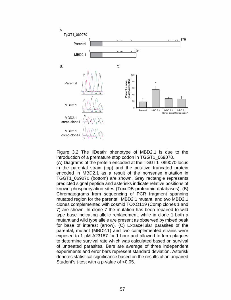
57
Figure 3.2 The iiDeath- phenotype of MBD2.1 is due to the introduction of a premature stop codon in TGGT1_069070. (A) Diagrams of the protein encoded at the TGGT1_069070 locus in the parental strain (top) and the putative truncated protein encoded in MBD2.1 as a result of the nonsense mutation in TGGT1_069070 (bottom) are shown. Gray rectangle represents predicted signal peptide and asterisks indicate relative positions of known phosphorylation sites (ToxoDB proteomic databases). (B) Chromatograms from sequencing of PCR fragment spanning mutated region for the parental, MBD2.1 mutant, and two MBD2.1 clones complemented with cosmid TOXO119 (Comp clones 1 and 7) are shown. In clone 7 the mutation has been repaired to wild type base indicating allelic replacement, while in clone 1 both a mutant and wild type allele are present as observed by mixed peak for base of interest (arrow). (C) Extracellular parasites of the parental, mutant (MBD2.1) and two complemented strains were exposed to 1 µM A23187 for 1 hour and allowed to form plaques to determine survival rate which was calculated based on survival of untreated parasites. Bars are average of three independent experiments and error bars represent standard deviation. Asterisk denotes statistical significance based on the results of an unpaired Student’s t-test with a p-value of <0.05.
58
resulting in an allelic replacement event. I have confirmed that Clone 7 still carries
the missense mutation in TGGT1_306020 corroborating that it is indeed a
derivative of the mutant strain MBD2.1 (data not shown).
Importantly, regardless of whether by random integration or by allelic
replacement, introduction of wildtype TGGT1_069070 restored ionophore
sensitivity to MBD2.1 (Figure 3.2, above). Treatment of extracellular parasites with
A23187 followed by plaque assay to determine number of viable parasites showed
that, while mutant MBD2.1 has increased survivability (54%±9%, p<0.05) as
compared to a parental strain (18%±11%), both MBD2.1 Comp Clone 1 and
MBD2.1 Comp Clone 7, were statistically as sensitive to iiDeath as the parental
strain (28%±5% and 26%±8% respectively, Figure 3.2, above). Thus, presence of
a wildtype copy of TGGT1_069070 restores ionophore sensitivity to mutant
MBD2.1. Based on this result and those described below I have focused my
studies on the protein product of TGGT1_069070 and have not as of yet explored
the role of the mutation found in TGGT1_306020 as it is unlikely to influence
iiDeath.
TGGT1_069070 encodes a novel dense granule protein, GRA41
In order to confirm the expression of TGGT1_069070 at the protein level
and to determine its localization, sequences encoding a carboxy-terminal (C-
terminal) triple-hemagglutinin (HA) tag were introduced into the endogenous locus
in the RH∆ku80∆hxgprt strain just before the stop codon, using a previously
described approach (Huynh and Carruthers 2009). Western blot analysis of protein
extract from the endogenously tagged strain revealed a doublet migrating at
59
approximately 30 kDa, which is higher than the predicted molecular weight of the
tagged protein, 24 KDa (Figure 3.3A, below). The doublet pattern was consistently
observed in extracts from both intracellular and extracellular parasites (Figure
3.3A, below). At present the reason for the migration pattern is not known, although
the protein is predicted to be phosphorylated, which could account for size
differences as it does for other T. gondii proteins such as GRA7 (Dunn, Ravindran
et al. 2008). The resulting strain grows normally in culture and does not exhibit
resistance to ionophore induced death, indicating that the tag does not interfere
with the protein’s function (Figure 3.3B, below). To explore the localization of
TGGT1_069070, I performed an immunofluorescence analysis (IFA) and found
that the encoded protein appears to localize within the PV, where it colocalizes
with the PV localized protein, GRA7 (Fischer, Stachelhaus et al. 1998, Jacobs,
Dubremetz et al. 1998) (Figure 3.3C, below). IFA of the engineered parasites with
antibodies against the parasite surface antigen SAG1 (Kasper, Bradley et al. 1984)
confirmed that the majority of the HA-tagged protein is present within the PV and
not within the parasites (Figure 3.3D, below). The majority of proteins in the PV
are secreted by intracellular parasites from the dense granules, whose contents
are more easily detected in extracellular parasites. To determine if
TGGT1_069070 is also a dense granule protein, I performed an IFA of extracellular
parasites with anti-HA antibodies to detect TGGT1_069070 and antibodies to
dense granule marker GRA7, which shows co-localization of both signals within
intracellular vesicles consistent with dense granules (Figure 3.3F, below). To
confirm and refine the results from immunofluorescence analysis, I,

60
Figure 3.3 TGGT1_069070 encodes a novel dense granule protein, GRA41
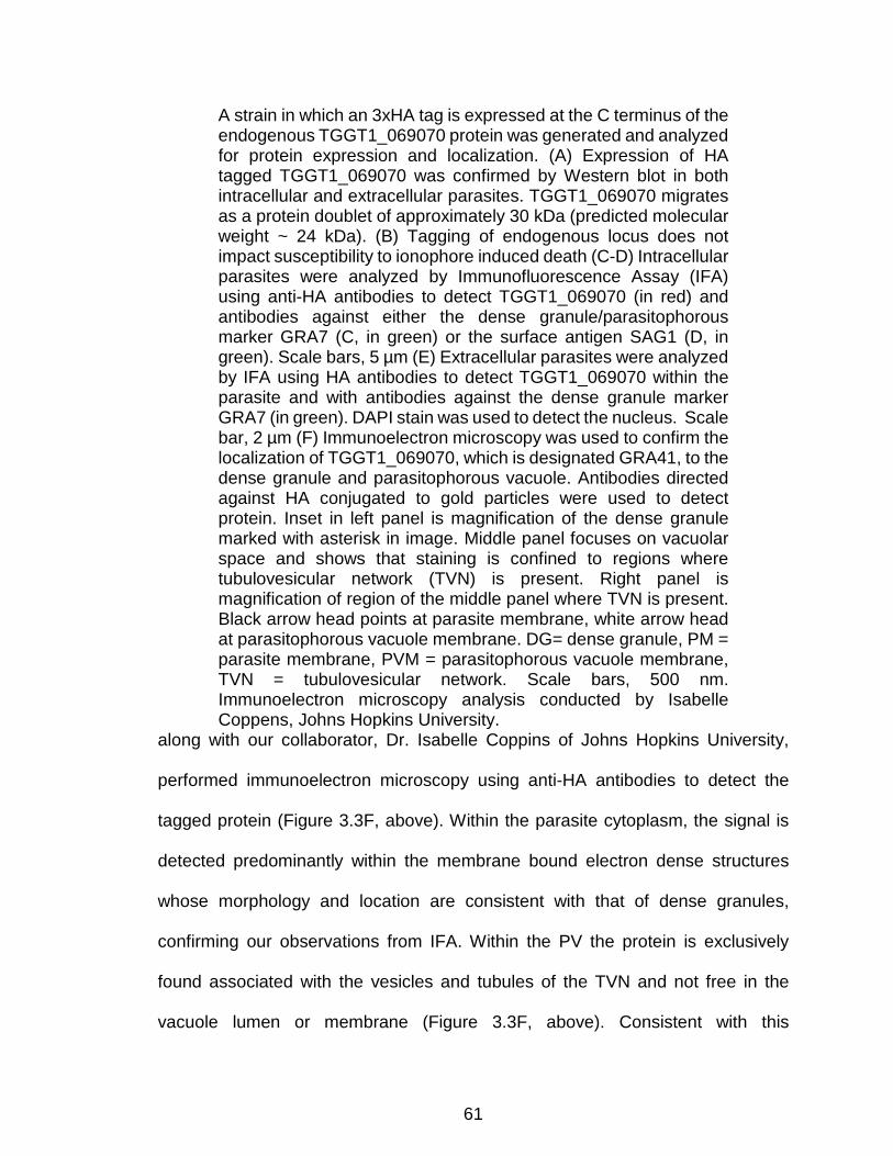
61
along with our collaborator, Dr. Isabelle Coppins of Johns Hopkins University,
performed immunoelectron microscopy using anti-HA antibodies to detect the
tagged protein (Figure 3.3F, above). Within the parasite cytoplasm, the signal is
detected predominantly within the membrane bound electron dense structures
whose morphology and location are consistent with that of dense granules,
confirming our observations from IFA. Within the PV the protein is exclusively
found associated with the vesicles and tubules of the TVN and not free in the
vacuole lumen or membrane (Figure 3.3F, above). Consistent with this
A strain in which an 3xHA tag is expressed at the C terminus of the endogenous TGGT1_069070 protein was generated and analyzed for protein expression and localization. (A) Expression of HA tagged TGGT1_069070 was confirmed by Western blot in both intracellular and extracellular parasites. TGGT1_069070 migrates as a protein doublet of approximately 30 kDa (predicted molecular weight ~ 24 kDa). (B) Tagging of endogenous locus does not impact susceptibility to ionophore induced death (C-D) Intracellular parasites were analyzed by Immunofluorescence Assay (IFA) using anti-HA antibodies to detect TGGT1_069070 (in red) and antibodies against either the dense granule/parasitophorous marker GRA7 (C, in green) or the surface antigen SAG1 (D, in green). Scale bars, 5 µm (E) Extracellular parasites were analyzed by IFA using HA antibodies to detect TGGT1_069070 within the parasite and with antibodies against the dense granule marker GRA7 (in green). DAPI stain was used to detect the nucleus. Scale bar, 2 µm (F) Immunoelectron microscopy was used to confirm the localization of TGGT1_069070, which is designated GRA41, to the dense granule and parasitophorous vacuole. Antibodies directed against HA conjugated to gold particles were used to detect protein. Inset in left panel is magnification of the dense granule marked with asterisk in image. Middle panel focuses on vacuolar space and shows that staining is confined to regions where tubulovesicular network (TVN) is present. Right panel is magnification of region of the middle panel where TVN is present. Black arrow head points at parasite membrane, white arrow head at parasitophorous vacuole membrane. DG= dense granule, PM = parasite membrane, PVM = parasitophorous vacuole membrane, TVN = tubulovesicular network. Scale bars, 500 nm. Immunoelectron microscopy analysis conducted by Isabelle Coppens, Johns Hopkins University.

62
membranous localization, partitioning with TX-114 showed the tagged protein
partitions almost exclusively with the detergent phase, consistent with a
membrane-associated protein (Figure 3.4, below). Based on these results, I have
concluded that the protein encoded at the TGGT1_069070 locus is a dense
granule derived protein that is secreted from the parasite into the PV where it
associates with the TVN. According to the currently published literature, the dense
granule proteins have been named from GRA1 to 40 so TGGT1_069070 is
designated as GRA41.
Figure 3.4 Western Blot Analysis of Triton-X 114 partitioning of parasite lysates from the GRA41 endogenously HA-tagged strain. GRA41 (detected with Rabbit anti-HA antibody) primarily partitions with the detergent (Det) rather than the aqueous (Aq) phase. ROP1 (detected with mouse anti-ROP1 antibody) was used as soluble protein control to label the aqueous phase and SAG1 (detected with mouse anti-SAG1 antibody) was used as a membrane protein control to label the detergent phase.
Complete knockout of GRA41 recapitulates iiDeath- phenotype
To further study the role of GRA41 in the parasite’s life cycle, I generated a
knockout of the gene by replacing the entire coding sequence with the selectable
marker hypoxanthine-guanine phosphoribosyltransferase (HXGPRT) through

63
double homologous recombination into the RH∆ku80∆hxgprt strain, hereafter
referred to as RH∆ku80 (Huynh and Carruthers 2009) (Figure 3.5A, below).
Parasites transfected with the knockout vector (pGRA41KO) and stably expressing
HXGPRT were selected by resistance to mycophenolic acid and xanthine and
cloned by limiting dilution. Through two separate attempts to generate knockout
strains, I established two independent clones (RH∆ku80∆gra41-1, aka KO1 and
RH∆ku80∆gra41-2, aka KO2) that were shown to have correct integration of the
construct by PCR (Figure 3.5B, below). To confirm that the genetic disruption of
the locus resulted in a functional knockout of GRA41, I measured relative transcript
abundance by quantitative PCR (qPCR), which failed to detect GRA41 message
in either of the knockout clones (Figure 3.5C, below). In addition, through qPCR, I
determined that the genetic disruption did not affect transcript levels of the genes
immediately upstream and downstream of GRA41 (Figure 3.5C, below). Based on
the nature of the genetic disruption expected from double recombination of the
knockout construct (pGRA41KO, Figure 3.5A, below) and the lack of detectable
transcript (Figure 3.5C, below), the two established clones represent functional
knockouts and are unlikely to produce any GRA41 protein. I then assessed
whether or not the complete knockout of GRA41 recapitulated the iiDeath-
phenotype seen in GRA41 truncation mutant MBD 2.1. Both RH∆ku80∆gra41-1
(45% ± 16% versus 11% ± 12% for the parental strain, p<0.05, Figure 3.5D, below)
and RH∆ku80∆gra41-2 (25% ± 5% versus 7% ± 5% for the parental strain, p<0.05,
Figure 3.5D, below) showed statistically significant increases in survival as
compared to the parental strain after 60 minutes of treatment with A23187. Thus,

64
the recapitulation of the phenotype seen in the mutant MBD2.1 by the complete
lack of GRA41 suggests that the iiDeath phenotype of MBD2.1 is likely due to the
absence of full-length GRA41 product and not a dominant negative effect imparted
by a possible truncated protein.
Figure 3.5 Complete knockout of GRA41 recapitulates the iiDeath- phenotype seen in MBD2.1 mutant. (A) Diagram shows strategy used to knockout GRA41 by double homologous recombination replacing the GRA41 locus with the HXGPRT selectable marker. Colored arrow heads indicate beginning and end of genomic regions used to drive homologous recombination of pGRA41KO vector with target locus in chromosome X (ChrX). Arrows indicate relative direction of transcription of the three genes depicted and black arrow heads represent primers p1, 2 and 3 used to corroborate integration. (B) PCR analysis of DNA from parental strain (Par) and two independent putative knockouts KO1 (top panels) and KO2 (bottom panels) using either a primer pair expected to produce an
65
Complete knockout of GRA41 results in decreased plaquing efficiency
While performing the iiDeath assays described above I noted that, although
the same number of untreated parasites were used for all strains, the knockout
parasites consistently established fewer plaques (discrete areas of host cell
distruction resulting from the invasion of a single parasite followed by multiple lytic
cycles) as compared to the parental. To determine whether the knockout strains
truly had reduced plaquing efficiency, which would suggest a defect in the lytic
cycle of the parasite, I infected cells with an equal number of freshly harvested,
untreated parasites of the parental and knockout strains and allowed them to
invade for two hours before removing extracellular parasites. Intracellular parasites
were allowed to form plaques, which were quantitated on the sixth day post
infection. Both clones showed a statistically significant decrease in the number of
plaques as compared to the parental without any noticeable differences in plaque
size (18%±12% of parental efficiency for RH∆ku80∆gra41-1, 63%±9% for
RH∆ku80∆gra41-2, p<0.05), although the phenotype of the second independently
generated clone was attenuated compared to the first (Figure 3.6A, below).
amplicon in both parental and knock strains (p1-p2, left panels) or a test primer pair that would produce a product only if the GRA41 gene were replaced (p2-p3, right panels). (C) qPCR analysis was used to monitor transcript levels of GRA41 and the two flanking genes TGGT1_236870 and TGGT1_236860 in the parental (Par) and two knockout strains (KO1 and KO2). Data is expressed as ∆∆Ct of each relative to that of parental and represents average of 3 independent experiments (D) Extracellular parasites of the parental and KO1 (left) and KO2 (right) mutant (MBD2.1 were exposed to 1 µM A23187 for 45 or 60 minutes and survival rate was determined as in Fig. 1. Bars are average of three independent experiments and error bars represent standard deviation. Asterisk denotes statistical significance based on the results of an unpaired Student’s t-test with a p-value of <0.05.
66
Consistent with this apparently variable phenotype, I observed that continuous
passage of the RH∆ku80∆gra41-1 in in vitro culture results in a relative loss of
phenotype over the course of a few weeks. Plaque assays conducted over the
course of two weeks showed the gradual increase in plaquing efficiency from 11%
to 56% for clone RH∆ku80∆gra41-1 (Figure 3.6B, below). Continued passage of
adapted parasites in culture resulted in a strain that grows robustly, exhibiting more
and larger-sized plaques as compared to the parental strain (data not shown).
Thus, the loss of GRA41 resulted in reduced plaquing efficiency of tachyzoites,
and the parasites adapted to this phenotype during continuous passage in culture,
suggesting a defect in one or more steps of the lytic cycle that is either lost or
compensated for over time.
Complementation of GRA41 knockout with the parental gene rescues both
iiDeath sensitivity and lytic cycle defects
While the fact that two independent knockout strains exhibited the same
plaquing phenotype support the idea that the lack of GRA41 affects parasite
propagation, there is always the possibility that secondary mutations are
responsible for the observed phenotype. The introduction of a wild-type allele of
GRA41, as was done for MBD2.1, would address this issue. However, the rapidity
with which this strain adapts to in vitro culture would complicate the interpretation
of phenotypic complementation, and a return to wild type levels of plaquing by a
complemented strain could be due to either the addition of the wild type allele or
simply to adaptation during the complementation process, which can take several

67
Figure 3.6 Complete knockout of GRA41 results in reduced plaquing efficiency which is rescued by complementation.
68
(A) Extracellular parasites of the parental and knockout strains were allowed to form plaques to assess plaquing efficiency, which was calculated by dividing the number of plaques formed for each knockout strain by that formed by the parental strain. (B) Extracellular parasites of RH∆ku80∆gra41-1, which had been maintained in culture for different amounts of time, were allowed to form plaques as in (A) to determine if knockout parasites were able to adapt to loss of GRA41 in respect to plaquing efficiency. (C) Diagram (left) shows the strategy for complementation of gra41 knockout by double homologous recombination of cosmid TOXO119 into the target locus. Parasites transformed with cosmid were exposed to 6-thioxanthine to select for loss of the HXGPRT cassette. Flow chart (right) shows the isolation of resulting clones. Indicated below each clone is the relative transcript level of gra41 (as described in Figure 3) and the presence/absence of the HXGPRT cassette, which was determined by sensitivity to MPA. (D) Extracellular parasites of the parental, knockout and complemented clones were allowed to grow for twenty-four hours before fixing and counting the number of vacuoles per field of view (vacuole formation). Bars are average of three independent experiments and errors bars represent standard deviation. Asterisk denotes statistical significance based on the results of an unpaired Student’s t-test with a p-value of <0.05.
weeks. Thus, a complementation strategy was devised that could control for
adaptation based on repairing the disrupted GRA41 locus by homologous
recombination using the same cosmid utilized to complement mutant MBD2.1
(Figure 3.6C, above). This approach took advantage of the fact that the HXGPRT
marker, which was used to replace GRA41 in the knockout strains (Figure 3.5A,
above), can be selected against with 6-thioxanthine (Donald, Carter et al. 1996).
Because 6-thioxanthine’s effect is static and not cidal (Pfefferkorn, Bzik et al. 2001)
I was able to transfect the knockout strain with the cosmid containing GRA41,
select for lack of HXGPRT for 2 weeks, clone by limiting dilution and obtain two
types of randomly selected clones: those with detectable levels of GRA41
transcript by qPCR and still HXGPRT+ as demonstrated by resistance to MPA/XAN
treatment (∆gra41A and B in Figure 3.6C, above), and those with measurable

69
levels of GRA41 transcript and now HXGPRT- (∆gra41C and D in Figure 3.6C,
above). Since all clones, both the ones with GRA41 and those without, had gone
through the same manipulations and been in culture for the same amount of time,
direct comparison of phenotypes could be performed with some control over
possible adaptation. Though the knockout strain also differs from the parental and
the complemented strain by the presence of HXGPRT, previous work has shown
that the loss of HXGPRT does not play a role in iiEgress, iiDeath or parasite
propagation (Arrizabalaga, Ruiz et al. 2004), supporting the conclusion that any
differences seen are due to the loss of GRA41. The ability of these parasites to
grow efficiently in in vitro culture was assessed by allowing parasites to infect a
confluent monolayer of host cells for two hours, then allowing them to grow for an
additional 22 hours before fixing them and counting the number of vacuoles per
field of view. The number of vacuoles formed by the KO clones over that by the
parental strain was used to convey the relative efficiency of vacuole formation.
While both clones still lacking GRA41 (∆gra41a and ∆gra41b) showed a
significantly decreased frequency of vacuole formation (49%±7% and 50%±33%
respectively, p<0.05), the two clones in which GRA41 expression was restored
(∆gra41c and ∆gra41d) had significantly enhanced ability to establish vacuoles
(125%±20% and 121%±37% respectively, Figure 3.6D, above). Thus, GRA41
plays a role in the efficiency of the parasite’s propagation cycle in addition to
sensitivity to iiDeath treatment.

70
Complete knockout of GRA41 affects timing of natural egress
To better define the phenotype of GRA41 loss of function I probed the ability
of RH∆ku80∆gra41-1 to complete the various steps of the lytic cycle. I first
performed a 30 minute post-invasion assay as described previously (Carruthers,
Giddings et al. 1999) to measure the parasite’s ability to attach and invade host
cells. A slight but statistically significant reduction in the percentage of invaded
parasites was seen from 63%±5% in the parental strain to 45%±11% in
RHΔku80Δgra41-1, though no difference was seen in attachment efficiency of the
GRA41 knockout strain (Figure 3.7, below). Thus, loss of GRA41 leads to a small
reduction in invasion efficiency. Since the level of reduction in invasion efficiency
is not sufficient to explain the 50 to 80% reduction plaquing efficiency, I looked at
post-invasion lytic cycle steps to determine why parasites were inefficient in
forming plaques after successful initial invasion. From the vacuole formation assay
used to assess successful complementation, it was apparent that there was a
decrease in the number of vacuoles present as early as 24 hours post-infection,
so I chose to see what was occurring before and after this time point during
Figure 3.7 Complete knockout of GRA41 results in decreased invasion efficiency (A) Attachment efficiency of RH∆Ku80∆GRA41 strain relative to parental (B) Percent of total parasites present in culture that successfully invaded. Average of three independent replicates. Error bars represent standard deviation. *, p<0.05

71
Figure 3.8 Complete knockout of GRA41 leads to premature egress of parasites. (A) Extracellular parasites of the parental, knockout and complemented strains were allowed to invade for two hours before removing parasites that remained outside. Invaded parasites were allowed to grow for a total of 12, 24 or 30 hours before fixing and counting the number of parasites per vacuole for a minimum of 100
72
vacuoles per strain. (B) The average number of parasites per vacuole for the data in (A) was calculated for each strain and graphed as a function of time. (C) The average number of vacuoles per field of view for the data in (A) was calculated for each strain/time point and normalized to the twelve-hour time point. (D) Percentage egress was calculated for each strain following 5, 10 or 20 minute treatment with 5 mM DTT. All data is the average of at least three independent experiments and errors bars represent standard deviation. Asterisk denotes statistical significance based on the results of an unpaired Student’s t-test with a p-value of <0.05.
parasite replication. Intracellular parasites from the parental, knockout and
complemented strains were manually extracted from host cells, harvested,
counted and allowed to invade confluent HFFs in multiple twenty-four well plates
for two hours. Each plate was fixed at twelve, twenty-four or thirty hours post-
infection and the number of parasites per vacuoles were determined for at least
100 vacuoles per sample (Figure 3.8, above). No statistically significant difference
in the average number of parasites per vacuole was seen at twelve or twenty-four
hours, suggesting that the knockout parasites were replicating at a normal rate
(Figure 3.8A and B, above). However, the knockout parasites showed a more than
two-fold decrease in the average number of parasites per vacuole at thirty hours
post infection (from 15±1 parasites/vacuole for parental to 4±0.3 parasites/vacuole
for knockout, p < 0.001, Figure 3.8B, above), while the complemented strain is not
significantly different from the parental (14±3 parasites/vacuole, p>0.05). This
decrease in the average number of parasite per vacuole from 24 to 30 hours post
infection for the knockout strain is due to a decrease in the proportion of large
vacuoles, which coincides with an increase in the number of vacuoles containing
1-2 parasites (Figure 3.8A, above). This pattern (i.e. decrease in large vacuoles
and increase in small ones) is consistent with egress and reinvasion, and suggests

73
that natural egress for the GRA41 knockout strain is occurring between 24 and 30
hours. This contrasts with what is observed with both the parental and
complemented parasite strain, which continue to divide within the original vacuoles
between 24 and 30 hours (Figure 3.8A, above).
In addition to quantifying the average number of parasites per vacuole, the
average number of vacuoles per field of view was also assessed for each strain to
see if vacuoles were being lost (due to egress without reinvasion or death) or
gained (due to egress with reinvasion). The average number of vacuoles per field
of view for both the parental and the complemented strains stayed relatively
constant overtime, while the knockout parasites first appeared to lose vacuoles
between 12 and 24 hours post infection (48±13% of the vacuoles twelve hours,
p<0.05, Figure 3.8C, above) and then gain vacuoles between 24 and 30 hours
post infection (195±48% of the vacuoles at twelve hours, p<0.05, Figure 3.8C,
above). The reduction in number of vacuoles between 12 and 24 hours post
infection is consistent with our previous data looking at parasites either shortly after
invasion or at twenty-four hours after infection. When looking shortly after invasion,
there was only a modest decrease in invasion efficiency for the knockout parasites
(Figure 3.7, above). However, there is a pronounced difference in the number of
vacuoles present at 24 hours between the strains, which would be explained by
losing vacuoles between 12 and 24 hours (Figure 3.6D, above). In conjunction,
these results are consistent with an early egress phenotype in the knockout
parasites, with the parasites egressing between 12 and 24 hours failing to reinvade
and the parasites egressing between 24 and 30 hours reinvading successfully.
74
Thus, analysis of the lytic cycle indicates that the loss of GRA41 does not impact
division rate of the parasites, but does lead to early egress of parasites, which
would account for a majority of the decrease in plaquing efficiency reported above.
Interestingly, the original MBD2.1 mutant (Black, Arrizabalaga et al. 2000)
as well as the GRA41 knockout strain (data not shown) appear to also undergo
egress at a faster rate than their parental strain after induction with the ionophore
A23187. Nonetheless, because of the high efficiency and fast rate of iiEgress the
effect is very slight and difficult to statistically validate. Accordingly, I tested the
sensitivity of the GRA41 knockout to a different inducer of egress, the redox
reagent DTT, which typically requires that parasites be infected for approximately
36 hours before induction and induces egress with slower kinetics than A23187
(Stommel, Cho et al. 2001). For this purpose, parasites were allowed to infect host
cells for 24 hours before inducing egress with 5 mM DTT. The GRA41 knockout
strain does show evidence of significant enhanced egress as compared to the wild-
type and complemented strains at both 10 and 20 minutes post-induction
(62%±19% versus 2±2% and 15±13% at 10 minutes, 85%±14% versus 5%±9%
and 14%±13% at 20 minutes, Figure 3.8D, above, p<0.05), indicating that the
GRA41 knockout strain is sensitive to induction of egress by DTT earlier in the
replication phase of the lytic cycle as compared to the parental and complemented
strains.
75
Chapter 4 : Functional Analysis of GRA41
In Chapter 3, I identified the novel dense granule protein, GRA41, as the
protein responsible for the resistance to iiDeath in mutant MBD2.1. In this chapter,
I aim to elucidate the mechanisms by which GRA41 might be impacting calcium
homeostasis by testing calcium levels in the parasite, calcium binding in the
recombinant GRA41 protein, identifying GRA41 interactors and looking for
additional phenotypes that could explain the resistance to iiDeath.
Ionophore induced death is due to decreased attachment and decreased
stimulated microneme secretion
To determine the exact reason why parasites exposed to ionophore for
extended periods of time were unable to form plaques in host cells, an ionophore
treatment of parental (RH∆hxgprt) parasites was conducted followed by an
invasion assay. Since the two distinct processes of attachment and invasion are
both required for successful entry into a host cell, it was necessary to distinguish
which step the ionophore affected. The invasion assay distinguishes between
attached and invaded parasites after a short incubation time by selective labeling
the parasite surface with antibodies. First, the attached parasites are labeled with
antibody directed to a parasite surface antigen without any permeabilization step
that would allow the antibody to cross the host cell membrane. The antibody is
then washed away before the host cell membrane is permeabilized and all
parasites are labeled with an additional antibody, which can be directed to any part
of the parasite. Attached parasites are labeled with both antibodies, which can be
detected with fluorophore-conjugated secondary antibodies, while invaded
76
parasites are only labeled with a single antibody. An invasion assay performed
after extracellular treatment of the parasites with 1 μM A23187 for one hour
showed that while ionophore-treated parasites showed no defect in invasion (data
not shown), they were severely affected in their ability to invade. Compared to
DMSO solvent control-treated parasites, ionophore treated parasites exhibited
nearly a 70% decrease in the number of attached parasites, indicating that
prolonged ionophore treatment leads to a defect in host cell attachment (Figure
4.1A, below). Since attachment has been shown to be mediated by secretion of
proteins from the micronemes, which can be stimulated by treatment with
ionophore, a micronemal secretion assay was conducted to verify that the defect
in attachment was due to a defect in micronemal secretion (Wan, Carruthers et al.
1997, Carruthers and Sibley 1999). To do this, extracellular parental (RH∆hxgprt)
parasites were exposed to ionophore or DMSO control for one hour before
separating parasites from secreted proteins by centrifugation. Both the DMSO and
ionophore pellets were then divided in half and subjected to an additional treatment
with either DMSO or ionophore. Subsequent supernatants and pellets were then
separated by SDS-PAGE followed by Western Blot in order to visualize
micronemal protein MIC2. As expected, the initial treatment with ionophore
resulted in increased micronemal secretion as compared to the DMSO control
(Figure 4.1B, below). For the parasites subjected to a one hour DMSO treatment,
similar results were seen, with ionophore treated parasites showing increased
secretion compared to the DMSO treated controls. However, the parasites that had
first undergone the ionophore induced death treatment showed no difference in

77
secretion when treated with ionophore as compared to the DMSO control, showing
that secretion of MIC2 is no longer enhanced in response to the induction of
calcium fluxes. The resulting pellets from the ionophore induced death treated
parasites were also included to show that there is still MIC2 remaining inside the
Figure 4.1 Ionophore treatment of extracellular parasites causes a decrease in host cell attachment and microneme secretion. (A) RH∆hxgprt parasites treated with 1 μM ionophore A23187 show decreased ability to attach to host cells as compared to DMSO treated control parasites. (B) Western Blot analysis of MIC2 secretion after extracellular treatment of parasites with either DMSO or 1 μM A23187 for one hour. Supernatants containing secreted proteins were separated by centrifugation before submitting parasites to a second treatment with either DMSO or 1 μM A23187. Micronemal secretion was assessed by Western blot analysis of the supernatants using a mouse monoclonal antibody directed against MIC2 (Wan et al, 1997).
parasite. Defining the consequences of iiDeath treatment on wild-type parasites
can help to clarify the phenotype by which the GRA41 mutant, MBD2.1, was
originally isolated. Though A23187 is capable of transporting calcium, it can also
transport other divalent cations. These divalent cations are exchanged for protons,
meaning this process is also pH-dependent (Reed and Lardy 1972). Here, I have
shown that the iiDeath treatment results in parasite death as a result of decreased
78
attachment and micronemal secretion. Since micronemal secretion (and
subsequently attachment) is triggered by a calcium increase in the parasite
cytosol, these results suggest that the iDeath phenomenon is a result of A23187’s
ability to transport calcium, rather than changes in pH or the transport of a different
divalent cation. This supports the hypothesis that GRA41 could be impacting
parasite sensitivity to iiDeath by a calcium-dependent mechanism.
Complete knockout of GRA41 leads to dysregulation of parasite calcium
Because GRA41 was initially identified as a result of a forward genetic
screen involving altered sensitivity to treatment with calcium ionophore, and its
complete knockout led to defects in events known to depend on calcium
homeostasis and fluxes, I wanted to compare free cytosolic calcium levels in
knockout and complemented strains with the parental (Figure 4.2, below). For this
purpose, we collaborated with Silvia Moreno’s laboratory at the University of
Georgia; all calcium experiments were conducted by a graduate student of Dr.
Moreno’s, Karla Marquez-Nogueras. To measure free cytosolic calcium within the
parasites, extracellular parasites of the parental, GRA41 knockout and
complemented strains were loaded with fluorescent calcium indicator Fura-2 and
fluorescence measurements were made before and after adding calcium to the
suspension buffer. Compared to the parental strain, the knockout has elevated
cytosolic calcium under calcium-free buffer conditions, which is not rescued by
complementation with wild-type GRA41 (Figure 4.2, below). This suggests that the
elevated free cytosolic calcium might not be a direct consequence of the loss of
GRA41. Instead it could be either part of the way the parasites adapt to its loss or
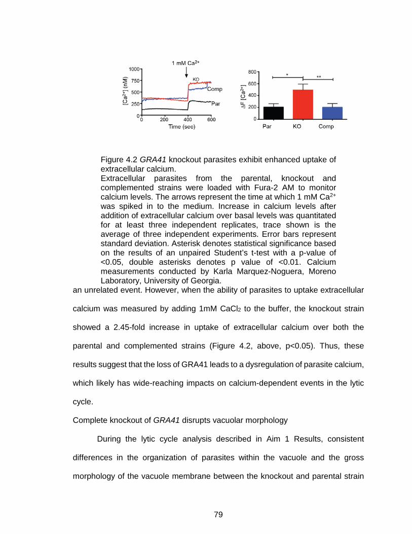
79
Figure 4.2 GRA41 knockout parasites exhibit enhanced uptake of extracellular calcium. Extracellular parasites from the parental, knockout and complemented strains were loaded with Fura-2 AM to monitor calcium levels. The arrows represent the time at which 1 mM Ca2+ was spiked in to the medium. Increase in calcium levels after addition of extracellular calcium over basal levels was quantitated for at least three independent replicates, trace shown is the average of three independent experiments. Error bars represent standard deviation. Asterisk denotes statistical significance based on the results of an unpaired Student’s t-test with a p-value of <0.05, double asterisks denotes p value of <0.01. Calcium measurements conducted by Karla Marquez-Noguera, Moreno Laboratory, University of Georgia.
an unrelated event. However, when the ability of parasites to uptake extracellular
calcium was measured by adding 1mM CaCl2 to the buffer, the knockout strain
showed a 2.45-fold increase in uptake of extracellular calcium over both the
parental and complemented strains (Figure 4.2, above, p<0.05). Thus, these
results suggest that the loss of GRA41 leads to a dysregulation of parasite calcium,
which likely has wide-reaching impacts on calcium-dependent events in the lytic
cycle.
Complete knockout of GRA41 disrupts vacuolar morphology
During the lytic cycle analysis described in Aim 1 Results, consistent
differences in the organization of parasites within the vacuole and the gross
morphology of the vacuole membrane between the knockout and parental strain

80
were seen, suggesting a role of GRA41 to maintain normal vacuolar morphology.
To investigate the possibility that the loss of GRA41 disrupted vacuolar
morphology, I visualized the parasites under high power magnification and noted
that many of the vacuoles in the knockout strain appeared to be collapsed around
the parasites within (Figure 4.3A, below). Quantitation of this “collapsed vacuole”
phenotype showed it was significantly more common in the GRA41 knockout strain
as compared to the parental and complemented strains (43±1% versus 10±6%
and 10±4%, respectively, p<0.05, Figure 4.3B, below). Furthermore, I noted that
the vacuoles in the knockout strain were more likely to have atypical numbers of
parasites. Typically, all parasites within the vacuole divide simultaneously into two
daughter parasites leading to a doubling of parasite number and a geometric
sequence of parasite number within each vacuole (i.e. 2, 4, 8, 16, etc.).
Nonetheless, 35±2% of the knockout vacuoles contained a number of parasites
that did not follow the normal pattern. This was significantly higher than both the
parental and complemented strains, with 12±3% and 7±2% respectively (Figure
4.3C, below, p<0.05). To investigate how this might be occurring, both parental
and GRA41 knockout parasites were stained for acetyl tubulin. The acetylation of
tubulin occurs during parasite replication and can be used to visualize the daughter
parasites as they are dividing (Varberg, Padgett et al. 2016). Using this method, I
was able to image GRA41 knockout parasites that showed evidence of more than
two daughters forming per mother parasites, which would explain the abnormal
number of parasites in some vacuoles (Figure 4.3D, below). The formation of
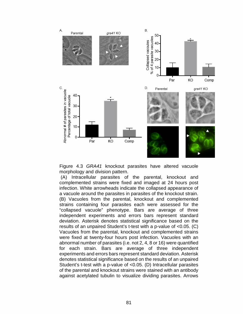
81
Figure 4.3 GRA41 knockout parasites have altered vacuole morphology and division pattern. (A) Intracellular parasites of the parental, knockout and complemented strains were fixed and imaged at 24 hours post infection. White arrowheads indicate the collapsed appearance of a vacuole around the parasites in parasites of the knockout strain. (B) Vacuoles from the parental, knockout and complemented strains containing four parasites each were assessed for the “collapsed vacuole” phenotype. Bars are average of three independent experiments and errors bars represent standard deviation. Asterisk denotes statistical significance based on the results of an unpaired Student’s t-test with a p-value of <0.05. (C) Vacuoles from the parental, knockout and complemented strains were fixed at twenty-four hours post infection. Vacuoles with an abnormal number of parasites (i.e. not 2, 4, 8 or 16) were quantified for each strain. Bars are average of three independent experiments and errors bars represent standard deviation. Asterisk denotes statistical significance based on the results of an unpaired Student’s t-test with a p-value of <0.05. (D) Intracellular parasites of the parental and knockout strains were stained with an antibody against acetylated tubulin to visualize dividing parasites. Arrows

82
show three daughter parasites for a single mother parasite in the knockout strain.
triplet and even quadruplet parasites has been observed in wild-type parasites at
low levels (Hu, Mann et al. 2002), suggesting that the knockout of GRA41 has
increased the frequency with which this happens.
Complete knockout of GRA41 leads to altered structure of the tubulovesicular
network
The “collapsed vacuole” and abnormal cell division are both similar to the
abnormal vacuole morphology and parasite organization associated with the
genetic disruption of GRA2, another secreted protein that localizes to the TVN and
that is critical for its formation (Mercier, Dubremetz et al. 2002). Accordingly, I
examined the structure of the TVN in the GRA41 knockout strain by transmission
electron microscopy (Figure 4.4, below). Host cells were infected with freshly
egressed tachyzoites for twenty-four hours prior to fixing and preparing samples
for thin sectioning. Thin sectioning and imaging were done with the assistance of
Dr. Barry Stein of the Indiana University-Bloomington Electron Microscopy Core
Facility. The parental, knockout and complemented strains all showed normal
morphology of the PV membrane, but the knockout strain appeared to have fewer
tubules within its network and an increase in the small vesicles from which the
tubules form (Figure 4.4, below). Additionally, the tubules that do form in the TVN
of the knockout strain appeared to be shorter than those of the parental and
complemented strains (Figure 4.4, below). Since the GRA41 knockout showed
similar disruption of the tubulovesicular network with the GRA41 knockout, I tested

83
Figure 4.4 GRA41 knockout parasites show altered morphology and size of tubulovesicular network (TVN) as compared to the parental and complemented strains Intracellular parasites were fixed at 24 hours post infection and prepared for imaging. Red arrowheads in left panels point at regions were imaged at higher magnification in the respective right panels.

84
to determine whether the GRA2 knockout (parental, knockout and complemented
strains graciously provided by Dr. Vern Carruthers, University of Michigan) would
mirror the resistance to iiDeath treatment of the GRA41 knockout. In contrast to
the GRA41 knockout, the GRA2 knockout did not show any increased survival over
its parental and complemented strains at either 45 or 60 minutes of treatment
(32±10% versus 26±4% and 30±13% at 45 minutes, respectively and 17±17%
versus 12±7% and 18±14% at 60 minutes, respectively, p>0.05, Figure 4.5,
below), suggesting that this phenomenon is restricted to the disruption of GRA41
and not a general feature of parasites with disrupted tubulovesicular networks.
Figure 4.5 Ionophore induced death phenotype of Gra2 parental, knockout and complemented strains. Percent survival of parasites treated with 1 μM A23187 versus DMSO solvent control for 45 or 60 minutes. Average of three independent replicates. Error bars represent standard deviation.
Incubation of recombinant GRA41 with calcium does not affect protein melting
temperature
To address question of whether GRA41 binds calcium and impacts calcium
homeostasis in this way, I isolated recombinant Histidine-tagged TgGRA41 from
transfected E. coli cells with the assistance of Dr. Sanofar Abdeen (Johnson
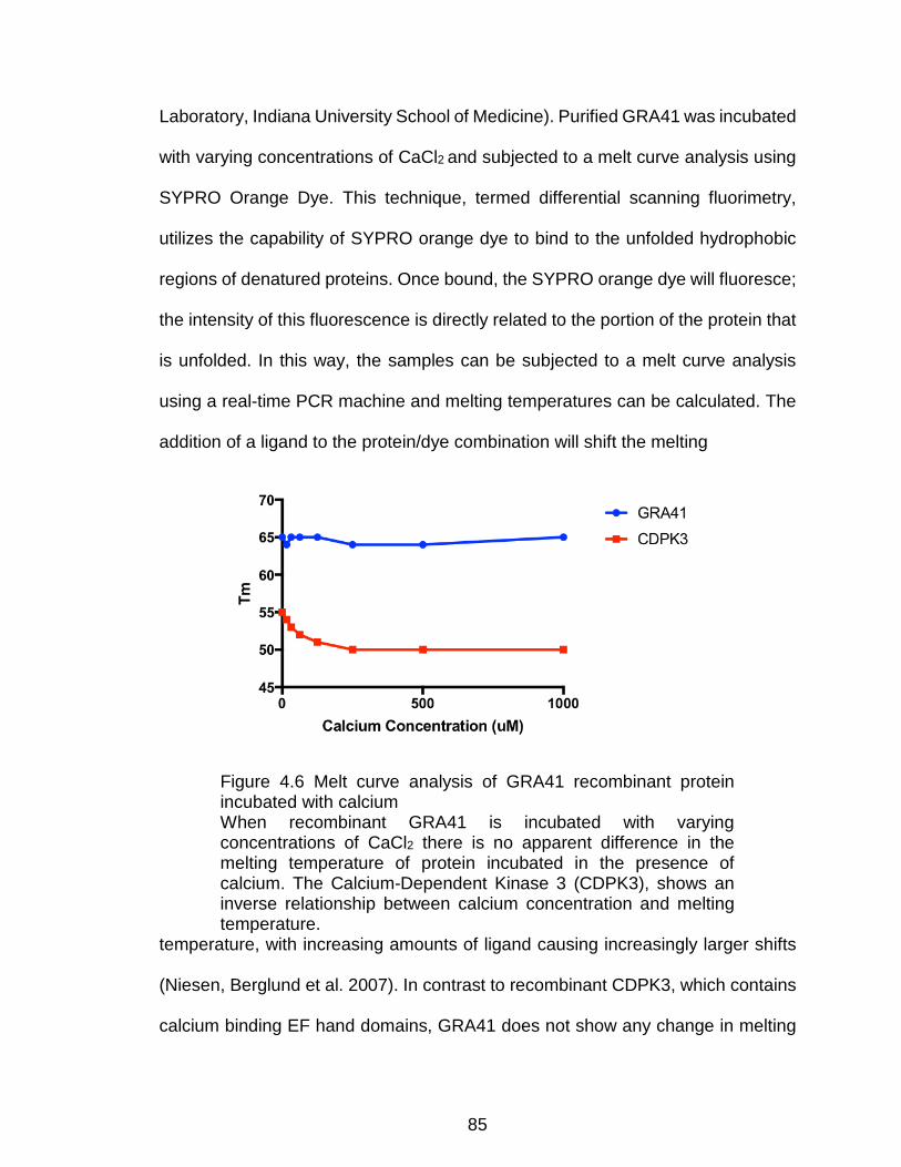
85
Laboratory, Indiana University School of Medicine). Purified GRA41 was incubated
with varying concentrations of CaCl2 and subjected to a melt curve analysis using
SYPRO Orange Dye. This technique, termed differential scanning fluorimetry,
utilizes the capability of SYPRO orange dye to bind to the unfolded hydrophobic
regions of denatured proteins. Once bound, the SYPRO orange dye will fluoresce;
the intensity of this fluorescence is directly related to the portion of the protein that
is unfolded. In this way, the samples can be subjected to a melt curve analysis
using a real-time PCR machine and melting temperatures can be calculated. The
addition of a ligand to the protein/dye combination will shift the melting
Figure 4.6 Melt curve analysis of GRA41 recombinant protein incubated with calcium When recombinant GRA41 is incubated with varying concentrations of CaCl2 there is no apparent difference in the melting temperature of protein incubated in the presence of calcium. The Calcium-Dependent Kinase 3 (CDPK3), shows an inverse relationship between calcium concentration and melting temperature.
temperature, with increasing amounts of ligand causing increasingly larger shifts
(Niesen, Berglund et al. 2007). In contrast to recombinant CDPK3, which contains
calcium binding EF hand domains, GRA41 does not show any change in melting

86
temperature when calcium is added to the sample, suggesting that the
recombinant protein is not capable of binding calcium. Though this doesnot rule
out the possibility that GRA41 is capable of binding calcium under certain
circumstances, it suggests that it is does not form a strong interaction with calcium
at the concentrations tested that is detectable using this assay.
Attempts to identify interactors of GRA41 by immunoprecipitation yield
inconsistent results
To begin to understand the mechanisms by which GRA41 might be
functioning, I performed a traditional co-immunoprecipitation using the Type I
endogenously HA-tagged strain to identify potential protein interaction partners.
Due to its location in the parasitophorous vacuole, intracellular parasites were
harvested along with the host cells and parasitophorous vacuoles by scraping and
Gene ID (ToxoDB.org)
Product Description (ToxoDB.org)
Computed Gene Ontology Function
(ToxoDB.org)
Expect Value
TGME49_227280 dense granule protein GRA3 None 2.70E-
05
TGME49_217460 glutaminyl-tRNA synthetase (GlnRS)
ATP binding, glutamine-tRNA ligase
activity
1.80E-04
TGME49_219540 tRNA-Ala synthetase
ATP binding, alanine-tRNA ligase activity, nucleic acid binding
2.40E-04
TGME49_299810 cysteine-tRNA synthetase (CysRS)
ATP binding, cysteine-tRNA ligase activity
2.40E-04
TGME49_233460 SAG-related sequence SRS29B None 2.70E-
04
TGME49_291960
rhoptry kinase family protein
ROP40 (incomplete catalytic triad)
transferase activity, transferring
phosphorus-containing groups
5.70E-04
TGME49_236210 peptidase M16 family potein,
putative
catalytic activity, metal ion binding,
metalloendopeptidase
6.20E-04

87
activity, zinc ion binding
TGME49_233480 SAG-related sequence SRS29C None 6.60E-
04
TGME49_251500 eukaryotic initiation factor-3, subunit 3,
putative protein binding 9.30E-
04
TGME49_308090 rhoptry protein ROP5
ATP binding, protein kinase activity 0.001
TGME49_309730 thioredoxin reductase
NADP binding, flavin adenine dinucleotide binding, glutathione-disulfide reductase activity, thioredoxin-disulfide reductase
activity
0.001
TGME49_219550
dihydrolipoyllysine-residue
succinyltransferase component of oxoglutarate
dehydrogenase
dihydrolipoyllysine-residue
succinyltransferase activity
0.0011
TGME49_290700 hypothetical protein None 0.0014 TGME49_319340 hypothetical protein None 0.0015
TGME49_295110 rhoptry protein ROP7
ATP binding, protein kinase activity 0.0017
TGME49_251540 dense granule protein GRA9 None 0.002
TGME49_309590 rhoptry protein ROP1 None 0.0051
TGME49_279100 hypothetical protein None 0.016
TGME49_297650 Ser/Thr
phosphatase family protein
acid phosphatase activity, hydrolase
activity 0.02
TGME49_226380 hypothetical protein None 0.022
TGME49_215775 rhoptry protein ROP8
ATP binding, protein kinase activity 0.027
resuspending in lysis buffer. The samples were further solubilized by sonication
before the lysates were incubated with anti-HA antibody. After the antibody was
allowed to bind to the GRA41 HA epitope tag, the entire antibody/antigen complex
Table 4.1 Proteins identified following immunoprecipitation of GRA41 from whole cell lysates Proteins are ranked by expect value, from smallest to largest.

88
was pulled out of the lysate using Protein G-coupled dynabeads, which bind to the
anti-HA antibody. The beads were washed and the contents eluted in sample
buffer before performing an SDS-PAGE separation and silver stain of the resultant
gel. After successful detection of bands at the correct molecular weight for the
GRA41 doublet observed by Western Blot analysis, three regions with significant
silver staining were excised and sent to the Indiana University Bloomington Mass
Spectrometry core facility for protein identification. The resulting protein list was
restricted to proteins not found in unrelated pulldowns (LaFavers and Arrizabalaga,
unpublished data), those which contained a signal peptide and those which had
an expect value of <0.0025. These proteins are listed from most to least likely in
Table 4.1, above. Of these proteins, only two are known dense granule proteins,
GRA3 and GRA9. Of the remaining proteins, five are known rhoptry proteins and
two are surface antigens. The remaining proteins are a mixture of proteins of
hypothetical function and uncharacterized proteins that have conserved functional
domains, such as a putative gluatminyl-tRNA synthetase. Additional efforts to
repeat these results were met with little success, as mass spectrometry analysis
of both prior and subsequent GRA41 pulldowns with samples submitted on
magnetic beads failed to identify GRA41 as a hit in the data analysis and were
disregarded. Due to these difficulties, a yeast two-hybrid approach was taken to
identify GRA41 candidate interactors.
GRA41 has several high confidence interactions in yeast two hybrid screen
A yeast two hybrid screen was conducted by Hybrigenics Services using
the GRA41 sequence with the signal peptide removed as a protein bait. Resulting

89
interactors were scored for statistical significance and ranked according to
expected values that take into account results from the GRA41 screen along with
all other screens against the T. gondii prey library. They are then assigned a global
predictive biological score from A (interactions with very high confidence) to F
(experimentally proven technical artifacts). Included in the table below are prey
proteins with a score of A, B (high confidence) and C (good confidence). Prey with
scores of D (moderate conference), E (interactions involving highly connected prey
domains, likely non-specific), and F were not included. Also removed were
common hits from unrelated nuclear proteins used as bait by the Sullivan
laboratory and an unrelated mitochondrial protein used as bait by the Arrizabalaga
lab against the same database (Garbuz, Harris, Huang, Srivasta, Arrizabalaga,
and Sullivan, unpublished data). The remaining hits included 11 proteins, three
ToxoDB.org Gene ID
Global
PBS
Product Description (ToxoDB.or
g)
Signal Peptid
e
# TM Domain
s
Predicted Localization (psort)
TGME49_270800 A
GAF domain-
containing protein
Yes 0 Nuclear/mitochondrial
TGME49_202680 A
peptidase M16, alpha
subunit, putative
Yes 0 Mitochondrial
TGME49_202370 A
T-complex protein 1, epsilon subunit (TCP-1-epsilon), putative
No 0 N/A
TGME49_270250 B dense
granule Yes 0 Extracellular
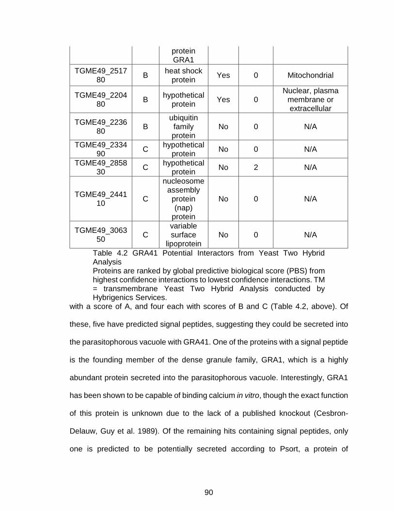
90
protein GRA1
TGME49_251780 B heat shock
protein Yes 0 Mitochondrial
TGME49_220480 B hypothetical
protein Yes 0 Nuclear, plasma
membrane or extracellular
TGME49_223680 B
ubiquitin family protein
No 0 N/A
TGME49_233490 C hypothetical
protein No 0 N/A
TGME49_285830 C hypothetical
protein No 2 N/A
TGME49_244110 C
nucleosome assembly
protein (nap)
protein
No 0 N/A
TGME49_306350 C
variable surface
lipoprotein No 0 N/A
with a score of A, and four each with scores of B and C (Table 4.2, above). Of
these, five have predicted signal peptides, suggesting they could be secreted into
the parasitophorous vacuole with GRA41. One of the proteins with a signal peptide
is the founding member of the dense granule family, GRA1, which is a highly
abundant protein secreted into the parasitophorous vacuole. Interestingly, GRA1
has been shown to be capable of binding calcium in vitro, though the exact function
of this protein is unknown due to the lack of a published knockout (Cesbron-
Delauw, Guy et al. 1989). Of the remaining hits containing signal peptides, only
one is predicted to be potentially secreted according to Psort, a protein of
Table 4.2 GRA41 Potential Interactors from Yeast Two Hybrid Analysis Proteins are ranked by global predictive biological score (PBS) from highest confidence interactions to lowest confidence interactions. TM = transmembrane Yeast Two Hybrid Analysis conducted by Hybrigenics Services.

91
hypothetical function with the gene ID TGME49_220480, which lacks sequence
homology to any proteins of known function. The remaining proteins are either
predicted to be trafficked to other locations within the cell, such as the
mitochondrion or nucleus, or do not contain a signal peptide. If these predicted
localizations are accurate, it is unlikely that GRA41 would ever encounter these
proteins within the parasite.
Using a combination of co-immunoprecipitation and yeast two-hybrid
analysis, I have generated a list of potential GRA41 interactors. Future studies
should focus on verifying potential interaction partners and using genetic
approaches to determine their impact on the phenotypes associated with the loss
of GRA41.
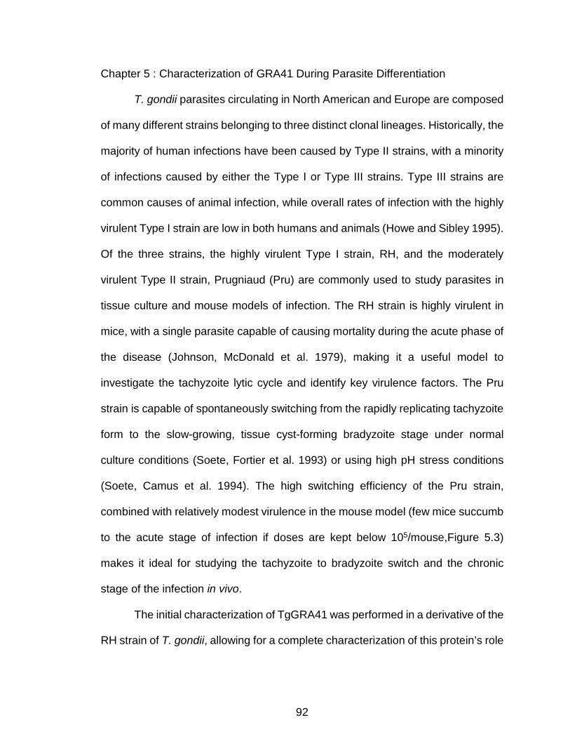
92
Chapter 5 : Characterization of GRA41 During Parasite Differentiation
T. gondii parasites circulating in North American and Europe are composed
of many different strains belonging to three distinct clonal lineages. Historically, the
majority of human infections have been caused by Type II strains, with a minority
of infections caused by either the Type I or Type III strains. Type III strains are
common causes of animal infection, while overall rates of infection with the highly
virulent Type I strain are low in both humans and animals (Howe and Sibley 1995).
Of the three strains, the highly virulent Type I strain, RH, and the moderately
virulent Type II strain, Prugniaud (Pru) are commonly used to study parasites in
tissue culture and mouse models of infection. The RH strain is highly virulent in
mice, with a single parasite capable of causing mortality during the acute phase of
the disease (Johnson, McDonald et al. 1979), making it a useful model to
investigate the tachyzoite lytic cycle and identify key virulence factors. The Pru
strain is capable of spontaneously switching from the rapidly replicating tachyzoite
form to the slow-growing, tissue cyst-forming bradyzoite stage under normal
culture conditions (Soete, Fortier et al. 1993) or using high pH stress conditions
(Soete, Camus et al. 1994). The high switching efficiency of the Pru strain,
combined with relatively modest virulence in the mouse model (few mice succumb
to the acute stage of infection if doses are kept below 105/mouse,Figure 5.3)
makes it ideal for studying the tachyzoite to bradyzoite switch and the chronic
stage of the infection in vivo.
The initial characterization of TgGRA41 was performed in a derivative of the
RH strain of T. gondii, allowing for a complete characterization of this protein’s role

93
during the tachyzoite lytic cycle. However, to determine the role of TgGRA41 in the
bradyzoite stage of infection, I utilized to the Pru strain to facilitate our analysis of
this stage. To do this, I endogenously tagged TgGRA41 in the Pru strain as
described for the RH strain to determine if TgGRA41 is expressed during the
bradyzoite stage of development and to analyze its localization at this time. I then
generated a knockout of the TgGRA41 in the Pru strain in order to determine the
impact of its loss during bradyzoite development and in vivo infection.
TgGRA41 is expressed in a Type II strain and localizes to the wall of bradyzoite
tissue cysts
In order to confirm that GRA41 is expressed in tachyzoites and bradyzoites
of a Type II strain, sequences encoding a carboxy-terminal (C-terminal) triple-
hemagglutinin (HA) tag were introduced into the endogenous locus in the
Pru∆Ku80∆hxgprt strain just before the stop codon, using a previously described
approach (Fox, Falla et al. 2011). Western blot analysis of protein extract from the
endogenously tagged strain revealed a single band migrating at approximately 20-
25 kDa, which is approximately the same size as the predicted molecular weight
of the tagged protein, 24 KDa (Figure 5.1A, below).
Since the doublet pattern found in Type I parasites (Figure 3.3A, above)was
not visible in the Type II strain, I conducted a sequence alignment using Clustal
Omega of the predicted protein sequences for Type I and Type II strains
(ToxoDB.org) to determine if any polymorphisms could account for the differences
in the banding patterns between the two strains (Figure 5.1B, below). Overall, there
is high identity between the two sequences (97%), with only 6 amino acids differing
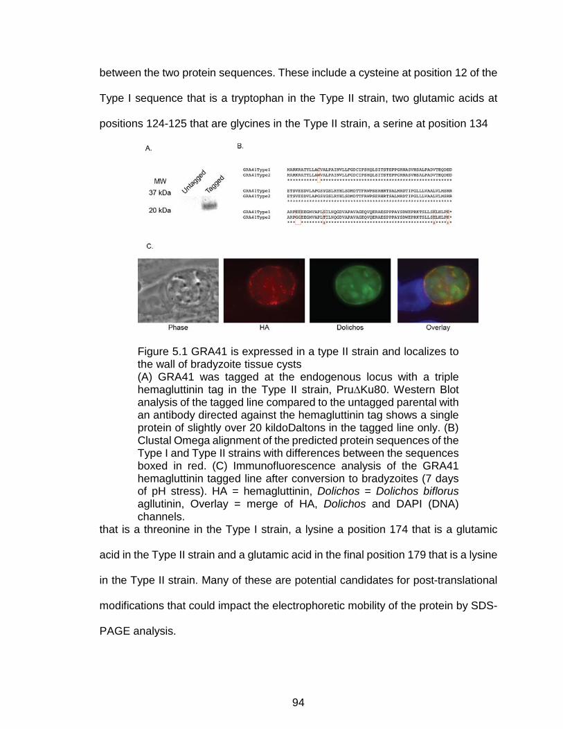
94
between the two protein sequences. These include a cysteine at position 12 of the
Type I sequence that is a tryptophan in the Type II strain, two glutamic acids at
positions 124-125 that are glycines in the Type II strain, a serine at position 134
Figure 5.1 GRA41 is expressed in a type II strain and localizes to the wall of bradyzoite tissue cysts (A) GRA41 was tagged at the endogenous locus with a triple hemagluttinin tag in the Type II strain, Pru∆Ku80. Western Blot analysis of the tagged line compared to the untagged parental with an antibody directed against the hemagluttinin tag shows a single protein of slightly over 20 kildoDaltons in the tagged line only. (B) Clustal Omega alignment of the predicted protein sequences of the Type I and Type II strains with differences between the sequences boxed in red. (C) Immunofluorescence analysis of the GRA41 hemagluttinin tagged line after conversion to bradyzoites (7 days of pH stress). HA = hemagluttinin, Dolichos = Dolichos biflorus agllutinin, Overlay = merge of HA, Dolichos and DAPI (DNA) channels.
that is a threonine in the Type I strain, a lysine a position 174 that is a glutamic
acid in the Type II strain and a glutamic acid in the final position 179 that is a lysine
in the Type II strain. Many of these are potential candidates for post-translational
modifications that could impact the electrophoretic mobility of the protein by SDS-
PAGE analysis.

95
The localization of GRA41 in a bradyzoite tissue cyst was then examined
by immunofluorescence microscopy conducted after tachyzoites were converted
to bradyzoite cysts by 7 days of pH stress with CO2 deprivation. GRA41 protein
was visualized by probing with an antibody directed against the hemagluttinin tag
of the endogenously tagged strain. The cyst wall of the bradyzoite was visualized
by staining with Fluorescein-conjugated DBA; individual bradyzoites are also
shown in the green channel due to the presence of a GFP-encoding gene under
the control of the bradyzoite-specific LDH2 promoter that is stably integrated into
the Pru∆ku80∆hxgprt strain (Cleary, Singh et al. 2002). Organelles containing DNA
(i.e. nucleus, apicoplast) are stained with DAPI. As expected for a dense granule
protein that is localized to the vacuole in tachyzoites, GRA41 is enriched in the
cyst wall of the newly formed bradyzoite tissue cyst (Figure 5.1C, above).
Knockout of GRA41 in a Type II Strain recapitulates resistance to ionophore
induced death
To further study the role of GRA41 in the bradyzoite stage of the parasite, I
generated a knockout of the gene by replacing the entire coding sequence with the
selectable marker hypoxanthine-guanine phosphoribosyltransferase (HXGPRT)
through double homologous recombination into the Pru∆ku80∆hxgprt parental
strain, hereafter referred to as Pru∆ku80 (Fox, Falla et al. 2011) (Figure 5.2A,
below). Parasites transfected with the knockout vector (pGRA41KO) and stably
expressing HXGPRT were selected by resistance to mycophenolic acid and
xanthine and cloned by limiting dilution. Clones were imaged live on a Leica
inverted DMI6000B microscope to determine whether or not they were expressing

96
GFP. Since a GFP expression cassette is present within the pGRA41KO vector
but is downstream of the 3’ flanking region, any GFP positive parasites are likely
to be the result of integration of the construct by single homologus recombination
rather than double homologous recombination. A single clone was isolated that
was GFP negative out of over 150 clones screened, which was then shown to have
correct integration of the construct by PCR (Figure 5.2B, below). Based on the
nature of the genetic disruption expected from double recombination of the
knockout construct (pGRA41KO, (Figure 5.2A, below)) the established clone
represents a functional knockout and is unlikely to produce any GRA41 protein. I
then assessed whether the complete knockout of GRA41 in the Pru∆ku80 strain
recapitulated the iiDeath- phenotype seen in GRA41 truncation mutant MBD 2.1
and the GRA41 Type I knockout.
Pru∆ku80∆gra41 showed statistically significant increases in survival as
compared to the parental strain after 10 or 30 minutes of treatment with A23187
(87% ± 5.5% versus 74% ± 5.2% for the parental strain at ten minutes and 48% ±
14% versus 28% ± 13% for the parental strain at thirty minutes, p<0.05, Figure
5.2C, below). Despite the differences in timing of the iiDeath phenotype as
compared to the RH strain, which has been shown previously (Lavine, Knoll et al.
2007), the Pru∆ku80∆gra41 recapitulates the decrease in sensitivity to ionophore
induced death found in the type I knockout, suggesting that Gra41 likely plays a
similar role in Type II strains as it does in Type I.
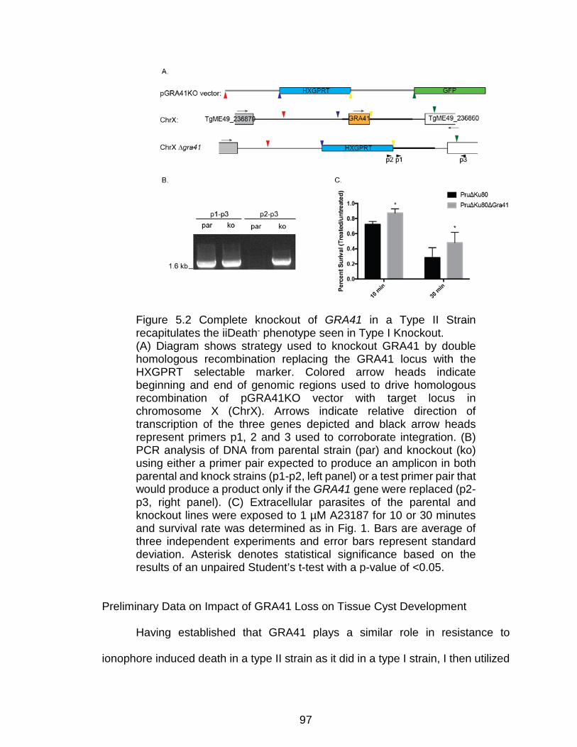
97
Preliminary Data on Impact of GRA41 Loss on Tissue Cyst Development
Having established that GRA41 plays a similar role in resistance to
ionophore induced death in a type II strain as it did in a type I strain, I then utilized
Figure 5.2 Complete knockout of GRA41 in a Type II Strain recapitulates the iiDeath- phenotype seen in Type I Knockout. (A) Diagram shows strategy used to knockout GRA41 by double homologous recombination replacing the GRA41 locus with the HXGPRT selectable marker. Colored arrow heads indicate beginning and end of genomic regions used to drive homologous recombination of pGRA41KO vector with target locus in chromosome X (ChrX). Arrows indicate relative direction of transcription of the three genes depicted and black arrow heads represent primers p1, 2 and 3 used to corroborate integration. (B) PCR analysis of DNA from parental strain (par) and knockout (ko) using either a primer pair expected to produce an amplicon in both parental and knock strains (p1-p2, left panel) or a test primer pair that would produce a product only if the GRA41 gene were replaced (p2-p3, right panel). (C) Extracellular parasites of the parental and knockout lines were exposed to 1 µM A23187 for 10 or 30 minutes and survival rate was determined as in Fig. 1. Bars are average of three independent experiments and error bars represent standard deviation. Asterisk denotes statistical significance based on the results of an unpaired Student’s t-test with a p-value of <0.05.
98
the type II GRA41 knockout to determine the role GRA41 plays in the tissue cyst
stage of the T. gondii life cycle. To do this, host cells were infected with parasites
for 2-4 hours before using an established pH stress protocol to induced conversion
to the bradyzoite stage (Weiss, Laplace et al. 1995). The pH stress media was
replaced every two days and the cells were kept in a CO2 deprivation incubator to
ensure high rates of stage switching were achieved. Conversion to bradyzoites
was measured utilizing rhodamine-conjugated DBA, which binds to glycosylated
proteins in the bradyzoite cyst wall (Scholytyseck, Mehlhorn et al. 1974, Tomita,
Bzik et al. 2013). Cells were fixed at four or seven days post infection to look for
defects in tissue cyst formation or maintenance. A minimum of 100 vacuoles were
scored as either DBA positive or negative. Small clusters of DBA negative
vacuoles containing 1-2 parasites that appeared to have remained as tachyzoites,
egressed and reinvaded nearby host cells were counted as a single DBA negative
vacuole, since these parasites likely came from one original vacuole. Though there
was no statistically significant difference in the percentage of DBA positive
vacuoles between the parental and knockout strains at four days post infection
(73%±16% and 53%±29% respectively, Figure 5.3A, below), there is a significant
difference between the two strains at seven days post infection (75%±7% and
20%±12%, respectively, **, p<0.01), suggesting that the knockout parasites form
bradyzoite cysts initially, but fail to maintain these bradyzoite cysts. Some of the
cysts formed by the knockout parasites had lost cyst wall staining at seven days
post infection, while many others had egressed and reinvaded, either as
tachyzoites or bradyzoites.
99
To further investigate bradyzoite formation under in vivo conditions, female
CBA/J mice were injected by Dr. Arrizabalaga with 103, 104 or 105 parasites per
mouse. Mice were allowed to progress from an acute infection to a chronic
infection for 28 days before brains and blood were harvested by myself and Tamila
Garbuz. The average number of tissue cysts per brain was measured by fixing and
staining brain homogenate with rhodamine-conjugated DBA for visualization by
fluorescent microscopy. Successful infection of mice containing no cysts was
verified by testing mouse serum against T. gondii antigens by dot blot analysis. At
a low dose (103 parasites/mouse), very few cysts were found in each mouse brain
for both the parental and the knockout strain and no differences were seen
between the two strains (Figure 5.3B, below). However, at a slightly higher dose
(104/mouse), the number of cysts is almost two-fold higher in the knockout as
compared to the parental (91 cysts versus 39, p<0.1, Figure 5.3B, below). Due to
a relatively small sample size, this difference is not statistically significant for a
cutoff of p<0.05, but is trending toward statistical significance. At the highest dose
(105/mouse), the knockout strain showed highly variable results, with two of the
mice not showing any evidence of cysts and two showing high numbers of cysts
as compared to the parental strain (Figure 5.3B, below). It is not clear whether or
not the lack of cysts in some of the mice at this dose is due to variability in the
counting method (since only a small percentage of the total brain material is
imaged), the failure of parasites to reach the brain or that cysts were too small and
could not be imaged with the magnification used for counting. When combined with
the in vitro switching

100
Figure 5.3 Differentiation of Type II Gra41 Knockout (A) Parental or GRA41 knockout parasites were allowed to form bradyzoite cysts under pH stress for 4 or 7 days before parasites were fixed and cyst walls were stained using rhodamine-conjugated DBA. Bars are average of three independent experiments and error bars represent standard deviation. Double asterisk denotes statistical significance based on the results of an
101
unpaired Student’s t-test with a p-value of <0.01. (B) Female CBA/J mice were infected with parental (WT) or GRA41 knockout (KO) parasites at the indicated doses and allowed to progress to chronic infection for 28 days before brains were harvested and cysts counted following staining with rhodamine-conjugated DBA. Individual mice are presented as data points in each treatment and error bars represent standard deviation. ‡ denotes statistical significance based on the results of an unpaired Student’s t-test with a p-value of <0.1.
data, the in vivo cyst formation data suggests that the bradyzoite cysts from
the knockout strain may have a defect in maintenance, leading to their
reconversion to tachyzoites, which then invade additional host cells and lead to the
formation of more cysts. However, without conducting an analysis at multiple time
points during bradyzoite infection with additional animals, the exact interpretation
remains unclear.

102
Chapter 6 : Discussion
Calcium-dependent signaling is central to the propagation of parasites of
the phylum Apicomplexa, including the causative agent of malaria, Plasmodium
falciparum, and the opportunistic pathogen T. gondii. Calcium-dependent events
underlie the motility, invasion, and egress of these parasites and thus calcium
homeostasis and fluxes are tightly controlled. Calcium signaling is proposed to be
an attractive drug target for controlling these parasitic infections, due to its
essential nature and uniqueness from vertebrate calcium-dependent signaling
pathway. While many of the effectors of calcium that drive events of the lytic cycle
in both Plasmodium species and T. gondii have been identified, much remains
unknown about how these calcium fluxes are regulated. In an effort to address this
knowledge gap our lab has used a forward genetics approach to identify genes
that influence T. gondii’s sensitivity to calcium fluxes, specifically those induced by
the calcium ionophore A23187. Surprisingly, I have discovered that a secreted
protein, which resides within the parasitophorous vacuole during parasite
intracellular growth, influences several calcium related events in both intra and
extracellular parasites. These results suggest a role for PV proteins in the
regulation of calcium homeostasis, timing of egress, and parasite division.
Additionally, preliminary results investigating the impact of this protein on
bradyzoite tissue cyst formation suggest this protein is also required for normal
cyst maintenance.
The novel dense granule protein GRA41 was first identified through the
sequencing of a mutant resistant to extracellular exposure to the ionophore

103
A23187. Normally, exposure to this calcium ionophore for over thirty minutes
renders extracellular parasites non-invasive, which, for this obligate intracellular
parasite, results in death. While this phenomenon, known as ionophore induced
death (iiDeath), has been known and exploited for decades (Mondragon and
Frixione 1996, Black, Arrizabalaga et al. 2000), the underlying mechanism is not
well understood. It has been proposed that prolonged exposure to elevated
calcium, which normally induces secretion of adhesive molecules required for
invasion, exhausts the contents of the secretory organelles that harbor such
proteins. Other plausible mechanisms include toxic effects from elevated
cytoplasmic calcium levels, and changes to the calcium sensitivity of the molecules
and machinery that regulate secretion. I have actually detected that following a
one-hour ionophore exposure, the levels of micronemal proteins do not change
compared to untreated parasites but the ability to elicit micronemal secretion upon
subsequent ionophore exposure is significantly affected (Figure 4.1, above). This
observation would indicate that iiDeath is not merely the consequence of the
exhaustion of proteins to be secreted, but of an alteration in the sensitivity to
calcium, which would lead to a reduction in active secretion. In this context, I
hypothesize that the resistance to iiDeath of the parasites lacking GRA41 is due
to an altered sensitivity to the ionophore, brought upon by the clear changes in
calcium regulation detected in the GRA41 knock out strain. Indeed, the gra41
mutant strain (MBD2.1) is over-sensitive to the effects of calcium chelation by
BAPTA-AM (Mondragon and Frixione 1996, Black, Arrizabalaga et al. 2000). It is
not yet clear whether the altered calcium regulation exhibited by the GRA41

104
knockout strain is due to a direct effect of lack of GRA41 or an indirect
consequence of how the parasites adapts to the loss of GRA41. The latter
explanation is supported by the observation that the mutant strain has an elevated
level of cytosolic calcium that is not corrected by genetic complementation.
Nonetheless, lack of GRA41 does seem to affect calcium regulation as the mutant
parasites exhibited dysregulated calcium uptake, a phenotype that is reverted by
complementation. While I have yet to elucidate the exact mechanism by which the
knockout parasites are adapting, I have observed that the premature egress
phenotype is very stable during culture, suggesting that the mutant and knockout
strains could adapt to this change in egress timing by modifying one of the other
steps in the lytic cycle (i.e. increasing attachment) which is also dependent on
calcium signaling processes. Though it is difficult to determine the specific function
of GRA41, the calcium uptake defect of extracellular parasites suggests that
GRA41 is involved in regulating calcium homeostasis. Higher levels of cytosolic
calcium and increased rates of calcium uptake by extracellular parasites shows
how the parasites responds to the loss of GRA41.
How a protein that resides outside of the parasite but within the
parasitophorous vacuole might affect calcium regulation within the cytoplasm of
the parasite is puzzling, especially considering that some of the phenotypes related
to its deletion, such as iiDeath resistance, manifest only once the parasites are
extracellular. It is plausible that the protein, which is in the dense granules before
secreted, has a role within this organelle that influences calcium homeostasis in
extracellular parasites. Nonetheless, there is no evidence that dense granules
105
accumulate significant amounts of calcium (Bonhomme, Pingret et al. 1993) or are
involved in the regulation of calcium homeostasis or fluxes. It is also conceivable
that GRA41, while in the PV, directly or indirectly influences the physiology of the
vacuole, which in turns affects that of the parasite. In such a model, the absence
of GRA41 would alter the physiology of the intracellular parasite and, once outside,
these parasites would react differently to external stimuli such as the calcium
ionophore.
Though the PV lumen has long been considered physiologically at
equilibrium with the host cell through a nonselective pore (Schwab, Beckers et al.
1994), whose components were recently identified (Gold, Kaplan et al. 2015),
several studies indicate that ionic concentration of the PV can vary in respect to
that of the host cytoplasm during intracellular growth. For instance, measurements
of calcium levels using non-ratiometric indicators in the PV have shown that
calcium is concentrated in the PV relative to the host cell cytoplasm and the
frequency of this calcium sequestration increases as egress approaches (Pingret,
Millot et al. 1996). Interestingly, one of the most abundant PV proteins, GRA1, has
been shown to bind calcium (Cesbron-Delauw, Guy et al. 1989), which suggests
a potential mechanism for this calcium sequestration. The calcium concentration
of the Plasmodium PV has been determined through the use of ratiometric
indicators to be approximately 40 μM, many fold higher than the 20 nM that would
be expected if the PV calcium levels were in equilibrium with the host red blood
cell’s cytosol (Murphy, Berkowitz et al. 1987, Gazarini, Thomas et al. 2003).
Additionally, the same study showed that depletion of calcium in the Plasmodium

106
PV leads to a depletion of intracellular calcium stores, which suggests that the
parasite depends on the sequestration of calcium in the PV to maintain its
intracellular pools of calcium (Gazarini, Thomas et al. 2003). It is possible that
similar mechanisms are regulating calcium levels within the T. gondii PV.
Interestingly, calcium is not the only ion which the parasite regulates the
concentration of within the PV; it has recently been shown that the pH of the
vacuole decreases as egress approaches and this increased acidification is
associated with enhanced parasite motility and protein secretion (Roiko, Svezhova
et al. 2014). While it is possible that one or more dense granule proteins that T.
gondii secretes into its vacuole are responsible for the sequestration of ions as
egress approaches, it is unlikely that this is occurring in Plasmodium species, since
the dense granule proteins described to date are restricted to the tissue-cyst
forming coccidia and are not found in other apicomplexans such as Plasmodium
(Mercier and Cesbron-Delauw 2015). Therefore, it is possible that these two
related organisms have evolved different strategies to utilize calcium signaling
within the context of the calcium-poor cytosol of their host cells.
It is unlikely that GRA41 binds calcium directly, since bioinformatic analysis
fails to identify any conserved calcium binding domains (SMART, NCBI, Prosite,
Interpro) and recombinant GRA41 protein fails to bind calcium in vitro (Figure 4.6,
above). Rather, I hypothesize that GRA41 might influence physiological calcium
homeostasis by its localization and functional role within the tubulovesicular
network (TVN). Loss of GRA41 leads to altered morphology of the TVN, which
interestingly has been proposed to play a role in calcium storage based on electron

107
microscopy studies (Bonhomme, Pingret et al. 1993). Nonetheless, other dense
granule proteins that localize to the membranes of the TVN are also critical for its
formation (Mercier, Dubremetz et al. 2002), but do not seem to appear to affect the
same of processes influenced by GRA41. The loss of the TVN in parasites lacking
GRA2 leads to a variety of phenotypes, including the formation of vacuoles
containing poorly organized parasites that egress erratically (Muniz-Hernandez,
Carmen et al. 2011), altered presentation of antigens to the host immune system
(Lopez, Bittame et al. 2015), decreased ingestion of host cytosolic proteins (Dou,
McGovern et al. 2014) and decreased virulence in mice (Mercier, Howe et al.
1998). However, I have shown that the loss of GRA2 does not phenocopy the
iiDeath resistance of the GRA41 knockout and forward genetic mutant, suggesting
that the disruption of the TVN might not be the underlying reason for the iiDeath
resistance of GRA41(Figure 4.5, above). Rather, it appears that the iiDeath
resistance of the GRA41 knockout strain is a direct effect of GRA41 by a novel
mechanism that has yet to be completely discerned. Nonetheless, there remains
the possibility that although the TVN is structurally affected in both the GRA41 and
GRA2 knockout strains, the nature, level and the functional consequences of that
effect might be different between the two mutant strains. For example, the loss of
GRA41 might prevent the formation of specific protein complexes within the PV
which are required for normal egress timing, which might explain why the loss of
GRA2 leads to erratic egress that occurs with normal timing.
At the current time, it is unclear which of the pleiotropic effects of GRA41
loss is the primary impact of its loss and which are simply downstream defects.
108
However, one of the most severe and distinct phenotypes of the GRA41 knockout
strain is a premature egress from the host cell. Natural egress from host cell can
be an active process dependent on calcium fluxes (Moudy, Manning et al. 2001).
Two alternative hypotheses to explain the premature egress are 1) that the altered
TVN, which might normally act as a mechanical barrier, allows for early exit and 2)
that the dysregulation of calcium homeostasis results in changes in the timing of
egress. The fact that other mutant parasites lacking a normal TVN, such as the
GRA2 knockout, don’t have a premature egress phenotype would argue against a
model in which the altered TVN, caused by gra41 disruption, allows for early
egress. To further separate these two phenotypes, complementation studies with
modified (i.e. truncated, mutated) forms of GRA41 in the knockout background
should be performed to determine if these two phenotypes can be functionally
linked to domains or residues within GRA41. Additionally, the only other dense
granule protein whose loss leads to an early egress phenotype similar to the
GRA41 knockout is GRA22. However, the GRA22 knockout does not exhibit any
obvious defect in TVN formation (Okada, Marmansari et al. 2013), suggesting that
GRA41 plays a structural role within the TVN that is independent from its role in
egress and, by inference, calcium-dependent signaling processes within the
parasite. It remains unclear whether or not these two proteins are interacting
together directly or within the same pathway to influence the timing of egress.
Though preliminary interaction studies failed to identify GRA22 as a potential
interactor as GRA41, future studies to expand and refine this list of GRA41
interactors and to create a strain lacking both GRA22 and GRA41 would address

109
some of these questions. Considering that tight control of calcium levels is needed
for egress, it is plausible that the early egress phenotype of the GRA41 knockout
is related to the dysregulation of calcium seen in this strain. Interestingly, the
MBD2.1 mutant, as well as the GRA41 knockout, show a slight but consistent
increase in the rate of calcium dependent iiEgress ((Black, Arrizabalaga et al.
2000) and unpublished data, LaFavers and Arrizabalaga). Additional support for a
connection between early egress and calcium dysregulation in the mutants, is the
fact that the GRA41 knockout is much more sensitive to DTT-induced egress than
the parental strain. The redox reagent DTT is proposed to induce egress by
activating NTPases located in the PV since it is capable of activating isolated
enzyme (Bermudes, Peck et al. 1994). Treatment with DTT has also been shown
to lead to calcium fluxes within the parasite, resulting in induced egress, which can
be blocked by chelating calcium, indicating that DTT-induced egress is also
calcium dependent (Stommel, Ely et al. 1997, Borges-Pereira, Budu et al. 2015).
Unlike calcium ionophores such as A23187, DTT is not capable of inducing egress
at early stages of parasite vacuole formation and requires longer growth times (~36
hours) for vacuoles to consistently egress. This suggests that DTT-induced egress
might be dependent on processes preceding natural egress and is therefore a
more accurate model for what is happening in natural uninduced egress (Stommel,
Cho et al. 2001). In this context, the ability of the GRA41 knockout strain to undergo
DTT-induced egress at 24 hours is consistent with its early egress phenotype. The
formation of more than two daughter parasites in the dividing GRA41 knockout
strain could also occur as a result of calcium dysregulation rather than simply being
110
an additional function of GRA41. Knockdown of the calcium-dependent kinase
CDPK7 has previously been shown to disrupt normal division (Morlon-Guyot, Berry
et al. 2014). Additionally, experiments to identify novel proteins in the inner
membrane complex (IMC) of the parasite, which serves as the scaffold for
daughter cell formation (Harding and Meissner 2014), identified a protein (ISC6,
TgGT1_267620, ToxoDB.org), that localizes to the IMC and contains a C2 calcium
binding domain (Chen, Moon et al. 2017), which could suggest a link between
calcium and daughter parasite formation in T. gondii.
Of note is the fact that GRA41 is not annotated as a protein encoding gene
in the latest version of the T. gondii genome database. This locus was denoted as
a gene in earlier annotations with both transcriptomic and proteomic corroborating
evidence. As I have shown there is clearly a protein encoding locus in that genomic
region; underscoring the limitation of genome annotations. One of the drawbacks
of GRA41 not being denoted as a protein in the current annotated genome is that
it is not considered when analyzing datasets that rely on the annotation. In the
many gene expression studies, mappings of post-translational modifications, and
sequencing of mutant strains that have been undertaken in the last few years,
GRA41 has not been included, significantly reducing our ability to understand its
function. For example, the presence of certain post-translational modifications
could provide clues to how GRA41 is associated with the TVN membrane. Though
GRA41 lacks an amphipathic alpha helix (Helical Wheel Predictions, RZ Lab) like
those found in other TVN-localized proteins such as GRA2 (Mercier, Cesbron-
Delauw et al. 1998), it does contain a predicted palmitoylation site at Cys26 (CSS-
111
Palm, Cuckoo Workgroup) that could explain membrane association. Though the
palmitoylome for T. gondii has been published (Foe, Child et al. 2015), GRA41
was not included in the database at the time and any potential palmitoylation
modifications were not mapped. As a result, future directions for the study of
GRA41 would include mapping post-translational modifications and determining
how they impact protein localization and function by mutational analysis.
In this context, the difference in electrophoretic mobility between the Type I
and Type II GRA41 proteins (i.e. migrating as a doublet in Type I and a singlet in
Type II strains) combined with the results of an alignment between the two proteins
could help to identify residues that are differentially modified between the two
strains. Though the modification responsible for the difference in electrophoretic
mobility is likely to not be essential for overall protein function, it is possible that it
could lead to subtle differences in protein function between the two strains. Such
differences could prove informative for both the study of GRA41 and differences
between Type I and Type II strains. Many of the amino acids which are different
between the Type I and Type II GRA41 amino acid sequences could be
responsible for the observed mobility difference. For example, the cysteine found
at position twelve in the Type I strain could participate in the formation of a disulfide
bond, which has been shown to lead to the production of a protein doublet by SDS-
PAGE analysis (Ruiz, Chng et al. 2010). This cysteine is also predicted as a
possible palmitoylation site by CSS-Palm, which would be lost if the amino acid at
this location were a tryptophan, as it is in the Type II strain (Ren, Wen et al. 2008).
The glutamic acid residues at positions 124, 125 and 179 may also be modified,

112
such as in the addition of a CO2 molecule to form gamma-carboxyglutamic acid.
The resulting modified amino acid has two negatively charged groups which have
been found to be important in coordinating divalent cations, such as calcium, in
blood clotting factors (Furie, Bouchard et al. 1999). Though serine residues are
capable of receiving many types of modifications, including phosphorylation and
glycosylation, threonine is also capable of accepting these types of modifications,
making the conversion of the serine to threonine at position 134 a somewhat less
likely candidate for the differences in protein migration observed (Blom, Sicheritz-
Ponten et al. 2004). Finally, lysines such as the one found at position 174 can be
modified by as many as 12 different post-translational modifications, including
acetylation, methylation and ubiquitination (Liu, Wang et al. 2014).
The discovery of GRA41 as a novel dense granule protein with a role in
calcium-dependent events helps to both answer and create new questions about
the role of the PV in these signaling processes. This work demonstrates that the
parasite is dependent on a dense granule protein for normal calcium homeostasis,
but it is still unclear how and if the parasite might regulate PV calcium levels
independent of the host cell cytosol in the presence of a nonselective PVM pore
(Schwab, Beckers et al. 1994). The explanation for this might be the existence of
additional calcium binding proteins that, in addition to GRA1 (Cesbron-Delauw,
Guy et al. 1989), could bind and sequester calcium within the PV. Future efforts to
directly measure calcium concentration in the PV of GRA41 mutant strains might
shed light on the specific function of this protein.
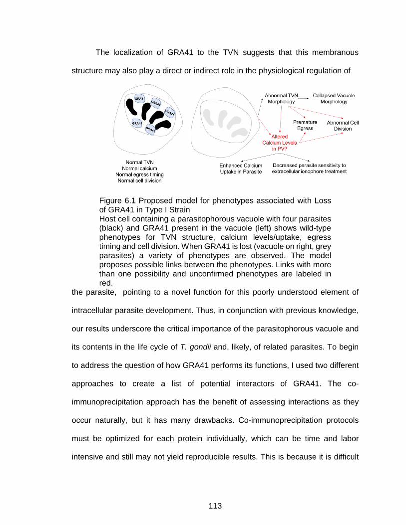
113
The localization of GRA41 to the TVN suggests that this membranous
structure may also play a direct or indirect role in the physiological regulation of
Figure 6.1 Proposed model for phenotypes associated with Loss of GRA41 in Type I Strain Host cell containing a parasitophorous vacuole with four parasites (black) and GRA41 present in the vacuole (left) shows wild-type phenotypes for TVN structure, calcium levels/uptake, egress timing and cell division. When GRA41 is lost (vacuole on right, grey parasites) a variety of phenotypes are observed. The model proposes possible links between the phenotypes. Links with more than one possibility and unconfirmed phenotypes are labeled in red.
the parasite, pointing to a novel function for this poorly understood element of
intracellular parasite development. Thus, in conjunction with previous knowledge,
our results underscore the critical importance of the parasitophorous vacuole and
its contents in the life cycle of T. gondii and, likely, of related parasites. To begin
to address the question of how GRA41 performs its functions, I used two different
approaches to create a list of potential interactors of GRA41. The co-
immunoprecipitation approach has the benefit of assessing interactions as they
occur naturally, but it has many drawbacks. Co-immunoprecipitation protocols
must be optimized for each protein individually, which can be time and labor
intensive and still may not yield reproducible results. This is because it is difficult
114
to capture transient or weak interactions, especially when strong detergents and
sonication are necessary to fully solubilize a target protein such as GRA41, which
has been shown to partition as a membrane protein (Figure 3.4, above). A single
successful co-immunoprecipitation was conducted on GRA41 using the strain
which has GRA41 endogenously tagged with HA and harvesting intracellular
parasites, which identified several potential interactors. Of these, some were
previously characterized dense granule and rhoptry proteins, while others are
proteins of hypothetical function that contain a signal peptide (characteristics
common to dense granule proteins). Two of these proteins that are most likely to
be bona fide interactors are GRA3 and GRA9, both of which at least partially
localized to the tubulovesicular network where GRA41 resides (Adjogble, Mercier
et al. 2004, Kim, Ahn et al. 2008). The large number of rhoptry proteins, which all
traffic to the membrane of the parasitophorous vacuole (Soldati, Lassen et al.
1998, El Hajj, Lebrun et al. 2007, Peixoto, Chen et al. 2010), are also potentially
bona fide interactors, since the tubulovesicular network can come into close
contact with the PVM. If these interactors are real, they could suggest a possible
function of GRA41 wherein it helps form the links between the TVN and the PVM.
If these links are required for the correct morphology of the TVN, they could explain
why loss of GRA41 leads to disruption of the TVN. It is important to note that none
of these interactions have been validated by targeted co-immunoprecipitations,
which would be necessary to demonstrate that they are true interactors.
Using the yeast two hybrid approach allows us to continue our search for
interactors while avoiding some of the pitfalls of the co-immunoprecipitation
115
approach. When samples do not need to be lysed and solubilized, there is no
concern that weak interactions are being missed. Since the yeast two-hybrid
approach takes place in a heterologous system, it will only give direct interactors,
rather than proteins that are part of the same complex as the target protein but do
not bind it directly. Additionally, because the prey fragments used in the library are
often smaller than the entire protein, it is possible to determine what region of the
prey protein that the bait protein is binding. Using this technique has generated a
list of 11 high confidence potential interactors for GRA41. Of these, probably the
most intriguing is GRA1, which is a dense granule protein that localizes to both the
lumen of the PV as well as the TVN. In addition to partially sharing localization with
GRA41, GRA1 also contains a motif that has been shown to bind calcium
(Cesbron-Delauw, Guy et al. 1989). It is possible that GRA1 acts to sequester
calcium within the vacuole and a potential interaction with GRA41 would allow it to
associate with the TVN. Of the remainder of the list, only a single protein of
hypothetical function is predicted to be secreted by Psort (Horton, Park et al. 2007),
though many of the remaining proteins do have signal peptides. Unfortunately,
none of the high confidence proteins on the yeast two-hybrid list overlap with the
high confidence proteins from the co-immunoprecipitation, likely due to the
differences and drawbacks of each approach. Since interactions of lower
confidence for both data sets were excluded from the analysis, it is possible that
are some genuine interactors that show up on both lists but are not listed as high
confidence due to low abundance of the potential interactor. The yeast two-hybrid
approach is likely to capture interactions that are possible based on protein
116
structure, but may not occur within the parasite because the proteins are localized
to different areas within the cell. They are also incapable of detecting indirect
interactions with multiple components of a protein complex. The co-
immunoprecipitation approach is limited by the optimization of the solubilization
and pull-down of the target antigen, but is more likely to be able to pull down large,
multi-protein complexes. To validate these potential interactors, it will be necessary
to show co-localization, which may require immunoelectron microscopy to
potential interactions between the PVM-localized rhoptries and the TVN-localized
GRA41. A proximity-labeling approach, such as BioID (Roux, Kim et al. 2013) or
APEX2 (Lam, Martell et al. 2015), could also help to verify transient or weak
interactions that show up in the yeast two-hybrid screen but are not seen using a
traditional co-immunoprecipitation approach.
A complete knockout of GRA41 in a Type II strain recapitulates the
resistance to iiDeath seen in both the Type I knockout and forward genetic mutant,
suggesting that the overall role in calcium processes is conserved between the two
strains. However, no significant defect in plaque formation was detected for the
knockout strain and additional phenotypes found in the Type I knockout strain were
not tested, so it remains unclear the exact function GRA41 might play in a Type II
strain. Determining whether or not the Type II knockout recapitulates all of the
phenotypes of the Type I knockout is particularly interesting in light of the
differences in electrophoretic mobility for the tagged proteins described above,
making it likely that GRA41 is differentially modified in the two different strains.

117
The most important question that generating a Type II knockout allows us
to answer is whether or not the loss of GRA41 plays any role in tissue cyst
formation or maintenance and what role it plays during in vivo infection. Here I
show preliminary data that the knockout of GRA41 shows a significant decrease
in the percentage of vacuoles with an intact cyst wall at 7 days when compared to
the parental strain. Since this phenotype is not seen at four days, it suggests that
the loss of GRA41 does not impair the parasite’s ability to form a cyst wall, but that
the cyst wall is not able to be maintained, even in the presence of continued pH
stress. It is unclear whether or not this failure to maintain the cyst wall is due to
defects in calcium signaling as seen in the Type I strain or structural defects in the
cyst wall. A pilot in vivo experiment shows that the loss of GRA41 shows highly
variable phenotypes according to the dose used to infect mice. At a low dose, cysts
counts are very low in both the parental and knockout-infected mice and no
difference is seen between the two groups. However, at a higher dose, there is a
2-fold increase in the number of cysts/mouse brain. This increase is also seen in
the cyst-containing brains of the mice that received the highest dose of knockout
parasites, though many of the brains in this group have no detectable cysts, so the
average number of cysts is equivalent in the parental and knockout strains. Follow-
up mouse experiments would need to address the issue of acute virulence by
increasing the highest dose to one that is lethal in at least some of the mice. This
would allow us to determine the impact that GRA41 has on virulence during the
acute infection. Given the high variability in the tissue cyst count data from the pilot
experiment, follow-up experiments should increase the number of animals

118
assessed to get a more accurate representation of total cyst numbers. Since the
preliminary results suggest that the knockout strain could have a defect in cyst
maintenance, which would lead to overall higher cyst counts due to the reactivation
of cysts to tachyzoites, followed by reinvasion and conversion back to bradyzoite
cysts, additional experiments examining cyst counts for mice infected with the
parental and knockout strains at later time points in the chronic infection might help
support this hypothesis. It is possible that some of the cysts of the knockout strain
are not able to be visualized with the current protocols due to small size, and it is
expected that looking at later time points might allow cysts to increase in size
and/or number. Overall, these results suggest that GRA41 plays an important role
in the bradyzoite stage of the life cycle and that differences between the Type I
and Type II proteins could provide important information about the role of specific
residues and potential posttranslational modifications in phenotypes already
characterized in Type I tachyzoites. Additionally, since very little is known about
the calcium regulation of bradyzoite formation, maintenance and reactivation, this
knockout could play a key role in elucidating the role of calcium in these processes.

119
Chapter 7 : Conclusion and Future Directions
Conclusion
Calcium-dependent signaling processes are critical for multiple steps of the
parasite’s lytic cycle and represent an appealing target for drug discovery
(Arrizabalaga and Boothroyd 2004). Former members of the Arrizabalaga
laboratory have used a forward genetic approach to identify and characterize some
of the key players in these signaling events (Black, Arrizabalaga et al. 2000,
Garrison, Treeck et al. 2012). Here I describe the characterization of a previously
generated mutant, MBD2.1, that is capable of surviving prolonged extracellular
treatment with a calcium ionophore, which renders wild-type parasites unable to
attach to host cells. I show that the mutation responsible for this phenotype is the
introduction of a premature stop codon at serine 92 of the protein of hypothetical
function, TgGT1_069070. Endogenous tagging at this locus reveals that the
encoded protein is secreted from the parasite organelle known as the dense
granules into the parasitophorous vacuole in which the parasite resides and
divides. Once in the vacuole, it traffics to the TVN. Its secretion from the dense
granule leads to its designation as a GRA protein and given the current published
GRA proteins in the field, it is specifically designated as GRA41.
A complete knockout of the locus encoding GRA41 leads to a recapitulation
of the resistance of extracellular ionophore treatment, while also showing a defect
in the efficiency of plaquing. This defect is rescued by complementing the knockout
strain with a wild-type copy of GRA41 and is demonstrated to be largely due to
premature egress of parasites from host cells. The GRA41 knockout strain also

120
exhibits increased uptake of calcium when extracellular, which was restored to
normal levels in the complemented strain. A morphological analysis of the
knockout strain revealed an improperly formed TVN and evidence of atypical cell
division. A knockout parasite strain with similar TVN disruption (Travier,
Mondragon et al. 2008) does not exhibit resistance to iiDeath treatment. Current
efforts to identify protein interaction partners have resulted in a list of unverified
potential interactors obtained by co-immunoprecipation and yeast two-hybrid
analysis.
Studies to investigate the function of GRA41 in the bradyzoite life stage
were conducted in the Type II Pru strain. Endogenous tagging of the Type II
GRA41 protein revealed that it localizes primarily to the tissue cyst wall and has a
unique electrophoretic migration pattern as compared to the Type I Gra41 protein.
The loss of GRA41 in the Type II strain recapitulated the resistance to ionophore
induced death seen in the original mutant and Type I knockout strain. This strain
also shows reduced tissue cyst wall staining of vacuoles following seven days of
bradyzoite differentiation by pH stress, though this difference is not seen after four
days of differentiation. A pilot study of chronic mouse infection with the Type II
knockout strain showed highly variable cyst counts at the highest dose and very
low cyst counts at the lowest dose, while the middle dose showed adequate cyst
counts in both the parental and the knockout with somewhat less variability than
was seen at the top dose. At this middle dose, the knockout strain was shown to
have an approximately two-fold higher cyst burden as compared to the parental
strain.
121
Overall, the forward genetic approach to identifying novel regulators of
calcium signaling processes has provided unexpected and intriguing results in the
identification of a novel dense granule protein that plays a role in calcium
sensitivity/uptake, the timing of egress and vacuolar morphology. Though the exact
mechanism by which this protein performs these important functions remains
unclear, these results provide many tantalizing clues and avenues for possible
future work in this area. An original goal of this work was to identify novel potential
drug targets to be able to develop inhibitors against for treatment of clinical
toxoplasmosis. Since GRA41 lacks homologues even among most other
apicomplexans, its uniqueness could be advantageous in developing specific
inhibitors that would not target host processes. However, the loss of GRA41 is not
lethal and, in fact, exhibits what could be considered a gain of function phenotype
in its premature egress from host cells and ability to withstand extended calcium
ionophore treatment. This suggests that a more viable strategy to target GRA41
for drug treatment could be to increase either its amount or stability. To test
whether or not this might be a viable strategy, we could generate a line which
overexpresses GRA41 under the promoter of a more highly expressed gene (i.e.
tubulin) and determine what impact his has on parasite growth.
After the initial identification and characterization of GRA41 from the forward
genetic approach, it is clear that the forward genetic approach has come with some
important advantages and limitations. This approach has allowed us to identify a
novel protein involved in calcium-dependent signaling processes that would have
easily been overlooked with a reverse genetic approach, due to its lack of
122
conserved domains. Additionally, the calcium-related phenotype by which the
mutant was isolated gave important clues to GRA41’s function. However, this
approach has a few key limitations. Specifically, the use of iiEgress/iiDeath
phenotypes for forward genetic selection has oftentimes resulted in the generation
of mutants in the calcium-dependent protein kinase 3 (TgCDPK3, (Garrison,
Treeck et al. 2012)), suggesting that this selection needs to be refined in order to
identify novel proteins involved in these processes. For example, the knockout of
GRA41 results in enhanced natural and DTT-induced egress, though the timing of
ionophore-induced egress is very similar to wild-type parasites. To identify more
proteins that might play a role in the timing of natural egress, the screen could be
modified by using DTT as the inducer of the egress rather than the calcium
ionophore. By isolating parasites that have delayed (i.e. do not egress following
DTT treatment at 35 hours post infection) or early (i.e. do egress following egress
treatment before 35 hours post infection) DTT-induced egress, it would be possible
to identify proteins that play a role in the timing of natural (uninduced) egress.
Future Directions
Does the loss of GRA41 impact calcium levels within the parasitophorous
vacuole?
Though this work has shown that the loss of GRA41 in a Type I strain leads
to enhanced calcium uptake in extracellular tachyzoites, it is unclear how a
secreted protein demonstrates many of its calcium-related phenotypes (decreased
sensitivity to ionophore treatment, enhanced calcium uptake) when the parasite is
extracellular. I hypothesize that the calcium levels within the parasitophorous
123
vacuole of parasites without GRA41 are higher than those seen in wild-type
parasites and that this impacts how the parasites within these vacuoles respond
to calcium. This is consistent with the premature egress seen in the GRA41
knockout, since calcium is an important trigger of parasite egress. However, I do
not have any direct evidence that the calcium levels within the PV are different
between the strains. To address this, I could load host cells containing parasites
with chemical calcium indicators that have been shown to traffic to the PV once
inside the host cell and use previously described methods to measure the calcium
concentration within the vacuoles (Pingret, Millot et al. 1996). This will help to
address an important question about how a secreted protein can play a role in the
calcium homeostasis within the parasite.
How does GRA41 localize to the tubulovesicular network?
The localization of GRA41 to a membranous network is unexpected, since
there is no strong prediction for a transmembrane domain within this protein.
Attempts to identify an amphipathic alpha helix, such as the one responsible for
localizing GRA2 to the TVN (Mercier, Cesbron-Delauw et al. 1998), have also been
unsuccessful. This makes it likely that the localization of GRA41 to this
membranous network is due to either a post-translational modification or its
interaction with another protein. To address the first possibility, and also provide
additional insight into other aspects of GRA41’s biology, I propose to conduct a
proteomic analysis to identify post-translational modifications of GRA41. Though
many proteomic data sets currently exist in the online T. gondii genome database,
GRA41 has not been annotated in any strain of the genome since v7.2 in 2012, so
124
any data sets created after this time did not include GRA41. The only known post-
translational modification of GRA41 are multiple phosphorylation marks from a
phosphoproteome analysis published before this time (Treeck, Sanders et al.
2011). The identification of post-translational modifications on GRA41 will help to
suggest potential modifications that could allow association with a membrane (i.e.
lipidation) or which may regulate its function. These residues could then be
mutated to see if they have any impact on the protein’s localization or function.
What parts of the protein are necessary for the different functions of GRA41?
Since GRA41 does not have any domains of known/predicted function, this
has made it difficult to ascribe specific functions to domains of the protein.
However, the identification of domains with specific functions within the protein can
help to understand the mechanisms by which GRA41 impacts vacuolar
morphology and calcium-dependent signaling processes. Previous attempts have
been made to generate a series of truncation mutants, but these resulted in
proteins with low or no expression within the parasite, making it difficult to
characterize their localization or potential phenotypes associated with these
truncations. Instead, I would take a complementation approach using the
previously generated knockout of GRA41. I currently have designed a
complementation construct consisting of GRA41 5’ and 3’ targeting regions
flanking a GRA41 coding sequence tagged with a triple HA tag. This construct
could be modified by scrambling the amino acid sequences of alpha helices,
mutating residues predicted/demonstrated to be post-translationally modified or
deleting short sequences from the protein. These could then be incorporated into
125
the knockout strain to assess their localization and ability to complement the
various phenotypes associated with the loss of GRA41.
What are the protein interaction partners of GRA41? Can any of these explain
the phenotypes associated with its loss?
Preliminary attempts to define the GRA41 interactome have generated an
initial list that will be expanded upon and validated. For this, I propose to take two
complementary approaches. Due to difficulties encountered with the traditional co-
immunoprecipitation approach, I would instead look to a proximity-based labeling
approach to identify potential interactors. In this context, APEX2 is an ascorbate
peroxidase that has been used to label compartments for electron microscopy
imaging and also to label proximal proteins with a biotin-phenol radical. It was
recently modified to have high catalytic activity, and can now be used even with
proteins that have low or medium endogenous expression without the requirement
of overexpression (Lam, Martell et al. 2015). A fusion between GRA41 and APEX
will be generated at the endogenous locus, similar to the addition of the
endogenous HA tag. In the presence of biotin phenol and hydrogen peroxide, this
fusion protein will add biotin groups to proteins within a 20-nm radius of the fusion
protein. These proteins can then be isolated using streptavidin-conjugated
agarose or magnetic beads and identified by mass spectrometry. Since the
labeling occurs in live parasites, this method is less likely to capture false positive
from interactors not found within the same location as the protein of interest. It is
also capable of detecting weak or transient interactions and there are no limits on
the types of lysis and wash buffers used, since it is not necessary to preserve
126
protein interactions. This method will be used to complement the data generated
from the co-immunoprecipitation and yeast two-hybrid analysis to prioritize
candidate proteins.
Candidate interactors identified in the current data and any additional data
sets generated from the APEX labeling experiments will be validated by verifying
colocalization with GRA41 when parasites are intracellular and extracellular (using
antibodies if available or by generating lines with the potential interactor tagged if
an antibody is not available). If colocalization is observed, reciprocal co-
immunoprecipitations will be performed in a strain tagged with both GRA41 and
the potential interactor. To account for the possibility that these interactions are
transient, proteins will first be cross-linked chemically using a reversible cross-
linker such as those of the N-hydroxysuccinimide class of crosslinkers to preserve
these interactions. An antibody will be used to pull down one of the proteins before
the crosslinking is reversed. A Western Blot analysis will be performed to detect
each protein in the elution fraction. If each pulldown shows both proteins are
coming down when one of them is the target, I will conclude they are interactors.
In the case of proteins with known functions, verification of these interactions could
provide important mechanistic insight into how GRA41 performs its functions. For
proteins whose function is not currently not known, this approach could help to
identify other novel PV proteins with similar roles to GRA41. These would then be
genetically disrupted to see if they share similar phenotypes to the knockout of
GRA41 or if they perform altogether different functions. In addition to investigating
the proteins found in this list, I also propose to look specifically at GRA22, since it

127
has been shown previously to have a premature egress phenotype, by generating
a single knockout of GRA22 and a double knockout of GRA41 and GRA22 for
phenotypic analysis.
Are there differences between the Type I and Type II GRA41 proteins that could
provide insight into protein function or parasite biology?
As I have already shown, though both the Type I and Type II GRA41
proteins are secreted proteins, they demonstrate an important difference in
electrophoretic mobility. The Type I GRA41 protein runs as a doublet on an SDS-
PAGE gel while the Type II protein runs as a single band. Further investigation
reveals that there are six amino acid changes between the two protein sequences
which could be responsible for the differences observed if any of them are shown
to be post-translationally modified within the Type I strain. The Type I and Type II
strains differ greatly in the in vivo virulence and polymorphisms within dense
granule and rhoptry proteins have been previously shown to be important
mediators of these virulence differences. I propose to test if the polymorphisms
between the Type I and Type II strains are important to GRA41 function by
conducting complementation studies in the knockout strains to determine whether
or not the Type II protein is capable of rescuing the Type I knockout’s phenotypes
and vice versa. A failure of the Type II protein to rescue the Type I knockout’s
phenotypes could help to narrow down key residues associated with certain
residues and areas of the protein. The specific residues that differ between the two
strains could be mutated to determine which, if any, impact the rescue of
phenotype. There are also additional phenotypes of the Type I knockout that have

128
not yet been assessed in the Type II knockout. Verifying whether or not the Type
II knockout retains the premature egress, TVN disruption and calcium uptake
phenotypes of the Type II knockout could prove very informative in identifying key
residues/domains involved in these various phenotypes.
The exact localization of the Type II protein remains unclear. Though it has
been shown to be secreted into the parasitophorous vacuole and localizes to the
tissue cyst wall, it is unclear if it localizes to the TVN of the vacuole or if it is found
free in the lumen of the PV. Preliminary studies to determine if the Type II GRA41
protein exists as a soluble or membrane-associated protein should first be
conducted. As expected for a TVN-associated protein, Type I GRA41 is found
within the membrane-associated fraction following partitioning with TX-114. If the
Type II protein does not have the same pattern, this may suggest that it has a
different localization within the vacuole than the Type I protein, which can be
verified by immunoelectron microscopy as was done for the Type I protein.
Additionally, very little is known about the TVN in cysts; if GRA41 continues to
localize to the TVN of Type II parasites, immunoelectron microscopy of bradyzoite
cysts expressing a tagged GRA41 could be informative in studying the TVN of
cysts.
Does the knockout of GRA41 in a Type II strain lead to a defect in bradyzoite
cyst maintenance?
Our preliminary data supports a hypothesis that the loss of GRA41 in a Type
II strain leads to a defect in the maintenance of bradyzoite cysts, both in vitro and
in vivo. However, there is much additional work to be done to test this hypothesis.
129
Most importantly, the generation of a complemented strain or an independently-
derived knockout is necessary to verify that the associated phenotypes are directly
due to the loss of GRA41. Preliminary attempts to generate such a strain resulted
in a population with a very low rate of successful incorporation and expression of
the complementation plasmid within the transfected population (<1%) and an
inability to isolate a positive clone. This was also seen when generating the Type
II knockout strain, and in this case, the addition of a GFP marker outside of the
homology arms that allow for incorporation into the target locus helped to eliminate
clones that had integrated the knockout plasmid randomly. Hundreds of clones
could be rapidly screened by live fluorescence imaging, rather than the methodical
screening of dozens of clones that is done by traditional immunofluorescence
analysis. Since the complementation construct has also been targeted to the
endogenous locus encoding GRA41, a similar approach could be used to quickly
screen for parasites that incorporated the plasmid in the correct location.
Additional work will also aim to further refine and characterize the
phenotypes associated with loss of GRA41 in a Type II strain. Given the observed
differences in sequence and electrophoretic mobility (and likely post-translational
modification), it is important to ask whether or not all of the phenotypes observed
in the Type I knockout are also found in the Type II knockout. In addition to this,
looking at bradyzoite formation both with a complemented strain and at additional,
later time points, will help to further clarify the role of GRA41 in cyst maintenance.
Based on the pilot in vivo study, future efforts to look at the impact of GRA41 on
cyst burden are warranted. Since I suspect that GRA41 might play a role in cyst

130
maintenance in vivo as well, additional time points harvested later during chronic
infection can help elucidate how the infection is progressing over time. Additionally,
none of the doses utilized in the pilot study led directly to any mouse death during
the acute infection, so a trial of a higher dose of parasites will be used to determine
if GRA41 plays a role in virulence in the mouse model.

131
Chapter 8 : References
Adjogble, K. D., C. Mercier, J. F. Dubremetz, C. Hucke, C. R. Mackenzie,
M. F. Cesbron-Delauw and W. Daubener (2004). "GRA9, a new Toxoplasma
gondii dense granule protein associated with the intravacuolar network of tubular
membranes." Int J Parasitol 34(11): 1255-1264.
Alexander, D. L., S. Arastu-Kapur, J. F. Dubremetz and J. C. Boothroyd
(2006). "Plasmodium falciparum AMA1 binds a rhoptry neck protein homologous
to TgRON4, a component of the moving junction in Toxoplasma gondii." Eukaryot
Cell 5(7): 1169-1173.
Alexander, D. L., J. Mital, G. E. Ward, P. Bradley and J. C. Boothroyd
(2005). "Identification of the moving junction complex of Toxoplasma gondii: a
collaboration between distinct secretory organelles." PLoS Pathog 1(2): e17.
Arrizabalaga, G. and J. C. Boothroyd (2004). "Role of calcium during
Toxoplasma gondii invasion and egress." Int J Parasitol 34(3): 361-368.
Arrizabalaga, G., F. Ruiz, S. Moreno and J. C. Boothroyd (2004).
"Ionophore-resistant mutant of Toxoplasma gondii reveals involvement of a
sodium/hydrogen exchanger in calcium regulation." J Cell Biol 165(5): 653-662.
Asai, T., W. J. O'Sullivan and M. Tatibana (1983). "A potent nucleoside
triphosphate hydrolase from the parasitic protozoan Toxoplasma gondii.
Purification, some properties, and activation by thiol compounds." J Biol Chem
258(11): 6816-6822.

132
Bartel, P., C. T. Chien, R. Sternglanz and S. Fields (1993). "Elimination of
false positives that arise in using the two-hybrid system." Biotechniques 14(6): 920-
924.
Beck, J. R., A. L. Chen, E. W. Kim and P. J. Bradley (2014). "RON5 is critical
for organization and function of the Toxoplasma moving junction complex." PLoS
Pathog 10(3): e1004025.
Beis, J. M., J. M. Andre, A. M. Datie and B. Brugerolle (2002). "Eye
movements and verbal reports in neglect patients during a letter reading task."
NeuroRehabilitation 17(2): 145-151.
Benmerzouga, I., L. A. Checkley, M. T. Ferdig, G. Arrizabalaga, R. C. Wek
and W. J. Sullivan, Jr. (2015). "Guanabenz repurposed as an antiparasitic with
activity against acute and latent toxoplasmosis." Antimicrob Agents Chemother
59(11): 6939-6945.
Bermudes, D., K. R. Peck, M. A. Afifi, C. J. Beckers and K. A. Joiner (1994).
"Tandemly repeated genes encode nucleoside triphosphate hydrolase isoforms
secreted into the parasitophorous vacuole of Toxoplasma gondii." J Biol Chem
269(46): 29252-29260.
Birch-Andersen, A., D. J. Ferguson and R. D. Pontefract (1976). "A
technique for obtaining thin sections of coccidian oocysts." Acta Pathol Microbiol
Scand B 84(4): 235-239.
Black, M. W., G. Arrizabalaga and J. C. Boothroyd (2000). "Ionophore-
resistant mutants of Toxoplasma gondii reveal host cell permeabilization as an
early event in egress." Mol Cell Biol 20(24): 9399-9408.

133
Black, M. W. and J. C. Boothroyd (2000). "Lytic cycle of Toxoplasma
gondii." Microbiol Mol Biol Rev 64(3): 607-623.
Blom, N., T. Sicheritz-Ponten, R. Gupta, S. Gammeltoft and S. Brunak
(2004). "Prediction of post-translational glycosylation and phosphorylation of
proteins from the amino acid sequence." Proteomics 4(6): 1633-1649.
Bonhomme, A., L. Pingret, P. Bonhomme, J. Michel, G. Balossier, M.
Lhotel, M. Pluot and J. M. Pinon (1993). "Subcellular calcium localization in
Toxoplasma gondii by electron microscopy and by X-ray and electron energy loss
spectroscopies." Microsc Res Tech 25(4): 276-285.
Boothroyd, J. C. and J. F. Dubremetz (2008). "Kiss and spit: the dual roles
of Toxoplasma rhoptries." Nat Rev Microbiol 6(1): 79-88.
Borges-Pereira, L., A. Budu, C. A. McKnight, C. A. Moore, S. A. Vella, M.
A. Hortua Triana, J. Liu, C. R. Garcia, D. A. Pace and S. N. Moreno (2015).
"Calcium Signaling throughout the Toxoplasma gondii Lytic Cycle: A STUDY
USING GENETICALLY ENCODED CALCIUM INDICATORS." J Biol Chem
290(45): 26914-26926.
Bougdour, A., E. Durandau, M. P. Brenier-Pinchart, P. Ortet, M. Barakat, S.
Kieffer, A. Curt-Varesano, R. L. Curt-Bertini, O. Bastien, Y. Coute, H. Pelloux and
M. A. Hakimi (2013). "Host cell subversion by Toxoplasma GRA16, an exported
dense granule protein that targets the host cell nucleus and alters gene
expression." Cell Host Microbe 13(4): 489-500.
Braun, L., M. P. Brenier-Pinchart, M. Yogavel, A. Curt-Varesano, R. L. Curt-
Bertini, T. Hussain, S. Kieffer-Jaquinod, Y. Coute, H. Pelloux, I. Tardieux, A.

134
Sharma, H. Belrhali, A. Bougdour and M. A. Hakimi (2013). "A Toxoplasma dense
granule protein, GRA24, modulates the early immune response to infection by
promoting a direct and sustained host p38 MAPK activation." J Exp Med 210(10):
2071-2086.
Brugerolle, G., G. Bricheux, H. Philippe and G. Coffea (2002). "Collodictyon
triciliatum and Diphylleia rotans (=Aulacomonas submarina) form a new family of
flagellates (Collodictyonidae) with tubular mitochondrial cristae that is
phylogenetically distant from other flagellate groups." Protist 153(1): 59-70.
Bullen, H. E., C. J. Tonkin, R. A. O'Donnell, W. H. Tham, A. T. Papenfuss,
S. Gould, A. F. Cowman, B. S. Crabb and P. R. Gilson (2009). "A novel family of
Apicomplexan glideosome-associated proteins with an inner membrane-anchoring
role." J Biol Chem 284(37): 25353-25363.
Caffaro, C. E., A. A. Koshy, L. Liu, G. M. Zeiner, C. B. Hirschberg and J. C.
Boothroyd (2013). "A nucleotide sugar transporter involved in glycosylation of the
Toxoplasma tissue cyst wall is required for efficient persistence of bradyzoites."
PLoS Pathog 9(5): e1003331.
Caldas, L. A., W. de Souza and M. Attias (2010). "Microscopic analysis of
calcium ionophore activated egress of Toxoplasma gondii from the host cell." Vet
Parasitol 167(1): 8-18.
Carruthers, V. B., O. K. Giddings and L. D. Sibley (1999). "Secretion of
micronemal proteins is associated with toxoplasma invasion of host cells." Cell
Microbiol 1(3): 225-235.

135
Carruthers, V. B. and L. D. Sibley (1997). "Sequential protein secretion from
three distinct organelles of Toxoplasma gondii accompanies invasion of human
fibroblasts." Eur J Cell Biol 73(2): 114-123.
Carruthers, V. B. and L. D. Sibley (1999). "Mobilization of intracellular
calcium stimulates microneme discharge in Toxoplasma gondii." Mol Microbiol
31(2): 421-428.
Cerede, O., J. F. Dubremetz, M. Soete, D. Deslee, H. Vial, D. Bout and M.
Lebrun (2005). "Synergistic role of micronemal proteins in Toxoplasma gondii
virulence." J Exp Med 201(3): 453-463.
Cesbron-Delauw, M. F., B. Guy, G. Torpier, R. J. Pierce, G. Lenzen, J. Y.
Cesbron, H. Charif, P. Lepage, F. Darcy, J. P. Lecocq and et al. (1989). "Molecular
characterization of a 23-kilodalton major antigen secreted by Toxoplasma gondii."
Proc Natl Acad Sci U S A 86(19): 7537-7541.
Chaturvedi, S., H. Qi, D. Coleman, A. Rodriguez, P. I. Hanson, B. Striepen,
D. S. Roos and K. A. Joiner (1999). "Constitutive calcium-independent release of
Toxoplasma gondii dense granules occurs through the NSF/SNAP/SNARE/Rab
machinery." J Biol Chem 274(4): 2424-2431.
Chen, A. L., A. S. Moon, H. N. Bell, A. S. Huang, A. A. Vashisht, J. Y. Toh,
A. H. Lin, S. M. Nadipuram, E. W. Kim, C. P. Choi, J. A. Wohlschlegel and P. J.
Bradley (2017). "Novel insights into the composition and function of the
Toxoplasma IMC sutures." Cell Microbiol 19(4).
Cleary, M. D., U. Singh, I. J. Blader, J. L. Brewer and J. C. Boothroyd (2002).
"Toxoplasma gondii asexual development: identification of developmentally

136
regulated genes and distinct patterns of gene expression." Eukaryot Cell 1(3): 329-
340.
Cochereau-Massin, I., P. LeHoang, M. Lautier-Frau, E. Zerdoun, L. Zazoun,
M. Robinet, P. Marcel, B. Girard, C. Katlama, C. Leport and et al. (1992). "Ocular
toxoplasmosis in human immunodeficiency virus-infected patients." Am J
Ophthalmol 114(2): 130-135.
Del Carmen, M. G., M. Mondragon, S. Gonzalez and R. Mondragon (2009).
"Induction and regulation of conoid extrusion in Toxoplasma gondii." Cell Microbiol
11(6): 967-982.
Docampo, R., W. de Souza, K. Miranda, P. Rohloff and S. N. Moreno
(2005). "Acidocalcisomes - conserved from bacteria to man." Nat Rev Microbiol
3(3): 251-261.
Donald, R. G., D. Carter, B. Ullman and D. S. Roos (1996). "Insertional
tagging, cloning, and expression of the Toxoplasma gondii hypoxanthine-xanthine-
guanine phosphoribosyltransferase gene. Use as a selectable marker for stable
transformation." J Biol Chem 271(24): 14010-14019.
Dou, Z. and V. B. Carruthers (2011). "Cathepsin proteases in Toxoplasma
gondii." Adv Exp Med Biol 712: 49-61.
Dou, Z., O. L. McGovern, M. Di Cristina and V. B. Carruthers (2014).
"Toxoplasma gondii ingests and digests host cytosolic proteins." MBio 5(4):
e01188-01114.

137
Dubey, J. P. (2006). "Comparative infectivity of oocysts and bradyzoites of
Toxoplasma gondii for intermediate (mice) and definitive (cats) hosts." Vet
Parasitol 140(1-2): 69-75.
Dunn, J. D., S. Ravindran, S. K. Kim and J. C. Boothroyd (2008). "The
Toxoplasma gondii dense granule protein GRA7 is phosphorylated upon invasion
and forms an unexpected association with the rhoptry proteins ROP2 and ROP4."
Infect Immun 76(12): 5853-5861.
El Hajj, H., M. Lebrun, S. T. Arold, H. Vial, G. Labesse and J. F. Dubremetz
(2007). "ROP18 is a rhoptry kinase controlling the intracellular proliferation of
Toxoplasma gondii." PLoS Pathog 3(2): e14.
El Hajj, H., M. Lebrun, M. N. Fourmaux, H. Vial and J. F. Dubremetz (2007).
"Inverted topology of the Toxoplasma gondii ROP5 rhoptry protein provides new
insights into the association of the ROP2 protein family with the parasitophorous
vacuole membrane." Cell Microbiol 9(1): 54-64.
Etheridge, R. D., A. Alaganan, K. Tang, H. J. Lou, B. E. Turk and L. D.
Sibley (2014). "The Toxoplasma pseudokinase ROP5 forms complexes with
ROP18 and ROP17 kinases that synergize to control acute virulence in mice." Cell
Host Microbe 15(5): 537-550.
Fentress, S. J. and L. D. Sibley (2011). "The secreted kinase ROP18
defends Toxoplasma's border." Bioessays 33(9): 693-700.
Ferguson, D. J. and W. M. Hutchison (1987). "The host-parasite relationship
of Toxoplasma gondii in the brains of chronically infected mice." Virchows Arch A
Pathol Anat Histopathol 411(1): 39-43.

138
Fischer, H. G., S. Stachelhaus, M. Sahm, H. E. Meyer and G. Reichmann
(1998). "GRA7, an excretory 29 kDa Toxoplasma gondii dense granule antigen
released by infected host cells." Mol Biochem Parasitol 91(2): 251-262.
Foe, I. T., M. A. Child, J. D. Majmudar, S. Krishnamurthy, W. A. van der
Linden, G. E. Ward, B. R. Martin and M. Bogyo (2015). "Global Analysis of
Palmitoylated Proteins in Toxoplasma gondii." Cell Host Microbe 18(4): 501-511.
Formstecher, E., S. Aresta, V. Collura, A. Hamburger, A. Meil, A. Trehin, C.
Reverdy, V. Betin, S. Maire, C. Brun, B. Jacq, M. Arpin, Y. Bellaiche, S. Bellusci,
P. Benaroch, M. Bornens, R. Chanet, P. Chavrier, O. Delattre, V. Doye, R. Fehon,
G. Faye, T. Galli, J. A. Girault, B. Goud, J. de Gunzburg, L. Johannes, M. P. Junier,
V. Mirouse, A. Mukherjee, D. Papadopoulo, F. Perez, A. Plessis, C. Rosse, S.
Saule, D. Stoppa-Lyonnet, A. Vincent, M. White, P. Legrain, J. Wojcik, J. Camonis
and L. Daviet (2005). "Protein interaction mapping: a Drosophila case study."
Genome Res 15(3): 376-384.
Fox, B. A., A. Falla, L. M. Rommereim, T. Tomita, J. P. Gigley, C. Mercier,
M. F. Cesbron-Delauw, L. M. Weiss and D. J. Bzik (2011). "Type II Toxoplasma
gondii KU80 knockout strains enable functional analysis of genes required for cyst
development and latent infection." Eukaryot Cell 10(9): 1193-1206.
Francia, M. E., S. Wicher, D. A. Pace, J. Sullivan, S. N. Moreno and G.
Arrizabalaga (2011). "A Toxoplasma gondii protein with homology to intracellular
type Na(+)/H(+) exchangers is important for osmoregulation and invasion." Exp
Cell Res 317(10): 1382-1396.

139
Frenal, K., V. Polonais, J. B. Marq, R. Stratmann, J. Limenitakis and D.
Soldati-Favre (2010). "Functional dissection of the apicomplexan glideosome
molecular architecture." Cell Host Microbe 8(4): 343-357.
Frenkel, J. K., J. P. Dubey and N. L. Miller (1970). "Toxoplasma gondii in
cats: fecal stages identified as coccidian oocysts." Science 167(3919): 893-896.
Fromont-Racine, M., J. C. Rain and P. Legrain (1997). "Toward a functional
analysis of the yeast genome through exhaustive two-hybrid screens." Nat Genet
16(3): 277-282.
Fruth, I. A. and G. Arrizabalaga (2007). "Toxoplasma gondii: induction of
egress by the potassium ionophore nigericin." Int J Parasitol 37(14): 1559-1567.
Fukasawa, Y., T. Oda, K. Tomii and K. Imai (2017). "Origin and Evolutionary
Alteration of the Mitochondrial Import System in Eukaryotic Lineages." Mol Biol
Evol 34(7): 1574-1586.
Furie, B., B. A. Bouchard and B. C. Furie (1999). "Vitamin K-dependent
biosynthesis of gamma-carboxyglutamic acid." Blood 93(6): 1798-1808.
Gaji, R. Y., D. E. Johnson, M. Treeck, M. Wang, A. Hudmon and G.
Arrizabalaga (2015). "Phosphorylation of a Myosin Motor by TgCDPK3 Facilitates
Rapid Initiation of Motility during Toxoplasma gondii egress." PLoS Pathog 11(11):
e1005268.
Gajria, B., A. Bahl, J. Brestelli, J. Dommer, S. Fischer, X. Gao, M. Heiges,
J. Iodice, J. C. Kissinger, A. J. Mackey, D. F. Pinney, D. S. Roos, C. J. Stoeckert,
Jr., H. Wang and B. P. Brunk (2008). "ToxoDB: an integrated Toxoplasma gondii
database resource." Nucleic Acids Res 36(Database issue): D553-556.

140
Garcia-Reguet, N., M. Lebrun, M. N. Fourmaux, O. Mercereau-Puijalon, T.
Mann, C. J. Beckers, B. Samyn, J. Van Beeumen, D. Bout and J. F. Dubremetz
(2000). "The microneme protein MIC3 of Toxoplasma gondii is a secretory adhesin
that binds to both the surface of the host cells and the surface of the parasite." Cell
Microbiol 2(4): 353-364.
Garrison, E., M. Treeck, E. Ehret, H. Butz, T. Garbuz, B. P. Oswald, M.
Settles, J. Boothroyd and G. Arrizabalaga (2012). "A forward genetic screen
reveals that calcium-dependent protein kinase 3 regulates egress in Toxoplasma."
PLoS Pathog 8(11): e1003049.
Gazarini, M. L., A. P. Thomas, T. Pozzan and C. R. Garcia (2003). "Calcium
signaling in a low calcium environment: how the intracellular malaria parasite
solves the problem." J Cell Biol 161(1): 103-110.
Gilbert, R. E. and M. R. Stanford (2000). "Is ocular toxoplasmosis caused
by prenatal or postnatal infection?" Br J Ophthalmol 84(2): 224-226.
Gold, D. A., A. D. Kaplan, A. Lis, G. C. Bett, E. E. Rosowski, K. M. Cirelli,
A. Bougdour, S. M. Sidik, J. R. Beck, S. Lourido, P. F. Egea, P. J. Bradley, M. A.
Hakimi, R. L. Rasmusson and J. P. Saeij (2015). "The Toxoplasma Dense Granule
Proteins GRA17 and GRA23 Mediate the Movement of Small Molecules between
the Host and the Parasitophorous Vacuole." Cell Host Microbe 17(5): 642-652.
Gould, S. B., R. F. Waller and G. I. McFadden (2008). "Plastid evolution."
Annu Rev Plant Biol 59: 491-517.
Gray, M. W. (2017). "Lynn Margulis and the endosymbiont hypothesis: 50
years later." Mol Biol Cell 28(10): 1285-1287.
141
Greenway, M. R. F., K. A. Sacco and M. C. Burton (2017). "In Deep:
Cerebral Toxoplasmosis." Am J Med 130(7): 802-804.
Haller, T., H. Volkl, P. Deetjen and P. Dietl (1996). "The lysosomal Ca2+
pool in MDCK cells can be released by ins(1,4,5)P3-dependent hormones or
thapsigargin but does not activate store-operated Ca2+ entry." Biochem J 319 ( Pt
3): 909-912.
Harding, C. R. and M. Meissner (2014). "The inner membrane complex
through development of Toxoplasma gondii and Plasmodium." Cell Microbiol
16(5): 632-641.
Hoppe, H. C. and K. A. Joiner (2000). "Cytoplasmic tail motifs mediate
endoplasmic reticulum localization and export of transmembrane reporters in the
protozoan parasite Toxoplasma gondii." Cell Microbiol 2(6): 569-578.
Horton, P., K. J. Park, T. Obayashi, N. Fujita, H. Harada, C. J. Adams-Collier
and K. Nakai (2007). "WoLF PSORT: protein localization predictor." Nucleic Acids
Res 35(Web Server issue): W585-587.
Howe, D. K. and L. D. Sibley (1995). "Toxoplasma gondii comprises three
clonal lineages: correlation of parasite genotype with human disease." J Infect Dis
172(6): 1561-1566.
Hu, K., T. Mann, B. Striepen, C. J. Beckers, D. S. Roos and J. M. Murray
(2002). "Daughter cell assembly in the protozoan parasite Toxoplasma gondii." Mol
Biol Cell 13(2): 593-606.
Hu, K., D. S. Roos and J. M. Murray (2002). "A novel polymer of tubulin
forms the conoid of Toxoplasma gondii." J Cell Biol 156(6): 1039-1050.

142
Huynh, M. H. and V. B. Carruthers (2006). "Toxoplasma MIC2 is a major
determinant of invasion and virulence." PLoS Pathog 2(8): e84.
Huynh, M. H. and V. B. Carruthers (2009). "Tagging of endogenous genes
in a Toxoplasma gondii strain lacking Ku80." Eukaryot Cell 8(4): 530-539.
Huynh, M. H., K. E. Rabenau, J. M. Harper, W. L. Beatty, L. D. Sibley and
V. B. Carruthers (2003). "Rapid invasion of host cells by Toxoplasma requires
secretion of the MIC2-M2AP adhesive protein complex." EMBO J 22(9): 2082-
2090.
Israelski, D. M. and J. S. Remington (1993). "Toxoplasmosis in patients with
cancer." Clin Infect Dis 17 Suppl 2: S423-435.
Jacobs, D., J. F. Dubremetz, A. Loyens, F. Bosman and E. Saman (1998).
"Identification and heterologous expression of a new dense granule protein
(GRA7) from Toxoplasma gondii." Mol Biochem Parasitol 91(2): 237-249.
Jewell, M. L., J. K. Frenkel, K. M. Johnson, V. Reed and A. Ruiz (1972).
"Development of Toxoplasma oocysts in neotropical felidae." Am J Trop Med Hyg
21(5): 512-517.
Johnson, A. M., P. J. McDonald and S. H. Neoh (1979). "Kinetics of the
growth of Toxoplasma gondii (RH strain) in mice." Int J Parasitol 9(1): 55-56.
Jones, J., A. Lopez and M. Wilson (2003). "Congenital toxoplasmosis." Am
Fam Physician 67(10): 2131-2138.
Karimi, G., A. Mardan and M. Zadsar (2014). "Toxoplasma and blood
transfusion." Iran J Parasitol 9(4): 597-598.
143
Karsten, V., H. Qi, C. J. Beckers, A. Reddy, J. F. Dubremetz, P. Webster
and K. A. Joiner (1998). "The protozoan parasite Toxoplasma gondii targets
proteins to dense granules and the vacuolar space using both conserved and
unusual mechanisms." J Cell Biol 141(6): 1323-1333.
Kasper, L. H., M. S. Bradley and E. R. Pfefferkorn (1984). "Identification of
stage-specific sporozoite antigens of Toxoplasma gondii by monoclonal
antibodies." J Immunol 132(1): 443-449.
Kim, J. Y., H. J. Ahn, K. J. Ryu and H. W. Nam (2008). "Interaction between
parasitophorous vacuolar membrane-associated GRA3 and calcium modulating
ligand of host cell endoplasmic reticulum in the parasitism of Toxoplasma gondii."
Korean J Parasitol 46(4): 209-216.
Kim, K., D. Soldati and J. C. Boothroyd (1993). "Gene replacement in
Toxoplasma gondii with chloramphenicol acetyltransferase as selectable marker."
Science 262(5135): 911-914.
Lam, S. S., J. D. Martell, K. J. Kamer, T. J. Deerinck, M. H. Ellisman, V. K.
Mootha and A. Y. Ting (2015). "Directed evolution of APEX2 for electron
microscopy and proximity labeling." Nat Methods 12(1): 51-54.
Landrith, T. A., T. H. Harris and E. H. Wilson (2015). "Characteristics and
critical function of CD8+ T cells in the Toxoplasma-infected brain." Semin
Immunopathol 37(3): 261-270.
Lau, Y. L., W. C. Lee, R. Gudimella, G. Zhang, X. T. Ching, R. Razali, F.
Aziz, A. Anwar and M. Y. Fong (2016). "Deciphering the Draft Genome of
Toxoplasma gondii RH Strain." PLoS One 11(6): e0157901.

144
Lavine, M. D., L. J. Knoll, P. J. Rooney and G. Arrizabalaga (2007). "A
Toxoplasma gondii mutant defective in responding to calcium fluxes shows
reduced in vivo pathogenicity." Mol Biochem Parasitol 155(2): 113-122.
Leander, B. S., O. N. Kuvardina, V. V. Aleshin, A. P. Mylnikov and P. J.
Keeling (2003). "Molecular phylogeny and surface morphology of Colpodella edax
(Alveolata): insights into the phagotrophic ancestry of apicomplexans." J Eukaryot
Microbiol 50(5): 334-340.
Lebrun, M., A. Michelin, H. El Hajj, J. Poncet, P. J. Bradley, H. Vial and J.
F. Dubremetz (2005). "The rhoptry neck protein RON4 re-localizes at the moving
junction during Toxoplasma gondii invasion." Cell Microbiol 7(12): 1823-1833.
Lecordier, L., C. Mercier, L. D. Sibley and M. F. Cesbron-Delauw (1999).
"Transmembrane insertion of the Toxoplasma gondii GRA5 protein occurs after
soluble secretion into the host cell." Mol Biol Cell 10(4): 1277-1287.
Lelu, M., I. Villena, M. L. Darde, D. Aubert, R. Geers, E. Dupuis, F. Marnef,
M. L. Poulle, C. Gotteland, A. Dumetre and E. Gilot-Fromont (2012). "Quantitative
estimation of the viability of Toxoplasma gondii oocysts in soil." Appl Environ
Microbiol 78(15): 5127-5132.
Liu, J., D. Pace, Z. Dou, T. P. King, D. Guidot, Z. H. Li, V. B. Carruthers and
S. N. Moreno (2014). "A vacuolar-H(+) -pyrophosphatase (TgVP1) is required for
microneme secretion, host cell invasion, and extracellular survival of Toxoplasma
gondii." Mol Microbiol 93(4): 698-712.

145
Liu, Z., Y. Wang, T. Gao, Z. Pan, H. Cheng, Q. Yang, Z. Cheng, A. Guo, J.
Ren and Y. Xue (2014). "CPLM: a database of protein lysine modifications."
Nucleic Acids Res 42(Database issue): D531-536.
Lopez, J., A. Bittame, C. Massera, V. Vasseur, G. Effantin, A. Valat, C.
Buaillon, S. Allart, B. A. Fox, L. M. Rommereim, D. J. Bzik, G. Schoehn, W.
Weissenhorn, J. F. Dubremetz, J. Gagnon, C. Mercier, M. F. Cesbron-Delauw and
N. Blanchard (2015). "Intravacuolar Membranes Regulate CD8 T Cell Recognition
of Membrane-Bound Toxoplasma gondii Protective Antigen." Cell Rep 13(10):
2273-2286.
Lourido, S. and S. N. Moreno (2015). "The calcium signaling toolkit of the
Apicomplexan parasites Toxoplasma gondii and Plasmodium spp." Cell Calcium
57(3): 186-193.
Lovett, J. L. and L. D. Sibley (2003). "Intracellular calcium stores in
Toxoplasma gondii govern invasion of host cells." J Cell Sci 116(Pt 14): 3009-
3016.
Luft, B. J. and J. S. Remington (1992). "Toxoplasmic encephalitis in AIDS."
Clin Infect Dis 15(2): 211-222.
Luo, S., F. A. Ruiz and S. N. Moreno (2005). "The acidocalcisome Ca2+-
ATPase (TgA1) of Toxoplasma gondii is required for polyphosphate storage,
intracellular calcium homeostasis and virulence." Mol Microbiol 55(4): 1034-1045.
Luo, S., M. Vieira, J. Graves, L. Zhong and S. N. Moreno (2001). "A plasma
membrane-type Ca(2+)-ATPase co-localizes with a vacuolar H(+)-

146
pyrophosphatase to acidocalcisomes of Toxoplasma gondii." EMBO J 20(1-2): 55-
64.
Martin-Iguacel, R., M. G. Ahlstrom, M. Touma, F. N. Engsig, N. B. Staerke,
M. Staerkind, N. Obel and L. D. Rasmussen (2017). "Incidence, presentation and
outcome of toxoplasmosis in HIV infected in the combination antiretroviral therapy
era." J Infect.
Mazzillo, F. F., K. Shapiro and M. W. Silver (2013). "A new pathogen
transmission mechanism in the ocean: the case of sea otter exposure to the land-
parasite Toxoplasma gondii." PLoS One 8(12): e82477.
McFadden, G. I. and E. Yeh (2017). "The apicoplast: now you see it, now
you don't." Int J Parasitol 47(2-3): 137-144.
Melo, E. J., M. Attias and W. De Souza (2000). "The single mitochondrion
of tachyzoites of Toxoplasma gondii." J Struct Biol 130(1): 27-33.
Mercier, C., K. D. Adjogble, W. Daubener and M. F. Delauw (2005). "Dense
granules: are they key organelles to help understand the parasitophorous vacuole
of all apicomplexa parasites?" Int J Parasitol 35(8): 829-849.
Mercier, C. and M. F. Cesbron-Delauw (2015). "Toxoplasma secretory
granules: one population or more?" Trends Parasitol 31(2): 60-71.
Mercier, C., M. F. Cesbron-Delauw and L. D. Sibley (1998). "The
amphipathic alpha helices of the toxoplasma protein GRA2 mediate post-secretory
membrane association." J Cell Sci 111 ( Pt 15): 2171-2180.
Mercier, C., J. F. Dubremetz, B. Rauscher, L. Lecordier, L. D. Sibley and M.
F. Cesbron-Delauw (2002). "Biogenesis of nanotubular network in Toxoplasma

147
parasitophorous vacuole induced by parasite proteins." Mol Biol Cell 13(7): 2397-
2409.
Mercier, C., D. K. Howe, D. Mordue, M. Lingnau and L. D. Sibley (1998).
"Targeted disruption of the GRA2 locus in Toxoplasma gondii decreases acute
virulence in mice." Infect Immun 66(9): 4176-4182.
Mets, M. B., E. Holfels, K. M. Boyer, C. N. Swisher, N. Roizen, L. Stein, M.
Stein, J. Hopkins, S. Withers, D. Mack, R. Luciano, D. Patel, J. S. Remington, P.
Meier and R. McLeod (1997). "Eye manifestations of congenital toxoplasmosis."
Am J Ophthalmol 123(1): 1-16.
Miranda, K., D. A. Pace, R. Cintron, J. C. Rodrigues, J. Fang, A. Smith, P.
Rohloff, E. Coelho, F. de Haas, W. de Souza, I. Coppens, L. D. Sibley and S. N.
Moreno (2010). "Characterization of a novel organelle in Toxoplasma gondii with
similar composition and function to the plant vacuole." Mol Microbiol 76(6): 1358-
1375.
Mondragon, R. and E. Frixione (1996). "Ca(2+)-dependence of conoid
extrusion in Toxoplasma gondii tachyzoites." J Eukaryot Microbiol 43(2): 120-127.
Montoya, J. G. and O. Liesenfeld (2004). "Toxoplasmosis." Lancet
363(9425): 1965-1976.
Mordue, D. G., N. Desai, M. Dustin and L. D. Sibley (1999). "Invasion by
Toxoplasma gondii establishes a moving junction that selectively excludes host
cell plasma membrane proteins on the basis of their membrane anchoring." J Exp
Med 190(12): 1783-1792.
148
Mordue, D. G., S. Hakansson, I. Niesman and L. D. Sibley (1999).
"Toxoplasma gondii resides in a vacuole that avoids fusion with host cell endocytic
and exocytic vesicular trafficking pathways." Exp Parasitol 92(2): 87-99.
Moreno, S. N., L. Ayong and D. A. Pace (2011). "Calcium storage and
function in apicomplexan parasites." Essays Biochem 51: 97-110.
Moreno, S. N. and L. Zhong (1996). "Acidocalcisomes in Toxoplasma gondii
tachyzoites." Biochem J 313 ( Pt 2): 655-659.
Morlon-Guyot, J., L. Berry, C. T. Chen, M. J. Gubbels, M. Lebrun and W.
Daher (2014). "The Toxoplasma gondii calcium-dependent protein kinase 7 is
involved in early steps of parasite division and is crucial for parasite survival." Cell
Microbiol 16(1): 95-114.
Moudy, R., T. J. Manning and C. J. Beckers (2001). "The loss of cytoplasmic
potassium upon host cell breakdown triggers egress of Toxoplasma gondii." J Biol
Chem 276(44): 41492-41501.
Muniz-Hernandez, S., M. G. Carmen, M. Mondragon, C. Mercier, M. F.
Cesbron, S. L. Mondragon-Gonzalez, S. Gonzalez and R. Mondragon (2011).
"Contribution of the residual body in the spatial organization of Toxoplasma gondii
tachyzoites within the parasitophorous vacuole." J Biomed Biotechnol 2011:
473983.
Murphy, E., L. R. Berkowitz, E. Orringer, L. Levy, S. A. Gabel and R. E.
London (1987). "Cytosolic free calcium levels in sickle red blood cells." Blood
69(5): 1469-1474.

149
Nagamune, K. (2009). "[Toxoplasma gondii as a plant: plant hormone and
calcium signaling]." Tanpakushitsu Kakusan Koso 54(8 Suppl): 1047-1052.
Nagamune, K., W. L. Beatty and L. D. Sibley (2007). "Artemisinin induces
calcium-dependent protein secretion in the protozoan parasite Toxoplasma
gondii." Eukaryot Cell 6(11): 2147-2156.
Nagamune, K., L. M. Hicks, B. Fux, F. Brossier, E. N. Chini and L. D. Sibley
(2008). "Abscisic acid controls calcium-dependent egress and development in
Toxoplasma gondii." Nature 451(7175): 207-210.
Nair, S. C., C. F. Brooks, C. D. Goodman, A. Sturm, G. I. McFadden, S.
Sundriyal, J. L. Anglin, Y. Song, S. N. Moreno and B. Striepen (2011). "Apicoplast
isoprenoid precursor synthesis and the molecular basis of fosmidomycin
resistance in Toxoplasma gondii." J Exp Med 208(7): 1547-1559.
Neuen-Jacob, E., C. Figge, G. Arendt, B. Wendtland, B. Jacob and W.
Wechsler (1993). "Neuropathological studies in the brains of AIDS patients with
opportunistic diseases." Int J Legal Med 105(6): 339-350.
Nichols, B. A. and M. L. Chiappino (1987). "Cytoskeleton of Toxoplasma
gondii." J Protozool 34(2): 217-226.
Niedelman, W., D. A. Gold, E. E. Rosowski, J. K. Sprokholt, D. Lim, A. Farid
Arenas, M. B. Melo, E. Spooner, M. B. Yaffe and J. P. Saeij (2012). "The rhoptry
proteins ROP18 and ROP5 mediate Toxoplasma gondii evasion of the murine, but
not the human, interferon-gamma response." PLoS Pathog 8(6): e1002784.
150
Niesen, F. H., H. Berglund and M. Vedadi (2007). "The use of differential
scanning fluorimetry to detect ligand interactions that promote protein stability."
Nat Protoc 2(9): 2212-2221.
Okada, T., D. Marmansari, Z. M. Li, A. Adilbish, S. Canko, A. Ueno, H.
Shono, H. Furuoka and M. Igarashi (2013). "A novel dense granule protein,
GRA22, is involved in regulating parasite egress in Toxoplasma gondii." Mol
Biochem Parasitol 189(1-2): 5-13.
Opitz, C. and D. Soldati (2002). "'The glideosome': a dynamic complex
powering gliding motion and host cell invasion by Toxoplasma gondii." Mol
Microbiol 45(3): 597-604.
Oray, M., P. C. Ozdal, Z. Cebeci, N. Kir and I. Tugal-Tutkun (2015).
"Fulminant Ocular Toxoplasmosis: The Hazards of Corticosteroid Monotherapy."
Ocul Immunol Inflamm: 1-10.
Pace, D. A., C. A. McKnight, J. Liu, V. Jimenez and S. N. Moreno (2014).
"Calcium entry in Toxoplasma gondii and its enhancing effect of invasion-linked
traits." J Biol Chem 289(28): 19637-19647.
Pappas, G., N. Roussos and M. E. Falagas (2009). "Toxoplasmosis
snapshots: global status of Toxoplasma gondii seroprevalence and implications
for pregnancy and congenital toxoplasmosis." Int J Parasitol 39(12): 1385-1394.
Park, Y. H. (2012). "Toxoplasma gondii in the peripheral blood of patients
with ocular toxoplasmosis." Br J Ophthalmol 96(5): 766; author reply 766.
Parker, S. L. and R. E. Holliman (1992). "Toxoplasmosis and laboratory
workers: a case-control assessment of risk." Med Lab Sci 49(2): 103-106.

151
Parussini, F., I. Coppens, P. P. Shah, S. L. Diamond and V. B. Carruthers
(2010). "Cathepsin L occupies a vacuolar compartment and is a protein maturase
within the endo/exocytic system of Toxoplasma gondii." Mol Microbiol 76(6): 1340-
1357.
Peixoto, L., F. Chen, O. S. Harb, P. H. Davis, D. P. Beiting, C. S.
Brownback, D. Ouloguem and D. S. Roos (2010). "Integrative genomic
approaches highlight a family of parasite-specific kinases that regulate host
responses." Cell Host Microbe 8(2): 208-218.
Pelletier, L., C. A. Stern, M. Pypaert, D. Sheff, H. M. Ngo, N. Roper, C. Y.
He, K. Hu, D. Toomre, I. Coppens, D. S. Roos, K. A. Joiner and G. Warren (2002).
"Golgi biogenesis in Toxoplasma gondii." Nature 418(6897): 548-552.
Pfefferkorn, E. R., D. J. Bzik and C. P. Honsinger (2001). "Toxoplasma
gondii: mechanism of the parasitostatic action of 6-thioxanthine." Exp Parasitol
99(4): 235-243.
Pfefferkorn, L. C. and E. R. Pfefferkorn (1980). "Toxoplasma gondii: genetic
recombination between drug resistant mutants." Exp Parasitol 50(3): 305-316.
Pingret, L., J. M. Millot, S. Sharonov, A. Bonhomme, M. Manfait and J. M.
Pinon (1996). "Relationship between intracellular free calcium concentrations and
the intracellular development of Toxoplasma gondii." J Histochem Cytochem
44(10): 1123-1129.
Porter, S. B. and M. A. Sande (1992). "Toxoplasmosis of the central nervous
system in the acquired immunodeficiency syndrome." N Engl J Med 327(23): 1643-
1648.

152
Pressman, B. C. (1976). "Biological applications of ionophores." Annu Rev
Biochem 45: 501-530.
Prole, D. L. and C. W. Taylor (2011). "Identification of intracellular and
plasma membrane calcium channel homologues in pathogenic parasites." PLoS
One 6(10): e26218.
Radke, J. R., B. Striepen, M. N. Guerini, M. E. Jerome, D. S. Roos and M.
W. White (2001). "Defining the cell cycle for the tachyzoite stage of Toxoplasma
gondii." Mol Biochem Parasitol 115(2): 165-175.
Rain, J. C., L. Selig, H. De Reuse, V. Battaglia, C. Reverdy, S. Simon, G.
Lenzen, F. Petel, J. Wojcik, V. Schachter, Y. Chemama, A. Labigne and P. Legrain
(2001). "The protein-protein interaction map of Helicobacter pylori." Nature
409(6817): 211-215.
Reed, P. W. and H. A. Lardy (1972). "A23187: a divalent cation ionophore."
J Biol Chem 247(21): 6970-6977.
Ren, J., L. Wen, X. Gao, C. Jin, Y. Xue and X. Yao (2008). "CSS-Palm 2.0:
an updated software for palmitoylation sites prediction." Protein Eng Des Sel
21(11): 639-644.
Rodrigues, C. O., F. A. Ruiz, P. Rohloff, D. A. Scott and S. N. Moreno
(2002). "Characterization of isolated acidocalcisomes from Toxoplasma gondii
tachyzoites reveals a novel pool of hydrolyzable polyphosphate." J Biol Chem
277(50): 48650-48656.
Rodrigues, C. O., D. A. Scott, B. N. Bailey, W. De Souza, M. Benchimol, B.
Moreno, J. A. Urbina, E. Oldfield and S. N. Moreno (2000). "Vacuolar proton

153
pyrophosphatase activity and pyrophosphate (PPi) in Toxoplasma gondii as
possible chemotherapeutic targets." Biochem J 349 Pt 3: 737-745.
Roiko, M. S., N. Svezhova and V. B. Carruthers (2014). "Acidification
Activates Toxoplasma gondii Motility and Egress by Enhancing Protein Secretion
and Cytolytic Activity." PLoS Pathog 10(11): e1004488.
Rome, M. E., J. R. Beck, J. M. Turetzky, P. Webster and P. J. Bradley
(2008). "Intervacuolar transport and unique topology of GRA14, a novel dense
granule protein in Toxoplasma gondii." Infect Immun 76(11): 4865-4875.
Rosowski, E. E., D. Lu, L. Julien, L. Rodda, R. A. Gaiser, K. D. Jensen and
J. P. Saeij (2011). "Strain-specific activation of the NF-kappaB pathway by GRA15,
a novel Toxoplasma gondii dense granule protein." J Exp Med 208(1): 195-212.
Roux, K. J., D. I. Kim and B. Burke (2013). "BioID: a screen for protein-
protein interactions." Curr Protoc Protein Sci 74: Unit 19 23.
Ruggiero, M. A., D. P. Gordon, T. M. Orrell, N. Bailly, T. Bourgoin, R. C.
Brusca, T. Cavalier-Smith, M. D. Guiry and P. M. Kirk (2015). "A higher level
classification of all living organisms." PLoS One 10(4): e0119248.
Ruiz, N., S. S. Chng, A. Hiniker, D. Kahne and T. J. Silhavy (2010).
"Nonconsecutive disulfide bond formation in an essential integral outer membrane
protein." Proc Natl Acad Sci U S A 107(27): 12245-12250.
Russell, D. G. and R. G. Burns (1984). "The polar ring of coccidian
sporozoites: a unique microtubule-organizing centre." J Cell Sci 65: 193-207.

154
Saeij, J. P., J. P. Boyle, M. E. Grigg, G. Arrizabalaga and J. C. Boothroyd
(2005). "Bioluminescence imaging of Toxoplasma gondii infection in living mice
reveals dramatic differences between strains." Infect Immun 73(2): 695-702.
San Miguel, J. M., D. Gutierrez-Exposito, A. Aguado-Martinez, E. Gonzalez-
Zotes, J. Pereira-Bueno, M. Gomez-Bautista, P. Rubio, L. M. Ortega-Mora, E.
Collantes-Fernandez and G. Alvarez-Garcia (2016). "Effect of Different
Ecosystems and Management Practices on Toxoplasma Gondii and Neospora
Caninum Infections in Wild Ruminants in Spain." J Wildl Dis 52(2): 293-300.
Scholytyseck, E., H. Mehlhorn and B. E. Muller (1974). "[Fine structure of
cyst and cyst wall of Sarcocystis tenella, Besnoitia jellisoni, Frenkelia sp. and
Toxoplasma gondii]." J Protozool 21(2): 284-294.
Schwab, J. C., C. J. Beckers and K. A. Joiner (1994). "The parasitophorous
vacuole membrane surrounding intracellular Toxoplasma gondii functions as a
molecular sieve." Proc Natl Acad Sci U S A 91(2): 509-513.
Schwartzman, J. D. and E. C. Krug (1989). "Toxoplasma gondii:
characterization of monoclonal antibodies that recognize rhoptries." Exp Parasitol
68(1): 74-82.
Sheiner, L., J. D. Fellows, J. Ovciarikova, C. F. Brooks, S. Agrawal, Z. C.
Holmes, I. Bietz, N. Flinner, S. Heiny, O. Mirus, J. M. Przyborski and B. Striepen
(2015). "Toxoplasma gondii Toc75 Functions in Import of Stromal but not
Peripheral Apicoplast Proteins." Traffic 16(12): 1254-1269.
Sheiner, L. and D. Soldati-Favre (2008). "Protein trafficking inside
Toxoplasma gondii." Traffic 9(5): 636-646.

155
Sibley, L. D., I. R. Niesman, T. Asai and T. Takeuchi (1994). "Toxoplasma
gondii: secretion of a potent nucleoside triphosphate hydrolase into the
parasitophorous vacuole." Exp Parasitol 79(3): 301-311.
Sibley, L. D., I. R. Niesman, S. F. Parmley and M. F. Cesbron-Delauw
(1995). "Regulated secretion of multi-lamellar vesicles leads to formation of a
tubulo-vesicular network in host-cell vacuoles occupied by Toxoplasma gondii." J
Cell Sci 108 ( Pt 4): 1669-1677.
Slavin, M. A., J. D. Meyers, J. S. Remington and R. C. Hackman (1994).
"Toxoplasma gondii infection in marrow transplant recipients: a 20 year
experience." Bone Marrow Transplant 13(5): 549-557.
Sloves, P. J., S. Delhaye, T. Mouveaux, E. Werkmeister, C. Slomianny, A.
Hovasse, T. Dilezitoko Alayi, I. Callebaut, R. Y. Gaji, C. Schaeffer-Reiss, A. Van
Dorsselear, V. B. Carruthers and S. Tomavo (2012). "Toxoplasma sortilin-like
receptor regulates protein transport and is essential for apical secretory organelle
biogenesis and host infection." Cell Host Microbe 11(5): 515-527.
Soete, M., D. Camus and J. F. Dubremetz (1994). "Experimental induction
of bradyzoite-specific antigen expression and cyst formation by the RH strain of
Toxoplasma gondii in vitro." Exp Parasitol 78(4): 361-370.
Soete, M., B. Fortier, D. Camus and J. F. Dubremetz (1993). "Toxoplasma
gondii: kinetics of bradyzoite-tachyzoite interconversion in vitro." Exp Parasitol
76(3): 259-264.

156
Soldati, D. and J. C. Boothroyd (1993). "Transient transfection and
expression in the obligate intracellular parasite Toxoplasma gondii." Science
260(5106): 349-352.
Soldati, D., A. Lassen, J. F. Dubremetz and J. C. Boothroyd (1998).
"Processing of Toxoplasma ROP1 protein in nascent rhoptries." Mol Biochem
Parasitol 96(1-2): 37-48.
Speer, C. A., S. Clark and J. P. Dubey (1998). "Ultrastructure of the oocysts,
sporocysts, and sporozoites of Toxoplasma gondii." J Parasitol 84(3): 505-512.
Speer, C. A., M. Tilley, M. E. Temple, J. A. Blixt, J. P. Dubey and M. W.
White (1995). "Sporozoites of Toxoplasma gondii lack dense-granule protein
GRA3 and form a unique parasitophorous vacuole." Mol Biochem Parasitol 75(1):
75-86.
Srinivas, S. P., A. Ong, L. Goon, L. Goon and J. A. Bonanno (2002).
"Lysosomal Ca(2+) stores in bovine corneal endothelium." Invest Ophthalmol Vis
Sci 43(7): 2341-2350.
Stommel, E. W., E. Cho, J. A. Steide, R. Seguin, A. Barchowsky, J. D.
Schwartzman and L. H. Kasper (2001). "Identification and role of thiols in
Toxoplasma gondii egress." Exp Biol Med (Maywood) 226(3): 229-236.
Stommel, E. W., K. H. Ely, J. D. Schwartzman and L. H. Kasper (1997).
"Toxoplasma gondii: dithiol-induced Ca2+ flux causes egress of parasites from the
parasitophorous vacuole." Exp Parasitol 87(2): 88-97.

157
Straub, K. W., E. D. Peng, B. E. Hajagos, J. S. Tyler and P. J. Bradley
(2011). "The moving junction protein RON8 facilitates firm attachment and host
cell invasion in Toxoplasma gondii." PLoS Pathog 7(3): e1002007.
Striepen, B., C. Y. He, M. Matrajt, D. Soldati and D. S. Roos (1998).
"Expression, selection, and organellar targeting of the green fluorescent protein in
Toxoplasma gondii." Mol Biochem Parasitol 92(2): 325-338.
Suss-Toby, E., J. Zimmerberg and G. E. Ward (1996). "Toxoplasma
invasion: the parasitophorous vacuole is formed from host cell plasma membrane
and pinches off via a fission pore." Proc Natl Acad Sci U S A 93(16): 8413-8418.
Tenter, A. M., A. R. Heckeroth and L. M. Weiss (2000). "Toxoplasma gondii:
from animals to humans." Int J Parasitol 30(12-13): 1217-1258.
Tomita, T., D. J. Bzik, Y. F. Ma, B. A. Fox, L. M. Markillie, R. C. Taylor, K.
Kim and L. M. Weiss (2013). "The Toxoplasma gondii cyst wall protein CST1 is
critical for cyst wall integrity and promotes bradyzoite persistence." PLoS Pathog
9(12): e1003823.
Tomita, T., T. Yamada, L. M. Weiss and A. Orlofsky (2009). "Externally
triggered egress is the major fate of Toxoplasma gondii during acute infection." J
Immunol 183(10): 6667-6680.
Tran, J. Q., J. C. de Leon, C. Li, M. H. Huynh, W. Beatty and N. S.
Morrissette (2010). "RNG1 is a late marker of the apical polar ring in Toxoplasma
gondii." Cytoskeleton (Hoboken) 67(9): 586-598.
Travier, L., R. Mondragon, J. F. Dubremetz, K. Musset, M. Mondragon, S.
Gonzalez, M. F. Cesbron-Delauw and C. Mercier (2008). "Functional domains of

158
the Toxoplasma GRA2 protein in the formation of the membranous nanotubular
network of the parasitophorous vacuole." Int J Parasitol 38(7): 757-773.
Treeck, M., J. L. Sanders, J. E. Elias and J. C. Boothroyd (2011). "The
phosphoproteomes of Plasmodium falciparum and Toxoplasma gondii reveal
unusual adaptations within and beyond the parasites' boundaries." Cell Host
Microbe 10(4): 410-419.
Tyler, J. S. and J. C. Boothroyd (2011). "The C-terminus of Toxoplasma
RON2 provides the crucial link between AMA1 and the host-associated invasion
complex." PLoS Pathog 7(2): e1001282.
van Dooren, G. G., C. Tomova, S. Agrawal, B. M. Humbel and B. Striepen
(2008). "Toxoplasma gondii Tic20 is essential for apicoplast protein import." Proc
Natl Acad Sci U S A 105(36): 13574-13579.
van Dooren, G. G., L. M. Yeoh, B. Striepen and G. I. McFadden (2016).
"The Import of Proteins into the Mitochondrion of Toxoplasma gondii." J Biol Chem
291(37): 19335-19350.
Varberg, J. M., L. R. Padgett, G. Arrizabalaga and W. J. Sullivan, Jr. (2016).
"TgATAT-Mediated alpha-Tubulin Acetylation Is Required for Division of the
Protozoan Parasite Toxoplasma gondii." mSphere 1(1).
Vivier, E. and A. Petitprez (1969). "[The outer membrane complex and its
development at the time of the formation of daughter cells in Toxoplasma gondii]."
J Cell Biol 43(2): 329-342.
Wan, K. L., V. B. Carruthers, L. D. Sibley and J. W. Ajioka (1997).
"Molecular characterisation of an expressed sequence tag locus of Toxoplasma
159
gondii encoding the micronemal protein MIC2." Mol Biochem Parasitol 84(2): 203-
214.
Warring, S. D., Z. Dou, V. B. Carruthers, G. I. McFadden and G. G. van
Dooren (2014). "Characterization of the chloroquine resistance transporter
homologue in Toxoplasma gondii." Eukaryot Cell 13(11): 1360-1370.
Weiss, L. M. and K. Kim (2000). "The development and biology of
bradyzoites of Toxoplasma gondii." Front Biosci 5: D391-405.
Weiss, L. M. and K. Kim (2014). Toxoplasma gondii the model
apicomplexan - perspectives and methods. Amsterdam, Elsevier/AP: xxi, 1085
pages.
Weiss, L. M., D. Laplace, P. M. Takvorian, H. B. Tanowitz, A. Cali and M.
Wittner (1995). "A cell culture system for study of the development of Toxoplasma
gondii bradyzoites." J Eukaryot Microbiol 42(2): 150-157.
Wilson, C. B., J. S. Remington, S. Stagno and D. W. Reynolds (1980).
"Development of adverse sequelae in children born with subclinical congenital
Toxoplasma infection." Pediatrics 66(5): 767-774.
Wojcik, J., I. G. Boneca and P. Legrain (2002). "Prediction, assessment and
validation of protein interaction maps in bacteria." J Mol Biol 323(4): 763-770.
Yung, S. and N. Lang-Unnasch (1999). "Targeting of a nuclear encoded
protein to the apicoplast of Toxoplasma gondii." J Eukaryot Microbiol 46(5): 79S-
80S.

Curriculum Vitae
Kaice Arminda LaFavers
Education
Doctor of Philosophy
Indiana University (Indianapolis, IN)
November 2017
Thesis: A Forward Genetic Approach to Identifying Novel Calcium Regulators in
Toxoplasma gondii
Mentor: Gustavo Arrizabalaga
Master of Public Health
Indiana University School of Medicine (Indianapolis, IN)
August 2012
Major: Epidemiology
Cumulative GPA: 3.979/4.000
Bachelor of Science
Purdue University (West Lafayette, IN)
May 2009
Majors: Microbiology, Honors Biochemistry, Genetic Biology; Cell,
Developmental and Molecular Biology
Minors: Chemistry, Forensic Science, Psychology
Cumulative GPA: 3.79/4.00

Research Experience
Graduate Student
Department of Pharmacology and Toxicology, Indiana University School of
Medicine (Indianapolis, IN)
Mentor: Gustavo Arrizabalaga, Ph.D.
May 2013 – November 2017
Area of study: Calcium signaling in the obligate intracellular parasite Toxoplasma
gondii
Undergraduate Researcher
Department of Biology, Purdue University (West Lafayette, IN)
Mentor: Richard Kuhn, Ph.D.
January 2006 – October 2008
Area of study: Non-structural protein function in Sindbis virus
Professional Experience
Biochemist
Kelly Scientific Resources, on site at Dow AgroSciences, LLC (Indianapolis, IN)
March 2011 – August 2012
• Created and maintained the structure of a protein “pharmacy” which
provided fully characterized, proprietary protein standards to internal and
external users.
• Worked with other members of the Plant Biology and Biochemistry
group to perform biochemical studies required to register genetically
modified plant products with global regulators.

• Developed proficiency in ELISA, SDS-PAGE, Western Blotting,
tissue extraction, and seed devitalization techniques
Teaching Experience
Research Mentor
Arrizabalaga Laboratory, Indiana University School of Medicine (Indianapolis, IN)
January 2016 – August 2017
• Mentored an undergraduate biology student on a project identifying
and characterizing novel calcium binding proteins in the T. gondii
parasitophorous vacuole.
Research Mentor
Arrizabalaga Laboratory, Indiana University School of Medicine (Indianapolis, IN)
May 2014 – July 2014
• Mentored a medical student as part of the Student Research
Program in Academic Medicine.
Tutor
Biology Resource Center (West Lafayette, Indiana)
January 2008 – May 2009
• Provided free one-on-one and group tutoring for students in core
biology courses
• Worked with biology professors to provide necessary resources for
underclassmen students to succeed
Publications
LaFavers KA, Marquez Nogueras K, Coppens I, Moreno S, Arrizabalaga
A. (2017) A novel dense granule protein, GRA41, regulates timing of egress and
calcium sensitivity in Toxoplasma gondii. Cellular Microbiology
Roiko MS, LaFavers KA, Arrizabalaga G. (2017) Toxoplasma gondii-
positive human sera recognize intracellular tachyzoites and bradyzoites with
diverse patterns of immunoreactivity, submitted.
Varberg J, LaFavers KA, Arrizabalaga G, Sullivan WJ. (2017)
Characterization of Plasmodium Atg3-Atg8 interaction inhibitors identifies novel
alternative mechanisms of action in Toxoplasma gondii, submitted.
Treeck M, Sanders JL, Gaji, RY, LaFavers KA, Child MA, Arrizabalaga G,
Elias JE, Boothroyd JC (2014) The calcium-dependent protein kinase 3 of
toxoplasma influences basal calcium levels and functions beyond egress as
revealed by quantitative phosphoproteome analysis. PLoS Pathogens. 10(6):
e1004197
Zhang J, Luo M, Marasco D, Logsdon D, LaFavers KA, Chen Q, Reed A,
Kelley MR, Gross ML, Georgiadis MM. (2013) Inhibition of apurinic/apyrimidinic
endonuclease I's redox activity revisited. Biochemistry. 52(17):2955-66
Grants
T32 AI060519 12/01/2014-12/31/2015
National Institutes of Health Role: Program trainee
Immunology and Infectious Disease Training Program

16PRE27260042 01/01/2016-12/31/2017
American Heart Association Role: Principal Investigator
The role of a novel dense granule protein in calcium-dependent signaling in
Toxoplasma gondii
Talks
LaFavers, KA, Gaji, R, Arrizabalaga, GA (2015) A Novel Dense Granule
Protein Plays a Role in Calcium-dependent Signaling Events in Toxoplasma
gondii. Talk given at 2015 Midwest Neglected Infectious Disease Meeting at
Notre Dame University.
LaFavers, KA, Gaji, R, Arrizabalaga, GA (2016) A Novel Dense Granule
Protein Plays a Role in Calcium-dependent Signaling Events in Toxoplasma
gondii. Teaser talk and poster given at 2016 Molecular Parasitology Meeting in
Woods Hole, MA.
Posters
LaFavers, KA, Gaji, R, Arrizabalaga, GA (2013) Genetic Dissection of
Calcium Signaling in Toxoplasma gondii. Poster presented at 2013 Midwest
Neglected Infectious Disease Meeting at Notre Dame University.
LaFavers, KA, Gaji, R, Arrizabalaga, GA (2013) Genetic Dissection of
Calcium Signaling in Toxoplasma gondii. Poster presented at the 2013 Molecular
Parasitology Meeting in Woods Hole, MA.
LaFavers, KA, Gaji, R, Arrizabalaga, GA (2014) A Novel Dense Granule
Protein Plays a Role in Calcium-dependent Signaling Events in Toxoplasma
gondii. Poster presented at 2014 Midwest Neglected Infectious Disease Meeting
at Notre Dame University.
LaFavers, KA, Arrizabalaga, GA (2015) A Novel Dense Granule Protein
Plays a Role in Calcium-dependent Signaling Events in Toxoplasma gondii.
Poster presented at The 13th International Congress on Toxoplasmosis and T.
gondii Biology.
Awards and Honors
Recipient of Paradise Travel Award to attend the 2013 Molecular
Parasitology Meeting in Woods Hole, MA.
Outstanding Poster Award for “A Novel Dense Granule Protein Plays a
Role in Calcium-dependent Signaling Events in Toxoplasma gondii” at The 13th
International Congress on Toxoplasmosis and T. gondii Biology in 2015.
Inducted into Phi Beta Kappa in May 2009.
Recipient of National Merit Scholar scholarship award in May 2005.
Recipient of Howard Hughes Research Internship in June 2006.