kakapo , a gene required for adhesion between and within
TRANSCRIPT

The Rockefeller University Press, 0021-9525/98/11/1271/12 $2.00The Journal of Cell Biology, Volume 143, Number 5, November 30, 1998 1271–1282http://www.jcb.org 1271
kakapo
, a Gene Required for Adhesion Between and WithinCell Layers in
Drosophila
, Encodes a Large CytoskeletalLinker Protein Related to Plectin and Dystrophin
Stephen L. Gregory and Nicholas H. Brown
Wellcome/CRC Institute and Department of Anatomy, University of Cambridge, Cambridge CB2 1QR, United Kingdom
Abstract.
Mutations in
kakapo
were recovered in ge-netic screens designed to isolate genes required for in-tegrin-mediated adhesion in
Drosophila
. We cloned the gene and found that it encodes a large protein (
.
5,000 amino acids) that is highly similar to plectin and BPAG1 over the first 1,000–amino acid region, and contains within this region an
a
-actinin type actin-bind-ing domain. A central region containing dystrophin-like repeats is followed by a carboxy domain that is distinct from plectin and dystrophin, having neither the inter-mediate filament-binding domain of plectin nor the dystroglycan/syntrophin-binding domain of dystro-phin. Instead, Kakapo has a carboxy terminus similar to the growth arrest–specific protein Gas2. Kakapo is
strongly expressed late during embryogenesis at the most prominent site of position-specific integrin adhe-sion, the muscle attachment sites. It is concentrated at apical and basal surfaces of epidermal muscle attach-ment cells, at the termini of the prominent microtubule bundles, and is required in these cells for strong attach-ment to muscles. Kakapo is also expressed more widely at a lower level where it is essential for epidermal cell layer stability. These results suggest that the Kakapo protein forms essential links among integrins, actin, and microtubules.
Key words: integrins • cell adhesion •
Drosophila
• cytoskeleton • extracellular matrix
T
he
integrin family of cell surface receptors wasnamed for its proposed role in integrating the extra-cellular matrix and the cytoskeleton (Hynes, 1987),
which remains one of the crucial functions of this diverseset of receptors. However, the mechanisms by which inte-grins become connected to the cytoskeleton are not yetclear despite the use of a variety of diverse experimentalapproaches to address this question.
One of the best-characterized subcellular sites of inte-grin function is the focal adhesion site, where integrins me-diate adhesion to the extracellular matrix and the cyto-skeleton becomes organized so that actin stress fibersterminate at the focal adhesions (Burridge et al., 1988;Craig, 1996). Two of the proteins that are concentrated atfocal adhesions—talin and
a
-actinin—have been shownbiochemically to interact directly with integrin cytoplasmictails (Horwitz et al., 1986; Otey et al., 1990), but it is notknown whether direct binding of these proteins to the in-tegrins is essential for the integrin–cytoskeletal linkage
within the cell. By using anti-integrin antibodies coupledto small beads to cluster integrins, Miyamoto and col-leagues (1995
a
) were able to show that before integrinsbind to their extracellular ligands, two proteins are associ-ated with the clustered integrins: focal adhesion kinaseand tensin. After ligand binding, many additional proteinscolocalize with the integrins, including the cytoskeletalproteins talin, vinculin, and actin filaments, as well asmany signaling molecules such as Src, Grb2, Csk, and Crk(Miyamoto et al., 1995
b
). A number of groups have suc-ceeded in identifying proteins that can bind directly to in-tegrin tails within the cell using two-hybrid screens in yeast(Shattil et al., 1995; Hannigan et al., 1996; Kolanus et al.,1996). However, these molecules, such as cytohesin and in-tegrin-linked kinase, do not appear to be components ofthe cytoskeleton, but instead are more likely to functionduring signaling. Thus, the direct link between integrinsand the cytoskeleton is not completely understood, partlybecause so many proteins colocalize with integrins at focaladhesions that it is difficult to determine which of the mo-lecular connections are essential for this link. This prob-lem is exacerbated by the fact that many of these proteinshave multiple binding sites for other colocalized proteins(e.g., Burridge et al., 1992).
Colocalization of signaling molecules with integrins
Address all correspondence to N.H. Brown, Wellcome/CRC Institute andDepartment of Anatomy, University of Cambridge, Tennis Court Road,Cambridge CB2 1QR, UK. Tel.: 44-1223-334128. Fax: 44-1223-334089.E-mail: [email protected]
D
ownloaded from
http://rupress.org/jcb/article-pdf/143/5/1271/1280954/9804097.pdf by guest on 25 January 2022

The Journal of Cell Biology, Volume 143, 1998 1272
raises an alternative possibility: that the role of integrins inlinking the extracellular matrix to the cytoskeleton is not astructural one but a signaling one, activating a signalingcascade that leads to linkage of the cytoskeleton to othertransmembrane proteins. At present it seems most likelythat the integrins perform both a structural and a signalingrole, but it is not known what is the relative importance ofthe two activities at particular sites of integrin function.
Recent progress in identifying the genes associated withhereditary forms of junctional epidermolysis bullosa, askin-blistering disease, has demonstrated the importanceof the
a
6
b
4
integrin in the adhesion of the epidermis to theunderlying dermis (for review see Uitto and Pulkkinen,1996). Two transmembrane proteins—the integrin
a
6
b
4
and bullous pemphigoid antigen 2 (BPAG2)
1
—bind tolaminin 5 and collagen type VII, and are linked to cytoker-atin filaments by plectin (also called HD1) and BPAG1.Plectin and HD1 have an intermediate filament-bindingdomain at their carboxy termini that is similar to thatfound in the desmosome component desmoplakin. Plectinand some isoforms of BPAG1 also have an actin-bindingdomain at the NH
2
terminus similar to the one found in
a
-actinin,
b
-spectrins, and dystrophin, suggesting that animportant function of this class of proteins is to providelinks among the different cytoskeletal filaments (Ruhr-berg and Watt, 1997). Mutations in the genes encodingthese proteins, or automimmune antisera against them,cause skin blistering (reviewed in Ruhrberg and Watt,1997), and the cellular nature of the defect is consistentwith the position of the protein in the link between the ex-tracellular matrix and the cytoskeleton. Thus, mutations inthe extracellular ligands or the
a
6
b
4
integrin subunitscause detachment of the epidermis from the dermis,whereas mutations in BPAG1 or plectin cause the basallayer of the epidermal cells to break in half, with the basalsurface remaining attached via hemidesomosomes to thedermis, and the apical surface remaining attached to therest of the epidermis by its desmosomal linkage (Guo etal., 1995; McLean et al., 1996). These observations supportthe model of integrins directly linking the extracellularmatrix to the cytoskeleton, although the fact that
b
4
has amuch longer cytoplasmic tail than the other
b
subunitsmay make this a specialized case.
To identify additional proteins that are required for in-tegrin-mediated adhesion, genetic screens have recentlybeen performed in
Drosophila
for mutations with thesame phenotype as mutations in the genes encoding theposition-specific (PS) integrins (Prout et al., 1997; Walshand Brown, 1998). The PS integrins are most similar to thevertebrate
b
1
family (reviewed in Brown, 1993). Thesescreens used the FLP-FRT method (Golic, 1991; Xu andRubin, 1993) to generate clones of cells that are homozy-gous mutant for newly generated mutations. The screen isbased on the fact that clones of cells mutant for the PS in-tegrin subunits cause a wing blister in the developing wingbecause the mutant cells fail to adhere to the opposinglayer of wild-type cells in the wing bilayer (e.g., Browerand Jaffe, 1989). Systematic screens for mutations that
cause the same defect identified 17 new complementationgroups that are likely to encode essential components ofintegrin-mediated adhesion. Here we show that this screenhas successfully identified proteins that are likely to linkintegrins to the cytoskeleton. We have cloned the
kakapo
locus and found that it encodes a large cytoskeletal proteinthat is similar at the amino terminus to the hemidesmo-some components plectin and BPAG1. In contrast to theseproteins, the Kakapo protein contains motifs from dystro-phin and the growth arrest protein Gas2 at its carboxy ter-minus, instead of containing an intermediate filament-binding domain. The pattern of expression of Kakapo andcertain aspects of its embryonic phenotype demonstratethat it is required for integrin-mediated adhesion in theembryo as well as in the wing. Thus, members of the plec-tin (or plakin) family of cytoskeletal linker proteins arenot restricted to linking the unusual
a
6
b
4
integrin to inter-mediate filaments, but are more widely involved in inte-grin adhesion events.
Materials and Methods
Drosophila Strains
The
kakapo
alleles used in this study were 18 of the 19 kakapo alleles iso-lated by Walsh and Brown (1998; one has been lost), and the lethal P-ele-ment insertions
l(2)k03010
(
kak
P1
) and
l(2)k03405
(
kak
P2
; I. Kiss collec-tion, Berkeley Drosophila Genome Project). We used two deficiencies for
kakDf(2R)MK1
(50B3-5 and 50D1-4; a kind gift from V. Hartenstein andT. Volk and
Df(2R)CX1
[Bloomington
Drosophila
Stock Center, Bloom-ington, IN]). Other
P
insertions that were tested and found not to be al-lelic to
kak
were
l(2)248
,
l(2)3105
,
l(2)4845
,
l(2)5488
,
l(2)k08121
,
l(2)k08708
,
l(2)k04204
,
l(2)10626
,
and
C6-2-29
(Bloomington Stock Cen-ter and Berkeley Drosophila Genome Project). To demonstrate that the
kak
mutations are associated with the P-element insertions in
kak
P1
and
kak
P2
, they were jumped out by crossing in
D
2-3 transposase, outcrossing,and screening for loss of the
w
1
marker, and then checked for viabilityover
Df(2R)CX1
. Both insertions reverted to viability over the deficiencyat a high frequency (data not shown).
Isolation and Sequence Analysis of cDNA andGenomic Clones
Genomic DNA adjacent to the site of insertion of
l(2)k03010
and
l(2)k03405
was isolated by cutting genomic DNA from heterozygous flieswith XbaI or EcoRI and ligating 10
m
g of each in 1 ml to circularize theDNA. The DNA was transformed into competent cells, and rescued plas-mids were selected for by Ampicillin. This procedure yielded genomicfragments that were used to screen a
l
genomic library (a kind gift of R.Blackman, University of Illinois, Urbana, IL) and 12–24 h embryonic andimaginal disc plasmid cDNA libraries (Brown and Kafatos, 1988). The siteof both P-element insertions map to the same nucleotide in the second in-tron of the
kakapo
gene (data not shown). The initial clones ended in in-tron sequence, so a 3
9
end fragment of the cDNA was used to walk towardthe 3
9
end of the gene. We were greatly assisted in our characterization ofthe
kakapo
transcript by sharing data with D. Strumpf and T. Volk (seeaccompanying paper), who were walking from the opposite end. ThecDNA clones were sequenced on both strands (Cambridge BiochemistryDepartment facility) by synthesizing 25 specific primers (Genosys, Pam-pisford, UK), and were assembled using Sequencher (Gene Codes Corp.,Ann Arbor, MI), MacVector, and AssemblyLign (Oxford MolecularGroup, Oxford, UK) into a contig of 17,420 bp for the form A transcript.The sequences of the NH
2
-terminal portion of the two isoforms of
kakapo
have accession numbers AJ011924 for form A and AJ011925 for form B.Database analysis was carried out using the BLAST server at Baylor Col-lege of Medicine (http://kiwi.bcm.tmc.edu:8088/). Alignments of related se-quences were carried out using ClustalW and by eye in MacVector. Phylo-genetic analysis of aligned sequences was carried out using the Dayhofmatrix and ProtDist in PHYLIP3.572 (J. Felsenstein, University of Wash-ington, Seattle, WA). Accession numbers of the related sequences used
1.
Abbreviations used in this paper:
BPAG2, bullous pemphigoid antigen2; PS, position-specific.
Dow
nloaded from http://rupress.org/jcb/article-pdf/143/5/1271/1280954/9804097.pdf by guest on 25 January 2022

Gregory and Brown
A Plectin/Dystrophin Homologue in Drosophila
1273
are: human plectin, Z54367; human BPAG1, I39160; mouse ACF7,U67203;
C
.
elegans
Kakapo, Z93398; human dystrophin, A27605; humanutrophin, S28381; mouse utrophin, Y12229;
Drosophila
b
-spectrin,Q00963; human
b
-spectrin, B27016;
Drosophila
b
H
-spectrin, A37792;
Drosophila
a
-actinin, A35598; human
a
-actinin 1, P12814; human
a
-acti-nin 2, P35609; human filamin, P21333.
Antibody Production and Purification
To generate polyclonal antisera against the Kakapo (Kak) protein, resi-dues 2–341 of Form A were expressed in bacteria as a fusion to maltose-binding protein using the pMALc-2 vector (New England Biolabs,Hitchin, UK). In this fusion, residues 2–143 are unique to Form A, andresidues 144–341 are found in both forms of Kakapo protein. To generatethis fusion, the 5
9
end of a cDNA clone was amplified using
Pwo
high-fidel-ity polymerase (Boehringer, Lewes, UK) with the primers GCAGGC-CTACATCGCATTCCTACT and CGCCTCGACAATGCTCTTAG. Thisfragment was cut with StuI and BamHI, and was cloned into pMALc-2 cutwith XmnI and BamHI in the strain DH5
a
.The fusion protein was purified from inclusion bodies as follows. Pro-
tein expression was induced in mid log cultures with 0.3 mM IPTG, and af-ter several hours the cells were harvested by centrifugation. 2 g of inducedcells were resuspended in 6 ml of lysis buffer (50 mM Tris-HCl pH 8, 1 mMEDTA, 100 mM NaCl) and protease inhibitor cocktail (Sigma ChemicalCo., Poole, UK). Lysozyme was added to 0.3 mg/ml, and the cell suspen-sion was incubated on ice for 20 min. Sodium deoxycholate was added to 1mg/ml, and the suspension was stirred at 37
8
C until it became viscous.Then, 40
m
g DNase was stirred in until the viscosity dropped. This lysatewas centrifuged at 15 krpm for 10 min in a Sorvall SS-34 rotor, and the su-pernatant was discarded. The pellet was vigorously resuspended in 9 ml oflysis buffer containing 0.5% Triton X-100 and 10 mM EDTA, incubated atroom temperature for 5 min, and centrifuged at 12 krpm for 10 min, andthe supernatant was discarded. The pellet was gently resuspended in 3 mlof denaturing buffer (8 M Urea, 100 mM NaCl, 50 mM Tris-HCl, pH 8, 1mM EDTA) and gently swirled at 30
8
C for 1 h. This suspension was cen-trifuged at 15 krpm for 15 min, and the supernatant was retained. This su-pernatant contained almost all the induced protein, and was quantitatedby Coomassie staining at
z
0.5 mg/ml.Polyclonal antisera were raised against this protein in two rabbits by
Eurogentec (Ougree, Belgium) using their standard protocol. To purifyspecific antibodies, 100 mg of fusion peptide was Western-blotted ontoPVDF membrane, blocked with 0.2% Tween-20 in PBS (PBTw) for 1 h,and then 5 ml of the final bleed serum was added and rocked with themembrane for 2 h. Nonbound proteins were removed with several 10-minwashes of PBTw followed by 30 min in 0.15 M NaCl and a final wash inPBS. Antibodies were eluted from the membrane strip by adding 360
m
l of0.1 M glycine-HCl, pH 2.6, for 10 min, and this solution was removed fromthe membrane and neutralized with 40
m
l of 1 M Tris-HCl, pH 8.Embryo lysates for Western blot analysis were made by homogenizing
stage 15–17 embryos in PBS plus 8 M Urea, 0.2% Triton X-100, 0.2%Nonidet NP-40, and 0.2
3
protease inhibitor cocktail (Sigma ChemicalCo.). Samples were run on a 4.2% separating gel with a 3.6% stacking gelin 0.2% SDS, 0.6% Tris, 2.88% glycine, and were then transferred toPVDF membrane in 0.3% SDS, 48 mM Tris, 29 mM glycine for 45 min at100V. Rainbow markers (Amersham, Little Chalfont, UK) were used toindicate mobilities of 250 and 160 kD. The filter was dried, and thenblocked in TBS plus 5% milk, 0.1% Tween-20 overnight at 4
8
C. Anti-Kakapo serum was diluted 1:500 in TBS plus 3% BSA, and was incubatedwith the filter for 24 h at 4
8
C
before adding biotinylated anti-rabbit at 1:500and detecting with the Vectastain reagents as recommended (VectorLabs, Burlingame, CA).
Embryo Immunostaining, Cuticle andMuscle Preparations
To get good staining with the anti-Kakapo antibody we found that we hadto fix the embryos with methanol rather than the more standard Formal-dehyde fixation (see Fig. 5,
e
and
f
for comparison). Embryos were col-lected from 14 to 16 h after laying, dechorionated in bleach, and fixed inglutaraldehyde-saturated heptane/methanol for 30 min essentially as de-scribed in Thomas and Kiehart (1994). After slow rehydration into PBSplus 0.2% Tween-20 (PBTw), embryos were treated with PBS plus 5%Triton X-100 for 1 h to assist in permeabilizing the cuticle. Subsequent in-cubations with antisera and washes were in PBTw. The primary antibod-ies used were: affinity-purified rabbit anti-Kak antisera (1:10); mouse
mAb DA1B6 anti-Fasciclin III (1:1; Brower et al., 1980); guinea pig anti-Coracle (1:50; the kind gift of R. Fehon; Fehon et al., 1994); mouse mAbCF6G11 anti-
b
PS
(1:100; Brower et al., 1984); mouse mAb19 anti-groovin(1:1; the kind gift of T. Volk; Volk and VijayRaghavan, 1994); mousemAb 22C10 (1:25; Fujita et al., 1982); mouse mAb anti-moesin (1:1; thekind gift of D. Kiehart); and mouse mAb anti-
a
Tubulin DM1A (1:50;Sigma Chemical Co.). Secondary antibodies used were FITC-conjugatedgoat anti–rabbit IgG and biotinylated horse anti–mouse IgG (VectorLabs, Inc., Burlingame, CA), both at 1:200, and a streptavidin Texas redconjugate at 1:200 (Amersham, Little Chalfont, UK). Confocal images ofembryos were obtained using a MRC1024 confocal microscope (Bio-RadLaboratories, Hemel Hempstead, UK).
Cuticles of mutant embryos were prepared by aging embryos for 36 h,and then dechorionating on adhesive tape and dissolving soft tissues withHoyers:lactate as described in Wieschaus and Nüsslein-Volhard (1986).Cuticles were photographed using an Axiophot microscope (Carl Zeiss,Thornwood, NY) on Tech-Pan film (Eastman Kodak Co., Rochester,NY), and were then scanned using a Nikon Coolscan film scanner (Instru-ment Group, Melville, NY). Embryonic muscles were visualized and pho-tographed using a Nikon polarized light microscope on hand-devitellin-ized 20–24-h embryos, mounting them in water, and then flattening themby removing excess water. All images were assembled using Photoshop4.0 (Adobe Systems, Mountain View, CA), and labels and drawings wereadded using FreeHand 5.0 (Macromedia, San Francisco, CA).
Results
Cloning kakapo
In a screen for mutations affecting processes requiring in-tegrin adhesion, we previously isolated 19 alleles of an em-bryonic lethal locus that we called
kopupu
(Walsh andBrown, 1998). This number of alleles is much larger thanthe number found in other genes on the same chromo-some arm (2–9 alleles/gene), demonstrating that
kakapo
ishighly mutable, and therefore is likely to be a large gene.Complementation testing revealed that our
kopupu
mu-tants are allelic to the previously named
kakapo
allelesisolated in a similar screen (Prout et al., 1997), so we nowrefer to this gene as
kakapo
. The
kakapo
(
kak
) locus wasoriginally mapped to
Df(2R)CX1
(Prout et al., 1997;Walsh and Brown, 1998), and using overlapping deficien-cies we further narrowed down the cytological intervalcontaining
kak
to 50B3-50D2, as it is still included within
Df(2R)MK1
. We then tested lethal P-element insertion al-leles that had been mapped to this cytological interval (seeMaterials and Methods), and found that two—
l(2)k03010
and
l(2)k03405
—that map to 50C9-10 are allelic to
kak
,and thus we renamed them
kak
P1
and
kak
P2
. The lethalityof both P-element lines can be reverted by jumping out theP-element, as scored by loss of the
w
1
marker (data notshown), demonstrating that the
kak
mutations are associ-ated with insertion of the
w
1
P-element. We recovered thegenomic DNA flanking the site of insertion by plasmidrescue, and found that both P-elements are inserted at ex-actly the same site.
Using the DNA flanking the P-element, five differentcDNA clones were recovered (Fig. 1 a). Two of the cDNAsextend to putative 59 ends, since each contains multiplestop codons before an ATG that initiates a long openreading frame. The two cDNAs encode alternate NH2-ter-minal sequences of 143 amino acids (form A) and 32amino acids (form B) before reaching shared sequences.These two cDNAs represent alternative starts of transcrip-tion (data not shown), and the P-elements are insertedinto the intron that separates the alternate starts from the
Dow
nloaded from http://rupress.org/jcb/article-pdf/143/5/1271/1280954/9804097.pdf by guest on 25 January 2022

The Journal of Cell Biology, Volume 143, 1998 1274
shared exons, and therefore are likely to affect both mRNAsby causing premature termination of the transcripts. Allfive cDNAs have the same 39 end; however, when we com-pleted the sequence of these clones, we noted that there isa splice donor site just before the end of the open readingframe, and that there is no obvious polyadenylation siteclose to the poly A tail (not shown). This result suggestedthat these clones may be copies of incompletely splicedmRNAs that initiate at a run of As in an intron, as previ-ously found in this oligo dT primed library (Brown et al.,1989), and was confirmed by finding a run of As at the endof these clones in the genomic DNA (not shown). We
therefore rescreened the libraries with the most 39 portionof the open reading frame, and recovered one new clone(Fig. 1 a, top) that, when sequenced, was found to be openthroughout its length. At this point, when we had recov-ered cDNAs and genomic sequence that together encodeda protein of 2,557 amino acids, we exchanged sequenceswith D. Strumpf and T. Volk and discovered that we werecloning the same gene, and that their cDNA sequenceoverlapped with ours and extended our cDNA sequence39 (see Strumpf and Volk, 1998). Together, the cDNA se-quences overlap to give mRNAs of 17,420 nt encoding a5,497–amino acid protein (form A) and 17,217 nt encodinga 5,385–amino acid protein (form B; Fig. 1 a).
The Kakapo Protein is Homologous to Plectin andDystonin at the NH2 Terminus, and to Dystrophin atthe COOH Terminus
Database searches using the predicted protein sequence ofKakapo revealed striking similarity to three distinctclasses of protein. The NH2-terminal region (residues 135–1200) most closely resembles the plakin class of cytoskele-tal cross-linker proteins including plectin, ACF7, andBPAG1/dystonin (Ruhrberg and Watt, 1997). Thesewidely expressed proteins have been implicated in cross-linking actin to intermediate filaments in hemidesmo-somes, in stabilizing neuronal structures (Wiche et al.,1991; Bernier et al., 1996; Yang et al., 1996), and when de-fective, cause epidermal blistering, ataxia, and neurode-generation (Guo et al., 1995; McLean et al., 1996; Andraet al., 1997; Dowling et al., 1997). The area of strongestsimilarity is with an actin-binding domain originally de-fined in a-actinin, but subsequently found in dystrophinsand spectrins as well as the plakin family (Dubreuil, 1991).Across this 240–amino acid region, Kakapo shares z65%amino acid identity with plectin and BPAG1 (Fig. 1 b).The high level of conservation suggests that this domain inKak does bind to actin. We also identified in the databasea related sequence from C. elegans (CeKak; within cosmidZK1151) that closely resembles Kak in its actin-bindingdomain, and phylogenetic analysis of these related actin-binding domains unambiguously places Kak among theplakins or plectin family rather than the spectrin or dystro-phin groups (Fig. 1 c). The close similarity to plakins con-tinues for the next 1,000 amino acids of Kak sharing z26%identity with plakins compared with 22% identity withspectrins or dystrophins. This similarity outside the actin-binding domain is to a region in the plakins with no knownenzymatic function, but includes a predicted globular headregion and much of the subsequent rod domain formedfrom a coiled coil of alpha helices (Tang et al., 1996). Allproteins so far described in the plakin family have a car-boxy-terminal domain that binds intermediate filaments,encoded by a single large final exon (Ruhrberg and Watt,1997). This does not appear to be the case for the Kak pro-tein, which, after residue 1200, has no further sequencesimilarity with plakins, and instead becomes similar to dys-trophin (Fig. 1 a).
The central region of Kakapo (amino acids 1,200–4,950)consists of 22 repeats of z109 amino acids. These repeatsare most closely related to those found in the central rodsection of the dystrophin family (Koenig et al., 1988), and
Figure 1. kakapo encodes a large protein containing an actin-binding domain at the amino terminus. (a) At the bottom of thispanel is a schematic of the kakapo form A mRNA, with the openreading frame indicated by the thicker box. The main features ofthe protein-coding region are marked. Above the mRNA areshown the cDNAs we recovered, with the alternative amino ter-minus in the form B cDNA indicated in grey, and two of thecDNAs isolated by Strumpf and Volk (1998) shown by dottedlines. The cDNA at the top was isolated in the second round ofscreening. The P-elements causing the kakP1 and kakP2 alleles areinserted in intron 2 of both form A and form B transcripts. (b)Alignment of the actin domain of Kakapo with human proteinscontaining a similar domain: plectin, BPAG1, dystrophin, b-spec-trin, a-actinin, and the distantly related filamin. Residues com-mon to greater than half of the sequences are boxed; shading andbold type indicate identical amino acids. (c) Phylogenetic tree ofrelated actin-binding domains, showing that Kakapo is most simi-lar to plectin, BPAG1, and ACF7. The sequences used are de-scribed in Materials and Methods.
Dow
nloaded from http://rupress.org/jcb/article-pdf/143/5/1271/1280954/9804097.pdf by guest on 25 January 2022

Gregory and Brown A Plectin/Dystrophin Homologue in Drosophila 1275
are part of the larger family of spectrin repeats (seeStrumpf and Volk, 1998 for further analysis). Dystrophinalso binds actin, and has been postulated to use the repeatsection as a flexible spacer between the cortical actin cy-toskeleton and membrane-bound dystroglycan proteins inmuscles (Koenig and Kunkel, 1990).
The carboxy terminus of dystrophin contains a cluster ofwidely conserved domains that bind calcium and mediateinteractions with a variety of membrane-bound and regu-latory proteins. Kak contains a related but distinct COOHterminus that retains a low level of similarity to the WWdomain and Ca11-binding EF hands in dystrophin (seeStrumpf and Volk, 1998), but does not have the conservedcysteines or final helices that characterize the dystrophinprotein interaction motifs (Brown and Lucy, 1997). In-stead, Kak has a region of similarity to Gas2, an actin-associated protein specifically expressed in growth-arrestedcultured cells (Brancolini et al., 1992). The region of simi-larity with Gas2 has not been assigned any specific func-tion, but it is retained in Gas2 deletions or protease cleavageproducts that give dramatic apoptosis-like rearrangementsof the actin cytoskeleton in cell culture (Brancolini et al.,1995). The joining together of segments of the kakapogene that are homologous to different types of proteinraised the concern that we had recovered a cDNA froman aberrant transcript that joined exons from adjacentgenes. However, in addition to the cDNA we isolated join-ing the plectin and dystrophin domains, our colleagues iso-lated a second cDNA that also joins these two regions(Strumpf and Volk, 1998), connecting them at a differentposition and demonstrating further alternative splicing ofthis gene. We also recovered two additional cDNAs con-necting the Gas2 domain to dystrophin region (data notshown). Thus, we are confident that these diverse domainsare linked together in a single protein in Drosophila, andlike plectin, dystonin, and dystrophin, multiple isoformsare produced (Brown and Lucy, 1997; Ruhrberg andWatt, 1997).
In the Embryo, Kakapo Is StronglyExpressed in the Epidermis at Sites of PSIntegrin-mediated Adhesion
Mutations in kakapo were isolated because they have thesame phenotype as mutations in the PS integrins: whenclones of cells lacking these gene products are produced inthe developing wing, those cells fail to adhere to the op-posing wing layer during pupal development, causing ablister in the adult wing. As a first step in determiningwhether kakapo also has a role in PS integrin-mediatedadhesion in the embryo, we wished to determine if theyare coexpressed in the embryo. We therefore raised anti-sera to a fusion protein containing the amino terminus ofKakapo form A (see Materials and Methods). Using affin-ity-purified anti-Kakapo antisera, we stained embryos andfound that Kakapo is strongly expressed in specific epider-mal cells (Fig. 2). These are specialized epidermal cellsthat attach to the muscles, linking the muscles to the exo-skeleton (cuticle; e.g., Prokop et al., 1998a). Muscle attach-ment requires the function of the PS integrins, which arestrongly expressed at the ends of the muscles and in theepidermal muscle attachment cells (see Brown, 1993).
Thus, Kakapo is strongly expressed in the same embryoniccells that express high levels of the PS integrins.
To demonstrate the specificity of our antibody, wetested it on kak mutant embryos. Embryos homozygousfor the kakP2 allele show no staining with our affinity-puri-fied anti-Kakapo antisera, demonstrating that these anti-bodies are specific for the gene product disrupted by theP-element insertion (Fig. 3, top). We confirmed that anti-bodies could penetrate and label mutant embryos by dou-ble staining with mAb19 (Volk and VijayRaghavan, 1994),which still labeled the mutant embryos (Fig. 3, bottom).We next used our antibody to determine if the Kakapoprotein found in embryos is the size predicted from thecDNA sequence (.600 kD). Western blot analysis of em-
Figure 2. Expression of Kakapo in the embryonic muscle attach-ment sites and internal tissues. Late stage 16 embryos stainedwith affinity-purified anti-Kakapo antisera. Lateral (a) anddorso-lateral (b) surface views of embryos showing expression inthe epidermal muscle attachment sites. To help orient the twoviews relative to each other, the dorsal attachment sites of thetransverse muscles in segment A7 are marked in both panels witharrowheads. In c the plane of focus is through the interior of theembryo, showing that there is no Kakapo staining in the muscles(see also Fig. 4 a and Fig. 5), and that two internal structures, thepharynx (ph) and proventriculus (pv), express Kakapo (shown inmore detail in Fig. 4).
Dow
nloaded from http://rupress.org/jcb/article-pdf/143/5/1271/1280954/9804097.pdf by guest on 25 January 2022

The Journal of Cell Biology, Volume 143, 1998 1276
bryo protein shows that the anti-Kak antibody recognizesprimarily a single high–molecular weight band in wild-typelysates that migrates much slower than the 250-kD marker(Fig. 3 b). Minor amounts of shorter proteins can be seenthat may be breakdown products or less-abundant alterna-tive forms. In lysates from embryos heterozygous forkakV168, a strong x-ray allele, we detected the wild-typeprotein and a truncated form (Fig. 3 b), showing that thedetected protein is modified by a kakapo mutation. Thus,the antiserum is specific for Kakapo gene products whenused for both immunofluorescence and Western blotting.
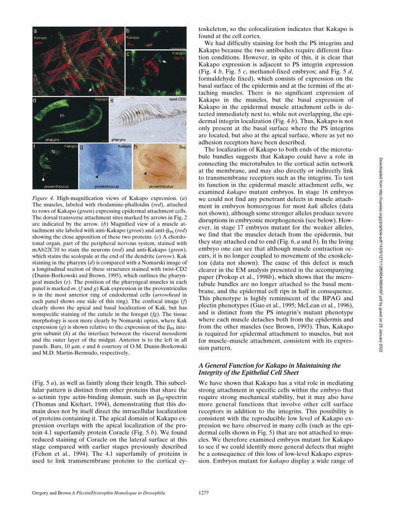
Further analysis of Kakapo expression reveals that theprotein is present at high levels in all of the epidermalmuscle attachment cells (Figs. 2 and 4 a): both those thatdirectly attach to the muscles and those that indirectly at-tach via the tendon matrix (Prokop et al., 1998a). We firstdetected strong Kakapo expression in these cells at mid-late stage 16, which is z4 h after the muscles first start toattach to the epidermis. During the last two stages of em-bryogenesis—16 and 17—attachment of the epidermalcells to the muscles or the tendon matrix is elaborated byexpansion of the hemiadherens junctions, accumulation oftendon matrix, and increased expression of b1 tubulin(Buttgereit et al., 1991; Prokop et al., 1998a). We do notdetect any expression of Kakapo in the muscles (Figs. 2 c,4 a, and 5), even though comparable hemiadherensjunctions, characterized by membrane-proximal electron-dense plaques into which cytoskeletal elements insert, areformed there and the adhesion is also integrin-dependent(Prokop et al., 1998a). This difference could indicate thatKakapo has a function that is incompatible with musclecontraction, such as forming stable anchoring structures.Kakapo is strongly expressed in two internal structures aswell: the pharynx and the proventriculus (Figs. 2 c and 4,d–h). In the pharynx, Kakapo is strongly expressed in theendodermal cell layers that attach to the pharyngeal mus-cles, as seen by comparing Kakapo in Fig. 4 d to a Nomar-ski image of these tissues stained for the muscles at a simi-lar stage (Fig. 4 e). As with the epidermal cells that attachto the somatic muscles (see below), Kakapo is found both
at the basal surface that contacts the mesoderm and at theapical surface, while the PS integrins are localized just atthe basal surface (Leptin et al., 1989). In the proventricu-lus, Kakapo is expressed in a ring of cells at the anteriormargin of the outer layer of this three-layered structure(Fig. 4, f and g). Expression of the PS integrins is foundprimarily at the interface between the outer endodermallayer of the proventriculus and the surrounding visceralmesoderm (Fig. 4 h). Therefore, in this tissue, the integrinsare expressed in more cells than Kakapo. It is not clearwhy strong expression of Kakapo might be needed at thissite, but one phenotype of integrin mutations is that the in-ner layers of the proventriculus become pulled out (Mar-tin-Bermudo et al., 1997), suggesting that some resistanceto mechanical stress is normally necessary to maintain theintegrity of the proventriculus structure. We can also de-tect modest Kakapo expression in the scolopale of thechordotonal organs of the peripheral nervous system (Fig.4 c). A function for the PS integrins in these cells has notbeen observed to date, but, like muscle attachment cells, itis a site of stabilized b1 microtubule based rigidity (Prokopet al., 1998b).
Kakapo Is Located at Both the Apical and BasalSurfaces of the Epidermal Muscle Attachment Cells
In the epidermal muscle-attachment cells, the PS integrinsare localized to the basal surface (Leptin et al., 1989),which contains large hemiadherens junctions (Prokop et al.,1998a). Microtubules extend from these basal junctions tothe apical hemiadherens junctions, which connect to theexoskeleton (cuticle). The microtubules appear to be serv-ing a similar structural role to that of keratin filaments inthe epidermal cells of vertebrates, as intermediate fila-ments have yet to be identified in Drosophila. When weexamined the subcellular localization of Kakapo in moredetail, we found that it is present at both apical and basalsurfaces of the muscle attachment cells (Fig. 5). Kakapocan be seen to be positioned at the termini of the microtu-bule bundles extending from the apical to the basal surface
Figure 3. Specificity of anti-Kakapo antisera. (a)Affinity-purified anti-Kakapo polyclonal antiseradoes not stain kakapo mutant embryos. wild type(left) and kakP2 mutant (right) stage 16 embryoswere double-labeled with anti-Kakapo (green)and mAb19 (red) to confirm that the antibodiespenetrate each embryo. The merged channels areshown in the middle, indicating that Kakapo ex-pression is absent in homozygous mutant em-bryos, while staining of the muscle attachmentcells with mAb19 is still observed. (b) Westernblots using this antiserum detect a high–molecu-lar weight product in wild-type embryo extracts(wt). Both wild-type and truncated proteins areobserved in extracts from embryos heterozygousfor the kakV168 mutation, confirming that kakapomutants are defective for this gene product, andthat the antiserum is specific for Kakapo protein.The position of the 250 and 160-kD marker pro-teins is shown on the right.
Dow
nloaded from http://rupress.org/jcb/article-pdf/143/5/1271/1280954/9804097.pdf by guest on 25 January 2022
Gregory and Brown A Plectin/Dystrophin Homologue in Drosophila 1277
(Fig. 5 a), as well as faintly along their length. This subcel-lular pattern is distinct from other proteins that share thea-actinin type actin-binding domain, such as bH-spectrin(Thomas and Kiehart, 1994), demonstrating that this do-main does not by itself direct the intracellular localizationof proteins containing it. The apical domain of Kakapo ex-pression overlaps with the apical localization of the pro-tein 4.1 superfamily protein Coracle (Fig. 5 b). We foundreduced staining of Coracle on the lateral surface at thisstage compared with earlier stages previously described(Fehon et al., 1994). The 4.1 superfamily of proteins isused to link transmembrane proteins to the cortical cy-
toskeleton, so the colocalization indicates that Kakapo isfound at the cell cortex.
We had difficulty staining for both the PS integrins andKakapo because the two antibodies require different fixa-tion conditions. However, in spite of this, it is clear thatKakapo expression is adjacent to PS integrin expression(Fig. 4 b, Fig. 5 c, methanol-fixed embryos; and Fig. 5 d,formaldehyde fixed), which consists of expression on thebasal surface of the epidermis and at the termini of the at-taching muscles. There is no significant expression ofKakapo in the muscles, but the basal expression ofKakapo in the epidermal muscle attachment cells is de-tected immediately next to, while not overlapping, the epi-dermal integrin localization (Fig. 4 b). Thus, Kakapo is notonly present at the basal surface where the PS integrinsare located, but also at the apical surface, where as yet noadhesion receptors have been described.
The localization of Kakapo to both ends of the microtu-bule bundles suggests that Kakapo could have a role inconnecting the microtubules to the cortical actin networkat the membrane, and may also directly or indirectly linkto transmembrane receptors such as the integrins. To testits function in the epidermal muscle attachment cells, weexamined kakapo mutant embryos. In stage 16 embryoswe could not find any penetrant defects in muscle attach-ment in embryos homozygous for most kak alleles (datanot shown), although some stronger alleles produce severedisruptions in embryonic morphogenesis (see below). How-ever, in stage 17 embryos mutant for the weaker alleles,we find that the muscles detach from the epidermis, butthey stay attached end to end (Fig. 6, a and b). In the livingembryo one can see that although muscle contraction oc-curs, it is no longer coupled to movement of the exoskele-ton (data not shown). The cause of this defect is muchclearer in the EM analysis presented in the accompanyingpaper (Prokop et al., 1998b), which shows that the micro-tubule bundles are no longer attached to the basal mem-brane, and the epidermal cell rips in half in consequence.This phenotype is highly reminiscent of the BPAG andplectin phenotypes (Guo et al., 1995; McLean et al., 1996),and is distinct from the PS integrin’s mutant phenotypewhere each muscle detaches both from the epidermis andfrom the other muscles (see Brown, 1993). Thus, Kakapois required for epidermal attachment to muscles, but notfor muscle–muscle attachment, consistent with its expres-sion pattern.
A General Function for Kakapo in Maintaining the Integrity of the Epithelial Cell Sheet
We have shown that Kakapo has a vital role in mediatingstrong attachment in specific cells within the embryo thatrequire strong mechanical stability, but it may also havemore general functions that involve other cell surfacereceptors in addition to the integrins. This possibility isconsistent with the reproducible low level of Kakapo ex-pression we have observed in many cells (such as the epi-dermal cells shown in Fig. 5) that are not attached to mus-cles. We therefore examined embryos mutant for Kakapoto see if we could identify more general defects that mightbe a consequence of this loss of low-level Kakapo expres-sion. Embryos mutant for kakapo display a wide range of
Figure 4. High-magnification views of Kakapo expression. (a)The muscles, labeled with rhodamine-phalloidin (red), attachedto rows of Kakapo (green) expressing epidermal attachment cells.The dorsal transverse attachment sites marked by arrows in Fig. 2are indicated by the arrow. (b) Magnified view of a muscle at-tachment site labeled with anti-Kakapo (green) and anti-bPS (red)showing the close apposition of these two proteins. (c) A chordo-tonal organ, part of the peripheral nervous system, stained withmAb22C10 to stain the neurons (red) and anti-Kakapo (green),which stains the scolopale at the end of the dendrite (arrow). Kakstaining in the pharynx (d) is compared with a Nomarski image ofa longitudinal section of these structures stained with twist-CD2(Dunin-Borkowski and Brown, 1995), which outlines the pharyn-geal muscles (e). The position of the pharyngeal muscles in eachpanel is marked m. (f and g) Kak expression in the proventriculusis in the most anterior ring of endodermal cells (arrowhead ineach panel shows one side of this ring). The confocal image (f)clearly shows the apical and basal localization of Kak, but hasnonspecific staining of the cuticle in the foregut (fg). The tissuemorphology is seen more clearly by Nomarski optics, where Kakexpression (g) is shown relative to the expression of the bPS inte-grin subunit (h) at the interface between the visceral mesodermand the outer layer of the midgut. Anterior is to the left in allpanels. Bars, 10 mm. e and h courtesy of O.M. Dunin-Borkowskiand M.D. Martin-Bermudo, respectively.
Dow
nloaded from http://rupress.org/jcb/article-pdf/143/5/1271/1280954/9804097.pdf by guest on 25 January 2022

The Journal of Cell Biology, Volume 143, 1998 1278
phenotypes, from almost normal development to severemorphological abnormalities. This range of phenotypes isalso found in embryos deficient for the locus Df(2R)MK1/Df(2R)MK1 or Df(2R)CX1/Df(2R)CX1 (data not shown),demonstrating that even the complete absence of thekakapo gene does not result in a consistent zygotic pheno-type. The variability of the phenotype may be due to a par-tial redundancy of function between Kakapo and anotherprotein, or variable contribution of maternal protein. Wefavor the former possibility, since generating germ line
clones of one of the kakapo alleles did not enhance thephenotype (Walsh and Brown, 1998), and we have notbeen able to detect Kakapo protein before gastrulation(data not shown).
Consistent with the variability found in embryos defi-cient for kakapo, we find that our kakapo alleles also dis-play diverse phenotypes. Most of the alleles isolated in ourscreen are embryonic lethal as homozygotes, although thetwo alleles examined previously develop normally throughstage 16 (Walsh and Brown, 1998). As shown above, at the
Figure 6. Mutations in Kakapo cause muscle de-tachment and widespread defects in the epider-mis. The first two panels show flat stage 17 em-bryos with the birefringent muscles and cuticlevisualized by polarizing optics: (a) wild-type em-bryo with two arrows marking the close attach-ment of the muscles to the epidermis. (b) InkakV104/kakV104 embryos, the muscles pull awayfrom the epidermis, but remain attached to eachother (positions on the epidermis equivalent tothose marked in a are marked by arrows). (c andd) Cuticle preparations, that reveal the underly-ing pattern of the epidermis: (c) wild-type cuticle;(d) strongest cuticle phenotype of kakP1/kakP1.(e–h) Late-stage 16 kakP2/kakP2 embryos stainedfor the 4.1 homologue Coracle (e and f; the sameembryo magnified) and membrane protein fasci-clin III (g and h; another example magnified),showing ruptures in the epidermis. Bar, 10 mm.
Figure 5. Kakapo is local-ized at the apical and basalsurfaces of the epidermal mus-cle attachment cells. Eachpanel shows a horizontal sec-tion of a late stage 16 em-bryo, showing two muscle at-tachment cells at the segmentborder as shown in the sche-matic drawing (e). Eachpanel is stained for Kakapoin green and a second antigenin red: (a) tubulin; (b) Cora-cle, a band 4.1 superfamilymember; and (c and d) a bPSintegrin subunit on embryosfixed either in methanol (c)or formaldehyde (d). Themerged images are shown atthe left. Bar, 10 mm.
Dow
nloaded from http://rupress.org/jcb/article-pdf/143/5/1271/1280954/9804097.pdf by guest on 25 January 2022

Gregory and Brown A Plectin/Dystrophin Homologue in Drosophila 1279
end of embryogenesis (stage 17), the mutant embryos havea phenotype where the epidermis detaches from the mus-cles (Fig. 6 b). By examining the phenotype of the differ-ent alleles, we found that some have much stronger pheno-types, indicative of a more general function. We examinedthe epidermally secreted cuticle, which reflects the patternof the underlying epidermis, of embryos homozygous forall our different x-ray–induced kakapo alleles and the in-sertion alleles kakP1 and kakP2. In the majority of the al-leles (17), the homozygous mutant embryos have a normalepidermal pattern, although z30% of these embryosshowed modest cuticular defects (not shown). It should bementioned that the screen for wing blister mutations mayhave selected for a particular type of weak allele if moresevere alleles cause drastic wing defects. One x-ray allele,kakV168, and the P-alleles kakP1 and kakP2, have a strongerphenotype: in z15% of the mutant embryos, germ bandretraction and head involution fail (Fig. 6, c and d). Ap-proximately 40% of embryos mutant for these alleles havenormal-looking cuticles, demonstrating that this pheno-type is also not fully penetrant.
Epidermal development was examined in more detail inthe embryos homozygous for strong kakapo alleles usingtwo markers for epidermal shape (Fig. 6, e–h). This exami-nation revealed defects in the integrity of the epidermalcell layer; namely, breaks in the ventral epidermis in themiddle abdominal segments of the fully germband-extendedembryos (Fig. 6 e). These breaks appear to arise at the siteof maximum strain during germ band retraction. Consis-
tent with this observation, germband retraction is arrestedin some embryos (Fig. 6, d and e). Other embryos success-fully undergo germband retraction, but retain a hole in theepidermis at this position of maximum strain (see Fig. 6, gand h), which could account for the disruptions observedin the cuticular denticle belts. The homophilic adhesionmolecule Fasiclin III is not present on the surface of thecells bordering the hole (Fig. 6 h). Such breaks in the epi-dermis are not observed in embryos mutant for the PS in-tegrins, suggesting that this Kakapo function involvesother cell adhesion molecules. We also observed morpho-genetic defects in the internal tissues (data not shown), butbecause they occur in embryos with severe epidermal de-fects, it is not yet certain whether this indicates a functionfor Kakapo in these tissues, or whether the internal defectsare a result of the epidermal disruption. The phenotypes inthe embryonic epidermis indicate that the low-level gen-eral expression of Kakapo is significant, and that Kakapomay play a general role in mediating adhesion, possibly bymediating interactions between transmembrane proteinsand the cytoskeleton.
DiscussionIdentification of intracellular proteins that are requiredfor integrin functioning is essential for an understanding ofhow integrins mediate adhesion. In this paper we have de-scribed the cloning and characterization of kakapo, a genethat was identified in screens for wing blister mutants(Prout et al., 1997; Walsh and Brown, 1998), and we showthat it encodes a cytoskeletal adaptor protein related toplectin, BPAG1, and dystrophin. We have demonstratedthat Kakapo is expressed in those epithelial cells wherestable adhesion is required in the developing embryo; it isrequired to maintain epidermal adhesion to the muscles,and more generally to maintain cohesion of the epidermalcell layer. From the our characterization of the sequence,pattern of expression, and phenotype of kakapo muta-tions, it appears that Kakapo has a similar function to plec-tin, and we propose a model in which Kakapo provideslinks among cortical actin, microtubules, and transmem-brane proteins such as integrins (Fig. 7).
The binding of Kakapo to actin is indicated by the pres-ence of a highly conserved actin-binding domain at theamino terminus of Kakapo. This domain is most similar tothe equivalent domain in plectin and BPAG1, comparedwith the domains found in dystrophins, b-spectrins, anda-actinins. This higher sequence conservation may indi-cate some functional diversity within this domain that isconserved in each subfamily, or it may simply representthe evolutionary history of conservative amino acid re-placements. What is clear is that this actin-binding domaindoes not dictate the intracellular localization of the pro-teins containing it. For example, the apical and basal local-ization of Kakapo is distinct from the general cortical lo-calization of both spectrin and the novel bH-spectrin ofDrosophila (Pesacreta et al., 1989; Martinez-Arias, 1993;Thomas and Kiehart, 1994).
Kakapo is closely related to plectin and BPAG1, twovertebrate proteins that are required for the link betweenthe integrin a6b4 and intermediate filaments at hemides-mosomes (Ruhrberg and Watt, 1997). However, this simi-
Figure 7. Model for the role of Kakapo in muscle/epidermal ad-hesion. A schematic of a muscle attachment site is shown with themuscles linked to each other and the epidermis by integrin–extra-cellular matrix linkage. At the basal surface of the epidermalcell, Kakapo (green) links the cortical actin cytoskeleton (red)and the microtubule bundles (blue) to integrins via an adaptorprotein (grey). At the apical surface, Kakapo links the cytoskele-ton to a currently unidentified receptor (purple).
Dow
nloaded from http://rupress.org/jcb/article-pdf/143/5/1271/1280954/9804097.pdf by guest on 25 January 2022

The Journal of Cell Biology, Volume 143, 1998 1280
larity does not extend through to the carboxy terminus ofthese proteins, which contain an intermediate filament-binding domain shared with the desmosome componentdesmoplakin. As Kakapo does not contain an intermedi-ate filament-binding domain, and so far no intermediatefilaments have been identified in Drosophila, it is unlikelyto have an identical function to plectin and BPAG1 andbind intermediate filaments. In Drosophila, stabilized mi-crotubule arrays appear to be used in place of intermedi-ate filaments to hold the cell rigid (Mogensen and Tucker,1988). This observation is particularly apparent in theadult wing, where transalar parallel arrays of microtubulesand microfilaments connect the apical cuticle with the in-tegrin-containing basal junctions (Mogensen and Tucker,1988; Fristrom et al., 1993), and in the larval epidermalcells that attach to the muscles (Prokop et al., 1998a). Thefact that Kakapo function is required for cell adhesion inboth sets of epithelial cells where stabilized microtubulesare found strongly suggests that Kakapo binds to these mi-crotubules rather than intermediate filaments. In contrastto a number of well-conserved actin-binding domains,many microtubule-binding proteins lack a shared microtu-bule-binding motif so that the lack of similarity betweenkakapo and a known microtubule-binding protein doesnot contradict our proposal that Kakapo binds to microtu-bules. It is also possible that Kakapo binds indirectly tomicrotubules though an interaction with a microtubule-associated protein.
In hemidesmosomes there appears to be a direct molec-ular interaction between integrins and plectin, since siteson the b4 cytoplasmic tail have been identified that di-rectly bind to plectin (Niessen et al., 1997). As the PS inte-grins have a more standard length of b subunit cytoplas-mic tail (47 amino acids vs. the 1019 amino acids of b4), anintervening linker protein may be required (indicated bythe grey sphere in Fig. 7) to connect the PS integrins toKakapo. We might expect this adaptor to be encoded byone of the other loci identified in the screen, such as rhea,which has a similar epidermal detachment phenotype(Prout et al., 1997). If this adaptor protein exists, it will beof interest to see if it is similar in sequence to the cytoplas-mic tail of b4.
Kakapo is not only similar in sequence to plectin andBPAG1, it is also similar to dystrophin. However, the pat-tern of expression of Kakapo is more similar to the expres-sion of BPAG1 than dystrophin, while plectin is expressedalmost ubiquitously. Like BPAG1, Kakapo is strongly ex-pressed in epidermal cells, and neither are expressed in themuscles, where dystrophin is strongly expressed. Further-more, we have shown the subcellular localization ofKakapo to be primarily at the site of the prominent hemi-adherens junctions that are present both apically and ba-sally in the epidermal muscle attachment site, as well assome decoration of the intervening cytoskeleton. Basalhemiadherens junctions consist of extensive plaques ofelectron-dense material where actin and microtubulesconnect to the extracellular matrix, while at the smallerapical junctions, microtubules are attached to the overly-ing cuticle (Prokop et al., 1998a). This subcellular localiza-tion resembles that seen for BPAG1 and plectin, whichlocalize to the inner plaque of hemidesmosomes and deco-rate intermediate filaments (Wiche et al., 1984; Guo et al.,
1995; Svitkina et al., 1996), and is quite distinct from thegeneral cortical staining of dystrophin in the muscles(Brown and Lucy, 1997). The staining we have observed isnot completely identical to that seen with an antibodyraised against the carboxy terminus of Kakapo, whichshows staining earlier in embryogenesis than our antibodyto the amino terminus, and looks more cortical (Strumpfand Volk, 1998). The Kakapo gene extends over 70 kb,and there may be additional isoforms of Kakapo yet to beidentified. This would be similar to the plectin and BPAGgenes that produce alternate isoforms, some of which lackthe actin-binding domain or rod sections (Yang et al.,1996; Elliott et al., 1997).
Finally, the phenotype of Kakapo is more similar toBPAG1 and plectin than to dystrophin. Mutations inBPAG1 or plectin cause skin blistering due to rupture ofepidermal cells and neuromuscular defects (Guo et al.,1995; Smith et al., 1996). The kakapo gene was identifiedby screening for a related phenotype in Drosophila; mu-tant cells cause blisters in the adult wing (Prout et al., 1997;Walsh and Brown, 1998). In the embryo, kakapo muta-tions cause detachment of the epidermis from the musclessimilar to skin blisters, and the ultrastructural phenotypeis remarkably similar, where mechanical stress leads tobreaking of the cells into apical and basal halves (Guo et al.,1995; Prokop et al., 1998b). In contrast, the muscles appearto develop normally, and have normal sarcomeric struc-ture (Fig. 6 and our unpublished results), in contrast to themuscular degeneration that occurs in the absence of dys-trophin (Brown and Lucy, 1997). The striking similarity ofsequence, expression pattern, and mutant phenotype pro-vide consistent evidence for the functional relationship ofKak with vertebrate hemidesmosomal proteins. Conse-quently, we propose that Kak plays a homologous role tothe vertebrate plakin family in the Drosophila hemi-adhe-rens junction, acting as an essential adaptor that distrib-utes the extension stress generated by muscle contractionfrom membrane-bound receptors into the cortical actinand stabilized microtubule arrays.
The identification of a new cytoskeletal linker protein asthe product of a gene required for integrin-mediated adhe-sion events strengthens the view that integrins do play animportant structural role in mediating adhesion. Our cur-rent model for the interaction between Kakapo and the PSintegrins is that they are connected indirectly through aprotein that serves a similar function to the extended cyto-plasmic domain of the b4 integrin subunit (as shown in Fig.7). However, this result is clearly an incomplete descrip-tion, because, unlike Kakapo, PS integrins are not essen-tial for the linkage between the plasma membrane andmicrotubules in the epidermal muscle attachment cells(Prokop et al., 1998a). The fact that Kakapo is required forthis microtubule–membrane linkage suggests that Kakapointeracts, directly or indirectly, with more transmembraneproteins than just the known integrins. This is also indi-cated by Kakapo localization at the apical surface, whichlacks the PS integrins. Whether these additional adhesionreceptors include novel integrins or alternative classes ofreceptor is an open question, but one that may be resolvedby cloning more of the loci identified in recent geneticscreens for adhesion defects (Prout et al., 1997; Walsh andBrown, 1998).
Dow
nloaded from http://rupress.org/jcb/article-pdf/143/5/1271/1280954/9804097.pdf by guest on 25 January 2022

Gregory and Brown A Plectin/Dystrophin Homologue in Drosophila 1281
The early phenotype we have documented where the in-tegrity of the epidermal cell layer is impaired also indi-cates the diversity of Kakapo function, because this is nota phenotype found in PS integrin mutants. In addition, theobservation that the differentiation of the epidermal mus-cle attachment cells is perturbed in kakapo mutant em-bryos (Strumpf and Volk, 1998) suggests that Kakapo maybe required to allow signaling events leading to cell differ-entiation. We do not think that the change in differentia-tion of the epidermal muscle attachment cells is sufficientto account for the epidermal detachment phenotype ofKakapo mutants, since although b1 tubulin expression isreduced (Strumpf and Volk, 1998), microtubule bundlesare still present in those cells, but are detached from theplasma membrane (Prokop et al., 1998b), indicating thatKakapo is essential for the link between the two. How-ever, the role of Kakapo in epidermal differentiation com-bined with its role in determining the subcellular localiza-tion of transmembrane adhesion proteins in neurons(Prokop et al., 1998b) suggests that one important func-tion of Kakapo is to organize receptors within the plasmamembrane. Thus, the Kakapo protein, which from its se-quence homology and phenotype seems to be an impor-tant component of the cytoskeleton, may potentially affectsignaling through the role of the cytoskeleton in organiz-ing membrane-bound receptors into functional signalingcomplexes.
The kakapo gene is the first of the novel genes to becloned from the screens for mutations required for inte-grin-mediated adhesion. The fact that kakapo encodes acytoskeletal linker protein related to proteins that are alsoimplicated in integrin-mediated adhesion demonstratesthe success of the genetic screening approach. Therefore,the characterization of the remaining 16 loci isolated fromthese screens should substantially contribute to our under-standing of the cellular machinery required for integrinadhesion.
We are grateful to T. Volk, D. Strumpf, and A. Prokop for sharing unpub-lished data, to O. Dunin-Borkowski and M.D. Martín-Bermudo for thestained embryo preparations shown in Fig. 4, e and h, and to R. Fehon, D.Kiehart, A. Beaton, and T. Volk for fly strains and antibodies. We alsothank J. Overton for technical assistance and M.D. Martín-Bermudo andS. Bray for helpful comments on the manuscript.
This work was supported by grants from the Wellcome Trust to N.H.Brown: a Senior Fellowship and Project Grant 050301.
Received for publication 24 April 1998 and in revised form 10 September1998.
References
Andra, K., H. Lassmann, R. Bittner, S. Shorny, R. Fassler, F. Propst, and G.Wiche. 1997. Targeted inactivation of plectin reveals essential function inmaintaining the integrity of skin, muscle, and heart cytoarchitecture. GenesDev. 11:3143–3156.
Bernier, G., M. Mathieu, Y. Derepentigny, S.M. Vidal, and R. Kothary. 1996.Cloning and characterization of mouse ACF7, a novel member of the Dysto-nin subfamily of actin binding proteins. Genomics. 38:19–29.
Brancolini, C., M. Benedetti, and C. Schneider. 1995. Microfilament reorgani-zation during apoptosis: the role Of Gas2, a possible substrate for Ice likeproteases. EMBO (Eur. Mol. Biol. Organ.) J. 14:5179–5190.
Brancolini, C., S. Bottega, and C. Schneider. 1992. Gas2, a growth arrest spe-cific protein, is a component of the microfilament network system. J. CellBiol. 117:1251–1261.
Brower, D.L., and S.M. Jaffe. 1989. Requirement for integrins during Dro-sophila wing development. Nature. 342:285–287.
Brower, D.L., R.J. Smith, and M. Wilcox. 1980. A monoclonal antibody specificfor diploid epithelial cells in Drosophila. Nature. 285:403–405.
Brower, D.L., M. Wilcox, M. Piovant, R.J. Smith, and L.A. Reger. 1984. Re-lated cell-surface antigens expressed with positional specificity in Drosophilaimaginal discs. Proc. Natl. Acad. Sci. USA. 81:7485–7489.
Brown, N.H. 1993. Integrins hold Drosophila together. BioEssays. 15:383–390.Brown, N.H., and F.C. Kafatos. 1988. Functional cDNA libraries from Dro-
sophila embryos. J. Mol. Biol. 203:425–437.Brown, N.H., D.L. King, M. Wilcox, and F.C. Kafatos. 1989. Developmentally
regulated alternative splicing of Drosophila integrin PS2 a transcripts. Cell.59:185–195.
Brown, S.C., and J.A. Lucy. 1997. Dystrophin: gene, protein, and cell biology.Cambridge University Press, Cambridge, UK. 338 pp.
Burridge, K., K. Fath, T. Kelly, G. Nuckolls, and C. Turner. 1988. Focal adhe-sions: transmembrane junctions between the extracellular matrix and the cy-toskeleton. Annu. Rev. Cell Biol. 4:487–525.
Burridge, K., C.E. Turner, and L.H. Romer. 1992. Tyrosine phosphorylation ofpaxillin and pp125(Fak) accompanies cell adhesion to extracellular matrix: arole in cytoskeletal assembly. J. Cell Biol. 119:893–903.
Buttgereit, D., D. Leiss, F. Michiels, and R. Renkawitz-Pohl. 1991. DuringDrosophila embryogenesis the b1 tubulin gene is specifically expressed inthe nervous system and the apodemes. Mech. Dev. 33:107–118.
Craig, S.W. 1996. Assembly of focal adhesions: progress, paradigms, and por-tents. Curr. Opin. Cell. Biol. 8:74–85.
Dowling, J., Y.M. Yang, R. Wollmann, L.F. Reichardt, and E. Fuchs. 1997. De-velopmental expression of BPAG1n: Insights into the spastic ataxia andgross neurologic degeneration in dystonia musculorum mice. Dev. Biol. 187:131–142.
Dubreuil, R.R. 1991. Structure and evolution of the actin cross linking proteins.BioEssays. 13:219–226.
Dunin-Borkowski, O.M., and N.H. Brown. 1995. Mammalian CD2 is an effec-tive heterologous marker of the cell surface in Drosophila. Dev. Biol. 168:689–693.
Elliott, C.E., B. Becker, S. Oehler, M.J. Castanon, R. Hauptmann, and G.Wiche. 1997. Plectin transcript diversity: Identification and tissue distribu-tion of variants with distinct first coding exons and rodless isoforms. Genom-ics. 42:115–125.
Fehon, R.G., I.A. Dawson, and S. Artavanis-Tsakonas. 1994. A Drosophila ho-mologue of membrane skeleton protein 4.1 is associated with septate junc-tions and is encoded by the coracle gene. Development. 120:545–557.
Fristrom, D., M. Wilcox, and J. Fristrom. 1993. The distribution of PS integrins,Laminin A and F-actin during key stages in Drosophila wing development.Development. 117:509–523.
Fujita, S.C., S.L. Zipursky, S. Benzer, A. Ferrus, and S.L. Shotwell. 1982. Mono-clonal antibodies against the Drosophila nervous system. Proc. Natl. Acad.Sci. USA. 79:7929–7933.
Golic, K.G. 1991. Site-specific recombination between homologous chromo-somes in Drosophila. Science. 252:958–961.
Guo, L.F., L. Degenstein, J. Dowling, Q.C. Yu, R. Wollmann, B. Perman, andE. Fuchs. 1995. Gene targeting of BPAG1 abnormalities in mechanicalstrength and cell migration in stratified epithelia and neurologic degenera-tion. Cell. 81:233–243.
Hannigan, G.E., C. Leung-Hagesteijn, L. Fitz-Gibbon, M.G. Coppolino, G.Radeva, J. Filmus, J.C. Bell, and S. Dedhar. 1996. Regulation of cell adhe-sion and anchorage-dependent growth by a new b1-integrin-linked proteinkinase. Nature. 379:91–96.
Horwitz, A., K. Duggan, C. Buck, M.C. Beckerle, and K. Burridge. 1986. Inter-action of plasma membrane fibronectin receptor with talin: a transmem-brane linkage. Nature. 320:531–533.
Hynes, R.O. 1987. Integrins: A family of cell surface receptors. Cell. 48:549–554.Koenig, M., and L.M. Kunkel. 1990. Detailed analysis of the repeat domain of
dystrophin reveals 4 potential hinge segments that may confer flexibility. J.Biol. Chem. 265:4560–4566.
Koenig, M., A.P. Monaco, and L.M. Kunkel. 1988. The complete sequence ofdystrophin predicts a rod shaped cytoskeletal protein. Cell. 53:219–228.
Kolanus, W., W. Nagel, B. Sciller, L. Zeitlmann, S. Godar, H. Stockinger, andB. Seed. 1996. aLb2 integrin/LFA-1 binding to ICAM 1 induced by cytohesin1, a cytoplasmic regulatory molecule. Cell. 86:233–242.
Leptin, M., T. Bogaert, R. Lehmann, and M. Wilcox. 1989. The function of PSintegrins during Drosophila embryogenesis. Cell. 56:401–408.
Martin-Bermudo, M.D., O.M. Dunin-Borkowski, and N.H. Brown. 1997. Speci-ficity of PS integrin function during embryogenesis resides in the a subunitextracellular domain. EMBO (Eur. Mol. Biol. Organ.) J. 16:4184–4193.
Martinez-Arias, A. 1993. Development and patterning of the larval epidermisof Drosophila. In The development of Drosophila melanogaster. M. Bate,and A. Martinez-Arias, editors. Cold Spring Harbor Laboratory Press, Plain-view, NY. 517–608.
McLean, W.H.I., L. Pulkkinen, F.J.D. Smith, E.L. Rugg, E.B. Lane, F. Bullrich,R.E. Burgeson, S. Amano, D.L. Hudson, K. Owaribe, et al. 1996. Loss ofplectin causes Epidermolysis Bullosa with muscular dystrophy cDNA clon-ing and genomic organization. Genes Dev. 10:1724–1735.
Miyamoto, S., S.K. Akiyama, and K.M. Yamada. 1995a. Synergistic roles for re-ceptor occupancy and aggregation in integrin transmembrane function. Sci-ence. 267:883–885.
Miyamoto, S., H. Teramoto, O.A. Coso, J.S. Gutkind, P.D. Burbelo, S.K. Aki-yama, and K.M. Yamada. 1995b. Integrin function: molecular hierarchies ofcytoskeletal and signaling molecules. J. Cell Biol. 131:791–805.
Dow
nloaded from http://rupress.org/jcb/article-pdf/143/5/1271/1280954/9804097.pdf by guest on 25 January 2022

The Journal of Cell Biology, Volume 143, 1998 1282
Mogensen, M.M., and J.B. Tucker. 1988. Intermicrotubular actin filaments inthe transalar cytoskeletal arrays of Drosophila. J. Cell Sci. 91:431–538.
Niessen, C.M., E.H.M. Hulsman, E.S. Rots, P. Sanchez-Aparicio, and A. Son-nenberg. 1997. Integrin a6b4 forms a complex with the cytoskeletal proteinHD1 and induces its redistribution in transfected COS 7 cells. Mol. Biol.Cell. 8:555–566.
Otey, C.A., F.M. Pavalko, and K. Burridge. 1990. An interaction between a-acti-nin and the b1 integrin subunit in vitro. J. Cell Biol. 111:721–729.
Pesacreta, T.C., T.J. Byers, R. Dubreuil, D.P. Kiehart, and D. Branton. 1989a.Drosophila spectrin: the membrane skeleton during embryogenesis. J. CellBiol. 108:1697–1709.
Prokop, A., M.D. Martin-Bermudo, M. Bate, and N.H. Brown. 1998a. In Dro-sophila embryos, the absence of the PS integrins or laminin A affects the ex-tracellular adhesion of hemiadherens and neuromuscular junctions, but nottheir intracellular assembly. Dev. Biol. 196:58–76.
Prokop, A., J. Uhler, J. Roote, and M. Bates. 1998b. The kakapo mutation af-fects terminal arborisation and central dendritic sprouting of Drosophilamotor neurons. J. Cell Biol. 143:1283–1294.
Prout, M., Z. Damania, J. Soong, D. Fristrom, and J.W. Fristrom. 1997. Autoso-mal mutations affecting adhesion between wing surfaces in Drosophila mela-nogaster. Genetics. 146:275–285.
Ruhrberg, C., and F.M. Watt. 1997. The plakin family: versatile organizers ofcytoskeletal architecture. Curr. Opin. Genet. Dev. 7:392–397.
Shattil, S.J., T.O. O’Toole, M. Eigenthaler, V. Thon, M. Williams, B.M. Babior,and M.H. Ginsberg. 1995. b3-endonexin, a novel polypeptide that interactsspecifically with the cytoplasmic tail of the integrin b3 subunit. J. Cell Biol.131:807–816.
Smith, F.J.D., R.A.J. Eady, I.M. Leigh, J.R. McMillan, E.L. Rugg, D.P. Kelsell,S.P. Bryant, N.K. Spurr, J.F. Geddes, G. Kirtschig, et al. 1996. Plectin defi-ciency results in muscular dystrophy with epidermolysis bullosa. Nat. Genet.13:450–457.
Strumpf, D., and T. Volk. 1998. Groovin, a novel Drosophila protein, is essen-tial for the restricted localization of the neuregulin-like factor, vein, at the
muscle–tendon junctional site. J. Cell Biol. 143:1259–1270.Svitkina, T.M., A.B. Verkhovsky, and G.G. Borisy. 1996. Plectin sidearms me-
diate interaction of intermediate filaments with microtubules and other com-ponents of the cytoskeleton. J. Cell Biol. 135:991–1007.
Tang, H.Y., A.F. Chaffotte, and S.M. Thacher. 1996. Structural analysis of thepredicted coiled coil rod domain of the cytoplasmic Bullous Pemphigoid An-tigen (BPAG1) empirical localization of the N-terminal globular domain rodboundary. J. Biol. Chem. 271:9716–9722.
Thomas, G.H., and D.P. Kiehart. 1994. bH Spectrin has a restricted tissue andsubcellular distribution during Drosophila embryogenesis. Development.120:2039–2050.
Uitto, J., and L. Pulkkinen. 1996. Molecular complexity of the cutaneous base-ment membrane zone. Mol. Biol. Rep. 23:35–46.
Volk, T., and K. VijayRaghavan. 1994. A central role for epidermal segmentborder cells in the induction of muscle patterning in the Drosophila embryo.Development. 120:59–70.
Walsh, E.P., and N.H. Brown. 1998. A screen to identify Drosophila genes re-quired for integrin mediated adhesion. Genetics. 150:791–805.
Wiche, G., B. Becker, K. Luber, G. Weitzer, M.J. Castanon, R. Hauptmann, C.Stratowa, and M. Stewart. 1991. Cloning and sequencing of rat plectin indi-cates a 466 kD polypeptide chain with a 3 domain structure based on a cen-tral alpha helical coiled coil. J. Cell Biol. 114:83–99.
Wiche, G., R. Krepler, U. Artlieb, R. Pytela, and W. Aberer. 1984. Identifica-tion of plectin in different human cell types and immunolocalization at epi-thelial basal cell surface membranes. Exp. Cell Res. 155:43–49.
Wieschaus, E., and C. Nüsslein-Volhard. 1986. Looking at embryos. In Dro-sophila: A Practical Approach. D.B. Roberts, editor. IRL Press, Oxford,UK. 199–286.
Xu, T., and G.M. Rubin, 1993. Analysis of genetic mosaics in developing andadult Drosophila tissues. Development. 117:1223–1237.
Yang, Y.M., J. Dowling, Q.C. Yu, P. Kouklis, D.W. Cleveland, and E. Fuchs.1996. An essential cytoskeletal linker protein connecting actin microfila-ments to intermediate filaments. Cell. 86:655–665.
Dow
nloaded from http://rupress.org/jcb/article-pdf/143/5/1271/1280954/9804097.pdf by guest on 25 January 2022