kamal, dr bushra (2015) analysis of skeleton in a …theses.gla.ac.uk/6092/1/2015kamal...
TRANSCRIPT

Glasgow Theses Service http://theses.gla.ac.uk/
Kamal, Dr Bushra (2015) Analysis of skeleton in a mouse model of Rett syndrome. PhD thesis. http://theses.gla.ac.uk/6092/ Copyright and moral rights for this thesis are retained by the author A copy can be downloaded for personal non-commercial research or study, without prior permission or charge This thesis cannot be reproduced or quoted extensively from without first obtaining permission in writing from the Author The content must not be changed in any way or sold commercially in any format or medium without the formal permission of the Author When referring to this work, full bibliographic details including the author, title, awarding institution and date of the thesis must be given

Analysis of skeleton in a mouse model of Rett syndrome
Dr Bushra Kamal MBBS
Thesis submitted in fulfilment of the requirement for the degree of Doctor of Philosophy Institute of Neuroscience and Psychology College of Medical, Veterinary and Life Science University of Glasgow Glasgow, G12 8QQ UK
© Bushra Kamal 2015

2
Acknowledgement
“In the name of Allah Almighty, the most merciful, the most beneficent” Man is a wonderful creature; he sees through the layers of fat (eyes), hears
through a bone (ears) and speaks through a lump of flesh (tongue) - Ali ibn
Abi Talib
I owe my deepest gratitude to my supervisors Dr Stuart Cobb and Prof. Anthony
Payne for their continuous guidance, advice and constructive criticism. I am also
extremely grateful to Prof. Elizabeth Tanner for the exciting learning opportunities,
she has provided over the period of 3 years.
I am very thankful to David Russell, Dr Robert Wallace for all their technical
assistance and fruitful collaboration.
I have been fortunate to know and work with fantastic people over the past few
years. Special thanks go to Dr Mark Bailey, Dr John Shaw-Dunn, Prof William
Cushley, Prof David Maxwell, Dr Stuart Macdonald, Dr Katherine price, Dr Paul
Rea, Paul Ross, for their kind help and support during the last four years.
I am indebted to Dr Kamal Gadalla, Lesley Maclnnes, Elaine Wales, Henry Gu,
Thishnapha Vudhironarit, Daniela Minchella, Eva Laura Bogaerts and Sophie Hall
for their constant help and personal support during the writing period of this thesis.
My deepest respect, great love and gratitude to my mother Shaeen Begum, the
most loving, kind, patient and intelligent woman I will ever know. This thesis is
dedicated to her. Finally, special thanks and great love goes to my lovely sister Dr
Sabin Kamal and dearest, sweet brother Mr Mustafa Kamal without their constant
love, prayers and support this whole journey of PhD would have not been
possible.

3
Abstract
Rett Syndrome (RTT) is an X-linked genetic disorder and a major cause of
intellectual disability in girls. Mutations in the methyl-CpG binding protein 2
(MECP2) gene, are the primary cause of the disorder. Despite the dominant
neurological phenotypes that characterise RTT, MECP2 is expressed ubiquitously
throughout the body and a number of peripheral phenotypes such as growth
retardation (reduced height and weight), skeletal deformities (scoliosis/kyphosis),
reduced bone mass and low energy fractures are also common yet under-reported
clinical features of the disorder.
In order to explore whether MeCP2 protein deficiency results in altered structural
and functional properties of bone and to test the potential reversibility of any such
defects, I have conducted series of histological, imaging and biomechanical tests
of bone using an accurate genetic (functional knockout) mouse model of RTT.
Initial experiments using a GFP reporter mouse line demonstrated the presence of
MeCP2 in bone cells and the effective silencing on the gene in functional knockout
mice. Different aspects of the study were conducted in different types of bone
tissues that were especially suited for individual assays. For instance,
biomechanical three point bending tests were conducted in long bone (femur)
whilst trabecular geometry measures were measured in spinal vertebrae.
Both hemizygous Mecp2stop/y male mice in which Mecp2 is silenced in all cells and
female Mecp2stop/+ mice in which Mecp2 is silenced in ~50% of cells as a
consequence of random X-chromosome inactivation (XCI), revealed, lighter and
smaller long bones and significant reductions in cortical bone mechanical
properties (~ 39.5% reduction in stiffness, 31% reduction in ultimate load and 37%
reduction in Young’s modulus respectively in Mecp2stop/y male mice; %) and
material properties (microhardess reduced 12.3% in Mecp2stop/y male mice and
14% inMecp2stop/+ female mice) as compared to age wild type control mice. Micro
structural analysis conducted using µCT also revealed a significant reduction in
cortical (54% reduction in cortical thickness, 30% in bone volume, 20% in total
area, and 38% in marrow area) and trabecular (~30% in trabecular thickness)
bone parameters as compared to age matched wild-type controls MeCP2-deficent
mice. Histological analysis using Sirius red staining as a marker of collagen

4
revealed a ~25% reduction in collagen content in MeCP2 deficient mice as
compared to age matched wild type controls.
In experiments designed to establish the potential for reversal of MeCP2-related
deficits, unsilencing of Mecp2 in adult mice by tamoxifen-induced and cre-
mediated excision of a stop cassette located at the endogenous Mecp2 locus
(male; Mecp2stop/y, CreER and female; Mecp2+/stop, CreER), resulted in a
restoration of biomechanical properties towards the wild-type levels. Specifically,
Male Mecp2stop/y, CreER mice displayed improvement in mechanical properties
(stiffness 40%, ultimate load 10%, young’s modulus 61% and micro hardness
12%) and structural bone parameter (trabecular thickness 80%) as compared to
Mecp2stop/y male mice. Female Mecp2+/stop, CreER, displayed a significant
improvement (19%) in microhardess measures as compared to Mecp2 deficient
mice.
Overall, the results of my studies show that MeCP2-deficiency results in overt, but
potentially reversible, alterations in the biomechanical integrity of bone and
highlights the importance of targeting skeletal phenotypes in considering the
development of pharmacological and gene-based therapies for Rett Syndrome.

5
Table of Contents
Acknowledgement ................................................................................................... 2
Abstract ................................................................................................................... 3
List of Figures ....................................................................................................... 12
List of Tables......................................................................................................... 14
Author’s Declaration .............................................................................................. 15
Abbreviations ........................................................................................................ 16
Chapter 1 .............................................................................................................. 20
General Introduction ............................................................................................. 20
1.1 Clinical picture of Rett syndrome ............................................................. 21
1.2 Rett syndrome and the MECP2 gene ...................................................... 26
1.3 MeCP2 Structure, Expression and Function ............................................ 28
1.3.1 MeCP2 Structure ............................................................................... 28
1.3.2 MeCP2 Expression ............................................................................ 30
1.3.3 MeCP2 molecular mechanism and function ...................................... 31
1.4 Bone phenotypes in Rett syndrome ......................................................... 35
1.4.1 MeCP2 expression and bone development` ..................................... 35
1.4.2 Factors affecting bone remodelling and their relevance to Rett syndrome Patients ......................................................................................... 36
1.5 Bone Structure and Composition ............................................................. 39

6
1.5.1 Bone tissue ....................................................................................... 39
1.5.2 Bone Matrix ....................................................................................... 40
1.5.3 Bone Cells ......................................................................................... 42
1.5.4 Osteoblast and Osteocyte ................................................................. 42
1.5.5 Osteoclast ......................................................................................... 47
1.6 Bone homeostasis, remodelling and mechanobiology ............................. 49
1.6.1 Mechanotransduction in bone tissue ................................................. 51
1.7 Bone development and growth ................................................................ 54
1.7.1 Intramembranous ossification ........................................................... 56
1.7.2 Endochondral ossification ................................................................. 56
1.8 Animal models of Rett Syndrome ............................................................ 57
1.9 Reversibility of RTT-like phenotype ......................................................... 59
1.9.1 Rescue of RTT like phenotype in Mecp2 knockout animal models ... 60
1.10 Therapeutic interventions for RTT ........................................................ 61
1.10.1 Reactivation of the normal allele .................................................... 61
1.10.2 Pharmacological approaches ......................................................... 62
1.10.3 Gene therapy ................................................................................. 63
1.11 Summary and Aims .............................................................................. 64
Chapter 2 .............................................................................................................. 66
General materials and methods ............................................................................ 66

7
2.1 Experimental Animals Models .................................................................. 66
2.2 Design of Mecp2 stop and rescue mouse mode ...................................... 66
2.2.1 lox- stop cassette and Mecp2 stop models ....................................... 66
2.2.2 Rescue of Mecp2 stop models .......................................................... 68
2.3 Breeding strategy of Mecp2- Stop mice ................................................... 72
2.4 Age of experimental animals .................................................................... 73
2.5 Establishment of expression of MeCP2 on bone cells ............................. 73
2.5.1 Methodology ...................................................................................... 74
2.6 General solutions ..................................................................................... 76
2.6.1 0.2 M PB ........................................................................................... 76
2.6.2 0.1 M PB ........................................................................................... 76
2.7 Dissection ................................................................................................ 76
2.7.1 Material ............................................................................................. 76
2.7.2 Method .............................................................................................. 77
2.8 Morphometric measurements .................................................................. 79
2.8.1 Whole body weights .......................................................................... 79
2.8.2 Individual bone weights ..................................................................... 80
2.8.3 Individual bone lengths ...................................................................... 80
2.9 Data handling and analysis ...................................................................... 81
Chapter 3 .............................................................................................................. 82
8
Biomechanical tests revealed genotype differences in bone properties ................ 82
3.1 Introduction .............................................................................................. 82
3.1.1 Fracture risk epidemiology in RTT patients ....................................... 82
3.1.2 Fracture site in RTT patients ............................................................. 83
3.1.3 Determinants of Fracture risk in RTT patients ................................... 83
3.1.4 Low energy fractures in RTT patients ................................................ 85
3.2 Biomechanical properties of bone ............................................................ 86
3.3 Animals models and bone biomechanics ................................................. 92
3.3.1 Load Types ....................................................................................... 93
3.3.2 Aim of the study................................................................................. 95
3.4 Material and Methods .............................................................................. 95
3.4.1 Three-point bending test ................................................................... 96
3.4.2 Micro indentation hardness test ........................................................ 97
3.4.3 Femoral neck fracture test ................................................................. 98
3.5 RESULTS .............................................................................................. 100
3.5.1 No difference in whole body weights of male and female cohorts ... 101
3.5.2 Reduced weight of femur and tibia in Mecp2-Stop male mice ......... 102
3.5.3 No significant difference in long bone (femur and tibia) weights in Mecp2-Stop female mice ............................................................................. 102
3.5.4 Significant reduction in tibial length of Stop male mice .................... 102

9
3.5.5 No significant difference in long bone (femur and tibia) length measures in Mecp2-Stop female mice ......................................................... 102
3.5.6 Significant reduction in biomechanical properties in Stop male mice and improvement in bone integrity of Rescue male mice............................. 103
3.5.7 Female mice tibia showed no difference in biomechanical properties of bones ....................................................................................................... 104
3.5.8 Male and Female Rescue mice showed a significant improvement in bone hardness ............................................................................................. 104
3.5.9 Male and Female Stop mice showed no significant difference in femur biomechanical properties ............................................................................. 105
3.6 Discussion ............................................................................................. 107
Chapter 4 ............................................................................................................ 112
Radiology based structural studies to assess trabecular and cortical bone parameters in a mouse model of Rett Syndrome ................................................ 112
4.1 Introduction ............................................................................................ 112
4.1.1 Bone structure and Bone strength ................................................... 114
4.1.2 µCT use in skeletal phenotypes ...................................................... 114
4.1.3 Aim of the study............................................................................... 115
4.2 Material and Methods ............................................................................ 115
4.2.1 Micro-computed tomography (µCT) ................................................ 115
4.2.2 Micro-computed tomography (µCT) for cortical bone measures ..... 117
4.2.3 Scanning Electron Microscopy (SEM) ............................................. 118
4.2.4 5th Lumbar vertebrae, µCT scan for trabecular parameters............. 119
4.3 Results ................................................................................................... 122

10
4.3.1 Micro CT revealed male Mecp2-Stop mice to display altered cortical bone properties. .................................................................................................. 122
4.3.2 Micro CT scans of heterozygous female Mecp2-Stop and Rescue mice showed no significant differences in cortical structure parameters ..... 124
4.3.3 Scanning electron microscopy revealed altered trabecular structure in Stop male mice ............................................................................................ 125
4.3.4 Micro CT scans showed improvement in trabecular bone thickness in Rescue male mice ....................................................................................... 126
4.3.5 Bone density measurements from μCT did not revealed any significant difference in Mecp2 stop mice. ................................................... 128
4.4 Discussion ............................................................................................. 129
Chapter 5 ............................................................................................................ 134
An analysis of the material composition of bone in an mouse model of Rett Syndrome ........................................................................................................... 134
5.1 Introduction ............................................................................................ 134
5.1.1 The material composition of bone: collagen and mineral ................ 134
5.1.2 The Cellular Machinery for bone homeostasis and turnover ........... 136
5.1.3 Aim of the study............................................................................... 136
5.2 Methods and Material ............................................................................ 137
5.2.1 Preparation of histological sections of bone .................................... 137
5.2.2 Quantitative measurement of collagen in bone ............................... 138
5.2.3 TRAP staining for osteoclast ........................................................... 144
5.2.4 Ash weight density .......................................................................... 146
5.3 Results ................................................................................................... 147

11
5.3.1 Mecp2 stop mice showed decrease in collagen content ................. 147
5.3.2 Osteoclast number did not showed any significant difference in Mecp2 stop mice ..................................................................................................... 149
5.3.3 Ash density analysis of bone tissues in Mecp2 stop mice ............... 150
5.4 Discussion ............................................................................................. 152
Chapter 6 ............................................................................................................ 159
General discussion ............................................................................................. 159
6.1 Major findings of the study ..................................................................... 159
6.2 Significance of the study ........................................................................ 167
6.3 Future studies ........................................................................................ 168
References.......................................................................................................... 170
12
List of Figures
Figure1-1 Systemic manifestations of Rett syndrome .......................................... 23
Figure1-2 The MECP2 gene location and MeCP2 protein structure with the most frequent sites of mutations .................................................................................... 27
Figure1-3 Splicing and composition pattern of MECP2 gene ............................... 29
Figure 1-4 Several different dysmorphic skeletal features of Rett syndrome. ...... 37
Figure1-5 Bone structure ................................................................................... 41
Figure1-6 Bone cells and contributing factors ...................................................... 44
Figure1-7 The ossification process in long bone .................................................. 54
Figure 1-8 Microscopic view of an epiphyseal disc showing cartilage production and bone replacement .......................................................................................... 55
Figure2-1 Representative diagram showing, the Cre ER/loxP system. ................. 69
Figure2-2 Experimental design of Tamoxifen regime (rescuing) of Mecp2stop/y mice .............................................................................................................................. 71
Figure 2-3 MECP2-GFP mouse model GENOTYPE CONSTRUCT .................... 74
Figure 2-4 MeCP2 is expressed widely in bone tissues ........................................ 75
Figure2-5 Dissection of femur and tibia ............................................................... 78
Figure2-6 Dissection of 5th Lumbar vertebrae ...................................................... 79
Figure2-7 Morphometric length measurements of femur and tibia ....................... 81
Figure3-1 Load-displacement curve for bone ...................................................... 88
Figure3-2 Load displacement curve showing various bone pathologies ............... 89
Figure3-3 Summary of contributing factors towards the bone strength ................ 90
Figure3-4 The Stress-strain curve for bone ......................................................... 91
Figure 3-5 Load Types (Compression, Tension, Torsion, Shear, Bending) ......... 93
Figure3-6 Three point bending test on right tibias ................................................ 96
Figure3-7 Microindentation Test for hardness ..................................................... 99
Figure3-8 Femur neck test ................................................................................. 100
Figure3-9 Bodyweight measurements in male and female mice cohort. ............ 101

13
Figure3-10 Three point bending test results in male mice cohort....................... 103
Figure3-11 Three point bending test measures in female cohorts ..................... 104
Figure3-12 Microindentation results in male and female cohorts ...................... 105
Figure3-13 Fracture neck test results of male and female cohort ...................... 106
Figure4-1 Micro CT scanning of Tibia ................................................................ 116
Figure4-2 Screen shot of image analysis while using the CT analyser software, displaying region of interest at mid diaphysis of tibia .......................................... 117
Figure4-3 Micro CT scan of 5th Lumbar vertebrae ............................................. 119
Figure4-4 Cortical bone parameter in Mecp2 Stop and Rescue male mice ........ 123
Figure4-5 Cortical bone parameters in Mecp2 Stop and Rescue Female mice. . 124
Figure 4-6 Scanning electron microscopy reveals pitted cortical bone and altered trabecular structure in distal femur of male MeCP2-deficient mice. .................... 125
Figure4-7 MicroCT scans of L5 vertebrae revealed thinner trabecular mass in MeCP2-deficient mice ......................................................................................... 126
Figure4-8 Trabecular bone parameters bar graphs of Mecp2 stop mice ............ 128
Figure4-9 Micro CT derived bone mineral density in Mecp2 stop mice 5th lumbar vertebrae ............................................................................................................. 129
Figure 5-1 Selection of image through image j Colour- ....................................... 141
Figure 5-2 Selection of different pixel colour clusters .......................................... 142
Figure 5-3 Percentage area measurement by Colour segmentation plugin ...... 143
Figure5-4 Region of interest selection for osteoclast count in male stop mice ... 145
Figure5-5 Collagen content analysis in Mecp2 stop mice ................................... 147
Figure 5-6 Comparison of %collagen content .................................................... 148
Figure5-7 Osteoclast number quantification analysis in Mecp2 Stop mice ........ 149
Figure5-8 Ash Content analysis in male and female stop mice.......................... 151
14
List of Tables
Table1-1 Revised diagnostic criteria for RTT 2010 ............................................... 25 Table1-2 Clinical criteria for diagnosis of “Classic and Atypical” RTT .................. 26 Table1-3 Bone hierarchical structure .................................................................. 39 Table3-1 Morphometric measurements of stop male and female mice .............. 102 Table 4-1: Trabecular bone parameters .............................................................. 120 Table 4-2: Density range calibration ................................................................... 122 Table 4-3 Lumbar vertebrae trabecular bone parameters ................................ 127

15
Author’s Declaration
I declare that the work presented in this thesis is entirely my own with all
exceptions being clearly indicated or/ and properly cited in the context.
Signature............................................................................................
Bushra Kamal
The work has not been presented in part or alone for any other degree
programme. Some of the work contained here has been submitted in part to be
published:
Bushra Kamal, David Russell, Anthony Payne, Diogo Constante, K. Elizabeth Tanner, Hanna Isaksson, Neashan Mathavan, Stuart R. Cobb, ( October 2014) “Bio-material properties of bone in a mouse model of Rett Syndrome”. Bone Journal, 71, pp 106-114. (doi:10.1016/j.bone.2014.10.008).
16
Abbreviations
AAV Adeno-associated virus
ANOVA Analysis of variance
B-ALP Bone specific alkaline phosphatase
BMC Bone mineral content
BMD Bone mineral density
BMP Bone morphogenic protein
BSU Basic structural unit
Ca CAM
Calcium Calmodulin
CP Cerebral Palsy
CpG COX
Cytosine-guanine dinucleotides Cycloxygenase
CTD C terminal domain
CTRs Calcitonin recptors
CTX CX
C – terminal telopeptide cross links Connexion
DNA 4’,6-diamindino-2-phenylindole
DXA ECM
Dual-energy X-ray absorptiometry Extracellular matrix

17
EDTA ERK FAK
Ethylenediaminetetraacetic acid Extracellular signal regulated kinase Focal adhesion kinase
GFP HDAC1 MAP M-CSF
Green fluorescent protein Histone deacetylase inhibitor Mitogen activated protein Macrophage colony stimulating factor
ID Inter domain
KO knock out
MBD Methyl binding domain
MECP2 Human Methyl-CpG-binding protein 2 gene
Mecp2 Mouse Methyl-CpG-binding protein 2 gene
MeCP2 Human Methyl-CpG-binding protein 2 protein
Mecp2 Mouse Methyl-CpG-binding protein 2 protein
MECP2_e1 Methyl-CpG-binding protein 2 isoform e1
MECP2_e2 Methyl-CpG-binding protein 2 isoform e2
NLS NO
Nuclear localising signal Nitric oxide
NTD N- terminal domain

18
OC Osteocalcin
ODF OI OPG
Osteoclast differentiation factors Osteogenesis imperfecta Osteoprotegerin
PBS PGE2
Phosphate buffer solution Prostaglandin E2
PINP N- terminal propeptides of collagen type 1
PTH Parathyroid hormone
RANK Receptor activator of nuclear factor- kB
RANKL Receptor activator of nuclear factor- kB ligand
RNA Ribonucleic acid
ROI Region of interest
RTT SA-CAT
Rett syndrome Stretch activated cation channel
SAXS Small-angle X-ray scattering
SD SUV39H
Standard deviation Suppressor of variegation 3-9 homolog1
SEM Scanning electron microscopy
TGF-β Tissue growth factor-β
19
TRAP TRANCE
Tartrate-resistant acid phosphatase Tumour necrosis factor related activation induced cytokine
TRD VEGF
Transcription repression domain Vascular endothelial growth factor
XCI X chromosome inactivation
µCT
X-ray microtomgraphy

20
Chapter 1
General Introduction
Andrea Rett, an Austrian physician, first noticed Rett syndrome (RTT) in 2 young
girls as they sat in the waiting room of his clinic. He observed that the children
were making the same repetitive hand-washing motions. In 1966, he conducted a
comparative review of 22 young females exhibiting similar symptoms and
compulsive stereotyped behaviours (Rett, 1966). He proposed the unusual brain
atrophy of RTT affected girls is linked to the hyper ammonia in childhood. Later on
after a gap of 17 years (in 1983) similar finding were described by Hagberg et al
(Hagberg et al., 1983). Dr Hagberg highlighted the findings presented by Andrea
Rett including the classical normal growth followed by developmental delay,
autism, gait abnormalities, atypical hand movements and microcephaly. He also
proposed that the particular occurrence of this syndrome in girls could be linked to
the X-linked genetic inheritance of this syndrome. However, of inspite of his great
interest, the cause of Rett syndrome remained unknown (Hagberg et al., 1983). Dr
Hagberg paper did manage to enhance the awareness of Rett syndrome in
worldwide scientific community. Three years later in 1986, Suzuki et al found same
clinical picture of RTT in their study of seven girls. They agreed with Rett’s and
Hagberg’s description of signs and symptoms of the disorder but they could not
found any abnormalities in blood chemistry of these young patients and found
ammonia levels to be normal in these females (Suzuki et al., 1986).
From 1983 until late 1990s, many studies were conducted not only to discover the
root cause of syndrome but also to define the clinical picture and different
treatment options especially in scientific communities of countries like Spain,
Germany, United Kingdom, Norway, Japan and Tunisia (Glaze et al., 1987;
Campos-Castelló et al., 1988; Keret et al., 1988; Roberts and Conner, 1988; Loder
et al., 1989; Holm and King, 1990; Yano et al., 1991; Witt Engerström, 1992;
Budden, 1995; Plöchl et al., 1996; Haas et al., 1997; Glasson et al., 1998).
Nearly three decades after the first discovery of RTT, in 1999 the disorder was

21
shown to be caused primarily by mutations in the X-linked gene, MECP2 (Amir et
al., 1999). Amir et al used a systemic gene screening approach and identified
mutations in the gene (MECP2) encoding X-linked methyl –CpG-binding protein 2
(MeCP2) as the primary cause of RTT. Their study also suggested aberrant
epigenetic regulation as potential mechanism underlying the RTT pathology.
The discovery of the genetic cause of RTT enhanced the scientific research
interest and number of groups had developed Mecp2 knockout mouse models
(Chen et al., 2001; Guy et al., 2001; Shahbazian et al., 2002b) in order to explore
the underlying biology and pathophysiology of Rett syndrome.
In addition to neurological phenotypes, a number of overt ‘peripheral’ phenotypes
are also common in RTT. For instance spinal deformity (principally scoliosis and
excessive kyphosis) is a very common feature with approximately 50-90% of
patients developing severe scoliosis, (Keret et al., 1988; Huang et al., 1994;
Lidström et al., 1994; Percy et al., 2010) many of whom require corrective surgery.
Other prominent skeletal anomalies include early osteoporosis, osteopenia,
increased risk of low energy fracture and hip deformities (Keret et al., 1988;
Roberts and Conner, 1988; Harrison and Webb, 1990; Leonard et al., 1999c;
Cepollaro et al., 2001; Downs et al., 2008a; Hofstaetter et al., 2010; Leonard et al.,
2010).
In my thesis I have used Mecp2 knockout mice to analyse these skeletal
anomalies. Various anatomical and biomechanical techniques have been
employed to evaluate the bone structure and strength. Additionally, I have tested
the reversibility of biomechanical phenotypes following un-silencing of the Mecp2
gene. Evaluation of the outcomes of these experiments should provide information
about challenges, benefits, drawback and prospects of gene-based therapies in
targeting bone phenotypes in Rett syndrome.
1.1 Clinical picture of Rett syndrome
Rett syndrome (RTT) is a severe neurodevelopmental disorder which almost
exclusively affects females, with prevalence between 1in 10,000 and 1 in 15,000
female births (Hagberg et al., 1983; Neul et al., 2010). Rett syndrome is an X-
linked dominant disorder as more than 95% of RTT cases arise de novo (Webb

22
and Latif, 2001). RTT is lethal in hemizygous males, who die around birth, and if
they do survive beyond birth then males present a different clinical picture from
that shown in young females with Rett syndrome (Hagberg et al., 1983; Webb and
Latif, 2001).
The uncertainty of diagnosis of Rett syndrome associated with the occasional
presence of apparently affected males, is further complicated by the very wide
clinical spectrum presented by females, which ranges from the severely affected
‘classical’ cases through a wide range of disability to a milder variant forms. This
variability has been partly ascribed to the particular type of mutation (Neul et al.,
2010) but also the degree to which skewing of X chromosome inactivation favours
the expression of the normal MECP2 allele (Webb and Latif, 2001).
The Rett syndrome not only affects the neurological system but also the
respiratory, gastrointestinal and skeletal systems (figure 1-1).
RTT was sometimes co-classified with other autism spectrum disorders with
features differentiating Rett syndrome from the former including an initial period of
6-18 month of apparently normal growth (low mean birth weight and head
circumference were also observed in few cases) (Leonard and Bower, 1998;
Huppke et al., 2003), followed by rapid destructive phase between 1-4 years
during which patients display loss of hand skills, impaired mobility and speech,
development of stereotypic hand movement (continuous repetitive wringing,
twisting, clapping hand automatism during wakefulness) and difficulties in social
interactions (figure 1-1).
This rapid deterioration phase is followed by plateau stage during which patients
get no worse or their intensity lessens. Late motor deterioration starts between 5
and 25 years of age and can last for decades (Engerström, 1992; Neul et al.,
2010).
Autistic features also form the frequent part of RTT clinical spectrum including
hypersensitivity to sound, expressionless face, and indifference to the surrounding
environment and un responsiveness to the social cues (Nomura, 2005) and mental
retardation (Chahrour and Zoghbi, 2007).

23
Figure1-1 Systemic manifestations of Rett syndrome

24
Musculoskeletal abnormalities of RTT includes scoliosis, which starts at an early
school age, and distal lower limb abnormalities (Hagberg et al., 2002). In a recent
study in an Australian cohort, investigators also showed the development of
scoliosis in majority (85.5%) of cases (Anderson et al., 2014).Radiographic studies
on RTT patients have demonstrated osteopenia (Leonard et al., 1995; Leonard et
al., 1999c; Cepollaro et al., 2001).
Females with Rett syndrome are at increased risk of fracture as it is stated that
about one third had sustained a fracture by the age of 15 years, compared with
only 15% of control population of age 20 years (Leonard et al., 1999c). In another
population based study the fracture risk in RTT patients was found to be nearly
four times the population rate (Downs et al., 2008a). Decrease in bone volume has
been reported in RTT patients, and it was concluded that the slow bone creation at
a young age eventually causes low bone density, pointing towards the possible
direct effect of MECP2 mutations on bone development (Gonnelli et al., 2008;
Leonard et al., 2010).
Rett syndrome patients also suffer from breathing difficulties including episodic
hyperventilation and apnoea during wakefulness (Kerr et al., 1997; Julu et al.,
2001). In a study on Australian cohort, abnormal breathing pattern were reported
for two thirds of women (66.4%) including 74.2% who suffered from
hyperventilation and 88.7% with apnoeic episodes (Anderson et al., 2014)
Epilepsy also occurs commonly during the plateau phase (Hagberg et al.,
2002).Risk of epilepsy is also thought to be genotype related (Jian et al., 2006).
Gastrointestinal problems including swallowing dysfunction, gastro oesophageal
reflux, constipation and distension are also observed in RTT patients (Reilly and
Cass, 2001; Hagberg, 2002; Oddy et al., 2007; Motil et al., 2012; Anderson et al.,
2014).

25
According to Neul et al RTT diagnostic criteria is as below:
Table1-1 Revised diagnostic criteria for RTT 2010
Clinical presentation and severity of RTT display a wide variation and patients may
exhibit all the essential features necessary for the RTT diagnosis or they may
show differences leading to their assignment in atypical RTT diagnosis (Neul et al.,
2010) See table below ( table1-2).

26
Table1-2 Clinical criteria for diagnosis of “Classic and Atypical” RTT
1.2 Rett syndrome and the MECP2 gene
The search for the genetic cause of Rett syndrome was seriously hampered by a
lack of familial cases as majority of cases of the syndrome were found to be
sporadic but with series of linkage analysis on the few familial cases the region of
interest was found to be Xq28 in 1998 (Lewis et al., 1992; Sirianni et al., 1998;
Webb et al., 1998; Xiang et al., 1998; Berg and Hagberg, 2001) (figure 1-2).
This discovery lead to intense screening of the Xq28 region for likely candidate
genes until in 1999 Amir et al finally published first report, establishing mutations in
MECP2 gene in 5 out of 21 cases of Rett syndrome (Amir et al., 1999).
Rett syndrome is not a heritable disorder and most common mutations in MECP2
arises de novo in germ cells, commonly on the paternal side (Trappe et al., 2001).
Since the original report by Amir et al, there have been a multiple confirmatory
studies to detect mutations in the MECP2 gene in the young girls with Rett
syndrome from worldwide and found that the more than 95% of RTT cases are
usually the result of dominantly acting , de novo (Girard et al., 2001) mutations in
the X-linked gene MECP2, which encodes methyl-CpG-binding protein 2 (MeCP2)
(Wan et al., 1999; Bienvenu et al., 2000; Cheadle et al., 2000; Girard et al., 2001;
Guy et al., 2011b; Zhang et al., 2012) other variant includes FOXG1,CDKL5 etc.
More than 600 pathogenic MECP2 mutations have been reported, including
missense, nonsense, frameshift and large deletion mutations (RettBase:
http://mecp2.chw.edu.au/mecp2/).

27
Studies have shown a genotype-phenotype relationship between phenotype and
MECP2 mutation and it is fascinating because it gives the opportunity to explore
mutations in a single gene (Amir et al., 2000; Ben-Ari and Spitzer, 2010).
Truncation mutations within the MECP2 gene for example show relation with more
severe RTT phenotypes (Weaving et al., 2003). Since MECP2 gene is an X-linked
gene, the X-chromosome inactivation patterns ( whether they are skewed or
random or whether the mutant allele is of paternal or maternal origin ) are linked to
the severity of RTT phenotypes and this has been established by various groups
(Ishii et al., 2001; Gibson et al., 2005; Xinhua Bao et al., 2008).
Figure1-2 The MECP2 gene location and MeCP2 protein structure with the most frequent sites of mutations (A) MECP2 gene is located in X-chromosome (Xq28), flanked by the RCP and
IRAK genes. (B) The schematic figure showing the distinct functional domains of
MeCP2. Apart from the N terminus, both MeCP2 isoforms are identical and
contain several functionally distinct domains: NTD, N-terminal domain; MBD,
methyl binding domain; ID, inter domain; TRD, transcription repression domain;
CTD, C-terminal domain; NLS; nuclear localisation signals. Most common point
mutations are also shown (red arrows).

28
Males typically inherit a mutant MECP2 allele, resulting in more severely affected
phenotype, presenting with infantile encephalopathy and usually not surviving
infancy. These differences between the heterozygous female and hemizygous
male RTT phenotype are due to the proportion of cells in the nervous system
expressing the mutant allele.
1.3 MeCP2 Structure, Expression and Function
1.3.1 MeCP2 Structure
MeCP2 is basically a nuclear protein with high affinity for DNA sequences
containing methylated 5’-CpG-3’ dinucleotides (Lewis et al., 1992). MeCP2
belongs to Methyl-CpG binding protein family that binds to methylated DNA
through their unique Methyl Binding Domain (MBD) (Singh et al., 2008).
In both human and mouse the MECP2/Mecp2 gene is composed of four major
exons (exon 1-4) and three introns (Intron 1-3). MeCP2 protein structure is
composed of five important domains, N-terminal Domain (NTD), Methyl Binding
Domain (MBD), Inter Domain (ID), Transcription Repression Domain (TRD) and C-
terminal Domain (CTD) and is approximately 53 kDa to 75 kDa in size (Nan et al.,
1996; Jones et al., 1998; Zachariah and Rastegar, 2012; Olson et al., 2014).
These domains combine to form a tertiary structure and this structural
arrangement of MeCP2 provides a better understanding of MeCP2
multifunctionality in vitro and in vivo (Adams et al., 2007) (Figure 1-2) MeCP2 has
two major splice isoforms, e1 and e2, that encode the proteins with different N-
termini. MECP2_e2, which is the first discovered isoform uses a translational start
site within exon 2, whereas the newer (and more abundant) isoform MECP2_e1
derives from mRNA in which exon 2 is found to be excluded (Mnatzakanian et al.,
2004) (figure 1-3).

29
Figure1-3 Splicing and composition pattern of MECP2 gene (A) Figure showing the splicing of Human MECP2 gene. Two mRNA isoforms are
generated; MECP2_e1 and MECP2_e2(B) The two isoforms generate two protein
isoforms of MeCP2 with differing N-termini due to the use of alternative translation
start sites (bent arrows). Yellow and green shadows refer to the amino acid
differences in the N-terminal of both MeCP2_e1 (GenBank accession no.
NM_001110792.1) and MeCP2_2 isoforms respectively (GenBank accession no.
NM_004992.3).
30
1.3.2 MeCP2 Expression
MeCP2 is widely expressed in many organs and its highest expression is found in
brain, lung and spleen, compared to the expression levels in liver, heart, kidney
and small intestines (Shahbazian et al., 2002b).
Mecp2 mRNA transcripts are highly expressed in skeletal muscle and heart, lung,
moderate in brain and low in liver and spleen (D'Esposito et al., 1996; Reichwald
et al., 2000; Adachi et al., 2005; Zhou et al., 2006).
The expression of MeCP2 in brain has been extensively studied, as the majority of
Rett syndrome phenotypes are neurological. However MeCP2 mis-expression
results in peripheral phenotypes as well for example the bone phenotype
(scoliosis/ limb movements), breathing and respiratory abnormalities, cardiac
problems, difficulty in feeding (Matarazzo et al., 2004; Smrt et al., 2007; Alvarez-
Saavedra et al., 2010). Over expression of MeCP2 in the mouse heart leads to
cardiac septum hypertrophy and the mutated expression of MeCP2 in the skeletal
tissue produces detrimental deformities (Alvarez-Saavedra et al., 2010).
In brain, both the distribution and levels of MeCP2 show regional variation as
recently demonstrated by studies in the adult murine brain regions, specifically in
the cortex, striatum, olfactory bulb, hippocampus, thalamus, cerebellum, olfactory
bulb and brain stem (Olson et al., 2014). The highest MeCP2 expression was
found in the cortex and cerebellum among the studied brain regions (Zachariah
and Rastegar, 2012).
Among the MeCP2 expressing cells, neurons show the highest MeCP2
expression, while lower amounts of MeCP2 are found in glial cell types (Ballas et
al., 2009; Zachariah and Rastegar, 2012). For normal maturation (Kishi and
Macklis, 2004; Singleton et al., 2011) and proper function of neurons a normal
MeCP2 expression is required (Shahbazian et al., 2002b; Nguyen et al., 2012).
MeCP2 expression has also been demonstrated in astrocytes, oligodendrocytes
and microglia (Ballas et al., 2009; Zachariah and Rastegar, 2012; Liyanage et al.,
2013; Olson et al., 2014).

31
1.3.3 MeCP2 molecular mechanism and function
MeCP2 is found to be a multifunctional protein as different domains of MeCP2
have been assigned to facilitate multiple functions either by direct DNA binding, or
by interaction with protein partners or recruiting other factors (Guy et al., 2011b).
Cells undergo differentiation mostly without alternating the sequence of the DNA
but rather the changes in their transcriptional activity. In mammals, the joint action
of chromatin remodelling complexes and epigenetic modifications at the level of
DNA and histones sets the different cell- and development-specific transcriptional
programs. Also the mammalian DNA is found to be covalently modified by the
supplementation of a methyl group to cytosines that occur predominantly in CpG
dinucleotides (Bird, 2002). Over the years lots of evidence has been gathered that
DNA Methylation plays a very important role in normal mammalian development
and also for the survival of differentiated cells (Jackson-Grusby et al., 2001; Goll
and Bestor, 2005). The methyl mark is interpreted by the family of methyl-CpG
binding proteins via a methyl-CpG-binding domain (MBD) (Hendrich and Bird,
1998). MeCP2 which is the founding member of the MBD family (Nan et al.,
1998a) mediates its interaction with chromatin remodelling complexes including
Swi-independent 3a (Sin3a) and Histone deacetylase inhibitor (HDAC1/2) ( (Jones
et al., 1998; Nan et al., 1998a), the histones methyltransferase, Suv39H (Fuks et
al., 2003), the DNA methyltransferase I (Kimura and Shiota, 2003) and the
silencing mediator for retinoid and thyroid hormone receptors (SMRT) (Stancheva
et al., 2003) through transcriptional repressor domain (TRD).
Over twenty years ago MeCP2 was first identified as a transcriptional repressor
that binds to methylated CpG dinucleotides (Lewis et al., 1992; Wakefield et al.,
1999). MeCP2 binds DNA directly through its N-terminal methyl-CpG binding
domain (MBD), whereas its C-terminal transcriptional repression domain (TRD)
allows it to interact with co repressors such as Sin3a, HDAC1, and HDAC2 (Nan et
al., 1998b). Recent studies have shown that MeCP2 is expressed at higher levels
than expected for classical site-specific transcriptional repressors. MeCP2 binds
as abundantly and widely throughout the genome as histone H1, which suggest
that the protein might have additional functions in chromatin biology (Skene et al.,
2010). Transcriptional studies in mouse brains as well as human embryonic stem
cell-derived neurons, have shown that most genes are actually down regulated in

32
RTT models that lack MeCP2 (Ben-Shachar et al., 2009; Li et al., 2013b). One
possible explanation to this is that MeCP2 acts as a “transcriptional noise
dampener”, such that loss of MeCP2 function results in the diversion of basal
transcriptional machinery to repetitive elements, indirectly leading to global
transcriptional down regulation (Skene et al., 2010).
Previous research has shown that membrane depolarization induces de novo
phophorylation of MeCP2 at serine amino-acid residue 421 (S421) that may
regulate Bdnf transcription (Chen et al., 2003; Zhou et al., 2006) although activity-
dependent DNA Methylation involving dissociation of the MeCP2 repression
complex may also regulate Bdnf transcription (Martinowich et al., 2003). Neuronal
activity induces differing phosphorylation states of MeCP2 and may be an
important mechanism through which MeCP2 regulates neuronal plasticity through
activity-dependent gene transcription. Tao et al have suggested that MeCP2
phosphorylation may provide a regulatory switch such that at rest S80
phosphorylation binds MeCP2 to chromatin but during depolarization S421
phosphorylation allows MeCP2 to dissociate from chromatin thereby providing a
transcriptionally permissive state (Tao et al., 2009).
MeCP2 is implicated as a key regulator of activity-dependent gene expression;
there is still much work needed to do, to identify the target genes involved in these
critical processes. Moreover there is a possibility of identification of other
phosphorylation sites on MeCP2, impacting its activity and ultimately gene
expression that mediates effects on short- and long term synaptic plasticity as well
as behavioural processes (Tao et al., 2009).
Significant insight into the functional consequences of MeCP2 in the brain has
come from the study of transgenic mice. Studies of mice with various temporal and
spatial deletions of Mecp2 have revealed numerous morphological changes and
alterations in synaptic transmission and plasticity that likely underlie the observed
cognitive and behavioural deficits reminiscent of human Rett syndrome (Moretti
and Zoghbi, 2006; Calfa et al., 2011; Na and Monteggia, 2011).
Various studies have identified and explored a role of MeCP2 in specific brain
areas. The anxiety and impaired motor coordination phenotypes observed in

33
Mecp2 mutant mice point to the amygdale and cerebellum as particular regions of
interest (Gemelli et al., 2006; Pelka et al., 2006).
1.3.3.1 MeCP2 as a Transcriptional Regulator
Although traditionally considered a global transcriptional repressor, the precise
role of MeCP2 as a transcriptional repressor (Nan et al., 1998a) or transcriptional
activator (Chahrour et al., 2008) is paradoxical. Therefore recent studies have
categorized MeCP2 as a genome-wide epigenetic modulator rather than a
transcriptional regulator (Della Ragione et al., 2012). As mentioned previously,
MeCP2 is a methyl binding domain protein which binds to DNA following the
addition of a methyl group to carbon-5 of the cytosine pryimidine ring (DNA
Methylation); principally at CpG dinucleotides (cytosine and guanine separated by
a phosphate). Once bound, the proteins are traditionally thought to involve a larger
repressor complex and chromatin remodelling proteins such as HDAC proteins
which suppresses gene transcription by chromatin compaction (Jones et al.,
1998). However, it is suggested the transcriptional repression of MeCP2 could be
chromatin independent too by means of inhibiting the basal transcriptional
machinery through interaction with general transcription factors IIB (Kaludov &
Wolffe 2000). Furthermore, repression is also thought to occur by MeCP2
mediated chromatin remodelling. This involves MeCP2 acting to form a loop of
inactive, methylated chromatin which regulates gene expression by containing
deacetylated histones which condense the DNA and restrict transcription (Horike
et al. 2005). Either way, mutations in MECP2 could affect any area of this process
resulting in a partially functioning protein or a complete breakdown of operation
MeCP2 involvement in chromatin structure.
In 2008 Chahrour et al decided to analyse the gene expression profiles in the
hypothalami of mice that have no Mecp2 present (Mecp2 null) or those that over
express MECP2 under the control of its endogenous promoter (MECP2-Tg) in the
hope of deciphering more information into the molecular mechanism of MeCP2
(Chahrour et al., 2008). Through the use of microarray analysis, a variety of genes
expressions were found misregulated in both mouse models. Surprisingly around
eighty five percent of these genes expressions were found upregulated in
transgenic hypothalami and dowregulated in Mecp2-null hypothalami suggesting

34
that many of these genes expression are likely activated by increased MeCP2
activity.
ChIP work with the antibody for Mecp2 confirmed that Mecp2 bound to the
promoter region of six of the activated genes (Sst, Oprk1, Mef2c, Gamt, Grpin1
and A2bp1). The same group also identified Mecp2 to bind to the promoter region
of the transcriptional activator CREB1 and also associate with this protein at the
promoters of activated target gene (Chahrour et al., 2008). This data collected
suggested in favour of the idea that MeCP2 has a role in activating target genes
and not just repressing them. One explanation for these results might be that
MeCP2 is repressing a transcriptional repressor therefore activation of the target
of this repressor would occur. However there is a possibility that changes
observed might be secondary to the physiological properties of the hypothalamus.
Overall these results propose a more complex mechanism of transcriptional
regulation by MeCP2 with a variety of genes being either positively or negatively
regulated.
MeCP2 has also been found to be involved in controlling chromatin structure
(Zlatanova, 2005; Chadwick and Wade, 2007). Significant differences have been
found in the chromo centres in Mecp2 –deficient and Mecp2-WT neurons, further
supporting role of MeCP2 in organisation of chromatin (Singleton et al., 2011).
MECP2 mutation causing Rett syndrome have been found to disrupt the functions
of higher order chromatin structure (Nikitina et al., 2007).
Recent studies have demonstrated that the DNA Methylation-dependent binding of
MeCP2 to the exons sequences modulates alternative splicing (Miyake et al.,
2013) . Altered RNA splicing of synaptic genes have been found in autism as well
as Rett syndrome (Smith and Sadee, 2011). MeCP2 plays a role to regulate the
alternative splicing of NMDA receptors subunit NR1 (Young et al., 2005).
1.3.3.2 MeCP2 role in other biological functions
Recent research has demonstrated now that the MeCP2 plays a role in regulating
protein synthesis and it is postulated that the reduced protein synthesis in MeCP2-
deficient cells is contributing to the RTT phenotypes detected in these cells (Li et
al., 2013b). This finding confirmed the involvement of MeCP2 in Rett syndrome
35
pathogenesis, the aforesaid functions are deteriorated in RTT patients (Kim et al.,
2011).
Recent biophysical studies have probed the binding specificity of MeCP2 and have
reported the interaction (via hydration within the major groove) with methylated
DNA and also the interaction with nucleosomes (Ho et al., 2008). Despite this
knowledge, the precise biological function of MeCP2 remains unclear. As
described previously proposed additional or alternative functions include selective
enhancement/activation of gene expression (Chahrour et al., 2008), chromatin
regulation (Nikitina et al., 2007), and RNA processing (Young et al., 2005).
In summary MeCP2 is distributed across the genome very much in parallel with
Methylation density, and to the exclusion, in neurons, of histon H1 (Skene et al.,
2010). This conclusion suggest that MeCP2 play a major role in the suppression of
transcription throughout very large scale genome-wide actions; in this way it may
be best to ascribe MeCP2’s function in terms of global dampening of
transcriptional noise.
1.4 Bone phenotypes in Rett syndrome
The frequent occurrence of bone anomalies like osteoporosis (Haas et al., 1997;
Leonard et al., 1999c), scoliosis (Amir et al., 2000; Ager et al., 2006; Bebbington et
al., 2012), increase risk of fracture (Downs et al., 2008a; Hofstaetter et al., 2010)
and generalized growth failure (Schultz et al., 1993) has raised questions between
the possible links of MECP2 gene mutations at chromosome Xq28 on bone growth
and attainment of peak bone mass.
1.4.1 MeCP2 expression and bone development`
Alternation of the normal pattern of expression of MeCP2 in skeletal tissues can
lead to detrimental effects on normal bone development and later on results into
severe malformations (Alvarez-Saavedra et al., 2010).
Although accumulating evidence suggests that the most of the RTT-like
phenotypes are caused specifically by dysfunction of mature neurons (Matarazzo
et al., 2004; Smrt et al., 2007) resulting from mis-expression of MeCP2 target
genes in the brain, however a role for MeCP2 in peripheral cells has not been

36
ruled out. For example in case of bone tissue, their usually observed decreases in
bone mineral density have been ascribed to abnormal activity of osteoblasts. The
commonly observed dysmorphic features (scoliosis/kyphosis) of MeCP2
duplication patients (Van Esch et al., 2005; Friez et al., 2006; Smyk et al., 2008)
could stem from MeCP2 dysfunction in peripheral tissues.
The slow bone creation at a young age in Rett syndrome patients may eventually
cause low bone density, showing that the influence of MECP2 is not restricted to
damaging brain tissues, but has a direct effect on bone development (Budden and
Gunness, 2001).
1.4.2 Factors affecting bone remodelling and their relevance to Rett syndrome Patients
Most of the females with RTT suffer from growth retardation (early deceleration of
head growth, followed by weight and height deceleration) (Schultz et al., 1993;
Reilly and Cass, 2001; Oddy et al., 2007; Jefferson et al., 2011). Inspite of this
description, other bone related symptoms such as fractures and bone mass are
not included in clinical scales evaluating severity scores in Rett syndrome
(Bebbington et al., 2008) .
In 1999, an Australian, population based study revealed that girls with Rett
syndrome showed a 4 times higher rate (Downs et al., 2008a) of fracture as
compare with a sample of control children (Leonard et al., 1999c). Moreover nearly
one third had sustained a fracture by the age of 15 years as compared with only
15% of girls and women in the general population of 20 years age (Cooley and
Jones, 2002).
Factors effecting the bone mineral density and increase fracture risk in the general
population include genetic predisposition (subjects with p.R168 and p.R270
mutations in MECP2 gene) (Downs et al., 2008a), hormonal factors (Huppke et al.,
2001), previous fractures, lack of soft tissue padding, lack of bone strength
(Zysman et al., 2006), weight bearing exercise, vitamin D levels (Motil et al., 2011)
and use of antiepileptic drugs (AECs) (Downs et al., 2008a; Leonard et al., 2010;
Jefferson et al., 2011).

37
Greater frequency of fractures of lower limb fractures have been reported within
RTT (Leonard et al., 1999c; Jones et al., 2002; Cooper et al., 2004; Downs et al.,
2008a; Roende et al., 2011b) and vertebral fractures were not found commonly
(Roende et al., 2011b).
Figure 1-4 Several different dysmorphic skeletal features of Rett
syndrome: (A, B) Bruxism and pragmatism in an adult patient; (C) severe
scoliosis in a 14-year-old patient; (D) segmental dystonia; (E, F) the same peculiar
dystonic feet posture in two different patients aged 2 and 16 years, respectively;
(G) dystonia of the left inferior limb that interferes with gait; (H, I) two different
patients with feet dystonia; (J) severe fixed feet in an adult patient; (K) dystonia of
the hands in a 12-year-old patient (L); hand athetosis in a 14-year-old patient.
(Temudo et al., 2008). Figure included with permission. Copyright © 2008,
Movement Disorder Society. License Number obtained after permission:
3510771178758. Licensed publish content by John Wiley and Sons.
38
Individuals with RTT are at risk of developing osteoporosis (Haas et al., 1997;
Leonard et al., 1999b; Motil et al., 2006) and mild hypercalciuria in Rett syndrome
has been reported (Motil et al., 2006). This suggests that the RTT-pathogenesis
might lie in bone resorption contributing to osteopenia.
Radiological studies on RTT patients (Carter et al., 1992; Cepollaro et al., 2001;
Gonnelli et al., 2008; Motil et al., 2008; Nazarian et al., 2008; Shapiro et al., 2010;
Jefferson et al., 2011; Roende et al., 2011a) have shown low bone mass from an
early age, with fewer studies showing similar findings in patients over 30 years of
age (Zysman et al., 2006; Motil et al., 2008; Shapiro et al., 2010).
Several different dysmorphic skeletal features have also been observed in
individuals suffering from Rett syndrome including severe scoliosis (Percy et al.,
2010; Riise et al., 2011), bruxism and pragmatism in an adult patient, (Temudo et
al., 2007), facial features at infancy and childhood includes microcephaly, flat
occiput/brachycephaly, broad face, hypertelorism, wide mouth and pointed chin
(Allanson et al., 2011) (figure 1-4 A-L).
Scoliosis is exceedingly common in Rett syndrome developing 50-90% of
individuals with the condition (Keret et al., 1988; Bassett and Tolo, 1990; Harrison
and Webb, 1990; Holm and King, 1990; Guidera et al., 1991b; Huang et al., 1994;
Lidström et al., 1994; Brunner and Gebhard, 2002; Ager et al., 2006; Downs et al.,
2009; Koop, 2011; Gabos et al., 2012). Scoliosis becomes apparent at an early
age and worsens rapidly during adolescence and continue to deteriorate further
even after the skeletal maturation (figure 1-4C).
Risk of Scoliosis is appeared to be link to specific chromosomal changes and
among the numerous mutations of MECP2, only two mutations (R294X and
R306C) are found to have reduced risk of developing scoliosis (Ager et al., 2006;
Percy et al., 2010). Rett syndrome patients tend to have long single curves in
which the pelvis might act as an end vertebra, resulting in pelvic obliquity (Riise et
al., 2011). Cases of milder skeletal phenotype found associated with balancing
double curves, usually of smaller magnitude (Riise et al., 2011).
Skeletal deformities and increased likelihood of fractures may reflect abnormalities
in adult bone structure or in the process of bone formation or in the cells and

39
mechanisms linked to bone turnover and remodelling. In next section I will briefly
review the basic biology of bone and its formation.
1.5 Bone Structure and Composition
Bone is a highly specialized and dynamic connective tissue and its properties
depend largely on the unique nature of its extracellular matrix. Throughout life, it is
being continuously removed and replaced.
1.5.1 Bone tissue
Bone is composed of a hierarchical structure and can be divided as follow (Yuan
et al., 2011):
Table1-3 Bone hierarchical structure
At the macroscopic level, bone is divided into cortical and cancellous bone
Trabecular bone in comparison to cortical bone is more active metabolically, is
remodelled more often than the cortical bone and on this basis considered
younger on average than the cortical bone. Every year 25% of trabecular bone is
replaced compared to only 2-3% of cortical bone (Swaminathan, 2001).
At the microscopic level, bone can be either described as lamellar or non-lamellar.
All the mature bone is mostly lamellar. Non lamellar bone is rarely present in the
normal human skeleton after the age of 4 or 5 years old (Buckwalter, 1995).

40
1.5.1.1 Cortical bone
The cortical bone forms the hard bone shell at the outer surface of each bone and
composed of a thick and a dense layer of calcified tissue (compact bone tissue)
(Fratzl, 2007). It is also known as compact bone tissue due to its minimal gaps and
spaces. In humans and many other mammals, the porosity of normal cortical bone
is below 3% at the optical microscopic level. Cortical bone is composed of
haversian systems known as osteons. Osteons are circular or oval in cross section
and contain central blood vessels in a cylindrical canal known as a Haversian
canal. The blood vessels are surrounded by three to eight concentrically arranged
lamellae. Osteons run parallel to the long axis of the bone (major loading
direction). Along their way they give off branches, Volkmann canals, which join
adjacent haversian canals. The most commonly suggested arrangement of
collagen fibres in lamellae of an osteon is that they lie in parallel in each lamella to
the next as a twisted helicoidally structure. Between the individual lamellae are
small spaces called lacunae and each contains a cell called an osteocyte. Each
central canal, with its surrounding lamellae, lacunae, osteocyte and a canaliculi,
forms a Haversian system (Giraud-Guille, 1988) (figure 1-4).
1.5.1.2 Trabecular bone
In contrast to cortical bone, trabecular bone (also known as cancellous bone) does
not contain osteons, although like cortical bone it is lamellar in structure. The
microstructure of the trabecular bone is made of a series of interconnecting rods or
occasionally plates of bone called trabeculae. As in cortical bone, the trabeculae
contain osteocytes that lie in lacunae and again radiating from the lacunae are
canaliculi containing osteocyte processes. Unlike osteocytes in the cortical bone,
those in trabeculae normally receive their nourishment directly from the blood
circulating through the marrow cavity. Haversian canals occur in very thick
trabeculae (Fratzl, 2007).
1.5.2 Bone Matrix
Bone matrix is composed of organic (collagens and non-collagenous proteins) and
inorganic (mineral crystals). The primary organic component of the bone matrix is
type 1 collagen, although minor amounts of other collagen types such as types III
and V have been reported as well (Niyibizi and Eyre, 1989, 1994). Type 1 collagen

41
comprises approximately 95% of the entire collagen content of bone and about
80% of the total proteins present in bone (Niyibizi and Eyre, 1994). The inorganic
component of bone matrix is known to consist largely of hydroxyapatite [Ca10
(PO4)6 (OH)2] and small but significant amounts of substitution ions such as HPO2-,
Na+, Mg2+, citrate, carbonate, K+ and others whose positions and configurations
are not completely known yet (Ziv and Weiner, 1994).
Figure1-5 Bone structure (A) Mouse tibia bone showing cortical shaft and trabecular part towards the
periphery. (B) Trabecular part of the bone composed of rods and plates. (C)
Transverse section of a typical long bone’s cortex. Modified and adopted Gray’s
anatomy 20th Edition.

42
1.5.3 Bone Cells
Bone cells are responsible for producing, modifying and maintaining a continuous
cellular layer that covers all available extracellular matrix surfaces. The majority of
these cells are in turn, connected in a network of cells which is dispersed thought
out the matrix (figure1-5). Four types of cells are commonly recognised, three
(osteoblast, osteoclast and bone lining cells) of which cover the surfaces of bone
tissue while the fourth type of cells (osteocyte) are encased within the mineralised
extracellular matrix (Miller and Jee, 1987; Burger and Klein-Nulend, 1999;
Hadjidakis and Androulakis, 2006).
The processes of cellular differentiation that gives rise to the skeleton are
regulated by genes, which first establish the pattern of skeletal structure in the
form of cartilage and mesochyme and then replace them with bone through the
differentiation of osteoblasts (Wellik and Capecchi, 2003).
1.5.4 Osteoblast and Osteocyte
Osteoblasts are a cuboidal, polar, basophilic cells covering (Lian and Stein, 1995)
the bone matrix at sites of active matrix formation. Undifferentiated mesenchymal
stem cells that have the potential to become osteoblasts usually reside in bone
canals, endosteurm, periosteum and marrow. Osteoblasts remain in their
undifferentiated form until they are stimulated to proliferate and differentiate into
mature osteoblasts. Osteoblasts produce extracellular matrix proteins and are
regulators of matrix mineralization during initial phase of bone formation and later
bone remodelling. In addition to bone formation, osteoblasts regulate osteoclast
differentiation and resorption activity by the secretion of cytokines or by direct cell
contact (Lian and Stein, 1995).
Osteoblast derives from pluripotent mesenchymal stem cells (Caplan, 1991;
Pittenger et al., 1999). Several specific transcription factors are responsible for the
commitment of pluripotent mesenchymal cells into the osteoblast cell lineage. One
of the most important of these is Cbfa1 (core-binding factor α1), a transcription
factor belonging to the runt-domain gene family, which plays a critical role in
osteoblast differentiation (Hoshi et al., 1999). Cbfa1-deficient mice are completely
lacking in bone formation (Hoshi et al., 1999). Another runt-related gene that plays
an important role in the commitment of multipotent mesenchymal cells to the

43
osteoblastic lineage and for osteoblast differentiation at an early stage is Runx-2.
Runx-2 is involved in the production of bone matrix proteins (Otto et al., 1997), as
it is able to up-regulate the expression of major bone matrix protein genes, such
as type 1 collagen, osteopontin, bone sialoprotein and osteocalcin (Miyoshi et al.,
1991; Ogawa et al., 1993). Runx-2 deficient mice are completely lacking in bone
formation, because of an absence of osteoblasts.
The progressive development of the osteoblast phenotype from an immature cell
to a mature osteoblastic cell synthesizing specific bone proteins is characterized
by a definite sequential expression of tissue specific genes that identifies three
periods of osteoblast development: proliferation, maturation and extra-cellular
matrix synthesis, and matrix mineralization.
During active proliferation phase, pre-osteoblasts express genes that support
proliferation and several genes encoding for extracellular matrix proteins such as
type 1 collagen and fibronectin. During this phase bone morphogenic proteins
(BMP), BMP-2 and BMP-5 play a significant role in increasing alkaline phosphatise
activity, osteocalcin synthesis (Takuwa et al., 1991; Yamaguchi et al., 2000) and
parathyroid hormone (PTH) responsiveness (Kodama et al., 1982; Takuwa et al.,
1991).
During the post proliferative phase, which is characterized by the high synthesis of
alkaline phosphatise, the extracellular matrix progresses into the mineralization
phase in which osteoblasts synthesize several proteins that are associated with
the mineralized matrix in vivo (Hauschka et al., 1989), including sialoprotein,
osteocalcin and osteopontin (Gerstenfeld et al., 1987; Owen et al., 1990).
Osteopontin is expressed during the active proliferation phase (Lian and Stein,
1995), and highest level of expression is achieved during mineralization.
Osteopontin might be involved in the control of the relationship between the cells
and extra-cellular matrix (Oldberg et al., 1986). Osteocalcin is maximally
expressed during the phase of mineralization in (Hauschka et al., 1989) and it is
involved in the regulation of mineral deposition and that it acts as a bone matrix
signal that promotes osteoblast differentiation and activation (DeFranco et al.,
1991; Chenu et al., 1994). Osteocalcin synthesis is regulated by various
hormones, 1, 25 OH Vitamin D, and growth factors e.g (Tissue growth factor -β.)
TGF-β.

44
Figure1-6 Bone cells and contributing factors Representative diagram showing different factors affecting bone cell’s
differentiation and regulation. Osteoblast derives from pluripotent mesenchymal
stem cells. Osteoblasts express receptors for various hormones including PTH, 1,
25(OH) 2D3, oestrogens, and glucocorticoids which are involved in the regulation
of osteoblast differentiation and activity. Vitamin D affects the metabolic activity of
osteoblasts through a series of Vitamin-D-responsive genes that reflect a more
mature osteoblast phenotype. Osteocytes are metabolically quiescent osteoblasts
embedded in bone matrix; they communicate with other bone cells through cell
processes and function as strain and stress sensors. Osteoclasts cells are
terminally differentiated multinucleated cells that are the principal, resorptive cells.
These multinucleated cells derived from hematopoietic stem cells. Factors
including vitamin D, PTH, oestrogen, calcitonin, thyroxin, and vitamin A are
involved in the regulation of Osteoclast differentiation and activity.

45
At the end of the synthesis and mineralization of the extracellular matrix, 50%-70%
of mature osteoblasts undergo apoptosis, whereas the remainder can differentiate
into lining cells or osteocytes or into the cells that deposit chondroid bone (Franz-
Odendaal et al., 2006). Osteocytes are metabolically quiescent osteoblasts
embedded in bone matrix; they communicate with other bone cells through cell
processes and function as strain and stress sensors (Lozupone et al., 1996)
(figure 1-6).
Osteoblast also synthesizes IGF-1, interleukin-1 (IL-1) and IL-6. IL-1 can affect
proliferation, collagen and osteocalcin synthesis and alkaline phosphatise
production (Kim et al., 2002).
Osteoblasts express receptors for various hormones including PTH (Demiralp et
al., 2002), 1,25(OH)2D3 (Lian et al., 1999), oestrogens (Boyce et al., 1999) and
glucocorticoids (Ishida and Heersche, 1998) which are involved in the regulation of
osteoblast differentiation and activity. Vitamin D affects the metabolic activity of
osteoblasts through a series of Vitamin-D-responsive genes that reflect a more
mature osteoblast phenotype (figure 1-6).
Activation and regulation of bone resorption requires an interaction between
osteoblasts and osteoclasts (Grano et al., 1990). In order to obtain mature
osteoclasts, the presence of osteoblasts was necessary and this phenomenon
was explained with the identification of RANK (receptor activator of nuclear factor
κB)/RANKL (RANK ligand)/OPG (osteoprotegerin) system (Yasuda et al., 1998).
RANKL is an essential factor for the recruitment, differentiation, activation and
survival of osteoclastic cells through binding to its specific receptor RANK, on the
surface of osteoclast. OPG is a soluble receptor of RANK and is synthesized by
osteoblasts. OPG inhibit osteoclast differentiation and activity (Lacey et al., 1998).
OPG-deficient mice exhibit an osteoporotic phenotype and presents an increased
number of osteoclasts (Bucay et al., 1998). Through the modulation of RANKL and
OPG, osteoblasts can control osteoclast differentiation and activity and
consequently bone remodelling.
Osteocytes are terminally differentiated cells of the osteogenic lineage that are
derived from mesenchymal precursor cells. Important markers of osteocytes
includes matrix extracellular phophoglycoprotein (Gowen et al., 2003), sclerostin

46
(Balemans et al., 2001), dentin matrix protein and phex protein (Feng et al., 2006).
The osteocytes are the most abundant cells in adult bone and are constantly
spaced throughout the mineralized matrix.
Mature osteocytes have a characteristic dendritic cell shape with processes
radiating from the cell body through the canaliculi in different directions. These
processes of osteocytes form an intercellular network through gap and adherent
junctions with surrounding osteocytes, the cells lining the bone surface and bone
marrow (figure 1-6). Osteocytes through this unique 3D network, are anatomically
placed in a prime position not only to sense deformations driven by stresses place
upon bone, but also to respond with passage of signal to the neighbouring
osteocytes (Vatsa et al., 2007).
Although it has not been determined which of the osteocyte cell parts are most
important for the function of the osteocyte as mechanosenor, it has been
suggested that fluid flow over dendritic processes in the lacunar canalicular
porosity can induce strains in the actin filament bundles of the cytoskeleton that
are more than an order of magnitude larger than tissue level strains (Han et al.,
2004). In single osteocytes mechanical stimulation of both cell body and cell
process resulted in up-regulation of intracellular NO production (Vatsa et al.,
2006). These results indicate that both cell body and cell process might play a role
in mechanosensing and Mechanotransduction in bone (Vatsa et al., 2006).
Furthermore it was shown that a mechanically stimulated single osteocyte can
pass on the information of local mechanical stimulus to the neighbouring cells in
the vicinity independent of intercellular connections, suggesting that this
communication happens through extracellular soluble factors (Vatsa et al., 2007).
Other studies have suggested an alternative mechanosensing structure i.e.,
osteocytes project a single cilia from their cell surface (Xiao et al., 2006).
Osteocyte cilia can translate fluid flow stimuli into a cellular response, indicating
that primary cilia might act as a mechanosensitive structure within the osteocytes
(Malone et al., 2007).
Osteocytes also play a pivotal role in bone remodeling. The final step of the
mechanical signal transduction pathway towards bone remodeling is the
transmission of molecules produced by osteocytes to the effector cells i.e.,

47
osteoblasts and osteoclasts. Due to the close physical proximity of osteocytes to
local osteoblasts and periosteal fibroblasts, it is highly plausible that soluble
factors produced by osteocytes act in a paracrine manner to affect these cells.
Soluble mediators released by osteocytes may regulate the properties of
neighbouring bone cell populations including their proliferation and differentiation.
Studies have supported the hypothesis that the osteocyte is an orchestrator of
different cell populations in bone in response to mechanical loading (Vezeridis et
al., 2006).
1.5.5 Osteoclast
Osteoclasts are terminally differentiated multinucleated cells that are the principal,
resorptive cells of bone, and play a critical role in the development of the skeleton
and regulation of its mass. These multinucleated cells derived from hematopoietic
stem cells (Suda et al., 1999; Teitelbaum, 2000; Nakamura et al., 2003) (figure 1-
6). Thus a premyeloid precursor can differentiate into either an osteoclast a
macrophage or a dendritic cell, depending on whether it is exposed to receptor
activator of NF-kBB ligand (RANKL; also called tumor necorsis factor-related
activation-induced cytokine (TRANCE), osteoprotegerin ligand (OPGL) or
osteoclast differentiation factor (ODF) macrophage colony-stimulating factor (M-
CSF) or granulocyte-macrophage colony-stimulating factor (GM-CSF),
respectively (Kong et al., 1999; Nutt et al., 1999; Suda et al., 1999). Osteoclast
lack many of the antigens that are characteristic of macrophages and inflammatory
polykaryons, in particular Fc and C3 receptors and express very high levels of
Tartrate-resistant acid phosphatase (TRAP) and vitronectin receptor (VNR). They
also express calcitonin receptors (CTRs) that are absent from macrophages. Most
uniquely osteoclasts ex vivo excavate bone within hours, but macrophages show
no excavation whatsoever, even on extended incubation on bone surfaces.
Many of Osteoclast responses are dependent on cells of the osteoblastic lineage;
osteoblastic cells, after diverse stimuli express osteoclast-resorption-stimulating
activity (ORSA) as the common pathway for osteoclast stimulation. Although
ORSA has been detected in culture supernatants from hormone stimulated
osteoblastic cells it has also frequently been detectable only when osteoblasts are
in contact with osteoclasts (Chambers, 1982; Chambers et al., 1984; Thomson et
al., 1986; Wesolowski et al., 1995). Studies have shown that osteoclasts formed in

48
vitro similarly depend on osteoblastic cells for activation of bone resorption
(Wesolowski et al., 1995; Jimi et al., 1996)
Several cytokines and hormones, including macrophage colony-stimulating factor
(M-CSF)(Yoshida et al., 1990; Fuller et al., 1993), interleukin 1 (IL-1)(Jimi et al.,
1995), RANKL (Lacey et al., 1998; Yasuda et al., 1998) and tumour necrosis factor
(TNF-a)(Kobayashi et al., 2000), regulate the differentiation, activation and survival
of osteoclast. They are essentially responsible for the mineralized matrix
degradation during physiological and pathological bone turnover.
Osteoclast resorption consists of several complicated processes: (1) the
proliferation of osteoclast progenitors; (2) differentiation of the progenitors into
mononuclear perfusion osteoclast (pOCs); (3) fusion of pOCs into multinucleated
osteoclasts; (4) adherement of osteoclasts to calcified tissue; (5) polarization; that
is, the development of a ruffled border and clear zone (actin ring), followed by the
secretion of acids and lysosomal enzymes into the space beneath the ruffled
border ; (6) finally apoptosis (Grano et al., 1990).
Presence or absence of mechanical stimulation is known to affect formation,
absorption, and maintenance of the bone, especially in association with multiplier
effects such as the systemic or hormonal disorders (Bourrin et al., 1995; Xie et al.,
1997). Animal studies have shown that the bone resorption is observed in the
palate of rats after experimental loading device and that the osteoclast resorption
is potentially a pressure threshold-regulated phenomenon (Mori et al., 1997; Sato
et al., 1998).
Bone resorption under compressive strain is attributed to the accumulation of
micro-damage that exceeds the bone’s capacity for repair and certain thresholds
of strains induced in bone govern its response (Nicolella et al., 2005). Interestingly
one of the recent studies has suggested that the osteoclastic resorption is
location-dependent and is sensitive to the local strain intensity (Fujiki et al., 2013).
Bone histomorphometric analysis from this study showed that osteoclasts were
localized in the bone subsurface adjacent to the loading site and the in the
periphery of the bone marrow space of the intracortical region. More than 90% of
the osteoclasts were observed in the areas with strain intensity higher than 85.0 μ
strain for the high stimulation group.

49
1.6 Bone homeostasis, remodelling and mechanobiology
Bone accumulates damage throughout life and is continuously removed and
replaced. The ongoing replacement of old bone tissue by new bone tissue is called
remodelling. The regulation of bone resorption serves two important functions
firstly the maintenance of the skeleton as a structural support system which is
constantly being remodelled and secondly a metabolic role in mineral homeostasis
as bone provides a reservoir of calcium and phosphate for the body. Remodelling
helps in removal of damaged and injured bone, replacing it with new tissue and
freeing up reservoirs of calcium required by other tissues. The premise of
mechanobiology, which includes the interaction of mechanical stimuli and
biological responses, is that biological processes, including bone remodelling, are
regulated by signals to cells generated by mechanical loading. Exactly how
external and muscle loads are transferred to the tissue, how the cells sense these
loads, via either through stress or strain levels, and how the signals are translated
into the cascade of biochemical reactions to produce cell expression are still
unknown.
Bone replacement is started with orthoclastic bone resorption followed soon after
by osteoblastic bone formation. These are usually regarded as independent
processes, but, in reality, resorption and formation are closely linked within
discrete temporary anatomic structures (Rho et al., 1998). These structures were
first named by Frost (Frost, 1969), who gave them the term ‘basic muticellular
units’ usually abbreviated to BMU. A fully developed BMU consists of a team of
osteoclasts in front forming the cutting cone, a team of osteoblasts behind forming
the close cone, some form of blood supply and associated connective tissue
(Parfitt, 1994). The lifespan of a single BMU is about 6-9 months during which
several generations of osteoclasts (average life of about 2 weeks) and osteoblasts
(average life of about 3 months) are formed.
The BMU basically moves in three directions, excavating and refilling a tunnel
through cortical bone or trench across the surface of trabecular bone. A cortical
bone BMU travels at about 20μm per day (Parfitt, 1994) while the cancellous BMU
travels about half this distance at about half the speed, thus functioning similar
timings as compact bone (Parfitt et al., 1996).

50
Two types of bone remodelling have been suggested: One that is not site
dependent and second that is targeted towards the specific sites (Parfitt et al.,
1996). The re-establishment of mineral content does not require that bone is
removed or replaced in a specific area. However, the repair of both fractures and
microfractures or any damage in bone by fatigue loading requires site dependent
remodelling. Whether this remodelling occurs, and if indeed it does how much of
bone turnover is targeted and how much is non-targeted is still unknown (Burr,
2002).
In adulthood the activities of osteoblasts and osteoclasts are balanced in adults
and remodelling has no overall effect on the amount of bone. However during
childhood there is more modelling than remodelling. Modelling helps in the
maintenance of the normal shape of bones during growth and is responsible for
the increase in bone circumference during growth. In late adulthood, bone loss
occurs as in menopausal osteoporosis, when the amount of bone resorption is
higher than the amount of bone formation. Trabecular bone is more active in
remodelling than the cortical bone. Bone modelling and remodelling are influenced
by parathyroid hormone (PTH), oestrogen, calcitonin, serotonin and leptin growth
factors which primarily act on osteoblasts modifying RANKL and OPG expression
but minimally affecting RANK expression. Other factors like cytokines, and
prostaglandins, hereditary and nutritional factors and physical activity also affects
bone remodelling (Seeman and Delmas, 2006; Shapiro, 2008).
Both PTH and 1,25 (OH)2D3 increase RANKL mRNA expression and decrease
OPG mRNA expression but these changes vary with maturation of osteoblast cells
(Thomas et al., 2001). Oestrogen acting directly on osteoblasts, has a dual effect;
it increase bone formation and reduce bone resorption by enhancing osteoblast
proliferation (Majeska et al., 1994), and further reduce osteoclast activity by
increasing OPG production in osteoblasts (Hofbauer et al., 1999). Calcitonin, a
known inhibitor of bone resorption, can act directly on osteoblasts by enhancing
proliferation, increasing OPG mRNA expression and inhibiting RANKL mRNA
(Tian et al., 2007). Leptin has also been reported to be expressed by osteoblasts
(Reseland et al., 2001). Leptin is a cytokine like hormone also secreted by
adiposities and controls food and energy expenditure. Leptin is a potent inhibitor of
bone formation and possibly act through a central relay (Elefteriou, 2008).
Destruction of Leptin receptors leads to an increased cancellous bone mass. This

51
finding confirms that leptin can control bone formation through the central nervous
system (Takeda et al., 2002). Following these observations, a number of central
nervous mediators which are able to modulate bone remodelling have been
identified in animal studies. Among these researches, the Neuropeptide Y system
(Baldock et al., 2002) supports the idea that the central nervous system is involved
in the control of bone remodelling.
1.6.1 Mechanotransduction in bone tissue
Several researches with different loading mechanisms showed that mechanical
stimuli have an influence at the cellular level of bone tissue. Prerequisite for this
influence is the transduction of mechanical signals into the cell; this phenomenon
is termed as Mechanotransduction. The mechanotransduction process is essential
for the maintenance of skeletal homeostasis in adults. This phenomenon is
considered to be regulated by hormones, osteoblastic cells and mechanical
stimuli. Mechanotransduction involves mechanoreceptors, such as integrins,
cadherins and stretch-activated Ca2+ channels, together with various signal
transduction pathways and ultimately regulates gene expression in the nucleus.
1.6.1.1 Cellular mechanism of Mechanotransduction
Initially transfer mechanism of the mechanical signal, deformation of the cell
membrane by stretch as well as shear stress mediated by the fluid flow in the
canaliculi is detected by the osteoblasts and osteocytes respectively (Duncan and
Turner, 1995; Pavalko et al., 2003). Osteoblasts are directly activated by loading,
which leads to matrix synthesis and proliferation, as well as being indirectly
activated by growth factors, and by the release of prostaglandins and NO by the
osteocytes (Mikuni-Takagaki, 1999).
Glycocalix is a primary sensor of mechanical loading signals that can transmit
force to plasma membrane or the submembrane cortex (actin cortical skeleton)
(Tarbell et al., 2005). Mechanotransduction sites within the plasma membrane
include lipid rafts, calveolae or at more remote regions of the membrane including
intercellular junctions (adherens junctions) and cell matrix contacts (Pavalko et al.,
2003). Mechanical strain also enhanced expression of cadherins, proteins of the
adherens junctions which interlink the cytoskeleton between neighbouring cells
(Rezzonico et al., 2003). Adhesion kinase interacts with the large conductance

52
calcium-activated hSlo K+ channel in the focal adhesion complexes of human
osteoblasts (Rezzonico et al., 2003). Previous research has shown GTPases,
such as RAS are activated by mechanical strain in osteoblast-like cells. Stretch-
activated Cation channels (SA-CAT) are also thought to be responsible for
Mechanotransduction in osteoblasts. It has also been studied that expression of
connexion (Cx) 43, a major component of gap junctions, is increased by
mechanical strain and regulated by prostaglandin E2 (PGE2) in an autocrine
manner (Cherian et al., 2005).
Signal transduction of mechanical stimuli is dependent on the structural integrity of
the microfilament component of the cytoskeleton. Research has shown that cyclic
forces induce an enhanced cytoskeleton anchorage of tyrosine-phosphorylated
proteins and an increased activation of FAK and mitogen-activated protein (MAP)
kinase. Attractive candidates for integrating mechanical signals includes, nuclear
matrix proteins including nuclear matrix protein (NMP) 4/cas interacting zinc finger
protein (CIZ). These transcription factors localize at adhesion plaques, transfer
into the nucleus, bind to consensus DNA sequences, and can activate promoters
of mechanosensitive genes. Mechanical stimulation leads to upregulation of
growth factors, such as insulin-like growth factor (IGF) I and II, vascular
endothelial growth factor (VEGF), transforming growth factor (TGF) β1 and bone
morphogenetic protein (BMP) 2 and 4, which act via autocrine and paracrine
mechanisms, through their tyrosine and serine/threonin receptors (Mikuni-
Takagaki, 1999; Hughes-Fulford, 2004). Furthermore BMP-2 induced signalling
pathway, leads to the expression of the three osteogenic master transcription
factors including Osterix, Runx2, and Dlx5 (Lee et al., 2003). Previous studies
have provided evidence of signal pathways interacting, such as for IGF-I with the
estrogen receptor (ER) signaling pathway in the proliferative response to
mechanical strain (Lau et al., 2006). A crosstalk among anabolic, intercellular
pathways may enhance the upregulation of these pathways, eventually leading to
the cellular response on mechanical loading.
Osteoblast differentiation is triggered by mechanical stimulation, which induces the
secretion of hormones and growth factors, thus affecting the differentiation and
proliferation potential of osteoblasts. Osteocytes are the mechanotransducer cells
of bone. It is osteocytes that combine actions of hormones and growth factors and
promote osteoblastogenic events in response to a range of mechanical stimuli,

53
including (oscillating) fluid flow, substrate strain membrane deformation, vibration,
altered gravity and compressive loading.
Nitric oxide (NO) secretion is another key load-sensing event. Research has
demonstrated secretion of NO by cyclically stretched primary osteoblasts. NO
binds to a regulatory site on Ras and potentially stimulates proliferation and ECM
production through the Ras-Raf-mitogen-activated protein kinase (MEK)-
extracellular signal-regulated kinase (ERK) cascade. Cyclo-oxygenase (Cox) 1,2
activation downstream of NO and ERK1,2 activation is also necessary for
induction of anabolic functional changes in osteoblast (Kapur et al., 2003).
Studies have shown that osteocytes are very sensitive to stress applied to intact
bone tissue. Imaging research involving computer simulation models have shown
that mechanosensors lying at the surface of bone, as osteoblasts and bone lining
cells do, would be less sensitive to changes in the loading pattern than the
osteocytes, lying within the calcified matrix (Skerry et al., 1989; el Haj et al., 1990;
Dallas et al., 1993; Lean et al., 1995; Terai et al., 1999; Tatsumi et al., 2007).
Furthermore targeted ablation of osteocytes in mice disturbs the adaptation of
bone to mechanical loading (Tatsumi et al., 2007).
When bones are loaded, the resulting deformation will cause a thin layer of
interstitial fluid surrounding the network of osteocytes to flow from regions under
high pressure to regions under low pressure (fluid flow hypothesis). This flow of
fluid is sensed by the osteocytes which in turn produce signalling molecules that
can regulate bone resorption through the osteoclasts, and bone formation through
osteoblasts leading to adequate bone remodeling (Cowin et al., 1995; You et al.,
2000). Previous study has shown that the cell shape and distribution of actin and
paxillin staining in osteocytes of mouse tibiae and calvariae were orientated
accordingly to the respective mechanical loading patterns applied in these bones,
suggesting that osteocytes might be able to directly sense matrix strains in bone
(Vatsa et al., 2008).
The conversion of physical force into biochemical information is essential to overall
development and physiology and goes beyond the skeletal system. Bone is
naturally designed to respond to and adapt to changes in mechanical loads. The
mechanisms by which overloading or underuse in mechanical stimuli cause bone

54
formation or resorption are the same, although the direction of changes is
different. There are no absolute levels of activity that constitute overuse or
underuse for example it is worth noting that overloading and underuse should be
defined as the increase and decrease respectively, in activity relative to that in
which skeleton is currently habituated (Skerry, 2008). One of important
manifestations of aberrant mecahncotransduction “cross talk” between osteoblasts
and osteoclasts is osteoporosis, in which an increased rate of bone resorption and
reduced bone formation per se is observed. RTT bone phenotype has been
frequently linked with osteoporosis. Osteoporosis can originate from disease,
hormonal or dietary deficiency and show a clinical spectrum of loss of bone
density, thinning of bone tissue and increased vulnerability to fractures. Similar
bone loss can also result from decreases in mechanical loading owing to inactivity/
extended bed rest or exposure to microgravity (Bucaro et al., 2004).
1.7 Bone development and growth
Bone development (ossification) involves two types of processes;
intramembranous ossification (flat bones) and endochondral ossification (long
bones). The main difference between the two types of development is the
presence of the cartilaginous phase in endochondral ossification (figue 1-7).
Figure1-7 The ossification process in long bone Progression of ossification from the cartilage model of the embryo to young adult.

55
Figure 1-8 Microscopic view of an epiphyseal disc showing cartilage production and bone replacement

56
1.7.1 Intramembranous ossification
A group of mesenchymal cells, under the influence of the local growth factors,
forms a condensation within the highly vascularised area of the embryonic
connective tissue by proliferating and differentiating directly into pre-osteoblasts
and then on osteoblasts(Shapiro, 2008). The osteoblasts then join together to form
an ossification centre. Subsequent mineralisation of the osteoid matrix begins
working outward from the ossification centre (figure 1-7). This leads to the early
trabeculae formation and the periosteum develop resulting in the formation of
woven bone.
1.7.2 Endochondral ossification
Most bones of the skeleton, including those of the limbs, vertebral column, pelvis
and base of skull, develop by endochondral ossification. In this process of
ossification the primitive mesenchymal cells differentiate into chondrocytes and
produce crude cartilage models of the adult bone destined to form at that site. An
avascular fibrous layer, the perichondrium, surrounds each cartilage model.
Chondrocytes near the centre becomes hypertrophic and matrix undergoes
mineralization (Boyce et al., 1999). Perichondrium then converts into periosteum
by invasion of capillaries and osteoclast, which later on establish a vascular
network. Pro osteoblasts also enter with the invading capillaries and differentiate
into osteoblasts, which deposit osteoid on remnants of the mineralized cartilage,
creating a primary ossification centre. Secondary ossification centres appear at
one or both ends and expand by endochondral ossification to form the epiphyses
of long bones (figure 1-7). As the epiphyses expand, they remain separated from
the primary ossification centre, now occupying the diaphysis and metaphysic of
the developing bone, by the physis or growth plate. Very limited growth in size of
epiphysis continues by endochondral ossification beneath the articular cartilage at
the articular-epiphyseal cartilage complex (Boyce et al., 1999). The epiphyseal
side ofthe growth plate soon becomes capped by a layer of trabecular bone, which
prevents further growth from that side but proliferation of chondrocytes in the
growth plate and endochondral ossification on the metaphyseal side continues till
maturity (Jubb, 1993) (figure 1-8).

57
1.8 Animal models of Rett Syndrome
Rett syndrome being a monogenic disorder has raised an interest in the scientific
community to investigate further the pathogenesis, causative factors and
therapeutic interventions (Neul and Zoghbi, 2004). Since Rett syndrome is caused
by MECP2 mutations, RTT can be modelled using Mecp2 knockout mice. Several
models of RTT have been created and many of them have shown RTT phenotype
similar to the one found in Rett syndrome patients. Summary of animal models
that have been used is as below:
1. One of the early mouse models was created by Chen et al by the deletion
of exon 3. In this Mecp2-/y (null) male mice model Mecp2 was knocked out
either globally or specifically in central nervous system (CNS)(Chen et al.,
2001). The mouse model showed nervousness, pila erection, body
trembling, and occasional hard respiration around age of 5 weeks but
showed a normal growth before that period. The heterozygous females,
Mecp2-/+ with mosaic network of cells expressing WT Mecp2 allele and cells
expressing mutant Mecp2 allele (absent Mecp2) displayed many of the
cardinal features that characterise RTT in humans, with initial period of
approximately 4 months of normal growth followed by weight gain, reduced
activity, ataxia and gait abnormalities during the later stage (Chen et al.,
2001). The same group of researchers created another mouse model by
knocking out Mecp2 specifically in postnatal neurons and the mouse model
displayed the similar milder phenotype as compared to the germ line Mecp2
deletion. Both of these models showed that RTT phenotype is caused
primarily by lack of functional copy of Mecp2.
2. Guy and colleagues (Guy et al., 2001) also have developed mouse models
mimicking the human RTT phenotype. In 2001 they created a Mecp2-/y
(null) mouse model in which Mecp2-exon3 and exon4 were deleted. This
mouse, unlike the mice created by Chen et al (Chen et al., 2001), did
display an apparent normal development in first 3-4 weeks after birth
corresponding to the characteristic normal growth in first 6-18 months in
RTT patients. After 4 weeks, the henizygous KO male mice developed gait

58
abnormalities, hypoactivity, and respiratory problems followed by premature
death around 12-14 weeks. Heterozygous female mice, Mecp2-/+ show
normal developmental period of 3 months followed by inertia development
and hind limb clasping. Some of the females remain symptomless as long
as one year while majority developed RTT like phenotype between 6-9
months which grew more severe but then stabilised.
3. More mouse models have been developed as well including Mecp2308
mouse model (Shahbazian et al., 2002a). This mouse model was created
with a truncated version of Mecp2 and recapitulated many features of RTT
phenotype but display an extended survival of up to one year as compared
to early morbidity and mortality shown in Stop mouse model.
4. In 2006, another group of researchers have generated a mouse line by
insertion of missense mutation in Mecp2 to replace amino-acid threonin
T158 with methionine to mimic one of the most common missense mutation
in human (Bienvenu and Chelly, 2006). This mouse model displayed a
milder RTT phenotype in comparison to Null mouse models and a slightly
extended survival (Goffin et al., 2012). The analysis of this mouse model
suggested that a single MECP2 mutation could be almost as lethal as the
absolute absence of the protein.
5. Guy et al in 2007 created a functional knockout mouse model through
silencing the endogenous Mecp2 gene by the insertion of a lox-stop
cassette (Dragatsis and Zeitlin, 2001) into intron 2 of Mecp2 gene (Guy et
al., 2007). Mecp2Stop/y male mice displayed tremor, hypoactivity, breathing
problems, gait problems, and general deterioration with death around 11
weeks of age. The same group of researchers used a modified approach of
this model to create a mouse model in which Cre recombinase and
modified oestrogen receptor (Cre-ER) were combined with the Mecp2lox/stop
allele. This combination allowed conditional activation (un-silencing) of
endogenous Mecp2 under its own promoter and regulator elements (Guy et
al., 2007). This Stop mouse model (Guy et al., 2007) mirrors the human
disease in that the inactivation is lethal in males and leads to delayed but
enduring phenotypes in females, making them a good model to test efficacy

59
of new therapeutic interventions. I have used this mouse model in my
current project and will be described in more detail in next chapter.
6. Another mouse model was created by Samaco et al (Samaco et al.,
2008)by silencing Mecp2 in tyrosine hydroxylase containing neurons. These
mice displayed motor abnormalities and respiratory problems with an
increased rate of apnoea. The observations obtained from this mouse
model points towards the possible link between dysfunction in aminergic
systems may be responsible for the breathing problems of RTT.
7. More recently a subtle RTT phenotype including autistic like repetitive
behaviours was observed in a mouse model created by Chao et al. In this
mouse model a Mecp2 was silenced specifically in inhibitory GABAergic
cells (Chao et al., 2010).
1.9 Reversibility of RTT-like phenotype
It is generally accepted that the abnormalities in brain development lead to
permanent neurological and psychiatric features due to the limited ability of brain
to generate new neurons or radically repair itself. However during the past decade
a number of animal models of such diseases including Down syndrome, fragile X
syndrome and Angelman syndrome (Dolen et al., 2007; Fernandez et al., 2007)
have started showing that some disease phenotypes can be rescued even in adult
mice (Ehninger D, 2008). The similar trend has been reported in RTT (Guy et al.,
2007; Derecki, 2012; Derecki NC, 2012).
Rett syndrome has been found to result from failure of neurons to mature or failure
to maintain a mature phenotype (Kishi and Macklis, 2004; Palmer et al., 2008).
Although MeCP2 is also present in astrocytes (Schmid et al., 2008) a deletion and
neuron specific expression of Mecp2 studies in mice showed a dominant mutant
phenotype is principally due to absence of MeCP2 in neurons (Chen et al., 2001;
Guy et al., 2001; Luikenhuis et al., 2004).
Whether RTT phenotype is reversible or even preventable needs much
consideration. Since neurons seems to require MeCP2 throughout their lives,
there is possibility that the introduction of normal MeCP2 or treatment strategies

60
targeting MeCP2-related signalling might restore function and thereby reverse
RTT phenotype. Another possibility is based on the assumption that may be
MeCP2 is essential for neuronal development during a specific time frame and
after which damage caused by its absence is permanent and are thus insensitive
to simple restoration of MeCP2 or other intervention beyond a critical period.
1.9.1 Rescue of RTT like phenotype in Mecp2 knockout animal models
Several mouse model studies have been conducted in order to test the reversibility
of RTT-like phenotype.
1.9.1.1 Global reintroduction of Mecp2 in mouse model studies
In one study it was shown that modest over expression of Mecp2 transgene under
a generic neuron-specific (tau) promoter in the Mecp2-null mice could prevent RTT
like phenotype on the other hand the severe overexpression of Mecp2 transgene
(2-4 fold of the WT level) in the same mouse model showed a profound motor
dysfunction (Luikenhuis et al., 2004). This study highlighted the importance of
maintain MeCP2 protein expression at an appropriate level. This is especially
important when considering potential gene therapies.
Similarly another study using an early brain-specific activation of Mecp2 under
either nestin (drive Mecp2 expression on pre-mitotic cells) or tau (drive Mecp2
expression on post mitotic neurons) promoter, suggested that the introduction of
Mecp2 to the nervous system under artificial promoter is sufficient to enable a
modest amelioration of RTT- like phenotype (Giacometti et al., 2007).
Guy and colleagues (Guy et al., 2007) have created a mouse model in which
endogenous Mecp2 gene is silenced by insertion of a lox-stop cassette but can be
conditionally activated. These mouse models showed robust symptom reversal
and dramatically enhanced survival if treated once symptoms had developed.
Another study by the same authors has shown an improvement in wide range of
respiratory and locomotors phenotypes along with the structural remodelling in the
brain following Mecp2 activation (Robinson et al., 2012a).

61
Another interesting study (McGraw et al., 2011) in which an adult mouse model of
RTT was created (tamoxifen-induced excision of a floxed Mecp2) suggested that
MeCP2 is critical for maintenance of neurological function in the adult nervous
system. Studies conducted by Robinson & colleagues and McGraw & colleagues
suggested, that potential therapies for RTT are likely to be required throughout life.
1.9.1.2 Restricted re expression of Mecp2 in mouse model studies
Studies, on more restricted expression of Mecp2, in which promoters other than
the endogenous Mecp2 promoter have been used, have shown a more modest
effect. Study conducted by Alvarez Saavedra et al in which they have used
Ca2/calmodulin dependent protein kinases II (CamKII) or enolase promoter in the
forebrain and cerebellum/striatum respectively, didn’t rescue the RTT like
phenotype in Mecp2-/y male mice (Alvarez-Saavedra et al., 2007). However
Mecp2-/+ female mice shown improvement in mobility and locomotors activity to
WT levels (Jugloff et al., 2008). The sustained deficits found in these mouse
models could be due to dysfunction of region or cell types in the brain still devoid
of Mecp2 or enhanced expression of exogenous promoters or other unknown
mechanism.
1.10 Therapeutic interventions for RTT
Currently several treatment strategies are employed in order to combat the
underlying pathology of Rett syndrome. MECP2 target approaches broadly
includes, activating a silent copy of MECP2, gene therapy, and pharmacological
approaches.
Guy et al (Guy et al., 2007) demonstrated reversibility of the Mecp2 knockout
phenotype, as described in previous section and raised interest in exploring
therapeutic approaches designed to reverse existing pathogenesis of RTT and to
prevent its onset.
1.10.1 Reactivation of the normal allele
As described earlier, MECP2 is located on the X chromosome and is subject to X
chromosome inactivation (XCI). Each cell in a heterozygous female RTT patient
expresses either the normal or mutant MECP2 allele, never both. The process of X

62
chromosome inactivation is random and results in an approximately 50:50 mixture
of cells, although there might be variations between individuals. Studies have
shown a mosaic pattern of brain cells expressing normal and mutant Mecp2 alleles
(Guy et al., 2007). The likely strategy will involve re activation of the inactive X to
allow expression of the normal allele in the same cells (Mohandas et al., 1981).
However this approach is unlikely applicable, as the re activation of entire inactive
X can lead to pathological levels of gene expression at many loci. Re-activation
needs to be targeted only at the MECP2 locus but currently no obvious resources
are there to target re-activation.
1.10.2 Pharmacological approaches
Identifying factors that are downstream of MeCP2 function and tackle those
pharmacologically seems to be a sensible approach to develop therapeutic
interventions in RTT. Nevertheless it is unlikely for drug molecules to replace the
yet unknown, function of MeCP2. For example brain biopsies from RTT patients
have reported decrease in monoamines (noradrenalin, serotonin and dopamine)
levels (Lekman et al., 1989). This reduction was also observed in Mecp2-null mice
(Roux et al., 2010). The in vitro application of noradrenalin to the brainstem in
Mecp2 null mice, which displayed irregular rhythms, stabilized the respiratory
network rhythmogenesis (Van Esch et al., 2005) suggesting a potential role of
monoamine in RTT therapy.
Similarly loss of MeCP2 is associated with other neurotransmitters like glutamate
(Maezawa and Jin, 2010), GABA (Chao et al., 2010) and various pharmacological
approaches have been employed for therapeutic benefits in RTT patients.
Another pharmacological approach would be to target immediate consequences of
the specific mutation responsible for the MeCP2 abnormal functions in the
particular patient. Many patients carry nonsense mutations in MECP2 (e.g
p.R168X, p.R255X, p.R270X) which are associated with premature stop codons
(PSCs). Antibiotics like aminoglycoside, which permit ribosomal read-through of
PSCs during translation, would enable production of full length functional protein
(Martin et al., 1989). Forty percent of typical Rett syndrome patients with MECP2
mutations have one of the nonsense mutations (Philippe et al., 2006),
aminoglycosides seem a promising avenue to achieve a full length functional

63
MeCP2. However the low read through efficiency together with the known toxicity
of these drugs indicates that currently available aminoglycoside drugs are unlikely
to represent a new therapeutic approach at present.
1.10.3 Gene therapy
Gene therapy is a promising approach for treating multiple disorders including
neurological, genetic and cancers (Blömer et al., 1996). Overall the gene therapy
involves delivering of new genetic instructions into target tissues to compensate
for missing or aberrant genes or to convey a new function. Gene therapy for
genetic disorders provides treatment at the molecular level to fix the primary
underlying cause of the disorder instead of tackling variable secondary effects.
As previously explained Rett syndrome is caused mainly by the MECP2 gene
mutations whose encoding sequences, isoforms and resultant protein products are
well studied. The lack of effective conventional therapeutic approaches and a lack
of understanding of the downstream effects of MeCP2 highlight the importance of
tackling RTT at the genetic level. Also the reported phenotype reversibility of RTT-
like phenotypes in Mecp2 knockout mice models makes it a very important
candidate for gene therapy. In RTT the major objective of this therapy will be to
deliver a working copy of MECP2 to as many affected brain cells as possible to
raise function (at both the molecular and cellular level) above a threshold required
for improvement of the clinical picture. One study has demonstrated the potential
for lentiviral transgene delivery to improve the phenotype of Mecp2 null neurons
derived from neuronal stem cells in culture (Rastegar et al., 2009).
Recently an improvement in survival and severity profile was reported in a Mecp2
null male mice (Gadalla et al., 2013). The researchers have used a neonatal
intracranial delivery approach of a single-stranded (ss) AAV9/chicken β-actin
(CBA)-MECP2 vector resulting into a significant improvement in the phenotype
severity score, in locomotor function and in exploratory activity. In another recent
study using a female mouse models it was shown that self-complementary AAV9,
bearing MeCP2 cDNA under control of a fragment of its own promoter
(scAAV9/MeCP2), is capable of reversing and stabilising RTT phenotype (Garg et
al., 2013). However encouraging these results may be, there are many challenges
to overcome for this approach to be successful. Some of these challenges include

64
finding an appropriate vector, transducing sufficient cells, avoidance of transgene
repression and over expression of exogenous MeCP2 in the mosaic female brain.
It will be important to assess how such novel therapies impact on non-CNS
aspects of RTT and thus it is important to characterised and investigate inherent
reversibility of bone phenotypes in animal models of Rett syndrome.
1.11 Summary and Aims
Rett syndrome (RTT), traditionally considered a neurodevelopmental disorder,
mainly affects girls and is due principally to mutations in the X-linked gene methyl-
CpG-binding protein 2 (MECP2). Whilst it is well established that the majority
(>95%) of classical RTT cases are due to mutations in the MECP2 gene, the
underlying function and regulation of MeCP2 protein remains unclear. MeCP2 is a
nuclear protein and is especially abundant in the brain. However, it is also
expressed throughout the body and in addition to the neurological phenotypes, a
number of overt peripheral phenotypes are also common in RTT. For instance,
spinal deformity (principally scoliosis) is a very common feature with ~50-90% of
patients developing severe scoliosis, many of whom require corrective surgery.
Other prominent skeletal anomalies include early osteoporosis, osteopenia, bone
fractures and hip deformities. Previous studies have found that Rett syndrome
patients have reduced bone mass. As a result, RTT patients have an increased
risk of fractures and commonly sustain low-energy fractures. Whilst MeCP2 is
known to be expressed in bone tissues and studies have suggested a role of the
protein in osteoclastogenesis, the role of MeCP2 in bone homeostasis is poorly
defined.
The exact mechanism by which disrupted MeCP2 function affects bone tissue is
not yet defined. Therefore the main aim of my PhD was to assess skeletal
phenotypes in a mouse model of Rett syndrome and to explore whether aspects of
bone-related pathologies were amenable to genetic recue of the Mecp2 gene.
Specific goals were as follow:
To establish whether silencing of the Mecp2 gene results in biomechanical
bone phenotypes in a Mecp2stop/y mouse model of Rett syndrome.

65
To establish if postnatal reactivation (genetic rescue) of MeCP2 gene
results in any reversal or prevention of RTT-related biomechanical bone
phenotypes.
Explore the effects of MECP2 protein mutation on bone structure, bone
mineral, bone collagen or bone cells.
The overall objective of my thesis research is thus to analyse the biomechanical
and anatomical properties of bone tissue in a mouse model of Rett syndrome and
explore whether such features are potentially reversible using gene-based
therapeutic approaches.

66
Chapter 2
General materials and methods
2.1 Experimental Animals Models
The Mecp2-stop mouse model [Mecp2stop/y (male) and Mecp2+/stop (female)] and
Mecp2 genetic rescue mice [Mecp2stop/y, CreER (male) and Mecp2+/stop, CreER
(female)] were created and supplied by Prof Adrian Bird’s laboratory at the
University of Edinburgh, Edinburgh, United Kingdom (UK) (Guy et al., 2007,
Robinson et al., 2012). These mice together with wild-type littermates were used
as part of a larger study to assess various neurological and brain morphological
phenotypes (Robinson et al., 2012). After the completion of such behavioural
phenotyping studies, adult male and female mice were killed by cervical
dislocation and were transcardially perfused with 4% paraformaldehyde (0.1M
phosphate buffered saline, pH 7.4) prior to shipping to the University of Glasgow,
Glasgow, UK for use in my PhD studies. Another subset of mice was used to
establish the expression of Mecp2 in bone cells (see section 2.5 for details)
2.2 Design of Mecp2 stop and rescue mouse mode
Guy and colleagues (Guy et al., 2007) created a mouse model in which the
endogenous Mecp2 gene was silenced by insertion of a lox-stop cassette flanked
by loxP sites. By crossing this line with mice expressing an inducible Cre
recombinase fused to a modified oestrogen receptor (CreER) (Hayashi and
McMahon, 2002), an additional cohort of mice enabled the conditional reactivation
of MeCP2 under the control of its endogenous promoter and regulatory elements
by Stop cassette deletion (figure 2-2) (Guy et at., 2007; Robinson et al., 2012).
2.2.1 lox- stop cassette and Mecp2 stop models
A strategy has been adopted in studies (Nagy, 2000) aiming to characterise the
function of gene products by rescuing lineage or developmental stage specific

67
knockout phenotypes by conditional gene repair. This approach is based on the
targeted insertion of a positive selection gene cassette (typically the neomycin
phosphotransferase gene, neo) flanked by loxP sites into an intron. Positive
selection cassettes have been known to have the potential to interfere with normal
expression of the targeted allele by promoter interference, disruption of normal
splicing patterns or by premature transcript termination (Meyers et al.,
1998).Insertion of neo (neomycin phosphotransferase) within an intron has its
potential drawbacks on the expression level of the target gene, including unaltered
expression, a reduction in targeted gene expression (generating a hypomorphic
allele), or complete inactivation (Meyers et al., 1998; Nagy et al., 1998; Dietrich et
al., 2000; Wolpowitz et al., 2000).
Usually conditional gene inactivation employs the Cre/loxP site-specific
recombination system, which provides a means to control the development and
tissue specific gene disruption, thus circumventing the early lethality found in
knockouts of developmentally critical genes (Sauer, 1998). The Cre/loxP system
has also been employed to activate conditionally transgene expression by
employing a floxed synthetic transcriptional/translational ‘stop’ cassette (STOP)
(Lakso et al., 1992). The STOP cassette consists of the 3’ portion of yeast His3
gene, an SV40 polyadenylation sequence and a false translation initiation codon
followed by a 5’ splice donor site. The floxed STOP cassette is inserted between
the promoters and coding sequences of a transgene, ensuring that few, if any,
transcripts containing the coding region are generated. In presence of Cre,
recombination at the loxP sites, Tamoxifen excises the STOP cassette, and there
by activating expression of the transgene (Lakso et al., 1992; Wakita et al., 1998)
(figure 2-1).
In summary, the one powerful use of Cre/loxP technology is in the conditional
removal or activation of gene function. In the former, Cre mediated recombination
leads to the precise excision of an essential region within a gene so that a
functional product is not produce and in the latter, Cre-mediated recombination
removes a functional barrier to the production of an active gene product, thereby
switching on gene activity.
Mecp2-stop mice [Mecp2stop/y (male) and Mecp2+/stop (female)] (Guy et al., 2007)
were created using a lox-stop cassette (Dragatsis and Zeitlin, 2001) which was

68
inserted into intron 2 of Mecp2 to generate an incomplete Mecp2 mRNA that
precludes translation of Mecp2 protein. Guy and colleagues performed the
western blots and in situ immunofluorescence to confirm the absence of
detectable MeCP2 protein in Mecp2lox-Stop/y (Stop/y) animals.
2.2.2 Rescue of Mecp2 stop models
Guy and colleagues (Guy et al., 2007) controlled the activation of Mecp2 by
combining a transgene expressing a fusion between Cre recombinase and a
modified oestrogen receptor (cre-ER) with the Mecp2lox/Stop allele (Hayashi and
McMahon, 2002).
2.2.2.1 Cre recombinase and modified oestrogen receptors
As described previously in section 2.3.1, Cre integrase from bacteriophage P1 to
catalyze recombination between its loxP target sites has gained popularity as an
essential tool for conditional gene activation or inactivation in mouse models
(Rossant and Nagy, 1995; Rossant and McMahon, 1999; Nagy, 2000).
Scientific community thought about the way in which the utility of Cre/loxP system
approach can be enhanced is by developing ways in which Cre activity can be
controlled and a number of groups has described various approaches to control
the spatial and temporal expression of the enzyme (Rossant and McMahon, 1999;
Nagy, 2000).
A fusion gene is created between Cre and mutant form of the ligand-binding
domain of the oestrogen receptor (ERTM). This mutation prevents binding of its
natural ligand (17b-estradiol) at normal physiological concentrations, but renders
the ERTM domain responsive to 4 hydroxyl (OH)-TM (Fawell et al., 1990;
Littlewood et al., 1995). Fusion of Cre with ERTM leads to the ERTM dependent
cytoplasmic sequestration of Cre by Hsp90 (Picard, 1994) and thus preventing
Cre-mediated recombination, a nuclear event.

69
Figure2-1 Representative diagram showing, the Cre ER/loxP
system.
CreER is tamoxifen inducible Cre recombinase. The Cre-ER protein in rescue
models remained in the cytoplasm unless exposed to the oestrogen analog
tamoxifen (TM), which causes it to translocate to the nucleus. Cre-mediated
recombination removes a functional barrier (loxP-Stop cassette) to the production
of an active gene product, thereby switching on gene activity.
Guy and colleagues rescue model was created by adopting the techniques used
by Hayashi et al in which they had created a more broadly useful strain of mouse
to generate a line in which Cre-ERTM is ubiquitously expressed. Crossing these
mice to an appropriate target strain permitted TM-dependent recombination in all
tissues, with precise temporal control, at embryonic and adult stages (Hayashi and
McMahon,2002).

70
2.2.2.2 Tamoxifen treatment
The Cre-ER protein in rescue models remained in the cytoplasm unless exposed
to the oestrogen analog tamoxifen (TM), which causes it to translocate to the
nucleus (figure 2-1). Guy and Colleagues verified this in their Stop mouse models
by southern blotting that the Cre-ER molecule did not spuriously enter the nucleus
in the absence of TM and cause unscheduled deletion of the lox-Stop cassette in
Mecp2lox-Stop/+, cre-ER (Stop/+, cre) females. Even after 10 months in the presence
of cytoplasmic Cre-ER, there was no sign of deleted allele found (Guy et al.,
2007).
The absence of spontaneous deletion of the lox-Stop cassette was evaluated
independently confirmed by the finding that Stop/y males showed identical survival
profiles in the presence or absence of Cre-ER. Therefore it was evaluated that in
the absence of TM, the Cre-ER molecule does not cause detectable deletion of
the lox-Stop cassette. The ability of Tamoxifen (TM) to delete the lox-Stop
cassette in Mecp2lox-Stop/y, cre-ER (Stop/y, cre) male mice rescue was also tested
and showed high levels of recombination efficiency (Guy et al., 2007).
2.2.2.3 Tamoxifen injection regime
The unsilencing of the mice (removal of Stop cassette) was achieved by tamoxifen
(100mg/kg; Sigma, UK) treatment, administered via intrapertoneal injection at an
injection volume of 5ml/kg (dissolved in corn oil) body weight. A treatment regime
of one injection of tamoxifen per week for 3 weeks followed by four daily injections
on consecutive days in the fourth week was employed (Guy et al., 2007; Robinson
et al., 2012a) (figure 2-3).
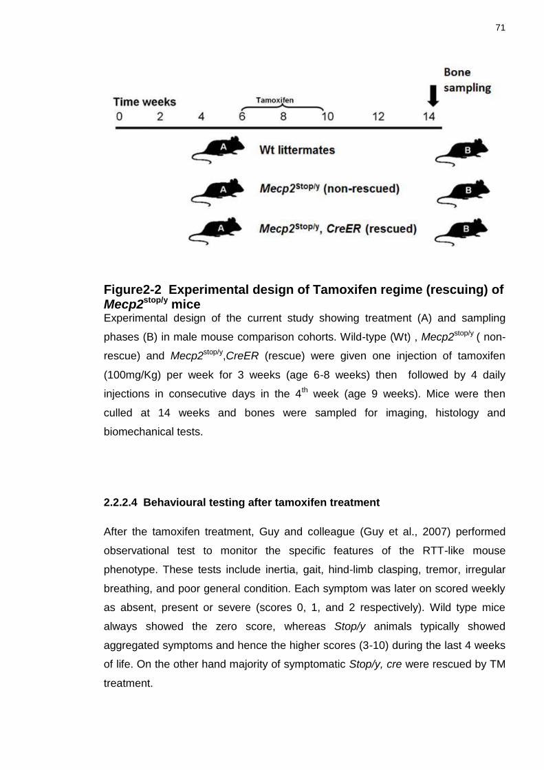
71
Figure2-2 Experimental design of Tamoxifen regime (rescuing) of Mecp2stop/y mice Experimental design of the current study showing treatment (A) and sampling
phases (B) in male mouse comparison cohorts. Wild-type (Wt) , Mecp2stop/y ( non-
rescue) and Mecp2stop/y,CreER (rescue) were given one injection of tamoxifen
(100mg/Kg) per week for 3 weeks (age 6-8 weeks) then followed by 4 daily
injections in consecutive days in the 4th week (age 9 weeks). Mice were then
culled at 14 weeks and bones were sampled for imaging, histology and
biomechanical tests.
2.2.2.4 Behavioural testing after tamoxifen treatment
After the tamoxifen treatment, Guy and colleague (Guy et al., 2007) performed
observational test to monitor the specific features of the RTT-like mouse
phenotype. These tests include inertia, gait, hind-limb clasping, tremor, irregular
breathing, and poor general condition. Each symptom was later on scored weekly
as absent, present or severe (scores 0, 1, and 2 respectively). Wild type mice
always showed the zero score, whereas Stop/y animals typically showed
aggregated symptoms and hence the higher scores (3-10) during the last 4 weeks
of life. On the other hand majority of symptomatic Stop/y, cre were rescued by TM
treatment.

72
These mice did showed milder symptoms and score (1-2) and were thought to
survive for up to 4 weeks from the date of first injection, instead they survived well
beyond the maximum recorded life span of Mecp2lox-Stop/y (17 weeks).
Heterozygous females may be the most accurate model for human RTT
(Kriaucionis and Bird, 2003) because both knockout Mecp2+/- and silenced
Mecp2Stop/+ females develop RTT like symptoms, including inertia, irregular
breathing, abnormal gait, and hind-limb clasping at 4-12 months of age. Similar to
the RTT patients, the phenotype stabilizes, and the mice have an apparently
normal life span. The mice do become obese with time which is not seen in RTT
patients.
Similar to the Stop/y, cre male mice, Guy et al, TM-treated Stop/+ females with
clear neurological symptoms were also used for behavioural testing. These mice
progressively reverted to a phenotype that scored at or very close to wild type. The
report from this study (Guy et al., 2007) demonstrated that the late onset
neurological symptoms in mature adult Stop/+,cre heterozygotes are reversible by
de novo expression of MeCP2.
Behavioural studies performed by Robinson and colleagues on these Stop/y, cre
and Stop/+, cre showed an improvement in structural deficits in cortical neurons,
rescue of respiratory phenotype and improvement in sensory motor tasks
(Robinson et al., 2012a). In my PhD project I have used these cohorts of these
mice to explore putative RTT-related bone phenotypes (see next chapters 3, 4 and
5).
2.3 Breeding strategy of Mecp2- Stop mice
Local Mecp2-Stop colonies at University of Aberdeen were established by
breeding heterozygous Mecp2Stop/+ mice in which the endogenous Mecp2 allele is
silenced by a targeted stop cassette (Mecp2tm2Bird, Jackson Laboratories Stock No.
006849) were crossed with hemizygous Cre ESR transgenic mice (CAG-
Cre/ESR1*, Jackson Laboratories Stock No. 004453) to create experimental
cohorts (Guy et al., 2007) ( table 2-1)

73
A breeding strategy of crossing C57BL6/J/CBA F1 animals and using the F2
offspring (Robinson et al., 2012a) was used. The genotype of the mice was
determined by polymerase chain reaction (Guy et al., 2007). Mice were housed in
groups with littermates, maintained on a 12-h light/dark cycle and provided with
food and water ad libitum. Experiments were carried out in accordance with the
European Communities Council Directive (86/609/EEC) and a project licence with
local ethical approval under the UK Animals (Scientific Procedures0 Act (1986).
2.4 Age of experimental animals
Age matched male mice cohort (Wild-type, Stop/y, Stop/y,cre) of mean age14
weeks ± 4 days and female mice cohort (Wild-type, Stop/+, Stop/+,cre) of mean
age of 20 months ± 5days were used in my PhD project experiments.
All mice were treated were treated by a single injection of tamoxifen (see section
2.3.2.3) at 6 week postnatal age followed by 2 further weekly does at 7 week age
and 8 week age with subsequent 4 consecutive daily doses in 9 week (figure 2-1)
. The mice were culled after 4 weeks of tamoxifen treatment.
Female (Stop/+, cre) mouse models were rescued with injection of tamoxifen at 18
months of age followed by 3 weekly injection and 4 consecutive daily doses. The
mice were culled 2 months after the tamoxifen treatment. Wild type control mice
were treated with tamoxifen in parallel with their Mecp2stop/y and Mecp2stop/y, Cre ER
littermates.
2.5 Establishment of expression of MeCP2 on bone cells
To establish MeCP2 expression in bone tissues, we have used an MeCP2-GFP
reporter line (McLeod et al., 2013). The heterozygous female mice aged 10-13
weeks were engineered to express a Mecp2-EGFP fusion by a targeted gene
knock-in in mouse ES cells (generated in Adrian Bird’s laboratory at the University
of Edinburgh; Mecp2tm3.1Bird, Jackson Laboratories stock no. 014610). Mecp2
status was detected in cells by the presence or absence of fluorescence in living
or fixed cells. Further on the experimental cohort was produced by breeding
Mecp2+/- females with Mecp2 GFP/Y males (supplied by Adrian Bird’s laboratory) on
a C57BL6/j background).

74
2.5.1 Methodology
In these mice, MeCP2 has a GFP tag cloned into the 3’ end of exon 4 to create a
C terminal GFP fusion product which enables the straightforward localisation of
endogenous MeCP2 protein via epifluorescence or laser scanning confocal
microscopy (figure 2-3).
Figure 2-3 MECP2-GFP mouse model GENOTYPE CONSTRUCT Schematic diagram showing GFP tagged MECP2 mouse model design. A GFP
tag is cloned into the 3’end of exon 4 to create a C terminal GFP fusion product.
NTD, N-terminal domain; MBD, methyl binding domain; ID, inter domain; TRD,
transcription repression domain; CTD, C-terminal domain; NLS; nuclear
localisation signals (red line).
Femur bone from male and female mice were dissected out and decalcified in
10% EDTA Solution (7.4 pH) (280g EDTA, 1.5 L of distilled water, 180ml of
ammonium hydroxide), for two weeks in a refrigerator at 4°C. The fresh 10%
EDTA solution was changed every other day, until the bones are properly
decalcified. Midshaft transverse section of 20μm thickness were carefully cut by
using a Leica VT1000 microtome (Leica Milton Keynes, UK) which is maintained
by Robert Kerr, West Medical Building, University of Glasgow, Glasgow, UK.
Images were taken by using laser scanning confocal microscopy (Zeiss LSM710,
Bio-Rad Radiance 2100, UK) using 20x and 40x objectives. Laser scanning
confocal microscopy is maintained by Andrew Todd, west medical building,
University of Glasgow, Glasgow, UK.
After the laser scanning confocal microscopy, we observed that all bone cells
express nuclear GFP fluorescence in wild type male (figure 2-4 Ai-iV) and female
mice. In contrast, GFP fluorescence is found absent in hemizygous Stop

75
(Mecp2stop/y) mice (figure 2-4 Bi-iv), in which Mecp2 is silenced by a stop cassette,
and is observed only in ~50% of bone cells nuclei in female heterozygous Stop
(Mecp2+/stop) mice in which one Mecp2 allele is silenced to mimic the mosaic
expression pattern seen in human Rett syndrome (Guy et al., 2007; Robinson et
al., 2012a).
Figure 2-4 MeCP2 is expressed widely in bone tissues (Ai) Low power and (ii-iv) high power micrographs of transverse sections taken
from mid shaft mouse femur showing GFP expression in all DAPI-labelled nuclei in
a male wild type (Mecp2+/y) mouse in which the native MeCP2 is tagged with a C-
terminal GFP. Note that MeCP2 is restricted to the nucleus of osteocytes as
indicated by the complete overlap with DAPI staining but present in all nuclei. (B)
GFP expression is not observed in stop mice in which MeCP2 expression is
functionally silenced by a neo-stop cassette. (C) Low power (i) and (ii-iv) high
power micrographs showing mosaic expression of GFP-tagged MeCP2 protein in
~ 50% of DAPI positive nuclei in a female heterozygous stop (Mecp2+/stop) mouse
in which one Mecp2 allele is functionally silenced. All scale bars: 100μm.

76
2.6 General solutions
All chemicals below without specified origins were supplied from Merck Ltd. (BDH Laboratories, UK) or Sigma-Aldrich Company Ltd, (Sigma, UK).
2.6.1 0.2 M PB
Materials
Solution A: 37.44 g of NaH2PO (2H2O) in 1200 ml ultrapure H2O
Solution B: 84.90 g of NaH2PO4 in 3000 ml ultrapure H2O
Methods Add 1120 ml of solution A to 2880 ml of Solution B and mix well. Adjust PH to 7.4 with either HCL or NaOH. Add 3000 ml distilled water to the final solution.
2.6.2 0.1 M PB
0.1 PB was made by 50/50 (v/v) dilution with distilled water.
2.7 Dissection
2.7.1 Material
Dissection of mice was carried out to obtain, right and left femur, tibia, humerus and lumbar 5 vertebrae from each mouse (n=6 per genotype).
50 mL Falcon tube containing 70% ethanol ( EtOH ) for sterilizing surgical
equipment.
Dissection board
Pins
Scissors
Plain and tooth forceps

77
2.7.2 Method
2.7.2.1 Tibia and Femur dissection
Each Mouse was pinned to the dissection board, lying on the dorsal back with
ventral surface of the body facing up. Hind limbs were sprayed down with 70%
ethanol / 30% H20. For each right and left leg, first nails were trimmed off with
small dissecting scissors. Then a cut is made in the skin around the full
circumference of the ankle. A second cut down the inside of the leg is made,
starting at the ankle cut and ending at the tip of the 3rd metatarsal. With the help of
small teethed forceps skin was peeled from the ankle towards the phalanges.
Another cut is made inside the leg, starting at the original ankle cut and continuing
along the tibia and femur. Skin was peeled off to the level of the hip. The medial
thigh muscles are dissected 2mm proximal to and along the course of the deep
femoral branch.
Dissection is continued laterally in 2 mm distance to and along the bundle of the
femoral nerve, artery and vein. The tendinous insertion sites of the medial thigh
muscles are clipped and the entire medial thigh muscle package excised. Excision
of calf muscle composed of gastrocnemicus, soleus and plantaris from the fascia.
To keep the bones in natural environment freshly dissected bones were
transferred in labelled tubes filled with 0.1 M Phosphate buffer solution (figure 2-5).

78
Figure2-5 Dissection of femur and tibia Images showing stepwise dissection of lower limb long bones (femur and tibia).
(A-D) dissection of mouse femur and tibia. (A); white arrow point towards the start
of incision area for dissection on left lower limb. (B); Skin and subcutaneous tissue
removed around femur and tibia (C); Skin, subcutaneous fat and muscle removed
around femur and tibia to expose the underlying bones; white arrow points toward
the skin, subcutaneous fat and medial thigh muscles (D); Image showing
dissected femur (blue arrow) and tibia (red arrow).
2.7.2.2 Lumbar vertebrae 5 dissection
Each Mouse was pinned to dissection board, with ventral surface of the body
facing up. Ventral body surface was sprayed down with 70% ethanol / 30% H20.
After the fur, skin and soft tissue removal, vertebrae were identified. With the help
of forceps, scissors and scalpel, Lumbar 5 vertebrae were carefully dissected out
from the rest of vertebrae. Excision of vertebral surrounding structure consists of
Para spinal ligaments and muscles (figure 2-6).

79
Figure2-6 Dissection of 5th Lumbar vertebrae Images showing stepwise dissection of 5th lumbar vertebrae (A-C). (A) image
taken after the dissection and removal of skin, subcutaneous tissue and organs.
(B);Image taken after removal of muscles and ligaments around the vertebral
column. (C); Image showing lumbar vertebrae. White arrow points towards the hip
bone as the identifying anatomical point for the lumbar vertebral dissection.
2.8 Morphometric measurements
After the dissection both right and left femur and tibia along with the 5th lumbar
vertebrae from each experimental genotype group were subjected to
morphometric measurement (see below) before further biomechanical, radiological
and histological analysis.
2.8.1 Whole body weights
Whole body weight measurements were taken using analytical balance (APX60,
Denver Instruments, UK) and accuracy was taken to be 0.0001g. These
measurements were required for the normalisation of individual bone weight
measurement. For results, see chapter 3 result section.

80
2.8.2 Individual bone weights
Femur, tibia and lumbar vertebrae wet weight measurements were obtained using
analytical balance (APX60, Denver Instruments, and UK) and accuracy was taken
to be 0.0001g. These measurements were taken to analyse if there are any gross
differences in individual weights of the bones of three comparison genotypes. For
results see chapter 3 and 4 result section.
2.8.3 Individual bone lengths
In order to obtain the individual bone lengths, femur and tibia were imaged in an
anteroposterior position and posteoranterior position, using a WolfVision VZ9.4F
(WolfVision Ltd, Maidenhead, UK).
Images were analysed for subsequent measurements using Axiovision 4.8
Software (Carl Zeiss Ltd, Cambridge, UK). Femur gross length measurement was
taken from the proximal anatomical point of the greater trochanter to the distal
anatomical point of medial condyle (Di Masso et al., 1998). Tibial gross length
measurements were taken from proximal anatomical point of centre of the
condyles to the distal anatomical point of medial malleolus (Di Masso et al., 2004)
(figure 2-7).
Right femurs were used for mechanical testing (the proximal part for the femoral
neck test and the midshaft part of femur bone was used for microindentation), see
details in chapter 3.
Left femurs were used for the bone histology (the proximal femur for Sirius red and
TRAP staining, the distal femur for scanning electron microscopy), see details in
chapter 4 and 5.
Right tibias were used for μCT, see chapter 4 and three-point bending tests, see
details in chapter 3 and left tibias used for the ash weight density measures, see
full text in chapter 3.
The 5th lumbar vertebrae were used for trabecular bone structure measures, see
chapter 4 for full detail.

81
Figure2-7 Morphometric length measurements of femur and tibia (A) Image of femur and tibia obtained using a WolfVision VZ9.4F Visualizer to
obtained length measurements (WolfVision Ltd, Maidenhead, UK). (B) Images
were analysed for subsequent length measurements using Axiovision 4.8 Software
(Carl Zeiss Ltd, Cambridge, UK). Femur gross length measurement was taken
from the proximal anatomical point of the greater trochanter to the distal
anatomical point of medial condyle. Tibial gross length measurements were taken
from proximal anatomical point of centre of the condyles to the distal anatomical
point of medial malleolus.
2.9 Data handling and analysis
For each biomechanical, structural and histological analysis data results have
been summarized using descriptive statistics. These were presented as mean ±
standard deviation (SD) values and compared by using one way ANOVA followed
by Tukey’s post hoc test. All statistical analyses were performed with the aid of
Prism 5.0 (Graph pad) software.
I have performed a series of biomechanical, morphological and histological on the
above mentioned dissected bones which will be described in detail in the next
three result chapters.

82
Chapter 3
Biomechanical tests revealed genotype differences in bone properties
3.1 Introduction
The majority of females with Rett syndrome are growth retarded (Schultz et al.,
1993; Neul et al., 2010; Roende et al., 2011b) and this postnatal pattern of altered
growth is one of RTT’s earliest clinical expressions. Head growth typically
decelerates during the first year of age and this further followed by a decline in
somatic (height and weight) growth (Armstrong et al., 1999; Neul et al., 2010).
Although not included in the clinical scales for diagnosis of Rett syndrome,
patients nevertheless do suffer from bone related symptoms such as increased
risk of fractures, spinal deformity (eg. scoliosis) (Roberts and Conner, 1988;
Guidera et al., 1991a; Cepollaro et al., 2001; Neul et al., 2010) and bone mass
anomalies (Leonard et al., 1999c; Budden and Gunness, 2001, 2003; Bebbington
et al., 2008; Shapiro et al., 2010; Jefferson et al., 2011; Roende et al., 2011a). The
effects of this high fracture incidence on the quality of life, care needs, and
outcome for RTT patients, are likely to be of importance. Children having fracture
episodes was reported to be one of the most strongly negative associations with
mother’s mental health status (Laurvick et al., 2006).
In my current study, I have looked at the bone biomechanical properties in an
accurate genetic mouse model of Rett Syndrome in order to establish the possible
impact of MeCP2 insufficiency on bone strength and propensity of fractures.
3.1.1 Fracture risk epidemiology in RTT patients
As described in the introductory chapter, multiple clinical studies suggest that
patients with Rett syndrome suffer from an increased risk of fracture (Leonard et
al., 1999c; Bebbington et al., 2008; Downs et al., 2008a).

83
It was found in one study that nearly one third of RTT patients had sustained a
fracture by the age of 15 years(Leonard et al., 1999c), compared with only 15% of
girls and women in the general population by the age of 20 year (Cooley and
Jones, 2002). Similar finding were found by Downs and Colleagues, who reported
38% of RTT patients in their study suffered from fracture episodes.
The incidence of any facture episode was 43.3 episodes per 1000 patient per
years and thus the fracture rate was found to be around 4 times higher in RTT
patients as compared to the girls of similar age (Downs et al., 2008a). Recently in
a case control study of a Danish RTT patient cohort, the investigators reported an
approximately 36% fracture events occurrence among cases as compared to the
27% in controls of same age. The first fracture event typically occurred (77.3% of
cases) below 15 years of age (Roende et al., 2011b).
Studies have been conducted in past to establish the site, cause (Leonard et al.,
1999c; Jones et al., 2002; Cooper et al., 2004; Downs et al., 2008a) and
mechanism (Roende et al., 2011b) of fractures within Rett syndrome patients but
exactly how and why RTT patients suffer from bone symptoms remains poorly
defined.
3.1.2 Fracture site in RTT patients
Greater frequency of fractures was found in lower limb long bones (Leonard et al.,
1999c; Jones et al., 2002; Cooper et al., 2004; Downs et al., 2008a; Roende et al.,
2011b) within RTT patients as compared to the controls, although fractures have
been reported in all regions (spine, ribs, sternum, clavicle, humerus, radius, ulna,
wrist bones, fingers, femur, tibia, fibula, patella, ankle bones, foot), including face.
The bone found most commonly to be fractured was femur followed by tibia,
humerus, ankle and wrist bones respectively (Downs et al., 2008a). Vertebral
fractures were not found to be common in Rett syndrome young population and
mostly found in elderly patients aged ~60 years (Roende et al., 2011b).
3.1.3 Determinants of Fracture risk in RTT patients
Genetic predispositions as the cause of RTT bone symptoms have been observed
by Downs and colleagues. In their study they categorized the cases according to
mutation types, including p.R106W, p.R133C, p.T158, p.R168X, p.R255X,

84
p.R270X, p.R294X, p.R306C, C-terminal deletion, early truncating and large
deletion mutations. The study found a threefold increased risk of fracture in
patients with p.R270X mutation and nearly twofold increase in patients with
p.R168X mutations. Individuals with p.R270X were found to be particularly
susceptible (Downs et al., 2008a). Interestingly this mutation in the nuclear
localization signal region of the transcription repression domain is the one that was
found to be most severe in a number of studies (Huppke et al., 2003; Colvin et al.,
2004). However no association between the mutation types and fracture
distribution was found by Roende and colleagues and they attributed this due to
the lack of actual number of different MECP2 mutations in their study cohort
(Roende et al., 2011b) as compared to Downs and colleagues. The issue
regarding genotype and fracture occurrence within RTT are in dire need of further
studies.
Low bone mass is seen in RTT (Haas et al., 1997; Leonard et al., 1999c;
Cepollaro et al., 2001; Motil et al., 2006; Zysman et al., 2006; Gonnelli et al., 2008;
Shapiro et al., 2010) and an association between low bone mass and fractures in
RTT has been found.
In Rett syndrome 80% of patients suffer from seizures (Jian et al., 2006),
prompting widespread use of antiepileptic drugs (AEDs). In a recent study it was
observed that 64% of the Rett syndrome patients are taking anti-epileptic drugs
(Anderson et al., 2014). Interestingly it has been observed that the risk of fracture
increases with antiepileptic drugs and was found elevated for valporate (anti-
epileptic drug) in particular, prescribed on its own or in combination with other
AEDs after one or more years (Leonard et al., 2010). In another case control study
RTT subjects with a diagnosis of epilepsy were found to have nearly 3 times the
risk of fracture and those receiving more than two AEDs had nearly twice the risk
of those who were receiving no medication (Downs et al., 2008a).
Risk of epilepsy in itself and development of scoliosis(Ager et al., 2006), in RTT
patients is also thought to be genotype related (Jian et al., 2006), pointing towards
the potential relationship between genotypes and bone strength acquisition.
Physical activity is often limited in RTT with patients have difficulty in standing and
walking, which declines further with age (Downs et al., 2008b). Association of risk
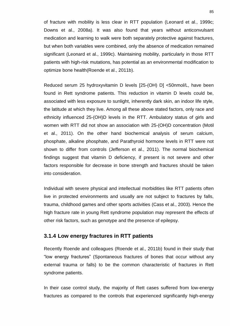
85
of fracture with mobility is less clear in RTT population (Leonard et al., 1999c;
Downs et al., 2008a). It was also found that years without anticonvulsant
medication and learning to walk were both separately protective against fractures,
but when both variables were combined, only the absence of medication remained
significant (Leonard et al., 1999c). Maintaining mobility, particularly in those RTT
patients with high-risk mutations, has potential as an environmental modification to
optimize bone health(Roende et al., 2011b).
Reduced serum 25 hydroxyvitamin D levels [25-(OH) D] <50nmol/L, have been
found in Rett syndrome patients. This reduction in vitamin D levels could be,
associated with less exposure to sunlight, inherently dark skin, an indoor life style,
the latitude at which they live. Among all these above stated factors, only race and
ethnicity influenced 25-(OH)D levels in the RTT. Ambulatory status of girls and
women with RTT did not show an association with 25-(OH)D concentration (Motil
et al., 2011). On the other hand biochemical analysis of serum calcium,
phosphate, alkaline phosphate, and Parathyroid hormone levels in RTT were not
shown to differ from controls (Jefferson et al., 2011). The normal biochemical
findings suggest that vitamin D deficiency, if present is not severe and other
factors responsible for decrease in bone strength and fractures should be taken
into consideration.
Individual with severe physical and intellectual morbidities like RTT patients often
live in protected environments and usually are not subject to fractures by falls,
trauma, childhood games and other sports activities (Cass et al., 2003). Hence the
high fracture rate in young Rett syndrome population may represent the effects of
other risk factors, such as genotype and the presence of epilepsy.
3.1.4 Low energy fractures in RTT patients
Recently Roende and colleagues (Roende et al., 2011b) found in their study that
“low energy fractures” (Spontaneous fractures of bones that occur without any
external trauma or falls) to be the common characteristic of fractures in Rett
syndrome patients.
In their case control study, the majority of Rett cases suffered from low-energy
fractures as compared to the controls that experienced significantly high-energy

86
fractures (fractures due to falls from one level to another or because of activities
involving speed). Cases tended to fracture mostly the lower limb by low energy
mechanisms, whereas controls tended to fracture mostly the upper limb by high
energy mechanisms.
In order to understand the mechanisms underlying the bone fractures an
enhanced knowledge of biomechanical properties is required. In the next section
of this chapter I am going to discuss a brief review of bone biomechanics.
3.2 Biomechanical properties of bone
As described in chapter 1, the organic and inorganic constituents together give
bone its unique properties. In general the mineral phase provides the “stiffness”.
The viscoelastic properties and resistance to fracture are attributed to the collagen
phase (Currey, 1979).
There is clinical and laboratory evidence that, in addition to bone mineral density,
the mechanical properties of bone tissue may play a critical role in bone strength
(Currey, 1979; Chavassieux et al., 2007). Alterations in these mechanical
properties would be expected to play a significant factor in bone fracture risk, even
though it has not been clear what mechanical properties are most important
(Currey, 2004).
Bone is a composite hierarchical material, therefore investigation of the
relationship between the materials properties and the geometry and mechanical
behaviour of whole bone is challenging and very complicated.

87
A thorough understanding of this relationship is of importance as it helps to
understand the normal behaviour of whole bones during physiological loading as
well as identifying areas of peak stresses which are more likely to fracture during
intense activity, and allows the prediction of effects of various pathological
processes and drug treatments (Sharir et al., 2008).
The hierarchical structure of bone material changes at different length scales. It is
also a graded material, as its composition, structure and mechanical properties
may vary continuously or in discrete steps from one location to another (Suresh,
2001).The mechanical behaviour of bone is determined by its geometry and the
properties of the material of which it is made.
There are a number of biomechanical parameters that can be used to characterize
the integrity of bone. One of the key relationships is between applied load to bone
and displacement in response to the load-displacement curve (figure 3-1). The
extrinsic stiffness or rigidity of the bone (S) is represented by slope of the elastic
region of the load-displacement curve. Other biomechanical properties of the bone
can be derived from the same graph including ultimate force (Fu ), work to failure
(area under the load-displacement curve , U) and ultimate displacement (du). Each
of these biomechanical parameters reflects a different property of bone. Ultimate
force also called ultimate load reflects the general integrity of the bone structure;
stiffness measure is closely related to the mineralization of the bone (figure 3-1).
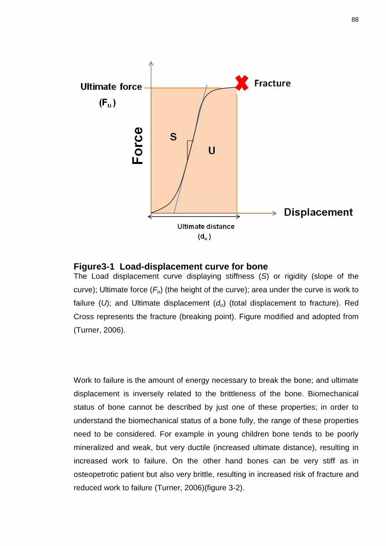
88
Figure3-1 Load-displacement curve for bone The Load displacement curve displaying stiffness (S) or rigidity (slope of the
curve); Ultimate force (Fu) (the height of the curve); area under the curve is work to
failure (U); and Ultimate displacement (du) (total displacement to fracture). Red
Cross represents the fracture (breaking point). Figure modified and adopted from
(Turner, 2006).
Work to failure is the amount of energy necessary to break the bone; and ultimate
displacement is inversely related to the brittleness of the bone. Biomechanical
status of bone cannot be described by just one of these properties; in order to
understand the biomechanical status of a bone fully, the range of these properties
need to be considered. For example in young children bone tends to be poorly
mineralized and weak, but very ductile (increased ultimate distance), resulting in
increased work to failure. On the other hand bones can be very stiff as in
osteopetrotic patient but also very brittle, resulting in increased risk of fracture and
reduced work to failure (Turner, 2006)(figure 3-2).

89
Figure3-2 Load displacement curve showing various bone pathologies Brittle bones displayed a reduced work to failure and inversely proportion to the
displacement. Young children bones are usually ductile, showing increased work
to failure and large ultimate displacement. Red Cross represents the fracture event
(breaking point). Figure modified and adopted from (Turner, 2006).
When load is converted into stress and deformation converts into strain, by the
relationship between stress and strain in bone follows a curve called the stress-
strain curve. The slope of the stress-strain curve within the elastic region is called
the elastic or Young’s modulus (E). Young’s modulus is a measure of the intrinsic
stiffness of the material (Turner, 2006)(figure 3-3).

90
Strength and stiffness are commonly used to define the bone health. Strength and
stiffness are not directly proportional to risk of fracture but the amount of energy
required to cause fracture is directly related to risk of fracture (Currey, 1979).
As described earlier, the bone tissue is a two-phase porous composite material
composed primarily of collagen and mineral, with mechanical properties
determined primarily by the amounts, arrangement, and molecular structure of
both of these constituents. The mineral part provides strength and stiffness to the
tissue (Turner, 2006). The collagen phase is tough and improves bone’s work to
failure or toughness. The ratio of mineral to collagen in bone does affect bone’s
strength and brittleness (Wang et al., 2002) (figure 3-3).
Figure3-3 Summary of contributing factors towards the bone strength

91
Bone hardness is represented by the resistance of the material to the load as the
structure is deformed. This response occurs in many material including bone,
tendons and ligaments (Choi and Goldstein, 1992). A hard material will respond
with a minimum deformation to the load increase. When the material fails in the
end of the elastic phase, it is considered a fragile material. The higher the load
imposed to the bone, the higher the deformation. In addition, if the load exceeds
the elastic limits of the material, there will be a permanent deformation and failure
of material (Choi and Goldstein, 1992) (figure 3-4).
Figure3-4 The Stress-strain curve for bone The slope of the curve is called Young’s modulus (E). The yield point represents a
transition, above which strains begin to cause permanent damage to the bone
structure. Red Cross represents the fracture event (breaking point). Figure
modified and adopted from (Turner, 2006).

92
3.3 Animals models and bone biomechanics
Animal models, in particular mice, offer the possibility of naturally achieving or
genetically engineering a skeletal phenotype and determining the resulting change
in bone’s mechanical properties. In contrary to traditional strength tests on small
animal bones, fracture mechanics tests display smaller variation and therefore
offer the possibility of reducing sample size (Vashishth, 2008).
Many types of mechanical tests have been proposed for mouse bones. As mouse
bones have the well-defined organizational hierarchy, mechanical tests scale the
natural length scale from mineral and protein levels to whole bone tests. Nano and
microindentation tests have been conducted on inbred or genetic knockout mouse
bones to determine the local elastic and viscoelastic properties associated with
bone protein and bone mineral modifications (Akhter et al., 2004a; Kavukcuoglu et
al., 2007; Ng et al., 2007; Tang et al., 2007).
At the whole bone level, three and four-point bending tests have been popular due
to the inherent simplicity of such tests in determining the mechanical properties
associated with changes in the structure and material due to exercise (Maloul et
al., 2006; Wergedal et al., 2006; Wallace et al., 2007), variations among different
inbred mouse strains (Wergedal et al., 2006) accelerated senescence and
ovariectomy and growth factor (Maloul et al., 2006). Less common biomechanics
tests for mouse bone includes femoral neck test which have been used in past to
determine genetic influence on bone and whole bone fracture studies (Hessle et
al., 2013).
In Three-point bending test, the tested bone is positioned onto two supports, and a
single-prolonged loading device is applied to the opposite surface at a point
precisely in the middle between the two supports. The alternative 4-point bending
method is similar except for the fact that the load is applied by two loading, located
equi-distance from the midpoint (Brodt et al., 1999)The main advantage of this test
is that the whole section of bone between two load-applying prongs is subjected to
a uniform moment. Nevertheless, bones are mostly tested in 3-point bending,
since their irregular surface geometry creates difficulty in having both prongs
contact the bone simultaneously.

93
Along with bending tests, indention technique has been used in order to determine
the mechanical properties in mouse bones (Hessle et al., 2013). The indentation
technique allows determining the hardness of a material. The hardness is the
resistance to the penetration of a hard indenter.
Figure 3-5 Load Types (Compression, Tension, Torsion, Shear, Bending) Bone as a structure may be loaded in tension, compression, bending, shear,
torsion, or a combination of these modes (figure 3-5). If the magnitude of applied
load does not exceed the bone’ elastic limits, fracture does not occur and the
bone, elastically deformed, returns to its prestrained state. The failure mode of
bone under circumstances of catastrophic overload is directly related to the
loading mode of the bone. That is, from an evaluation of the fracture
characteristics, it is possible to speculate what loading modes produced the
fracture.
3.3.1 Load Types
The skeletal system is subjected to a variety of different types of forces on such a
way that the bone receives loads in different directions. There are loads produced
by the weight sustentation, by the gravity, by muscle forces and by external forces.

94
The loads are applied in different directions producing forces that may vary from
five different types: compression, tension, shear, curvature or torsion.
TENSION
Because bone as a structural component of the musculoskeletal system must
withstand large axial loads (both compressive and tensile) to sustain weight
bearing and locomotion, it by adaptation, exhibits greater strength when subjected
to tension directed longitudinally versus tension directed transversely. This
observation is essentially a restatement of Wolff’s law and helps to explain bone’s
anisotropic mechanical behaviour (i.e., varying strength as a function of load
direction) (figure 3-5).
COMPRESSION
Compressive forces on a structure tend to shorten and widen it. As in pure
tension, maximal stresses occur on a plane perpendicular to the applied load;
however, the stress distribution and resultant fracture mechanics in compressive
failure are often very complicated. Unlike failure in tension, compressive failure in
bone does not always proceed along the theoretic perpendicular plane of
maximum stress, but rather once a crack is initiated it may propagate obliquely
across the osteons following the line of maximum shear stress.
SHEAR
Tensile and compressive forces act perpendicular or normal to a structure’s
surface. In contrast, shear forces act parallel to the surface and tend to deform a
structure in an angular manner.
BENDING
Bending is a loading mode that results in the generation of maximum tensile forces
on the convex surface of the bent member and maximum compressive forces on
the concave side. Between the two surfaces, that is, through the cross section of
the member, there is a continuous gradient of stress distribution from tension to
compression. Because mature healthy bone is stronger in compression than in
tension, failures usually begins on the tension surface. In very young animals or

95
severely osteoporotic bone, however folding or buckle fractures are sometimes
noted on the concave or compression side of the bone, indicating in a
compressive mode subsequent to bending.
TORSION
Torsion loading is a geometric variation of shear and acts to twist a structure about
an axis (the neutral axis). The amount of deformation is measured in terms of
shear angle, alpha. As in bending, in which maximum tensile and compressive
stresses occur on the surface and distant from the neutral axis, torsional loading
produces maximum shear stresses over the entire surface, and these stresses are
proportional to the distance from the neutral axis.
The skeletal system injury can be produced by applying a high-magnitude single
strength of one these types of load or by repeated application of low-magnitude
single strength of one these types of load or by repeated application of low-
magnitude loads over a long period. The second type of injury in the bone is called
stress fracture; fatigue fracture or bone distension. These fractures occur because
of cumulative microtrauma imposed on the skeletal system, when the placement of
loads on the system is so frequent that the process of bone repair cannot be equal
to the breakdown of the bone tissue(Egan, 1987).
3.3.2 Aim of the study
Given that Rett syndrome patients have a demonstrated increased risk of fracture
and reduced bone strength, the aim of this study was to analyse the
biomechanical properties of bones in Mecp2-Stop mice modelling Rett Syndrome
compared to wild-type and genetically rescued mice. We aimed to explore
potential genotype-related deficiencies in mechanical properties of bone and
hypothesize reduced bone strength in Mecp2-Stop mice with improved functional
integrity following genetic rescue.
3.4 Material and Methods
Right tibial and femoral shafts from each comparison genotypes of male and
female cohorts were subjected to mechanical testing (three point bending and
96
microindentation tests) after they are scanned by Sky scan 1172/A X-ray
computed microtomgraphy (μCT). The mechanical tests were selected to test the
cortical and cancellous parts of the bone. The tests were performed using a
Zwick/Roell z2.0 testing machine (Leominster, UK) with a 100N load cell.
3.4.1 Three-point bending test
For three-point bending test (Hessle et al., 2013), tibias were placed on the lower
supports, at 8 mm separation, with the posterior surface of the tibia facing down.
Load was applied on the shaft of the tibia using the Zwick/Roell testing machine
until the fracture occurred (figure 3-5)
Figure3-6 Three point bending test on right tibias (A)Tibia was placed on the lower supports, with posterior surface facing down.
Load was applied until the fracture occurred. Supports were separated 8mm. (B)
Test was performed using the Zwick/Roell testing machine with a 100N load cell
until fracture occurred. These tests were performed in Mechanical Engineering
Lab, Rankine Building, Department of Engineering University of Glasgow.
From each bone tested, a load-displacement graph was obtained using the
testXpert® II software installed in computer linked to the Zwick/Roell z2.0 testing
machine. The stiffness was calculated by measuring the slope in the graph of
force-displacement and the ultimate load by measuring the maximum force that

97
the bone was able to resist. The second moment of area was calculated using the
equation that considers the bone as a hollow cylinder:
) (eq.1)
Where I is the second moment of area, the is outside diameter and the is the
internal diameter; both of these measurements were taken from the μCT
Young’s modulus measure was also estimated from the geometry of the loading
device and the measured stiffness of the bone. Such an estimate is based on
beam theory and the approximation of the shaft of the tested bone as a uniform
hollow tube.
For Young’s modulus following equation was used:
................................... (eq.2)
Where is the stiffness, determine from the load-displacement curve. is
the separation of the supports and is the second moment of area of the tibias.
This is a commonly used method to estimate the mechanical properties of the
material bone within the whole bone (Sharir et al., 2008).
3.4.2 Micro indentation hardness test
Right femur diaphysis from male and female mice cohort were cut transversely at
midpoint of each bone shaft, using a diamond saw (IsoMet Plus Precision Saw;
Buehler Ltd.). The midpoint of each bone was measured using venire calliper.
Distal mid-shaft sections were used for the micro indentation test in each mouse
bone. Proximal part of each right femur per mouse per genotype was used in
femur neck test (see below).
Bone sections were air dried and embedded in metallurgical mounting resin
prepared by using, VARI-SET Acrylic powder/liquid system (VARI-SET 10/20
liquid, Meta Prep, UK). The moulds were then allowed to solidify at room
temperature for 24 hours. The bone cross-section surface was subsequently hand
polished using silicon carbide papers with decreasing grain size (240, 400, 600,

98
800, and 1200) impregnated with diamond pastes (15, 6 and 1µm) to produce a
smooth polished surface.
Vickers test method (Dall’Ara et al., 2007; Hessle et al., 2013) was employed while
using the microindentation hardness test. Seven indentations with an applied load
of 25gf were done, along the transverse section of each specimen, using a Wilson
Wolport Micro-Vickers 401MVA machine UK (figure 3-6). The indenter of the
machine is held in place for each indentation for 10 or 15 seconds according to the
average value found in the literature. Furthermore, to avoid overlapping of
deformation from one indentation to another a minimum distance of 2 diagonals
between indentations was established (ASTM E384).The Vickers pyramid number
(HV) was calculated with an equation where the load (L) is in grams force and the
average of the two diagonals (d) is in millimetres:
HV=1.854
...................................................... (eq.3) (figure 3-7)
3.4.3 Femoral neck fracture test
The femoral neck fracture test was used to test the mechanical properties
(stiffness and ultimate load) at the proximal part of femur. The shaft of the femur
was fixed in a mechanical chuck and placed in the Zwick/Roell z2.0 mechanical
testing machine (Akhter et al., 2004b). The bone sample was clamped at a 9˚
angle lateral to the vertical axis of the bone (Hessle et al., 2013). Load was applied
to the femoral head until fracture occurred. After each bone being tested the load-
displacement graph is obtained by using the testXpert® II software installed in
computer linked to the Zwick/Roell z2.0 testing machine Stiffness and ultimate
load measurement were calculated in a similar method as described earlier (see
section3.4.2 above) (figure 3-7).

99
Figure3-7 Microindentation Test for hardness (A) Image showing Wilson Wolport Micro-Vickers 401MVA machine used for the
micro indentation test. (B) Resin mould, with embedded bone tissue, white arrow
pointing towards the bone sample held in clippers. (Ci) mouse femur, dotted line
black line showed the midpoint of femur, distal half was used for the
microindentation test. (Cii) transverse section of the bone sample showing the
diamond shape indentation marks on the bone samples (arrows). (Ciii) Insert
shows higher power image of region indicated by box in Cii. Scales bars as
shown.

100
Figure3-8 Femur neck test (A) Image showing proximal femur mounted at 9° to the horizontal plane in the
mechanical chuck. Load was applied on the head of the femur until the fracture is
produced. (B) Image showing Zwick Roell testing machine used for the femur neck
test.Results
3.5 RESULTS
In order to determine any gross skeletal abnormalities caused by MeCP2
deficiency, the tibia and femur of male Mecp2stop/y mice together with wild-type
littermates were examined for gross morphometric analyses; measurements are
summarized below (table 3-1).
Morphometric measurements revealed no significant difference in average body
weights of male and female genotypes. Weight measures of femur and tibia
showed a significant reduction in Stop mice and Rescue mice showed a significant
treatment effect and improvement in weight of femur bone. No difference in femur
length was observed. All data given as mean ± SD for each group of samples (n=5

101
per genotype). Significance was assessed by one way ANOVA with Tukey’s post
hoc test. Abbreviations: * p<0.05, ** p<0.01. Symbol¶ (a comparison is made
between Wild-type control and Stop).Symbol ɸ (a comparison is made between
Stop and Rescue). Symbol ψ (a comparison is made between Wild-type control
and Rescue). For ease of understanding, the male mice comparison genotypes
will be addressed as Wt (control); Stop (Mecp2stop/y); and Rescue (Mecp2stop/y,
CreER) mice and female genotypes cohort as Wt (control); Stop (Mecp2+/stop); and
Rescue (Mecp2+/stop, CreER) mice in subsequent chapters.
3.5.1 No difference in whole body weights of male and female cohorts
No difference in whole body weights was observed genotypes in male mice (figure 3-9).
Figure3-9 Bodyweight measurements in male and female mice cohort. Bar graphs showing, male (A) and female (B) mice cohort analysis results for
whole body weight measurements (A) In male mice whole body weight
comparison among genotypes showed no significant difference (WT= 30.73 ± 3.75
g; Stop = 28.50 ± 3.75g; Rescue= 28.21 ± 2.37g; n=6 per genotype; p<0.05,
ANOVA with Tukey’s post hoc test) (p<0.05). (B) Similar trend was seen in female
mice cohort (p<0.05) (WT= 32.72 ± 5.59g; Stop= 41.70± 7.15g; Rescue= 39.47 ±
9.77g; n=6 per genotype; p<0.05, ANOVA with Tukey’s post hoc test).
Abbreviation: ns = not significant, Plots show mean ± SD.

102
3.5.2 Reduced weight of femur and tibia in Mecp2-Stop male mice
Mecp2stop/y mouse femurs showed a significantly (p<0.05) reduced weight in
comparison with wild-type (Wt) littermate controls and Mecp2stop/y, CreER. A
similar trend of reduction in whole bone weight was observed in Mecp2 Stop mice
tibias, weight measures (table 3-1).
Table 3-1 Morphometric measurements of stop male and female mice
3.5.3 No significant difference in long bone (femur and tibia) weights in Mecp2-Stop female mice
In contrast of the measurements made in male tissues, the female comparison
cohort did not show any significant p>0.05 difference in femur weight measures
between genotypes and similarly no reduction in female tibia weight was found
(table 3-1).
3.5.4 Significant reduction in tibial length of Stop male mice
There was an accompanying reduction p<0.05 in tibial length in male Mecp2 Stop
mice but no significant difference in femoral length between comparison groups
(table 3-1).
3.5.5 No significant difference in long bone (femur and tibia) length measures in Mecp2-Stop female mice
Female Mecp2 Stop mice showed no significant difference in tibial and femur
length among the comparing genotypes (table 3-1).

103
3.5.6 Significant reduction in biomechanical properties in Stop male mice and improvement in bone integrity of Rescue male mice.
Following the morphometric analysis, mechanical tests were applied in order to
explore possible differences in the biomechanical properties of MeCP2-deficient
mice bones. Results from this test revealed a reduced structural stiffness (p<0.01),
ultimate load (p<0.01) and Young’s modulus (p<0.05) measures in male
Mecp2stop/y mice. Samples from Rescue mice revealed that stiffness, ultimate load
and Young’s modulus measures were not different from wild-type values (figure 3-
10).
Figure3-10 Three point bending test results in male mice cohort
Male mice measures of (A) cortical bone stiffness (WT= 106.8 ± 17.88 N/mm; Stop
= 64.7 ± 10.50 N/mm; Rescue= 90.7 ± 14.83 N/mm , n=5 per genotype; p<0.01,
ANOVA with Tukey’s post hoc test) (B) Ultimate load (WT= 17.50 ± 2.45 N; Stop=
12.09 ± 1.94 N; Rescue=15.7 ± 0.08 N; n=5 per genotype; p<0.01, ANOVA with
Tukey’s post hoc test) and (C) Young’s modulus (WT= 12.1 ± 2.37 kN/mm²; Stop=
7.6 ± 1.60 kN/mm²; Rescue = 12.4 ± 3.49 kN/mm2; n=5 per genotype; p<0.05,
one way ANOVA with Tukey’s post hoc test) were significantly reduced in
Mecp2stop/y (Stop) mice as compared to wild-type (WT), and genetically rescued
Mecp2stop/y; CreER (rescue) mice. Abbreviation: ns = not significant; * p<0.05, **
p<0.01. Plots show mean ± SD.

104
3.5.7 Female mice tibia showed no difference in biomechanical properties of bones
The same tests when conducted on tibia from female Mecp2 Stop mice showed no
significant difference in stiffness, ultimate load and Young’s modulus (measure of
stress-strain relationship) measures (figure3-11).
Figure3-11 Three point bending test measures in female cohorts Bar plot showing measures of (A) cortical bone stiffness, (WT= 98.62 ± 7.24
N/mm; Stop = 84.48 ± 10.07 N/mm; Rescue= 85.17 ± 13.00 N/mm , n=5 per
genotype;, ANOVA with Tukey’s post hoc test) (B) ultimate load (WT= 16.60 ±
3.77 N; Stop= 12.68 ± 1.86 N; Rescue=12.63 ± 3.25 N; n≤5 per genotype;
p0>0.05, ANOVA with Tukey’s post hoc test) and (C) Young’s modulus (WT=
12.29 ± 4.33 kN/mm²; Stop= 13.94 ± 2.50 kN/mm²; Rescue = 14.67 ± 0.70
kN/mm2; n=3-5 per genotype; p0>0.05, one way ANOVA with Tukey’s post hoc
test) in female wild-type (Wt), Mecp2Stop/+ (Stop) and Mecp2stop/+ (Rescue) mice.
Abbreviation: ns = not significant; Plots show mean ± SD.
3.5.8 Male and Female Rescue mice showed a significant improvement in bone hardness
Results from male mice showed significantly reduced bone hardness in male Stop
mice as compared to wild-type littermates. Moreover, tamoxifen-treated Rescue
mice did not differ significantly from wild-type and showed a significant treatment
effect when compared with the Mecp2Stop/y cohort (WT= 73.7 ± 1.3 HV, Stop = 65.4
± 1.2 HV, Rescue = 72.1 ± 4.7 HV, n = 5 per genotype, p<0.01, ANOVA with
Tukey’s post hoc test). A significant deficit in bone hardness was also observed in
female Mecp2 Stop mice femurs (WT= 72.8 ± 6.3 HV, Stop = 63.2 ± 3.0 HV,

105
Rescue = 75.7 ± 2.2 HV, n=3-5 per genotype; p<0.01, ANOVA with Tukey’s post
hoc test). Again, rescue mice showed a significant treatment effect and measures
were not found significantly different, from wild-type (figure 3.12).
Figure3-12 Microindentation results in male and female cohorts (A) Bar chart showing a significant reduced cortical bone hardness in Mecp2stop/y
(Stop) mice when compared with the wild-type (Wt) controls (p<0.05, one way
ANOVA with Tukey’s post hoc test, n = 5 femurs per genotype). In contrast, micro
hardness measures in rescued Mecp2stop/y, CreER (rescue) mice were not different
from controls (p>0.05; n=5 femurs). (B) Microindentation hardness test results in
female mice showed a similar pattern with reduced cortical bone hardness in
Mecp2+/stop (Stop) mice when compared with wild-type(Wt) controls and rescued
mice (n= 3-5 femurs per genotype, one way ANOVA with Tukey’s post hoc test).
Abbreviations: ns = not significant, * = p<0.05, **= p<0.01, ***=p<0.001). Plots
show mean ± SD.
3.5.9 Male and Female Stop mice showed no significant difference in femur biomechanical properties
This test was conducted to assess possible group differences in the mechanical
properties of the femoral neck. Femurs were mounted and force applied as shown
in figure 3-7. In male mice, no significant differences were observed in stiffness
and ultimate load measures of biomechanical properties.
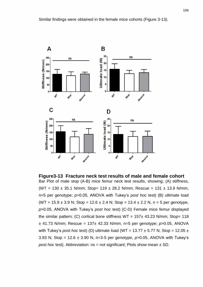
106
Similar findings were obtained in the female mice cohorts (Figure 3-13).
Figure3-13 Fracture neck test results of male and female cohort Bar Plot of male stop (A-B) mice femur neck test results, showing; (A) stiffness,
(WT = 130 ± 35.1 N/mm; Stop= 119 ± 28.2 N/mm; Rescue = 131 ± 13.9 N/mm,
n=5 per genotype; p>0.05, ANOVA with Tukey’s post hoc test) (B) ultimate load
(WT = 15.9 ± 3.9 N; Stop = 12.6 ± 2.4 N; Stop = 13.4 ± 2.2 N, n = 5 per genotype,
p>0.05, ANOVA with Tukey’s post hoc test) (C-D) Female mice femur displayed
the similar pattern; (C) cortical bone stiffness WT = 157± 43.23 N/mm; Stop= 118
± 41.73 N/mm; Rescue = 137± 42.33 N/mm, n=5 per genotype; p>0.05, ANOVA
with Tukey’s post hoc test) (D) ultimate load (WT = 13.77 ± 5.77 N; Stop = 12.05 ±
3.93 N; Stop = 12.6 ± 3.90 N, n=3-5 per genotype, p>0.05, ANOVA with Tukey’s
post hoc test). Abbreviation: ns = not significant; Plots show mean ± SD.

107
3.6 Discussion
As describe earlier, MeCP2 is a nuclear protein, abundant in post mitotic cells of
the brain but also widely expressed throughout the body (Shahbazian et al.,
2002b; Braunschweig et al., 2004; Zhou et al., 2006). The results of my
fluorescence confocal microscope images confirmed this nuclear expression of
Mecp2 in all bone cells nuclei stained with DAPI (4’,6-Diamindino-2 Phenylindole
Dilactate, a blue fluorescent nucleic acid stain) of male wild type GFP tagged
mice( figure 2-4Ai-iv) .
Further on, confocal images from hemizygous stop male mice (figure 2-4 Bi-iv) in
which Mecp2 is silenced by a stop cassette, GFP tagged Mecp2 nuclear
expression was found absent in all bone cells. On the other hand heterozygous
Mecp2+/stop (Stop) female (figure 2-4 Ci-iv) showed only ~50% of nuclear bone
cells expression of Mecp2 in mice in which one Mecp2 allele is silenced to mimic
the mosaic expression pattern seen in human Rett syndrome (Guy et al., 2007;
Robinson et al., 2012a).
For the morphometric and biomechanical analysis I have used the lower limb long
bones (femur and tibia) as these were found to be the most commonly effected
bones in RTT patients in terms of increase rate of fracture was found in these
bones (Downs et al., 2008a; Roende et al., 2011b).
The results of morphometric analysis of long bones revealed that Mecp2 stop
male have an abnormal skeletal phenotype that shares components of the clinical
skeletal features of RTT patients (Neul et al., 2010). Long bone morphometric
analysis showed that Mecp2 stop male mice have lighter (a significant reduction of
14% in femur weight and 13% in tibial weight) and shorter bones (a significant
reduction of 10% in tibial lengths) as compared to age matched wild type controls
(table 3-1). These findings were consistent with the growth retardation found in
RTT patients (Schultz et al., 1993; Neul et al., 2010).
Although there was no significant difference found in whole body weight measures
of three comparison groups in male stop mice cohorts. The basis for this apparent
discrepancy was the observation that male stop mouse model often had more
subcutaneous fat than their wild type matched littermates detected during the

108
dissection. These findings of my study were also found consistent with the findings
of O’Connor and colleagues study on RTT bone phenotype using the Mecp2 null
mouse model.
In Robinson and colleagues study of morphological and phenotype reversal in
Mecp2 stop male mice, an observation of reduced skeletal size along with the
presence of a kyphotic curvature of the spine had been made (Robinson et al.,
2012a). In my project I have used the same stop mice. Kyphotic posturing
frequently observed in the stop mice is comparable to the ‘S’ type scoliotic
curvature of the spine that is more common among RTT patients (Koop, 2011;
Riise et al., 2011). These skeletal dysmorphic findings in stop mouse model were
also found consistent with the bone phenotype of Mecp2 null mouse model by
O’Connor et al. One of the unique features of my study was the use of Rescue
mice. Morphometric analysis of rescue mice in which Mecp2 gene has been
reactivated showed a significant improvement in femur bone weight (15%) as
compared to stop mice. A similar trend of increase in tibial weight (10%) as
compared to wild type was also observed but it does not reach statistical
significance owing to the power of my study. Tibial and femur length
measurements remained reduced.
In contrast to male stop mice, adult female heterozygous Mecp2+/stop mice did not
showed any significant differences in gross tibia and femur length/weight
measures. However an interesting similar trend as found in male stop was also
noted in female stop mice cohort displaying lighter bones (7% reduction in femur
weight and 5% reduction in tibial weight) and in case of tibia shorter bones (5%) as
compared to age match wild type but the values did not reached the statistical
significance (table 3-1).
My study is the first study in which female mice have been used to explore the
bone phenotype of RTT. Female Mecp2+/stop are a gender appropriate and
accurate genetic model of RTT yet display a more subtle and delayed onset (4-12
months) of neurological features (Guy et al., 2007) compared to hemizygous male
mice who become symptomatic by the age of 6-8 weeks.
A major finding of the current study was the demonstrated robust deficits in
mechanical properties and micro-hardness of bone seen in the male Mecp2 Stop

109
mice. This is the first time that biomechanical tests have been performed on stop
mice modelling RTT bone phenotype. Such deficiencies in mechanical and
material properties were profound (39.5% reduction in stiffness in the three point
bending test; 37% decrease in Young’s modulus, 31% in load and 12.3%
reduction in micro hardness) (figure 3-10) and could explain the occurrence of low
energy fractures reported in Rett syndrome patient (Leonard et al. 1999; Zysman
et al. 2006; Downs et al. 2008; Leonard et al. 2010). Whilst I have not observed
overt signs of spontaneous fractures in experimental colonies of mice, such a
magnitude of reduced bone stiffness and load properties could mirror the 4 times
increased risk of fracture in Rett patients compared to the population rate (Downs
et al. 2008). Given that the mice are housed under standard laboratory conditions
and there is not opportunity for traumatic bone insult, it is perhaps not surprising
that spontaneous fractures are not apparent.
Male rescue mice interestingly showed a significant improvement in bone stiffness
(40%), ultimate load (10%),Young’s modulus (61%) and microindentation (12%)
when the gene is reactivated as compared to male stop mice. These findings were
quite encouraging and potentiated our hypothesis of genetic basis of RTT bone
phenotype (figure 3-10).
Mechanical properties and micro hardness test was also performed in female
heterozygous cohort. This is the first time that female mice modelling RTT bone
phenotype has been used.
Biomechanical analysis of heterozygous stop female showed similar trend (15%
reduction in stiffness and 24% reduction in ultimate load as compared to age
matched wild-type control) as the results of their morphometric analysis results but
like morphometric findings, these values does not reach the statistical significance
(figure 3-11).
The finding that a similar significant reduction as male stop mice values, in micro
hardness (14%) measure was seen in female mice that are heterozygous and
mosaic for the mutant allele is important and demonstrates that the bone deficits
are not restricted to the more severe male RTT-like phenotype but are seen in a
gender and MeCP2 expression pattern appropriate model of RTT.

110
A very interesting finding in female heterozygous rescue mice was found to be a
significant improvement in microhardness (19%) when the gene is reactivated as
compared to the stop female. These findings were quite encouraging and
displayed that the bone deficits rescue is not restricted to the more severe male
RTT-like phenotype but rescue effects can be seen in a gender and MeCP2
expression pattern appropriate model of RTT (figure 3-12).
Analysis of femoral neck fracture showed no difference between genotypes. Male
stop mice showed a decrease of 9% in stiffness and 21% decrease in load
measures but the values did not reach the statistical significance. Male rescue
mice displayed a 10% improvement in stiffness and 6% improvement in ultimate
load measures but these values were also not found statistically different (figure 3-
13).
Female stop mice showed a similar trend of decrease in stiffness values of 15%
and load value of 13% as compared to age matched wild type control, while
female rescue mice showed a 16% improvement in stiffness as compared to stop
mice and no improvement are seen in ultimate load values. Similar to male stop
and rescue mice measures of femur neck test all these measures did not reached
the statistical significance (figure 3-14).
It is possible that the complex microstructure of bone in the femoral neck (cf. the
simple cortical shaft geometry) is a confounding factor and limits the sensitivity of
this test. Indeed, we also noted greater variance in this test than in the other
biomechanical tests which may limit our ability to resolve subtle changes in this
parameter. Nevertheless, this test has been used in other rodent models to show
deficits in femoral neck integrity (Hessle et al., 2013).
An important finding of the current study and one with therapeutic implications is
that the observed deficits in cortical bone material and biomechanical properties
were rescued by delayed postnatal activation of the Mecp2 gene. This finding
mirrors the improvements seen in multiple non-bone phenotypes seen in the
Mecp2Stop/y mice after delayed activation of the Mecp2 gene including survival,
normalized bodyweight, locomotor and behavioural activities and well as
morphological features within the brain (Guy et al., 2007; Robinson et al., 2012a).

111
These results suggest that the bone abnormalities present in RTT patients may
be at least partially reversible using gene-based therapies that are currently being
developed (Gadalla et al., 2013; Garg et al., 2013) should the animal studies
translate to clinical studies. However, it is also possible that significant
amelioration of bone phenotypes may also be achieved using pharmacological
strategies. Pharmacological approaches are being investigated in RTT, both in
pre-clinical studies as well as clinical trials (Gadalla et al., 2011; Gadalla et al.,
2013; Garg et al., 2013). Of particular importance for this approach with respect to
bone phenotypes is to identify the mechanisms by which MeCP2 deficiency results
in altered bone properties. Whilst we show that MeCP2 is expressed in osteocytes
(figure 3.8), the protein is widely expressed throughout the body and it is possible
that metabolic and endocrine perturbations elsewhere in the body (Motil et al.,
2006; Motil et al., 2011; Roende et al., 2014) may also impact on bone
homeostasis.
However the results obtained from our biomechanical tests study were quite
encouraging, the decrease in bone strength in Mecp2 deficient mouse and the
subsequent improvement of bone integrity when the gene is switch back lead us to
explore further into the mechanism by which Mecp2 is causing this deficiency in
bone strength. The experiments performed in this regard will be discussed in detail
in the next chapters.

112
Chapter 4
Radiology based structural studies to assess trabecular and cortical bone parameters in a mouse model of Rett Syndrome
4.1 Introduction
Radiographic and ultrasound studies have been conducted in Rett syndrome
patients to better understand the underlying pathology that may account for the
reduced bone strength and increased risk of fractures in RTT patients (Leonard et
al., 1995; Leonard et al., 1999b; Cepollaro et al., 2001). The majority of these
imaging studies have been conducted to investigate the bone mineral density and
bone mineral content in RTT patients. However there remains to be a detailed
study exploring the effect of MECP2 mutations on bone structural geometry in
humans. One reason for this may be the lack of appreciation of bone phenotypes
in what is considered a largely neurological disorder. Another difficulty might be
the application of radiological test in patients with many other confounding
impairments. For instance, patients with scoliosis who require spinal rod
placements have implanted metal, which interferes with the ability of Dual-energy
X-ray absorptiometry (DXA) in provision of accurate assessment of bone
parameters.
Bone mass has been investigated in detail in RTT patients (Haas et al., 1997;
Leonard et al., 1999c; Cepollaro et al., 2001; Motil et al., 2006; Zysman et al.,
2006; Gonnelli et al., 2008; Shapiro et al., 2010) as it is shown to be a strong
predictor of fracture risk in adults (Hui et al., 1988) and children (Hui et al., 1988;
Flynn et al., 2007). Although prospective measures of BMD in the lumbar spine of
children with cerebral palsy did not predict subsequent fracture risk (Henderson,
1997).

113
Neurological disabilities found in RTT patients evolves over several years with
potential co-morbidities such as poor nutrition due to problems with swallowing
(Oddy et al., 2007), surgical procedures (Kerr et al., 2003) for the correction of
bone deformities (scoliosis) and certain anticonvulsant medications usage
(Leonard et al., 2010), that together may limit the development of normal bone
mass. The availability of murine RTT models now permits an assessment of the
effects of MECP2 mutation on bone mass independent of theses contributing
factors. The ultra structure and density of bone in mice with and without the Mecp2
protein have been investigated in a study by O’Connor and colleagues (O'Connor
et al., 2009b). This study showed, growth retardation, abnormal growth plates
(irregular shape chondrocytes) and decreased cortical and trabecular bone
parameters. Another study conducted on the same Mecp2 null mouse model by
Shapiro and colleagues found differences in cortical thickness, mineralization of
the medullary cavity in long bones and spinal bone density (Shapiro et al., 2010).
O’Connor and colleague in their Mecp2 knockout mouse model also found
modestly lower values of bone mineral density (BMD) and bone mineral content
(BMC) but this decrease did not achieve statistical significance and hence bone
mineral density changes as a cause of bone anomalies seen in RTT murine model
is still not fully understood.
Rett syndrome bone phenotypes have been frequently compared to osteoporosis
which has been defined as ‘a systemic skeletal disease characterized by low bone
mass and micro-architectural deterioration of bone tissue with a resulting increase
in fragility and risk of fractures’ (Zysman et al., 2006). Hence, a comprehensive
approach to investigate bone material and structural properties is required to
better understand the bone phenotype in RTT.
Previous studies investigating various osteoporotic processes in murine model
have used femur, tibia and lumbar 5 and 6 vertebrae (Sheng et al., 2002; Rubin et
al., 2004) as sample biopsies to better understand underlying structural
pathologies in a range of conditions. The work presented in the current chapter
was an analysis of long bones and vertebrae in order to explore aspects of cortical
and trabecular bone structure of Mecp2 stop mice.

114
4.1.1 Bone structure and Bone strength
Predicting fracture propensity for fracture requires a proper understanding of the
relationship between bone structure and the mechanical properties of bone. The
material composition and structural design of bone determines its strength.
In recent years the concept of bone strength has moved beyond density alone and
has expanded to include an amalgamation of all the factors that determine how
well the skeleton can resist fracturing, such as micro architecture, accumulated
microscopic damage, the quality of collagen, the size of mineral crystals and the
rate of bone turn over (Chavassieux et al., 2007).
In particularly, the supremacy of bone structure has been found over tissue-level
material properties. The net response of osteoblasts for bone formation and
osteoclasts for bone resorption are reflected by changes in the trabecular structure
and bone volume fraction (Nazarian et al., 2008).
Studies in the past have concluded that a unified consideration of the relationship
of bone tissue mineralization and trabecular structure can predict the mechanical
properties of normal and pathologic bones (Cody et al., 1991; Kim et al., 2007).
4.1.2 µCT use in skeletal phenotypes
Histological and radiological studies are usually employed to understand the bone
micro architecture. Micro computerised tomography (µCT) has now become the
gold standard for the evaluation of bone morphology and micro architecture in
mice and other small animals (Martín-Badosa et al., 2003). The accuracy of µCT
morphology measurements has been evaluated both in animals (Waarsing et al.,
2004; Bonnet et al., 2009) and in humans (Kuhn et al., 1990; Müller et al., 1998)
specimens. These studies have shown that 2D and 3D morphologic
measurements by µCT generally are correlated highly than these from 2D
histomorphometry.

115
4.1.3 Aim of the study
The results obtained from the analyses of biomechanical properties described in
the previous chapter were encouraging in that they demonstrated a reduction in
bone strength in Mecp2 mice and also an amelioration of this phenotype in stop
mice genetic rescue. This suggested that at the functional level in stop mice, bone
deficits were overt but reversible. The aim of the experiments described in the
current chapter was to explore further the potential structural alterations in bone of
Mecp2 deficient mice that might account for some of the observed biomechanical
deficits. I hypothesised that the reduction in bone strength, seen in Mecp2 stop
mice is due to alternations in bone structure and bone mineral density (BMD)
levels. In order to analyse these features I have used the µCT scanning
technology to analyse the ultra structure of bones from wild-type, Mecp2 stop and
genetically rescued mice of male and female cohorts.
4.2 Material and Methods
The structural properties of bone samples from wild-type, Mecp2-stop and
genetically recued mice were assessed using scanning electron microscopy and
were also visualised using a µCT scanner (see below). The result analysis
obtained were then used to investigate cortical and trabecular bone structure.
4.2.1 Micro-computed tomography (µCT)
After the gross morphometric measurements (chapter 3) a subset of bone samples
was scanned using µCT. The samples were scanned prior to any biomechanical
tests being performed. All scans were conducted using the micro computed
tomography facility at the Orthopaedic Research group, University of Edinburgh,
Edinburgh, UK. We have used the Sky Scan 1172/A X-ray Computed
Microtomgraphy system (µCT) maintained by Robert Wallace, Chancellors
Building, Orthopaedic Research Group, Edinburgh, UK.
The micro CT scanner is composed of a sealed micro focus X-ray tube, air cooled
with a spot size lens and a camera. The maximum length of object that is capable
of fitting into this device is 40mm. Bone specimens were scanned in wet form by
placing them first in a vial containing water to maintain hydration (figure 4.1).

116
The sample vial was subsequently fixed to the machine stub (mounting plate) with
masking tape, which in turn was securely fastened to the holder in the µCT x-ray
chamber. This must be secured correctly as any movement of the sample could
render the scan unusable.
Multiple projection images were obtained with a rotation of 0.45°- 0.1° between
each image. Given a series of projection images a stack of 2D sections was
reconstructed for each specimen. CT-Analyser v1.8.1.3 (Skyscan, Kontich,
Belgium) and NRecon v1.6.6.0 (Skyscan, Kontich, Belgium) were used for the task
of reconstruction and whole data processing as described in user’s manual and
according to protocols followed by local technical faculty. Image slices obtained
and stored in the .bmp format with indexed grey levels ranging from 0 (black) to
255 (white). Understandably, this is a resource intensive computer task and as
such a dedicated computer was used for this process (2x 3GHz Quad core CPUs,
8Gb Ram, NVIDIA Quadro FX 570).
Figure4-1 Micro CT scanning of Tibia (A) Mouse tibia in the test tube filled with water to maintain hydration during the CT
scanning (B) Scanned image of tibia placed in test tube taken during the µCT
scan. Images were scanned at a voxel resolution of 34μm for male and female
stop tibia, using a sky scan micro CT machine. The X-ray tube was operated at
54KV and 185μA. The Sky scan 1172 micro CT scanner at Chancellor’s building,
Orthopaedic Research Group facility at the University of Edinburgh, Edinburgh UK
was used for the trabecular and cortical bone parameters analysis.

117
4.2.2 Micro-computed tomography (µCT) for cortical bone measures
In order to obtain accurate internal and external diameter measures for calculating
second moment of area and for cortical bone structural parameter evaluation, right
tibias from male (n=5) and female cohorts (n3-5) were scanned at a voxel
resolution of 34μm using a sky scan micro CT machine. The X-ray tube was
operated at 54KV and 185μA. For cortical bone parameter analyses, 2mm
midshaft region of interest (ROIs) were selected from tibial diaphysis, starting from
the anatomical point of tibiofibular junction in each bone specimen.
Figure4-2 Screen shot of image analysis while using the CT analyser software, displaying region of interest at mid diaphysis of tibia Representative screen shot showing mouse tibia with a region of interest at the
mid-diaphysis, while CT scan images of tibia were analysed using the CT analyser
software v 1.8.1.3. A region of interest (2mm) was selected from tibial shaft per
bone specimen per genotype. A lower grey threshold value of 113 and upper grey
threshold value of 255 was used as thresholding values. Care was taken that all
the reconstruction parameters selected were applied identically to all bone scans.
A

118
Images obtained were reconstructed and analysed using the NRecon software v
1.6.6.0 and CT analyser software v 1.8.1.3. The data from each scan was then
split by region of interest. A lower grey threshold value of 113 and upper grey
threshold value of 255 was used as thresholding values in each cortical bone
sample. All the reconstruction parameters selected were applied identically to all
bone scans (figure 4-2).
Individual two dimensional object analyses were performed on six sections per
bone specimen within each comparison genotype group to calculate the inner and
outer perimeters of bone. An average of six values per specimen then used as the
final measure for the inner and outer perimeter and subsequently cortical
thickness measurement was derived from these values. Three dimensional
analyses further used to calculate marrow area, cortical area, total area and bone
volume.
4.2.3 Scanning Electron Microscopy (SEM)
The Undecalcified, left distal femur metaphyseal region from both male mice (n=5)
and female (n=3-5) mice were selected to observe any trabecular structural
differences using scanning electron microscope (SEM) Stereoscan 250 MK3,
Cambridge, UK) at the Anatomy Department, University of Glasgow, Glasgow, UK.
4.2.3.1 Sample preparation for SEM
Distal parts of left femur per bone per genotype from both male and female mice
were cut with diamond saw (IsoMet plus Precision Saw; Buehler Ltd.) transversely,
3mm above the condyles. The 3mm distance from the medial condyle was
measured with vernier calliper before cutting. Bones were then stored in 2.5%
paraformaldehyde in 0.1M sodium phosphate buffer (water, pH7.4) at 4°C for 48h.
Adherent soft tissue was removed by immersion in 3% hydrogen peroxide solution
for 48h. After rinsing with distilled water, specimens were defatted in 50:50
methanol/chloroform for 24h at room temperature and transferred to a 5% trypsin
solution (0.1M PB, pH 7.4) at room temperature for 48h. After cleaning with

119
distilled water, specimens were desiccated. Samples were gold coated by using a
sputter coater (Polaron E5000, East Sussex, UK). An extra coating with silver
paint was done to have the proper imaging. Images were obtained using a
scanning electron microscope (Stereoscan 250 MK3, Cambridge, UK).
4.2.4 5th Lumbar vertebrae, µCT scan for trabecular parameters
5th Lumbar vertebrae from the mice from each genotype were scanned at a
resolution of 5µm.
Figure4-3 Micro CT scan of 5th Lumbar vertebrae The L5 vertebrae (n=6) from each genotype of male mice cohort (WT, Stop, and
Rescue) were dissected out. The micro CT scans of each vertebra were taken
using the sky scanner µCT facility. (A) Lateral view of L5 vertebrae scanned at
5um resolution. A cylindrical shape region of interest (ROI), comprising of 150
slices was taken from the body of the vertebrae. The shell properties of cortical
bone were not included. (B) Reconstructed image of 5th Lumbar vertebrae,
showing the selection region of interest (ROI) within the body of vertebrae. A lower
grey threshold value of 81 and upper grey threshold value of 252 was used as
thresholding values in each trabecular bone sample. Scale bar=200μm.

120
A higher resolution was needed to scan the trabecular bone and hence 5 μm
selected for the mouse trabecular bone parameters as compared to the cortical
bone parameters(Ito, 2005). The X-ray tube was operated at 41kV and 240μA.
Table 4-1: Trabecular bone parameters
Abbreviation Description Definition Standard Unit
TV Total volume Volume of the entire region of interest
mm-3
BV Bone volume Volume of the region segmented as bone
mm-3
BS Bone surface Surface of the region segmented as bone
mm-3
BV/TV Bone volume fraction
Ratio of the segmented bone volume to the total volume of the region of interest.
%
BS/TV Bone surface density
Ratio of the segmented bone surface to the volume of the region of interest.
mm-2/ mm-
3
BS/BV Specific bone surface
Ratio of the segmented bone surface to the segmented bone volume.
mm-2/ mm-
3
Conn.D Connectivity density
A measure of the degree of connectivity of the trabeculae normalised by TV.
1/ mm-3
SMI Structure model index
An indicator of the structure of trabeculae, 0=parallel plates, 3= cylindrical rods
Tb.N Trabecular number
Average number of trabeculae per unit length
1/mm
Tb.Th Trabecular thickness
Mean thickness of trabeculae assessed using Direct 3D methods.
mm
Tb.Sp Trabecular separation
Mean distance between trabeculae mm
DA Degree of anisotropy
Length of longest divided by shortest mean Intercept length vector.
a
Note: Modified and adopted from (Bouxsein et al. 2010).
A lower grey threshold value of 81 and upper grey threshold value of 252 was
used as thresholding values in each trabecular bone sample. A cylindrical region
of interest (150 slices or 0.774mm) was selected from the centre of each vertebral
body excluding the cortical shell area, in order to analyse only the trabecular
parameter specifically (figure 4.3). Images reconstructed and analysed using the
NRecon 1.6.6.0 and CT-Analyser 1.8.1.3 software. Vertebral body lengths were
determined by measuring a line drawn at a 90° angle from the proximal part of the

121
vertebral body to the distal part. Three dimensional analysis was performed for the
following parameters: trabecular thickness, trabecular separation, trabecular bone
volume, trabecular porosity, as well as degree of anisotropy (DA) and structure
model index (SMI); for details of these parameter see (table 4-1
4.2.4.1 Density calibration of 5th lumbar vertebrae in µCT scanner
Bone mineral density (BMD) was standardized to the volumetric density of calcium
hydroxyapatite (CaHA) in terms of g.cm-3. For bone mineral density (BMD)
calibration, the Skyscan CT- analyser, was calibrated by means of phantom rods,
with known BMD values of 0.25 and 0.75 g.cm-3 CaHA respectively.
Trabecular (medullary) density can refer to the density of a defined volume of bone
plus soft tissue.
Hounsfield units (HU) are a standard unit of x-ray CT density, in which air and
water are ascribed values of -1000 and 0 respectively. The Skyscan CT-analyser
software provides for an integrated calibration of datasets into these two density
scales (HU and BMD). Both require the appropriate calibration phantom scans and
measurements.
For density calibration, a scan of free standing tibia, within a tube of water was
performed. Then the two BMD rods (under the same conditions as the bone scan)
with BMD values of values of 0.25 and 0.75 g.cm-3 CaHA were scanned as well.
Reconstruction of the scan of the bone in the water tube was done by using the
NRecon 1.6.6.0 and CT-Analyser 1.8.1.3 softwares. Reconstruction parameter
were selected and same parameter were applied throughout the scans of each
sample bone per genotype. Also care was taken with the selection of the lower
and upper contrast limits.

122
With the scans and reconstruction of the bones and the calibrating phantoms
complete, the HU and BMD calibration was implemented in CT-analyser.Following
density range calibration was selected (Table 3-2).
Table 4-2: Density range calibration
Calibration unit Min Value Max Value
Index 0 255
HU -1000 7292
4.3 Results
µCT analysis was used to examine the three dimensional structure of wild-type
and Mecp2 stop male and female tibias. Micro CT analysis of male Mecp2-Stop
mice tibia revealed considerable differences in cortical bone parameters while
several trends were noteworthy in the trabecular bone.
4.3.1 Micro CT revealed male Mecp2-Stop mice to display altered cortical bone properties.
One of the major structural findings of my current study was the reduction found in
cortical bone parameters results obtained from the µCT analysis of male Mecp2
mice cortical bone. These results were consistent with the reduced biomechanical
strength findings and also correlate with the reduced cortical bone parameters
seen in RTT patients.
A significant difference in male stop mice cortical bone parameters was found in
cortical bone thickness (54%), outer perimeter (20%), inner perimeter (12%),
marrow area (38%), total area (20%) and bone volume (30%) values as compared
to wild type control mice However no significant difference was seen in cortical
area values of Mecp2 stop mice (figure 4-4 i-vii). Rescue mice didn’t show any
improvement in bone cortical parameters and values obtained, remain reduced
when a comparison is made with WT control values.

123
Figure4-4 Cortical bone parameter in Mecp2 Stop and Rescue male mice Bar graphs (i-vii) showing a significant decrease in cortical thickness, (WT= 0.41±0.17mm, Stop=
0.19±0.07mm, Rescue= 0.21±0.08mm; n=5 per genotype; p<0.05, ANOVA with Tukey’s post hoc
test outer perimeter (WT= 1.65±0.22mm, Stop= 1.32±0.07mm, Rescue= 1.38±0.05mm; n=5 per
genotype; p<0.05, ANOVA with Tukey’s post hoc test), inner perimeter (WT= 1.26±0.24mm, Stop=
1.12±0.07mm, Rescue= 1.08±0.05mm; n=5 per genotype; p<0.05, ANOVA with Tukey’s post hoc
test), marrow area (WT=0.48±0.14mm², Stop=0.30±0.02mm², Rescue=0.29±0.03mm²; n=5 per
genotype; p<0.05, ANOVA with Tukey’s post hoc test),total area (WT=1.26±0.17mm²,
Stop=1.05±0.11mm², Rescue=0.98±0.05mm²; n=5 per genotype; p<0.05, ANOVA with Tukey’s
post hoc test), and bone volume (WT= 1.75±0.21 mm³, Stop=1.39±0.19 mm³, Rescue= 1.39±0.11
mm³; n=5 per genotype; p<0.05, ANOVA with Tukey’s post hoc test). No significant difference was
seen in cortical area (WT=0.81±0.08 mm², Stop=0.75±0.13 mm², Rescue= 0.69±0.045 mm²; n=5
per genotype; p>0.05, ANOVA with Tukey’s post hoc test) values of Mecp2 stop mice as compared
to wild-type controls. Abbreviation: ns = not significant; * p<0.05, ** p<0.01. Plots show mean ± SD.

124
4.3.2 Micro CT scans of heterozygous female Mecp2-Stop and Rescue mice showed no significant differences in cortical structure parameters
Figure4-5 Cortical bone parameters in Mecp2 Stop and Rescue Female mice. Bar graphs (A-G) showing no significant difference (p>0.05) in results of outer perimeter (WT=
1.44±0.05mm, Stop= 1.42±0.17mm, Rescue= 1.35±0.05mm; n=3-5 per genotype; p0>.05, ANOVA
with Tukey’s post hoc test), inner perimeter (WT= 1.21±0.09mm, Stop= 1.18±0.12mm, Rescue=
1.12±0.12mm; n=3-5 per genotype; p>0.05, ANOVA with Tukey’s post hoc test) cortical thickness
(WT= 0.22±0.08mm, Stop= 0.21±0.07mm, Rescue= 0.22±0.06mm; n=3-5 per genotype; p>0.05,
ANOVA with Tukey’s post hoc test), cortical area (WT=0.82±0.09 mm², Stop=0.71±0.08 mm²,
Rescue= 0.76±0.14 mm²; n=3-5 per genotype; p>0.05, ANOVA with Tukey’s post hoc test), marrow
area (WT=0.39±0.09mm², Stop=0.40±0.10mm², Rescue=0.29±0.07mm²; n=3-5 per genotype;
p>0.05, ANOVA with Tukey’s post hoc test), total area (WT=1.21±0.14mm², Stop=1.11±0.17mm²,
Rescue=1.06±0.18mm²; n=3-5 per genotype; p>0.05, ANOVA with Tukey’s post hoc test), and
bone volume (WT= 1.73±0.22 mm³, Stop=1.51±0.21mm³, Rescue= 1.54±0.31 mm³; n=3-5 per
genotype; p>0.05, ANOVA with Tukey’s post hoc test) as compared to wild-type controls

125
4.3.3 Scanning electron microscopy revealed altered trabecular structure in Stop male mice
Qualitative analysis using scanning electron microscopy (SEM) of the distal femur
(n=5 per genotype) revealed porous structure in cortical bone (3 of 5 mice) as well
as alterations in the architecture of trabecular bone in Mecp2stop/y mice (figure 4-6).
The central metaphyseal region in Mecp2stop/y mice showed a sparse trabecular
mass consisting of short, thin trabecular rod and plate structures. In contrast, a
more robust trabecular structure, with a network of shorter and thicker rods and
plates was found in wild-type control tissue (figure 4-6). The porosity and altered
trabecular structure was less evident in rescued Mecp2stop/y, CreER mice.
Figure 4-6 Scanning electron microscopy reveals pitted cortical bone and altered trabecular structure in distal femur of male MeCP2-deficient mice. Scanning electron micrographs of distal femur in (Ai) wild-type (Wt) and (Bi)
Mecp2stop/y (stop).Higher powered images of cortical (ii) and metaphyseal (iii)
regions (areas indicated in A) reveal a more porous structure in cortical bone
(arrows in Bi indicate pores) and a sparse trabecular structure in Mecp2stop/y mice
when compared with representative with Wt controls. (Ci-iii) Representative
micrograph from a Mecp2stop/y, CreER (rescue) mouse.

126
4.3.4 Micro CT scans showed improvement in trabecular bone thickness in Rescue male mice
Three dimensional μCT scan analyses were performed to obtain a quantitative
measure of trabecular architecture in wild-type, Stop and Rescue mouse 5th
lumbar vertebrae. A significant reduction of 5th lumbar, trabecular thickness
(~30%) was observed in Stop male mouse tissues compared to the wild-type
controls. Interestingly rescue male mice 5th lumbar μCT scan results, showed a
significant increase (+80%, p<0.01) in trabecular rod and plates thickness
compared to Mecp2stop/y mice (Wt=0.073 ± 0.01mm; Stop = 0.051 ± 0.02mm;
Rescue= 0.09 ± 0.02 mm; n=6 per genotype; p<0.01, ANOVA with Tukey’s post
hoc test) suggesting a significant treatment effect (figure 4.6).
Figure4-7 MicroCT scans of L5 vertebrae revealed thinner trabecular mass in MeCP2-deficient mice (A) Bar plot showing quantitative analysis of trabecular thickness (arrows in B-D).
Note the reduced thickness in Mecp2stop/y samples (p<0.05; n=6 per genotype). (B-
D) Micrographs showing representative trabecular samples from wild-type (Wt),
Mecp2stop/y (Stop) and Mecp2stop/y, CreER (rescue) mice. (E) Scale bar: B-D,
50µm. Abbreviations: ns = not significant, * = p<0.05, ** = p<0.01; one way
ANOVA with Tukey’s post hoc test). Plots show mean ± SD.

127
Length of the vertebral bodies revealed a significant difference (WT= 4.157 ±
0.52mm, Stop= 3.48 ± 0.39mm, Rescue= 3.14 ± 0.37mm; n=6 per genotype;
p<0.05, ANOVA with Tukey’s post hoc test) in Mecp2 stop and rescue mice
vertebral body length measurements as compared to age matched wild-
typecontrol. No significant difference was observed in trabecular separation,
trabecular bone volume, trabecular porosity, bone mineral density (BMD), degree
of anisotropy (DA) and structure model index (SMI) between comparison
genotypes of male mice cohort. All findings are summarized in figure 4.6 and table
4.3.
Table 4-3 Lumbar vertebrae trabecular bone parameters Body of 5th Lumbar vertebrae was selected as region of interest (ROI) and was
analysed to assess the trabecular part of the bone. All data given as mean ± SD
for each group of samples (n=6 per genotype). Significance was assessed by one
way ANOVA with Tukey’s post hoc test. Abbreviations: * p<0.05, ** p<0.01.
Symbol¶ (a comparison is made between Wild-type control and Stop male mice).
Symbol ɸ (a comparison is made between Stop and Rescue male mice).

128
Figure4-8 Trabecular bone parameters bar graphs of Mecp2 stop mice Bar graphs (A-E); displaying trabecular bone parameters, showed an apparent
trend of decrease in bone volume fraction (%), connectivity density (1/mm3),
trabecular separation and structure model index measurements but these values
does not reach any statistical significant (p>0.05) difference; n=6; Abbreviations:
ns = not significant ; one way ANOVA with Tukey’s post hoc test). Plots show
mean ± SD.
4.3.5 Bone density measurements from μCT did not revealed any significant difference in Mecp2 stop mice.
In order to analyse bone density measurements, 5th Lumbar vertebrae were
scanned, and no significant difference was not observed in male stop mice
genotypes (WT= 0.96±0.06; Stop= 0.92±0.07; Rescue=0.94±0.06 n≤7 per
genotype; p>0.05, ANOVA with Tukey’s post hoc test). All data given as, mean ±
SD (figure 4-9). To further confirm these finding we have performed and
experiment to calculate the ash content. See chapter 5 for full details.

129
Figure4-9 Micro CT derived bone mineral density in Mecp2 stop mice 5th lumbar vertebrae Bone mineral density (BMD) values in Stop and rescue mice cohorts. (A) BMD
values derived from the CT scan after density calibration showed no significant
difference p>0.05 among the comparison genotypes; n=6, one way ANOVA with
Tukey’s post hoc test. Abbreviations: ns = not significant; one way ANOVA with
Tukey’s post hoc test). Plots show mean ± SD.
4.4 Discussion
The main finding of the current chapter was the demonstration that MeCP2
deficient in bone results in significant changes in bone both at macro and
microstructure levels. The alternation in cortical and trabecular bone parameters in
the structure found could account for the biomechanical defects reported in the
previous results. Radiological study using µCT revealed some interesting finding in
Mecp2 stop and rescue hemizygous male and heterozygous female mice.
The cortical bone parameters analysis of male Mecp2 stop mice revealed a 54%
decrease in cortical thickness, 20% reduction in total area and outer perimeter
values, 12% reduction in inner perimeter along with the 38% reduction in marrow
and 30% reduction in bone volume measures in a bone (tibia) that is 90% the
length of the age-matched wild-type group (figure 4-4). This cortical bone thinning
found in Mecp2 stop mice was consistent with what is expected in an osteoporotic
model and it is known that reductions in bone strength and increases in cortical

130
micro damage affect the fragility of bone (Seeman, 2008a). Interestingly, RTT
bone phenotype has been frequently related to osteoporosis because of the
reduced level of bone strength, increase in fracture risk and reduced bone mineral
density reported in RTT patients (Zysman et al., 2006; Roende et al., 2011b). My
findings of cortical bone parameters were also found consistent with the one
reported in RTT patients by Leonard and colleagues. They reported a decrease in
total area of 20%, medullary area of 47%, cortical thickness of 30% and cortical
area of 20% in patients suffering from Rett syndrome (Leonard et al., 1999b).
However in my study Mecp2 stop showed a modest reduction of only 8% in
cortical area which did not reached statistical significance owing to the high
variance found in stop mice.
These values of reduction in cortical bone parameters were also consistent with
my earlier biomechanical test results analysis and pointed towards the potential
underlying alternation in ultra-structural arrangement as the possible mechanism
of reduced bone strength values seen in Mecp2 stop mice. My findings of cortical
bone parameters were also found consistent with the O’Conner’s micro CT
analysis of cortical bone of Mecp2 null mouse model, in which they showed a
similar reduction of 20% in total area and a similar significant but modest reduction
of 17% reduction in cortical thickness, 7% reduction in outer perimeter and 14%
reduction in marrow area (O'Connor et al., 2009b) as compared to Mecp2 male
stop mouse model. The slight variations in results of my study and O’Conner’s
RTT bone analysis could be because of the high variance found in Mecp2 stop
mice statistical analysis, or difference of age, strain or type of mutation among the
mouse models (O'Connor et al., 2009b). There was a difference of age between
the two mouse models, Mecp2 null mouse model was much younger 8 weeks as
compared to stop mouse model which was of 14 weeks. Hence the enhanced
reduction of cortical bone parameter in my stop mouse models could be the result
of worsening of bone phenotype with age. This point is also further supported by
the fact that bone phenotype (reduction in cortical thickness, cortical area of bone,
total area and bone mineral density values) in RTT patients have also been
reported to deteriorates with age (Leonard et al., 1999c; Motil et al., 2008).
Finding of 30% reduction in bone volume by μCT in current study in particular is
very interesting as it is consistent with the bone histomorphometric analysis of iliac
crest biopsies of 5 RTT children. This histological analysis in RTT patients showed

131
decreased bone volume, decreased osteoclast surface and number, and a
reduced rate of bone formation suggesting decreased osteoblast function in RTT
patients (Budden and Gunness, 2003).
One of the unique features of the current study was the use of rescue male mice
for the structural analysis of bone. Rescue mice did showed a significant
improvement in biomechanical properties but failed to show improvement in
cortical bone structural parameters (figure 4-4). These findings surfaced the need
to explore further the possible causes of improvement in bone strength identified in
rescue male mice, outside the realms of structural entity of bones and hence we
carried out the extracellular matrix analysis (see chapter 5).
In this study we also explored the cortical parameters in heterozygous stop female
mice. This is the first time that female mouse model has been used to explore the
structural properties of Mecp2 deficient mice. Unlike male mouse, female mouse
displayed a modest decrease in cortical thickness (5%), total area (7%), cortical
area (13%) and bone volume (14%). This decrease in values didn’t reach
statistical significance because of the high variance and with the number of
animals that I had available for this study. This subtle decrease in cortical bone
parameters was found consistent with my biomechanical tests results of stop
female mice. Female stop rescue mice similar to the male stop mice did not
showed any significant improvement in cortical structural parameter.
After the cortical bone analyses, I also wanted to explore the trabecular structure
of the bone. For this reason distal femoral metaphyseal region was scanned and
imaged using the scanning electron microscopy. Qualitative analysis by scanning
electron microscopy did reveal altered trabecular architecture (thin trabeculae) in
Mecp2 stop mice, consistent with the overall osteoporotic picture and suggesting
clear structural differences between genotypes which would be consistent with
reduce bone integrity results obtained after the biomechanical analysis. The
cortical area surrounding the central rod and plate mass showed characteristic pits
in Mecp2stop/y which were much less numerous in wild-type controls. These could
result from increased nutrient foramina or poorly laden osteoporotic bone due to
osteoblast dysfunction. This is further supported by the known fact that the
increasing porosity of cortical bone effectively trabecularizes the cortex and hence
leads to osteoporotic bone phenotype (Brown et al., 1987; Foldes et al., 1991).

132
The quantitative μCT on the trabecular portion of L5 vertebrae were carried out
and the results were found consistent with the SEM osteoporotic findings in that
the trabecular thickness was significantly reduced in Mecp2 stop mice (figure 4-6).
The trabeculae in vertebral bodies of Mecp2 stop mouse were found significantly
thinner and but displayed a trend of increase in number and hence reduced
trabecular separation, although the enhance in trabecular number and trabecular
separation was not found statistically significant due to the high statistical variance
shown in Mecp2 stop mice. This discrepancy could also be because of the overall,
decrease in length of vertebral body in Mecp2 stop mice as compared to the age
matched wild type group and hence the apparent increase in number and reduced
trabecular separation. Nonetheless, the significant thinner trabecular finding in
vertebral bodies and thinning of cortical thickness along with decrease in total area
and bone volume found in cortical bone supported the overall osteoporotic picture
seen commonly in RTT patients (Zysman et al., 2006; Roende et al., 2011a;
Roende et al., 2011b).
An interesting finding consistent with the functional tests results on long bones
was seen, and trabecular thickness was normalized to wild type levels upon
unsilencing Mecp2 in the Rescue cohort. This is indicative of a pronounced
phenotypic rescue and evidence of structural remodelling upon activation of
MeCP2 analogous to structural rescuing demonstrated in the brain by Robinson
and colleagues (Robinson et al., 2012b).
Other parameters of the trabecular bone showed loss of bone volume fraction
percentage (10%), connectivity density (17%) and structural model index (30%
reduction, indicating more plate like trabecular structure rather than rod like)
(Hildebrand and Rüegsegger, 1997) in male stop mice but these values didn’t
reached the statistical significance owing to the high variance among the
comparison groups (figure 4-7). The number, thickness, spacing, distribution and
connectivity (i.e., connection) of trabeculae reflect the trabecular network and
determine bone strength (Chavassieux et al., 2007). It is also known that for the
same defect in trabecular density, loss of connectivity has more deleterious effects
on bone strength than thinned but well-connected trabeculae (Weinstein and
Hutson, 1987; van der Linden et al., 2001). In my trabecular bone analysis I found
significant reduction in trabecular thickness and loss of connectivity in Mecp2 stop
5th lumbar vertebrae, though the latter does not reach statistical significance. As

133
stated earlier, overall the decrease in trabecular thickness, bone volume fraction %
in vertebral bodies points towards an osteopenic phenotype frequently reported in
RTT patients and seen in RTT bone phenotype animal models as these results
were also found consistent with µCT and histomorphometric analysis reported by
O’Connor and colleague using the Mecp2 null mouse model (O'Connor et al.,
2009b).
Surprisingly, bone mineral density (µCT) values didn’t show any difference in
comparison genotypes in male stop mice 5th lumbar vertebrae (figure 4-8).
Reduced bone mass is commonly associated with osteoporotic phenotypes (Hui et
al., 1988; Leonard et al., 1999c; Cummings et al., 2002; Ager et al., 2006; Flynn et
al., 2007) and bone mineral density, differences have been reported in Mecp2-null
mice (Shapiro et al., 2010). The lack of observed differences (density) in the
current study could be due to differences between mouse models (strain, mutation
type, age). However my findings of bone mineral density were consistent with the
ones reported in Mecp2 null of bone mineral density and bone mineral content.
They did found a modest difference in BMD and BMC, but this decrease did not
reach statistical significance due to the small number of animal they used in the
study (O'Connor et al., 2009a).
Interestingly among the indicators of cortical bone loss, the percentage cortical
area is considered to be the most directly related to bone mass (Leonard et al.,
1999c). In my current study no significant difference was observed both in cortical
area and bone mineral density suggesting that the primary cause of reduced bone
strength might be the result of cellular, osteoblast decrease activity as seen by
reduction in bone volume or because of the increase osteoclast number/activity or
probable defect in organic part of the bone. Based on these findings I had
performed histological experiments. See Chapter 5 for further details.
134
Chapter 5
An analysis of the material composition of bone in an mouse model of Rett Syndrome
5.1 Introduction
A number of clinical studies have investigated potential properties of bones that
might underlie reduced bone strength seen in RTT patients. Such studies have
adopted both static and dynamic histomorphometric approaches (Budden and
Gunness, 2001, 2003; Zysman et al., 2006; Motil et al., 2008). Overall, these
studies have so far revealed consistent decreases in bone volume, accompanied
by reduction in bone formation rates (Budden and Gunness, 2003; O'Connor et al.,
2009b).However significant changes in osteoid thickness and number per bone
surface as well as absolute osteoclast number has remained inconclusive (Budden
and Gunness, 2003; O'Connor et al., 2009b; Rastegar et al., 2009). And hence the
exact cellular mechanism leading to bone phenotypes in RTT remains poorly
defined.
5.1.1 The material composition of bone: collagen and mineral
Bone is a specialized connective tissue and is composed of an organic matrix of
type 1 collagen. The unique feature of collagen component is its mineralization
with an inorganic phase comprising of calcium hydroxyapatite-like crystals. The
organic matrix of bone tissue provides flexibility, whereas increasing amount of
mineral contributes towards material stiffness (Cooper et al., 2004).
Collagen molecules are structural macromolecules present in the extracellular
matrix. They include as a part of their structure one or several domains that have a
characteristic triple helical conformation. Most common types includes I II, III,V,
and XI with less common subtypes including types IV and VIII (van der Rest and
Garrone, 1991). Type 1 collagen is the most ubiquitously distributed and most
abundant of the collagen family of protein.
135
Structure wise, collagen is a heterotrimer which is composed of two alpha1 chains
and one alpha 2 chain (Dalgleish, 1997). The type 1 collagen is encoded by
COL1A1 and COL1A2 respectively. Collagen abnormalities can result from
mutations in these genes with over 200 of such mutations having been reported
(Chavassieux et al., 2007). Mutations at these loci can lead to pathologies such as
osteogenesis imperfect (OI) and Ehlers-Danlos syndrome. Mutations at these loci
have also been reported to be linked with osteoporosis and Marfan’s syndrome
(Dalgleish, 1997; Chavassieux et al., 2007). Bone phenotype in OI, has been
particularly linked with RTT bone phenotype.
As described in the introductory chapter, the basic structural units (BSUs) in bone
matrix are not uniformly mineralized. More recently completed BSUs are less
densely mineralized than older BSUs that have had more time to undergo
secondary mineralization (crystal enlargement) (Boskey, 2003). Even within a
BSU, the organisation is formed as a composite. The higher and lower density
lamellae with collagen fibers oriented in different directions creates a structure that
serves to prevent the occurrence of cracks and limits crack progression in skeletal
tissue. Loss of the lamellar organization as seen in woven bone in Paget’s disease
and loss of heterogeneity in tissue mineral density as frequently seen in prolong
use of bisphosphonate may affect bone’s ability to prevent crack occurrence and
progression (Boivin and Meunier, 2002). Keeping this in mind, the mechanism of
low energy fractures seen in RTT patients can be explained however research in
terms of collagen component of bone pathologies seen in RTT patients is still
poorly define.
Recently a case control study (Roende et al., 2014) has been conducted by
Roende and colleagues, which revealed a decrease in the bone formation marker
N-terminal propeptides of collagen type 1 (PINP), pointing towards the potential
role of collagen along with mineral component as the contributing factors to altered
bone integrity seen in Rett Syndrome. However, analogous and controlled studies
assessing collagen content and composition have so far not been conducted in
animal mouse models of RTT.

136
5.1.2 The Cellular Machinery for bone homeostasis and turnover
As mentioned earlier the cellular activities of bone modelling and remodelling
determine the material composition and structure of bone. Bone modelling
represents the formation of new bone phase whereas bone remodelling
encompasses both a resorptive phase and a bone formation phase. The whole
process of bone modelling and remodelling contributes to the bone strength
(Chavassieux et al., 2007). Bone cells (see section 1.5.3) plays a vitol role in this
regards, osteoclast in particular starts the remodelling by first differentiation under
the stimulation by osteoblast cells (Nakashima, 2014).
Receptor activator of nuclear factor-kB (RANK) LIGAND (RANKL) is expressed
and secreted by osteoblast precursor cell and binds RANK expressed by
osteoclasts, thus promoting the differentiation and activity of osteoclasts.
Osteoblasts secrete osteoprotegerin (OPG), which binds to RANKL and inhibit the
RANK-RANKL interaction (Nakashima, 2014). RANKL knockout mice have display
severe osteoporosis and an analysis of cell types reveal that they lack osteoclasts
despite the presence of osteoprogenitors (OPG). In contrast to the consequences
of reduced RANKL expression, increased expression of RANKL may explain
disorders associated with increased / excessive resorption such as multiple
myeloma. Interestingly OPG-deficient mice showed a sever osteoporosis as well
resulting from increase in osteoclastic activity and formation (Horowitz et al., 2001;
Kon et al., 2001; Chavassieux et al., 2007).
5.1.3 Aim of the study
Results from the previous chapters showing altered biomechanical as well as
material (microhardess) properties of MeCP2-deficient bone suggest that there are
likely to be alteration in bone composition. In the current set of experiments I
hypothesised that alterations in the protein or mineralisation of MeCP2 deficient
bone may explain the earlier biomechanical and material bone phenotypes
observed in the mouse model of Rett Syndrome. Thus, the specific aim of the
experiments in this chapter was to determine whether deletion and restoration of
MeCP2 is accompanied by detectable changes in bone mineralisation, collagen
content and osteoclast number / activity.

137
5.2 Methods and Material
5.2.1 Preparation of histological sections of bone
Whilst the distal parts of male MeCP2 stop/y, wild-type and rescued mouse left
femurs were used for scanning electron microscopy imaging described in chapter
4, the proximal aspects of left femurs (n=5 per genotype) were used for
histological analysis. Because of the limited availability of tissue samples in my
study I have selected two important experiments as the initial histological analysis.
Firstly, I have looked at the collagen content as this parameter has never been
analysed in both RTT patients or animal studies before. Also since I did not find
any difference in inorganic part (mineral content) I wanted to explore the organic
part of the bone. Furthermore since collagen forms the primary organic component
of bone matrix it was appropriate to start an initial analysis by measuring the
collagen content first. Osteoblast is involved in the synthesis of collagen hence the
results obtained could also be the indirect measure of osteoblast function.
Secondly the results of previous experiments of trabecular thinning seen in Mecp2
stop mice and significant improvement seen in rescue mice raised questions as to
whether increased/decreased osteoclast activity in the bone tissue is the primary
cause of under lying pathology. Alternate sections were stained with either Sirius
red staining for collagen content (see section 5.2.2.1) or tartrate-resistant acid
phosphatase (TRAP) staining for osteoclasts (see section 5.2.3.1).
5.2.1.1 Decalcification of proximal parts of stop femur
The bone samples were first decalcified in 12% EDTA, (pH 8.0, 5N NaOH) for 14
days. The fresh solution was added every second day over the 14 day period. The
specimens were then kept in the decalcifying solution in a refrigerator at 4°C prior
to tissue sectioning.
5.2.1.2 Processing of tissues for histology
Following fixation by 10% neutral buffered formalin, tissues were placed in plastic
cassettes and processed using a Leica TP1020 tissue processor (Leica Milton
Keynes, UK) maintained by David Russell, Laboratory of Human Anatomy,
University of Glasgow. The overnight processing programme took tissues through

138
graded alcohols (70%, 90%, 100%; 1hr, 1hr, 1.5hrs), amyl acetate and molten
paraffin wax. Tissues were then transferred for embedding
5.2.1.3 Embedding tissues in paraffin
Embedding was performed on a Tissue Tek embedding centre maintained by
David Russell, Laboratory of human anatomy, University of Glasgow. Molten wax
was poured into moulds and bone samples were carefully oriented to provide a
longitudinal (LS) sections on cutting. Wax hardens around the plastic embedding
cassettes and makes up the finished block, allowing blocks to be easily cut.
5.2.1.4 Cutting of histological sections
Longitudinal sections of 5 μm thickness (150 slices in total) were cut using a
microtome (Leica RM2035, Milton Keynes, UK) maintained by David Russell,
Laboratory of human anatomy, University of Glasgow.
5.2.2 Quantitative measurement of collagen in bone
A variety of stains are available to stain collagen including, Masson’s trichrome
which is one of the commonly used staining methods employed for collagen and
collagen related pathologies (O'Brien et al., 2000; Lazzarini et al., 2005). Other
studies have used sirius red staining because of its specific binding to collagen
(Puchtler et al., 1973; Junqueira et al., 1979; Malkusch et al., 1995; Wright et al.,
2003; Hui et al., 2004; Goodman et al., 2007; Kliment et al., 2011; Huang et al.,
2013).
However trichrome stain has limitation as it is not specific to collagen, in contrast,
the sirius red stain is based on the application of a single dye that has been shown
to specifically stain collagen types I, II and III and is highly sensitive in detecting
small amounts of collagen (Junqueira et al., 1979).
Moreover in a recent study (Huang et al., 2013) a collagen proportional area
(CPA) images, stained with trichrome staining was compared with sirius red stain
and it was found that the reproducibility of collagen proportional area image
analysis stained with sirius red stain was superior to that achieved with trichrome.
Moreover the mean CPA stained with sirius red was found to be significantly
greater than CPA stained with trichrome and this was found to be consistent with

139
their morphological findings that sirius red staining detected more collagen then
did trichrome staining.
In my current study I have used Picro-sirius red method of staining (after (Puchtler
et al., 1973; Junqueira et al., 1979) to detect the percentage of Picro-sirius red
stained collagen content in each bone specimen per genotype as sirius red
staining for collagen is one of the best understood techniques of collagen staining
(for methodology see section 5.2.2.1).
After staining, percentage collagen content was calculated by pixel counting
technology (see section 5.2.2.2 for details). Pixel counting technology is potentially
a highly accurate technology that calculates collagen area however the reliability
could be influenced by threshold settings, magnification and image resolution
(Huang et al., 2013). In order to obtain reliable analysis, both during the staining
and image analysis I and technical staff were blinded to the identification of the
sample.
5.2.2.1 Sirius Red staining for collagen
After cutting, the bone sections were de-waxed with Histoclear (Fisher), for 15
mins and hydrated through 100%, 90% and 70% alcohols. Nuclei were stained
with Mayer’s haematoxylin for 8 minutes, and then slides were washed for 10
minutes.
Specimens were then stained in Picro-sirius red solution [prepared by adding 0.5g
of Sirius red F3B (Sigma-Aldrich, C.1.35782) in 500 ml of saturated aqueous
solute ion of picric acid]. This gives near optimal staining, which does not increase
with longer incubation. Tissue samples were washed in two changes of acidified
water [prepared by adding 5 ml of acetic acid (glacial) in 1 litre of water (tap or
distilled)]. Specimens were then dehydrated in increasing ethanol concentrations,
cleared in Histoclear and mounted in DPX (consists of distyrene, a plasticizer, and
Xylene) (Sigma Aldrich). To standardize staining, care was taken that all sections
were stained in a single batch (figure 5-6).

140
5.2.2.2 Region of interest and image analyses of bone sections for collagen content
To assess total collagen content, bright field images were sampled using a 40X
objective lens on an Axioskop50 microscope (Zeiss, Cambridge, UK) with Carl
Zeiss AxioCam MRc camera (Zeiss, Cambridge, UK) and images were further
analysed using Zeiss Axiovision v.4.8.3.0 software and colour segmentation
plugin.
Care was taken to obtain all images with same magnification, thresholding and
image resolution. A demarcation, line was drawn across each specimen image, at
the beginning of third trochanter across the whole bone section to standardise and
specify the exact area from which region of interests (219.31μm x 164.56μm) were
selected each, from both medial (femoral head side) and lateral side (trochanter
side) per bone per genotype (figure 5-6).
The Regions of interests were further analysed and quantified by using the Image
J 1.47v, colour segmentation plugin. This plugin helps to segment (different
colours) within the examining bone image. The Positive sirius red stained pixels
were selected and quantified by using Imagej (colour segmentation plugin) with
pixel area count and reported as a percentage of the total tissue area (219.31μm
x164.56μm) specified in each bone per genotype (Malkusch et al., 1995; Kiernan,
2008) (figure 5-6, Bi-Biii). The detailed steps of methodology are as below:
Steps
1. To calculate the area of red pixels corresponding to the Sirius red stained
collagen fibres, images were opened in image j 1.47v (figure 5-1).
2. Under the Colour segmentation, POINTCROSS tool was selected, in the
toolbar section of image j software.

141
Figure 5-1 Selection of image through image j Colour- Segmentation plugin
3. First cluster of high intensity red pixels was defined by clicking on the
image.
4. Subsequent clusters of white, blue (nuclei) pixels of osteocyte cells and
remaining faint red pixels were selected by clicking on the image (figure 5-
2).

142
Figure 5-2 Selection of different pixel colour clusters
5. Under the data organization section dependant channels option was
selected.
6. Further on algorithm of K-Means clustering was carried out. After this
selection, Run option was clicked and an output image was obtained per
sample.

143
Figure 5-3 Percentage area measurement by Colour segmentation plugin
7. The subsequent output images gave the percentage area measurements of
each defined colour cluster.
8. The % area of red pixels corresponding to collagen fibres, relative to total
tissue area, was estimated using a colour segmentation plugin in Image J
(Biomedical imaging Group, EPFL, Switzerland
:http://bigwww.epfl.ch/sage/soft/colorsegmentation/) using independent
colour channels and the K-means algorithm (figure 3-3).

144
5.2.3 TRAP staining for osteoclast
Alternate sections from the proximal femur longitudinal sections were stained with
Tartrate-resistant acid phosphatase (TRAP) staining to assess resorption activity
(osteoclast number per bone surface).
5.2.3.1 Tartrate-resistant acid phosphatase (TRAP) staining for osteoclasts
After cutting, the bone sections were de-waxed with Histoclear, for 15 mins and
hydrated through 100%, 90% and 70% alcohols. TRAP solution was prepared by
adding in 100 ml of distilled water, 1.15 g of sodium Tartrate, 1.22 g of sodium
acetate. Solution was adjusted to pH5 by using 1M HCl before 5 mg Fast Red TR
10 mg Naphol AS-MX (Sigma Aldrich, N-4875) was added. Specimens were then
stained in TRAP solution for 1 hour at 37°C. Tissue samples were washed in two
changes of tap water. Nuclei were stained with Mayer’s haematoxylin for 8
minutes, and then slides were washed for 10 minutes for blueing of nuclei. To
standardize staining, care was taken that all sections were stained in a single
batch. A slide was treated with the same solution minus the substrate, as a
negative control. Method of staining adopted from O’Connor et al (O'Connor et al.,
2009b).
5.2.3.2 Region of interest and image analyses of bone sections for osteoclast number
To quantify osteoclast number in histological sections, images were sampled by
bright field microscopy using a 40X objective lens on an Axioskop50 microscope
(Zeiss, Cambridge, UK). A rectangle area of 1.47 mm2 was selected as the region
of interest below the anatomical point of femoral trochanter in each bone specimen
per genotype. The TRAP stained cells were independently counted by at least two
blinded reviewers, and each multinucleated and TRAP stained cell was counted as
one osteoclast. Total numbers of osteoclasts were counted within the region of
interest both on the medial and lateral side per bone specimen per genotype. For
each sample an average number of osteoclasts were counted using the method

145
described by Sawyer and colleagues (Sawyer et al., 2003) and adopted by
O’Connor and colleagues (O'Connor et al., 2009b). Osteoclast were defined as
TRAP stained, multinucleated, light blue stained cells containing foaming
cytoplasm lying close to an eroded lacuna or on the bone surface (figure 5-4).
TRAP-positive osteoclasts adjacent to bone showing one nucleus or no nuclei at
all in the plane of section were also included in the count as the osteoclasts. The
number of TRAP stained cells was independently counted by two blind reviewers,
assuming each TRAP stained cell was one osteoclast. Average number of
osteoclast counts was calculated for each bone per genotype and total mean
values for all three genotypes were compared.
Figure5-4 Region of interest selection for osteoclast count in male stop mice (A) Low power bright field micrograph showing, the method for selection of regions
of interest (ROI) for the quantification of osteoclast number. A rectangular region
of interest (1.47 mm2) was selected below the anatomical point of beginning of
trochanter. (B) High power (40x) image of region of interest) showing osteoclasts
(black arrows) (A) Scale bar= 1000μm (B) Scale bar= 50μm

146
5.2.4 Ash weight density
In order to confirm the density findings obtained from CT scan measures, I
performed an ash weight density experiment. For this experiment, Left tibias from
both male and female mice cohorts were place in Pyrex crucibles and dried at
100°C for 24h in a muffled furnace at the Organic Chemistry Department,
University of Glasgow, Glasgow, UK. Weight measurement of the crucibles was
taken separately at the beginning of experiment by using the analytical balance
(APX60 Analytical Balance, Denver Instruments). Weight measures were taken
after the bones have been dried and ashed in muffled furnace.
Dry weight measures were obtained (APX60 Analytical Balance, Denver
Instruments) while the tibias were still in the crucibles to avoid any tissue loss as
the mouse bones are understandably very small and fragile to handle, especially
after being heated at high temperature . The specimens were then reduced to ash
at 650°C for 24h and the ash weight measurement were taken in a same manner
as dry weight measures. The ratio of ash weight and dry weight was used to
obtain the final ash content (Kriewall et al., 1981; Keene et al., 2004). The ash
content can be expressed on either a dry or wet basis:
X 100
X 100
Where refers to the mass (weight) of the ashed sample and and
refer to the original masses (weights) of the dried and wet bone samples.

147
5.3 Results
5.3.1 Mecp2 stop mice showed decrease in collagen content
Sirius red staining was conducted as an initial experiment to assess any gross
defects in overall collagen content of the bone samples from Mecp2stop/y mice and
wilt-type and genetic rescue controls. The percentage college content values were
derived by quantifying pixel area count of positive sirius red stain, localization in
each region of interest per bone per genotype. Final values were reported as a
percentage of the total tissue area (1.47mm2) specified in all genotypes. Stop male
mice showed a significant decrease in collagen content compared to Wt mice (WT
= 65.1 ± 8.6%; Stop= 48.8 ± 9.1 %; Rescue = 55.63 ± 11.4 %; n = 5 specimens
per bone per genotype, p<0.01, one way ANOVA with Tukey’s post hoc test). An
interesting finding of reduced collagen content and increased pale stained
extracellular are in tissue sample of Mecp2 stop was also made as compared to
the age matched wild type and rescue mice (figure 5-5).
Figure5-5 Collagen content analysis in Mecp2 stop mice Bar chart showing percentage Collagen content in Mecp2Stop/y (Stop) mice is
reduced as compared to wild-type (Wt; p<0.01); n = 5 specimens (5 sections
from each medial and lateral side) per bone per genotype. No significant treatment
effect was seen in rescue mice. Abbreviations: ns = not significant; one way
ANOVA with Tukey’s post hoc test). Plots show mean ± S.D.

148
Figure 5-6 Comparison of %collagen content (A) Low power micrograph showing position of (boxes) region of interest (ROI)
selected for the quantification of collagen. Regions of interest are selected from
both the medial and lateral side of each proximal femur at a site demarcated by
the beginning of trochanter. A line was drawn across the section so that both the
sections are selected at the same level. (B) Higher power bright field micrograph
showing sirius red stained region of interest. (Ci-iii) Representative, high power (40
x objective) micrographs showing representative regions of interest from each
experimental cohort/genotype. Images of region of interest were reconstructed for
the quantification of percentage college content by using image j (colour
segmentation plugin). (A) Scale bar= 500μm (B) Scale bar= 50μm
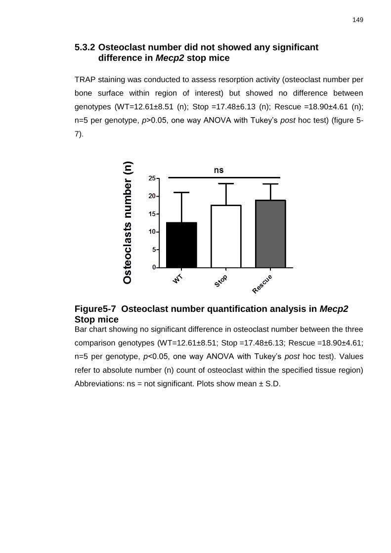
149
5.3.2 Osteoclast number did not showed any significant difference in Mecp2 stop mice
TRAP staining was conducted to assess resorption activity (osteoclast number per
bone surface within region of interest) but showed no difference between
genotypes (WT=12.61±8.51 (n); Stop =17.48±6.13 (n); Rescue =18.90±4.61 (n);
n=5 per genotype, p>0.05, one way ANOVA with Tukey’s post hoc test) (figure 5-
7).
Figure5-7 Osteoclast number quantification analysis in Mecp2 Stop mice Bar chart showing no significant difference in osteoclast number between the three
comparison genotypes (WT=12.61±8.51; Stop =17.48±6.13; Rescue =18.90±4.61;
n=5 per genotype, p<0.05, one way ANOVA with Tukey’s post hoc test). Values
refer to absolute number (n) count of osteoclast within the specified tissue region)
Abbreviations: ns = not significant. Plots show mean ± S.D.

150
5.3.3 Ash density analysis of bone tissues in Mecp2 stop mice
The analysis of mechanical (reduced bone strength), material (reduced bone
hardness measure), structural (thinning of cortical and trabecular bone)
experiments revealed an osteoporotic picture of the bone tissue similar to the one
frequently reported in RTT patients.
A surprising result was the finding of no significant difference in bone mineral
density measures obtained after the μCT (see chapter 4). Decrease in bone
mineral density has been frequently reported in RTT patients (Budden and
Gunness, 2001; Cepollaro et al., 2001; Budden and Gunness, 2003; Shapiro et al.,
2010; Jefferson et al., 2011; Roende et al., 2013b). In order to re confirm the bone
mineral density values derived from μCT, I performed ash weight density test. Ash
weight density is frequently used to asses’ ash content. Studies have been done to
compare the results from bone mineral density and ash weight tests. I also
performed this experiment to confirm the findings of my earlier experiment.
Left tibia was used to analyse the percentage mineralization (ash content) see
above section 4.2.5 for methodology. Results showed no significant difference in
percentage ash content measured on the basis of dry weight measures and in
percentage ash content values measured on the basis of wet weight measures
between genotypes of male mice (figure 5-8).

151
Figure5-8 Ash Content analysis in male and female stop mice (Ai-Aii) Bar charts showed no difference in percentage ash content in male mice
cohort (dry basis) (WT= 62.15± 5.22%; Stop =62.57± 4.25%; Rescue =61.75 ±
3.72%; n =6 per genotype, p>0.05, ANOVA with Tukey’s post hoc test) and (wet
basis) (WT=41.54 ± 11.35%; Stop =40.88 ± 7.92%; Rescue =41.23 ± 5.36%; n=6
per genotype, p>0.05, ANOVA with Tukey’s post hoc test). (Bi-Bii) Percentage ash
content values in Stop female mice showed similar pattern of no significant
difference, in % ash content (dry basis) (WT= 45.58± 6.37%; Stop =45.49± 3.78%;
Rescue =49.55 ± 5.128%; n=3-5 per genotype, p>0.05, ANOVA with Tukey’s post
hoc test) and % ash content (wet basis) (WT= 17.27 ± 26.27%; Stop=15.00 ±
21.99%; Rescue =17.59 ± 26.36%; n=3-5 per genotype, p>0.05, ANOVA with
Tukey’s post hoc test). Abbreviations: ns = not significant, Plots show mean ± S.D.
152
A similar trend of percentage ash content results was observed in female cohort
tibias. No significant difference was found in heterozygous Stop female cohort of
values of percentage ash content calculated on either dry weight basis or wet
weight basis All data given as, mean ± SD (figure 5-8).
5.4 Discussion
The major finding of the current set of experiments was the observation that the
collagen content is appreciably reduced in bone sections stained with sirius red
from MeCP2-defience mice as compared to wild-type littermate mice. Sirius red
stain was first described by Junqueira et al. in 1979 (Junqueira et al., 1979) and is
a dye that binds to the [Gly-x-y] triple-helix structure found in all collagen fibers.
This property of sirius red stain can be utilized to assess collagen in various tissue
sections under bright field and polarized light microscope (Junqueira et al., 1979;
Whittaker et al., 1994) and in cell culture (Walsh et al., 1992).
In my initial analysis, to observe any gross changes in collagen content, I have
used bright field microscope to assess the overall content of collagen in a
specified region of interest per bone per genotype. The Sirius red stained images
were further analysed by using the colour segmentation plugin of Imagej. The
colour segmentation is an Imagej plugin that allows to segment an colour image or
a stack of colour by pixels clustering. I have used the colour segmentation plugin
to segment the bone cells from the extracellular content. The high intensity sirius
red stained collagen content area was identified in each image per slide per bone
by using Imagej software and the collagen content was calculated as a percentage
of the specified area of each image (expressed in pixels).
The results obtained showed mean colour area percentage of collagen in Mecp2
stop mice to be 48.8% which was significantly decreased compared with the
control group (p<0.01). This reduction in % collagen content was found consistent
with the mean colour area percentage of collagen of 47.7% seen in histological
sections of female albino rat (ovariectomy induced) models of osteoporosis (Naim,
2011). The overall picture of reduced collagen content and increase pale stained
space within the extracellular bone matrix found in my study adds to the
osteoporotic picture of RTT bone phenotype. However bone histomorphometric
analysis of collagen content in children and adolescents with RTT is unknown.

153
The magnitude of the reduction in my study was in the range of ~25% and thus it
can be predicted to have a profound effect on overall material and mechanical
properties of bones in these mice. Interestingly, there was a trend towards a
reversal of collagen level deficits in genetically rescued mice (~15% increase
compared to knockout mice) but this change did not achieve statistical
significance. Hence, this study did not provide direct evidence that restoration of
MeCP2 can results in improvement of organic part of extracellular matrix. In RTT
patients bone anomalies have always being considered and investigated in terms
of decrease in bone mineral density, while little attention has been paid to the
organic part of extracellular matrix.
As described earlier the organic (collagenous and non-collagenous proteins) and
inorganic phase (minerals) both contributes towards the bone strength (Marotti et
al., 1994). Collagen fibrils are stiffened by integration of the mineral phase. The
presence of mineral phase increases bone strength; but in woven bone, which is
constituted by unorganized collagen fibrils, the mechanical properties are
decreased despite a high mineral content (Marotti et al., 1994). As majority of
collagen content consists of type 1 form of collagen, my finding of decrease in
collagen content are also consistent with the results of a recent analysis of
biochemical bone marker, in which all the bone markers including N-terminal ,
propeptides of collagen type 1(PNIP), the C-terminal telopeptide cross links (CTX);
and osteocalcin (see section 1.5.4) were found decreased in blood samples taken
from Rett patients (Roende et al., 2014).
Osteoblast plays a major role in collagen homeostasis as it synthesizes and
secretes the C-terminal propeptides of the alpha 1 and alpha 2 chains of type 1
collagen (Johansen et al., 1992). Hence the reduction in collagen content from the
current study can be interpreted as the decreased osteoblast function in Mecp2
stop mice.
Interestingly, MeCP2 has been found to regulate the expression of RANKL gene
(Kitazawa and Kitazawa, 2007) expressed by osteoblasts (Alvarez-Saavedra et
al., 2010)(see section 1.5.4 and 5.1.2 for details of RANKL and its role in bone
homeostasis) further supporting the potential involvement of MeCP2 in
skeletogenesis. Also MeCP2 overexpression, duplication and triplication has been
found to result in diminished ossification resulting in severe kyphosis, a distorted
154
sternum, spina bifida and dysmorphic feature in male TTM20dTg mice (kyphosis,
scoliosis).
Interestingly, RTT bone phenotype, other than osteoporosis has also been linked
to share features with a disorder known as Osteogenesis imperfecta (OI). OI is an
inherited disorder characterized by increased bone fragility with recurrent fractures
mainly caused by the defects in collagen synthesis (Smith et al., 1975; Camacho
et al., 1999). Nonetheless the possible role of collagen defects, in RTT bone
phenotype needs more attention. This current study has limitation as I have
calculated the gross values of collagen content by using the bright field
microscopy. In future studies a polarized light microscopic (Lillian and Whittaker,
2005) examination to identify the collagen structure in detail can give further
details.
Histological experiment to count osteoclast number was also performed to analyse
the resorptive activity of the bone (O'Connor et al., 2009b). Connor and colleague
also looked at the absolute osteoclast number in their Mecp2 null mouse model
and did not find any difference. They relate the non-significance of their findings to
the small number of animals they have used in their study. I wanted to look at the
osteoclast number to see if there is any significant difference in the resorption
activity in bone that is causing all the apparent increase in bone fragility in Mecp2
stop mice. The multinucleated osteoclast-like cells form pits on bone or dentine
slice express high concentrations of 5 isozyme of tartrate-resistant acid
phosphatase (TRAP) (Alatalo et al., 2000).
TRAP is a histochemical marker of the osteoclast. It is also characteristic of
macrophages and other cells of mononhitiocytic lineage (Alatalo et al., 2000;
Hayman et al., 2000). This enzyme partially dephosphorlylates the bone matrix
phophoproteins osteopontin and bone sialoprotein, which have been implicated in
cell attachment (Ek-Rylander et al., 1994). The TRAP “knockout” mouse has
shown that the enzyme is essential for normal mineralization of cartilage in
developing bones and for maintaining the integrity and turnover of the adult
skeleton (Hayman et al., 1996).
As an initial experiment to analyse the bone resorption activity I have calculated
the number of TRAP stained osteoclast within the specified region of interest
155
selected per bone per genotype. TRAP is confirmed as a valid marker for
identification of osteoclasts on the other hand TRAP activity is an osteoclastic
marker of weak sensitivity. This may be due to known fact that synthesis of
enzyme is not being unique to osteoclasts (Ballanti et al., 1997). Osteoclasts are
heterogeneous with respect to cellular size and shape. They used to be
recognised mainly on the basis of multinuclearity and cell width but since these
criteria do not recognize mononucleated osteoclasts, this methodology has been
questioned (Palle et al., 1989).
Tartrate-resistant acid phosphatase (TRAP) is detectable in large amounts in the
lysosomes of osteoclasts and its activity is now considered an established
cytochemical marker useful for recognizing polynucleated as well as
mononucleated osteoclasts in bone sections (Baron et al., 1986). At present, the
number of osteoclasts is determined by counting the number of TRAP-positive
multinucleated osteoclast-like cells (Takahashi et al., 1988).
The results obtained displayed no significant difference of osteoclast number
among the three comparison genotypes of male stop mice. However if we look at
the analysis bar graph an interesting trend of 30% increased osteoclast number in
stop mice and 50% increase in rescue mice as compared to wild type control mice
was found indicating towards the slight increase in bone resorptive activity as
compared to the age matched wild type control. The non-significance of this
experiment results could be the result of high biological variance seen in tissue
samples analysis (figure 5-4).
Our results of no significant difference in osteoclast number were inacordance to
the finding obtained by O’Connor et al. Similar to our finding they did not find any
significant difference in osteoclast number per bone surface (WT= 6.34±2.43,
Mecp2 null= 6.60±2.30) in their TRAP stained tibial section of Mecp2 null mouse
mode. In another study conducted on five RTT girls revealed a decrease in
osteoclast number and surface (Budden and Gunness, 2003). However this study
had the limitations that patients and controls were not age matched and hence
conclusion about resorption activity or rates cannot be inferred from these surface
estimates. On the other hand Motil and colleagues study has displayed increase in
bone turnover (increased resorption to formation) by age (Motil et al., 2008).
Resorptive activity measures in RTT bone phenotype are still unclear and future

156
bone histomorphometric studies need to be done to fully understand the cellular
mechanism of bone resorption in RTT patients.
After the histological experiments in organic part of bone I also looked at the
inorganic (mineral) content of the bone of male and female stop mice by
conducting the ash weight density measurements. Although I already had obtained
the bone mineral density measurement from μCT scans analysis, I performed this
experiment to confirm and validate the finding of μCT mineralisation calculations
as various report in RTT patients have reported decrease in bone mineral density
(Haas et al., 1997; Leonard et al., 1999b; Leonard et al., 1999c; Cepollaro et al.,
2001; Motil et al., 2006; Zysman et al., 2006; Gonnelli et al., 2008; Alvarez-
Saavedra et al., 2010; Shapiro et al., 2010).
No significant difference was seen in wet and dry ash weight density values of
male and female stop mice and the results from the experiment confirmed the
findings of μCT. In adults mammals 20% of bone weight is water, 45% is ash and
35% is organic matrix (Carter and Spengler, 1978). On a dry weight bases,
mineral content is 65 to 70% and organic matrix is 30 to 35%. In this current study
Mecp2 stop male mice showed a 62% of ash content on dry basis which was
comparable to wild type and rescue values and 41% ash content based on wet
basis and consistent with the normal range. Compare to male mice, female stop
mice displayed 45% ash content analysis on dry weight basis and 15% on wet
weight basis, this apparent decrease in density measures obtained from male and
female stop cohorts was gender and age appropriate (Henry and Eastell, 2000).
Overall similar to male stop mice cohort, female mice cohort showed no significant
difference among the three comparison genotypes. O’ Connor and colleagues in
their Mecp2 null mouse model, had displayed a modest reduction of bone mineral
density and bone mineral content values (O'Connor et al., 2009b) but results does
not reach the statistical significance. Also a recent study (Roende et al., 2014) on
bone metabolism of patients with RTT has revealed some interesting insights.
Bone metabolism can be characterized by biochemical markers of bone formation,
resorption, mineralization and turnover (Szulc et al., 2000; Jürimäe, 2010).
Roende and colleagues characterise bone metabolism in RTT patients, by
comparing biochemical bone markers levels in RTT patients with healthy controls.
They found that both markers of bone formation the N-terminal propeptides of
157
collagen type 1 (PINP); markers of bone resorption, the C-terminal telopeptide
cross links (CTX); of bone turnover, osteocalcin (OC); and of bone mineralization,
bone specific alkaline phosphatase (B-ALP) were significantly reduced.
Interestingly they also found no significant association between bone markers
levels and volumetric bone mineral density calculations obtained from DXA scans
of lumbar spine and femur neck of the patients. The difference of bone mineral
density value obtained from this study and previous studies (Cepollaro et al., 2001;
Motil et al., 2006; Gonnelli et al., 2008) could be because the Roende et al
adjusted their analysis of biochemical bone markers for age and puberty while
previous studies have only analysed biochemical markers of bone formation in
RTT children and young adults less than of 25 years of age. Studies
(Rauchenzauner et al., 2007; Tuchman et al., 2008; Jürimäe, 2010) on
biochemical bone markers in healthy persons have reported high levels in early
childhood, peaking in puberty and decreasing to stable levels in the mid-20s.
Children with disabilities and limited mobility are at increased risk of osteoporosis
(Aronson and Stevenson, 2012). RTT patients display osteoporotic bone
phenotype (Zysman et al., 2006) and have been compared with cerebral palsy
(CP) patients in past to analyse the bone mineral density values (Haas et al.,
1997). Interestingly biochemical markers (OC, B-ALP, and N- telopeptides) in
cerebral palsy (CP) patients with motor deficiencies have shown a wide variety in
serum levels (Henderson et al., 1995; Henderson et al., 2002) and no significant
association with measures of bone mass as BMD z scores of the lumbar spine and
distal femur region have been found (Henderson et al., 2002).
Moreover another study comparing CP and healthy children reported no significant
difference in both formation and resorption markers (Chen et al., 2011). Thus the
reduced level of bone turnover seen in children and adolescents with RTT could
be a direct effect of MECP2 gene mutation, although levels of bone markers did
not differ between different mutation groups in patients with RTT (Roende et al.,
2014). This could be due to an overall general effect of MECP2 on regulation of
growth and bone turnover. This is corroborated by my findings of decrease cortical
bone volume, alteration in trabecular structure, reduced bone strength in Mecp2
stop mouse model along with the growth plate abnormalities, decreased femoral
trabecular and cortical bone volume and decreased bone mineral apposition rates
seen in other Mecp2 mouse models (O'Connor et al., 2009a; Shapiro et al., 2010).

158
The results obtained from this study are quite interesting. As described earlier, in
my initial experiments I did find a difference in bone strength and structural
parameters in Mecp2 stop mice. In this current study, I found a possible defect
(reduced collagen content) in organic part and not the traditionally considered
inorganic part (ash weight density) of extracellular matrix to be responsible for
bone fragility.
Future experiments need to be more focused on the organic part of extracellular
matrix in order to determine the possible mechanisms of bone fragility commonly
seen in RTT patients.

159
Chapter 6
General discussion
The overall aim of the experiments described in this thesis was to explore the
effects of MeCP2 protein on bone tissue using various biomechanical, radiological
and anatomical techniques applied to mice in which the Mecp2 gene was
functionally silenced. A secondary theme was to assess whether such features are
potentially reversible by genetic rescue of the Mecp2 gene. This has important
therapeutic implications as it would predict whether future gene-based therapeutic
interventions are likely to impact on bone phenotypes. In this chapter I will
integrate the major findings of this thesis, the significance of these findings and
finally, I will discuss possible future experiments to extend this work and derive the
maximum benefits from the results I have obtained to date.
6.1 Major findings of the study
My first aim was to see whether inactivating the Mecp2 gene in male and female
mice will result in overt bone phenotypes. My study is only the second study to
explore RTT-like bone phenotype in mice and the first study to investigate such
phenotypes in female mice as well as to investigate biomechanical properties of
bone. Female Mecp2+/stop mice are a gender appropriate and accurate genetic
model of RTT yet they display a more subtle and delayed onset (4-12 months) of
neurological features (Guy et al., 2007; Robinson et al., 2012a) compared to
hemizygous male mice. Male Mecp2-null mice typically become symptomatic by
the age of 5-8 weeks (Guy et al., 2007) and because of this rapid and severe
disease trajectory are more commonly used in preclinical studies (Gadalla et al.,
2011). In keeping with this pattern, the results of my work revealed a more
pronounced bone phenotype in male stop mice and subtle or undetectable bone
phenotypes in heterozygous female mice.
At the outset of the project there was no published literature on the expression of
MeCP2 in bone. Therefore, an important initial experiment was to assess this in a

160
MeCP2-GFP reporter mouse line in which MeCP2 protein with an C-terminal GFP
tag (McLeod et al., 2013) is expressed under its endogenous promoter and
regulation. In male mice the MeCP2-GFP was observed to co-localise with DAPI in
the nucleus but was absent elsewhere. This is consistent with MeCP2 displaying a
similar nuclear-specific expression pattern as reported in brain neurons as well as
other tissues. These experiments also showed heterozygous Mecp2+/stop mice to
display punctate nuclear GFP fluorescence in ~50% of nuclei and no detectable
MeCP2-GFP in the other cells. The heterozygous Mecp2 stop female mice in
which one Mecp2 allele is inactivated mimic the mosaic expression seen in human
Rett syndrome (figure 3-8).
Having established the presence of MeCP2 in bone and the efficient silencing of
the gene in the Mecp2-stop mouse line the results of my subsequent anatomical
and biomechanical phenotyping revealed that Mecp2 stop male mice display a
range of abnormal skeletal phenotypes that shares many of the features seen in
clinical cased of RTT. Morphometric analysis revealed that the long bones of
Mecp2 stop male mice are lighter and shorter as compared to the age matched
wild type group. This finding correlates with the growth retardation pattern seen
commonly in RTT patients (Neul et al., 2010). Male stop mice are also known to
display skeletal dysmorphic feature of kyphosis, which is comparable to
pathogenomic ‘S’ type of curvature seen in RTT patients (Ager et al., 2006; Koop,
2011; Riise et al., 2011).
One of the major finding of the current study came after the biomechanical
analysis of male and female stop mice. The robust deficits were seen in
mechanical properties were profound (39.5% reduction in stiffness in the three
point bending test; 31% in load and 12.3% reduction in micro hardness) and could
explain the weakness in MeCP2 deficient bone that accounts for the increasing
occurrence of low energy fractures reported in Rett syndrome patients (Roende et
al., 2011b). Frequent findings of reduction in bone strength (brittle bones) and
hence increase incidence of fracture has given human RTT bone phenotype an
overall osteoporotic bone phenotype picture (Haas et al., 1997; Zysman et al.,
2006). However, the current studies do not discriminate whether the observed

161
defects arise from absence of MeCP2 protein or MeCP2 per se (ie. a primary
pathology) or whether global silencing of Mecp2 throughout the body results in
endocrine or systems defects elsewhere in the body that results a secondary
pathology within the skeletal system. This question could potentially be address by
using bone-linage specific cre lines. In terms of the potential secondary pathology,
it is important to consider that the male mouse tissues used in the study were
harvested at a time point at which the Mecp2-stop mice where showing fairly
severe and advanced clinical signs (tremor, abnormal breathing, reduced
locomotor activity and general poor condition) and these non-bone-related
phenotypes may be an important factor. In contrast, the female heterozygous
mice, despite being tested at a much later (many months rather than weeks)
display similar non-bone phenotypes, albeit with much reduced severity (Guy et
al., 2007).
Another important finding of my work came after the structural analysis of cortical
and trabecular bone. Robust significant differences were seen in Mecp2 stop
mouse cortical and trabecular bone including a pronounced 54% reduction in
cortical thickness, 30% in bone volume, 20% in total area, 38% in marrow area
and 30% in trabecular thickness. Such findings are consistent with what is
expected in an osteoporotic model (Seeman, 2008b).
Female stop mice bone phenotype results remained consistent with their overall
subtle bone phenotypes and didn’t show an overt difference in morphometric
measurements of long bone length and weight measurements. Within the nervous
system there is evidence for both cell autonomous and non-cell autonomous
effects of MeCP2-deficiency including morphological changes in dendritic
architecture (Ballas et al., 2009). Whilst it is clear from the results of the current
experiments that various bone phenotypes are more subtle, absent or below the
level of detection in hemizygous females compared to equivalent measures in
males, it is unclear whether this is indeed due to the fact that only ~50% of bone
cells are expressing MeCP2 or whether dysfunction in other systems are
contributing to the subtle phenotypes. Nonetheless, a similar significant reduction
(14%) in micro hardness and a trend towards reduced biomechanical properties
162
seen in female mice that are heterozygous and mosaic for the mutant allele,
demonstrate that the bone deficits are not restricted to the more severe male RTT-
like phenotype but are also seen in a gender and MeCP2 expression pattern
appropriate model of RTT.
An important finding of the current study and one with therapeutic significance is
that the bone anomalies observed in terms of cortical bone material and
biomechanical properties were rescued by delayed postnatal activation of the
Mecp2 gene. Male rescue mice displayed a robust improvement in mechanical
(stiffness 40%, ultimate load 10%, young’s modulus 61% and micro hardness
12%) and structural bone parameters (trabecular thickness 80%) as compared to
Mecp2 stop male mice. Similarly another major finding was the rescue of female
bone phenotype in female stop mice. Female rescue mice displayed a significant
improvement in bone material properties (micro hardness 19%) and a trend of
improvement in mechanical properties (stiffness, load) as compared to stop mice.
These finding of rescue of bone phenotype in stop mice were consistent with the
improvements seen in multiple non-bone phenotypes seen in the Mecp2Stop/y mice
after delayed activation of the Mecp2 gene including survival, normalized
bodyweight, locomotor and behavioural activities (Guy et al., 2007; Robinson et
al., 2012a).
These results were quite significant as they suggest that the bone anomalies seen
in RTT patients may be at least partially reversible using gene-based approaches
currently under development (Gadalla et al., 2013; Garg et al., 2013). However, it
is also possible that significant amelioration of bone phenotypes may also be
achieved by using pharmacological strategies (Gadalla et al., 2011; Garg et al.,
2013). In order to apply all these therapeutic intervention most important aspect is
to identify the mechanisms by which MeCP2 deficiency results in altered bone
mechanical, material and structural properties. In my study I have found that
MeCP2 is expressed in osteocytes (figure 3-8), but the protein is widely expressed
throughout the body and it is possible that metabolic and endocrine factors can
influence the bone homeostasis (Motil et al., 2006; Motil et al., 2011; Motil et al.,
2012).

163
A surprising finding of the current study was the absence of any significant
difference in ash weight density or bone mineral density (µCT) in stop male and
female cohorts. Our findings of no significant difference in bone mineral density
(BMD) in Mecp2 mice were similar to the one reported in a Mecp2 null mouse
model (O'Connor et al., 2009a) while differs with other animal study in which bone
mineral density were found reduced in Mecp2 null mouse model (Shapiro et al.,
2010). These are the only two animal model studies conducted in past to explore
the RTT bone phenotype using the Mecp2 null mouse model. The relationship
between RTT bone phenotype and bone mineral density values in animal studies
is still unclear.
Reduced bone mass is commonly associated with osteoporotic phenotypes and
indeed these have been reported in RTT patients (Hui et al., 1988; Leonard et al.,
1999c; Cummings et al., 2002; Ager et al., 2006; Flynn et al., 2007). Several
clinical, density x-ray absorptiometry (DXA) studies (Haas et al., 1997; Leonard et
al., 1999a; Cepollaro et al., 2001; Motil et al., 2008; Shapiro et al., 2010) in RTT
patients have shown low absolute values of BMC (g) and or BMD (g/cm2)
compared to age-matched controls. The problem with use of DXA scan for
assessment of bone mineral values is that the size adjusted absolute DXA values
of aBMD (g/cm2) may lead to interpretation of a relatively lower bone density
among RTT patients than is actually the case (Roende et al., 2011a). Nonetheless
these findings of bone mineral density from both µCT and ash content analysis are
very interesting. They points towards the further need and importance of
exploration of other possible factors (e.g. cellular dysfunction and alterations in the
extracellular protein matrix) involved in the robust reduction of bone strength and
structural parameters seen in Mecp2 stop mice. Furthermore reversal of bone
integrity (bone stiffness, hardness, trabecular thickness) seen in rescue mice after
the gene reactivation leads to assess the mechanisms by which bone structure
and properties are dynamically regulated by MeCP2 levels. As stated, it is also
necessary to assess whether the influence of MeCP2 on bone homeostasis is a
primary or secondary mechanism.
A number of studies have been conducted to investigate the role of specific genes,
gene pathways and biochemical networks involved in the regulation of bone

164
homeostasis (Elefteriou et al., 2014; Quiros-Gonzalez and Yadav, 2014).
Studies involving leptin and neuropeptide Y2 have disclosed unrecognized
interactions between the central nervous system, peripheral neurotransmitters and
osteoblast function (Allison et al., 2007; Abdala et al., 2013; Elefteriou et al., 2014;
Quiros-Gonzalez and Yadav, 2014). Several reports suggest that MeCP2 is an
important regulator of neuronal gene expression (Skene et al., 2010; Guy et al.,
2011b)). Neurological studies suggest that MeCP2 can affect osteoblast function
by altering osteoblast chromatin structure as already seen in brain tissue or by
altering cell maturation as observed in RTT neuronal tissues (Budden and
Gunness, 2003; Chadwick and Wade, 2007). However, the precise role played by
MeCP2 in the nucleus remains unclear (Chahrour et al., 2008; Skene et al., 2010;
Guy et al., 2011a; Li et al., 2013a), but it is generally considered to regulate gene
expression.
Another important finding of my studies and one which may relate to aberrant
gene expression is the effect of MeCP2 deficiency on collagen content. As
collagen is the most abundant gene product and structural determinant in bone, I
conducted an initial analysis of collagen content and distribution using sirius red
staining. The decreased levels (25% as compared to age matched wild type
genotype) of intense sirius red stain observed in the MeCP2-deficient mice is
consistent with reduced PINP (bone formation markers) levels in human RTT
(Roende et al., 2013a) and the patches of reduced staining resemble those
features characteristic of early osteoporosis (Leonard et al., 1999a). Whilst my
work suggests that deregulation of collagen may be a significant potential
mechanism underlying RTT-related bone phenotypes, further studies would be
required to assess whether altered collagen level or altered balance of different
collagen subtypes may result as a direct consequence of MeCP2 deficiency.
In addition to structural protein, we also investigated the resorptive properties of
the bone in terms of TRAP staining. The lack of any difference in osteoclast
number between genotypes is consistent with a previous report (O'Connor et al.,
2009a) and suggests the possible absence of any primary defect in bone
remodelling.

165
My findings of osteoclasts were also consistent with the bone histomorphometric
analysis performed by Budden and colleagues (Budden and Gunness, 2003).
Although no difference in resorptive parameter (osteoclast number) was found,
conclusions about the resorption activity or rates cannot be inferred from these
surface estimates.
Overall, based on all these major findings of my study it could be said that Mecp2
stop mice display, osteopathic features of RTT (reduced bone strength, decrease
in cortical and trabecular thickness, decrease in collagen content and tendency
towards spinal curvature) which are also similar to those reported in collagen type
1 genetic disorder (osteogenesis imperfecta; brittle bone disease) (Dogba et al.,
2013) pointing towards the possible importance of collagen defects in RTT. The
RTT bone phenotype has previously been linked with osteogenesis imperfecta
(OI) in past (Loder et al., 1989). Indeed, one patient in this study who suffered
from increased rate of fractures had originally been given a primary diagnosis of
osteogenesis imperfecta before the Rett syndrome was diagnosed.
Animal’s studies and studies in human subjects suggest that skeletal fragility in
osteogenesis (OI) is due to the defect in collagen synthesis, whereas the
abnormalities in bone turnover and mineral are inconsistent. These findings of
reduced collagen and no significant difference in bone mineral density were similar
to the one I have observed in my analysis of skeletal phenotype of RTT. The
collagen abnormalities seen in OI are the result of the two type 1 collagen genes,
mutation COLIA1 and COLIA2. Over 200 mutation types have been reported. Two
main classes of type 1 collagen mutations have been described (Seeman and
Delmas, 2006). The first “null allele” mutation affects the pro-alpha1 or pro-alpha2
allele that impairs transcription and mRNA stability and produce low amounts of
the secreted heterodimer. The abnormal heterodimers are incorporated into
matrix, resulting in a quantitative and qualitative abnormal bone matrix (Seeman
and Delmas, 2006). These results of quantitative abnormality of bone matrix were
consistent with my finding of quantitative analysis of Mecp2 stop mice showing
reduction of 30% in collagen content and a recent study on bone biochemical
markers (Roende et al., 2013a).
166
Whatever the mutation in OI, there is less bone synthesized (Seeman and
Delmas, 2006), a feature that may parallel the bone phenotypes seen in RTT
(Budden and Gunness, 2003; O'Connor et al., 2009a). Moreover abnormal
collagen fibrils may be unable to provide nucleating and scaffolding sites for
mineral propagation. Such mechanism point towards a hypothesis that low mineral
density seen in RTT patients could result from a direct alteration in collagen
homeosynthesis.
As mentioned earlier, although RTT-like bone phenotypes are frequently linked to
an osteoporosis bone picture (Zysman et al., 2006), the biomechanical, structural
and histological finding from my study has features consistent with both
osteoporosis as well as those of OI bone phenotypes.
A mouse model having well-defined genetic mutations on the COLIA2 gene
produces aplpha1 collagen homotrimers and non-functional pro alpha2 chains.
These oim/oim mouse modelling human OI, displayed a bone phenotype
characterized by spontaneous fractures and limb deformities (Camacho et al.,
1999), both of these features are commonly reported in RTT(Guidera et al., 1991a;
Roende et al., 2011b). OI, oim/oim mouse model (Camacho et al., 1999) also had
displayed mechanical defects (30% decrease in stiffness, 20% decrease in
collagen content, reduced trabecular thickness and an unchanged mineral content,
but with a decreased mineral crystallinity). Other than mineral crystalline results,
all the features of RTT bone phenotype in my Mecp2 stop mouse study were
qualitatively and indeed quantitatively similar (39.5% reduced stiffness, ~30%
decrease in trabecular thickness, 25% decrease in collagen content and no
difference in bone mineral deficits) suggesting that RTT bone phenotype shares
common features with OI bone phenotype and that specific material properties
such as mineral crytallinity and collagen content commonly seen in oim mice,
could also be indicative and possibly predictive of bone fragility seen in RTT
patients. Leonard and colleagues (Leonard et al., 1999a) did mentioned these
similarities between OI bone phenotype and RTT bone phenotype but since then
not much attention has been given both by human and animal based trials to
explore further qualitative and quantitative dysfunction in organic part of
extracellular matrix as a possible causes of bone fragility. To investigate the
mineral crystalline structure directly, I have already initiated synchrotron X-ray
nanomechanical (SAXS) imaging of mineralized fibre composites on Mecp2 stop

167
mice humerus. I have performed this research experiment at MAX Lab at Lund
University (Sweden). Results and analysis are in the process. In situ synchrotron
X-ray scattering and diffraction, in combination with micromechanical testing can
provide quantitative information on the nanoscale mechanics of bio mineralized
composites of bone. In bone carbonated apatite forms a composite fibril with type
1 collagen of diameter ~ 50-200nm, which forms plywood-like lamellae in the bone
with widths ~5-10μm that in turn form cylindrical osteons at the scale of ~100-
200μm. Due to this well known hierarchical organization the structural and
mechanical properties of the mineral/protein composite at small (submicron)
scales are crucial to the mechanical function of the entire organ (Reznikov et al.,
2014).
6.2 Significance of the study
Given the very limited previous attempts to explore the effects of Mecp2 on
skeletal tissue, my current studies brings significant advance understandings that
Mecp2 has a role in skeletal tissue regulation since inactivation of Mecp2 resulted
in RTT like bone phenotype in stop mouse and reactivation of Mecp2 resulted in
improvement of some of these defects identified in stop mice. My study is the first
study to use female stop mice which is a genetically accurate mouse model of
RTT, and the results obtained from stop mice bone phenotype analysis will
contribute towards the pre clinical trials of gene therapy intervention.
Fractures due to osteoporosis are a major cause of long term dysfunction and
even death (Heaney, 2003) in individuals with physical and mental disabilities
(Gray et al., 1992; Lingam and Joester, 1994). Lack of soft tissue padding,
inappropriate postural reactions, and lack of bone strength as major contributors to
fractures and these factors are common among individual with RTT. For these
reasons a thorough knowledge of mechanisms by which Mecp2 regulates and
participates in bone homeostasis is required. Biomechanical and structural
findings from my study will contribute towards the better understanding of these
parameters and their link with MeCP2.
My study results along with other recent studies (Roende et al., 2013a) have found
a defect most probably linked to bone formative factors (decrease in collagen
content and no difference in osteoclast number) rather than bone resorptive

168
measures. In this scenario, use of pharmacological antiresorptive measures
seems less useful by further decreasing the activity of osteoclasts and the
osteoblast. In contrast anabolic bone treatment stimulating bone formation may be
more relevant, but caution should be taken due to possible medical side effects
regarding risk of inducing uncontrolled bone formation in childhood (Tashjian and
Goltzman, 2008).
Also my study is the first study to provide initial quantitative analysis of collagen
protein in RTT bone phenotype. Majority of the clinical trials in past have been
focused to explore the inorganic part of the bone and not much research has been
done to explore the organic part of the bone. My study highlights the importance of
more insights into the collagen as underlying mechanism for the RTT bone
phenotype
And lastly the most important significance of this study is the use of adult male and
female rescue mice. Reversal studies (Guy et al., 2007; Robinson et al., 2012a;
Gadalla et al., 2013; Garg et al., 2013) in stop mice have shown potential
reversibility of RTT-like phenotypes. In this study I have shown the first evidence
that RTT bone phenotype can be prevented/ improved by genetic manipulation.
These data in rescue mice however at the proof of concept level should have an
impact on the future therapeutic approaches not only just for RTT bone phenotype
but for better bone health in other related bone pathologies (osteoporosis,
osteogenesis imperfecta etc) for which gene based therapies might eventually
have therapeutic potential.
6.3 Future studies
The nature of this project has been to explore the consequence of inactivation of
MeCP2 on bone tissue using an animal model of RTT. This represents a logical
trajectory in reaching the ultimate goal of exact mechanism by which MECP2 is
linked with the skeletal tissue:
In my current study I have used a functional knockout mouse model in
which Mecp2 is silenced throughout the body including the nervous system.
In order to discriminate where the bone phenotypes I have identified result
primarily from local intrinsic bone dysfunction or whether central (nervous

169
system) dysfunction result in secondary bone phenotypes, experiments with
different mouse cre lines may be informative. For instance mouse lines
have been developed in which Mecp2 is activated or inactivated only in the
nervous system or only in peripheral tissues or other organ systems
(Alvarez-Saavedra et al., 2007; Alvarez-Saavedra et al., 2010; Chao et al.,
2010; Nguyen et al., 2012). An analysis of bone measures in these models
would be highly informative.
The role of collagen in the pathogenesis of RTT bone phenotype has not
been explored yet. Basic validation of altered collagen level by techniques
like immunoblotting would strengthen this conclusion whereas a better
understanding of collagen (matrix) structure in bone from RTT model using
approaches such as Small energy X-Ray (SAXS), a nanomechanical
imaging of mineralized fibre composites can provide useful insights about
the exact structural changes that might result from MeCP2 insufficiency
In my current study I have used an adult mouse model, in future, animal
studies need to be done to explore the possible effects of MECP2 on the
bone mass formation and later on ossification of bone, using the embryonic
and early postnatal murine models. These experiments will give a better
understanding of the exact time points over which the deleterious effects of
MeCP2 deficiency start impacting bone health/properties. This will also help
in the development of future therapeutic strategies and time point at which
they should be implemented.
Based on the findings of this study, the reduced bone size reduced mechanical
properties of bone, altered structural parameters and extracellular organic deficits
in an adult stop mouse model points towards the fact that MeCP2 has a general
role in regulating bone growth. Improved knowledge of how it involved in bone
metabolism is important to assist directions for prevention and treatment in order
to improve bone health in RTT.

170
References
Abdala AP, Lioy DT, Garg SK, Knopp SJ, Paton JF, Bissonnette JM (2013) Effect of Sarizotan, a 5-HT1a and D2-Like Receptor Agonist, on Respiration in Three Mouse Models of Rett Syndrome. Am J Respir Cell Mol Biol.
Adachi M, Keefer EW, Jones FS (2005) A segment of the Mecp2 promoter is
sufficient to drive expression in neurons. Hum Mol Genet 14:3709-3722. Adams VH, McBryant SJ, Wade PA, Woodcock CL, Hansen JC (2007) Intrinsic
disorder and autonomous domain function in the multifunctional nuclear protein, MeCP2. J Biol Chem 282:15057-15064.
Ager S, Fyfe S, Christodoulou J, Jacoby P, Schmitt L, Leonard H (2006) Predictors
of scoliosis in Rett syndrome. J Child Neurol 21:809-813. Akhter MP, Fan Z, Rho JY (2004a) Bone intrinsic material properties in three
inbred mouse strains. Calcif Tissue Int 75:416-420. Akhter MP, Wells DJ, Short SJ, Cullen DM, Johnson ML, Haynatzki GR, Babij P,
Allen KM, Yaworsky PJ, Bex F, Recker RR (2004b) Bone biomechanical properties in LRP5 mutant mice. Bone 35:162-169.
Alatalo SL, Halleen JM, Hentunen TA, Mönkkönen J, Väänänen HK (2000) Rapid
screening method for osteoclast differentiation in vitro that measures tartrate-resistant acid phosphatase 5b activity secreted into the culture medium. Clin Chem 46:1751-1754.
Allanson JE, Hennekam RC, Moog U, Smeets EE (2011) Rett syndrome: a study
of the face. Am J Med Genet A 155A:1563-1567. Allison SJ, Baldock PA, Herzog H (2007) The control of bone remodeling by
neuropeptide Y receptors. Peptides 28:320-325. Alvarez-Saavedra M, Sáez MA, Kang D, Zoghbi HY, Young JI (2007) Cell-specific
expression of wild-type MeCP2 in mouse models of Rett syndrome yields insight about pathogenesis. Hum Mol Genet 16:2315-2325.
Alvarez-Saavedra M, Carrasco L, Sura-Trueba S, Demarchi Aiello V, Walz K, Neto
JX, Young JI (2010) Elevated expression of MeCP2 in cardiac and skeletal tissues is detrimental for normal development. Hum Mol Genet 19:2177-2190.
Amir RE, Van den Veyver IB, Wan M, Tran CQ, Francke U, Zoghbi HY (1999) Rett
syndrome is caused by mutations in X-linked MECP2, encoding methyl-CpG-binding protein 2. Nat Genet 23:185-188.
Amir RE, Van den Veyver IB, Schultz R, Malicki DM, Tran CQ, Dahle EJ, Philippi
A, Timar L, Percy AK, Motil KJ, Lichtarge O, Smith EO, Glaze DG, Zoghbi

171
HY (2000) Influence of mutation type and X chromosome inactivation on Rett syndrome phenotypes. Ann Neurol 47:670-679.
Anderson A, Wong K, Jacoby P, Downs J, Leonard H (2014) Twenty years of
surveillance in Rett syndrome: what does this tell us? Orphanet J Rare Dis 9:87.
Armstrong DD, Dunn JK, Schultz RJ, Herbert DA, Glaze DG, Motil KJ (1999)
Organ growth in Rett syndrome: a postmortem examination analysis. Pediatr Neurol 20:125-129.
Aronson E, Stevenson SB (2012) Bone health in children with cerebral palsy and
epilepsy. J Pediatr Health Care 26:193-199. Baldock PA, Sainsbury A, Couzens M, Enriquez RF, Thomas GP, Gardiner EM,
Herzog H (2002) Hypothalamic Y2 receptors regulate bone formation. J Clin Invest 109:915-921.
Balemans W et al. (2001) Increased bone density in sclerosteosis is due to the
deficiency of a novel secreted protein (SOST). Hum Mol Genet 10:537-543. Ballanti P, Minisola S, Pacitti MT, Scarnecchia L, Rosso R, Mazzuoli GF, Bonucci
E (1997) Tartrate-resistant acid phosphate activity as osteoclastic marker: sensitivity of cytochemical assessment and serum assay in comparison with standardized osteoclast histomorphometry. Osteoporos Int 7:39-43.
Ballas N, Lioy DT, Grunseich C, Mandel G (2009) Non-cell autonomous influence
of MeCP2-deficient glia on neuronal dendritic morphology. Nat Neurosci 12:311-317.
Baron R, Neff L, Tran Van P, Nefussi JR, Vignery A (1986) Kinetic and
cytochemical identification of osteoclast precursors and their differentiation into multinucleated osteoclasts. Am J Pathol 122:363-378.
Bassett GS, Tolo VT (1990) The incidence and natural history of scoliosis in Rett
syndrome. Dev Med Child Neurol 32:963-966. Bebbington A, Downs J, Percy A, Pineda M, Zeev BB, Bahi-Buisson N, Leonard H
(2012) The phenotype associated with a large deletion on MECP2. Eur J Hum Genet.
Bebbington A, Anderson A, Ravine D, Fyfe S, Pineda M, de Klerk N, Ben-Zeev B,
Yatawara N, Percy A, Kaufmann WE, Leonard H (2008) Investigating genotype-phenotype relationships in Rett syndrome using an international data set. Neurology 70:868-875.
Ben-Ari Y, Spitzer NC (2010) Phenotypic checkpoints regulate neuronal
development. Trends Neurosci 33:485-492. Ben-Shachar S, Chahrour M, Thaller C, Shaw CA, Zoghbi HY (2009) Mouse
models of MeCP2 disorders share gene expression changes in the cerebellum and hypothalamus. Hum Mol Genet 18:2431-2442.

172
Berg M, Hagberg B (2001) Rett syndrome: update of a 25 year follow-up investigation in Western Sweden--sociomedical aspects. Brain Dev 23 Suppl 1:S224-226.
Bienvenu T, Chelly J (2006) Molecular genetics of Rett syndrome: when DNA
methylation goes unrecognized. Nat Rev Genet 7:415-426. Bienvenu T, Carrie A, de Roux N, Vinet MC, Jonveaux P, Couvert P, Villard L,
Arzimanoglou A, Beldjord C, Fontes M, Tardieu M, Chelly J (2000) MECP2 mutations account for most cases of typical forms of Rett syndrome. Hum Mol Genet 9:1377-1384.
Bird A (2002) DNA methylation patterns and epigenetic memory. Genes Dev 16:6-
21. Blömer U, Naldini L, Verma IM, Trono D, Gage FH (1996) Applications of gene
therapy to the CNS. Hum Mol Genet 5 Spec No:1397-1404. Boivin G, Meunier PJ (2002) The degree of mineralization of bone tissue
measured by computerized quantitative contact microradiography. Calcif Tissue Int 70:503-511.
Bonnet N, Laroche N, Vico L, Dolleans E, Courteix D, Benhamou CL (2009)
Assessment of trabecular bone microarchitecture by two different x-ray microcomputed tomographs: a comparative study of the rat distal tibia using Skyscan and Scanco devices. Med Phys 36:1286-1297.
Boskey A (2003) Mineral changes in osteopetrosis. Crit Rev Eukaryot Gene Expr
13:109-116. Bourrin S, Palle S, Genty C, Alexandre C (1995) Physical exercise during
remobilization restores a normal bone trabecular network after tail suspension-induced osteopenia in young rats. J Bone Miner Res 10:820-828.
Boyce BF, Hughes DE, Wright KR, Xing L, Dai A (1999) Recent advances in bone
biology provide insight into the pathogenesis of bone diseases. Lab Invest 79:83-94.
Braunschweig D, Simcox T, Samaco RC, LaSalle JM (2004) X-Chromosome
inactivation ratios affect wild-type MeCP2 expression within mosaic Rett syndrome and Mecp2-/+ mouse brain. Hum Mol Genet 13:1275-1286.
Brodt MD, Ellis CB, Silva MJ (1999) Growing C57Bl/6 mice increase whole bone
mechanical properties by increasing geometric and material properties. J Bone Miner Res 14:2159-2166.
Brown JP, Delmas PD, Arlot M, Meunier PJ (1987) Active bone turnover of the
cortico-endosteal envelope in postmenopausal osteoporosis. J Clin Endocrinol Metab 64:954-959.
Brunner R, Gebhard F (2002) [Neurogenic spinal deformities. I. Conservative and
surgical treatment of spinal deformities]. Orthopade 31:51-57.

173
Bucaro MA, Fertala J, Adams CS, Steinbeck M, Ayyaswamy P, Mukundakrishnan K, Shapiro IM, Risbud MV (2004) Bone cell survival in microgravity: evidence that modeled microgravity increases osteoblast sensitivity to apoptogens. Ann N Y Acad Sci 1027:64-73.
Bucay N, Sarosi I, Dunstan CR, Morony S, Tarpley J, Capparelli C, Scully S, Tan
HL, Xu W, Lacey DL, Boyle WJ, Simonet WS (1998) osteoprotegerin-deficient mice develop early onset osteoporosis and arterial calcification. Genes Dev 12:1260-1268.
Buckwalter JA, Glimcher, M.J, Cooper,R.R and Recker,R. (1995) Bone biology.
JBone Joint SurgAm 8:1276-1289. Budden SS (1995) Management of Rett syndrome: a ten year experience.
Neuropediatrics 26:75-77. Budden SS, Gunness ME (2001) Bone histomorphometry in three females with
Rett syndrome. Brain Dev 23 Suppl 1:S133-137. Budden SS, Gunness ME (2003) Possible mechanisms of osteopenia in Rett
syndrome: bone histomorphometric studies. J Child Neurol 18:698-702. Burger EH, Klein-Nulend J (1999) Mechanotransduction in bone--role of the
lacuno-canalicular network. FASEB J 13 Suppl:S101-112. Burr DB (2002) Targeted and nontargeted remodeling. Bone 30:2-4. Calfa G, Percy AK, Pozzo-Miller L (2011) Experimental models of Rett syndrome
based on Mecp2 dysfunction. Exp Biol Med (Maywood) 236:3-19. Camacho NP, Hou L, Toledano TR, Ilg WA, Brayton CF, Raggio CL, Root L,
Boskey AL (1999) The material basis for reduced mechanical properties in oim mice bones. J Bone Miner Res 14:264-272.
Campos-Castelló J, Peral Guerra M, Riviere Gómez A, Oliete García F, Herranz
Tanarro J, Toledano Barrero M, Espinar Sierra J, Cristobal Sassot S, Lautre Ecenarro MJ, Franco Carcedo C (1988) [Rett's syndrome: study of 15 cases]. An Esp Pediatr 28:286-292.
Caplan AI (1991) Mesenchymal stem cells. J Orthop Res 9:641-650. Carter DR, Spengler DM (1978) Mechanical properties and composition of cortical
bone. Clin Orthop Relat Res:192-217. Carter DR, Bouxsein ML, Marcus R (1992) New approaches for interpreting
projected bone densitometry data. J Bone Miner Res 7:137-145. Cass H, Reilly S, Owen L, Wisbeach A, Weekes L, Slonims V, Wigram T,
Charman T (2003) Findings from a multidisciplinary clinical case series of females with Rett syndrome. Dev Med Child Neurol 45:325-337.
Cepollaro C, Gonnelli S, Bruni D, Pacini S, Martini S, Franci MB, Gennari L, Rossi
S, Hayek G, Zappella M, Gennari C (2001) Dual X-ray absorptiometry and

174
bone ultrasonography in patients with Rett syndrome. Calcif Tissue Int 69:259-262.
Chadwick LH, Wade PA (2007) MeCP2 in Rett syndrome: transcriptional repressor
or chromatin architectural protein? Curr Opin Genet Dev 17:121-125. Chahrour M, Zoghbi HY (2007) The story of Rett syndrome: from clinic to
neurobiology. Neuron 56:422-437. Chahrour M, Jung SY, Shaw C, Zhou X, Wong ST, Qin J, Zoghbi HY (2008)
MeCP2, a key contributor to neurological disease, activates and represses transcription. Science 320:1224-1229.
Chambers TJ (1982) Osteoblasts release osteoclasts from calcitonin-induced
quiescence. J Cell Sci 57:247-260. Chambers TJ, Athanasou NA, Fuller K (1984) Effect of parathyroid hormone and
calcitonin on the cytoplasmic spreading of isolated osteoclasts. J Endocrinol 102:281-286.
Chao HT, Chen H, Samaco RC, Xue M, Chahrour M, Yoo J, Neul JL, Gong S, Lu
HC, Heintz N, Ekker M, Rubenstein JL, Noebels JL, Rosenmund C, Zoghbi HY (2010) Dysfunction in GABA signalling mediates autism-like stereotypies and Rett syndrome phenotypes. Nature 468:263-269.
Chavassieux P, Seeman E, Delmas PD (2007) Insights into material and structural
basis of bone fragility from diseases associated with fractures: how determinants of the biomechanical properties of bone are compromised by disease. Endocr Rev 28:151-164.
Cheadle JP, Gill H, Fleming N, Maynard J, Kerr A, Leonard H, Krawczak M,
Cooper DN, Lynch S, Thomas N, Hughes H, Hulten M, Ravine D, Sampson JR, Clarke A (2000) Long-read sequence analysis of the MECP2 gene in Rett syndrome patients: correlation of disease severity with mutation type and location. Hum Mol Genet 9:1119-1129.
Chen CL, Ke JY, Wang CJ, Wu KP, Wu CY, Wong AM (2011) Factors associated
with bone density in different skeletal regions in children with cerebral palsy of various motor severities. Dev Med Child Neurol 53:131-136.
Chen RZ, Akbarian S, Tudor M, Jaenisch R (2001) Deficiency of methyl-CpG
binding protein-2 in CNS neurons results in a Rett-like phenotype in mice. Nat Genet 27:327-331.
Chen WG, Chang Q, Lin Y, Meissner A, West AE, Griffith EC, Jaenisch R,
Greenberg ME (2003) Derepression of BDNF transcription involves calcium-dependent phosphorylation of MeCP2. Science 302:885-889.
Chenu C, Colucci S, Grano M, Zigrino P, Barattolo R, Zambonin G, Baldini N,
Vergnaud P, Delmas PD, Zallone AZ (1994) Osteocalcin induces chemotaxis, secretion of matrix proteins, and calcium-mediated intracellular signaling in human osteoclast-like cells. J Cell Biol 127:1149-1158.

175
Cherian PP, Siller-Jackson AJ, Gu S, Wang X, Bonewald LF, Sprague E, Jiang JX (2005) Mechanical strain opens connexin 43 hemichannels in osteocytes: a novel mechanism for the release of prostaglandin. Mol Biol Cell 16:3100-3106.
Choi K, Goldstein SA (1992) A comparison of the fatigue behavior of human
trabecular and cortical bone tissue. J Biomech 25:1371-1381. Cody DD, Goldstein SA, Flynn MJ, Brown EB (1991) Correlations between
vertebral regional bone mineral density (rBMD) and whole bone fracture load. Spine (Phila Pa 1976) 16:146-154.
Colvin L, Leonard H, de Klerk N, Davis M, Weaving L, Williamson S, Christodoulou
J (2004) Refining the phenotype of common mutations in Rett syndrome. J Med Genet 41:25-30.
Cooley HM, Jones G (2002) Symptomatic fracture incidence in southern
Tasmania: does living in the country reduce your fracture risk? Osteoporos Int 13:317-322.
Cooper C, Dennison EM, Leufkens HG, Bishop N, van Staa TP (2004)
Epidemiology of childhood fractures in Britain: a study using the general practice research database. J Bone Miner Res 19:1976-1981.
Cowin SC, Weinbaum S, Zeng Y (1995) A case for bone canaliculi as the
anatomical site of strain generated potentials. J Biomech 28:1281-1297. Cummings SR, Bates D, Black DM (2002) Clinical use of bone densitometry:
scientific review. JAMA 288:1889-1897. Currey J (2004) Incompatible mechanical properties in compact bone. J Theor Biol
231:569-580. Currey JD (1979) Changes in the impact energy absorption of bone with age. J
Biomech 12:459-469. D'Esposito M, Quaderi NA, Ciccodicola A, Bruni P, Esposito T, D'Urso M, Brown
SD (1996) Isolation, physical mapping, and northern analysis of the X-linked human gene encoding methyl CpG-binding protein, MECP2. Mamm Genome 7:533-535.
Dalgleish R (1997) The human type I collagen mutation database. Nucleic Acids
Res 25:181-187. Dallas SL, Zaman G, Pead MJ, Lanyon LE (1993) Early strain-related changes in
cultured embryonic chick tibiotarsi parallel those associated with adaptive modeling in vivo. J Bone Miner Res 8:251-259.
Dall’Ara E, Öhman C, Baleani M, Viceconti M (2007) The effect of tissue condition
and applied load on Vickers hardness of human trabecular bone. Journal of Biomechanics 40:3267-3270.

176
DeFranco DJ, Glowacki J, Cox KA, Lian JB (1991) Normal bone particles are preferentially resorbed in the presence of osteocalcin-deficient bone particles in vivo. Calcif Tissue Int 49:43-50.
Della Ragione F, Filosa S, Scalabrì F, D'Esposito M (2012) MeCP2 as a genome-
wide modulator: the renewal of an old story. Front Genet 3:181. Demiralp B, Chen HL, Koh AJ, Keller ET, McCauley LK (2002) Anabolic actions of
parathyroid hormone during bone growth are dependent on c-fos. Endocrinology 143:4038-4047.
Derecki CJ, Lu Z, Xu E, Abbott SB, Guyenet PG, Kipnis J (2012) Wild-type
microglia arrest pathology in a mouse model of Rett syndrome. Nature 484:105-109.
Derecki NC CJ, Lu Z, Xu E, Abbott SB, Guyenet PG, Kipnis J (2012) Wild-type
microglia arrest pathology in a mouse model of Rett syndrome. Nature 484:105-109.
Di Masso RJ, Celoria GC, Font MT (1998) Morphometric skeletal traits, femoral
measurements, and bone mineral deposition in mice with agonistic selection for body conformation. Bone 22:539-543.
Di Masso RJ, Silva PS, Font MT (2004) Muscle-bone relationships in mice
selected for different body conformations. J Musculoskelet Neuronal Interact 4:41-47.
Dietrich P, Dragatsis I, Xuan S, Zeitlin S, Efstratiadis A (2000) Conditional
mutagenesis in mice with heat shock promoter-driven cre transgenes. Mamm Genome 11:196-205.
Dogba MJ, Bedos C, Durigova M, Montpetit K, Wong T, Glorieux FH, Rauch F
(2013) The impact of severe osteogenesis imperfecta on the lives of young patients and their parents - a qualitative analysis. BMC pediatrics 13:153.
Dolen G, Osterweil E, Rao BS, Smith GB, Auerbach BD, Chattarji S, Bear MF
(2007) Correction of fragile X syndrome in mice. Neuron 56:955-962. Downs J, Young D, de Klerk N, Bebbington A, Baikie G, Leonard H (2009) Impact
of scoliosis surgery on activities of daily living in females with Rett syndrome. J Pediatr Orthop 29:369-374.
Downs J, Bebbington A, Woodhead H, Jacoby P, Jian L, Jefferson A, Leonard H
(2008a) Early determinants of fractures in Rett syndrome. Pediatrics 121:540-546.
Downs JA, Bebbington A, Jacoby P, Msall ME, McIlroy O, Fyfe S, Bahi-Buisson N,
Kaufmann WE, Leonard H (2008b) Gross motor profile in rett syndrome as determined by video analysis. Neuropediatrics 39:205-210.
Dragatsis I, Zeitlin S (2001) A method for the generation of conditional gene repair
mutations in mice. Nucleic Acids Res 29:E10.

177
Duncan RL, Turner CH (1995) Mechanotransduction and the functional response of bone to mechanical strain. Calcif Tissue Int 57:344-358.
Egan JM (1987) A constitutive model for the mechanical behaviour of soft
connective tissues. J Biomech 20:681-692. Ehninger D LW, Fox K, Stryker MP, Silva AJ (2008) Reversing nerodevelopmental
disorders in adults. Neuron 60:950-960. Ek-Rylander B, Flores M, Wendel M, Heinegård D, Andersson G (1994)
Dephosphorylation of osteopontin and bone sialoprotein by osteoclastic tartrate-resistant acid phosphatase. Modulation of osteoclast adhesion in vitro. J Biol Chem 269:14853-14856.
El Haj AJ, Minter SL, Rawlinson SC, Suswillo R, Lanyon LE (1990) Cellular
responses to mechanical loading in vitro. J Bone Miner Res 5:923-932. Elefteriou F (2008) Regulation of bone remodeling by the central and peripheral
nervous system. Arch Biochem Biophys 473:231-236. Elefteriou F, Campbell P, Ma Y (2014) Control of bone remodeling by the
peripheral sympathetic nervous system. Calcif Tissue Int 94:140-151. Engerström IW (1992) Rett syndrome: the late infantile regression period--a
retrospective analysis of 91 cases. Acta Paediatr 81:167-172. Fawell SE, Lees JA, White R, Parker MG (1990) Characterization and
colocalization of steroid binding and dimerization activities in the mouse estrogen receptor. Cell 60:953-962.
Feng JQ, Ward LM, Liu S, Lu Y, Xie Y, Yuan B, Yu X, Rauch F, Davis SI, Zhang
S, Rios H, Drezner MK, Quarles LD, Bonewald LF, White KE (2006) Loss of DMP1 causes rickets and osteomalacia and identifies a role for osteocytes in mineral metabolism. Nat Genet 38:1310-1315.
Fernandez F, Morishita W, Zuniga E, Nguyen J, Blank M, Malenka RC, Garner CC
(2007) Pharmacotherapy for cognitive impairment in a mouse model of Down syndrome. Nat Neurosci 10:411-413.
Flynn J, Foley S, Jones G (2007) Can BMD assessed by DXA at age 8 predict
fracture risk in boys and girls during puberty?: an eight-year prospective study. J Bone Miner Res 22:1463-1467.
Foldes J, Parfitt AM, Shih MS, Rao DS, Kleerekoper M (1991) Structural and
geometric changes in iliac bone: relationship to normal aging and osteoporosis. J Bone Miner Res 6:759-766.
Franz-Odendaal TA, Hall BK, Witten PE (2006) Buried alive: how osteoblasts
become osteocytes. Dev Dyn 235:176-190. Fratzl P (2007) Biomimetic materials research: what can we really learn from
nature's structural materials? J R Soc Interface 4:637-642.

178
Friez MJ, Jones JR, Clarkson K, Lubs H, Abuelo D, Bier JA, Pai S, Simensen R, Williams C, Giampietro PF, Schwartz CE, Stevenson RE (2006) Recurrent infections, hypotonia, and mental retardation caused by duplication of MECP2 and adjacent region in Xq28. Pediatrics 118:e1687-1695.
Frost HM (1969) Tetracycline-based histological analysis of bone remodeling.
Calcif Tissue Res 3:211-237. Fujiki K, Aoki K, Marcián P, Borák L, Hudieb M, Ohya K, Igarashi Y, Wakabayashi
N (2013) The influence of mechanical stimulation on osteoclast localization in the mouse maxilla: bone histomorphometry and finite element analysis. Biomech Model Mechanobiol 12:325-333.
Fuks F, Hurd PJ, Wolf D, Nan X, Bird AP, Kouzarides T (2003) The methyl-CpG-
binding protein MeCP2 links DNA methylation to histone methylation. J Biol Chem 278:4035-4040.
Fuller K, Owens JM, Jagger CJ, Wilson A, Moss R, Chambers TJ (1993)
Macrophage colony-stimulating factor stimulates survival and chemotactic behavior in isolated osteoclasts. J Exp Med 178:1733-1744.
Gabos PG, Inan M, Thacker M, Borkhu B (2012) Spinal fusion for scoliosis in Rett
syndrome with an emphasis on early postoperative complications. Spine (Phila Pa 1976) 37:E90-94.
Gadalla KK, Bailey ME, Cobb SR (2011) MeCP2 and Rett syndrome: reversibility
and potential avenues for therapy. Biochem J 439:1-14. Gadalla KK, Bailey ME, Spike RC, Ross PD, Woodard KT, Kalburgi SN,
Bachaboina L, Deng JV, West AE, Samulski RJ, Gray SJ, Cobb SR (2013) Improved survival and reduced phenotypic severity following AAV9/MECP2 gene transfer to neonatal and juvenile male Mecp2 knockout mice. Mol Ther 21:18-30.
Garg SK, Lioy DT, Cheval H, McGann JC, Bissonnette JM, Murtha MJ, Foust KD,
Kaspar BK, Bird A, Mandel G (2013) Systemic delivery of MeCP2 rescues behavioral and cellular deficits in female mouse models of Rett syndrome. J Neurosci 33:13612-13620.
Gemelli T, Berton O, Nelson ED, Perrotti LI, Jaenisch R, Monteggia LM (2006)
Postnatal loss of methyl-CpG binding protein 2 in the forebrain is sufficient to mediate behavioral aspects of Rett syndrome in mice. Biol Psychiatry 59:468-476.
Gerstenfeld LC, Chipman SD, Glowacki J, Lian JB (1987) Expression of
differentiated function by mineralizing cultures of chicken osteoblasts. Dev Biol 122:49-60.
Giacometti E, Luikenhuis S, Beard C, Jaenisch R (2007) Partial rescue of MeCP2
deficiency by postnatal activation of MeCP2. Proc Natl Acad Sci U S A 104:1931-1936.

179
Gibson JH, Williamson SL, Arbuckle S, Christodoulou J (2005) X chromosome inactivation patterns in brain in Rett syndrome: implications for the disease phenotype. Brain Dev 27:266-270.
Girard M, Couvert P, Carrié A, Tardieu M, Chelly J, Beldjord C, Bienvenu T (2001)
Parental origin of de novo MECP2 mutations in Rett syndrome. Eur J Hum Genet 9:231-236.
Giraud-Guille MM (1988) Twisted plywood architecture of collagen fibrils in human
compact bone osteons. Calcif Tissue Int 42:167-180. Glasson EJ, Bower C, Thomson MR, Fyfe S, Leonard S, Rousham E,
Christodoulou J, Ellaway C, Leonard H (1998) Diagnosis of Rett syndrome: can a radiograph help? Dev Med Child Neurol 40:737-742.
Glaze DG, Frost JD, Zoghbi HY, Percy AK (1987) Rett's syndrome. Correlation of
electroencephalographic characteristics with clinical staging. Arch Neurol 44:1053-1056.
Goffin D, Allen M, Zhang L, Amorim M, Wang IT, Reyes AR, Mercado-Berton A,
Ong C, Cohen S, Hu L, Blendy JA, Carlson GC, Siegel SJ, Greenberg ME, Zhou Z (2012) Rett syndrome mutation MeCP2 T158A disrupts DNA binding, protein stability and ERP responses. Nat Neurosci 15:274-283.
Goll MG, Bestor TH (2005) Eukaryotic cytosine methyltransferases. Annu Rev
Biochem 74:481-514. Gonnelli S, Caffarelli C, Hayek J, Montagnani A, Cadirni A, Franci B, Lucani B,
Rossi S, Nuti R (2008) Bone ultrasonography at phalanxes in patients with Rett syndrome: a 3-year longitudinal study. Bone 42:737-742.
Goodman ZD, Becker RL, Pockros PJ, Afdhal NH (2007) Progression of fibrosis in
advanced chronic hepatitis C: evaluation by morphometric image analysis. Hepatology 45:886-894.
Gowen LC, Petersen DN, Mansolf AL, Qi H, Stock JL, Tkalcevic GT, Simmons HA,
Crawford DT, Chidsey-Frink KL, Ke HZ, McNeish JD, Brown TA (2003) Targeted disruption of the osteoblast/osteocyte factor 45 gene (OF45) results in increased bone formation and bone mass. J Biol Chem 278:1998-2007.
Grano M, Colucci S, Cantatore FP, Teti A, Zambonin Zallone A (1990) Osteoclast
bone resorption is enhanced in the presence of osteoblasts. Boll Soc Ital Biol Sper 66:1051-1057.
Gray B, Hsu JD, Furumasu J (1992) Fractures caused by falling from a wheelchair
in patients with neuromuscular disease. Dev Med Child Neurol 34:589-592. Guidera KJ, Borrelli J, Raney E, Thompson-Rangel T, Ogden JA (1991a)
Orthopaedic manifestations of Rett syndrome. J Pediatr Orthop 11:204-208. Guidera KJ, Borrelli J, Raney E, Thompson-Rangel T, Ogden JA (1991b)
Orthopaedic manifestations of Rett syndrome. J Pediatr Orthop 11:204-208.

180
Guy J, Cheval H, Selfridge J, Bird A (2011a) The role of MeCP2 in the brain. Annu Rev Cell Dev Biol 27:631-652.
Guy J, Cheval H, Selfridge J, Bird A (2011b) The role of MeCP2 in the brain. Annu
Rev Cell Dev Biol 27:631-652. Guy J, Hendrich B, Holmes M, Martin JE, Bird A (2001) A mouse Mecp2-null
mutation causes neurological symptoms that mimic Rett syndrome. Nat Genet 27:322-326.
Guy J, Gan J, Selfridge J, Cobb S, Bird A (2007) Reversal of neurological defects
in a mouse model of Rett syndrome. Science 315:1143-1147. Haas RH, Dixon SD, Sartoris DJ, Hennessy MJ (1997) Osteopenia in Rett
syndrome. J Pediatr 131:771-774. Hadjidakis DJ, Androulakis II (2006) Bone remodeling. Ann N Y Acad Sci
1092:385-396. Hagberg B (2002) Clinical manifestations and stages of Rett syndrome. Ment
Retard Dev Disabil Res Rev 8:61-65. Hagberg B, Aicardi J, Dias K, Ramos O (1983) A progressive syndrome of autism,
dementia, ataxia, and loss of purposeful hand use in girls: Rett's syndrome: report of 35 cases. Ann Neurol 14:471-479.
Hagberg B, Hanefeld F, Percy A, Skjeldal O (2002) An update on clinically
applicable diagnostic criteria in Rett syndrome. Comments to Rett Syndrome Clinical Criteria Consensus Panel Satellite to European Paediatric Neurology Society Meeting, Baden Baden, Germany, 11 September 2001. Eur J Paediatr Neurol 6:293-297.
Han Y, Cowin SC, Schaffler MB, Weinbaum S (2004) Mechanotransduction and
strain amplification in osteocyte cell processes. Proc Natl Acad Sci U S A 101:16689-16694.
Harrison DJ, Webb PJ (1990) Scoliosis in the Rett syndrome: natural history and
treatment. Brain Dev 12:154-156. Hauschka PV, Lian JB, Cole DE, Gundberg CM (1989) Osteocalcin and matrix Gla
protein: vitamin K-dependent proteins in bone. Physiol Rev 69:990-1047. Hayashi S, McMahon AP (2002) Efficient recombination in diverse tissues by a
tamoxifen-inducible form of Cre: a tool for temporally regulated gene activation/inactivation in the mouse. Dev Biol 244:305-318.
Hayman AR, Bune AJ, Bradley JR, Rashbass J, Cox TM (2000) Osteoclastic
tartrate-resistant acid phosphatase (Acp 5): its localization to dendritic cells and diverse murine tissues. J Histochem Cytochem 48:219-228.
Hayman AR, Jones SJ, Boyde A, Foster D, Colledge WH, Carlton MB, Evans MJ,
Cox TM (1996) Mice lacking tartrate-resistant acid phosphatase (Acp 5)

181
have disrupted endochondral ossification and mild osteopetrosis. Development 122:3151-3162.
Heaney RP (2003) Advances in therapy for osteoporosis. Clin Med Res 1:93-99. Henderson RC (1997) Bone density and other possible predictors of fracture risk
in children and adolescents with spastic quadriplegia. Dev Med Child Neurol 39:224-227.
Henderson RC, Lin PP, Greene WB (1995) Bone-mineral density in children and
adolescents who have spastic cerebral palsy. J Bone Joint Surg Am 77:1671-1681.
Henderson RC, Lark RK, Gurka MJ, Worley G, Fung EB, Conaway M, Stallings
VA, Stevenson RD (2002) Bone density and metabolism in children and adolescents with moderate to severe cerebral palsy. Pediatrics 110:e5.
Hendrich B, Bird A (1998) Identification and characterization of a family of
mammalian methyl-CpG binding proteins. Mol Cell Biol 18:6538-6547. Henry YM, Eastell R (2000) Ethnic and gender differences in bone mineral density
and bone turnover in young adults: effect of bone size. Osteoporos Int 11:512-517.
Hessle L, Stordalen GA, Wenglén C, Petzold C, Tanner EK, Brorson SH,
Baekkevold ES, Önnerfjord P, Reinholt FP, Heinegård D (2013) The skeletal phenotype of chondroadherin deficient mice. PLoS One 8:e63080.
Hildebrand T, Rüegsegger P (1997) Quantification of Bone Microarchitecture with
the Structure Model Index. Comput Methods Biomech Biomed Engin 1:15-23.
Ho KL, McNae IW, Schmiedeberg L, Klose RJ, Bird AP, Walkinshaw MD (2008)
MeCP2 binding to DNA depends upon hydration at methyl-CpG. Mol Cell 29:525-531.
Hofbauer LC, Khosla S, Dunstan CR, Lacey DL, Spelsberg TC, Riggs BL (1999)
Estrogen stimulates gene expression and protein production of osteoprotegerin in human osteoblastic cells. Endocrinology 140:4367-4370.
Hofstaetter JG, Roetzer KM, Krepler P, Nawrot-Wawrzyniak K, Schwarzbraun T,
Klaushofer K, Roschger P (2010) Altered bone matrix mineralization in a patient with Rett syndrome. Bone 47:701-705.
Holm VA, King HA (1990) Scoliosis in the Rett syndrome. Brain Dev 12:151-153. Horowitz MC, Xi Y, Wilson K, Kacena MA (2001) Control of osteoclastogenesis
and bone resorption by members of the TNF family of receptors and ligands. Cytokine Growth Factor Rev 12:9-18.
Hoshi K, Komori T, Ozawa H (1999) Morphological characterization of skeletal
cells in Cbfa1-deficient mice. Bone 25:639-651.

182
Huang TJ, Lubicky JP, Hammerberg KW (1994) Scoliosis in Rett syndrome. Orthop Rev 23:931-937.
Huang Y, de Boer WB, Adams LA, MacQuillan G, Rossi E, Rigby P, Raftopoulos
SC, Bulsara M, Jeffrey GP (2013) Image analysis of liver collagen using sirius red is more accurate and correlates better with serum fibrosis markers than trichrome. Liver Int 33:1249-1256.
Hughes-Fulford M (2004) Signal transduction and mechanical stress. Sci STKE
2004:RE12. Hui AY, Liew CT, Go MY, Chim AM, Chan HL, Leung NW, Sung JJ (2004)
Quantitative assessment of fibrosis in liver biopsies from patients with chronic hepatitis B. Liver Int 24:611-618.
Hui SL, Slemenda CW, Johnston CC (1988) Age and bone mass as predictors of
fracture in a prospective study. J Clin Invest 81:1804-1809. Huppke P, Held M, Laccone F, Hanefeld F (2003) The spectrum of phenotypes in
females with Rett Syndrome. Brain Dev 25:346-351. Huppke P, Roth C, Christen HJ, Brockmann K, Hanefeld F (2001)
Endocrinological study on growth retardation in Rett syndrome. Acta Paediatr 90:1257-1261.
Ishida Y, Heersche JN (1998) Glucocorticoid-induced osteoporosis: both in vivo
and in vitro concentrations of glucocorticoids higher than physiological levels attenuate osteoblast differentiation. J Bone Miner Res 13:1822-1826.
Ishii T, Makita Y, Ogawa A, Amamiya S, Yamamoto M, Miyamoto A, Oki J (2001)
The role of different X-inactivation pattern on the variable clinical phenotype with Rett syndrome. Brain Dev 23 Suppl 1:S161-164.
Ito M (2005) Assessment of bone quality using micro-computed tomography
(micro-CT) and synchrotron micro-CT. J Bone Miner Metab 23 Suppl:115-121.
Jackson-Grusby L, Beard C, Possemato R, Tudor M, Fambrough D, Csankovszki
G, Dausman J, Lee P, Wilson C, Lander E, Jaenisch R (2001) Loss of genomic methylation causes p53-dependent apoptosis and epigenetic deregulation. Nat Genet 27:31-39.
Jefferson AL, Woodhead HJ, Fyfe S, Briody J, Bebbington A, Strauss BJ, Jacoby
P, Leonard H (2011) Bone mineral content and density in Rett syndrome and their contributing factors. Pediatr Res 69:293-298.
Jian L, Nagarajan L, de Klerk N, Ravine D, Bower C, Anderson A, Williamson S,
Christodoulou J, Leonard H (2006) Predictors of seizure onset in Rett syndrome. J Pediatr 149:542-547.
Jimi E, Shuto T, Koga T (1995) Macrophage colony-stimulating factor and
interleukin-1 alpha maintain the survival of osteoclast-like cells. Endocrinology 136:808-811.

183
Jimi E, Nakamura I, Amano H, Taguchi Y, Tsurukai T, Tamura M, Takahashi N, Suda T (1996) Osteoclast function is activated by osteoblastic cells through a mechanism involving cell-to-cell contact. Endocrinology 137:2187-2190.
Johansen JS, Williamson MK, Rice JS, Price PA (1992) Identification of proteins
secreted by human osteoblastic cells in culture. J Bone Miner Res 7:501-512.
Jones IE, Williams SM, Dow N, Goulding A (2002) How many children remain
fracture-free during growth? a longitudinal study of children and adolescents participating in the Dunedin Multidisciplinary Health and Development Study. Osteoporos Int 13:990-995.
Jones PL, Veenstra GJ, Wade PA, Vermaak D, Kass SU, Landsberger N,
Strouboulis J, Wolffe AP (1998) Methylated DNA and MeCP2 recruit histone deacetylase to repress transcription. Nat Genet 19:187-191.
Jubb KVF, Peter C. Kennedy, Nigel Palmer (1993) Pathology of domestic animals,
5 Edition. Edinburgh: Elsevier saunders. Jugloff DG, Vandamme K, Logan R, Visanji NP, Brotchie JM, Eubanks JH (2008)
Targeted delivery of an Mecp2 transgene to forebrain neurons improves the behavior of female Mecp2-deficient mice. Hum Mol Genet 17:1386-1396.
Julu PO, Kerr AM, Apartopoulos F, Al-Rawas S, Engerström IW, Engerström L,
Jamal GA, Hansen S (2001) Characterisation of breathing and associated central autonomic dysfunction in the Rett disorder. Arch Dis Child 85:29-37.
Junqueira LC, Bignolas G, Brentani RR (1979) Picrosirius staining plus
polarization microscopy, a specific method for collagen detection in tissue sections. Histochem J 11:447-455.
Jürimäe J (2010) Interpretation and application of bone turnover markers in
children and adolescents. Curr Opin Pediatr 22:494-500. Kapur S, Baylink DJ, Lau KH (2003) Fluid flow shear stress stimulates human
osteoblast proliferation and differentiation through multiple interacting and competing signal transduction pathways. Bone 32:241-251.
Kavukcuoglu NB, Denhardt DT, Guzelsu N, Mann AB (2007) Osteopontin
deficiency and aging on nanomechanics of mouse bone. J Biomed Mater Res A 83:136-144.
Keene BE, Knowlton KF, McGilliard ML, Lawrence LA, Nickols-Richardson SM,
Wilson JH, Rutledge AM, McDowell LR, Van Amburgh ME (2004) Measures of bone mineral content in mature dairy cows. J Dairy Sci 87:3816-3825.
Keret D, Bassett GS, Bunnell WP, Marks HG (1988) Scoliosis in Rett syndrome. J
Pediatr Orthop 8:138-142. Kerr AM, Webb P, Prescott RJ, Milne Y (2003) Results of surgery for scoliosis in
Rett syndrome. J Child Neurol 18:703-708.

184
Kerr AM, Armstrong DD, Prescott RJ, Doyle D, Kearney DL (1997) Rett syndrome: analysis of deaths in the British survey. Eur Child Adolesc Psychiatry 6 Suppl 1:71-74.
Kiernan JA (2008) Histological and Histochemical Methods. Therory and Practice.
Oxfordshire, UK: Scion. Kim CH, Kang BS, Lee TK, Park WH, Kim JK, Park YG, Kim HM, Lee YC (2002)
IL-1beta regulates cellular proliferation, prostaglandin E2 synthesis, plasminogen activator activity, osteocalcin production, and bone resorptive activity of the mouse calvarial bone cells. Immunopharmacol Immunotoxicol 24:395-407.
Kim DG, Hunt CA, Zauel R, Fyhrie DP, Yeni YN (2007) The effect of regional
variations of the trabecular bone properties on the compressive strength of human vertebral bodies. Ann Biomed Eng 35:1907-1913.
Kim KY, Hysolli E, Park IH (2011) Neuronal maturation defect in induced
pluripotent stem cells from patients with Rett syndrome. Proc Natl Acad Sci U S A 108:14169-14174.
Kimura H, Shiota K (2003) Methyl-CpG-binding protein, MeCP2, is a target
molecule for maintenance DNA methyltransferase, Dnmt1. J Biol Chem 278:4806-4812.
Kishi N, Macklis JD (2004) MECP2 is progressively expressed in post-migratory
neurons and is involved in neuronal maturation rather than cell fate decisions. Mol Cell Neurosci 27:306-321.
Kitazawa R, Kitazawa S (2007) Methylation status of a single CpG locus 3 bases
upstream of TATA-box of receptor activator of nuclear factor-kappaB ligand (RANKL) gene promoter modulates cell- and tissue-specific RANKL expression and osteoclastogenesis. Mol Endocrinol 21:148-158.
Kliment CR, Englert JM, Crum LP, Oury TD (2011) A novel method for accurate
collagen and biochemical assessment of pulmonary tissue utilizing one animal. Int J Clin Exp Pathol 4:349-355.
Kobayashi K, Takahashi N, Jimi E, Udagawa N, Takami M, Kotake S, Nakagawa
N, Kinosaki M, Yamaguchi K, Shima N, Yasuda H, Morinaga T, Higashio K, Martin TJ, Suda T (2000) Tumor necrosis factor alpha stimulates osteoclast differentiation by a mechanism independent of the ODF/RANKL-RANK interaction. J Exp Med 191:275-286.
Kodama HA, Amagai Y, Koyama H, Kasai S (1982) A new preadipose cell line
derived from newborn mouse calvaria can promote the proliferation of pluripotent hemopoietic stem cells in vitro. J Cell Physiol 112:89-95.
Kon T, Cho TJ, Aizawa T, Yamazaki M, Nooh N, Graves D, Gerstenfeld LC,
Einhorn TA (2001) Expression of osteoprotegerin, receptor activator of NF-kappaB ligand (osteoprotegerin ligand) and related proinflammatory cytokines during fracture healing. J Bone Miner Res 16:1004-1014.

185
Kong YY, Yoshida H, Sarosi I, Tan HL, Timms E, Capparelli C, Morony S, Oliveira-dos-Santos AJ, Van G, Itie A, Khoo W, Wakeham A, Dunstan CR, Lacey DL, Mak TW, Boyle WJ, Penninger JM (1999) OPGL is a key regulator of osteoclastogenesis, lymphocyte development and lymph-node organogenesis. Nature 397:315-323.
Koop SE (2011) Scoliosis and Rett syndrome. Dev Med Child Neurol 53:582-583. Kriaucionis S, Bird A (2003) DNA methylation and Rett syndrome. Hum Mol Genet
12 Spec No 2:R221-227. Kriewall TJ, McPherson GK, Tsai AC (1981) Bending properties and ash content
of fetal cranial bone. J Biomech 14:73-79. Kuhn JL, Goldstein SA, Feldkamp LA, Goulet RW, Jesion G (1990) Evaluation of a
microcomputed tomography system to study trabecular bone structure. J Orthop Res 8:833-842.
Lacey DL et al. (1998) Osteoprotegerin ligand is a cytokine that regulates
osteoclast differentiation and activation. Cell 93:165-176. Lakso M, Sauer B, Mosinger B, Lee EJ, Manning RW, Yu SH, Mulder KL,
Westphal H (1992) Targeted oncogene activation by site-specific recombination in transgenic mice. Proc Natl Acad Sci U S A 89:6232-6236.
Lau KH, Kapur S, Kesavan C, Baylink DJ (2006) Up-regulation of the Wnt,
estrogen receptor, insulin-like growth factor-I, and bone morphogenetic protein pathways in C57BL/6J osteoblasts as opposed to C3H/HeJ osteoblasts in part contributes to the differential anabolic response to fluid shear. J Biol Chem 281:9576-9588.
Laurvick CL, Msall ME, Silburn S, Bower C, de Klerk N, Leonard H (2006) Physical
and mental health of mothers caring for a child with Rett syndrome. Pediatrics 118:e1152-1164.
Lazzarini AL, Levine RA, Ploutz-Snyder RJ, Sanderson SO (2005) Advances in
digital quantification technique enhance discrimination between mild and advanced liver fibrosis in chronic hepatitis C. Liver Int 25:1142-1149.
Lean JM, Jagger CJ, Chambers TJ, Chow JW (1995) Increased insulin-like growth
factor I mRNA expression in rat osteocytes in response to mechanical stimulation. Am J Physiol 268:E318-327.
Lee MH, Kwon TG, Park HS, Wozney JM, Ryoo HM (2003) BMP-2-induced
Osterix expression is mediated by Dlx5 but is independent of Runx2. Biochem Biophys Res Commun 309:689-694.
Lekman A, Witt-Engerström I, Gottfries J, Hagberg BA, Percy AK, Svennerholm L
(1989) Rett syndrome: biogenic amines and metabolites in postmortem brain. Pediatr Neurol 5:357-362.
Leonard H, Bower C (1998) Is the girl with Rett syndrome normal at birth? Dev
Med Child Neurol 40:115-121.
186
Leonard H, Thomson M, Bower C, Fyfe S, Constantinou J (1995) Skeletal abnormalities in Rett syndrome: increasing evidence for dysmorphogenetic defects. Am J Med Genet 58:282-285.
Leonard H, Thomson MR, Glasson EJ, Fyfe S, Leonard S, Bower C,
Christodoulou J, Ellaway C (1999a) A population-based approach to the investigation of osteopenia in Rett syndrome. Dev Med Child Neurol 41:323-328.
Leonard H, Thomson M, Glasson E, Fyfe S, Leonard S, Ellaway C, Christodoulou
J, Bower C (1999b) Metacarpophalangeal pattern profile and bone age in Rett syndrome: further radiological clues to the diagnosis. Am J Med Genet 83:88-95.
Leonard H, Thomson MR, Glasson EJ, Fyfe S, Leonard S, Bower C,
Christodoulou J, Ellaway C (1999c) A population-based approach to the investigation of osteopenia in Rett syndrome. Dev Med Child Neurol 41:323-328.
Leonard H, Downs J, Jian L, Bebbington A, Jacoby P, Nagarajan L, Ravine D,
Woodhead H (2010) Valproate and risk of fracture in Rett syndrome. Arch Dis Child 95:444-448.
Lewis JD, Meehan RR, Henzel WJ, Maurer-Fogy I, Jeppesen P, Klein F, Bird A
(1992) Purification, sequence, and cellular localization of a novel chromosomal protein that binds to methylated DNA. Cell 69:905-914.
Li Y, Wang H, Muffat J, Cheng AW, Orlando DA, Loven J, Kwok SM, Feldman DA,
Bateup HS, Gao Q, Hockemeyer D, Mitalipova M, Lewis CA, Vander Heiden MG, Sur M, Young RA, Jaenisch R (2013a) Global transcriptional and translational repression in human-embryonic-stem-cell-derived Rett syndrome neurons. Cell Stem Cell 13:446-458.
Li Y, Wang H, Muffat J, Cheng AW, Orlando DA, Lovén J, Kwok SM, Feldman DA,
Bateup HS, Gao Q, Hockemeyer D, Mitalipova M, Lewis CA, Vander Heiden MG, Sur M, Young RA, Jaenisch R (2013b) Global transcriptional and translational repression in human-embryonic-stem-cell-derived Rett syndrome neurons. Cell Stem Cell 13:446-458.
Lian JB, Stein GS (1995) Development of the osteoblast phenotype: molecular
mechanisms mediating osteoblast growth and differentiation. Iowa Orthop J 15:118-140.
Lian JB, Stein GS, Stein JL, van Wijnen AJ (1999) Regulated expression of the
bone-specific osteocalcin gene by vitamins and hormones. Vitam Horm 55:443-509.
Lidström J, Stokland E, Hagberg B (1994) Scoliosis in Rett syndrome. Clinical and
biological aspects. Spine (Phila Pa 1976) 19:1632-1635. Lillian R, Whittaker P (2005) Collagen and piecrosirius red staining: A polarized
light assessment of fibrillar hue and spatial distribution. BrazilJmorpholSci 22:97-1c04.
187
Lingam S, Joester J (1994) Spontaneous fractures in children and adolescents with cerebral palsy. BMJ 309:265.
Littlewood TD, Hancock DC, Danielian PS, Parker MG, Evan GI (1995) A modified
oestrogen receptor ligand-binding domain as an improved switch for the regulation of heterologous proteins. Nucleic Acids Res 23:1686-1690.
Liyanage VR, Zachariah RM, Rastegar M (2013) Decitabine alters the expression
of Mecp2 isoforms via dynamic DNA methylation at the Mecp2 regulatory elements in neural stem cells. Mol Autism 4:46.
Loder RT, Lee CL, Richards BS (1989) Orthopedic aspects of Rett syndrome: a
multicenter review. J Pediatr Orthop 9:557-562. Lozupone E, Palumbo C, Favia A, Ferretti M, Palazzini S, Cantatore FP (1996)
Intermittent compressive load stimulates osteogenesis and improves osteocyte viability in bones cultured "in vitro". Clin Rheumatol 15:563-572.
Luikenhuis S, Giacometti E, Beard CF, Jaenisch R (2004) Expression of MeCP2 in
postmitotic neurons rescues Rett syndrome in mice. Proc Natl Acad Sci U S A 101:6033-6038.
Maezawa I, Jin LW (2010) Rett syndrome microglia damage dendrites and
synapses by the elevated release of glutamate. J Neurosci 30:5346-5356. Majeska RJ, Ryaby JT, Einhorn TA (1994) Direct modulation of osteoblastic
activity with estrogen. J Bone Joint Surg Am 76:713-721. Malkusch W, Rehn B, Bruch J (1995) Advantages of Sirius Red staining for
quantitative morphometric collagen measurements in lungs. Experimental lung research 21:67-77.
Malone AM, Anderson CT, Tummala P, Kwon RY, Johnston TR, Stearns T,
Jacobs CR (2007) Primary cilia mediate mechanosensing in bone cells by a calcium-independent mechanism. Proc Natl Acad Sci U S A 104:13325-13330.
Maloul A, Rossmeier K, Mikic B, Pogue V, Battaglia T (2006) Geometric and
material contributions to whole bone structural behavior in GDF-7-deficient mice. Connect Tissue Res 47:157-162.
Marotti G, Muglia MA, Palumbo C (1994) Structure and function of lamellar bone.
Clin Rheumatol 13 Suppl 1:63-68. Martin R, Mogg AE, Heywood LA, Nitschke L, Burke JF (1989) Aminoglycoside
suppression at UAG, UAA and UGA codons in Escherichia coli and human tissue culture cells. Mol Gen Genet 217:411-418.
Martinowich K, Hattori D, Wu H, Fouse S, He F, Hu Y, Fan G, Sun YE (2003) DNA
methylation-related chromatin remodeling in activity-dependent BDNF gene regulation. Science 302:890-893.
188
Martín-Badosa E, Amblard D, Nuzzo S, Elmoutaouakkil A, Vico L, Peyrin F (2003) Excised bone structures in mice: imaging at three-dimensional synchrotron radiation micro CT. Radiology 229:921-928.
Matarazzo V, Cohen D, Palmer AM, Simpson PJ, Khokhar B, Pan SJ, Ronnett GV
(2004) The transcriptional repressor Mecp2 regulates terminal neuronal differentiation. Mol Cell Neurosci 27:44-58.
McGraw CM, Samaco RC, Zoghbi HY (2011) Adult neural function requires
MeCP2. Science 333:186. McLeod F, Ganley R, Williams L, Selfridge J, Bird A, Cobb SR (2013) Reduced
seizure threshold and altered network oscillatory properties in a mouse model of Rett syndrome. Neuroscience 231:195-205.
Meyers EN, Lewandoski M, Martin GR (1998) An Fgf8 mutant allelic series
generated by Cre- and Flp-mediated recombination. Nat Genet 18:136-141. Mikuni-Takagaki Y (1999) Mechanical responses and signal transduction
pathways in stretched osteocytes. J Bone Miner Metab 17:57-60. Miller SC, Jee WS (1987) The bone lining cell: a distinct phenotype? Calcif Tissue
Int 41:1-5. Miyake K, Yang C, Minakuchi Y, Ohori K, Soutome M, Hirasawa T, Kazuki Y,
Adachi N, Suzuki S, Itoh M, Goto YI, Andoh T, Kurosawa H, Oshimura M, Sasaki M, Toyoda A, Kubota T (2013) Comparison of Genomic and Epigenomic Expression in Monozygotic Twins Discordant for Rett Syndrome. PLoS One 8:e66729.
Miyoshi H, Shimizu K, Kozu T, Maseki N, Kaneko Y, Ohki M (1991) t(8;21)
breakpoints on chromosome 21 in acute myeloid leukemia are clustered within a limited region of a single gene, AML1. Proc Natl Acad Sci U S A 88:10431-10434.
Mnatzakanian GN, Lohi H, Munteanu I, Alfred SE, Yamada T, MacLeod PJ, Jones
JR, Scherer SW, Schanen NC, Friez MJ, Vincent JB, Minassian BA (2004) A previously unidentified MECP2 open reading frame defines a new protein isoform relevant to Rett syndrome. Nat Genet 36:339-341.
Mohandas T, Sparkes RS, Shapiro LJ (1981) Reactivation of an inactive human X
chromosome: evidence for X inactivation by DNA methylation. Science 211:393-396.
Moretti P, Zoghbi HY (2006) MeCP2 dysfunction in Rett syndrome and related
disorders. Curr Opin Genet Dev 16:276-281. Mori S, Sato T, Hara T, Nakashima K, Minagi S (1997) Effect of continuous
pressure on histopathological changes in denture-supporting tissues. J Oral Rehabil 24:37-46.
189
Motil KJ, Schultz RJ, Abrams S, Ellis KJ, Glaze DG (2006) Fractional calcium absorption is increased in girls with Rett syndrome. J Pediatr Gastroenterol Nutr 42:419-426.
Motil KJ, Ellis KJ, Barrish JO, Caeg E, Glaze DG (2008) Bone mineral content and
bone mineral density are lower in older than in younger females with Rett syndrome. Pediatr Res 64:435-439.
Motil KJ, Barrish JO, Lane J, Geerts SP, Annese F, McNair L, Percy AK, Skinner
SA, Neul JL, Glaze DG (2011) Vitamin d deficiency is prevalent in girls and women with rett syndrome. J Pediatr Gastroenterol Nutr 53:569-574.
Motil KJ, Caeg E, Barrish JO, Geerts S, Lane JB, Percy AK, Annese F, McNair L,
Skinner SA, Lee HS, Neul JL, Glaze DG (2012) Gastrointestinal And Nutritional Problems Occur Frequently Throughout Life In Girls And Women With Rett Syndrome. J Pediatr Gastroenterol Nutr.
Müller R, Van Campenhout H, Van Damme B, Van Der Perre G, Dequeker J,
Hildebrand T, Rüegsegger P (1998) Morphometric analysis of human bone biopsies: a quantitative structural comparison of histological sections and micro-computed tomography. Bone 23:59-66.
Na ES, Monteggia LM (2011) The role of MeCP2 in CNS development and
function. Horm Behav 59:364-368. Nagy A (2000) Cre recombinase: the universal reagent for genome tailoring.
Genesis 26:99-109. Nagy A, Moens C, Ivanyi E, Pawling J, Gertsenstein M, Hadjantonakis AK, Pirity
M, Rossant J (1998) Dissecting the role of N-myc in development using a single targeting vector to generate a series of alleles. Curr Biol 8:661-664.
Naim MM (2011) Histological assessment of zoledronic acid (Aclasta) in protection
against induced osteoporosis in female albino rats. The Egyptian journal of Histology 34:129-138.
Nakamura I, Rodan GA, Duong lT (2003) Regulatory mechanism of osteoclast
activation. J Electron Microsc (Tokyo) 52:527-533. Nakashima T (2014) [Coupling and communication between bone cells]. Clin
Calcium 24:853-861. Nan X, Tate P, Li E, Bird A (1996) DNA methylation specifies chromosomal
localization of MeCP2. Mol Cell Biol 16:414-421. Nan X, Ng HH, Johnson CA, Laherty CD, Turner BM, Eisenman RN, Bird A
(1998a) Transcriptional repression by the methyl-CpG-binding protein MeCP2 involves a histone deacetylase complex. Nature 393:386-389.
Nan X, Ng HH, Johnson CA, Laherty CD, Turner BM, Eisenman RN, Bird A
(1998b) Transcriptional repression by the methyl-CpG-binding protein MeCP2 involves a histone deacetylase complex. Nature 393:386-389.

190
Nazarian A, von Stechow D, Zurakowski D, Müller R, Snyder BD (2008) Bone volume fraction explains the variation in strength and stiffness of cancellous bone affected by metastatic cancer and osteoporosis. Calcif Tissue Int 83:368-379.
Neul JL, Zoghbi HY (2004) Rett syndrome: a prototypical neurodevelopmental
disorder. Neuroscientist 10:118-128. Neul JL, Kaufmann WE, Glaze DG, Christodoulou J, Clarke AJ, Bahi-Buisson N,
Leonard H, Bailey ME, Schanen NC, Zappella M, Renieri A, Huppke P, Percy AK, Consortium R (2010) Rett syndrome: revised diagnostic criteria and nomenclature. Ann Neurol 68:944-950.
Ng AH, Wang SX, Turner CH, Beamer WG, Grynpas MD (2007) Bone quality and
bone strength in BXH recombinant inbred mice. Calcif Tissue Int 81:215-223.
Nguyen MV, Du F, Felice CA, Shan X, Nigam A, Mandel G, Robinson JK, Ballas N
(2012) MeCP2 is critical for maintaining mature neuronal networks and global brain anatomy during late stages of postnatal brain development and in the mature adult brain. J Neurosci 32:10021-10034.
Nicolella DP, Bonewald LF, Moravits DE, Lankford J (2005) Measurement of
microstructural strain in cortical bone. Eur J Morphol 42:23-29. Nikitina T, Shi X, Ghosh RP, Horowitz-Scherer RA, Hansen JC, Woodcock CL
(2007) Multiple modes of interaction between the methylated DNA binding protein MeCP2 and chromatin. Mol Cell Biol 27:864-877.
Niyibizi C, Eyre DR (1989) Bone type V collagen: chain composition and location
of a trypsin cleavage site. Connect Tissue Res 20:247-250. Niyibizi C, Eyre DR (1994) Structural characteristics of cross-linking sites in type V
collagen of bone. Chain specificities and heterotypic links to type I collagen. Eur J Biochem 224:943-950.
Nomura Y (2005) Early behavior characteristics and sleep disturbance in Rett
syndrome. Brain Dev 27 Suppl 1:S35-S42. Nutt SL, Heavey B, Rolink AG, Busslinger M (1999) Commitment to the B-
lymphoid lineage depends on the transcription factor Pax5. Nature 401:556-562.
O'Brien MJ, Keating NM, Elderiny S, Cerda S, Keaveny AP, Afdhal NH, Nunes DP
(2000) An assessment of digital image analysis to measure fibrosis in liver biopsy specimens of patients with chronic hepatitis C. Am J Clin Pathol 114:712-718.
O'Connor RD, Zayzafoon M, Farach-Carson MC, Schanen NC (2009a) Mecp2
deficiency decreases bone formation and reduces bone volume in a rodent model of Rett syndrome. Bone 45:346-356.

191
O'Connor RD, Zayzafoon M, Farach-Carson MC, Schanen NC (2009b) Mecp2 deficiency decreases bone formation and reduces bone volume in a rodent model of Rett syndrome. Bone 45:346-356.
Oddy WH, Webb KG, Baikie G, Thompson SM, Reilly S, Fyfe SD, Young D,
Anderson AM, Leonard H (2007) Feeding experiences and growth status in a Rett syndrome population. J Pediatr Gastroenterol Nutr 45:582-590.
Ogawa E, Inuzuka M, Maruyama M, Satake M, Naito-Fujimoto M, Ito Y, Shigesada
K (1993) Molecular cloning and characterization of PEBP2 beta, the heterodimeric partner of a novel Drosophila runt-related DNA binding protein PEBP2 alpha. Virology 194:314-331.
Oldberg A, Franzén A, Heinegård D (1986) Cloning and sequence analysis of rat
bone sialoprotein (osteopontin) cDNA reveals an Arg-Gly-Asp cell-binding sequence. Proc Natl Acad Sci U S A 83:8819-8823.
Olson CO, Zachariah RM, Ezeonwuka CD, Liyanage VR, Rastegar M (2014) Brain
region-specific expression of MeCP2 isoforms correlates with DNA methylation within Mecp2 regulatory elements. PLoS One 9:e90645.
Otto F, Thornell AP, Crompton T, Denzel A, Gilmour KC, Rosewell IR, Stamp GW,
Beddington RS, Mundlos S, Olsen BR, Selby PB, Owen MJ (1997) Cbfa1, a candidate gene for cleidocranial dysplasia syndrome, is essential for osteoblast differentiation and bone development. Cell 89:765-771.
Owen TA, Aronow M, Shalhoub V, Barone LM, Wilming L, Tassinari MS, Kennedy
MB, Pockwinse S, Lian JB, Stein GS (1990) Progressive development of the rat osteoblast phenotype in vitro: reciprocal relationships in expression of genes associated with osteoblast proliferation and differentiation during formation of the bone extracellular matrix. J Cell Physiol 143:420-430.
Palle S, Chappard D, Vico L, Riffat G, Alexandre C (1989) Evaluation of the
osteoclastic population in iliac crest biopsies from 36 normal subjects: a histoenzymologic and histomorphometric study. J Bone Miner Res 4:501-506.
Palmer A, Qayumi J, Ronnett G (2008) MeCP2 mutation causes distinguishable
phases of acute and chronic defects in synaptogenesis and maintenance, respectively. Mol Cell Neurosci 37:794-807.
Parfitt AM (1994) Osteonal and hemi-osteonal remodeling: the spatial and
temporal framework for signal traffic in adult human bone. J Cell Biochem 55:273-286.
Parfitt AM, Mundy GR, Roodman GD, Hughes DE, Boyce BF (1996) A new model
for the regulation of bone resorption, with particular reference to the effects of bisphosphonates. J Bone Miner Res 11:150-159.
Pavalko FM, Norvell SM, Burr DB, Turner CH, Duncan RL, Bidwell JP (2003) A
model for mechanotransduction in bone cells: the load-bearing mechanosomes. J Cell Biochem 88:104-112.

192
Pelka GJ, Watson CM, Radziewic T, Hayward M, Lahooti H, Christodoulou J, Tam PP (2006) Mecp2 deficiency is associated with learning and cognitive deficits and altered gene activity in the hippocampal region of mice. Brain 129:887-898.
Percy AK, Lee HS, Neul JL, Lane JB, Skinner SA, Geerts SP, Annese F, Graham
J, McNair L, Motil KJ, Barrish JO, Glaze DG (2010) Profiling scoliosis in Rett syndrome. Pediatr Res 67:435-439.
Philippe C, Villard L, De Roux N, Raynaud M, Bonnefond JP, Pasquier L, Lesca G,
Mancini J, Jonveaux P, Moncla A, Chelly J, Bienvenu T (2006) Spectrum and distribution of MECP2 mutations in 424 Rett syndrome patients: a molecular update. Eur J Med Genet 49:9-18.
Picard D (1994) Regulation of protein function through expression of chimaeric
proteins. Curr Opin Biotechnol 5:511-515. Pittenger MF, Mackay AM, Beck SC, Jaiswal RK, Douglas R, Mosca JD, Moorman
MA, Simonetti DW, Craig S, Marshak DR (1999) Multilineage potential of adult human mesenchymal stem cells. Science 284:143-147.
Plöchl E, Sperl W, Wermuth B, Colombo JP (1996) [Carnitine deficiency and
carnitine therapy in a patient with Rett syndrome]. Klin Padiatr 208:129-134. Puchtler H, Waldrop FS, Valentine LS (1973) Polarization microscopic studies of
connective tissue stained with picro-sirius red FBA. Beitr Pathol 150:174-187.
Quiros-Gonzalez I, Yadav VK (2014) Central genes, pathways and modules that
regulate bone mass. Arch Biochem Biophys. Rastegar M, Hotta A, Pasceri P, Makarem M, Cheung AY, Elliott S, Park KJ,
Adachi M, Jones FS, Clarke ID, Dirks P, Ellis J (2009) MECP2 isoform-specific vectors with regulated expression for Rett syndrome gene therapy. PLoS One 4:e6810.
Rauchenzauner M, Schmid A, Heinz-Erian P, Kapelari K, Falkensammer G,
Griesmacher A, Finkenstedt G, Högler W (2007) Sex- and age-specific reference curves for serum markers of bone turnover in healthy children from 2 months to 18 years. J Clin Endocrinol Metab 92:443-449.
Reichwald K, Thiesen J, Wiehe T, Weitzel J, Poustka WA, Rosenthal A, Platzer M,
Strätling WH, Kioschis P (2000) Comparative sequence analysis of the MECP2-locus in human and mouse reveals new transcribed regions. Mamm Genome 11:182-190.
Reilly S, Cass H (2001) Growth and nutrition in Rett syndrome. Disabil Rehabil
23:118-128. Reseland JE, Syversen U, Bakke I, Qvigstad G, Eide LG, Hjertner O, Gordeladze
JO, Drevon CA (2001) Leptin is expressed in and secreted from primary cultures of human osteoblasts and promotes bone mineralization. J Bone Miner Res 16:1426-1433.

193
Rett A (1966) [On a unusual brain atrophy syndrome in hyperammonemia in childhood]. Wien Med Wochenschr 116:723-726.
Reznikov N, Shahar R, Weiner S (2014) Three-dimensional structure of human
lamellar bone: the presence of two different materials and new insights into the hierarchical organization. Bone 59:93-104.
Rezzonico R, Cayatte C, Bourget-Ponzio I, Romey G, Belhacene N, Loubat A,
Rocchi S, Van Obberghen E, Girault JA, Rossi B, Schmid-Antomarchi H (2003) Focal adhesion kinase pp125FAK interacts with the large conductance calcium-activated hSlo potassium channel in human osteoblasts: potential role in mechanotransduction. J Bone Miner Res 18:1863-1871.
Rho JY, Kuhn-Spearing L, Zioupos P (1998) Mechanical properties and the
hierarchical structure of bone. Med Eng Phys 20:92-102. Riise R, Brox JI, Sorensen R, Skjeldal OH (2011) Spinal deformity and disability in
patients with Rett syndrome. Dev Med Child Neurol 53:653-657. Roberts AP, Conner AN (1988) Orthopaedic aspects of Rett's syndrome: brief
report. J Bone Joint Surg Br 70:674. Robinson L, Guy J, McKay L, Brockett E, Spike RC, Selfridge J, De Sousa D,
Merusi C, Riedel G, Bird A, Cobb SR (2012a) Morphological and functional reversal of phenotypes in a mouse model of Rett syndrome. Brain.
Robinson L, Guy J, McKay L, Brockett E, Spike RC, Selfridge J, De Sousa D,
Merusi C, Riedel G, Bird A, Cobb SR (2012b) Morphological and functional reversal of phenotypes in a mouse model of Rett syndrome. Brain 135:2699-2710.
Roende G, Ravn K, Fuglsang K, Andersen H, Nielsen JB, Brøndum-Nielsen K,
Jensen JE (2011a) DXA measurements in Rett syndrome reveal small bones with low bone mass. J Bone Miner Res 26:2280-2286.
Roende G, Ravn K, Fuglsang K, Andersen H, Vestergaard A, Brondum-Nielsen K,
Jensen J-EB, Nielsen JB (2011b) Patients With Rett Syndrome Sustain Low-Energy Fractures. Pediatric Research 69:359-364.
Roende G, Petersen J, Ravn K, Fuglsang K, Andersen H, Nielsen JB, Brondum-
Nielsen K, Jensen JE (2013a) Low bone turnover phenotype in Rett syndrome: Results of biochemical bone marker analysis. Pediatr Res.
Roende G, Petersen J, Ravn K, Fuglsang K, Andersen H, Nielsen JB, Brøndum-
Nielsen K, Jensen JE (2013b) Low bone turnover phenotype in Rett syndrome: Results of biochemical bone marker analysis. Pediatr Res.
Roende G, Petersen J, Ravn K, Fuglsang K, Andersen H, Nielsen JB, Brøndum-
Nielsen K, Jensen JE (2014) Low bone turnover phenotype in Rett syndrome: results of biochemical bone marker analysis. Pediatr Res 75:551-558.

194
Rossant J, Nagy A (1995) Genome engineering: the new mouse genetics. Nat Med 1:592-594.
Rossant J, McMahon A (1999) "Cre"-ating mouse mutants-a meeting review on
conditional mouse genetics. Genes Dev 13:142-145. Roux JC, Panayotis N, Dura E, Villard L (2010) Progressive noradrenergic deficits
in the locus coeruleus of Mecp2 deficient mice. J Neurosci Res 88:1500-1509.
Rubin MA, Rubin J, Jasiuk I (2004) SEM and TEM study of the hierarchical
structure of C57BL/6J and C3H/HeJ mice trabecular bone. Bone 35:11-20. Samaco RC, Fryer JD, Ren J, Fyffe S, Chao HT, Sun Y, Greer JJ, Zoghbi HY,
Neul JL (2008) A partial loss of function allele of methyl-CpG-binding protein 2 predicts a human neurodevelopmental syndrome. Hum Mol Genet 17:1718-1727.
Sato T, Hara T, Mori S, Shirai H, Minagi S (1998) Threshold for bone resorption
induced by continuous and intermittent pressure in the rat hard palate. J Dent Res 77:387-392.
Sauer B (1998) Inducible gene targeting in mice using the Cre/lox system.
Methods 14:381-392. Sawyer A, Lott P, Titrud J, McDonald J (2003) Quantification of tartrate resistant
acid phosphatase distribution in mouse tibiae using image analysis. Biotech Histochem 78:271-278.
Schmid RS, Tsujimoto N, Qu Q, Lei H, Li E, Chen T, Blaustein CS (2008) A
methyl-CpG-binding protein 2-enhanced green fluorescent protein reporter mouse model provides a new tool for studying the neuronal basis of Rett syndrome. Neuroreport 19:393-398.
Schultz RJ, Glaze DG, Motil KJ, Armstrong DD, del Junco DJ, Hubbard CR, Percy
AK (1993) The pattern of growth failure in Rett syndrome. Am J Dis Child 147:633-637.
Seeman E (2008a) Bone quality: the material and structural basis of bone
strength. J Bone Miner Metab 26:1-8. Seeman E (2008b) Bone quality: the material and structural basis of bone
strength. J Bone Miner Metab 26:1-8. Seeman E, Delmas PD (2006) Bone quality--the material and structural basis of
bone strength and fragility. N Engl J Med 354:2250-2261. Shahbazian M, Young J, Yuva-Paylor L, Spencer C, Antalffy B, Noebels J,
Armstrong D, Paylor R, Zoghbi H (2002a) Mice with truncated MeCP2 recapitulate many Rett syndrome features and display hyperacetylation of histone H3. Neuron 35:243-254.

195
Shahbazian MD, Antalffy B, Armstrong DL, Zoghbi HY (2002b) Insight into Rett syndrome: MeCP2 levels display tissue- and cell-specific differences and correlate with neuronal maturation. Hum Mol Genet 11:115-124.
Shapiro F (2008) Bone development and its relation to fracture repair. The role of
mesenchymal osteoblasts and surface osteoblasts. Eur Cell Mater 15:53-76.
Shapiro JR, Bibat G, Hiremath G, Blue ME, Hundalani S, Yablonski T, Kantipuly A,
Rohde C, Johnston M, Naidu S (2010) Bone mass in Rett syndrome: association with clinical parameters and MECP2 mutations. Pediatr Res 68:446-451.
Sharir A, Barak MM, Shahar R (2008) Whole bone mechanics and mechanical
testing. Vet J 177:8-17. Sheng MH, Baylink DJ, Beamer WG, Donahue LR, Lau KH, Wergedal JE (2002)
Regulation of bone volume is different in the metaphyses of the femur and vertebra of C3H/HeJ and C57BL/6J mice. Bone 30:486-491.
Singh J, Saxena A, Christodoulou J, Ravine D (2008) MECP2 genomic structure
and function: insights from ENCODE. Nucleic Acids Res 36:6035-6047. Singleton MK, Gonzales ML, Leung KN, Yasui DH, Schroeder DI, Dunaway K,
LaSalle JM (2011) MeCP2 is required for global heterochromatic and nucleolar changes during activity-dependent neuronal maturation. Neurobiol Dis 43:190-200.
Sirianni N, Naidu S, Pereira J, Pillotto RF, Hoffman EP (1998) Rett syndrome:
confirmation of X-linked dominant inheritance, and localization of the gene to Xq28. Am J Hum Genet 63:1552-1558.
Skene PJ, Illingworth RS, Webb S, Kerr AR, James KD, Turner DJ, Andrews R,
Bird AP (2010) Neuronal MeCP2 is expressed at near histone-octamer levels and globally alters the chromatin state. Molecular cell 37:457-468.
Skerry TM (2008) The response of bone to mechanical loading and disuse:
fundamental principles and influences on osteoblast/osteocyte homeostasis. Arch Biochem Biophys 473:117-123.
Skerry TM, Bitensky L, Chayen J, Lanyon LE (1989) Early strain-related changes
in enzyme activity in osteocytes following bone loading in vivo. J Bone Miner Res 4:783-788.
Smith R, Francis MJ, Bauze RJ (1975) Osteogenesis imperfecta. A clinical and
biochemical study of a generalized connective tissue disorder. Q J Med 44:555-573.
Smith RM, Sadee W (2011) Synaptic signaling and aberrant RNA splicing in
autism spectrum disorders. Front Synaptic Neurosci 3:1.

196
Smrt RD, Eaves-Egenes J, Barkho BZ, Santistevan NJ, Zhao C, Aimone JB, Gage FH, Zhao X (2007) Mecp2 deficiency leads to delayed maturation and altered gene expression in hippocampal neurons. Neurobiol Dis 27:77-89.
Smyk M, Obersztyn E, Nowakowska B, Nawara M, Cheung SW, Mazurczak T,
Stankiewicz P, Bocian E (2008) Different-sized duplications of Xq28, including MECP2, in three males with mental retardation, absent or delayed speech, and recurrent infections. Am J Med Genet B Neuropsychiatr Genet 147B:799-806.
Stancheva I, Collins AL, Van den Veyver IB, Zoghbi H, Meehan RR (2003) A
mutant form of MeCP2 protein associated with human Rett syndrome cannot be displaced from methylated DNA by notch in Xenopus embryos. Mol Cell 12:425-435.
Suda T, Takahashi N, Udagawa N, Jimi E, Gillespie MT, Martin TJ (1999)
Modulation of osteoclast differentiation and function by the new members of the tumor necrosis factor receptor and ligand families. Endocr Rev 20:345-357.
Suresh S (2001) Graded materials for resistance to contact deformation and
damage. Science 292:2447-2451. Suzuki H, Matsuzaka T, Hirayama Y, Sakuragawa N, Arima M, Tateno A, Tojo M,
Suzuki Y (1986) Rett's syndrome: progression of symptoms from infancy to childhood. J Child Neurol 1:137-141.
Swaminathan R (2001) Biochemical markers of bone turnover. Clin Chim Acta
313:95-105. Szulc P, Seeman E, Delmas PD (2000) Biochemical measurements of bone
turnover in children and adolescents. Osteoporos Int 11:281-294. Takahashi N, Yamana H, Yoshiki S, Roodman GD, Mundy GR, Jones SJ, Boyde
A, Suda T (1988) Osteoclast-like cell formation and its regulation by osteotropic hormones in mouse bone marrow cultures. Endocrinology 122:1373-1382.
Takeda S, Elefteriou F, Levasseur R, Liu X, Zhao L, Parker KL, Armstrong D,
Ducy P, Karsenty G (2002) Leptin regulates bone formation via the sympathetic nervous system. Cell 111:305-317.
Takuwa Y, Ohse C, Wang EA, Wozney JM, Yamashita K (1991) Bone
morphogenetic protein-2 stimulates alkaline phosphatase activity and collagen synthesis in cultured osteoblastic cells, MC3T3-E1. Biochem Biophys Res Commun 174:96-101.
Tang B, Ngan AH, Lu WW (2007) An improved method for the measurement of
mechanical properties of bone by nanoindentation. J Mater Sci Mater Med 18:1875-1881.
Tao J, Hu K, Chang Q, Wu H, Sherman NE, Martinowich K, Klose RJ, Schanen C,
Jaenisch R, Wang W, Sun YE (2009) Phosphorylation of MeCP2 at Serine
197
80 regulates its chromatin association and neurological function. Proc Natl Acad Sci U S A 106:4882-4887.
Tarbell JM, Weinbaum S, Kamm RD (2005) Cellular fluid mechanics and
mechanotransduction. Ann Biomed Eng 33:1719-1723. Tashjian AH, Goltzman D (2008) On the interpretation of rat carcinogenicity
studies for human PTH(1-34) and human PTH(1-84). J Bone Miner Res 23:803-811.
Tatsumi S, Ishii K, Amizuka N, Li M, Kobayashi T, Kohno K, Ito M, Takeshita S,
Ikeda K (2007) Targeted ablation of osteocytes induces osteoporosis with defective mechanotransduction. Cell Metab 5:464-475.
Teitelbaum SL (2000) Bone resorption by osteoclasts. Science 289:1504-1508. Temudo T, Ramos E, Dias K, Barbot C, Vieira JP, Moreira A, Calado E, Carrilho I,
Oliveira G, Levy A, Fonseca M, Cabral A, Cabral P, Monteiro JP, Borges L, Gomes R, Santos M, Sequeiros J, Maciel P (2008) Movement disorders in Rett syndrome: an analysis of 60 patients with detected MECP2 mutation and correlation with mutation type. Mov Disord 23:1384-1390.
Temudo T et al. (2007) Stereotypies in Rett syndrome: analysis of 83 patients with
and without detected MECP2 mutations. Neurology 68:1183-1187. Terai K, Takano-Yamamoto T, Ohba Y, Hiura K, Sugimoto M, Sato M, Kawahata
H, Inaguma N, Kitamura Y, Nomura S (1999) Role of osteopontin in bone remodeling caused by mechanical stress. J Bone Miner Res 14:839-849.
Thomas GP, Baker SU, Eisman JA, Gardiner EM (2001) Changing RANKL/OPG
mRNA expression in differentiating murine primary osteoblasts. J Endocrinol 170:451-460.
Thomson BM, Saklatvala J, Chambers TJ (1986) Osteoblasts mediate interleukin
1 stimulation of bone resorption by rat osteoclasts. J Exp Med 164:104-112. Tian QX, Huang GY, Zhou JL, Liu QH, DU XR (2007) [Effects of calcitonin on
osteoblast cell proliferation and OPG/RANKL expression: experiment with mouse osteoblasts]. Zhonghua Yi Xue Za Zhi 87:1501-1505.
Trappe R, Laccone F, Cobilanschi J, Meins M, Huppke P, Hanefeld F, Engel W
(2001) MECP2 mutations in sporadic cases of Rett syndrome are almost exclusively of paternal origin. Am J Hum Genet 68:1093-1101.
Tuchman S, Thayu M, Shults J, Zemel BS, Burnham JM, Leonard MB (2008)
Interpretation of biomarkers of bone metabolism in children: impact of growth velocity and body size in healthy children and chronic disease. J Pediatr 153:484-490.
Turner CH (2006) Bone strength: current concepts. Ann N Y Acad Sci 1068:429-
446.

198
van der Linden JC, Homminga J, Verhaar JA, Weinans H (2001) Mechanical consequences of bone loss in cancellous bone. J Bone Miner Res 16:457-465.
van der Rest M, Garrone R (1991) Collagen family of proteins. FASEB J 5:2814-
2823. Van Esch H, Bauters M, Ignatius J, Jansen M, Raynaud M, Hollanders K,
Lugtenberg D, Bienvenu T, Jensen LR, Gecz J, Moraine C, Marynen P, Fryns JP, Froyen G (2005) Duplication of the MECP2 region is a frequent cause of severe mental retardation and progressive neurological symptoms in males. Am J Hum Genet 77:442-453.
Vashishth D (2008) Small animal bone biomechanics. Bone 43:794-797. Vatsa A, Smit TH, Klein-Nulend J (2007) Extracellular NO signalling from a
mechanically stimulated osteocyte. J Biomech 40 Suppl 1:S89-95. Vatsa A, Semeins CM, Smit TH, Klein-Nulend J (2008) Paxillin localisation in
osteocytes--is it determined by the direction of loading? Biochem Biophys Res Commun 377:1019-1024.
Vatsa A, Mizuno D, Smit TH, Schmidt CF, MacKintosh FC, Klein-Nulend J (2006)
Bio imaging of intracellular NO production in single bone cells after mechanical stimulation. J Bone Miner Res 21:1722-1728.
Vezeridis PS, Semeins CM, Chen Q, Klein-Nulend J (2006) Osteocytes subjected
to pulsating fluid flow regulate osteoblast proliferation and differentiation. Biochem Biophys Res Commun 348:1082-1088.
Waarsing JH, Day JS, Weinans H (2004) An improved segmentation method for in
vivo microCT imaging. J Bone Miner Res 19:1640-1650. Wakefield RI, Smith BO, Nan X, Free A, Soteriou A, Uhrin D, Bird AP, Barlow PN
(1999) The solution structure of the domain from MeCP2 that binds to methylated DNA. J Mol Biol 291:1055-1065.
Wakita T, Taya C, Katsume A, Kato J, Yonekawa H, Kanegae Y, Saito I, Hayashi
Y, Koike M, Kohara M (1998) Efficient conditional transgene expression in hepatitis C virus cDNA transgenic mice mediated by the Cre/loxP system. J Biol Chem 273:9001-9006.
Wallace JM, Rajachar RM, Allen MR, Bloomfield SA, Robey PG, Young MF, Kohn
DH (2007) Exercise-induced changes in the cortical bone of growing mice are bone- and gender-specific. Bone 40:1120-1127.
Walsh BJ, Thornton SC, Penny R, Breit SN (1992) Microplate reader-based
quantitation of collagens. Anal Biochem 203:187-190. Wan M, Lee SS, Zhang X, Houwink-Manville I, Song HR, Amir RE, Budden S,
Naidu S, Pereira JL, Lo IF, Zoghbi HY, Schanen NC, Francke U (1999) Rett syndrome and beyond: recurrent spontaneous and familial MECP2 mutations at CpG hotspots. Am J Hum Genet 65:1520-1529.

199
Wang X, Shen X, Li X, Agrawal CM (2002) Age-related changes in the collagen network and toughness of bone. Bone 31:1-7.
Weaving LS, Williamson SL, Bennetts B, Davis M, Ellaway CJ, Leonard H, Thong
MK, Delatycki M, Thompson EM, Laing N, Christodoulou J (2003) Effects of MECP2 mutation type, location and X-inactivation in modulating Rett syndrome phenotype. Am J Med Genet A 118A:103-114.
Webb T, Latif F (2001) Rett syndrome and the MECP2 gene. J Med Genet 38:217-
223. Webb T, Clarke A, Hanefeld F, Pereira JL, Rosenbloom L, Woods CG (1998)
Linkage analysis in Rett syndrome families suggests that there may be a critical region at Xq28. J Med Genet 35:997-1003.
Weinstein RS, Hutson MS (1987) Decreased trabecular width and increased
trabecular spacing contribute to bone loss with aging. Bone 8:137-142. Wellik DM, Capecchi MR (2003) Hox10 and Hox11 genes are required to globally
pattern the mammalian skeleton. Science 301:363-367. Wergedal JE, Ackert-Bicknell CL, Tsaih SW, Sheng MH, Li R, Mohan S, Beamer
WG, Churchill GA, Baylink DJ (2006) Femur mechanical properties in the F2 progeny of an NZB/B1NJ x RF/J cross are regulated predominantly by genetic loci that regulate bone geometry. J Bone Miner Res 21:1256-1266.
Wesolowski G, Duong LT, Lakkakorpi PT, Nagy RM, Tezuka K, Tanaka H, Rodan
GA, Rodan SB (1995) Isolation and characterization of highly enriched, prefusion mouse osteoclastic cells. Exp Cell Res 219:679-686.
Whittaker P, Kloner RA, Boughner DR, Pickering JG (1994) Quantitative
assessment of myocardial collagen with picrosirius red staining and circularly polarized light. Basic Res Cardiol 89:397-410.
Witt Engerström I (1992) Age-related occurrence of signs and symptoms in the
Rett syndrome. Brain Dev 14 Suppl:S11-20. Wolpowitz D, Mason TB, Dietrich P, Mendelsohn M, Talmage DA, Role LW (2000)
Cysteine-rich domain isoforms of the neuregulin-1 gene are required for maintenance of peripheral synapses. Neuron 25:79-91.
Wright M, Thursz M, Pullen R, Thomas H, Goldin R (2003) Quantitative versus
morphological assessment of liver fibrosis: semi-quantitative scores are more robust than digital image fibrosis area estimation. Liver Int 23:28-34.
Xiang F, Zhang Z, Clarke A, Joseluiz P, Sakkubai N, Sarojini B, Delozier-Blanchet
CD, Hansmann I, Edström L, Anvret M (1998) Chromosome mapping of Rett syndrome: a likely candidate region on the telomere of Xq. J Med Genet 35:297-300.
Xiao Z, Zhang S, Mahlios J, Zhou G, Magenheimer BS, Guo D, Dallas SL, Maser
R, Calvet JP, Bonewald L, Quarles LD (2006) Cilia-like structures and
200
polycystin-1 in osteoblasts/osteocytes and associated abnormalities in skeletogenesis and Runx2 expression. J Biol Chem 281:30884-30895.
Xie Q, Ainamo A, Tilvis R (1997) Association of residual ridge resorption with
systemic factors in home-living elderly subjects. Acta Odontol Scand 55:299-305.
Xinhua Bao, Shengling Jiang, Fuying Song, Hong Pan, Meirong Li, Wu XR (2008)
X chromosome inactivation in Rett Syndrome and its correlations with MECP2 mutations and phenotype. J Child Neurol 23:22-25.
Yamaguchi A, Komori T, Suda T (2000) Regulation of osteoblast differentiation
mediated by bone morphogenetic proteins, hedgehogs, and Cbfa1. Endocr Rev 21:393-411.
Yano S, Yamashita Y, Matsuishi T, Abe T, Yamada S, Shinohara M (1991) Four
adult Rett patients at an institution for the handicapped. Pediatr Neurol 7:289-292.
Yasuda H, Shima N, Nakagawa N, Yamaguchi K, Kinosaki M, Mochizuki S,
Tomoyasu A, Yano K, Goto M, Murakami A, Tsuda E, Morinaga T, Higashio K, Udagawa N, Takahashi N, Suda T (1998) Osteoclast differentiation factor is a ligand for osteoprotegerin/osteoclastogenesis-inhibitory factor and is identical to TRANCE/RANKL. Proc Natl Acad Sci U S A 95:3597-3602.
Yoshida H, Hayashi S, Kunisada T, Ogawa M, Nishikawa S, Okamura H, Sudo T,
Shultz LD (1990) The murine mutation osteopetrosis is in the coding region of the macrophage colony stimulating factor gene. Nature 345:442-444.
You J, Yellowley CE, Donahue HJ, Zhang Y, Chen Q, Jacobs CR (2000)
Substrate deformation levels associated with routine physical activity are less stimulatory to bone cells relative to loading-induced oscillatory fluid flow. J Biomech Eng 122:387-393.
Young JI, Hong EP, Castle JC, Crespo-Barreto J, Bowman AB, Rose MF, Kang D,
Richman R, Johnson JM, Berget S, Zoghbi HY (2005) Regulation of RNA splicing by the methylation-dependent transcriptional repressor methyl-CpG binding protein 2. Proc Natl Acad Sci U S A 102:17551-17558.
Yuan F, Stock SR, Haeffner DR, Almer JD, Dunand DC, Brinson LC (2011) A new
model to simulate the elastic properties of mineralized collagen fibril. Biomech Model Mechanobiol 10:147-160.
Zachariah RM, Rastegar M (2012) Linking epigenetics to human disease and Rett
syndrome: the emerging novel and challenging concepts in MeCP2 research. Neural Plast 2012:415825.
Zhang X, Bao X, Zhang J, Zhao Y, Cao G, Pan H, Wei L, Wu X (2012) Molecular
characteristics of Chinese patients with Rett syndrome. Eur J Med Genet 55:677-681.

201
Zhou Z, Hong EJ, Cohen S, Zhao WN, Ho HY, Schmidt L, Chen WG, Lin Y, Savner E, Griffith EC, Hu L, Steen JA, Weitz CJ, Greenberg ME (2006) Brain-specific phosphorylation of MeCP2 regulates activity-dependent Bdnf transcription, dendritic growth, and spine maturation. Neuron 52:255-269.
Ziv V, Weiner S (1994) Bone crystal sizes: a comparison of transmission electron
microscopic and X-ray diffraction line width broadening techniques. Connect Tissue Res 30:165-175.
Zlatanova J (2005) MeCP2: the chromatin connection and beyond. Biochem Cell
Biol 83:251-262. Zysman L, Lotan M, Ben-Zeev B (2006) Osteoporosis in Rett syndrome: A study
on normal values. ScientificWorldJournal 6:1619-1630. ................................................................................