keratinizing dysplasia and select variants of head & neck
TRANSCRIPT

5/22/2018
1
Keratinizing Dysplasia and
Select Variants of Head & Neck
Squamous Cell Carcinoma
Napa Valley Pathology Conference
Silverado Resort & Spa
May 18, 2018
Bruce M. Wenig, MD
Moffitt Cancer Center
Tampa, FL
Head & Neck Squamous Cell Lesions
Outline
• Keratinizing Dysplasia
• Select Variants of Squamous cell
carcinoma
Vocal cord Floor of Mouth
Buccal Mucosa
Normal Squamous Epithelium

5/22/2018
2
Oral Leukoplakia
Vocal Cord Leukoplakia
Laryngeal Speckled Leukoplakia
5/22/2018
3
Epithelial Alterations
Histopathology
• (Hyper)keratosis
• Hyperplasia
• Dysplasia:
– Spectrum of architectural and cytological
epithelial changes caused by a gradual
accumulation of genetic changes with an
increased likelihood of progression to squamous
cell carcinoma
Criteria for Dysplasia
2017 WHO Blue Book
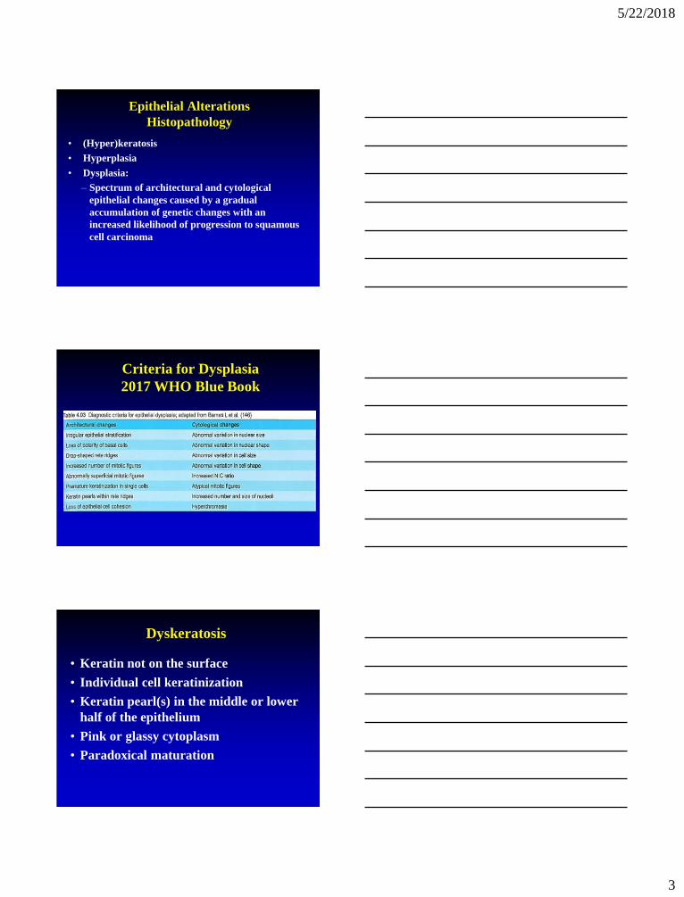
Dyskeratosis
• Keratin not on the surface
• Individual cell keratinization
• Keratin pearl(s) in the middle or lower
half of the epithelium
• Pink or glassy cytoplasm
• Paradoxical maturation
5/22/2018
4
Dyskeratosis
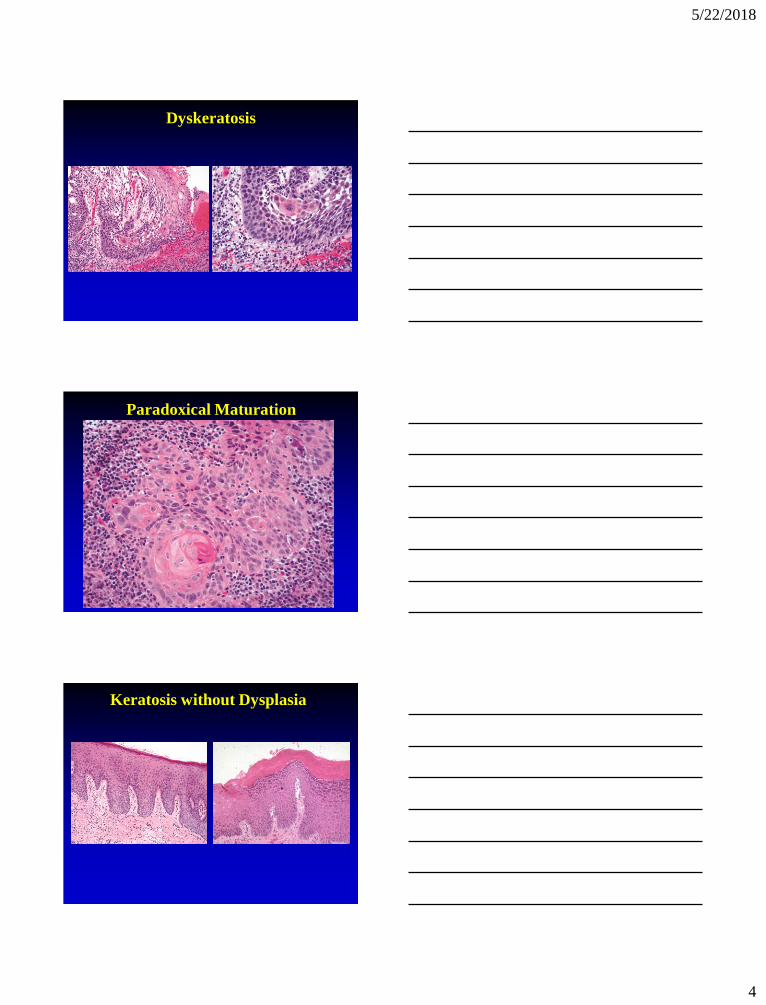
Paradoxical Maturation
Keratosis without Dysplasia
5/22/2018
5
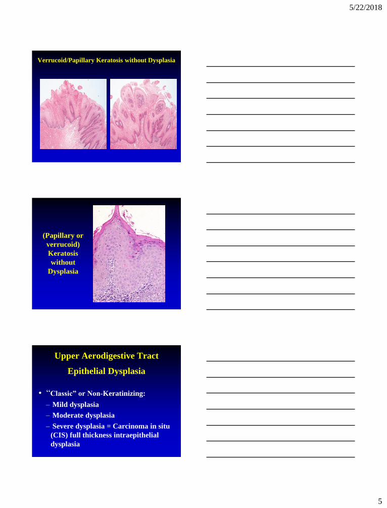
Verrucoid/Papillary Keratosis without Dysplasia
(Papillary or
verrucoid)
Keratosis
without
Dysplasia
Upper Aerodigestive Tract
Epithelial Dysplasia
• “Classic” or Non-Keratinizing:
– Mild dysplasia
– Moderate dysplasia
– Severe dysplasia = Carcinoma in situ
(CIS) full thickness intraepithelial
dysplasia

5/22/2018
6
CIS (Nonkeratinizing)
• Uncommon as isolated lesion in H&N
• Occurs in mucosal sites that are usually clinically quiescent (e.g.,
supraglottic larynx, oro- & nasopharynx) only seen in
association with invasive SCC
Upper Aerodigestive Tract
Epithelial Dysplasia
• Keratinizing >>>> Nonkeratinizing:
– Mild dysplasia
– Moderate dysplasia
– Severe dysplasia
Keratinizing Mild Dysplasia
5/22/2018
7
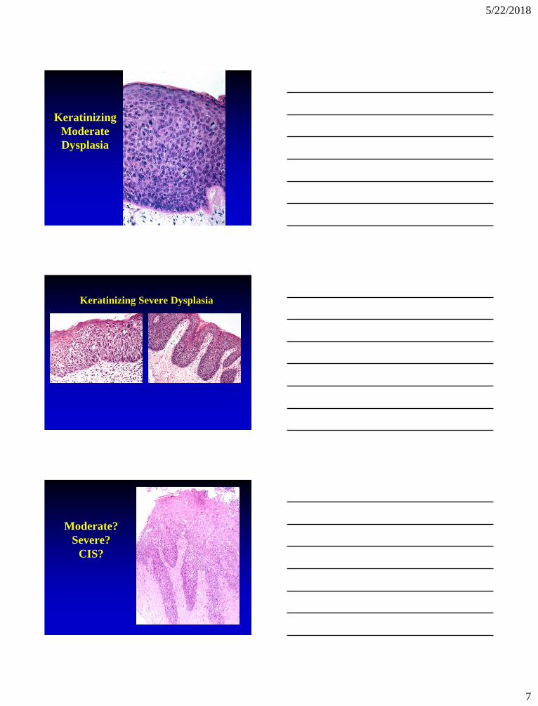
Keratinizing
Moderate
Dysplasia
Keratinizing Severe Dysplasia
Moderate?
Severe?
CIS?

5/22/2018
8
“Drop Off” Carcinoma
Carcinoma In Situ (CIS)
• In the absence of full thickness intra-
epithelial dysplasia is the use of CIS
justified?
• Does keratinizing severe dysplasia = CIS?
• Is it important to separate moderate and
severe dysplasia/CIS?
Upper Aerodigestive Tract
Keratinizing Dysplasia
• Goal of any grading system is:
– Reproducible and Applicable
– Convey to the clinician the potential
risk for progression of disease

5/22/2018
9
Upper Aerodigestive Tract
Grading Keratinizing Dysplasia
• Imprecise and subjective
• Preferred grading based on degree and extent of cellular and maturation alterations
– mild dysplasia
– moderate dysplasia
– severe dysplasia

5/22/2018
10
Grading Keratinizing Dysplasia
• No statistical difference in progression to invasive SCC between keratinizing moderate dysplasia and keratinizing severe dysplasia/CIS
• Justification to 2-Tier grading scheme:
– Low-grade Dysplasia = Mild dysplasia
– High-grade Dysplasia = Moderate Dysplasia, Severe Dysplasia, CIS
• Better reproducibility
Binary Grading – Laryngeal Dysplasia
2017 WHO Blue Book

5/22/2018
11
Binary Grading – Oral Dysplasia
2017 WHO Blue Book
Keratinizing Dysplasia
Etiology
• Tobacco (smoking, chewing)
• Alcohol
• Areca nut, with or without tobacco, causes
oral submucous fibrosis with a relatively high
frequency of oral dysplasia
• High risk human papillomavirus? Generally
not considered a risk factor
Keratinizing Dysplasia
IHC Staining
• p16, p53 and Ki67 (MIB1):
– p16 of limited diagnostic utility in keratinizing dysplasias of the UADT
– p53: increase expression
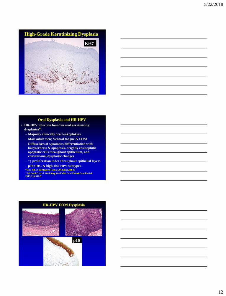
– Ki67: increase intraepithelial proliferation rate through all epithelial layers
• Overall of limited utility
5/22/2018
12
High-Grade Keratinizing Dysplasia
Ki67
Ki67
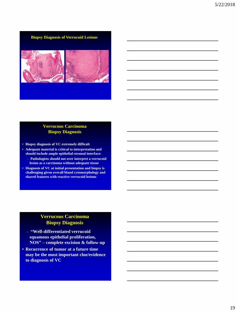
Oral Dysplasia and HR-HPV
• HR-HPV infection found in oral keratinizing
dysplasias*:
– Majority clinically oral leukoplakias
– Most adult men; Ventral tongue & FOM
– Diffuse loss of squamous differentiation with
karyorrhexis & apoptosis, brightly eosinophilic
apoptotic cells throughout epithelium, and
conventional dysplastic changes
– ↑↑ proliferation index throughout epithelial layers
– p16+IHC & high-risk HPV subtypes*Woo SB, et al. Modern Pathol 2013;26:1288-97
* McCord C, et al. Oral Surg Oral Med Oral Pathol Oral Radiol
2013;115:541-9
HR-HPV FOM Dysplasia
p16

5/22/2018
13
HNSCC
Factors Associated with Prognosis
• Adequacy of resection (surgical margins)
• Pattern of invasion: cohesive v dyscohesive
• Tumor size, depth of invasion (DOI), location
• LVI, neurotropism and soft tissue invasion
• Regional metastasis - Extranodal Extension
• Distant metastasis
• Angiogenesis; Host immune response
• Second malignancy
Depth of Invasion (DOI)
• T1: Tumor ≤ 2cm & DOI ≤5mm
• T2: Tumor ≤ 2cm & DOI >5mm & ≤10mm or tumor >2cm but not ≤4cm, & DOI ≤10mm
• T3: Tumor >4cm or any tumor DOI >10mm
Extranodal Extension

5/22/2018
14
Variants of Squamous Cell
Carcinoma of the Upper
Aerodigestive Tract
Squamous Cell Carcinoma Variants
• Verrucous Carcinoma
• Viral-Associated Carcinomas (HPV; EBV)
• Spindle Cell Squamous Carcinoma
• Papillary (Exophytic) SCC
• Basaloid Squamous Cell Carcinoma
• Adenosquamous Carcinoma
• Lymphoepithelial-like Carcinoma
• Adenoid SCC (angiosarcoma-like or acantholytic)
• Other variants:
– NUT carcinoma
– SMARCB1 (INI-1) Deficient Carcinoma
Verrucous Carcinoma (VC)
• Highly differentiated variant of
squamous cell carcinoma with locally
destructive but not metastatic
capabilities

5/22/2018
15
Verrucous Carcinoma
Clinical Features
• M > F; generally occurs in older age groups
(6th – 7th decades of life)
• Sites:
– oral cavity (4%) > larynx (1-3%) > other
(sinonasal tract; nasopharynx)
• Symptoms vary according to site
Verrucous Carcinoma
Etiology
• Tobacco (smoking, chewing) use
• HPV may play an active role in the multistep progression to cancer by binding (via protein products) to the RB gene product removing regulatory block in the cell cycle (Science 1989;243:934-7
• Recent studies using highly sensitive and specific
molecular methods suggest that VC is not associated
with human papillomavirus infection

5/22/2018
16
Hybrid Carcinoma
• Tumor showing mixed histology including verrucous carcinoma and conventional SCC
• Oral cavity > larynx >>> other sites
• Biologic risk that of conventional SCC
– potential for metastasis
• Treatment that of conventional SCC
Hybrid Carcinoma

5/22/2018
17
Hybrid Carcinoma
Hybrid Carcinoma
Hybrid Carcinoma vs VC with Dysplasia
or Minimal Invasion
• Patel KR, et al. Head Neck Pathol 2015;9:65-73:
– VC (n=18)
– VC with dysplasia or minimal invasion (VCDMI) (n=26) ≤ 2 mm
– VC & SCC (n=14) → >2mm depth of invasion
• Prognosis:
– VC or VCDMI: limited recurrences, no metastases, no deaths
– VC&SCC: 50% recurrence; 14% nodal metastases; 36% DOD

5/22/2018
18
Biopsy Diagnosis of Verrucoid Lesions
Biopsy Diagnosis of Verrucoid Lesions
Biopsy Diagnosis of Verrucoid Lesions
5/22/2018
19
Biopsy Diagnosis of Verrucoid Lesions
Verrucous Carcinoma
Biopsy Diagnosis
• Biopsy diagnosis of VC extremely difficult
• Adequate material is critical to interpretation and
should include ample epithelial-stromal interface:
– Pathologists should not over interpret a verrucoid
lesion as a carcinoma without adequate tissue
• Diagnosis of VC at initial presentation and biopsy is
challenging given overall bland cytomorphology and
shared features with reactive verrucoid lesions
Verrucous Carcinoma
Biopsy Diagnosis
– “Well-differentiated verrucoid
squamous epithelial proliferation,
NOS” – complete excision & follow-up
• Recurrence of tumor at a future time
may be the most important clue/evidence
to diagnosis of VC
5/22/2018
20
Verrucous Carcinoma
Differential Diagnosis
• “Conventional” squamous cell carcinoma
• Reactive verrucoid hyperplasia
• Proliferative verrucous leukoplakia
(PVL)
• Papilloma
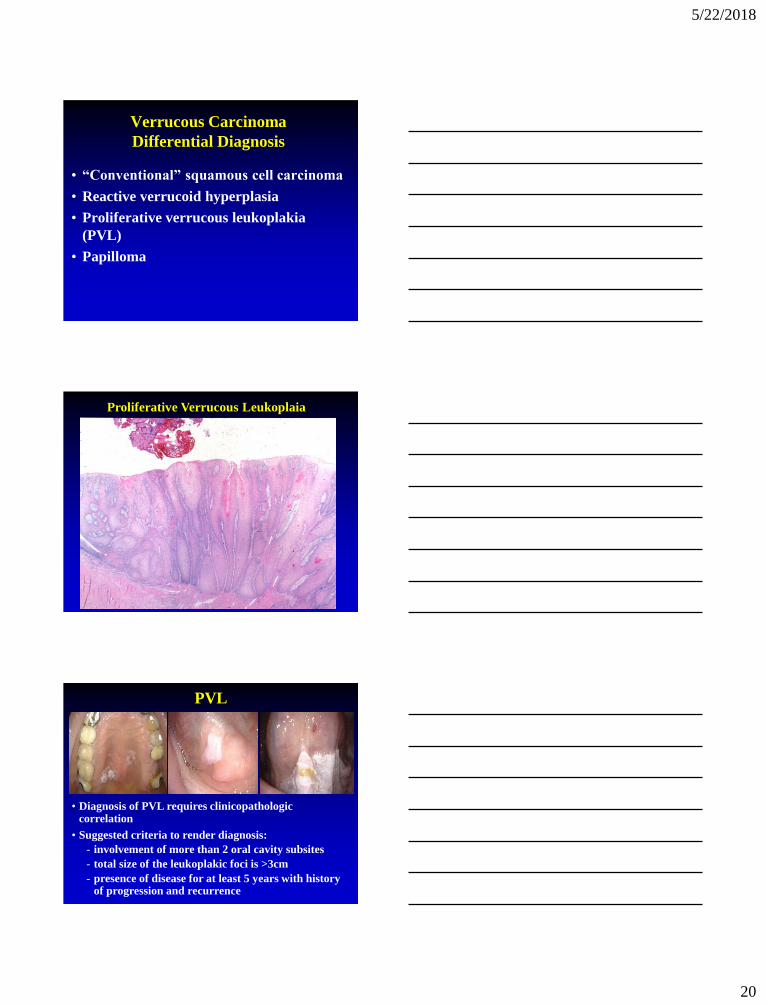
Proliferative Verrucous Leukoplaia
PVL
• Diagnosis of PVL requires clinicopathologic correlation
• Suggested criteria to render diagnosis:
- involvement of more than 2 oral cavity subsites
- total size of the leukoplakic foci is >3cm
- presence of disease for at least 5 years with history of progression and recurrence

5/22/2018
21
Verrucous Carcinoma
Treatment and Prognosis
• Surgery is the treatment of choice
• Radiotherapy used in select settings
• Excellent prognosis:
– for laryngeal VC: 5-yr survival rates of 86-95%
• Local recurrence but no metastases
– may cause extensive destruction if left untreated
• Does not metastasize
• Hybrid carcinoma has potential for metastasis and
should be treated as conventional SCC
Viral-Associated Neoplasms of the H&N
• Human papillomavirus (HPV):
- Papilloma (Low-risk)
- Oropharyngeal carcinoma (High-risk)
• Epstein-Barr virus (EBV):
- Nasopharyngeal carcinoma
- Hematolymphoid tumors
- Smooth muscle tumors
• Merkel cell polyoma virus:
- Merkel cell carcinoma
• Human herpes virus 8:
- Kaposi sarcoma
• Human immunodeficiency virus (HIV)
- HNSCC
Viral-Associated Neoplasms of the Head & Neck
• Oropharyngeal HPV-associated squamous cell
carcinoma (WHO 2017 – SCC, HPV-positive):
- Clinicopathologic features
- Morphologic variants
- Ancillary testing & CAP Recommendations
- Non-squamous malignant neoplasms
• Nasopharyngeal EBV-associated squamous cell
carcinoma
• Metastatic cervical carcinoma with unknown primary
tumor

5/22/2018
22
HPV-positive SCC vs HPV-negative SCC
HPV-positive SCC HPV-negative SCC
Incidence Increasing Stable to decreasing
Age/Gender Younger; M=F Older; M>F
Race Caucasian >>>> African
American
African American >
Caucasian
Risk factors HPV Smoking, alcohol
Primary location Oropharynx (BOT; tonsil) All UADT mucosal sites
Histology Nonkeratinizing SCC Keratinizing SCC
p16 Positive Negative
AJCC staging Lower T, higher N Higher T, lower N
ChemoXRT
response
Good with low rate of
recurrence
Good with high rate
recurrence
Prognosis Better disease-free & overall
survival (worse if smokers)
Worse disease-free and
overall survival
Cervical Neck Lymph Node Stations
Site Specific Lymph Node Drainage
5/22/2018
23
MRI
T1 T2
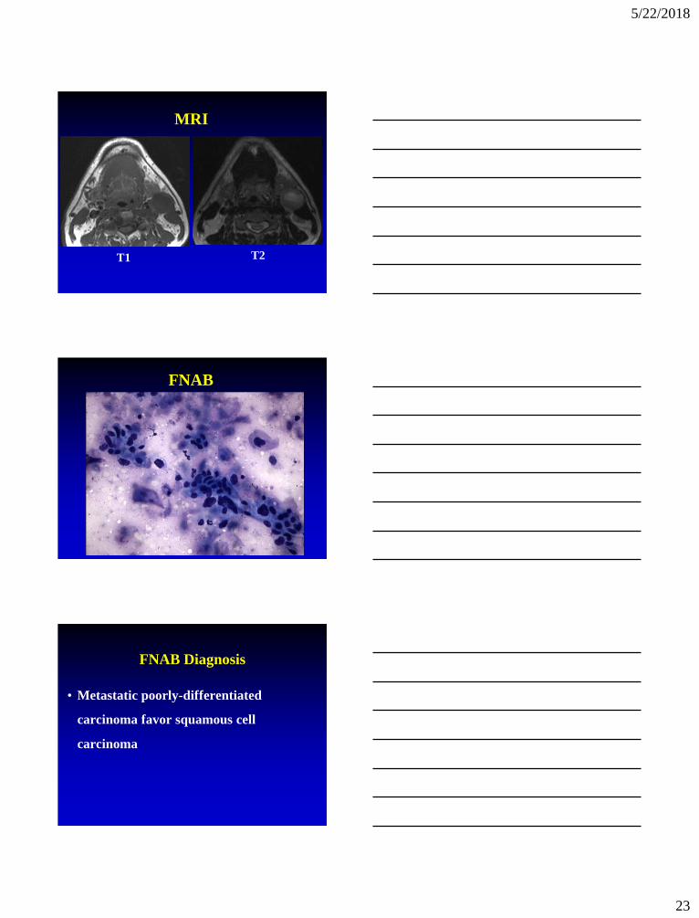
FNAB
FNAB Diagnosis
• Metastatic poorly-differentiated
carcinoma favor squamous cell
carcinoma

5/22/2018
24
PET/CT
BOT
Biopsy
BOT
Biopsy

5/22/2018
25
CAM5.2
p16
Diagnosis
• Oropharyngeal (Tonsillar) Carcinoma:
- Poorly-differentiated squamous cell
carcinoma
- Squamous cell carcinoma with basaloid
features
- Nonkeratinizing carcinoma - recapitulate
tonsillar crypt epithelium so in fact are
differentiated and NOT poorly-differentiated
cancers and should not be graded as such
Nonkeratinizing Carcinoma
↓
Human Papillomavirus (HPV)
↓
Oropharyngeal Carcinoma
(SCC, HPV-positive WHO 2017)

5/22/2018
26
Invasive Oropharyngeal SCC,
Predominantly Nonkeratinizing

5/22/2018
27
p16 ISH HPV16
Oropharyngeal SCC, HPV-Positive
Metastatic SCC, HPV-positive c/w Oropharyngeal Origin
p16
Metastatic SCC, HPV-positive c/w Oropharyngeal Origin

5/22/2018
28
Hybrid Oropharyngeal SCC
Hybrid Oropharyngeal SCC, HPV-positive
p16
Oropharyngeal SCC, HPV-Positive
Morphologic Spectrum
• Nonkeratinizing
• Hybrid
• Papillary SCC (PSCC)
• Basaloid SCC (BSCC)
• Spindle cell SCC (Sarcomatoid carcinoma)
• Adenosquamous (Ciliated cell) carcinoma
• Lymphoepithelial-like

5/22/2018
29
PSCC
PSCC
PSCC

5/22/2018
30
PSCC
PSCC
p16
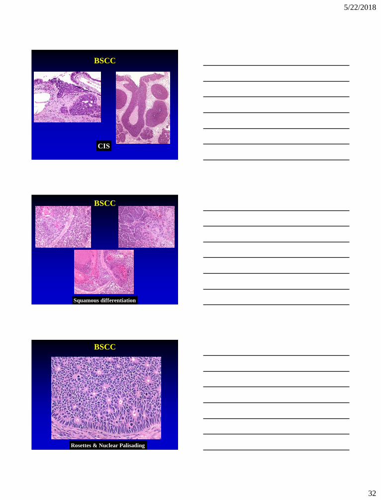
BSCC

5/22/2018
31
BSCC
BSCC
Nuclear palisading
Lobular growth,
comedonecrosis
Reduplicated basement
membrane-like material
BSCC
5/22/2018
32
CIS
BSCC
Squamous differentiation
BSCC
BSCC
Rosettes & Nuclear Palisading

5/22/2018
33
Perineural invasion
BSCC
BSCC
IHC Findings
• IHC:
– Cytokeratins
– p63/p40 (diffusely positive)
– Variable reactivity for S100 protein, NSE
– Mesenchymal: Vimentin, SMA
– Negative for neuroendocrine, melanocytic and lymphoid markers
– p16:
• Most non-oropharyngeal HPV-negative
• Most oropharyngeal HPV-positive
BSCC
Treatment and Prognosis
• Aggressive management:
– Complete surgical resection
– Radiotherapy and chemotherapy
• HPV-negative:
– dismal prognosis
• Active smokers and those with nodal metastases at
presentation have worse prognosis
• Lymphatic and hematogenous spread:
– Regional lymph nodes (50-70%)
– Lung, bone, skin and brain

5/22/2018
34
BSCC
• HPV-positive:
– Better overall prognosis than histologically similar non-HPV associated head and neck BSCC (Am J Surg Pathol 2008;32:1044-50)
• Any tumor appearing to arise in the
larynx/hypopharynx but that involves the
oropharynx should be tested for HPV (p16)
Adenosquamous (Ciliated Cell) Carcinoma
Adenosquamous (Ciliated Cell) Carcinoma

5/22/2018
35
p16 p16
Adenosquamous (Ciliated Cell) Carcinoma
Ciliated HPV-Associated Carcinoma
(aka Ciliated Adenosquamous Carcinoma)
• Bishop JA, Westra WH. Am J Surg Pathol
2015;39:1591-1595
• Radkay-Gonzalez L, et al. Head Neck
Pathol 2016;10:167-175
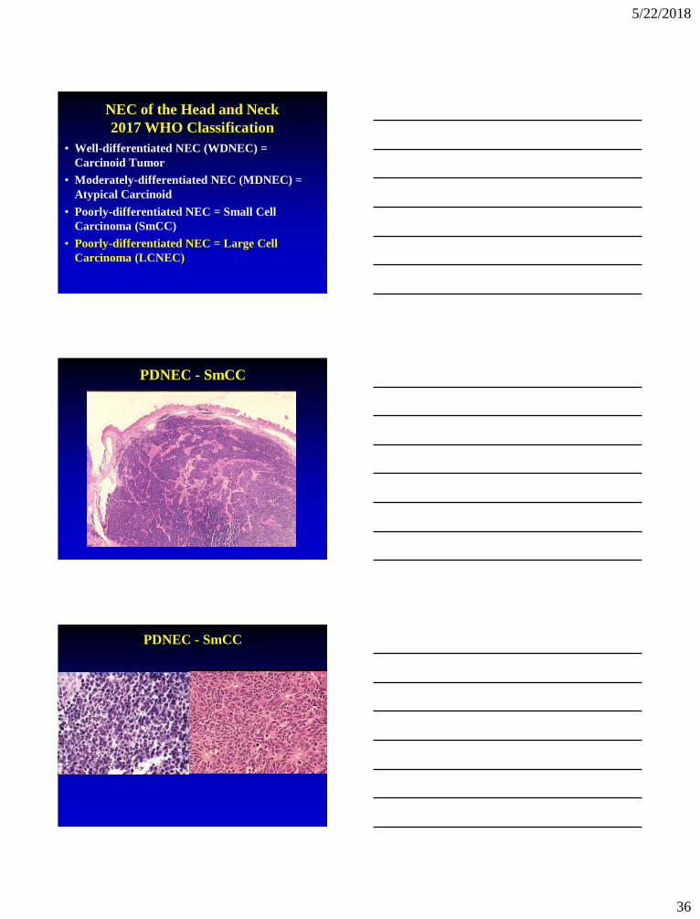
Neuroendocrine Carcinoma (NEC)
Definition
• Heterogeneous group of malignant
neoplasms with divergent
differentiation along epithelial and
neuroendocrine cell lines
5/22/2018
36
NEC of the Head and Neck
2017 WHO Classification
• Well-differentiated NEC (WDNEC) =
Carcinoid Tumor
• Moderately-differentiated NEC (MDNEC) =
Atypical Carcinoid
• Poorly-differentiated NEC = Small Cell
Carcinoma (SmCC)
• Poorly-differentiated NEC = Large Cell
Carcinoma (LCNEC)
PDNEC - SmCC
PDNEC - SmCC

5/22/2018
37
CAM5.2 SYN
TTF1
PDNEC - SmCC
p63/p40: negative
CK5/6: negative
p16
Oropharyngeal SmCC(HPV-Associated SmCC)
HPV-Related Small Cell Carcinoma
of the Oropharynx
• Bishop & Westra. AJSP 2011;35:1679-1684
• Kraft S, Faquin WC, Krane JF. AJSP
2012;36:321-330
- 17 cases
- M > F; 6th-7th decades
- Tonsil, base of tongue, neck
- Smoking history
- Presentation with neck metastases
including occult primary
5/22/2018
38
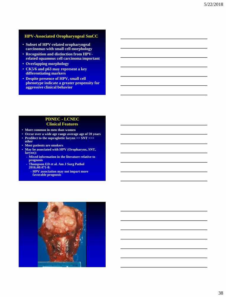
HPV-Associated Oropharyngeal SmCC
• Subset of HPV-related oropharyngeal carcinomas with small cell morphology
• Recognition and distinction from HPV-related squamous cell carcinoma important
• Overlapping morphology
• CK5/6 and p63 may represent a key differentiating markers
• Despite presence of HPV, small cell phenotype indicate a greater propensity for aggressive clinical behavior
PDNEC - LCNEC
Clinical Features
• More common in men than women
• Occur over a wide age range average age of 59 years
• Predilect to the supraglottic larynx >> SNT >>> other
• Most patients are smokers
• May be associated with HPV (Oropharynx, SNT, larynx):
- Mixed information in the literature relative to prognosis
- Thompson ED et al. Am J Surg Pathol 2016;40:471-8:
- HPV association may not impart more favorable prognosis

5/22/2018
39
PDNEC - LCNEC
PDNEC - LCNEC - IHC
CAM5.2 SYN
CHR CD56
Criteria for (Laryngeal) LCNECLewis J, et al. Head Neck Pathol 2010;4:198-207
Requisite criteria Other typical
features
Tumor cells with moderate to abundant
cytoplasm
Nuclei with
prominent nucleoli
Features of neuroendocrine differentiation
(organoid nesting, trabecular growth,
rosettes, and peripheral palisading)
Cellular
pleomorphism
Mitotic activity > 10/10 hpf (2 mm2) Large areas of
necrosis
Confirmation of neuroendocrine
differentiation using immunohistochemical
staining

5/22/2018
40
PDNEC - LCNEC
Treatment and Prognosis
• Chemoradiotherapy
• Many patients have disseminated disease at presentation obviating option of laryngectomy and neck dissection
• Commonly present with advanced stage (stages III and IV):
– may be metastatic to cervical lymph nodes at presentation
– may be metastatic to distant sites at presentation (e.g., liver)
• 5-year disease specific survival (DSS) of 15-21%
Head and Neck NECs
• Larynx most common site; less common sites include SNT, salivary gland, others
• M > F; generally occurs in the 6th-7th decades of life
• Larynx:
– Supraglottic larynx overwhelmingly the most common site of occurrence
– History of cigarette smoking > 60%
– MDNEC >>> SmCC >> LCNEC >>> WDNEC
• SNT and Salivary Gland (Parotid):
– SmCC >>> LCNEC >> MDNEC > WDNEC
HPV Testing in HNSCC: Guidelines from CAP
Lewis JS, et al. Arch Pathol Lab med
2018;142:559-597
• Staining with IHC p16:
- should be used as an initial screening method
- nuclear & cytoplasmic positivity
- > 70% cut off
• 14 Guideline statements
- Strong recommendation
- Recommendation
- Expert consensus opinion
- No recommendation

5/22/2018
41
CAP Testing Guidelines for
High Risk (HR)-HPV in H&N SCC
• #1: Strong recommendation – should perform HR-HPV on all patients with newly diagnosed OPSCC, including all histologic subtypes; on primary tumor or on regional LN metastasis when clinical findings c/w OP origin
• #2: Recommendation – For oropharyngeal tissue specimens (i.e., noncytology), pathologists should perform HR-HPV testing by surrogate marker p16 IHC. Additional HPV-specific testing may be done at the discretion of the pathologist and/or treating clinician, or in the context of a clinical trial
CAP Testing Guidelines for
High Risk (HR)-HPV in H&N SCC
• #3: Expert Consensus Opinion – Pathologists should not
routinely perform HR-HPV testing on patients with non-SCCs
of the oropharynx (neuroendocrine carcinomas; salivary
gland carcinomas)
• #4: Recommendation – Pathologists should not routinely
perform HR-HPV testing on patients with nonoropharyngeal
primary tumors of the H&N
• #5: Recommendation – Pathologists should routinely perform
HR-HPV testing on patients with metastatic SCC of unknown
primary in a cervical upper or mid jugular chain lymph node.
An explanatory note on the significance of a positive HPV
result is recommended
CAP Testing Guidelines for
High Risk (HR)-HPV in H&N SCC
• #6: Expert Consensus Opinion – For tissue specimens (i.e., noncytology) from patients presenting with metastatic SCC of unknown primary in a cervical upper or mid jugular chain lymph node, pathologists should perform p16 IHC
NOTE: Additional HR-HPV testing on p16-positive cases should be performed for tumors located outside of level II or III (noncytology testing) in the neck and/or for tumors with keratinizing morphology
5/22/2018
42
CAP Testing Guidelines for
High Risk (HR)-HPV in H&N SCC
• #7: Expert Consensus Opinion – Pathologists should perform HR-HPV testing on head and neck fine needle aspiration (FNA) SCC samples from all patients with known OPSCC not previously tested for HR-HPV, with suspected OPSCC, or with metastatic SCC of unknown primary
NOTE: No recommendation is made for or against any specific testing methodology for HR-HPV testing in FNA samples. If the result of HR-HPV testing on the FNA sample is negative, testing should be performed on tissue if it becomes available. If pathologists use cytology samples for p16 IHC testing, they should validate the criteria (i.e., cutoff) for a positive result
CAP Testing Guidelines for
High Risk (HR)-HPV in H&N SCC• #9: Expert Consensus Opinion – Pathologists should
not routinely perform low-risk HPV testing on
patients with head and neck carcinomas
• #10: Expert Consensus Opinion - Pathologists should
not repeat HPV testing on patients with locally
recurrent, regionally recurrent, or persistent tumor if
primary tumor HR-HPV status has already been
established. If initial HR-HPV status was never
assessed or results are unknown, testing is
recommended. HPV testing may be performed on a
case-by-case basis for diagnostic purposes if there is
uncertainty regarding whether the tumor in question
is a recurrence or a new primary SCC
CAP Testing Guidelines for
High Risk (HR)-HPV in H&N SCC
• #11: Expert Consensus Opinion – Pathologists should not routinely perform HR-HPV testing on patients with distant metastases if primary tumor HR-HPV status has been established
• #12: Expert Consensus Opinion – Pathologists should report primary OPSCCs that test positive for HR-HPV or its surrogate marker p16 as HPV-positive/p16-positive
• #13: Expert Consensus Opinion – Pathologists should not provide a tumor grade or differentiation status for HPV-positive/p16-positive OPSCCs

5/22/2018
43
Oropharyngeal Reticulated
Epithelium
p16AE1/AE3
Oropharyngeal Reticulated
Epithelium Immunohistochemistry
Reticulated Epithelium
Westra WH: Head & Neck Pathol 2012;6:S48-S54

5/22/2018
44
Carcinoma involving Tonsillar Crypt ≠ CIS
p16
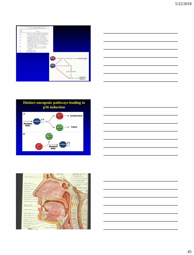
p16 Function
p16 functions to activate RB-dependent
cell cycle arrest
5/22/2018
45
Distinct oncogenic pathways leading to
p16 induction

5/22/2018
46
Nasopharyngeal Carcinoma (NPC)
WHO Classification (2017)
• Keratinizing SCC:
– well-, moderately, poorly-differentiated (WHO 1)
• Nonkeratinizing SCC:
– Differentiated type (Transitional Cell or Cylindrical Cell Carcinoma; WHO 2)
– Undifferentiated type (Lymphoepithelioma; WHO 3)
• Basaloid SCC
NPC
KeratinizingNonkeratinizing
Differentiated
Nonkeratinizing
Undifferentiated
PercentApproximately
25%
Least common
< 15%
Most common
> 60%
Sex/AgeM > F; 4th- 6th
decades
M > F; 4th - 6th
decades
M > F; 4th - 6th
decades; may
occur in children
EBVWeak
association
Strong
association
Strong
association
XRT
Respons
e
Radio-
responsiveness
is not good
Radioresponsive Radioresponsive
5-Yr
survival 20-40% 75% 75%
NPC, nonkeratinizing differentiated
EBER

5/22/2018
47
NPC, nonkeratinizing undifferentiated
NPC, nonkeratinizing undifferentiated
NPC, nonkeratinizing undifferentiated
EBERp63AE1/AE3

5/22/2018
48
Metastatic Cervical Carcinoma with an
Unknown Primary Tumor (MCCUP)
• Definition: - Overt neck mass harboring a cytologically or histologically proven metastatic carcinoma in the absence of signs and symptoms of a primary neoplasm or of a clinically detectable mass:
• no history of previous malignancy or cancer ablation of any indeterminate lesion
• no history of definite symptoms related to a specific organ system
• no clinical or laboratory evidence of a primary neoplasm
Luna MA. Chapter 17. In: Barnes L, ed. Surgical Pathology of the H&N. 2009
Waldeyer Tonsillar Tissues

5/22/2018
49
Luna MA. Chapter 17. In: Barnes L, ed. Surgical Pathology of the H&N. 2009
Branchiogenic Carcinoma Criteria*
• Cervical tumor occurs along line extending from anterior to the tragus along the anterior border of the SCM to the clavicle
• Histology c/w origin from tissue known to be present in branchial vestige
• No primary source for carcinoma on at least 5-year f/u
• Histologic evidence of carcinoma arising in wall of epithelial-lined cyst
*Martin et al. Ann Surg 1950;132:825-832
Branchial Cleft Cyst
• Benign lateral neck cyst most often of 2nd
branchial cleft apparatus
• Bimodal age: 20-40 (75%); <5 yrs (20%):
- rare (≤5%) in ages >40 years
• Painless cervical swelling typically near angle of mandible along border of SCM

5/22/2018
50
Branchial Cleft Cyst
p16 p16p16
Case History
• 39 year old female presented with an
enlarging right sided neck mass at
Level IIA (subdigastric lymph node)
• There was no past or current history of
malignancy
• The lymph node was excised
Level IIA Lymph Node

5/22/2018
51
Level IIA Lymph Node
Level IIA Lymph Node
Case
p63AE1/AE3

5/22/2018
52
Case
Other IHC
• Hematolymphoid markers
(CD45; CD20) negative
• Melanoma markers (S100
protein, HMB45, others) negative
Case
p16EBER
Case
Diagnosis
• Metastatic HPV-associated
lymphoepithelial-like carcinoma
consistent with oropharyngeal origin
• Endoscopic biopsies of UADT sites
including oro- and nasopharynx were
performed

5/22/2018
53
Right TonsilBiopsy
Right TonsilBiopsy
AE1/AE3
p16
p63
EBER
Lymphoepithelial-like Carcinoma of the
Oropharynx: A morphologic variant of
HPV-related head and neck carcinoma
Singhi AD, Stelow EB, Mills SE, Westra WH.
Am J Surg Pathol 2010;34:800-805

5/22/2018
54
Squamous Cell Lesions
Summary
• Overview of intraepithelial alterations of the
upper aerodigestive tract:
– focus on keratinizing dysplasia
– 2 Tier grading system:
• Low-grade (mild dysplasia)
• High-grade (moderate & severe dysplasia
and CIS)
• Verrucous carcinoma diagnosis
–
Squamous Cell Lesions
Summary• Viral carcinogenesis causally associated with HNSCC
• Classification:
- SCC, HPV-positive (oropharynx)
- SCC, EBV-positive (nasopharynx)
• Overall better prognosis than non-viral associated HNSCC
• Overlapping morphology between HPV+ and EBV+ cancers:
- When confronted with MCCUP, both p16 and EBER should be performed
• No correlation to size of primary neoplasm (millimeters) and size of metastasis (centimeters)
- tiny foci may give rise to large metastases
• Relative to oropharyngeal cancers concept of CIS is not applicable:
- lesions that morphologically appear to be CIS may metastasize
AJCC Staging 8th Edition
• HPV-mediated (p16+) Oropharyngeal Cancer (Chapter 10):
- Descriptor “poorly-differentiated” at odds with known
improved prognosis so use should be avoided
- Use of the designation “oropharyngeal SCC,
nonkeratinizing type” is recommended
- Histologic grading is not relevant
- Presence of keratinization in a p16+/HR HPV+ carcinoma
does not exclude using this staging system
- Cervical lymph node metastases (to level II/III) from
unknown primary tumor (pT0) that is p16+ and histology is
consistent with HPV-mediated carcinogenesis are staged
according to the guidelines in this chapter
- Diagnosis of malignant transformation of branchial cleft
cyst “should be rejected”

5/22/2018
55
Squamous Cell Lesions
Summary
• New WHO Classification of H&N PDNEC includes
small cell and large cell types
• PDNEC may also metastasize without a known primary
particularly of oropharyngeal origin:
- May be HPV+ (not associated with more favorable
prognosis)
H. Lee Moffitt
Cancer Center and
Research Institute
Questions?