kidney stones keywords: at the end of medical school, the ... · describe the best imaging study to...
TRANSCRIPT
NATIONAL MEDICAL STUDENT CURRICULUM
This document was amended in July 2016 to reflect literature that was released since the original publication of this content in May 2013. This document will continue to be periodically updated to reflect the growing body of literature related to this topic.
KIDNEY STONES
KEYWORDS: Nephrolithiasis, Urinary Stones, Urolithiasis, Hypercalciuria, Hyperoxaluria, Hypocitraturia, Hyperuricosuria, Cystinuria At the end of medical school, the medical student will understand and be able to…
1. List the major risk factors for the most common types of kidney stones. 2. Contrast differences between the clinical presentations of acute renal colic versus an
acute abdomen. 3. Name the five most common kidney stone chemical compositions and describe the
recommended medical prophylaxis options for each of them. 4. Describe the best imaging study to diagnose kidney and ureteral stones. 5. Describe three types of medications that are effective for relief of renal colic pain. 6. List three clinical situations that warrant urgent surgical decompression of a ureteral
stone. 7. List two types of medications that may help medical expulsion therapy of a distal
ureteral stone. 8. List the three common surgical techniques to manage renal and ureteral stones that
fail to pass with observation. 9. Identify the factors that help predict the likelihood of spontaneous stone passage.
INTRODUCTION Urinary stone prevalence is estimated at 3% in all individuals, and it affects up to 12% of the population during their lifetime. Urinary stone recurrence rates approach 50% at 10 years and white males have the highest incidence in the U.S. The incidence of stones in women is increasing and approaches 50% of the male rate. The overall incidence of nephrolithiasis is also increasing. There is traditionally a high incidence of urinary stones in the Southeastern and South Central United States, termed the “Stone Belt”, which probably reflects the hot weather climate and relative dehydration that occurs in these areas. Prior to the development of modern urologic techniques for treatment, mortality from untreated staghorn (infection) calculi was 27%. Currently, overall mortality from stone disease is rare, although there is still a significant rate (28%) of renal deterioration with certain stone types. PATHOPHYSIOLOGY Urinary calculi may have various compositions which include, in order of decreasing frequency: calcium oxalate, uric acid, struvite or infection (triple phosphate = magnesium ammonium calcium phosphate), calcium phosphate and cystine. There are other less common stones, such as xanthine and drug-related stones, as well. Calculi are typically composed of
2
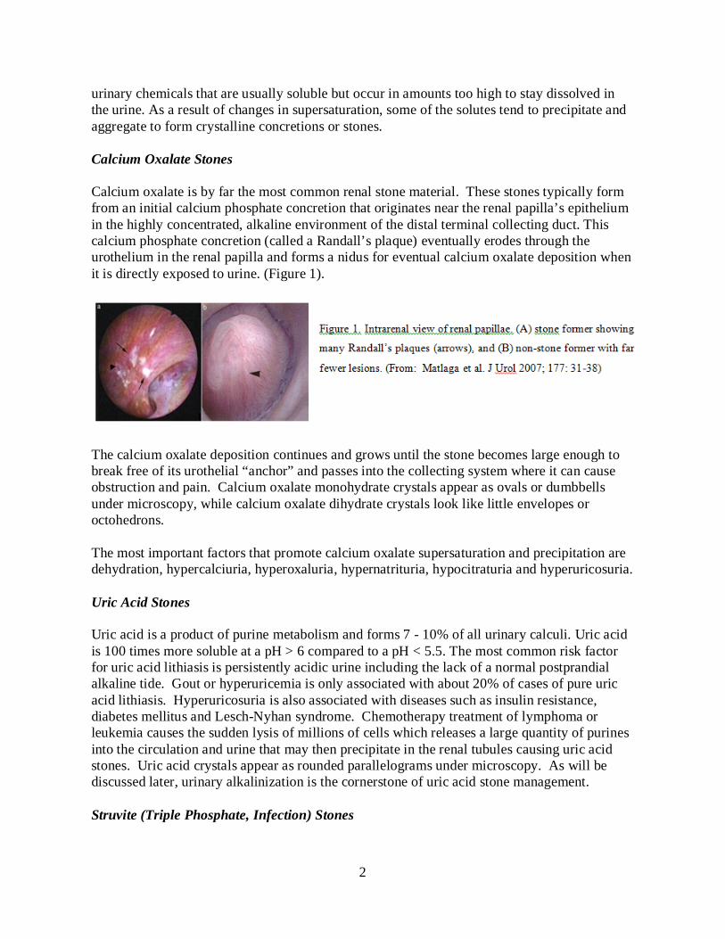
urinary chemicals that are usually soluble but occur in amounts too high to stay dissolved in the urine. As a result of changes in supersaturation, some of the solutes tend to precipitate and aggregate to form crystalline concretions or stones. Calcium Oxalate Stones Calcium oxalate is by far the most common renal stone material. These stones typically form from an initial calcium phosphate concretion that originates near the renal papilla’s epithelium in the highly concentrated, alkaline environment of the distal terminal collecting duct. This calcium phosphate concretion (called a Randall’s plaque) eventually erodes through the urothelium in the renal papilla and forms a nidus for eventual calcium oxalate deposition when it is directly exposed to urine. (Figure 1).
The calcium oxalate deposition continues and grows until the stone becomes large enough to break free of its urothelial “anchor” and passes into the collecting system where it can cause obstruction and pain. Calcium oxalate monohydrate crystals appear as ovals or dumbbells under microscopy, while calcium oxalate dihydrate crystals look like little envelopes or octohedrons. The most important factors that promote calcium oxalate supersaturation and precipitation are dehydration, hypercalciuria, hyperoxaluria, hypernatrituria, hypocitraturia and hyperuricosuria. Uric Acid Stones Uric acid is a product of purine metabolism and forms 7 - 10% of all urinary calculi. Uric acid is 100 times more soluble at a pH > 6 compared to a pH < 5.5. The most common risk factor for uric acid lithiasis is persistently acidic urine including the lack of a normal postprandial alkaline tide. Gout or hyperuricemia is only associated with about 20% of cases of pure uric acid lithiasis. Hyperuricosuria is also associated with diseases such as insulin resistance, diabetes mellitus and Lesch-Nyhan syndrome. Chemotherapy treatment of lymphoma or leukemia causes the sudden lysis of millions of cells which releases a large quantity of purines into the circulation and urine that may then precipitate in the renal tubules causing uric acid stones. Uric acid crystals appear as rounded parallelograms under microscopy. As will be discussed later, urinary alkalinization is the cornerstone of uric acid stone management. Struvite (Triple Phosphate, Infection) Stones
3
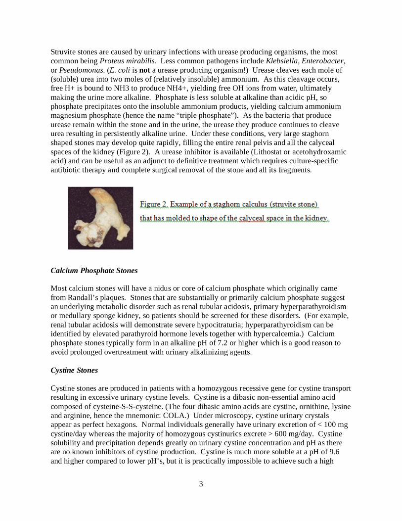
Struvite stones are caused by urinary infections with urease producing organisms, the most common being Proteus mirabilis. Less common pathogens include Klebsiella, Enterobacter, or Pseudomonas. (E. coli is not a urease producing organism!) Urease cleaves each mole of (soluble) urea into two moles of (relatively insoluble) ammonium. As this cleavage occurs, free H+ is bound to NH3 to produce NH4+, yielding free OH ions from water, ultimately making the urine more alkaline. Phosphate is less soluble at alkaline than acidic pH, so phosphate precipitates onto the insoluble ammonium products, yielding calcium ammonium magnesium phosphate (hence the name “triple phosphate”). As the bacteria that produce urease remain within the stone and in the urine, the urease they produce continues to cleave urea resulting in persistently alkaline urine. Under these conditions, very large staghorn shaped stones may develop quite rapidly, filling the entire renal pelvis and all the calyceal spaces of the kidney (Figure 2). A urease inhibitor is available (Lithostat or acetohydroxamic acid) and can be useful as an adjunct to definitive treatment which requires culture-specific antibiotic therapy and complete surgical removal of the stone and all its fragments.
Calcium Phosphate Stones Most calcium stones will have a nidus or core of calcium phosphate which originally came from Randall’s plaques. Stones that are substantially or primarily calcium phosphate suggest an underlying metabolic disorder such as renal tubular acidosis, primary hyperparathyroidism or medullary sponge kidney, so patients should be screened for these disorders. (For example, renal tubular acidosis will demonstrate severe hypocitraturia; hyperparathyroidism can be identified by elevated parathyroid hormone levels together with hypercalcemia.) Calcium phosphate stones typically form in an alkaline pH of 7.2 or higher which is a good reason to avoid prolonged overtreatment with urinary alkalinizing agents. Cystine Stones Cystine stones are produced in patients with a homozygous recessive gene for cystine transport resulting in excessive urinary cystine levels. Cystine is a dibasic non-essential amino acid composed of cysteine-S-S-cysteine. (The four dibasic amino acids are cystine, ornithine, lysine and arginine, hence the mnemonic: COLA.) Under microscopy, cystine urinary crystals appear as perfect hexagons. Normal individuals generally have urinary excretion of < 100 mg cystine/day whereas the majority of homozygous cystinurics excrete > 600 mg/day. Cystine solubility and precipitation depends greatly on urinary cystine concentration and pH as there are no known inhibitors of cystine production. Cystine is much more soluble at a pH of 9.6 and higher compared to lower pH’s, but it is practically impossible to achieve such a high
4
urinary pH by oral alkalinizing agents alone (and certainly not without the risk of calcium phosphate stone formation). Renal Physiology with Obstruction All stones may produce obstruction and pain. Pain is thought to occur from ureteral dilation from the obstruction and/or renal capsular distension. With acute unilateral obstruction, in the setting of a normal contralateral kidney, the affected kidney responds in two phases to the blockage:
• Initial 2 hours: There is increased renal pelvic pressures and renal blood flow. As renal pelvic pressure increases, glomerular filtration rate (GFR) decreases, as GFR represents the sum of net hydrostatic and oncotic pressures across the glomerulus.
• At 6 - 24 hours: Renal pelvic pressures remain elevated, but renal blood flow
diminishes. • After 24 hours: Renal pelvic pressures trend down towards baseline (but remain
elevated) and renal blood flow continues to diminish. If persistent, the obstruction eventually leads to renal ischemia.
Thus, obstruction from urinary stones threatens GFR, reduces renal blood flow and, if the obstruction is not relieved, renal ischemia which leads eventually to irreversible renal impairment. In general, with high-grade obstruction, renal impairment will occur within two weeks.
CLINICAL PRESENTATION The classic presentation of a renal stone is acute, colicky flank pain radiating to the groin or scrotum, often associated with nausea and vomiting. As the stone descends in the ureter, pain may localize to the abdomen overlying the stone. Renal and ureteral colic are often considered the most severe pain ever experienced by patients, and many female stone patients describe the pain as even more intense than that of childbirth. As the stone approaches the ureterovesical junction, lower quadrant pain, urinary urgency, frequency and dysuria are common, mimicking bacterial cystitis. A family history of renal calculi is present in 55% of patients with recurrent stones. Stones occur three times more frequently in men with a family history of stones. The physical exam typically shows a distressed patient, often writhing and constantly moving while trying to find a comfortable position. In contrast, patients with an acute abdomen typically have board-like abdominal rigidity and do not wish to move at all. Costovertebral angle or lower quadrant tenderness may be present. A distal ureteral calculus at the ureterovesical junction in a woman may sometimes be palpated directly on vaginal exam. Gross or microscopic hematuria is present in approximately 85% of patients. Importantly, the absence of hematuria with acute flank pain does not preclude renal or ureteral calculi as there may be complete obstruction. Hydronephrosis and renal capsular distension may also produce

5
nausea and vomiting. Thus, the typical symptoms of urinary stones producing acute renal colic may mimic other acute abdominal conditions (Table 1), making rapid and accurate diagnosis important.
TABLE 1: DIFFERENTIAL DIAGNOSIS OF ACUTE RENAL COLIC IN ADULTS Renal or ureteral stone Hydronephrosis (ureteropelvic junction obstruction, sloughed papilla) Bacterial cystitis or pyelonephritis Lobar pneumonia Rib fractures Acute abdomen (bowel, biliary, pancreas or aortic abdominal aneurysm sources) Gynecologic (ectopic pregnancy, ovarian cyst, torsion or rupture) Radicular pain (L1 herpes zoster, sciatica) Referred pain (orchitis) DIAGNOSTIC EVALUATION The current gold standard for confirming urinary stones in the setting of acute flank pain is an unenhanced, non-contrast helical computed tomography (CT) scan of the abdomen and pelvis. This study surpasses the intravenous pyelogram (IVP) which had been the standard imaging test for decades. A prospective trial of 106 adult patients with acute flank pain imaged all patients with both an unenhanced helical CT and IVP. CT and IVP showed a ureteral stone in 96% vs. 87% of patients, respectively, which was significantly different. Of those patients without stones, the CT and IVP were negative in 100% versus 94% of cases, also significant. Thus, the positive and negative predictive values for CT were 100% and 91%, and for IVP, 97% and 74%, respectively. In ambulatory settings where CT is not available, a plain abdominal radiograph (KUB) is useful as approximately 75 - 90% of urinary stones are radiopaque. A KUB is also recommended as an adjunct to any initial CT scan positive for urinary stones. The KUB provides an easy way to track progress of the stone over time, quickly establishes its radio-opacity when its location is known and is usually better than CT for determining stone shape. Ultrasound appears to be vastly inferior to unenhanced CT for stones and is insensitive for ureteral calculi. However, ultrasound is the recommended first imaging test when a urinary calculus is suspected in a pregnant woman. Ultrasound is also useful when done together with the KUB to help identify non-calcified stones that the KUB alone might miss, to estimate the degree of urinary obstruction/hydronephrosis and to measure the Renal Resistive Index which is elevated when the kidney is obstructed. (Renal Resistive Index = (Peak Systolic Velocity – End Diastolic Velocity)/Peak Systolic Velocity. Normal = <0.65 while readings >0.70 suggest medical renal disease or obstruction.) An IVP or contrast CT is now recommended only when medication metabolite stones are suspected, such as with some HIV medications like Crixivan (Indinivir), as these stones would not otherwise be visible.
6
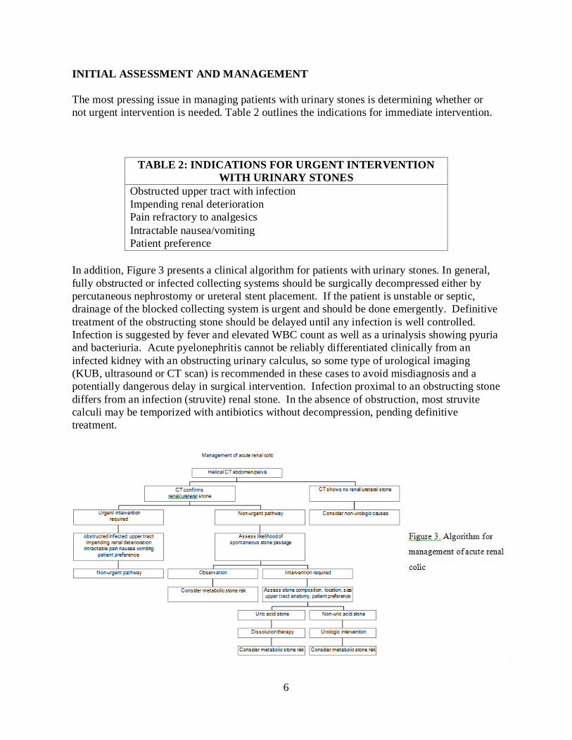
INITIAL ASSESSMENT AND MANAGEMENT The most pressing issue in managing patients with urinary stones is determining whether or not urgent intervention is needed. Table 2 outlines the indications for immediate intervention.
TABLE 2: INDICATIONS FOR URGENT INTERVENTION WITH URINARY STONES
Obstructed upper tract with infection Impending renal deterioration Pain refractory to analgesics Intractable nausea/vomiting Patient preference
In addition, Figure 3 presents a clinical algorithm for patients with urinary stones. In general, fully obstructed or infected collecting systems should be surgically decompressed either by percutaneous nephrostomy or ureteral stent placement. If the patient is unstable or septic, drainage of the blocked collecting system is urgent and should be done emergently. Definitive treatment of the obstructing stone should be delayed until any infection is well controlled. Infection is suggested by fever and elevated WBC count as well as a urinalysis showing pyuria and bacteriuria. Acute pyelonephritis cannot be reliably differentiated clinically from an infected kidney with an obstructing urinary calculus, so some type of urological imaging (KUB, ultrasound or CT scan) is recommended in these cases to avoid misdiagnosis and a potentially dangerous delay in surgical intervention. Infection proximal to an obstructing stone differs from an infection (struvite) renal stone. In the absence of obstruction, most struvite calculi may be temporized with antibiotics without decompression, pending definitive treatment.
7
High-grade obstruction (moderate or severe hydronephrosis) in a solitary or transplanted kidney is an example of impending renal deterioration and requires rapid resolution of the blockage with drainage or surgery. Patient preference is a relative indication for urgent intervention. Pain Since most stone patients present with pain, analgesia must also be addressed. Traditionally, narcotics and nonsteroidal anti-inflammatory drugs (NSAIDs) are commonly used for pain relief. In most randomized, blinded studies of NSAIDs versus narcotics, NSAIDs have shown equal or greater efficacy of pain relief and a shorter time to reach adequate analgesia with equal or fewer side effects. NSAIDs may pose a threat to renal function with decreased blood flow from obstruction, particularly if patients have pre-existing renal impairment. Also, if surgical intervention is warranted, NSAIDs cause platelet inhibition and risk increased surgical bleeding. Renal colic may also be managed with antidiuretic hormone (desmopressin, DDAVP) and intravenous acetaminophen. Intractable renal colic pain is effectively controlled by decompressing the obstruction via percutaneous nephrostomy or ureteral stenting. Expectant Management When urgent intervention is unnecessary, the next clinical decision is whether patients may be followed expectantly in anticipation of passing their stone spontaneously versus elective intervention. Stone size and location are key determinants to predict spontaneous passage. The ureter is the smallest diameter structure of the urinary tract and is the area most prone to obstruction by a stone; especially the ureterovesical junction or UVJ. The majority of stones < 5 mm in diameter are likely to pass spontaneously but the likelihood of spontaneous stone passage decreases as stone size increases (Table 3).
TABLE 3: CHANCE OF PASSING URETERAL STONES
Stone size (mm) Number of days to pass stone (mean)
% Likelihood of eventual need for intervention
2 or less 8 3 3 12 14
4-6 22 50
> 6 -- 99%
Two-thirds of ureteral stones pass spontaneously within four weeks of the onset of symptoms. Spontaneous stone passage within the distal ureter may be facilitated with drugs that enhance expulsion and reduce ureteral spasm. Such medical expulsive therapy (MET) includes calcium channel blockers and alpha blockers like tamsulosin which are typically used in combination with NSAIDs. MET is most effective for small, distal ureteral stones where it appears to shorten the duration of ureteral obstruction and increases the likelihood of spontaneous stone passage by about 30%. Corticosteroids (e.g., prednisone) have also been studied in
8
combination with alpha blockers and may help with stone expulsion; however, anecdotal reports of avascular necrosis of the hip limit its clinical use. Patients rarely have complete obstruction and thus the risk of renal deterioration from observation for a small stone is presumed low. However, a ureteral stone that has not passed or moved within 1 - 2 months is unlikely to pass spontaneously with further observation alone. An observation period of 2 - 4 weeks is reasonable in most circumstances even in symptomatic patients. With observation, close follow-up is needed to ensure stone passage, to follow stone growth and to watch for new infections. Asymptomatic patients who have stones < 5 mm in size may be followed unless symptoms, infection, impending renal deterioration or stone growth warrant intervention. As precise stone chemical composition is typically not known on initial presentation, it is important to encourage patients to catch and submit their stone or fragments for chemical analysis so that recurrent stone episodes may be more efficiently managed with knowledge of prior stone composition. MEDICAL AND SURGICAL MANAGEMENT For those in whom intervention is warranted, treatment is based on stone characteristics such as chemical composition, intra-renal location, number and size as well as upper tract anatomy and other factors such as patient risk factors and co-morbidities, patient size and body habitus, equipment availability, surgeon’s judgment and patient preference (Table 4).
TABLE 4: OPTIONS FOR STONE INTERVENTION Oral stone dissolution (Uric acid stones only) Extracorporeal Shock Wave Lithotripsy (ESWL) Ureteroscopy Percutaneous Nephrolithotomy (PCNL) Open or Laparoscopic Lithotomy
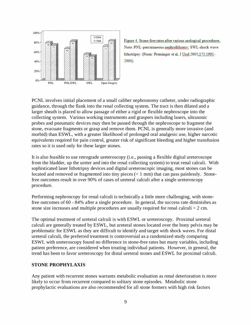
Uric acid calculi are unique in that they may be completely managed and dissolved medically. Urinary alkalinization with potassium citrate (or alternatively sodium citrate or sodium bicarbonate) will dissolve uric acid stones. Sufficient alkalinization therapy should be given to increase the pH to at least 6.5. Maintaining the pH at this level usually results in dissolution of pure uric acid stones in 2 - 6 weeks. Progress can be followed with ultrasound. Renal calculi < 3 cm in maximal diameter that are visible on KUB are generally best treated by Extracorporeal Shock Wave Lithotripsy (ESWL) (Figure 3). ESWL generates shock waves extra corporeally, focuses them on the stone and then fragments it. The patient then passes these very small fragments painlessly in their urine. Success varies based on the number and density of the stones being treated, the specific ESWL machine used, the total number and rate of shocks given, stone size, chemical composition and the stone’s precise intra-renal location. ESWL is less successful for renal calculi located in the lower pole compared to all other renal locations, likely from the effects of gravity on fragment clearance. Patients with lower pole stones are more likely to be stone-free if treated by ureteroscopy or percutaneous nephrolithotomy (PCNL) than by ESWL (Figure 4). Renal calculi in all other locations > 3 cm are best treated by percutaneous nephrolithotomy (PCNL), with or without adjunctive ESWL.
9
PCNL involves initial placement of a small caliber nephrostomy catheter, under radiographic guidance, through the flank into the renal collecting system. The tract is then dilated and a larger sheath is placed to allow passage of either a rigid or flexible nephroscope into the collecting system. Various working instruments and graspers including lasers, ultrasonic probes and pneumatic devices may then be passed through the nephroscope to fragment the stone, evacuate fragments or grasp and remove them. PCNL is generally more invasive (and morbid) than ESWL, with a greater likelihood of prolonged oral analgesic use, higher narcotic equivalents required for pain control, greater risk of significant bleeding and higher transfusion rates so it is used only for these larger stones. It is also feasible to use retrograde ureteroscopy (i.e., passing a flexible digital ureteroscope from the bladder, up the ureter and into the renal collecting system) to treat renal calculi. With sophisticated laser lithotripsy devices and digital ureteroscopic imaging, most stones can be located and removed or fragmented into tiny pieces (< 1 mm) that can pass painlessly. Stone-free outcomes result in over 90% of cases of ureteral calculi after a single ureteroscopy procedure. Performing nephroscopy for renal calculi is technically a little more challenging, with stone-free outcomes of 60 - 84% after a single procedure. In general, the success rate diminishes as stone size increases and multiple procedures are usually required for renal calculi > 2 cm. The optimal treatment of ureteral calculi is with ESWL or ureteroscopy. Proximal ureteral calculi are generally treated by ESWL, but ureteral stones located over the bony pelvis may be problematic for ESWL as they are difficult to identify and target with shock waves. For distal ureteral calculi, the preferred treatment is controversial as a randomized study comparing ESWL with ureteroscopy found no difference in stone-free rates but many variables, including patient preference, are considered when treating individual patients. However, in general, the trend has been to favor ureteroscopy for distal ureteral stones and ESWL for proximal calculi. STONE PROPHYLAXIS Any patient with recurrent stones warrants metabolic evaluation as renal deterioration is more likely to occur from recurrent compared to solitary stone episodes. Metabolic stone prophylactic evaluations are also recommended for all stone formers with high risk factors
10
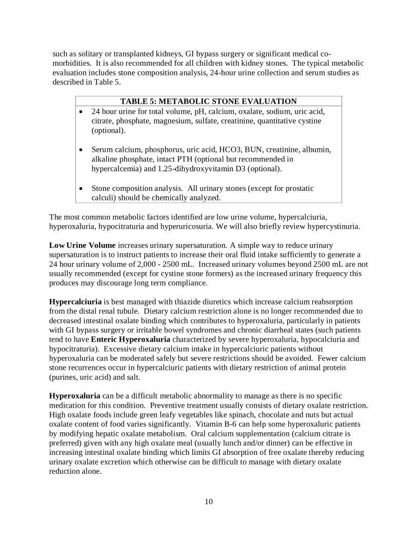
such as solitary or transplanted kidneys, GI bypass surgery or significant medical co-morbidities. It is also recommended for all children with kidney stones. The typical metabolic evaluation includes stone composition analysis, 24-hour urine collection and serum studies as described in Table 5.
TABLE 5: METABOLIC STONE EVALUATION
• 24 hour urine for total volume, pH, calcium, oxalate, sodium, uric acid, citrate, phosphate, magnesium, sulfate, creatinine, quantitative cystine (optional).
• Serum calcium, phosphorus, uric acid, HCO3, BUN, creatinine, albumin,
alkaline phosphate, intact PTH (optional but recommended in hypercalcemia) and 1.25-dihydroxyvitamin D3 (optional).
• Stone composition analysis. All urinary stones (except for prostatic
calculi) should be chemically analyzed.
The most common metabolic factors identified are low urine volume, hypercalciuria, hyperoxaluria, hypocitraturia and hyperuricosuria. We will also briefly review hypercystinuria. Low Urine Volume increases urinary supersaturation. A simple way to reduce urinary supersaturation is to instruct patients to increase their oral fluid intake sufficiently to generate a 24 hour urinary volume of 2,000 - 2500 mL. Increased urinary volumes beyond 2500 mL are not usually recommended (except for cystine stone formers) as the increased urinary frequency this produces may discourage long term compliance. Hypercalciuria is best managed with thiazide diuretics which increase calcium reabsorption from the distal renal tubule. Dietary calcium restriction alone is no longer recommended due to decreased intestinal oxalate binding which contributes to hyperoxaluria, particularly in patients with GI bypass surgery or irritable bowel syndromes and chronic diarrheal states (such patients tend to have Enteric Hyperoxaluria characterized by severe hyperoxaluria, hypocalciuria and hypocitraturia). Excessive dietary calcium intake in hypercalciuric patients without hyperoxaluria can be moderated safely but severe restrictions should be avoided. Fewer calcium stone recurrences occur in hypercalciuric patients with dietary restriction of animal protein (purines, uric acid) and salt. Hyperoxaluria can be a difficult metabolic abnormality to manage as there is no specific medication for this condition. Preventive treatment usually consists of dietary oxalate restriction. High oxalate foods include green leafy vegetables like spinach, chocolate and nuts but actual oxalate content of food varies significantly. Vitamin B-6 can help some hyperoxaluric patients by modifying hepatic oxalate metabolism. Oral calcium supplementation (calcium citrate is preferred) given with any high oxalate meal (usually lunch and/or dinner) can be effective in increasing intestinal oxalate binding which limits GI absorption of free oxalate thereby reducing urinary oxalate excretion which otherwise can be difficult to manage with dietary oxalate reduction alone.
11
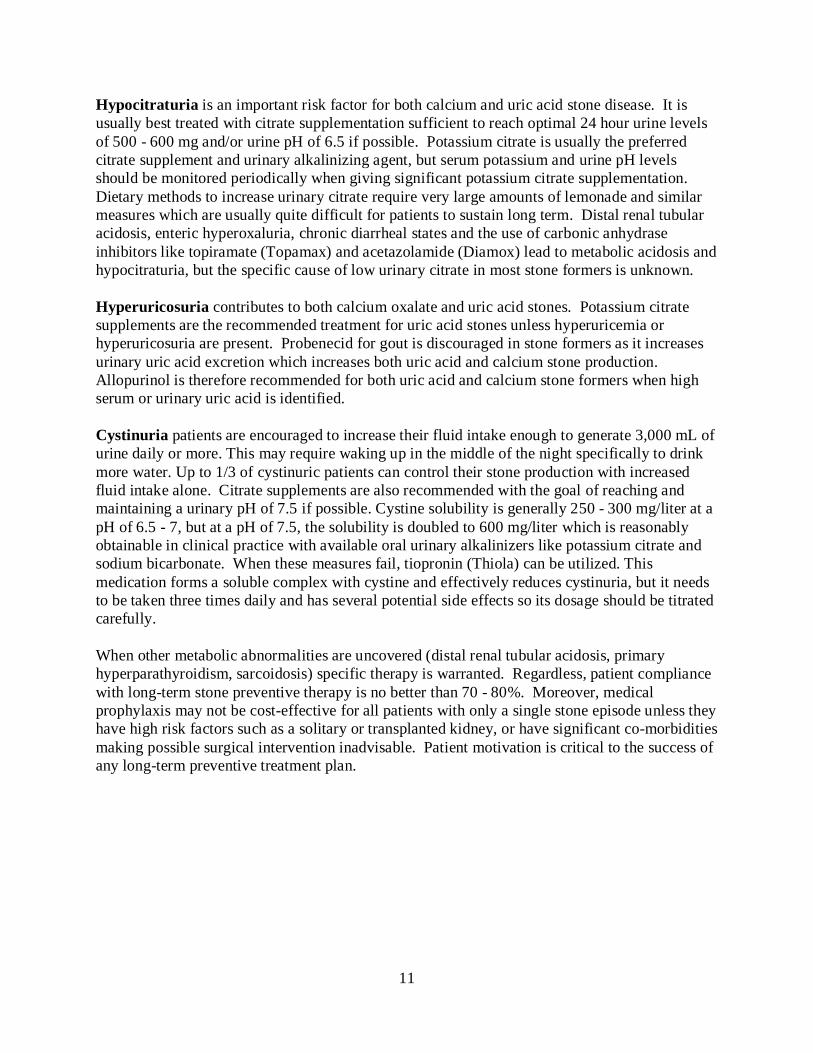
Hypocitraturia is an important risk factor for both calcium and uric acid stone disease. It is usually best treated with citrate supplementation sufficient to reach optimal 24 hour urine levels of 500 - 600 mg and/or urine pH of 6.5 if possible. Potassium citrate is usually the preferred citrate supplement and urinary alkalinizing agent, but serum potassium and urine pH levels should be monitored periodically when giving significant potassium citrate supplementation. Dietary methods to increase urinary citrate require very large amounts of lemonade and similar measures which are usually quite difficult for patients to sustain long term. Distal renal tubular acidosis, enteric hyperoxaluria, chronic diarrheal states and the use of carbonic anhydrase inhibitors like topiramate (Topamax) and acetazolamide (Diamox) lead to metabolic acidosis and hypocitraturia, but the specific cause of low urinary citrate in most stone formers is unknown. Hyperuricosuria contributes to both calcium oxalate and uric acid stones. Potassium citrate supplements are the recommended treatment for uric acid stones unless hyperuricemia or hyperuricosuria are present. Probenecid for gout is discouraged in stone formers as it increases urinary uric acid excretion which increases both uric acid and calcium stone production. Allopurinol is therefore recommended for both uric acid and calcium stone formers when high serum or urinary uric acid is identified. Cystinuria patients are encouraged to increase their fluid intake enough to generate 3,000 mL of urine daily or more. This may require waking up in the middle of the night specifically to drink more water. Up to 1/3 of cystinuric patients can control their stone production with increased fluid intake alone. Citrate supplements are also recommended with the goal of reaching and maintaining a urinary pH of 7.5 if possible. Cystine solubility is generally 250 - 300 mg/liter at a pH of 6.5 - 7, but at a pH of 7.5, the solubility is doubled to 600 mg/liter which is reasonably obtainable in clinical practice with available oral urinary alkalinizers like potassium citrate and sodium bicarbonate. When these measures fail, tiopronin (Thiola) can be utilized. This medication forms a soluble complex with cystine and effectively reduces cystinuria, but it needs to be taken three times daily and has several potential side effects so its dosage should be titrated carefully. When other metabolic abnormalities are uncovered (distal renal tubular acidosis, primary hyperparathyroidism, sarcoidosis) specific therapy is warranted. Regardless, patient compliance with long-term stone preventive therapy is no better than 70 - 80%. Moreover, medical prophylaxis may not be cost-effective for all patients with only a single stone episode unless they have high risk factors such as a solitary or transplanted kidney, or have significant co-morbidities making possible surgical intervention inadvisable. Patient motivation is critical to the success of any long-term preventive treatment plan.
12
SUMMARY
1. Urinary calculi typically present with renal colic and hematuria frequently accompanied by nausea and vomiting.
2. Gross or microscopic hematuria frequently accompanies renal colic but may be absent in 15% of cases.
3. The unenhanced CT is the single best initial diagnostic imaging test. If positive, an immediate KUB is very helpful for determining stone shape and density as well as for follow-up and tracking.
4. Clinicians should initially assess the need for urgent intervention as well as the likelihood of spontaneous stone passage.
5. Urologic intervention must be individualized. 6. Metabolic risk of stone recurrences should be addressed in repeat stone formers, children
and in some motivated first-time stone formers.
REFERENCES Preminger GM, Assimos DG, LingemanJE, Nakada SY, Pearle MS, Wolf JS Jr: Report on the Management of Staghorn Calculi. http://www.auanet.org/content/clinical-practice-guidelines/clinical-guidelines.cfm?sub=uc accessed May 7, 2011
Preminger GM, Tiselius H-G, Assimos DG, et al: 2007 Guideline for the management of ureteral calculi.http://www.auanet.org/content/clinical-practice-guidelines/clinical-guidelines.cfm?sub=sc accessed May7, 2011
Preminger GM, Ferrandino MN, Haleblian GE, et al: Urology Core Curriculum: Surgical Stone Management. http://www.auanet.org/eforms/elearning/core/?topic=14 accessed May 7, 2011
Matlaga BR, Coe FL, Evan AP, Lingeman JE: The role of Randall’s plaques in the pathogenesis of calcium stones. J Urol 2007; 177: 31-8.
Miller OF, Kane CJ: Time to stone passage for observed ureteral calculi: a guide for patient education. J Urol 1999; 162: 688-91.
Preminger GM, Assimos DG, Lingeman JE, et al: Chapter 1: AUA Guideline on management of staghorn calculi: diagnosis and treatment recommendations. J Urol 2005; 173: 1991-2000.
Segura JW, Preminger GM, Assimos DG et al: Ureteral stones clinical guidelines panel summary report on the management of ureteral calculi. J Urol 1997; 158: 1915.
Pak CY, Resnick MI: Medical therapy and new approaches to management of urolithiasis. Urol Cl N Am 2000; 27: 243-53.
13
Ngo TC and Assimos DG: Uric acid nephrolithiasis: recent progress and future directions. Rev Urol 2007; 9(1): 17-27, PMID: 1831527
Hamm LL, Hering-Smith KS. Pathophysiology of hypocitraturic nephrolithiasis. Endocrinol Metab Clin North Am. 2002; 31: 885–893.
Dent CC, Rose GA. Amino acid metabolism in cystinuria. Q J Med New Series. 1974. 214: 507-12.
Leslie SW: Hypercalciuria. Emedicine/Medscape. 2013. http://emedicine.medscape.com/article/2182757-overview
AUTHORS
Gina Badalato, MD Houston, TX Disclosures: Nothing to disclose Stephen W. Leslie, MD FACS Omaha, NE Disclosures: Nothing to disclose Joel Teichman, MD Vancouver, BC Disclosures: Urigen, Investment Interest. © 2012, 2016 American Urological Association Education and Research, Inc.® All Rights Reserved