knee & ankle joints
TRANSCRIPT

KNEE & ANKLE JOINTS
Khaleel Alyahya, PhD, MedKing Saud University
@khaleelya

KNEE JOINT
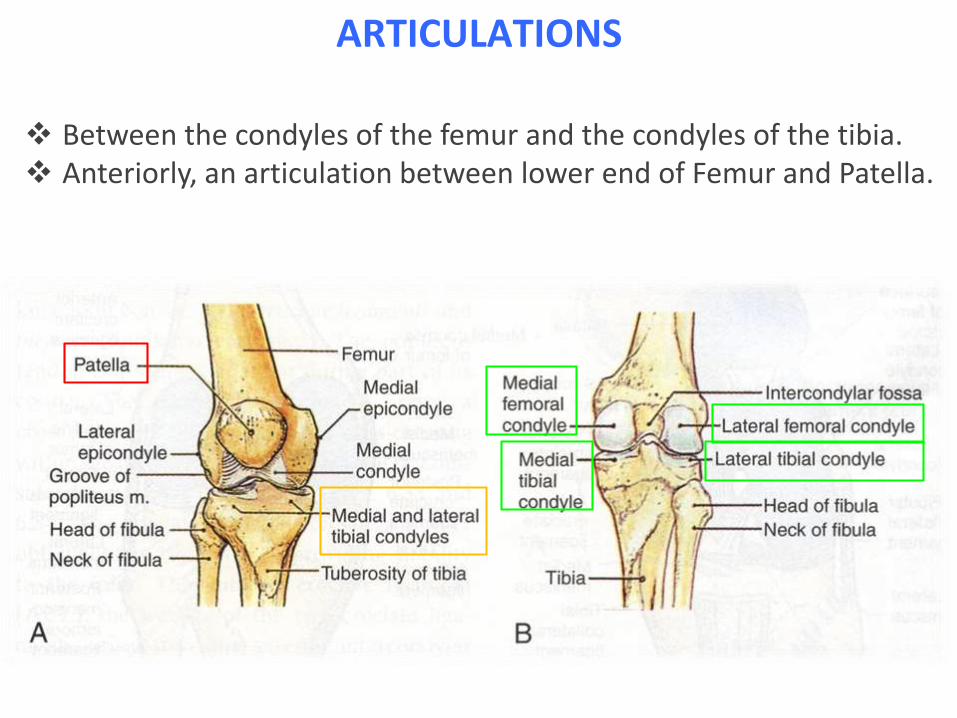
ARTICULATIONS
Between the condyles of the femur and the condyles of the tibia. Anteriorly, an articulation between lower end of Femur and Patella.

TYPES
The joint between thefemur & the tibia is asynovial modified hingejoint, with some degree ofrotatory movement.
The joint between thefemur and patella is asynovial plane gliding joint.

CAPSULE
Attached to the margins of the articular surfaces and surrounds the sidesand the posterior aspect of the joint. The capsule is absent anteriorly.
Replaced anteriorly by quadriceps tendon, patella & ligamentum patellae.
Consists of two layers: the outer fibrous membrane that contain ligaments. the inner synovial membrane that secretes the lubricating.

The capsule is strengthened on each side of the patella by expansions of the tendons of vastus lateralis and medialis, and posteriorly by the expansion of the semimembranous muscle and oblique popliteal ligament.
CAPSULE
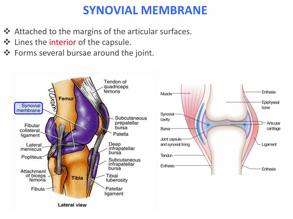
SYNOVIAL MEMBRANE
Attached to the margins of the articular surfaces. Lines the interior of the capsule. Forms several bursae around the joint.

EXTRACAPSULAR LIGAMENTS
Ligamentum Patellae: between the lower border of the
patella & the tuberosity of thetibia.
It is a continuation of the tendon ofquadriceps femoris.
Lateral (Fibular) Collateral Ligament: between the lateral condyle of
femur and the head of the fibula. Medial (Tibial) Collateral Ligament:
between the medial condyle of thefemur and medial side of the shaftof the tibia.
Oblique Popliteal Ligament: strengthens the posterior side of
the capsule

EXTRACAPSULAR LIGAMENTS
Oblique Popliteal Ligament: strengthens the posterior side of the capsule

INTRACAPSULAR LIGAMENTS
Anterior Cruciate Ligament: between the anterior intercondylar
area of the tibia and the posterior part of the medial surface of the lateral femoral condyle.
Function: prevents posteriordisplacement of the femur on thetibia. In flexed knee, prevents the tibia
from being pulled anteriorly.

Posterior Cruciate Ligament: between the posterior intercondylar
area of the tibia and the anteriorpart of the lateral surface of themedial femoral condyle.
Function: prevents anteriordisplacement of the femur on thetibia. In flexed knee, prevents the tibia
from being pulled posteriorly.
INTRACAPSULAR LIMANETS

MENISCI
C-shaped intracapsular ligaments

The peripheral border is thick andattached to the capsule, the innerborder is thin and free.
Each meniscus is attached to theupper surface of the tibia byanterior and posterior horns.
The medial meniscus is firmlyattached to the medial collateralligament.
Function: to deepen the articularsurface of the tibial condyles andto serve as shock absorber.
MENISCI

MENISCI

BURSAE
ANTERIOR
Suprapatellar: beneath the quadricepsfemoris. Prepatellar: between the skin and the
patella. Superficial infrapatellar: between the
skin and the lower part of ligamentumpatellae. Deep infrapatellar: between
ligamentum patellae and the tibia.
BURSAE
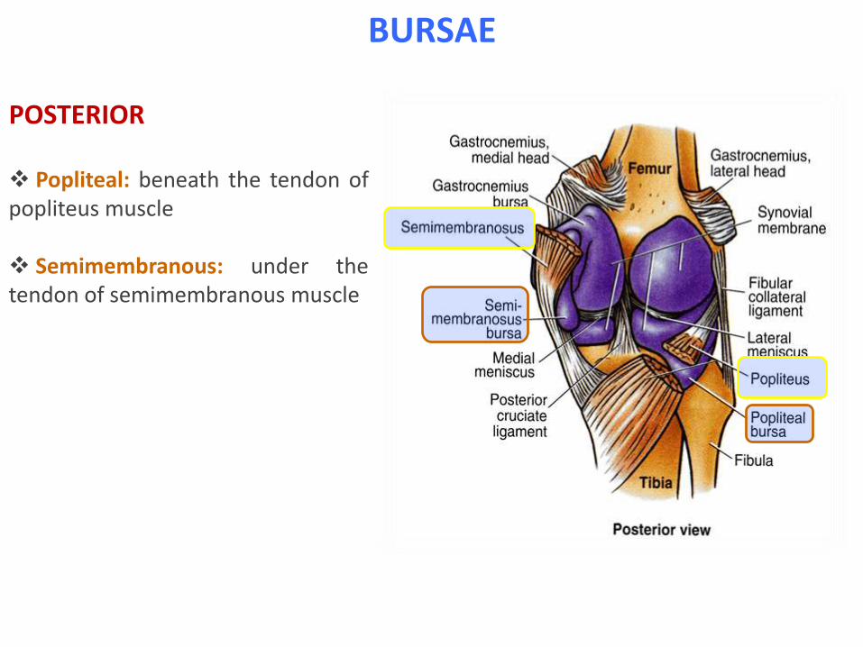
POSTERIOR
Popliteal: beneath the tendon ofpopliteus muscle
Semimembranous: under thetendon of semimembranous muscle

MOVEMENT Flexion:
Biceps femoris, semitendinosus and semimembranosus, assisted by gracilis, sartorius and popliteus. Limited by contact with the back of thigh.
Extension: Quadriceps femoris. Limited by tension of the joint ligaments.
Medial Rotation: Sartorius, gracilis and semitendinosus.
Lateral Rotation:
Biceps femoris.
Stability of the joint: Dependent on the tone of the muscles and the strength of the ligaments.

Locked position: In full extension, the femur is
medially rotated, producing tightening of all of the ligaments and compression of the menisci.
Unlocking: by contraction of the popliteus
muscle, producing lateral rotation of the femur.
It is necessary to start flexion.
MOVEMENT

BLOOD SUPPLY & INNERVATION
Blood Supply The femoral artery and the poplitealartery help form the arterial networksurrounding the knee joint.
NERVE SUPPLY Femoral Obturator Tibial Common peroneal

KNEE INJURIES
Common injury: when the knee is twisted while
running
In sports that place great pressure onthe knees, especially with twisting forces,it is common to tear one or moreligaments or cartilages. Some of the most common kneeinjuries are those to the medialside: medial knee injuries. Anterior Cruciate Ligament (ACL) is themost commonly injured ligament of theknee. The injury is common during sports.Twisting of the knee is a common causeof over-stretching or tearing the ACL


ANKLE JOINT

ARTICULATIONS
Between the distal end of the tibia, the two malleoli and the body of the talus.
Type: synovial hinge joint.

LIGAMENTS
Medial (Deltoid) Ligament: from the tip of the medial
malleolus to: 1. Talus2. Plantar calcaneonavicular
ligament3. Tuberosity of the navicular
bone.

Lateral Ligament: Anterior Talofibular Ligament:
from the lateral malleolus tothe lateral surface of the talus.
Calcaneofibular Ligament: from the lateral malleolus to
the lateral surface of thecalcaneum.
Posterior Talofibular Ligament: from the lateral malleolus to
the posterior tubercle of thetalus.
LIGAMENTS

MOVEMENTS
Dorsiflexion: Tibialis anterior. Extensor hallucis longus. Extensor digitorum longus. Peroneus tertius.
Plantar flexion: Gastrocnemius. Soleus. Plantaris. Peroneus longus. Peroneus brevis. Tibialis posterior. Flexor digitorum longus. Flexor hallucis longus.

INJURIES
Sprained Ankle: Usually inversion injury. Severe sprains lead to torn lateral ligament and fracture
of the lateral malleolus. Result in instability of the ankle joint.

QUESTION?