kreatech diagnostics solid tumors - forsiden - ah … · kreatech diagnostics solid tumors...
TRANSCRIPT

KREATECH DiAgnosTiCsSolid TumorS
REPEAT-FREE™ PosEiDon™ SolutionS for DiagnoSiS in SoliD tumor SampleS

2 For more information please visit our website: www.kreatech.com
FISH4u™
New CuStom Probe ServICe For YouFrom KreAteCH
Our flexible custom probe service FISH4U™ gives you access to your probe of choice. We can develop completely new probe designs to your specifications or you can just request an existing DNA probe from our portfolio to be labeled with an alternative color from our range.
Custom made probes• Leading-edge probe design• Designed to meet your specifications
rePeAt-Free™ PoSeIDoN™ FISH DNA Probes • A clearer background• Greater signal intensity
Check out our custom probe serviceContact customer service by mail:[email protected] visit our website:
www.kreatech.com
Disclaimer FISH4U:
The Customer acknowledges that no licenses or other rights are provided to the
Customer under any patents, patent applications, trade secrets or other proprietary
rights of Kreatech, except the right to conduct research.
Kreatech retains ownership of the Product and Kreatech is free to commercialize the
Product to any third party except if customer has rights or desires to receive rights to
such probe as designed, developed and produced by Kreatech.
If the Customer desires to use the Product for commercial purposes and/or acquire
exclusive rights in the Product, the Customer agrees, in advance of such use, to
negotiate in good faith with Kreatech to establish the terms of such an agreement.
FisH4U™
3For more information please visit our website: www.kreatech.com
FisH4U™
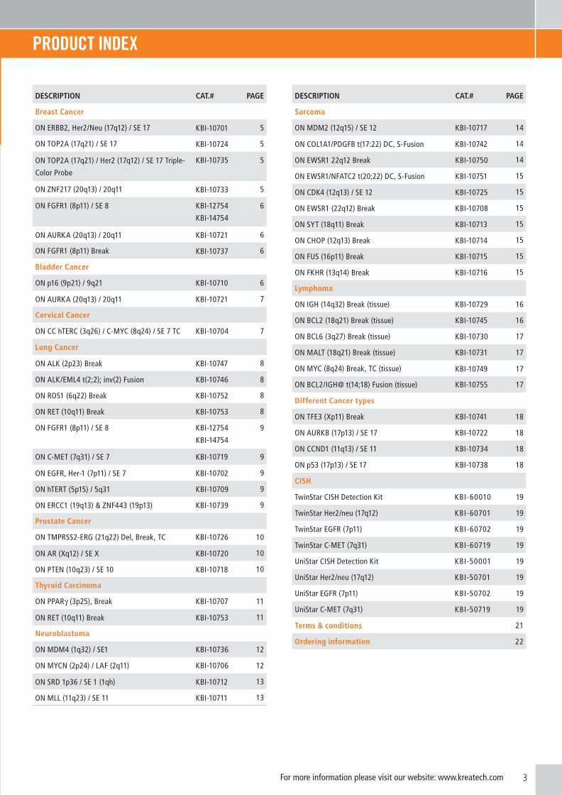
PRoDUCT inDEx
DESCRIPTION CAT.# PAGE
Breast Cancer
ON ERBB2, Her2/Neu (17q12) / SE 17 KBI-10701 5
ON TOP2A (17q21) / SE 17 KBI-10724 5
ON TOP2A (17q21) / Her2 (17q12) / SE 17 Triple-
Color Probe
KBI-10735 5
ON ZNF217 (20q13) / 20q11 KBI-10733 5
ON FGFR1 (8p11) / SE 8 KBI-12754
KBI-14754
6
ON AURKA (20q13) / 20q11 KBI-10721 6
ON FGFR1 (8p11) Break KBI-10737 6
Bladder Cancer
ON p16 (9p21) / 9q21 KBI-10710 6
ON AURKA (20q13) / 20q11 KBI-10721 7
Cervical Cancer
ON CC hTERC (3q26) / C-MYC (8q24) / SE 7 TC KBI-10704 7
Lung Cancer
ON ALK (2p23) Break KBI-10747 8
ON ALK/EML4 t(2;2); inv(2) Fusion KBI-10746 8
ON ROS1 (6q22) Break KBI-10752 8
ON RET (10q11) Break KBI-10753 8
ON FGFR1 (8p11) / SE 8 KBI-12754
KBI-14754
9
ON C-MET (7q31) / SE 7 KBI-10719 9
ON EGFR, Her-1 (7p11) / SE 7 KBI-10702 9
ON hTERT (5p15) / 5q31 KBI-10709 9
ON ERCC1 (19q13) & ZNF443 (19p13) KBI-10739 9
Prostate Cancer
ON TMPRSS2-ERG (21q22) Del, Break, TC KBI-10726 10
ON AR (Xq12) / SE X KBI-10720 10
ON PTEN (10q23) / SE 10 KBI-10718 10
Thyroid Carcinoma
ON PPARγ (3p25), Break KBI-10707 11
ON RET (10q11) Break KBI-10753 11
Neuroblastoma
ON MDM4 (1q32) / SE1 KBI-10736 12
ON MYCN (2p24) / LAF (2q11) KBI-10706 12
ON SRD 1p36 / SE 1 (1qh) KBI-10712 13
ON MLL (11q23) / SE 11 KBI-10711 13
DESCRIPTION CAT.# PAGE
Sarcoma
ON MDM2 (12q15) / SE 12 KBI-10717 14
ON COL1A1/PDGFB t(17:22) DC, S-Fusion KBI-10742 14
ON EWSR1 22q12 Break KBI-10750 14
ON EWSR1/NFATC2 t(20;22) DC, S-Fusion KBI-10751 15
ON CDK4 (12q13) / SE 12 KBI-10725 15
ON EWSR1 (22q12) Break KBI-10708 15
ON SYT (18q11) Break KBI-10713 15
ON CHOP (12q13) Break KBI-10714 15
ON FUS (16p11) Break KBI-10715 15
ON FKHR (13q14) Break KBI-10716 15
Lymphoma
ON IGH (14q32) Break (tissue) KBI-10729 16
ON BCL2 (18q21) Break (tissue) KBI-10745 16
ON BCL6 (3q27) Break (tissue) KBI-10730 17
ON MALT (18q21) Break (tissue) KBI-10731 17
ON MYC (8q24) Break, TC (tissue) KBI-10749 17
ON BCL2/IGH@ t(14;18) Fusion (tissue) KBI-10755 17
Different Cancer types
ON TFE3 (Xp11) Break KBI-10741 18
ON AURKB (17p13) / SE 17 KBI-10722 18
ON CCND1 (11q13) / SE 11 KBI-10734 18
ON p53 (17p13) / SE 17 KBI-10738 18
CISH
TwinStar CISH Detection Kit KBI-60010 19
TwinStar Her2/neu (17q12) KBI-60701 19
TwinStar EGFR (7p11) KBI-60702 19
TwinStar C-MET (7q31) KBI-60719 19
UniStar CISH Detection Kit KBI-50001 19
UniStar Her2/neu (17q12) KBI-50701 19
UniStar EGFR (7p11) KBI-50702 19
UniStar C-MET (7q31) KBI-50719 19
Terms & conditions 21
Ordering information 22

4 For more information please visit our website: www.kreatech.com
1
2
2
3
REPEAT-FREETM
POSEIDON ProbeR
EPEAT-FR
EETM
POSEID
ON
Prob
e
1
2
REPEA
T-FREE
POSEID
ON
Prob
e
REPEA
T-FREE
POSEID
ON
Prob
e
REP
EAT-
FREE
POSE
IDO
N P
rob
e
REP
EAT-
FREE
POSE
IDO
N P
rob
e
REP
EAT-
FREE
POSE
IDO
N P
rob
e
REP
EAT-
FREE
POSE
IDO
N P
rob
e
REP
EAT-
FREE
POSE
IDO
N P
rob
eR
EPEA
T-FR
EEPO
SEID
ON
Pro
be
REP
EAT-
FREE
POSE
IDO
N P
rob
e
1 2Target Area NON-Target AreaTarget
Sequence
RepeatSequence
RepeatSequence
NON-TargetSequence
Hybridize probe
Wash and image
3
Co
t1 Prob
eC
ot1 Pro
be
Co
t1 Prob
e
1
2
2
3 1
2
1 2Target Area NON-Target AreaTarget
Sequence
RepeatSequence
RepeatSequence
NON-TargetSequence
Hybridize probe
Wash and image
3
+ Cot1 DNA
Conventional FISH Probe
Conventional FISH Probe
Conventional FISH Probe
Conventional FISH Probe
Cot1 ProbeCot1 ProbeCot1 Probe
Cot1 ProbeCot1 ProbeCot1 ProbeCot1 Probe
Cot1 ProbeCot1 Probe
Cot1 ProbeCot1 Probe
Cot1 ProbeCot1 ProbeCot1 ProbeCot1 Probe
Cot1 ProbeCot1 Probe
Cot1 ProbeCot1 Probe
Cot1 ProbeCot1 ProbeCot1 ProbeCot1 ProbeCot1 ProbeCot1 ProbeCot1 ProbeCot1 Probe
Cot1 ProbeCot1 Probe
Cot1 ProbeCot1 Probe
Co
nve
nti
on
al
FISH
Pro
be
Co
nve
nti
on
al
FISH
Pro
be
Co
t1 Prob
eC
ot1 Pro
be
Co
nven
tion
al FISH
Prob
e
Co
nven
tion
al FISH
Prob
e
Co
nven
tion
al FISH
Prob
eC
on
ventio
nal
FISH Pro
be
Co
nven
tion
al FISH
Prob
e
Co
t1 Prob
eC
ot1 Pro
be
Co
t1 Prob
e
Co
t1 Prob
eCo
t1 Prob
e
Co
t1 Prob
e
Co
t1 Prob
e
Co
nven
tion
al FISH
Prob
e
Co
t1 P
rob
e
Co
t1 P
rob
e
Co
t1 P
rob
eC
ot1
Pro
be
Co
t1 P
rob
e
Co
nve
nti
on
al
FISH
Pro
be
Co
t1 P
rob
e
Co
t1 P
rob
e
Co
t1 P
rob
e
Co
t1 P
rob
e
Co
t1 P
rob
eC
ot1
Pro
be
Co
t1 P
rob
eC
on
ven
tio
nal
FI
SH P
rob
e
Co
t1 P
rob
e
Co
t1 P
rob
e
Co
t1 P
rob
e
REPEAT-FREE™ PosEiDon™ FisH PRoCEDURE
ConVEnTionAL FisH PRoCEDURE
REPEAT-FREE™ (RF) POSEIDON™ FISH PROBES are the latest advancements in DNA FISH probes. The RF technology used to construct the RF POSEIDON™ FISH probes is based on subtractive hybridization that specifically removes all repetitive elements that are dispersed throughout the target area of interest. Eliminating these repeat sequences leads to more specific binding kinetics and eliminates the need to use Cot1 DNA during pre-annealing. This results in FISH assays with reduced
backgrounds and brighter signals. Traditional FISH is time consuming with standard hybridizations being 16 hours on average. With the use of RF POSEIDON™ probes, hybridization may be shortened to as little as 4 hours depending on the combination of probe and sample type used. RF technology can have a positive impact on the readability, sensitivity and specificity of your FISH assays.
➢ BRigHTER CoLoRs – more Sensitive➢ REDUCED BACKgRoUnD – more Specific➢ FAsTER HYBRiDiZATion – Saves Time ➢ READY-To-UsE FoRMATs – Simplified Workflow➢ BRoAD PRoDUCT RAngE – Wide Choice➢ CUsToM PRoBEs AVAiLABLE – ultimate Flexibility
FEATUREs oF THE REPEAT-FREE™ PosEiDon™ FisH PRoBEs:
We are continually expanding our RF POSEIDON™ product range, which currently consist of more than 400 FISH probes. In order to meet the needs of as many laboratories and researchers as possible, our FISH probes have been developed through collaborations with leading scientists and partners worldwide.
inTRoDUCTion
5For more information please visit our website: www.kreatech.com
BREAsT CAnCER
17q12
ERBB2 460 KB
D17S700
RH 112805
17
D17Z1
Additional Breast Cancer Probes
PRoDUCT nAME DEsCRiPTion CAT. #
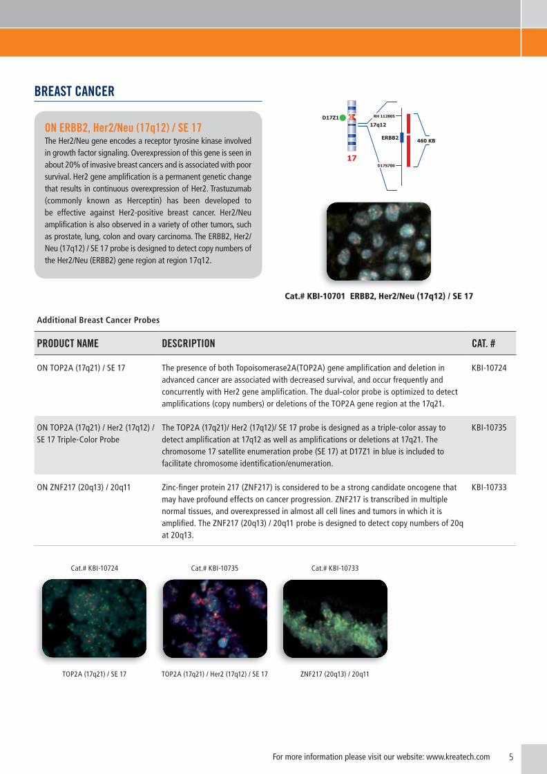
ON TOP2A (17q21) / SE 17 The presence of both Topoisomerase2A(TOP2A) gene amplification and deletion in advanced cancer are associated with decreased survival, and occur frequently and concurrently with Her2 gene amplification. The dual-color probe is optimized to detect amplifications (copy numbers) or deletions of the TOP2A gene region at the 17q21.
KBI-10724
ON TOP2A (17q21) / Her2 (17q12) / SE 17 Triple-Color Probe
The TOP2A (17q21)/ Her2 (17q12)/ SE 17 probe is designed as a triple-color assay to detect amplification at 17q12 as well as amplifications or deletions at 17q21. The chromosome 17 satellite enumeration probe (SE 17) at D17Z1 in blue is included to facilitate chromosome identification/enumeration.
KBI-10735
ON ZNF217 (20q13) / 20q11 Zinc-finger protein 217 (ZNF217) is considered to be a strong candidate oncogene that may have profound effects on cancer progression. ZNF217 is transcribed in multiple normal tissues, and overexpressed in almost all cell lines and tumors in which it is amplified. The ZNF217 (20q13) / 20q11 probe is designed to detect copy numbers of 20q at 20q13.
KBI-10733
Cat.# KBI-10724 Cat.# KBI-10735 Cat.# KBI-10733
TOP2A (17q21) / SE 17 TOP2A (17q21) / Her2 (17q12) / SE 17 ZNF217 (20q13) / 20q11
on ERBB2, Her2/neu (17q12) / sE 17The Her2/Neu gene encodes a receptor tyrosine kinase involved in growth factor signaling. Overexpression of this gene is seen in about 20% of invasive breast cancers and is associated with poor survival. Her2 gene amplification is a permanent genetic change that results in continuous overexpression of Her2. Trastuzumab (commonly known as Herceptin) has been developed to be effective against Her2-positive breast cancer. Her2/Neu amplification is also observed in a variety of other tumors, such as prostate, lung, colon and ovary carcinoma. The ERBB2, Her2/Neu (17q12) / SE 17 probe is designed to detect copy numbers of the Her2/Neu (ERBB2) gene region at region 17q12.
Cat.# KBI-10701 ERBB2, Her2/Neu (17q12) / SE 17

6 For more information please visit our website: www.kreatech.com
Additional Breast Cancer Probes
PRoDUCT nAME DEsCRiPTion CAT. #
ON FGFR1 (8p11) / SE 8 Fibroblast growth factor receptor 1 (FGFR1) gene amplification is found in up to 10% of breast cancers. FGFR1 gene amplification is significantly more prevalent in patients > 50 years of age and in tumors that lack HER2 expression. FGFR1 amplification might act as a valuable biomarker for R&D and provide an attractive approach for clinical stratification.
KBI-12754 KBI-14754
ON AURKA (20q13) / 20q11 Aurora kinase A (AURKA) gene amplification has been detected in approximately 12% of primary breast tumors, as well as in bladder, ovarian, colon, prostate, neuroblastoma and cervical cancer cell lines. Recent research into new drug development has focused on the importance of aurora kinases for tumor suppression. The AURKA (20q13) / 20q11 probe is designed to detect copy numbers of the AURKA gene region at region 20q13.
KBI-10721
ON FGFR1 (8p11) Break The FGFR1 (8p11) break-apart probe is designed to detect translocations involving the FGFR1 gene region at 8p11.
KBI-10737
Cat.# KBI-12754 / KBI-14754 Cat.# KBI-10721 Cat.# KBI-10737
FGFR1 (8p11) / SE 8 Amplification1 AURKA (20q13) / 20q112 FGFR1 (8p11) Break
1 Images kindly provided by Dr. Otte, Vienna. 2 Material kindly provided by Dr. Carvalho, Amsterdam.
BLADDER CAnCER
Cat.# KBI-10710 p16 (9p21) / 9q21
p16400 KB
p15
9p21D9S1749
D9S975
9
9q21
on p16 (9p21) / 9q21Homozygous and hemizygous deletions of 9p21 are the earliest and most common genetic alteration in bladder cancer. The p16 (CDKN2A) gene has been identified as tumor suppressor gene. In bladder cancer, the region harboring the p16 gene is frequently deleted. The loss of DNA sequences on chromosomal bands 9p21-22 has been documented also in a variety of malignancies including leukemias, gliomas, lung cancers, and melanomas.The p16 (9p21) / 9q21 probe is designed to detect copy numbers of the p16 gene region at region 9p21.

7For more information please visit our website: www.kreatech.com
CERViCAL CAnCER
Cervical cancer, a potentially preventable disease, remains the second most common malignancy in women worldwide.
370 KBhTERC3q26.2
RH17919
RH10606
3
C-MYC
8q24
540 KB
SHGC-144185
RH77966
8
2 Material kindly provided by Dr. Carvalho, Amsterdam. 3 Image kindly provided by Dr. Weimer, Kiel.
Cat.# KBI-10704 Cervical Cancer hTERC (3q26) / C-MYC (8q24) / SE 7 Triple-Color3
on CC hTERC (3q26) / C-MYC (8q24) / sE 7 TC The most consistent chromosomal gain in aneuploid tumors of cervical squamous cell carcinoma mapped to chromosome arm 3q, includes the human telomerase gene locus (hTERC) at 3q26. High-level copy number increases were also mapped to chromosome 8. Integration of HPV (Human Papilloma Virus) DNA sequences into C-MYC chromosomal regions have been repeatedly observed in cases of invasive genital carcinomas and in cervical cancers. The hTERC (3q26) / C-MYC (8q24) / SE 7 probe is designed to detect copy numbers of the hTERC gene region at region 3q26. The C-MYC (8q24) specific DNA probe is optimized to detect copy numbers of the C-MYC gene region at 8q24.
Additional Bladder Cancer Probes
PRoDUCT nAME DEsCRiPTion CAT. #
ON AURKA (20q13) / 20q11 Aurora kinase A (AURKA) gene amplification has been detected in approximately 12% of primary breast tumors, as well as in bladder, ovarian, colon, prostate, neuroblastoma and cervical cancer cell lines. Recent research into new drug development has focused on the importance of aurora kinases for tumor suppression. The AURKA (20q13) / 20q11 probe is designed to detect copy numbers of the AURKA gene region at region 20q13.
KBI-10721
Cat.# KBI-10721
AURKA (20q13) / 20q112

8
LUng CAnCER
Lung cancer remains the leading cause of cancer death, annually resulting in more than one million cases worldwide. About 1.6 million new cases are diagnosed each year and prognoses are poor. Non-small cell lung cancer (NSCLC), the most common form (~80%) of lung cancer, has a 5-year survival rate of approximately 15%, mainly of late-stage detection. A personalized medicine approach for treatment of NSCLC is emerging. Promising results have been obtained with specific anaplastic lymphoma kinase or ALK inhibitors like crizotinib (XALKORI®)in patients carrying the fusion gene ALK-EML4.
Additional Lung Cancer Probes
PRoDUCT nAME DEsCRiPTion CAT. #
ON ALK/EML4 t(2;2); inv(2) Fusion The inversion in 2p21 and 2p23 leading to a fusion of the kinase domain of ALK (anaplastic lymphoma kinase) and EML4 (echinoderm microtubule associated protein like 4) has been described in 5-7% of NSCLC cases. ALK and EML4 are ~12 MB apart in opposite directions; a simple inversion generates the fusion gene. The ALK/EML4 t(2;2); inv(2) Fusion probe is designed as a dual-color assay to detect the fusion of the ALK gene with the EML4 gene by paracentric inversion with breakage and reunion occurring at bands 2p21 and 2p23.
KBI-10746
ON ROS1 (6q22) Break ROS1 (c-ros oncogene 1, receptor tyrosine kinase) gene rearrangements represent 1-2% of aberrations in NSCLC. ROS1 gene rearrangements define a unique molecular subset of NSCLC. These are mainly non-overlapping with other oncogenic aberrations (e.g. EGFR mutations, KRAS mutations, ALK rearrangements, etc.). The ROS1 (6q22) Break captures all ROS1 rearrangements described to date including the GOPC-ROS1 fusion.
KBI-10752
ON RET (10q11) Break RET gene rearrangement represents 1-2% of aberrations in NSCLC, and can include KIF5B-RET or CCDC6-RET fusions. RET gene rearrangements define a unique molecular subset of NSCLC. These are mainly non-overlapping with other oncogenic aberrations (e.g. ALK- and ROS1 rearrangements, etc.).The RET (10q11) Break captures all RET rearrangements including KIF5B-RET Fusion.
KBI-10753
4 Image kindly provided by Prof. Terris, Dr. Just, Paris.
on ALK (2p23) BreakTranslocations of the ALK (anaplastic lymphoma kinase) gene at 2p23 have originally been associated with anaplastic lymphomas, B-cell lymphomas, neuroblastomas and myofibroblastic tumors. To date at least 21 translocation partners have been described, however 80% of the translocations involve the NPM1 gene (5q35). More recently, ALK rearrangements have been described in non-small cell lung cancer (NSCLC) cases. Promising results have been obtained with specific anaplastic lymphoma kinase or ALK inhibitors like crizotinib (XALKORI®) in patients carrying the fusion gene ALK-EML4. The ALK (2p23) Break probe is optimized to detect translocations involving the ALK gene region at 2p23.
Cat# KBI-10747 ALK (2p23) Break4
2p23
SHGC-17159
360 KB
D2S392
340 KBALK
2
D2S405
RH109933

9For more information please visit our website: www.kreatech.com
5 Presented at the ADAPT meeting 2012, Washington, DC.6 Images kindly provided by Dr. Otte, Vienna.
Additional Lung Cancer Probes
PRoDUCT nAME DEsCRiPTion CAT. #
ON FGFR1 (8p11) / SE 8 Amplification of the fibroblast growth factor receptor type 1 gene (FGFR1) has been observed in numerous cancer types including Squamous Cell Carcinoma (SCC) of the lung. FGFR1 amplifications represent ±4% of aberrations found in NSCLC and represent around 17% of aberrations in SCC of the lung. With the development of new therapeutic strategies, FGFR1 amplification could act as a valuable biomarker for R&D and provide an attractive tool for clinical stratification.
KBI-12754 KBI-14754
ON C-MET (7q31) / SE 7 The C-MET proto-oncogene is a receptor-like tyrosine kinase that drives a physiological cellular program important for development, cell movement, cell repair, cellular growth. Aberrant execution of the program has been associated with neoplastic transformation, invasion and metastasis. Activation of C-MET has been reported in a significant percentage of human cancers and is amplified during the transition between primary tumors and metastasis. The C-MET (7q31) / SE 7 probe is designed to detect copy numbers of the C-MET gene region at region 7q31.
KBI-10719
ON EGFR, Her-1 (7p11) / SE 7 Epidermal growth factor receptor (EGFR) has been found to act as a strong prognostic indicator in NSCLC, head and neck, ovarian, cervical, bladder and oesophageal cancers. In these cancers, increased EGFR expression was associated with reduced recurrence-free or overall survival. The EGFR (7p11) / SE 7 probe is designed to detect copy numbers of the EGFR (Her-1) gene region at region 7p11.
KBI-10702
ON hTERT (5p15) / 5q31 Amplification of the hTERT gene at band 5p15 has been observed in a variety of cancers, particularly lung cancer, cervical tumors, and breast carcinomas. The hTERT (5p15) / 5q31 probe is designed as a dual-color assay to detect amplification at 5p15. The CDC25C/EGR1 (5q31) gene region probe is included as internal control.
KBI-10709
ON ERCC1 (19q13) & ZNF443 (19p13)
Excision repair cross-complementing rodent repair deficiency, complementation group 1 (ERCC1) gene has been shown to be an important marker to predict responsiveness to cisplatin-based chemotherapy. Low ERCC1 gene expression correlates with prolonged survival after cisplatin-based chemotherapy. The ERCC1 (19q13) & ZNF443 (19p13) probe has been designed to detect copy numbers of the ERCC1 gene region at 19q13.
KBI-10739
Cat# KBI-10746 Cat.# KBI-10752 Cat.# KBI-10753 Cat.# KBI-12754 / KBI-14754
ALK/EML4 t(2;2); inv(2) Fusion5 ROS1 (6q22) Break RET (10q11) Break FGFR1 (8p11) / SE 8 Amplification6
10 For more information please visit our website: www.kreatech.com
PRosTATE CAnCER
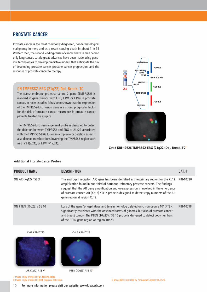
Prostate cancer is the most commonly diagnosed, nondermatological malignancy in men; and as a result causing death in about 1 in 35 Western men, the second leading cause of cancer death in men behind only lung cancer. Lately, great advances have been made using geno-mic technologies to develop predictive models that anticipate the risk of developing prostate cancer, prostate cancer progression, and the response of prostate cancer to therapy.
Additional Prostate Cancer Probes
PRoDUCT nAME DEsCRiPTion CAT. #
ON AR (Xq12) / SE X The androgen receptor (AR) gene has been identified as the primary region for the Xq12 amplification found in one-third of hormone-refractory prostate cancers. The findings suggest that the AR gene amplification and overexpression is involved in the emergence of prostate cancer. AR (Xq12) / SE X probe is designed to detect copy numbers of the AR gene region at region Xq12.
KBI-10720
ON PTEN (10q23) / SE 10 Loss of the gene ‘phosphatase and tensin homolog deleted on chromosome 10’ (PTEN) significantly correlates with the advanced forms of gliomas, but also of prostate cancer and breast tumors. The PTEN (10q23) / SE 10 probe is designed to detect copy numbers of the PTEN gene region at region 10q23.
KBI-10718
Cat# KBI-10720 Cat.# KBI-10718
AR (Xq12) / SE X8 PTEN (10q23) / SE 109
21q22
ERG
TMPRSS2
600 KB
700 KB
D21S1820
D21S1439
21
ETS2
700 KBRH123924
D21S349
GAP 2.3 MB
For more information please visit our website: www.kreatech.com
7 Image kindly provided by Dr. Teixeira, Porto.8 Image kindly provided by Prof. Trapman, Rotterdam. 9 Image kindly provided by Portuguese Cancer Inst., Porto.
on TMPRss2-ERg (21q22) Del, Break, TC The transmembrane protease serine 2 gene (TMPRSS2) is involved in gene fusions with ERG, ETV1 or ETV4 in prostate cancer. In recent studies it has been shown that the expression of the TMPRSS2-ERG fusion gene is a strong prognostic factor for the risk of prostate cancer recurrence in prostate cancer patients treated by surgery.
The TMPRSS2-ERG rearrangement probe is designed to detect the deletion between TMPRSS2 and ERG at 21q22 associated with the TMPRSS2-ERG fusion in a triple-color deletion assay. It also detects translocations involving the TMPRSS2 region such as ETV1 t(7;21), or ETV4 t(17;21).
Cat.# KBI-10726 TMPRSS2-ERG (21q22) Del, Break, TC7

11For more information please visit our website: www.kreatech.com
THYRoiD CARCinoMA
Papillary thyroid carcinoma (PTC) is the most frequent primary carcinoma of the thyroid gland. The follicular carcinomas are associated with endemic goiter and a diet with low iodine intake. PTC, conversely, are multifocal and are associated with prior radiation and high iodine intake.
325 KB
PPARg
3p25
350 KB
D35456
D351351
3
Cat.# KBI-10707 PPARγ (3p25) Break10
For more information please visit our website: www.kreatech.com
Additional Thyroid Carcinoma Probes
PRoDUCT nAME DEsCRiPTion CAT. #
ON RET (10q11) Break RET rearrangements are found in approximately 10-20% of sporadic papillary thyroid cancers (PTCs). Different RET rearrangements have been described in PTCs. The majority of the cases harbor in 60-70% the RET/CCDC6 (PTC1) fusion, in 20-30% the RET/NCOA4 (PTC3) fusion and in 5% the RET/PRKAR1A (PTC2) fusion. These oncogenic rearrangements consist of various 5' partners fused to the kinase domain of RET, leading to constitutive activation of the RET receptor tyrosine kinase.
KBI-10753
Cat.# KBI-10753
RET (10q11) Break
9 Image kindly provided by Portuguese Cancer Inst., Porto.
Cat.# KBI-10707 PPAR (3p25) Break
on PPARγ (3p25), BreakFollicular thyroid carcinoma is associated with the chromosomal translocation t(2;3)(q13;p25), fusing PAX8 (2q13) with the nuclear receptor, peroxisome proliferator-activated receptor γ (PPARγ). The immediate vicinity of PPAR is a breakpoint hot spot region, leading to recurrent alterations of this gene in thyroid tumors of follicular origin including carcinomas as well as adenomas with or without involvement of PAX8. The PPARγ (3p25) Break probe is designed to detect translocations involving the PPARγ gene region at 3p25.
10 Image kindly provided by Dr. Valent, Paris.

12 For more information please visit our website: www.kreatech.comFor more information please visit our website: www.kreatech.com
11 Image kindly provided by Pasteur Workshop 2008, Paris.
on MYCn (2p24) / LAF (2q11) Amplification of the human N-myc protooncogene, MYCN, is frequently seen either in extrachromosomal double minutes or in homogeneously staining regions of aggressively growing neuroblastomas. MYCN amplification has been defined by the INRG as > 4-fold MYCN signals compared to 2q reference probe signals.
The MYCN (2p24) probe is designed to detect copy numbers of the MYCN gene region at 2p24. The LAF gene region probe at 2q11 is included to facilitate chromosome identification.
Cat.# KBI-10706 MYCN (2p24) / LAF (2q11)11
2p24.3
RH112907
450 KB
D2S2676
MYCN
2
375 KBLAF4
2q11.2
2
nEURoBLAsToMA
According to the International Neuroblastoma Risk Grouping (INRG) Biology Committee MYCN remains the only genomic factor to be used currently for treatment stratification. Common data elements to be obtained by all groups include tumor cell ploidy and copy number/LOH status at chromosome bands 1p36, 11q23, and 17q23-25.
Cat.# KBI-10736 MDM4 (1q32) / SE 111
on MDM4 (1q32) / sE1 Studies showed an increased MDM4 (also known as MDMX, murine double minute gene) copy number in 65% of human retinoblastomas compared to other tumors, qualifying MDM4 as a specific chemotherapeutic target for treatment of this tumor. The MDM4 (1q32) / SE 1 probe is designed to detect amplification at 1q32.
1q32
1
MDM4
D1S504
440 KB
RH2406

13For more information please visit our website: www.kreatech.com
Additional Neuroblastoma Probes
PRoDUCT nAME DEsCRiPTion CAT. #
ON SRD 1p36 / SE 1 (1qh) Neuroblastomas frequently have deletions of chromosome 1p and amplification of the N-myc oncogene. These deletions tend to be large and extend to the telomere, and a common region within sub-band 1p36.3 is consistently lost. Inactivation of a tumor suppressor gene within 1p36.3 is believed to be associated with an increased risk for disease relapse. The 1p36 specific DNA probe covers the recently described smallest region of consistent deletion (SRD) between D1S2795 and D1S253.
KBI-10712
ON MLL (11q23) / SE 11 Deletions of the long arm of chromosome 11 (11q) have been noted in primary neuroblastomas. The MLL (11q23) / SE 11 probe is designed to detect amplification or deletion involving the MLL gene region at 11q23.
KBI-10711
Cat.# KBI-10712 Cat.# KBI-10711
SRD 1p36 / SE 1 (1qh) MLL (11q23) / SE 11
For more information please visit our website: www.kreatech.com

14 For more information please visit our website: www.kreatech.com
Additional Sarcoma Probes
PRoDUCT nAME DEsCRiPTion CAT. #
ON COL1A1/PDGFB t(17:22) DC,S-Fusion
The diagnosis of primary soft tissue and bone tumors is often challenging as they are relatively rare. The misdiagnosis between dermatofibroma (DF) and dermatofibrosar-coma protuberans (DFSP) or giant cell fibroblastoma (GCF) might result in improper primary management. DFSP and GCF have in most cases diagnosed today a transloca-tion involving the COL1A1 (collagen, type I, alpha 1) gene at 17q21 and the PDGFB (platelet-derived growth factor beta polypeptide) gene at 22q13. Also, a supernumerary ring chromosome derived from the translocation r(17;22) can be present.
KBI-10742
ON EWSR1 (22q12) Break Ewing’s sarcoma is the second most frequent primary bone cancer. In most cases a translocation involving the EWSR1 gene at 22q12 and the FLI1 gene at 11q24 is observed, but several other translocation partners (ERG, ETV1, FEV, and E1A3) can also be involved. The EWSR1 (22q12) Break Probe is optimized to detect translocations involving the EWSR1 gene region at 22q12 in a dual-color, split assay on metaphase/interphase spreads and paraffin embedded tissue sections.
KBI-10750
12 Image kindly provided by Mr. Griffioen, Arnhem.
on MDM2 (12q15) / sE 12Well-differentiated liposarcoma/atypical lipomatous tumor and dedifferentiated liposarcoma are among the most common malignant soft tissue tumors presented in older adults. These tumors can be difficult to distinguish from benign lipomatous neoplasms and other high-grade sarcomas.
Amplification of the MDM2 gene has been identified in lipomatous neoplasms. The use of fluorescence in situ hybridization in identifying MDM2 amplification has made the MDM2 amplification probe a valuable diagnostic tool in well- differentiated liposarcomas/atypical lipomatous tumors.
Fibrosarcoma is a rare soft-tissue tumor composed of fascicles of spindled fibroblast-like cells. Gains and high-level amplifications of 12q14–22 were the most common genomic imbalances, and reflected MDM2 amplification, thereby indicating the importance of this gene in the evolution of fibrosarcomas. The MDM2 (12q15) / SE 12 probe is optimized to detect copy numbers of the MDM2 gene region at region 12q15.
528 KBMDM212q15
D12S1382E
D12S1497
12
D12Z3
Cat.# KBI-10717 MDM2 (12q15) / SE 1212
sARCoMA
Soft Tissue Sarcoma (STS) and bone sarcomas are rare malignancies of mesenchymal origin that account for less than 1% of all adult solid tumors. Sarcomas provide a particular diagnostic dilemma, not only due to their rarity, but also due to their wide diversity, with more than 50 histological subtypes currently recognized. This heterogeneity in classification is accompanied by a broad spectrum of biologic behavior, from locally aggressive but non-metastasizing tumors to those which are highly aggressive and rapidly metastasizing.
Molecular Alterations in Sarcomas may be roughly categorized into three types:1. Relatively simple karyotype with a defining translocation
or amplification of a particular locus.2. Simple or complex karyotype with a specific oncogenic mutation.3. Complex karyotype with multiple chromosomal rearrangements,
duplications, and deletions. Source: www.cancer.net

15For more information please visit our website: www.kreatech.com
Additional Sarcoma Probes
PRoDUCT nAME DEsCRiPTion CAT. #
ON EWSR1/NFATC2 t(20;22) DC,S-Fusion
Ewing’s sarcoma is the second most frequent primary bone cancer. In most cases a trans-location involving the EWSR1 gene at 22q12 and the FLI1 gene at 11q24 is observed. Several other translocation partners of the ETS gene family can also be involved. The first non-ETS family translocation partner described is the NFATC2 gene (nuclear factor of activated T-cells, cyto-plasmic, calcineurin-dependent 2) at 20q13. The EWSR1/NFATC2 single fusion probe is best used to analyze the specific trans-locations of the EWSR1 and NFATC2 gene on formalin fixed paraffin embedded tissue for routine clinical diagnosis.
KBI-10751
ON CDK4 (12q13) / SE 12 The Chromosomal region 12q13-15 is recurrently amplified in sarcoma and gliomas. Putative target genes located in this region include MDM2 and CDK4. Independent CDK4 amplifications has been described, suggesting two different amplified regions one including MDM2, the other CDK4. The CDK4 (12q13) / SE 12 probe is designed to detect copy numbers of the CDK4 gene region at 12q13.
KBI-10725
ON SYT (18q11) Break The characteristic chromosomal abnormality in synovial sarcoma t(X;18)(p11.2;q11.2) is present in 90% of patients. This translocation results in the fusion of the chromosome 18 SYT gene to either of two distinct genes, SSX1 or SSX2, located on the X chromosome. The SYT (18q11) Break probe is designed to detect translocation of the SYT (SS18) gene.
KBI-10713
ON CHOP (12q13) Break Most patients with round cell/myxoid liposarcoma have an acquired t(12;16)(CHOP-FUS) or t(12;22)(CHOP-EWS) translocation, both of which involve the CHOP gene at 12q13. The CHOP (12q13) Break probe is designed to detect translocations involving the CHOP gene region at 12q13.
KBI-10714
ON FUS (16p11) Break The FUS gene was originally shown to be rearranged in myxoid liposarcomas harboring a t(12;16)(q13;p11) translocation. FUS has been shown to be involved with ERG in acute myeloid leukemia carrying a t(16;21), with ATF1 in band 12q13 in angiomatoid fibrous histiocytoma, and with CREB3L2 in fibromyxoid sarcoma. The FUS (16p11) Break probe is designed to detect translocation involving the FUS gene.
KBI-10715
ON FKHR (13q14) Break Identification of translocations t(2;13) (q35;q14) and t(1;13) (p36;q14) and the associated PAX3 - FKHR and PAX3 - FKHR fusion transcripts are a valuable diagnostic adjunct and important prognostic parameter in alveolar rhabdomyosarcoma. The FKHR (13q14) Break probe is designed to detect translocations involving the FKHR gene region at 13q14.
KBI-10716
Cat.# KBI-10742 Cat.# KBI-10750 Cat.# KBI-10751 Cat.# KBI-10725
COL1A1/PDGFB t(17:22) DC, S-Fusion ON EWSR1 (22q12) Break EWSR1/NFATC2 t(20;22) DC,S-Fusion
CDK4 (12q13) / SE 1213
13 Image kindly provided by Dr. Sapi, Hungary.
16 For more information please visit our website: www.kreatech.com
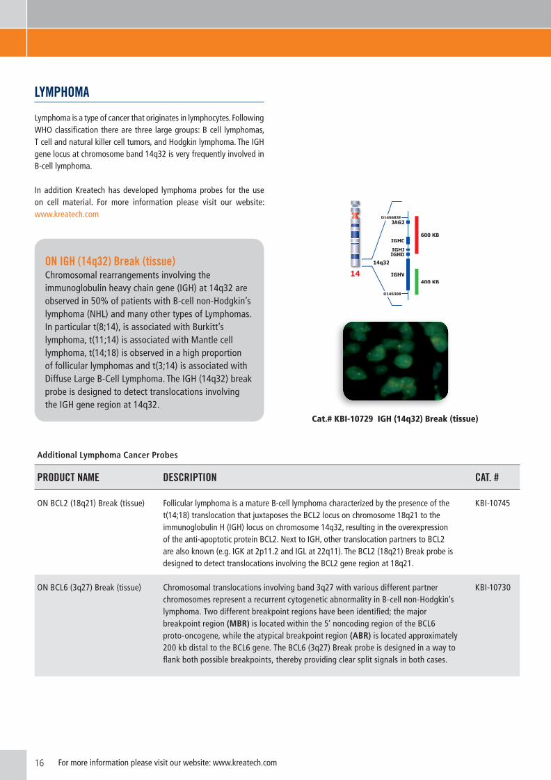
LYMPHoMA Lymphoma is a type of cancer that originates in lymphocytes. Following WHO classification there are three large groups: B cell lymphomas, T cell and natural killer cell tumors, and Hodgkin lymphoma. The IGH gene locus at chromosome band 14q32 is very frequently involved in B-cell lymphoma.
In addition Kreatech has developed lymphoma probes for the use on cell material. For more information please visit our website: www.kreatech.com
Additional Lymphoma Cancer Probes
PRoDUCT nAME DEsCRiPTion CAT. #
ON BCL2 (18q21) Break (tissue) Follicular lymphoma is a mature B-cell lymphoma characterized by the presence of the t(14;18) translocation that juxtaposes the BCL2 locus on chromosome 18q21 to the immunoglobulin H (IGH) locus on chromosome 14q32, resulting in the overexpression of the anti-apoptotic protein BCL2. Next to IGH, other translocation partners to BCL2 are also known (e.g. IGK at 2p11.2 and IGL at 22q11). The BCL2 (18q21) Break probe is designed to detect translocations involving the BCL2 gene region at 18q21.
KBI-10745
ON BCL6 (3q27) Break (tissue) Chromosomal translocations involving band 3q27 with various different partner chromosomes represent a recurrent cytogenetic abnormality in B-cell non-Hodgkin’s lymphoma. Two different breakpoint regions have been identified; the major breakpoint region (MBR) is located within the 5’ noncoding region of the BCL6 proto-oncogene, while the atypical breakpoint region (ABR) is located approximately 200 kb distal to the BCL6 gene. The BCL6 (3q27) Break probe is designed in a way to flank both possible breakpoints, thereby providing clear split signals in both cases.
KBI-10730
on igH (14q32) Break (tissue)Chromosomal rearrangements involving the immunoglobulin heavy chain gene (IGH) at 14q32 are observed in 50% of patients with B-cell non-Hodgkin’s lymphoma (NHL) and many other types of Lymphomas. In particular t(8;14), is associated with Burkitt’s lymphoma, t(11;14) is associated with Mantle cell lymphoma, t(14;18) is observed in a high proportion of follicular lymphomas and t(3;14) is associated with Diffuse Large B-Cell Lymphoma. The IGH (14q32) break probe is designed to detect translocations involving the IGH gene region at 14q32.
Cat.# KBI-10729 IGH (14q32) Break (tissue)
14q32
JAG2
IGHV
IGHD
IGHC
IGHJ
D14S683E
600 KB
D14S308
400 KB14

17For more information please visit our website: www.kreatech.com
Additional Lymphoma Cancer Probes
PRoDUCT nAME DEsCRiPTion CAT. #
ON MALT (18q21) Break (tissue) Low grade malignant lymphomas arising from mucosa associated lymphoid tissue (MALT) represent a distinct clinicopathological entity. The three major translocations seen in MALT lymphomas are t(11;18)(q21;q21)/API2-MALT1, t(14;18)(q32;q21)/ IGH-MALT1 and t(1;14)(p22;q32)/IGH-BCL10. The MALT (18q21) Break probe is designed to analyze translocation of the MALT gene region at 18q21.
KBI-10731
ON MYC (8q24) Break, TC (tissue)
Rearrangements of the proto oncogene C-myc (or MYC) have been consistently found in tumor cells of patients suffering from Burkitt’s lymphoma. In cases with the common t(8;14) chromosomal translocation, the c-myc gene is translocated to chromosome 14 and rearranged with the immunoglobulin heavy chain genes; the breakpoint occurs 5’ to the c-myc gene and may disrupt the gene itself separating part of the first untranslated exon from the remaining two coding exons. In Burkitt’s lymphoma showing the variant t(2;8) or t(8;22) translocations, the genes coding for the k and l immunoglobulin light chain are translocated to chromosome 8. The MYC (8q24) break-apart probe is optimized to detect rearrangements involving the 8q24 locus in a triple-color, split assay on formalin fixed paraffin embedded tissue.
KBI-10749
ON BCL2/IGH@ t(14;18) Fusion (tissue)
Follicular lymphoma is a mature B-Cell lymphoma, characterized by the presence of the t(14;18) translocation that juxtaposes the BCL2 locus on chromosome 18q21 to the immunoglobulin H (IGH) locus on chromosome 14q32, resulting in the overexpression of the antiapoptotic protein BCL2.
KBI-10755
Cat# KBI-10745 Cat.# KBI-10730 Cat.# KBI-10749 Cat# KBI-10755
BCL2 (18q21) Break (tissue) BCL6 (3q27) Break (tissue) MYC (8q24) Break, TC (tissue) BCL2/IGH@ t(14;18) Fusion (tissue)

18 For more information please visit our website: www.kreatech.com
DiFFEREnT CAnCER TYPEs
Cat.# KBI-10741 TFE3 (Xp11) Break14
on TFE3 (xp11) BreakAbnormalities of Xp11.2 region have often been observed in papillary renal cell carcinomas and are sometimes the sole cytogenetic abnormality present. The transcription factor binding to IGHM enhancer 3 (TFE3) gene, can be fused to various other chromosomal regions by translocation. Known fusion partners are NONO (Xq12), PRCC (1q21), SFPQ (1p34), CLTC (17q23) and ASPSCR1 (17q25). The TFE3 (Xp11) Break probe is designed to detect translocations involving the TFE3 gene region at Xp11.2.
425 KB
525 KB
X
TFE3
Xp11.2
DXS6784
DXS1522
14 Image kindly provided by Dr. Desangles, Paris.
Additional different Cancer Probes
PRoDUCT nAME DEsCRiPTion CAT. #
ON AURKB (17p13) / SE 17 Aurora kinase B (AURKB) gene amplification is predictive for the aggressive recurrence of many different types of tumors, including hepatocellular carcinoma and oral squamous cell carcinoma. Recently new drugs have been under investigation for their capacity of interfering with the aurora kinases activity related to tumor-suppressor effects. The AURKB (17p13) / SE 17 probe is designed to detect copy numbers of the AURKB gene region at region 17p13.
KBI-10722
ON CCND1 (11q13) / SE 11 Amplification of CCND1 (also named Cyclin D1 or BCL1) plays a pivotal role in the development of a subset of human cancers including parathyroid adenoma, breast cancer, colon cancer, lymphoma, melanoma, and prostate cancer. The CCND1 (11q13) / SE 11 probe is designed to detect copy numbers of the CCND1 gene region at region 11q13.
KBI-10734
ON p53 (17p13) / SE 17 Allelic loss, usually by deletion, and inactivation of p53 have been reported in numerous tumor types and are associated with poor prognosis in B-cell chronic lymphocytic leukaemia (CLL). The p53 (17p13) specific DNA probe is designed to detect copy numbers of the p53 tumor suppressor gene region at 17p13.
KBI-10738
Cat.# KBI-10722 Cat.# KBI-10734 Cat.# KBI-10738
AURKB (17p13) / SE 17 CCND1 (11q13) / SE 11 p53 (17p13) / SE 17

19For more information please visit our website: www.kreatech.com
CHRoMogEniC in SiTu hybridizaTion
PRoDUCT nAME DEsCRiPTion CAT. #
TwinStar CISH Detection Kit For the use with POSEIDON™ DNA probes labeled in red and green KBI-60010
TwinStar Her2/neu (17q12) DNA probes specific for Her-2/Neu and SE 17, TwinStar CISH Detection Kit KBI-60701
TwinStar EGFR (7p11) DNA probes specific for EGFR and and SE 7, TwinStar CISH Detection Kit KBI-60702
TwinStar C-MET (7q31) DNA probes specific for C-MET and and SE 7, TwinStar CISH Detection Kit KBI-60719
UniStar CISH Detection Kit UniStar CISH Detection Kit for the use with POSEIDON™ DNA Probes labeled in red KBI-50001
UniStar Her2/neu (17q12) DNA probes specific for Her-2/Neu and SE 17, UniStar CISH Detection Kit KBI-50701
UniStar EGFR (7p11) DNA probes specific for EGFR and and SE 7, UniStar CISH Detection Kit KBI-50702
UniStar C-MET (7q31) DNA probes specific for C-MET and and SE 7, UniStar CISH Detection Kit KBI-50719
Amplified Her-2/Neu (ERBB2) on a breast cancer specimen visualized with the TwinStar dual color CISH kit.15
Glioblastoma specimen showing amplified EGFR. The slide was hybridized with the EGFR POSEIDON™ fluorescent probe followed by conversion of the red EGFR signal into a colorimetric signal using the UniStar CISH Detection Kit.16
TwinstarTwinstar is based on a novel assay based on its well established REPEAT-FREE™ POSEIDON™ DNA probes, enabling the clinician to consecutively perform FISH and CISH on the same sample. Twinstar is designed for dual-color CISH offering the possibility to study ratio of genes in a light microscope, such as for the Her-2 gene in relation to the centromeric region of chromosome 17. Each kit includes the corresponding FISH probes and the specific TwinStar detection module converting both signals into chromogenic signals via a colorimetric assay system.
The TwinStar CISH Detection Kit is a module of all TwinStar assays including all reagents required to perform dual-color CISH. The detection module includes two proprietary substrates converting both fluorescent signals into distinct colorimetric signals, the red fluor is converted into a red signal and the green fluor into a grayish-green signal. TwinStar provides a universal solution capable of transforming all of Kreatech´s dual-color FISH probes into chromogenic signals.
15 Images kindly provided by Dr. Beiske, Norway.16 Images kindly provided by Dr. Beiske, Norway.
UnistarEach of the UniStar kits include the corresponding REPEAT-FREE™ POSEIDON™ DNA probes for copy number detection of the gene of interest, as well as a control probe for performing FISH in a dual-color assay.
In addition, they include a specific detection module converting the signal of the critical probe into a chromogenic single-color signal via a colorimetric assay system.
Tissue Slide Pretreatment Probe Hybridization
A.
B.
CISH
CISH
FISH Analysis

20 For more information please visit our website: www.kreatech.com
ordering and DeliveryOrders for Kreatech products can be placed via e-mail, fax or over the telephone with one of our Sales and/or Customer Service representatives on the basis of a purchase order number. Purchase orders for regularly stocked items received by Kreatech by or before 14:00 CET Monday through Thursday are shipped the same working day. Purchase orders received after 14:00 CET are shipped the following working day. In the event that an order contains a non-stocked item, or if, for any reason, Kreatech is unable to completely fulfill an order, a partial shipment will be made unless instructed otherwise. You will be notified immediately of any delay. Delivery is made EXW Kreatech, Amsterdam (Incoterms 2010).
Prices, Payment and shipping termsTerms of sales are net 30 days from the date shown on the invoice. Prices are subject to change without notice. All orders under € 400 will be charged with the shipping costs. There is no minimum order quantity; however Kreatech reserves the right to charge an additional processing fee of € 15 for orders received under € 200.
All orders are subject to review by Kreatech and additional information may be requested. Kreatech reserves the right to refuse shipment of material to anyone.
standing order / Volume DiscountsFor your convenience, Kreatech can schedule routine shipments of any single purchase order. Discounted prices may be available when purchasing large volumes. Inquiries should be directed to your local Sales Representative.
Methods of payment including Credit Card ordersTo better serve your needs with convenience and efficiency, Kreatech also accepts MasterCard and VISA as payment for orders placed via fax. Please provide the name on your credit card, account number, expiration date and security code when placing credit card orders.
The Kreatech Customer satisfaction PolicyIf you are not completely satisfied with the performance of any product, Kreatech will, at your option, either credit your account or send a replacement product to you at our expense. Requirements for this are that the product has not expired, it has been stored and processed according to Kreatech product literature and that the user followed all appropriate Kreatech protocols when using the product.
Returned goods PolicyNo returns will be approved without the authorization of Kreatech. Unauthorized returns will not be accepted and will be returned at the sender’s expense. Complete documentation must accompany all authorized returned goods. If errors in ordering goods are made by Kreatech, Kreatech will exchange goods at Kreatech’s expense.
Product UseAll products manufactured and/or distributed by Kreatech should be used in accordance with the products labeled intended use.Kreatech's Poseidon™ Repeat-Free™ FISH products are supplied for
in vitro diagnostics (or for research use only if not CE marked). They are NOT intended to be used as drugs, food, additives, cosmetics or household chemicals. Kreatech warrants that, at the time of shipment, the products sold are free from defects in materials and conform to the specifications described in Kreatech’s literature. Kreatech makes no other warranty, expressed or implied, with respect to the products. This includes any warranty of merchantability or suitability for any particular purpose.
LiabilityKreatech accepts liability for damages suffered by its customers, up to a maximum amount of € 5.000,- if these damages are directly and exclusively due to gross negligence and/or willful misconduct by Kreatech.
Kreatech does not accept liability for (i) damages resulting from the use/stocking of Kreatech products by its customers contrary to Kreatech’s instructions, (ii) damages resulting from off label use of the Kreatech products, (iii) the results of any diagnosis or research made on the basis of or with the Kreatech products or (iv) any consequential or indirect damages suffered by its customers.
Patents and TrademarksThese products or the use of these products is subject to intellectual property rights. FISH and CISH probes are produced under an exclusive license from Veridex LLC for its REPEAT-FREE™ technology. FISH probes are labeled with the Kreatech’s proprietary Universal Linkage system (ULS™). The fluorophores used in the PlantinumBright™ Nucleic Acid Labeling Kits (-415; -547;-590;-647) are subject to patents and know-how, owned or controlled, and manufactured by DYOMICS GmbH. The fluorophores used in the ULS™Cy5 and ULS™Cy3 labeling compounds are subject to patents, owned or controlled, and manufactured by GE Healthcare. The ULS™ technology and products are covered by one or more of the following US patents and corresponding patent claims outside the US owned by KREATECH: US 5,580, 990; 5714,327; 5,985,566; 6,133,038;US RE 40,557E, 6,797,818; 7,217,813.KREATECH is a trade name of Kreatech Biotechnology B.V.; ULS™, PlatinumBright™, KreaPure™, KreaBlock™ and POSEIDON™ are trademarks of KREATECH Biotechnology B.V.REPEAT-FREE™ is a trademark of Veridex LLC. ThermoBrite™ and CytoFuge™ 2 are trademarks of StatSpin, a division of IRIS Sample Processing.
Nothing disclosed herein is to be construed as a recommendation to use any of KREATECH's products in violation of any patents. Furthermore, KREATECH does not warrant that the product, either alone, or in combination with other products, is immune from charges of patent infringement. KREATECH will not be held responsible for patent infringements or other violations that may occur from the use of any of its products.
For full General Sale and Supply Terms, see www.kreatech.com
TERMs & ConDiTions

21For more information please visit our website: www.kreatech.com
oRDERing inFoRMATion CoLoR TEsTs CAT# PAgE
BREAsT CAnCERON ERBB2, Her2/Neu (17q12) / SE 17 red/green 10 KBI-10701 5
ON TOP2A (17q21) / SE 17 red/green 10 KBI-10724 5
ON TOP2A (17q21) / Her 2 / SE 17 red/green 10 KBI-10735 5
ON ZNF217 (20q13) / 20q11 red/green 10 KBI-10733 5
ON FGFR1 (8p11) / SE 8 red/green 20 KBI-12754 6
ON FGFR1 (8p11) / SE 8 red/green 50 KBI-14754 6
ON AURKA (20q13) / 20q11 red/green 10 KBI-10721 6
ON FGFR1 (8p11) Break red/green 10 KBI-10737 6
BLADDER CAnCERON p16 (9p21) / 9q21 red/green 10 KBI-10710 6
ON AURKA (20q13) / 20q11 red/green 10 KBI-10721 7
CERViCAL CAnCERON CC hTERC (3q26) / C-MYC (8q24) / SE 7 TC red/green 10 KBI-10704 7
LUng CAnCERON ALK (2p23) Break red/green 10 KBI-10747 8
ON ALK/EML4 t(2;2); inv(2) Fusion red/green 10 KBI-10746 8
ON ROS1 (6q22) Break red/green 10 KBI-10752 8
ON RET (10q11) Break red/green 10 KBI-10753 8
ON FGFR1 (8p11) / SE 8 red/green 20 KBI-12754 9
ON FGFR1 (8p11) / SE 8 red/green 50 KBI-14754 9
ON C-MET (7q31) / SE 7 red/green 10 KBI-10719 9
ON EGFR, Her-1 (7p11) / SE 7 red/green 10 KBI-10702 9
ON hTERT (5p15) / 5q31 red/green 10 KBI-10709 9
ON ERCC1 (19q13) / ZNF443 (19p13) red/green 10 KBI-10739 9
PRosTATE CAnCERON TMPRSS2-ERG (21q22) Del, Break, TC red/green 10 KBI-10726 10
ON AR (Xq12) / SE X red/green 10 KBI-10720 10
ON PTEN (10q23) / SE 10 red/green 10 KBI-10718 10
THYRoiD CARCinoMAON PPARγ (3p25), Break red/green 10 KBI-10707 11
ON RET (10q11) Break red/green 10 KBI-10753 11
PRoDUCT AnD oRDERing inFoRMATion

22 For more information please visit our website: www.kreatech.com
PRoDUCT AnD oRDERing inFoRMATion
oRDERing inFoRMATion CoLoR TEsTs CAT# PAgE
nEURoBLAsToMAON MDM4 (1q32) / SE1 red/green 10 KBI-10736 12
ON MYCN (2p24) / LAF (2q11) red/green 10 KBI-10706 12
ON SRD 1p36 / SE 1 (1qh) red/green 10 KBI-10712 13
ON MLL (11q23) / SE 11 red/green 10 KBI-10711 13
sARCoMAON MDM2 (12q15) / SE 12 red/green 10 KBI-10717 14
ON COL1A1/PDGFB t(17:22) DC, S-Fusion red/green 10 KBI-10742 14
ON EWSR1 22q12 Break red/green 10 KBI-10750 14
ON EWSR1/NFATC2 t(20;22) DC, S-Fusion red/green 10 KBI-10751 15
ON CDK4 (12q13) / SE 12 red/green 10 KBI-10725 15
ON EWSR1 (22q12) Break red/green 10 KBI-10708 15
ON SYT (18q11) Break red/green 10 KBI-10713 15
ON CHOP (12q13) Break red/green 10 KBI-10714 15
ON FUS (16p11) Break red/green 10 KBI-10715 15
ON FKHR (13q14) Break red/green 10 KBI-10716 15
LYMPHoMAON IGH (14q32) Break red/green 10 KBI-10729 16
ON BCL2 (18q21) Break red/green 10 KBI-10745 16
ON BCL6 (3q27) Break red/green 10 KBI-10730 17
ON MALT (18q21) Break red/green 10 KBI-10731 17
ON MYC (8q24) Break, TC (tissue) red/green/blue 10 KBI-10749 17
ON BCL2/IGH@ t(14;18) Fusion (tissue) red/green 10 KBI-10755 17
DiFFEREnT CAnCER TYPEsON TFE3 (Xp11) Break red/green 10 KBI-10741 18
ON AURKB (17p13) / SE 17 red/green 10 KBI-10722 18
ON CCND1 (11q13) / SE 11 red/green 10 KBI-10734 18
ON p53 (17p13) / SE 17 red/green 10 KBI-10738 18
CisHTwinStar CISH Detection Kit 10 KBI-60010 19
TwinStar Her2/neu (17q12) 10 KBI-60701 19
TwinStar EGFR (7p11) 10 KBI-60702 19
TwinStar C-MET (7q31) 10 KBI-60719 19
UniStar CISH Detection Kit 10 KBI-50001 19
UniStar Her2/neu (17q12) 10 KBI-50701 19
UniStar EGFR (7p11) 10 KBI-50702 19
UniStar C-MET (7q31) 10 KBI-50719 19

23For more information please visit our website: www.kreatech.com
noTEs

© 2
013
KREA
TECH
Dia
gnos
tics
P
ublis
hed
3 M
ay 2
013
InternationalKREATECH Diagnostics Vlierweg 201032 LG AmsterdamThe Netherlands
Phone: + 31 (0)20 691 9181Fax: +31 (0)20 630 4247
E-mail: [email protected]: www.kreatech.com
BeneluxKREATECH Diagnostics Vlierweg 201032 LG AmsterdamThe Netherlands
Phone: + 31 (0)6 4850 0107Fax: +31 (0)35 656 4806
E-mail: [email protected]: www.kreatech.com
FranceKREATECH Diagnostics20 Avenue de la Paix67080 Strasbourg Cedex
Phone: + 33 (0)1 4372 0079Fax: +33 (0)1 4348 8244
E-mail: [email protected]: www.kreatech.com
GermanyKREATECH Diagnostics Vlierweg 201032 LG AmsterdamThe Netherlands
Phone: + 49 (0)223 3713 5979Fax: + 31 (0)20 630 4247
E-mail: [email protected]: www.kreatech.com
United KingdomKREATECH Diagnostic52 New Town, UckfieldEast Sussex, TN22 5DE
Phone: +44 (0)208 350 5430Fax: +44 (0)208 711 3132
E-mail: [email protected]: www.kreatech.com
Disclaimer Marketing Materials:The content of this brochure is explicitly not intended for the North American region. If you are a resident in this region please contact the North American sales office to obtain the appropriate product information for your country of residence. For more information please visit our website: www.kreatech.com.