kv1.3 channel blockade with the vm24 scorpion toxin
TRANSCRIPT

SHORT REPORT Open Access
Kv1.3 channel blockade with the Vm24scorpion toxin attenuates the CD4+ effectormemory T cell response to TCR stimulationJosé Ignacio Veytia-Bucheli1,2, Juana María Jiménez-Vargas1, Erika Isabel Melchy-Pérez1,Monserrat Alba Sandoval-Hernández1,2, Lourival Domingos Possani1* and Yvonne Rosenstein1*
Abstract
Background: In T cells, the Kv1.3 and the KCa3.1 potassium channels regulate the membrane potential and calciumhomeostasis. Notably, during TEM cell activation, the number of Kv1.3 channels on the cell membrane dramaticallyincreases. Kv1.3 blockade results in inhibition of Ca2+ signaling in TEM cells, thus eliciting an immunomodulatoryeffect. Among the naturally occurring peptides, the Vm24 toxin from the Mexican scorpion Vaejovis mexicanus is themost potent and selective Kv1.3 channel blocker known, which makes it a promissory candidate for its use in theclinic. We have shown that addition of Vm24 to TCR-activated human T cells inhibits CD25 expression, cellproliferation and reduces delayed-type hypersensitivity reactions in a chronic inflammation model. Here, we usedthe Vm24 toxin as a tool to investigate the molecular events that follow Kv1.3 blockade specifically on human CD4+
TEM cells as they are actively involved in inflammation and are key mediators of autoimmune diseases.
Methods: We combined cell viability, activation, and multiplex cytokine assays with a proteomic analysis to identifythe biological processes affected by Kv1.3 blockade on healthy donors CD4+ TEM cells, following TCR activation inthe presence or absence of the Vm24 toxin.
Results: The peptide completely blocked Kv1.3 channels currents without impairing TEM cell viability, and in responseto TCR stimulation, it inhibited the expression of the activation markers CD25 and CD40L (but not that of CD69), as wellas the secretion of the pro-inflammatory cytokines IFN-γ and TNF and the anti-inflammatory cytokines IL-4, IL-5, IL-9, IL-10, and IL-13. These results, in combination with data from the proteomic analysis, indicate that the biologicalprocesses most affected by the blockade of Kv1.3 channels in a T cell activation context were cytokine-cytokine receptorinteraction, mRNA processing via spliceosome, response to unfolded proteins and intracellular vesicle transport, targetingthe cell protein synthesis machinery.
Conclusions: The Vm24 toxin, a highly specific inhibitor of Kv1.3 channels allowed us to define downstream functions ofthe Kv1.3 channels in human CD4+ TEM lymphocytes. Blocking Kv1.3 channels profoundly affects the mRNA synthesismachinery, the unfolded protein response and the intracellular vesicle transport, impairing the synthesis and secretion ofcytokines in response to TCR engagement, underscoring the role of Kv1.3 channels in regulating TEM lymphocytefunction.
Keywords: Kv1.3 potassium channel, Vm24 toxin, Effector memory T cells, Proteomics, Autoimmune disease
* Correspondence: [email protected]; [email protected] de Medicina Molecular y Bioprocesos, Instituto deBiotecnología, Universidad Nacional Autónoma de México, Av. Universidad2001, Col. Chamilpa, 62210 Cuernavaca, Morelos, MexicoFull list of author information is available at the end of the article
© The Author(s). 2018 Open Access This article is distributed under the terms of the Creative Commons Attribution 4.0International License (http://creativecommons.org/licenses/by/4.0/), which permits unrestricted use, distribution, andreproduction in any medium, provided you give appropriate credit to the original author(s) and the source, provide a link tothe Creative Commons license, and indicate if changes were made. The Creative Commons Public Domain Dedication waiver(http://creativecommons.org/publicdomain/zero/1.0/) applies to the data made available in this article, unless otherwise stated.
Veytia-Bucheli et al. Cell Communication and Signaling (2018) 16:45 https://doi.org/10.1186/s12964-018-0257-7

BackgroundIon transport through ion channels is essential toregulate the membrane potential, the signaling bycalcium (Ca2+), magnesium, zinc and other divalentcations, as well as downstream events such as geneexpression, apoptosis, proliferation, development, andmigration [1, 2]. Immune cells express a variety ofion channels and transporters that allow the flux ofions across the plasma membrane and the membraneof intracellular organelles. In T cells, the interactionof the T cell receptor (TCR) with its cognate antigenleads to an increase in the intracellular Ca2+ concen-tration, regulating numerous downstream signalingpathways that control clonal expansion, differenti-ation and cytokine production [1]. Following intra-cellular Ca2+ stores depletion, the electrochemicalpotential required for Ca2+ entry through Ca2+
release-activated Ca2+ channels (CRAC) in theplasma membrane is regulated by the efflux ofpotassium cations to the extracellular space, aprocess controlled by potassium channels [3]. In Tlymphocytes, the voltage-gated potassium channelKv1.3 and the calcium-activated potassium channelKCa3.1 regulate the membrane potential and calciumhomeostasis [4] by operating at different levels ofthe Ca2+ signaling pathway. Furthermore, the Kv1.3and the KCa3.1 potassium channels cluster with theCRAC calcium channel at the immunological syn-apse and regulate its function [3, 4]. The Kv1.3 chan-nels four identical subunits contain a voltage sensorand are activated by membrane depolarization [5].Notably, upon activation, the number of Kv1.3 chan-nels of effector memory T (TEM) cells dramaticallyincreases, while that of KCa3.1 channels remains con-stant, underscoring a role of Kv1.3 channels in thedecision making process of TEM lymphocytes [6].TEM lymphocytes rapidly and copiously produce andrelease inflammatory and cytotoxic mediators suchas IFN-γ, IL-4, and perforin. They lack CCR7 andCD62L, two receptors involved in homing to thelymph nodes, but the expression of the receptors forinflammatory cytokines CCR1, CCR3 and, CCR5 al-lows them to recirculate between the blood and in-flammatory foci [7–11]. Blocking Kv1.3 channels inTEM cells has been reported to reduce the influx ofCa2+, resulting in a TEM-specific immunomodulatoryeffect [4, 12, 13], without compromising naïve andcentral memory T (TCM) lymphocytes’ effector func-tions, such as protection against pathogens, and Tand B cells crosstalk for the generation of high affin-ity protective antibodies and isotype switching [14,15]. Autoreactive cells found in multiple sclerosis,rheumatoid arthritis and type I diabetes mellitus le-sions exhibit a TEM phenotype and are key mediators
in the pathogenesis of these autoimmune diseases [6, 16],stressing the need for restraining these cells.Potent Kv1.3 channel blockers have been found in ani-
mal venoms. These molecules interact with the channelthough a pharmacophore called “functional dyad”, con-sisting of a blocking lysine and an aromatic residue lo-cated around 7 Å apart. The lysine interacts with acidicresidues on the channel selectivity filter, blocking theconduction pore, and preventing the passage of ionsthrough it [17]. Several scorpion toxins (margatoxin,noxiustoxin, kaliotoxin, charybdotoxin, agitoxin-2,OSK1, hongotoxin, anuroctoxin), anemone toxins (ShK)and even peptides from parasitic worms (AcK1, BmK1)have been shown to block the Kv1.3 potassium channelwith pico- or nanomolar affinities. Particularly, theanemone ShK peptide has been reported to suppress theTEM cell proliferation and pro-inflammatory cytokinesecretion without affecting naïve or TCM lymphocytes[6, 16, 18]. Unfortunately, these toxins are promiscu-ous and affect other related potassium channels(Kv1.1, Kv1.2, Kv1.6, Kv1.7) necessary for the activityof neurons and muscle cells, eventually causing severeadverse effects and even death [17, 19–24].Among the naturally occurring peptides, the 36 amino
acid toxin Vm24, isolated from the Mexican scorpionVaejovis mexicanus, is the most potent (Kd = 2.9 pM)and selective (> 1500-fold affinity over other assayed po-tassium channels) Kv1.3 channel blocker known, whatmakes it a very promissory candidate for its use in theclinic [23]. This peptide, similar to other scorpion ionchannel modulating toxins, has a cysteine-stabilized α/βstructural motif, formed by a short α-helix joined byfour disulfide bridges to a triple-stranded antiparallelβ-sheet. This structural motif, in combination with theC-terminal amidation, confers stability to the toxin [25].We have shown that addition of Vm24 to TCR-activatedhuman T cells inhibits calcium-mediated cell signalingand generates a dose-dependent inhibition of CD25 ex-pression and cell proliferation. Furthermore, it reducesdelayed-type hypersensitivity reactions in rats in achronic inflammation model [23]. To achieve a deeperunderstanding of the role of Kv1.3 ion channels in theimmune response, to identify the biological processes af-fected by Kv1.3 blockade, and to better characterize thepotential pharmacological use of the Vm24 peptide, weevaluated the cytokine secretion and proteomic profilesof CD4+ TEM cells isolated from healthy donorsfollowing TCR activation, in the presence or absence ofthe Vm24 toxin.
MethodsVm24 and ShK toxinsThe Vm24 peptide, the generous gift of Dr. GeorginaGurrola-Briones, was prepared by chemical synthesis
Veytia-Bucheli et al. Cell Communication and Signaling (2018) 16:45 Page 2 of 15
according to previously published work [25]. The ShK toxinwas purchased from Alomone Labs (Jerusalem, Israel).
CD4+ TEM lymphocytes purificationThis procedure was approved by the Bioethics Com-mittee of the Instituto de Biotecnología. Buffy coatsfrom anonymized healthy donors were obtained fromthe Centro Estatal de la Transfusión Sanguínea (Cuer-navaca, Morelos, Mexico). Mononuclear cells wereseparated through Ficoll-Paque PLUS (GE HealthcareBio-Sciences AB, Uppsala, Sweden) density gradientcentrifugation. Cells obtained were resuspended inRPMI-1640 medium (HyClone, GE Healthcare LifeSciences, Logan, UT, USA) supplemented with 10%fetal calf serum (By Productos, Guadalajara, Jalisco,Mexico) and incubated in 100 mm tissue-culturetreated polystyrene dishes (8 × 10⁷ cells/dish) at 37 °Cin 5% CO2 overnight. Non-adherent cells were recov-ered in arrest medium (RPMI-1640 medium supple-mented with 2% fetal calf serum), and incubated inthe same medium at 37 °C in 5% CO2 for 24 h.CD4+ TEM lymphocytes were purified by magneticcell sorting (negative selection) with the CD4+ Ef-fector Memory T Cell Isolation Kit (Miltenyi BiotecGmbH, Bergisch Gladbach, Germany). Briefly,non-CD4+ TEM cells were labeled with a monoclonalantibody cocktail (biotin-conjugated anti-CD8, CD14,CD15, CD16, CD19, CD34, CD36, CD45RA, CD56,CD123, CD235a, TCRγ/δ and APC-conjugatedanti-CCR7). Subsequently, the preparation was incu-bated with anti-biotin and anti-APC secondary anti-bodies conjugated with magnetic MicroBeads. Thecell suspension was transferred to an LD Column(Miltenyi Biotec GmbH) placed on a MidiMACS Sep-arator (Miltenyi Biotec GmbH) permanent magnet.The CD4+ TEM lymphocytes were recovered by elu-tion, and purity (CD3, CD4, CD45RO and CCR7 ex-pression) was determined by flow cytometry.
Electrophysiological studiesBlockade of Kv1.3 potassium channels by the Vm24toxin was evaluated on purified CD4+ TEM lymphocytes.Whole-cell currents were measured in voltage-clampedcells using a MultiClamp 700B (Molecular Devices, LLC,Sunnyvale, CA, USA) amplifier connected to a computerwith Digidata 1440A (Molecular Devices, LLC) digitizerhardware. For data analysis, the pCLAMP 10 (MolecularDevices, LLC) software package was used. Cells were ob-served with an Eclipse TS100 (Nikon Instruments Inc.,Melville, NY, USA) inverted microscope. Pipettes werepulled from G120 T-4 borosilicate glass capillaries(Warner Instruments, LLC, Hamden, CT, USA) in twostages, which resulted in electrodes with 3 to 5 MΩ re-sistance in the bath. The bath solution consisted of
145 mM NaCl, 5 mM KCl, 1 mM MgCl2, 2.5 mM CaCl2,5.5 mM glucose and 10 mM HEPES (pH 7.35). The pip-ette filling solution contained 140 mM KF, 2 mM MgCl2,1 mM CaCl2, 10 mM HEPES and 11 mM EGTA(pH 7.22). For currents measurements from Kv1.3 chan-nels, a depolarizing pulse to + 50 mV was applied for 14milliseconds from a holding potential of − 120 mV. Theprotocol was repeated every 15 s. The Vm24 toxin wasperfused to the cells at a concentration of 1 nM.
CD4+ TEM lymphocytes stimulationCD4+ TEM lymphocytes were divided in five groups: a)unstimulated cells, b) unstimulated cells + Vm24(1 nM), c) anti-CD3 stimulated cells, d) anti-CD3 stimu-lated cells + Vm24 (1 nM), and e) anti-CD3 stimulatedcells + ShK (1 nM). For TCR-specific stimulation,anti-human CD3e (clone OKT3, home purified) mono-clonal antibody was bound to the surface of 24-wellpolystyrene cell culture plates at 2 μg/cm2, for two hoursat 37 °C in phosphate-buffered saline (PBS). Wells werewashed three times with PBS to remove the unboundantibody and 1 mL of the cell suspension (1 × 10⁶ cells/mL) per well was plated. When indicated, cells were in-cubated with the Vm24 or ShK toxins (1 nM) five mi-nutes before the onset of stimulation. Plates wereincubated at 37 °C in 5% CO2 for the indicated times. Asexperiments were performed with cells from differentdonors, the expression of the activation marker CD25(evaluated by flow cytometry) of cells stimulated in thepresence or absence of Vm24, was used as internalquality control for all samples.
Cytokine secretion profileThe supernatants from CD4+ TEM lymphocytes activatedas indicated above were collected, and the secretion ofcytokines was evaluated with the LEGENDplex HumanTh Cytokine Panel 13-plex (BioLegend, San Diego, CA,USA) and flow cytometry, following the manufacturer’sinstructions. Data were collected on a BD FACSCanto II(BD Biosciences, San Jose, CA, USA) flow cytometerwith the BD FACSDiva (version 6.1.3, BD Biosciences)software, and analyzed with the LEGENDplex Data Ana-lysis Software (BioLegend).
Quantitative proteomic analysisThe quantitative proteomic analysis was performed onCD4+ TEM lymphocytes from three independent donors.Cells from each donor were divided in four conditions(2 × 10⁶ cells/condition): a) unstimulated cells, b) un-stimulated cells + Vm24 (1 nM), c) anti-CD3 stimulatedcells, and d) anti-CD3 stimulated cells + Vm24 (1 nM),yielding a total of 12 samples. After a 24 h incubationperiod, cells were washed with PBS and disrupted in 200μL of lysis solution (2 M urea, 7 M thiourea, 4% CHAPS,
Veytia-Bucheli et al. Cell Communication and Signaling (2018) 16:45 Page 3 of 15
50 mM DTT) supplemented with cOmplete (RocheDiagnostics GmbH, Mannheim, Germany) protease in-hibitor cocktail and PhosSTOP (Roche DiagnosticsGmbH) phosphatase inhibitor cocktail. The preparationswere incubated on ice for 20 min and centrifuged at17,000 x g at 4 °C for 5 min to remove insoluble mater-ial. Total protein in the supernatant was quantified withthe 2-D Quant Kit (GE Healthcare Bio-Sciences Corp,Piscataway, NJ, USA), and 30 μg of protein were takenfrom each sample and brought to 250 μL with 10× TEbuffer (100 mM Tris-HCl, 10 mM EDTA, pH 8.0), 100μL of 0.3% sodium deoxycholate were added and proteinwas sequentially precipitated with 72% trichloroaceticacid, followed by 90% acetone as previously reported[26, 27]. Samples were vacuum-dried and sent to theProteomics Facility of the Institut de Recherches Clini-ques (Montreal, Canada). Proteins were reduced withDTT, alkylated with iodoacetamide and digested withtrypsin, and the resulting peptides were analyzed bynano-liquid chromatography coupled to tandem massspectrometry (MS/MS) as previously reported [26, 27].The Scaffold (version 4.4.7, Proteome Software, Inc.,Portland, OR, USA) software was used to validate MS/MS-based peptide and protein identifications. Peptideidentifications were accepted if they exceeded the spe-cific database search engine thresholds, calculated as-10log (p), where p is the probability that the observedmatch between the experimental data and the databasesequence is a random event. Mascot (Matrix ScienceInc., Boston, MA, USA) identification requires that ionscores be at least greater than the associated identityscores and 20, 15 and 15 for peptides with double, triple,and quadruple charges, respectively. X! Tandem (TheGlobal Proteome Machine, [28]) identifications requiredat least –log (expected) scores greater than 2.0. Peptideidentifications were accepted if they could be establishedat greater than 95% probability as specified by the Pepti-deProphet algorithm [29]. Protein identifications wereaccepted if they could be established at greater than 99%probability [30]. Proteins that contained similar peptidesand could not be differentiated based on MS/MS ana-lysis alone were grouped to satisfy the principles of par-simony. For protein quantitation, the exponentiallymodified protein abundance index (emPAI) value wasused [31].
Interaction networksFor protein physical/functional interaction networks iden-tification and functional enrichments specific for Bio-logical Process (GO) and KEGG Pathways, the STRINGdatabase (version 10.5, [32]) was used. For interaction net-works, proteins were linked based on neighborhood,gene fusion, co-expression, co-occurrence, experimen-tal evidences, existing databases and text-mining
criteria with a minimum required interaction score of0.7 (high confidence).
Flow cytometryCell viability was assessed with the Fixable ViabilityDye eFluor 780 (Life Technologies, Carlsbad, Califor-nia, USA). Cells were incubated with the dye (500μL of a 1:100,000 dilution in PBS) at 4 °C for30 min in the dark, before analysis by flow cytome-try. Changes in forward scatter (FSC) and positivestaining with the viability dye were considered asmarkers of cell death. For fluorescent antibody stain-ing, cells were washed with FACS solution (PBS sup-plemented with 0.5% bovine serum albumin and0.1% sodium azide). Fc receptors were blocked with100 μL of 10% human serum at 4 °C for 30 min,and cells were stained in a 150 μL final volume withthe PE anti-human CD3d (clone 7D6, Caltag Labora-tories, Burlingame, CA, USA), PerCP anti-humanCD3e (clone SK7, BioLegend), TC anti-human CD4(clone S3.5, Invitrogen Camarillo, CA, USA), PEanti-human CD45RO (clone UCHL1, BioLegend),APC anti-human CCR7 (clone G043H7, BioLegend),APC anti-human CD25 (clone BC96, BioLegend), PEanti-human CD69 (clone FN50, BioLegend), andAPC anti-human CD40L (clone 24–31, eBioscience,San Diego, CA, USA) fluorescent antibodies whereindicated at 4 °C for 30 min and washed. For IRF4and Hsp90 detection, cells were fixed with 2% para-formaldehyde in PBS at 37 °C for 10 min, washed and in-cubated on ice with 500 μL of permeabilization buffer(0.5% Triton X-100, 0.2 μg/mL EDTA and 1% bovineserum albumin in PBS) for 15 min. After removal of thedetergent solution, cells were further permeabilized with90% methanol for an additional hour at − 20 °C andstained with PE anti-IRF4 (clone IRF4.3E4, BioLegend) oranti-Hsp90α/β (clone 3H3C27, BioLegend) antibodies,followed by Alexa Fluor 647 goat anti-mouse IgG (Invitro-gen, Eugene, OR, USA) second step. Samples wereacquired on a BD FACSCanto II (BD Biosciences) flowcytometer with the BD FACSDiva (BD Biosciences)software and analyzed using the FlowJo (version 8.7,FlowJo, LLC, Ashland, OR, USA) software.
Statistical analysisData analysis and graphics creation were performed withthe OriginPro 7 (OriginLab Corporation, Northampton,MA, USA) software. The heat map was generated usingthe open-source software MultiExperiment Viewer (ver-sion 4.9.0, [33]). For statistical significance, one-wayANOVA test (p < 0.05) was used as a first threshold fordifferences across treatment groups, followed by aFisher’s Least Significant Difference (LSD) post-hoc test(p < 0.05).
Veytia-Bucheli et al. Cell Communication and Signaling (2018) 16:45 Page 4 of 15
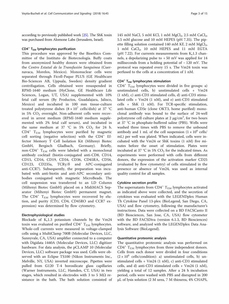
ResultsKv1.3 channel blockade by the Vm24 toxin does notcompromise cell viability but decreases the activation ofTEM cellsKv1.3 is the dominating potassium channel in TEM cells,and a potential target to treat chronic inflammation byselectively compromising TEM cells without undermin-ing the function of naïve and TCM cells that rather de-pend on the KCa3.1 channels [6]. We previously reportedthat by specifically blocking the Kv1.3 channel, the Vm24toxin inhibited T cell activation and proliferation [8]. Asthese results were obtained with total T cells, here weevaluated the capacity of the Vm24 peptide to specific-ally inhibit the CD4+ TEM cells function. As a controlfor Kv1.3 channel blockade, we used the ShK toxin fromthe Caribbean Sea anemone Stichodactyla helianthus, arecognized Kv1.3 channels inhibitor [21].CD4+ TEM cells isolated from peripheral blood of
healthy donors were positive (> 95%) for the CD3, CD4and CD45RO surface markers and negative for theCCR7 chemokine receptor (Fig. 1a, b). These markers
were verified before and after activation, and under allconditions, the terminally differentiated CD4+ TEM cellsmaintained their phenotype (CD45RO+ and CCR7−), aspreviously reported [34, 35]. Since the main goal of thisstudy was to perform a functional analysis ofTCR-activated TEM cells in the presence of the Vm24peptide, we first evaluated whether blocking Kv1.3 com-promised cell viability.Following a 24, 48 and 96 h culture period in the pres-
ence of the Vm24 peptide or the ShK toxin, CD4+ TEM
cells viability was not impaired either in quiescent orOKT3-activated cells. As death positive control, 30% di-methyl sulfoxide (DMSO) was added to the cells for30 min (Fig. 1c and Additional file 1a, b).The ability of the synthetic Vm24 toxin to block CD4+
TEM cells Kv1.3 channels was measured by patch clampin whole cell mode. Consistent with our previous report[23], the Vm24 toxin (1 nM) completely inhibited thecurrent of Kv1.3 channels in CD4+ TEM lymphocytes.Moreover, the Kv1.3 channel current was not recoveredafter a 10 min wash-out period, indicative of the slow
Fig. 1 Kv1.3 channel blockade does not compromise cell viability. Purified CD4+ TEM cells were stained for the (a) CD3, CD4 and (b) CD45RO andCCR7 surface markers. (c) Cell viability was assessed following a 24 h culture period with the Fixable Viability Dye eFluor 780. Changes in FSC andpositive staining with the viability dye were considered as cell death markers. For death positive control, 30% dimethyl sulfoxide (DMSO) wasadded to the cells for 30 min. Data from three independent experiments are shown as mean ± SEM (standard error of mean). (d) Kv1.3 channelscurrents were measured by patch clamp in whole cell mode. Currents were evoked by a depolarizing pulse to + 50 mV from a − 120 mV holdingpotential. The Vm24 toxin was perfused to the cells at a concentration of 1 nM
Veytia-Bucheli et al. Cell Communication and Signaling (2018) 16:45 Page 5 of 15
dissociation rate of the channel-toxin complex (Fig. 1d).Although concentrations below 100 pM of the Vm24toxin completely block Kv1.3 currents [23], a 1 nM con-centration was used to ensure complete blockade of thechannels throughout the entire culture time. Consider-ing that the estimated Kd for KCa3.1 channels (alsopresent in T cells) is at least 4500-fold higher than thatfor the Kv1.3 channels [23], it is expected that theremaining current on KCa3.1 channels is still 95% in thepresence of 1 nM Vm24, strongly indicating that the ef-fects observed on the TEM cells result exclusively ofKv1.3 channel blockade.Finally, we evaluated the effect of blocking Kv1.3 chan-
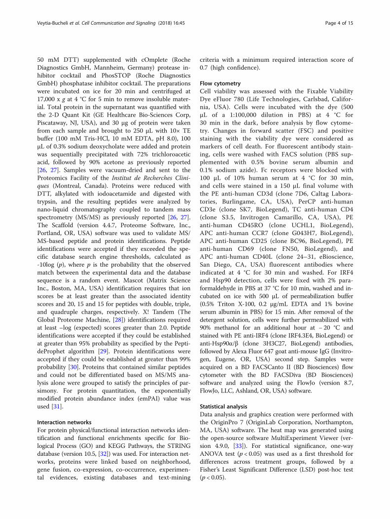
nels on the expression of the early activation markersCD25 (IL2RA), CD40L (CD40LG) and CD69 (CD69) inresponse to TCR ligation for 24 h. The Vm24 toxinsignificantly prevented the TCR-mediated CD25 upregula-tion, similar to the ShK toxin (Fig. 2a, b). Vm24 alsoprevented the upregulation of CD40L in all subjects(Fig. 2c, d), although this was not statistically signifi-cant (p = 0.06). Contrary to what we expected, CD69 upreg-ulation was not affected by Vm24 (Fig. 2e, f), suggestingthat the Kv1.3 channels do not participate in the cytoplasmto the plasma membrane translocation of this molecule.Altogether these data indicate that the Vm24 toxin did notcompromise cell viability, though it effectively blocked theKv1.3 channels on TEM cells, resulting in diminishedactivation.
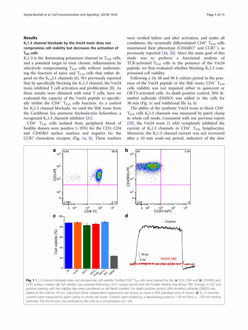
Kv1.3 channel blockade by the Vm24 toxin decreases theproduction of pro- and anti-inflammatory cytokinesTo characterize the impact of blocking the Kv1.3 channelon TCR activated CD4+ TEM lymphocytes function, weanalyzed their cytokine secretion profile in the presenceof the Vm24 and ShK peptides. Cells were left unstimu-lated or were activated for 24 h with plate-bound OKT3in the presence or absence of the toxins, and thepresence of cytokines in the supernatant was evaluatedwith a multiplex assay. Cytokines that showed at least1.5-fold change and statistically significant differences (p< 0.05), were identified. Consistent with previous reports[7, 8, 36], stimulating purified CD4+ TEM cells throughthe TCR induced the secretion of high quantities of thepro-inflammatory cytokine IFN-γ (IFNG) as well as thatof the anti-inflammatory cytokines IL-4 (IL4), IL-5 (IL5),IL-9 (IL9), IL-10 (IL10) and IL-13 (IL13), but reducedlevels of IL-2 (IL2). The level of the pro-inflammatorycytokine TNF (TNF) was also markedly increased in allsamples, yet not in a statistically significant manner, prob-ably reflecting variability between individuals (Fig. 3a). Al-though CD4+ TEM cells are the principal IL-17-producingpopulation [37], under our experimental conditions (24 hpost-OKT3 stimulation), we detected very low levels ofIL-17A (IL17A) and IL-17F (IL17F), likely resulting of the
fact that this is a family of late expression cytokines, withsecretion peaking around day 6 after activation (Fig. 3a).Pairwise comparison between the OKT3 and the OKT3 +Vm24-treated group showed that activating the cells inthe presence of the Vm24 toxin resulted in significantlylower levels in the secretion of IFN-γ, IL-4, IL-5, IL-9,IL-10 and IL-13. Vm24 also reduced the TNF increase byat least 50% in all subjects, although this was not statisti-cally significant, probably due also to inter-individual vari-ation (Fig. 3b). Similar to Vm24, the ShK toxin loweredcytokine production resulting of TCR-mediated activationof CD4+ TEM cells.
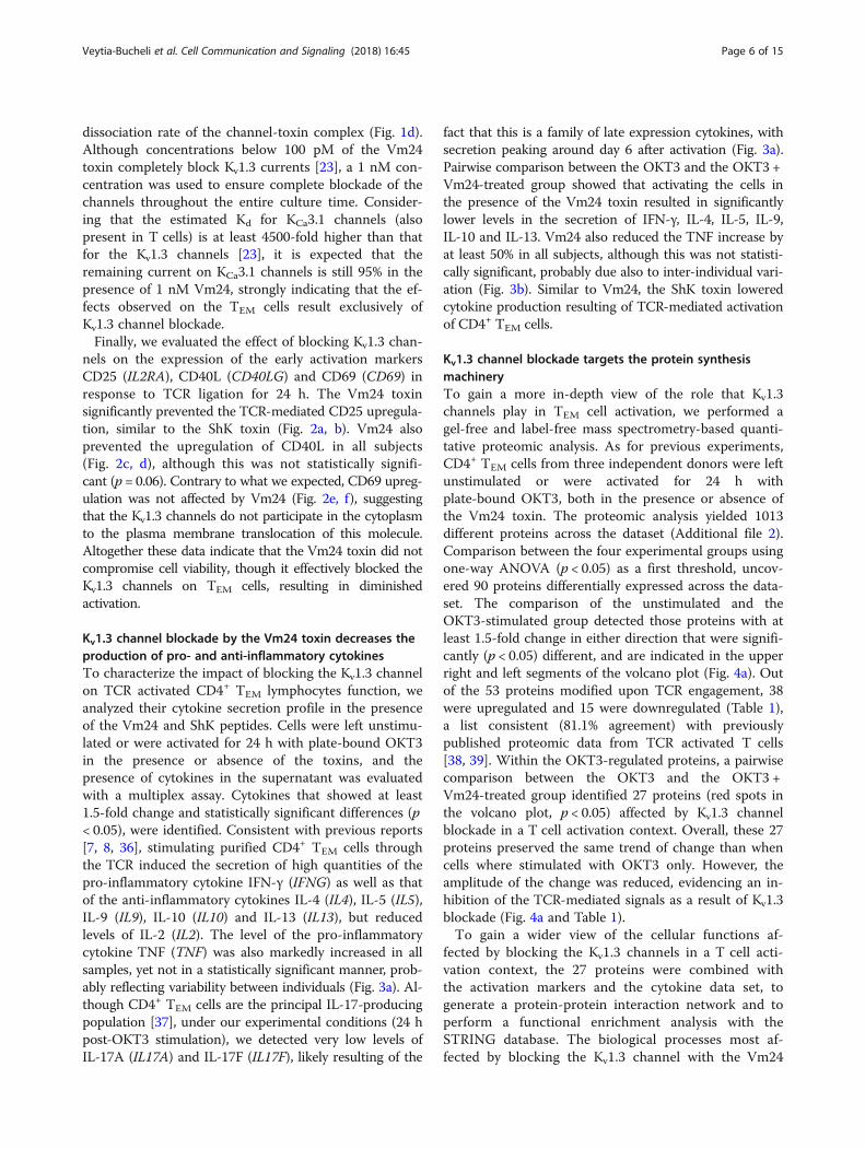
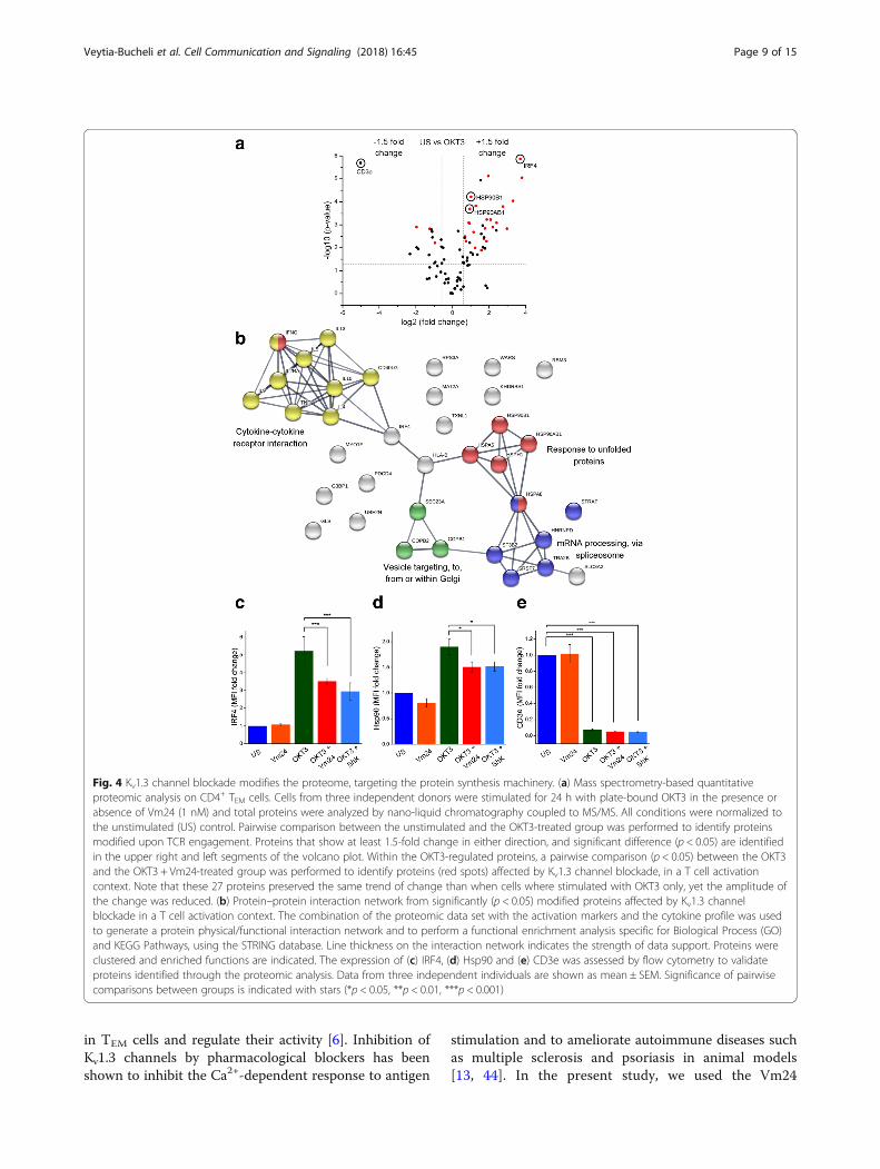
Kv1.3 channel blockade targets the protein synthesismachineryTo gain a more in-depth view of the role that Kv1.3channels play in TEM cell activation, we performed agel-free and label-free mass spectrometry-based quanti-tative proteomic analysis. As for previous experiments,CD4+ TEM cells from three independent donors were leftunstimulated or were activated for 24 h withplate-bound OKT3, both in the presence or absence ofthe Vm24 toxin. The proteomic analysis yielded 1013different proteins across the dataset (Additional file 2).Comparison between the four experimental groups usingone-way ANOVA (p < 0.05) as a first threshold, uncov-ered 90 proteins differentially expressed across the data-set. The comparison of the unstimulated and theOKT3-stimulated group detected those proteins with atleast 1.5-fold change in either direction that were signifi-cantly (p < 0.05) different, and are indicated in the upperright and left segments of the volcano plot (Fig. 4a). Outof the 53 proteins modified upon TCR engagement, 38were upregulated and 15 were downregulated (Table 1),a list consistent (81.1% agreement) with previouslypublished proteomic data from TCR activated T cells[38, 39]. Within the OKT3-regulated proteins, a pairwisecomparison between the OKT3 and the OKT3 +Vm24-treated group identified 27 proteins (red spots inthe volcano plot, p < 0.05) affected by Kv1.3 channelblockade in a T cell activation context. Overall, these 27proteins preserved the same trend of change than whencells where stimulated with OKT3 only. However, theamplitude of the change was reduced, evidencing an in-hibition of the TCR-mediated signals as a result of Kv1.3blockade (Fig. 4a and Table 1).To gain a wider view of the cellular functions af-
fected by blocking the Kv1.3 channels in a T cell acti-vation context, the 27 proteins were combined withthe activation markers and the cytokine data set, togenerate a protein-protein interaction network and toperform a functional enrichment analysis with theSTRING database. The biological processes most af-fected by blocking the Kv1.3 channel with the Vm24
Veytia-Bucheli et al. Cell Communication and Signaling (2018) 16:45 Page 6 of 15

toxin in a T cell activation context werecytokine-cytokine receptor interaction, mRNA pro-cessing, response to unfolded proteins and intracellu-lar vesicle transport (Fig. 4b).All proteins in the connected clusters were upregulated
upon TCR engagement, and the addition of Vm24 partiallyprevented their upregulation. This data highlights the roleof Kv1.3 channels in reinforcing the TCR-mediated signals,and unravels regulatory functions for these channels in the
protein synthesis machinery of TEM cells, further com-promising their effector functions at different levels.Only the TCR-mediated downregulation of four pro-
teins (RPS3A, PDCD4, UBE2N and MYO1F) was pre-vented by the addition of Vm24 (Fig. 4a upper leftsegment of the volcano plot and Table 1). These proteinsare not part of any of the indicated functional clusters.The comparison of the unstimulated and the
Vm24-only treated group, using the same analytical
Fig. 2 Kv1.3 channel blockade decreases the expression of CD25 and CD40L, but not that of CD69. CD4+ TEM cells were stimulated through theTCR with plate-bound OKT3 in the presence or absence of Vm24 or ShK (1 nM) toxins. After 24 h of culture, cells were stained for (a) CD25,(c) CD40L and (e) CD69. The histogram of a representative donor for each marker is shown. (b, d, f) Data from 3 to 6 independentexperiments are shown as mean ± SEM. The color coding for histograms and bars is maintained. Significance of pairwise comparisonsbetween groups is indicated with stars (*p < 0.05, **p < 0.01, ***p < 0.001)
Veytia-Bucheli et al. Cell Communication and Signaling (2018) 16:45 Page 7 of 15

criteria (at least 1.5-fold change in either direction andp < 0.05), showed that incubating the unstimulated cellswith the Vm24 toxin resulted in the upregulation ofthree proteins and the downregulation of 11 proteins(Additional file 3). Although these proteins are not in-volved together in a clear canonical biological processaccording to the enrichment analysis, they participate intranscriptional activation, DNA repair, RNA stability,ribosomal function, and cytokinesis, suggesting that, inresting TEM lymphocytes, Kv1.3-mediated signaling alsocontrols essential biological processes.To validate the proteomic data set, we analyzed the
expression level of interferon regulatory factor 4 (IRF4)and heat shock protein 90 (Hsp90), as their expressionaugmented in a TCR-dependent activation context, andwhen T cells were activated with OKT3 in the presenceof the Vm24 toxin this increase was reduced. IRF4, atranscription factor linking the “cytokine-cytokine recep-tor interaction” cluster with the “intracellular vesicletransport” and “response to unfolded proteins” clusters,is expressed in all CD4+ T cell subsets following TCRengagement and it is crucial for cytokine production byeffector cells [40]. Hsp90, a fundamental member of the“response to unfolded protein” cluster, participates inthe correct folding of the numerous nascent proteinsproduced during the T lymphocyte effector response andin the regulation of NF-κB signaling and inflammatory re-sponses [41]. Both, IRF4 (Fig. 4c and Additional file 4a)
and Hsp90 (Fig. 4d and Additional file 4b) followed thesame expression pattern by mass spectrometry and flowcytometry: a marked upregulation following TCR ligationand a partial reduction of this increase in the presence ofeither Vm24 or ShK toxins, indicating that Kv1.3 channelfunction is important for their upregulation. We also eval-uated CD3e (CD3E) since TCR stimulation either by anti-gen, anti-CD3 antibodies, or pharmacological activators ofprotein kinase C, results in increased TCR-CD3 internal-ization and therefore, a down-modulation of its surfacelevels [42, 43]. Under our experimental conditions, theOKT3-dependent TEM cell activation resulted in an al-most complete disappearance of CD3e from the TEM cellssurface, regardless of the presence of Vm24 or ShK, sug-gesting that the molecular events regulating CD3edown-modulation are not dependent on the activity of theKv1.3 channels (Fig. 4e and Additional file 4c).Altogether, our results identified a number of proteins
targeted by the Kv1.3 channel-dependent signaling inTCR-activated CD4+ TEM lymphocytes, uncoveringregulatory roles for Kv1.3 channels in mRNA processing,response to unfolded proteins, intracellular vesicle trans-port and cytokine-cytokine receptor interaction.
DiscussionThe potassium channels Kv1.3 and KCa3.1 promote thesustained Ca2+ influx necessary for complete T cell acti-vation. Particularly, Kv1.3 channels are highly expressed
Fig. 3 Kv1.3 channel blockade decreases the production of pro- and anti-inflammatory cytokines. (a) Heat map representation of cytokines levelsin CD4+ TEM cells supernatants. Cells were stimulated for 24 h with plate-bound OKT3 in the presence or absence of Vm24 or ShK (1 nM) toxinsand supernatants were collected and analyzed with a multiplex assay. All conditions were normalized to the unstimulated (US) control, and therelative abundance (fold change) of each cytokine is indicated by a gradient of color from blue (low abundance) to red (high abundance). Theheat map was generated with the data from three independent donors using the Manhattan distance metric and hierarchical clustering basedon average linkage. To identify cytokines modified upon TCR engagement, pairwise comparison between the unstimulated and the OKT3-treatedgroup was performed. Proteins that show at least 1.5-fold change and significant difference (p < 0.05) are identified in the heat map with stars(*p < 0.05, **p < 0.01, ***p < 0.001). (b) Cytokines that showed increased levels following OKT3 stimulation (Fig. 3a) are plotted. Fold change ofOKT3, OKT3 + Vm24 and OKT3 + ShK groups normalized to the unstimulated control are shown. The dotted line indicates the protein levels in thesupernatants of unstimulated cells. Data from three independent donors are plotted, and significance of pairwise comparisons between the OKT3and the OKT3 + Vm24 group are indicated with stars (*p < 0.05, **p < 0.01, ***p < 0.001)
Veytia-Bucheli et al. Cell Communication and Signaling (2018) 16:45 Page 8 of 15
in TEM cells and regulate their activity [6]. Inhibition ofKv1.3 channels by pharmacological blockers has beenshown to inhibit the Ca2+-dependent response to antigen
stimulation and to ameliorate autoimmune diseases suchas multiple sclerosis and psoriasis in animal models[13, 44]. In the present study, we used the Vm24
Fig. 4 Kv1.3 channel blockade modifies the proteome, targeting the protein synthesis machinery. (a) Mass spectrometry-based quantitativeproteomic analysis on CD4+ TEM cells. Cells from three independent donors were stimulated for 24 h with plate-bound OKT3 in the presence orabsence of Vm24 (1 nM) and total proteins were analyzed by nano-liquid chromatography coupled to MS/MS. All conditions were normalized tothe unstimulated (US) control. Pairwise comparison between the unstimulated and the OKT3-treated group was performed to identify proteinsmodified upon TCR engagement. Proteins that show at least 1.5-fold change in either direction, and significant difference (p < 0.05) are identifiedin the upper right and left segments of the volcano plot. Within the OKT3-regulated proteins, a pairwise comparison (p < 0.05) between the OKT3and the OKT3 + Vm24-treated group was performed to identify proteins (red spots) affected by Kv1.3 channel blockade, in a T cell activationcontext. Note that these 27 proteins preserved the same trend of change than when cells where stimulated with OKT3 only, yet the amplitude ofthe change was reduced. (b) Protein–protein interaction network from significantly (p < 0.05) modified proteins affected by Kv1.3 channelblockade in a T cell activation context. The combination of the proteomic data set with the activation markers and the cytokine profile was usedto generate a protein physical/functional interaction network and to perform a functional enrichment analysis specific for Biological Process (GO)and KEGG Pathways, using the STRING database. Line thickness on the interaction network indicates the strength of data support. Proteins wereclustered and enriched functions are indicated. The expression of (c) IRF4, (d) Hsp90 and (e) CD3e was assessed by flow cytometry to validateproteins identified through the proteomic analysis. Data from three independent individuals are shown as mean ± SEM. Significance of pairwisecomparisons between groups is indicated with stars (*p < 0.05, **p < 0.01, ***p < 0.001)
Veytia-Bucheli et al. Cell Communication and Signaling (2018) 16:45 Page 9 of 15
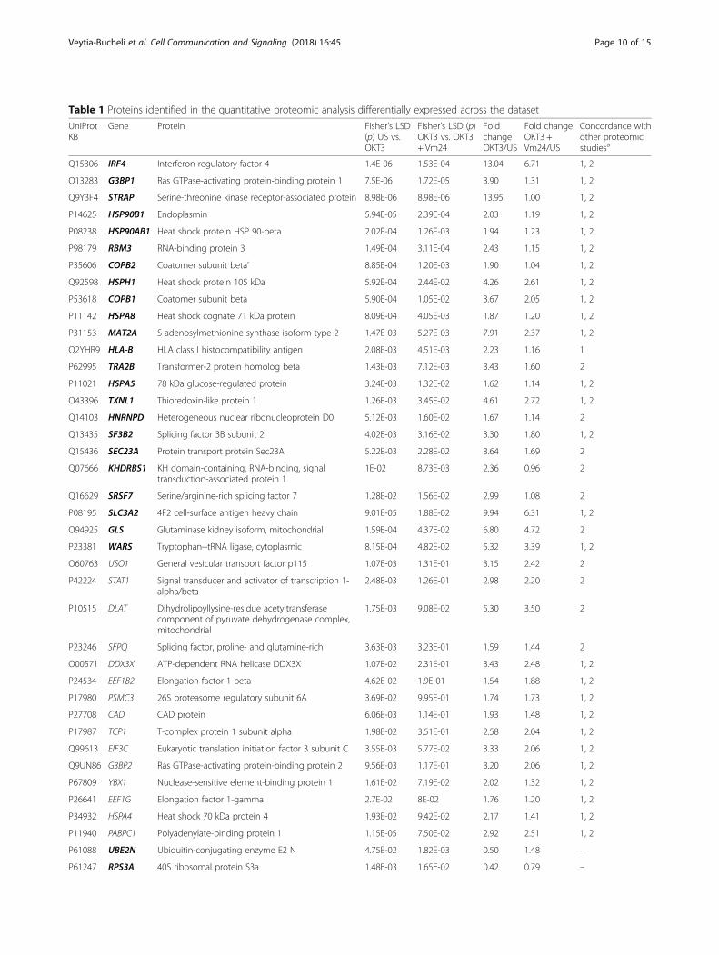
Table 1 Proteins identified in the quantitative proteomic analysis differentially expressed across the dataset
UniProtKB
Gene Protein Fisher’s LSD(p) US vs.OKT3
Fisher’s LSD (p)OKT3 vs. OKT3+ Vm24
FoldchangeOKT3/US
Fold changeOKT3 +Vm24/US
Concordance withother proteomicstudiesa
Q15306 IRF4 Interferon regulatory factor 4 1.4E-06 1.53E-04 13.04 6.71 1, 2
Q13283 G3BP1 Ras GTPase-activating protein-binding protein 1 7.5E-06 1.72E-05 3.90 1.31 1, 2
Q9Y3F4 STRAP Serine-threonine kinase receptor-associated protein 8.98E-06 8.98E-06 13.95 1.00 1, 2
P14625 HSP90B1 Endoplasmin 5.94E-05 2.39E-04 2.03 1.19 1, 2
P08238 HSP90AB1 Heat shock protein HSP 90-beta 2.02E-04 1.26E-03 1.94 1.23 1, 2
P98179 RBM3 RNA-binding protein 3 1.49E-04 3.11E-04 2.43 1.15 1, 2
P35606 COPB2 Coatomer subunit beta’ 8.85E-04 1.20E-03 1.90 1.04 1, 2
Q92598 HSPH1 Heat shock protein 105 kDa 5.92E-04 2.44E-02 4.26 2.61 1, 2
P53618 COPB1 Coatomer subunit beta 5.90E-04 1.05E-02 3.67 2.05 1, 2
P11142 HSPA8 Heat shock cognate 71 kDa protein 8.09E-04 4.05E-03 1.87 1.20 1, 2
P31153 MAT2A S-adenosylmethionine synthase isoform type-2 1.47E-03 5.27E-03 7.91 2.37 1, 2
Q2YHR9 HLA-B HLA class I histocompatibility antigen 2.08E-03 4.51E-03 2.23 1.16 1
P62995 TRA2B Transformer-2 protein homolog beta 1.43E-03 7.12E-03 3.43 1.60 2
P11021 HSPA5 78 kDa glucose-regulated protein 3.24E-03 1.32E-02 1.62 1.14 1, 2
O43396 TXNL1 Thioredoxin-like protein 1 1.26E-03 3.45E-02 4.61 2.72 1, 2
Q14103 HNRNPD Heterogeneous nuclear ribonucleoprotein D0 5.12E-03 1.60E-02 1.67 1.14 2
Q13435 SF3B2 Splicing factor 3B subunit 2 4.02E-03 3.16E-02 3.30 1.80 1, 2
Q15436 SEC23A Protein transport protein Sec23A 5.22E-03 2.28E-02 3.64 1.69 2
Q07666 KHDRBS1 KH domain-containing, RNA-binding, signaltransduction-associated protein 1
1E-02 8.73E-03 2.36 0.96 2
Q16629 SRSF7 Serine/arginine-rich splicing factor 7 1.28E-02 1.56E-02 2.99 1.08 2
P08195 SLC3A2 4F2 cell-surface antigen heavy chain 9.01E-05 1.88E-02 9.94 6.31 1, 2
O94925 GLS Glutaminase kidney isoform, mitochondrial 1.59E-04 4.37E-02 6.80 4.72 2
P23381 WARS Tryptophan--tRNA ligase, cytoplasmic 8.15E-04 4.82E-02 5.32 3.39 1, 2
O60763 USO1 General vesicular transport factor p115 1.07E-03 1.31E-01 3.15 2.42 2
P42224 STAT1 Signal transducer and activator of transcription 1-alpha/beta
2.48E-03 1.26E-01 2.98 2.20 2
P10515 DLAT Dihydrolipoyllysine-residue acetyltransferasecomponent of pyruvate dehydrogenase complex,mitochondrial
1.75E-03 9.08E-02 5.30 3.50 2
P23246 SFPQ Splicing factor, proline- and glutamine-rich 3.63E-03 3.23E-01 1.59 1.44 2
O00571 DDX3X ATP-dependent RNA helicase DDX3X 1.07E-02 2.31E-01 3.43 2.48 1, 2
P24534 EEF1B2 Elongation factor 1-beta 4.62E-02 1.9E-01 1.54 1.88 1, 2
P17980 PSMC3 26S proteasome regulatory subunit 6A 3.69E-02 9.95E-01 1.74 1.73 1, 2
P27708 CAD CAD protein 6.06E-03 1.14E-01 1.93 1.48 1, 2
P17987 TCP1 T-complex protein 1 subunit alpha 1.98E-02 3.51E-01 2.58 2.04 1, 2
Q99613 EIF3C Eukaryotic translation initiation factor 3 subunit C 3.55E-03 5.77E-02 3.33 2.06 1, 2
Q9UN86 G3BP2 Ras GTPase-activating protein-binding protein 2 9.56E-03 1.17E-01 3.20 2.06 1, 2
P67809 YBX1 Nuclease-sensitive element-binding protein 1 1.61E-02 7.19E-02 2.02 1.32 1, 2
P26641 EEF1G Elongation factor 1-gamma 2.7E-02 8E-02 1.76 1.20 1, 2
P34932 HSPA4 Heat shock 70 kDa protein 4 1.93E-02 9.42E-02 2.17 1.41 1, 2
P11940 PABPC1 Polyadenylate-binding protein 1 1.15E-05 7.50E-02 2.92 2.51 1, 2
P61088 UBE2N Ubiquitin-conjugating enzyme E2 N 4.75E-02 1.82E-03 0.50 1.48 –
P61247 RPS3A 40S ribosomal protein S3a 1.48E-03 1.65E-02 0.42 0.79 –
Veytia-Bucheli et al. Cell Communication and Signaling (2018) 16:45 Page 10 of 15

toxin, a highly specific blocker of the Kv1.3 channelsto identify the cellular processes that depend on theactivity of these channels in TCR-activated humanTEM lymphocytes as well as to validate the use of theVm24 peptide to downregulate TEM cell function.Consistent with previous reports [23], and with the high
dependency of TEM lymphocytes on Kv1.3 channel functionfor sustained activation, the addition of Vm24 toCD3-activated TEM lymphocytes resulted in a pronouncedinhibition of CD25 and CD40L expression, probably as aconsequence of the lack of activation of calcineurin and thesubsequent translocation of the NFAT transcription factorto the CD25 and CD40L promoters [45] [46]. Interestingly,under our experimental conditions, Vm24 did not preventthe OKT3-induced expression of CD69, a negative regula-tor of chronic inflammation [47, 48].In consonance with the inhibition of the expression of
early activation markers, the addition of the Vm24 pep-tide to OKT3-activated TEM lymphocytes inhibited thesecretion of the pro-inflammatory cytokines IFN-γ andTNF, as well as that of the Th2 cytokines IL-4, IL-5,IL-9, IL-10 and IL-13, all of which are dependent on theavailability of NFAT [49–59].We previously reported that administration of the
Vm24 peptide lessened the severity of inflammation
in a delayed-type hypersensitivity model [23]. Otherstudies in animal models of human diseases (allergicasthma, experimental autoimmune encephalomyelitis)have shown that the abatement of the Kv1.3 channelcurrents in vivo (by knocking out the channel or bytreatment with ShK derivatives) ameliorates the pro-gression of the disease and decreases the productionof the effector cytokines IFN-γ, IL-4, IL-5 and IL-17,but enhances that of the anti-inflammatory cytokineIL-10 [60, 61]. Contrary to these reports, under ourexperimental conditions, the blockade of Kv1.3 chan-nels in isolated human CD4+ TEM cells from healthydonors reduced IL-10 production, consistent with adownstream inhibition of NFATc2 and IRF4 recruit-ment, two transcription factors that synergisticallyaugment the activity of the Th2-specific enhancerCNS-9 (a cis-regulatory element upstream of theIL-10 gene locus) [62, 63]. Whether this discrepancyreflects species-specific differences or differences thatresult from different experimental settings (knockingout the channel or an in vivo T cell activation versusin vitro anti-CD3 CD4+ TEM activation) remains to beinvestigated.Also, it is important to consider that although IL-4,
IL-5, IL-10 and IL-13 are classified as anti-inflammatory
Table 1 Proteins identified in the quantitative proteomic analysis differentially expressed across the dataset (Continued)
UniProtKB
Gene Protein Fisher’s LSD(p) US vs.OKT3
Fisher’s LSD (p)OKT3 vs. OKT3+ Vm24
FoldchangeOKT3/US
Fold changeOKT3 +Vm24/US
Concordance withother proteomicstudiesa
Q53EL6 PDCD4 Programmed cell death protein 4 1.26E-03 2.79E-02 0.26 0.67 1, 2
O00160 MYO1F Unconventional myosin-If 6.13E-03 3.84E-02 0.52 0.84 –
Q5JSL3 DOCK11 Dedicator of cytokinesis protein 11 1.63E-03 8.98E-01 0.45 0.44 1
Q92522 H1FX Histone H1x 1.97E-03 6.06E-02 0.46 0.72 –
P07737 PFN1 Profilin-1 3.51E-03 5.64E-01 0.47 0.55 1
O43390 HNRNPR Heterogeneous nuclear ribonucleoprotein R 4.43E-03 1.9E-01 0.65 0.78 –
Q13561 DCTN2 Dynactin subunit 2 1.12E-02 2.14E-01 0.27 0.57 –
P16104 H2AFX Histone H2AX 4.24E-02 9.79E-02 0.52 0.89 –
O94906 PRPF6 Pre-mRNA-processing factor 6 2.05E-02 8.37E-02 0.41 0.82 –
P62906 RPL10A 60S ribosomal protein L10a 9.61E-03 7.94E-02 0.26 0.70 –
P07766 CD3E T-cell surface glycoprotein CD3 epsilon chain 2.02E-06 1 0.03 0.03 1
Q9UGI8 TES Testin 9.03E-03 4.25E-01 0.66 0.74 –
Q6JBY9 RCSD1 CapZ-interacting protein 1.86E-02 2.26E-01 0.20 0.56 1
Proteins with at least 1.5-fold change in either direction that were significantly (p < 0.05) different comparing the unstimulated and the OKT3-stimulated group.Concordance with other T cell activation proteomic studies is indicated. Within the OKT3-regulated proteins, comparison between the OKT3 and the OKT3 +Vm24-treated group is indicated and proteins that showed a statistically significant reduction on the amplitude of the TCR-mediated change as a result of theaddition of the Vm24 toxin are indicated in bolda) Concordance with other proteomic studies:1Tan H, Yang K, Li Y, Shaw TI, Wang Y, Blanco DB, et al. Integrative Proteomics and Phosphoproteomics Profiling Reveals Dynamic Signaling Networks andBioenergetics Pathways Underlying T Cell Activation. Immunity. 2017;46:488–5032Ron-Harel N, Santos D, Ghergurovich JM, Sage PT, Reddy A, Lovitch SB, et al. Mitochondrial Biogenesis and Proteome Remodeling Promote One-CarbonMetabolism for T Cell Activation. Cell Metab. 2016;24:104–17
Veytia-Bucheli et al. Cell Communication and Signaling (2018) 16:45 Page 11 of 15

cytokines due to their down-modulatory effect on in-flammatory phenomena mediated by Th1/Th17 cells,these cytokines mediate type I hypersensitivity inflam-matory conditions and significantly contribute to thepathogenesis mediated by immune complexes, throughtheir important effect on the activation and proliferationof B cells and antibody synthesis [64].Furthermore, consistent with the fact that the signal-
ing pathways that activate the transcription of thosegenes are also dependent on NFAT availability, our datasuggest that the inhibition of the Kv1.3 channel exerts adown-regulatory effect on the different TEM lymphocytesubsets, including Th1 and Th2 cells. In this regard, itwould be expected that Kv1.3 blockers could act as awide spectrum immunosuppressive molecule, with a sig-nificant effect on different immune-mediated conditions.The proteomic analysis revealed that, in agreement
with the large secretory demand of TCR-activated TEM
cells, the protein synthesis machinery is prepared to gen-erate a robust immune response by regulating the ex-pression level of transcription factors specific forinflammatory mediators, such as IRF4, as well as that ofproteins involved in the splicing machinery, the unfoldedprotein response and vesicular transport of the novelsynthesized mediators. IRF4 is expressed across all T cellsubsets within a few hours following TCR engagement,and it is necessary for optimal T cell proliferation in re-sponse to mitogenic stimuli. In cooperation with tran-scriptional partners such as NFAT, it controls theexpression of IL-2, IL-4, IL-5, IL-9, IL-10, IL-13, IL-17,IL-21, IFN-γ and TNF [40, 65–67]. Furthermore, the cal-cineurin inhibitor cyclosporine A [65] as well as defectsin CRAC channels function [68] result in impaired IRF4expression, implicating NFAT in IRF4 upregulation. Ourdata show that in a T cell activation context, the expres-sion level of IRF4 is diminished following Vm24 treat-ment, suggesting that blocking the Kv1.3 channelhinders downstream events such as NFAT activation andIRF4 expression, resulting in impaired cytokineproduction, in consonance with a previously assignedfunction for Kv1.3-dependent signals in secretory func-tions [14, 69].The unfolded protein response allows cells to manage
the endoplasmic reticulum stress resulting of the in-creased folding demand imposed by the requirements ofactivated T lymphocytes engaged in secretory functions.Especially, the molecular chaperone Hsp90 regulates thestability and function of IRE1, an endoplasmic reticulumtransmembrane kinase that activates the unfoldedprotein response to maintain the endoplasmic reticulumfunction [70]. Upon treatment with the Vm24 peptide orthe ShK toxin, the expression level of the Hsp90 chaperoneas well as that of other members of the heat shock familyof proteins was strongly diminished. In addition, we found
that Kv1.3 channels-dependent signals are necessary for theup-regulation of the amino acid transporter SLC3A2. Nutri-ent transporters such as SLC3A2 ensure and coordinate thesupply of nutrients necessary for the increased metabolicrequirements of effector lymphocytes. Thus, Kv1.3 channelsparticipate in regulating basic metabolic requirements inTEM cells. In concordance with our results, a recent prote-omic study performed on activated microglia also revealedthat blocking Kv1.3 channels attenuates biological processesrelated to the regulation of the immune response and theintracellular protein transport [71]. Assessing if this extendsto other cell types becomes critical to appreciate better thepotential side effects of blocking the Kv1.3 channels to con-trol autoimmune disorders.Interestingly, we found that blocking the Kv1.3 chan-
nels with Vm24 on OKT3-activated CD4+ TEM cellsdoes not entirely block the anti-CD3 induced cyto-kines synthesis or proteomic changes; it prevents thechanges from reaching the highest level. Residual po-tassium fluxes from the few KCa3.1 channels presenton these cells may contribute to the “incomplete” in-hibition [35]. In support of the relative abundance ofKv1.3 channels as compared to that of KCa3.1 channelson TEM cells, our data indicate that Kv1.3 channels arethe major providers of potassium efflux; they are notindispensable for CD4+ TEM cell response, yet they arecentral for regulating the amplitude of the response.Although less is known about the function of the
Kv1.3 channels in human CD8+ cells, the decrease ofKv1.3 currents by genetic or pharmacological approachesinhibits the differentiation of TCM to TEM cells and inthe latter, it severely impairs the proliferation, the secre-tion of IL-2, TNF and granzyme B (but not of IFN-γ), ul-timately dampening their ability to kill target cells [69,72]. Interestingly, NFATc1-deficient CD8+ T cells showdiminished RNA levels of granzyme B and of genes en-coding cytokines and chemokines in addition to genescontrolling glycolysis [73], further underscoring the im-portance of Kv1.3 channels on the calcium/calcineurin/NFAT network.In addition to exploring the function of Kv1.3 channels
in activated CD4+ TEM cells, the experiments we per-formed allowed us to compare the efficacy of two Kv1.3channel blockers: the Vm24 toxin, isolated from theMexican scorpion Vaejovis mexicanus and the ShKtoxin, from the sea anemone Stichodactyla helianthus.Their capacity to hinder TEM cells functions showed thatboth toxins were very effective to block the Kv1.3 potas-sium channels-dependent signaling as they both inhib-ited the production of INF-γ, TNF, IL-4, IL-5, IL9, IL10and IL13 following TCR ligation. Although not statisti-cally significant, the inhibitory capacity of the ShK pep-tide was always higher than that of Vm24. The ShKpeptide is a very potent (Kd = 10 pM) Kv1.3 blocker.
Veytia-Bucheli et al. Cell Communication and Signaling (2018) 16:45 Page 12 of 15

However, it has a low selectivity for the Kv1.3 channels(only 2.8-fold affinity over other channels), as a result ofwhich it is very toxic to mammals [21, 74]. Dalazatide®(formerly ShK-186), a molecule derived from the ShKtoxin, is a Kv1.3 channel blocker that has gone into clin-ical phase trials, has been shown to reduce the levels ofplasmatic and TEM inflammation markers, and to im-prove psoriatic skin lesions from mild to moderateplaque psoriasis patients [13, 16, 75]. In comparison withDalazatide®, the Vm24 peptide has the advantage of agreater potency (Kd 2.9 vs. 69 pM) and selectivity (1500vs. 100-fold affinity over other channels) towards Kv1.3channels [21, 23], which improves the safety index inconditions where the blood-brain barrier (such as mul-tiple sclerosis) is compromised and the presence of highconcentrations of non-specific blockers (such as neur-onal Kv1.1 channels blockers) in the central nervoussystem can bring severe neurotoxicity. Interestingly, al-though Dalazatide® was well tolerated during clinical tri-als, the most common adverse events were temporaryhypoesthesia and paresthesia involving hands, feet, orperioral area which may be prevented with moreselective Kv1.3 blockers such as Vm24 or othersecond-generation blockers derived from ShK toxin thathave been recently developed [76].
ConclusionsOur results show that blocking Kv1.3 channels with theVm24 peptide profoundly affects the mRNA synthesis ma-chinery, the unfolded protein response and the intracellularvesicle transport, thus impairing the synthesis and secretionof cytokines in response to TCR engagement, highlightingthe importance of Kv1.3 channels for TEM cell function. AsTEM cells are considered to be main players in the path-ology of autoimmune diseases, further studies are neededto better characterize the molecular mechanisms affectedby the blockade of Kv1.3 channels by toxins such as theVm24 peptide.
Additional files
Additional file 1: Kv1.3 channel blockade does not compromise cellviability. (a) Cell viability of cells treated under the same experimentalconditions as for Fig. 1 was assessed following a 24, 48 and 96 h cultureperiod with the Fixable Viability Dye eFluor 780. The dot plot of arepresentative donor after 24 h of culture is shown. Changes in forwardscatter (FSC) and positive staining with the viability dye were consideredas cell death markers. As death positive control, 30% dimethyl sulfoxide(DMSO) was added to the cells for 30 min. The cell death area isenclosed in the gate. (b) Data from three independent experiments withCD4+ TEM cells from independent donors are shown as mean ± SEM.(TIF 6948 kb)
Additional file 2: Proteins identified in the mass spectrometry-basedquantitative proteomic analysis. Peptide identifications were accepted ifthey could be established at greater than 95% probability as specified bythe PeptideProphet algorithm. Protein identifications were accepted ifthey could be established at greater than 99% probability. (XLSX 427 kb)
Additional file 3: Proteins differentially expressed when incubating theunstimulated cells with the Vm24 toxin. Proteins identified with the massspectrometry-based quantitative proteomic analysis, with at least 1.5-foldchange in either direction and that were significantly (p < 0.05) different,comparing the unstimulated and the Vm24-treated group. (PDF 150 kb)
Additional file 4: Validation of proteomic analysis results by flowcytometry. CD4+ TEM cells were stimulated through the TCR with plate-bound OKT3 in the presence or absence of Vm24 or ShK (1 nM) toxins, asindicated in the methods section. After 24 h of culture, cells were stainedfor (a) IRF4, (b) Hsp90 and (c) CD3e. The histogram of one representativedonor for each staining is shown. (TIF 4252 kb)
AbbreviationsNF-κB: nuclear factor kappa-light-chain-enhancer of activated B cells;CRAC: Ca2+ release-activated Ca2+ channels; DMSO: Dimethyl sulfoxide;emPAI: Exponentially modified protein abundance index; Fisher’sLSD: Fisher’s Least Significant Difference post-hoc test; FSC: Forward scatter;Hsp90: Heat shock protein 90; IRE1: Serine/threonine-protein kinase/endoribonuclease IRE1; IRF4: Interferon regulatory factor 4; Kd: Dissociationconstant; MS/MS: Tandem mass spectrometry; NFAT: Nuclear factor ofactivated T-cells; SEM: Standard error of mean; TCR: T cell receptor;TEM: Effector memory T cells
AcknowledgementsThe authors are indebted to Dr. Georgina Gurrola-Briones for the generousthe gift of the Vm24 toxin used in this work and to Dr. Rita Restano-Cassulinifor support during the electrophysiological (patch clamp) experiments withVm24. The authors thank Dr. Denis Faubert and Dr. Rosario Vera-Estrella fortheir helpful suggestions for MS analysis and Estefanía Alemán-Navarro, M.Sc., María Teresa Romero-Gutierrez, M. Sc., Dr. Ernesto Ortiz, and Dr. RobertoGonzález-Amaro for help with data analysis. JIV is the recipient of a graduatestudent fellowship from CONACYT (fellowship holder number 289448).
FundingThis work was partially supported by grants from the Consejo Nacional deCiencia y Tecnología (CONACYT) SEP-CONACYT 237864 (LDP) and SEP-CONACYT 220990 (YR). The funding sources had no role in the design of thestudy, in collection, analysis, and interpretation of the data or in writing ofthe manuscript.
Authors’ contributionsJIVB, JMJV, LDP and YR designed the experiments and wrote the manuscriptwith input from all other authors; JIVB and EIMP conducted the experiments;JIVB, JMJV, MASH and YR undertook the analysis. All authors read andapproved the final manuscript.
Ethics approval and consent to participateNot applicable.
Consent for publicationNot applicable.
Competing interestsThe Vm24 peptide and its use are protected by an international patentapplication WO2008139243 A1 in the name of Universidad NacionalAutónoma de Mexico (UNAM). The same subject was protected in severalcountries. In addition, a license was granted to a Mexican pharmaceuticalcompany for commercial exploitation; however, the authors kept the right tocontinuing research in the field involving this peptide. The financing of thiswork came from the declared funding institutions only. No other possibleconflicts of interest are involved.
Publisher’s NoteSpringer Nature remains neutral with regard to jurisdictional claims inpublished maps and institutional affiliations.
Author details1Departamento de Medicina Molecular y Bioprocesos, Instituto deBiotecnología, Universidad Nacional Autónoma de México, Av. Universidad
Veytia-Bucheli et al. Cell Communication and Signaling (2018) 16:45 Page 13 of 15
2001, Col. Chamilpa, 62210 Cuernavaca, Morelos, Mexico. 2Posgrado enCiencias Bioquímicas, Universidad Nacional Autónoma de México, MexicoCity, Mexico.
Received: 1 June 2018 Accepted: 2 August 2018
References1. Feske S, Wulff H, Skolnik EY. Ion channels in innate and adaptive immunity.
Annu Rev Immunol. 2015;33:291–353.2. Cahalan MD, Chandy KG. The functional network of ion channels in T
lymphocytes. Immunol Rev. 2009;231:59–87.3. Feske S. Calcium signalling in lymphocyte activation and disease. Nat Rev
Immunol. 2007;7:690–702.4. George Chandy K, Wulff H, Beeton C, Pennington M, Gutman GA, Cahalan
MD. K+ channels as targets for specific immunomodulation. TrendsPharmacol Sci. 2004;25:280–9.
5. Gutman GA. International Union of Pharmacology. LIII. Nomenclature andmolecular relationships of voltage-gated potassium channels. PharmacolRev. 2005;57:473–508.
6. Wulff H, Calabresi PA, Allie R, Yun S, Pennington M, Beeton C, et al. Thevoltage-gated Kv1.3 K+ channel in effector memory T cells as new targetfor MS. J Clin Invest. 2003;111:1703–13.
7. Sallusto F, Lenig D, Förster R, Lipp M, Lanzavecchia A. Two subsets ofmemory T lymphocytes with distinct homing potentials and effectorfunctions. Nature. 1999;401:708–12.
8. Sallusto F, Geginat J, Lanzavecchia A. Central memory and effector memoryT cell subsets: function, generation, and maintenance. Annu Rev Immunol.2004;22:745–63.
9. Reinhardt RL, Khoruts A, Merica R, Zell T, Jenkins MK. Visualizing thegeneration of memory CD4 T cells in the whole body. Nature. 2001;410:101–5.
10. Schenkel JM, Masopust D. Tissue-resident memory T cells. Immunity. 2014;41:886–97.
11. Gerlach C, Moseman EA, Loughhead SM, Alvarez D, Zwijnenburg AJ,Waanders L, et al. The chemokine receptor CX3CR1 defines three antigen-experienced CD8 T cell subsets with distinct roles in immune surveillanceand homeostasis. Immunity. 2016;45:1270–84.
12. Beeton C, Barbaria J, Giraud P, Devaux J, Benoliel A-M, Gola M, et al.Selective blocking of voltage-gated K+ channels improves experimentalautoimmune encephalomyelitis and inhibits T cell activation. J Immunol.2001;166:936–44.
13. Tarcha EJ, Chi V, Munoz-Elias EJ, Bailey D, Londono LM, Upadhyay SK, et al.Durable pharmacological responses from the peptide ShK-186, a specificKv1.3 channel inhibitor that suppresses T cell mediators of autoimmunedisease. J Pharmacol Exp Ther. 2012;342:642–53.
14. Beeton C. Targeting effector memory T cells with a selective peptideinhibitor of Kv1.3 channels for therapy of autoimmune diseases. MolPharmacol. 2005;67:1369–81.
15. Matheu MP, Beeton C, Garcia A, Chi V, Rangaraju S, Safrina O, et al. Imagingof effector memory T cells during a delayed-type hypersensitivity reactionand suppression by Kv1.3 channel block. Immunity. 2008;29:602–14.
16. Beeton C, Wulff H, Standifer NE, Azam P, Mullen KM, Pennington MW, et al.Kv1.3 channels are a therapeutic target for T cell-mediated autoimmunediseases. Proc Natl Acad Sci. 2006;103:17414–9.
17. Quintero-Hernández V, Jiménez-Vargas JM, Gurrola GB, Valdivia HH, PossaniLD. Scorpion venom components that affect ion-channels function. Toxicon.2013;76:328–42.
18. Hu L, Pennington M, Jiang Q, Whartenby KA, Calabresi PA. Characterizationof the Functional Properties of the Voltage-Gated Potassium Channel Kv1.3in Human CD4+ T Lymphocytes. J Immunol. 2007;179:4563–70.
19. Carbone E, Wanke E, Prestipino G, Possani LD, Maelicke A. Selectiveblockage of voltage-dependent K+ channels by a novel scorpion toxin.Nature. 1982;296:90–1.
20. Chhabra S, Chang SC, Nguyen HM, Huq R, Tanner MR, Londono LM, et al.Kv1.3 channel-blocking immunomodulatory peptides from parasitic worms:implications for autoimmune diseases. FASEB J. 2014;28:3952–64.
21. Chi V, Pennington MW, Norton RS, Tarcha EJ, Londono LM, Sims-Fahey B,et al. Development of a sea anemone toxin as an immunomodulator fortherapy of autoimmune diseases. Toxicon. 2012;59:529–46.
22. Grissmer S, Nguyen AN, Aiyar J, Hanson DC, Mather RJ, Gutman GA, et al.Pharmacological characterization of five cloned voltage-gated K+ channels,types Kv1.1, 1.2, 1.3, 1.5, and 3.1, stably expressed in mammalian cell lines.Mol Pharmacol. 1994;45:1227–34.
23. Varga Z, Gurrola-Briones G, Papp F, de la Vega RC R, Pedraza-Alva G, TajhyaRB, et al. Vm24, a natural immunosuppressive peptide, potently andselectively blocks Kv1.3 potassium channels of human T cells. MolPharmacol. 2012;82:372–82.
24. Wulff H, Castle NA, Pardo LA. Voltage-gated potassium channels astherapeutic targets. Nat Rev Drug Discov. 2009;8:982–1001.
25. Gurrola GB, Hernández-López RA, de la Vega RC R, Varga Z, CVF B, Salas-Castillo SP, et al. Structure, function, and chemical synthesis of Vaejovismexicanus peptide 24: a novel potent blocker of Kv1.3 potassium channelsof human T lymphocytes. Biochemistry. 2012;51:4049–61.
26. Barkla BJ, Vera-Estrella R, Pantoja O. Protein profiling of epidermal bladdercells from the halophyte Mesembryanthemum crystallinum. Proteomics.2012;12:2862–5.
27. Murillo A, Vera-Estrella R, Barkla BJ, Méndez E, Arias CF. Identification of hostcell factors associated with Astrovirus replication in Caco-2 cells. J Virol.2015;89:10359–70.
28. Beavis RC. Using the global proteome machine for protein identification. In:Nedelkov D, Nelson RW, editors. New and emerging proteomic techniques.New Jersey: Humana Press; 2006. p. 217–28.
29. Keller A, Nesvizhskii AI, Kolker E, Aebersold R. Empirical statistical model toestimate the accuracy of peptide identifications made by MS/MS anddatabase search. Anal Chem. 2002;74:5383–92.
30. Nesvizhskii AI, Keller A, Kolker E, Aebersold R. A statistical model foridentifying proteins by tandem mass spectrometry. Anal Chem. 2003;75:4646–58.
31. Ishihama Y, Oda Y, Tabata T, Sato T, Nagasu T, Rappsilber J, et al.Exponentially modified protein abundance index (emPAI) for estimation ofabsolute protein amount in proteomics by the number of sequencedpeptides per protein. Mol Cell Proteomics. 2005;4:1265–72.
32. Szklarczyk D, Franceschini A, Wyder S, Forslund K, Heller D, Huerta-Cepas J,et al. STRING v10: protein–protein interaction networks, integrated over thetree of life. Nucleic Acids Res. 2015;43:D447–52.
33. Howe E, Holton K, Nair S, Schlauch D, Sinha R, Quackenbush J. BiomedicalInformatics for Cancer Research. In: Ochs MF, Casagrande JT, Davuluri R V.,editors. Biomedical Informatics for Cancer Research. Boston, MA: SpringerUS; 2010. p. 267–277.
34. Li Y, Kurlander RJ. Comparison of anti-CD3 and anti-CD28-coated beadswith soluble anti-CD3 for expanding human T cells: differing impact on CD8T cell phenotype and responsiveness to restimulation. J Transl Med.2010;8:104.
35. Chiang EY, Li T, Jeet S, Peng I, Zhang J, Lee WP, et al. Potassium channelsKv1.3 and KCa3.1 cooperatively and compensatorily regulate antigen-specific memory T cell functions. Nat Commun. 2017;8:14644.
36. Farber DL, Yudanin NA, Restifo NP. Human memory T cells: generation,compartmentalization and homeostasis. Nat Rev Immunol. 2014;14:24–35.
37. Liu H, Rohowsky-Kochan C. Regulation of IL-17 in human CCR6+ effectormemory T cells. J Immunol. 2008;180:7948–57.
38. Tan H, Yang K, Li Y, Shaw TI, Wang Y, Blanco DB, et al. Integrativeproteomics and Phosphoproteomics profiling reveals dynamic signalingnetworks and bioenergetics pathways underlying T cell activation.Immunity. 2017;46:488–503.
39. Ron-Harel N, Santos D, Ghergurovich JM, Sage PT, Reddy A, Lovitch SB, et al.Mitochondrial biogenesis and proteome remodeling promote one-carbonmetabolism for T cell activation. Cell Metab. 2016;24:104–17.
40. Huber M, Lohoff M. IRF4 at the crossroads of effector T-cell fate decision.Eur J Immunol. 2014;44:1886–95.
41. Knowlton AA. NFκB, heat shock proteins, HSF-1, and inflammation.Cardiovasc Res. 2006;69:7–8.
42. Boyer C, Auphan N, Luton F, Malburet J-M, Barad M, Bizozzero J-P, et al. Tcell receptor/CD3 complex internalization following activation of a cytolyticT cell clone: evidence for a protein kinase C-independent staurosporine-sensitive step. Eur J Immunol. 1991;21:1623–34.
43. José ES, Borroto A, Niedergang F, Alcover A, Alarcón B. Triggering the TCRcomplex causes the Downregulation of nonengaged receptors by a signaltransduction-dependent mechanism. Immunity. 2000;12:161–70.
44. Kundu-Raychaudhuri S, Chen Y-J, Wulff H, Raychaudhuri SP. Kv1.3 inpsoriatic disease: PAP-1, a small molecule inhibitor of Kv1.3 is effective
Veytia-Bucheli et al. Cell Communication and Signaling (2018) 16:45 Page 14 of 15

in the SCID mouse psoriasis – Xenograft model. J Autoimmun. 2014;55:63–72.
45. Schuh K, Twardzik T, Kneitz B, Heyer J, Schimpl A, Serfling E. The interleukin2 receptor α chain/CD25 promoter is a target for nuclear factor of activatedT cells. J Exp Med. 1998;188:1369–73.
46. Tsytsykova AV, Tsitsikov EN, Geha RS. The CD40L promoter contains nuclearfactor of activated T cells-binding motifs which require AP-1 binding foractivation of transcription. J Biol Chem. 1996;271:3763–70.
47. Martín P, Gómez M, Lamana A, Marín AM, Cortés JR, Ramírez-Huesca M,et al. The leukocyte activation antigen CD69 limits allergic asthma and skincontact hypersensitivity. J Allergy Clin Immunol. 2010;126:355–65.e3.
48. Sancho D, Gómez M, Sánchez-Madrid F. CD69 is an immunoregulatorymolecule induced following activation. Trends Immunol. 2005;26:136–40.
49. Sica A, Dorman L, Viggiano V, Cippitelli M, Ghosh P, Rice N, et al. Interactionof NF-κB and NFAT with the interferon-γ promoter. J Biol Chem. 1997;272:30412–20.
50. Kaminuma O, Kitamura F, Kitamura N, Hiroi T, Miyoshi H, Miyawaki A, et al.Differential contribution of NFATc2 and NFATc1 to TNF- gene expression inT cells. J Immunol. 2008;180:319–26.
51. Goldfeld AE. Identification of a novel cyclosporin-sensitive element in thehuman tumor necrosis factor alpha gene promoter. J Exp Med. 1993;178:1365–79.
52. McCaffrey PG, Goldfeld AE, Rao A. The role of NFATp in cyclosporin A-sensitive tumor necrosis factor-α gene transcription. J Biol Chem. 1994;269:30445–50.
53. Szabo SJ, Gold JS, Murphy TL, Murphy KM. Identification of cis-actingregulatory elements controlling interleukin-4 gene expression in T cells:roles for NF-Y and NF-ATc. Mol Cell Biol. 1993;13:4793–805.
54. Chuvpilo S, Schomberg C, Gerwig R, Heinfling A, Reeves R, Grummt F, et al.Multiple closely-linked NFAT/octamer and HMG I(Y) binding sites are part ofthe interleukin-4 promoter. Nucleic Acids Res. 1993;21:5694–704.
55. Rooney JW, Hodge MR, McCaffrey PG, Rao A, Glimcher LH. A commonfactor regulates both Th1- and Th2-specific cytokine gene expression. EMBOJ. 1994;13:625–33.
56. Stranick KS, Zambas DN, Uss AS, Egan RW, Billah MM, Umland SP.Identification of transcription factor binding sites important in theregulation of the human Interleukin-5 gene. J Biol Chem. 1997;272:16453–65.
57. Lee HJ, Masuda ES, Arai N, Arai K, Yokota T. Definition of cis -regulatoryelements of the mouse Interleukin-5 gene promoter. J Biol Chem. 1995;270:17541–50.
58. Jash A, Sahoo A, Kim G-C, Chae C-S, Hwang J-S, Kim J-E, et al. Nuclear factorof activated T cells 1 (NFAT1)-induced permissive chromatin modificationfacilitates nuclear factor-κB (NF-κB)-mediated Interleukin-9 (IL-9)transactivation. J Biol Chem. 2012;287:15445–57.
59. Macian F. Gene expression elicited by NFAT in the presence or absence ofcooperative recruitment of Fos and Jun. EMBO J. 2000;19:4783–95.
60. Gocke AR, Lebson LA, Grishkan IV, Hu L, Nguyen HM, Whartenby KA, et al.Kv1.3 deletion biases T cells toward an Immunoregulatory phenotype andrenders mice resistant to autoimmune encephalomyelitis. J Immunol. 2012;188:5877–86.
61. Koshy S, Huq R, Tanner MR, Atik MA, Porter PC, Khan FS, et al. Blocking Kv 1.3 channels inhibits Th2 lymphocyte function and treats a rat model ofasthma. J Biol Chem. 2014;289:12623–32.
62. Im S-H, Hueber A, Monticelli S, Kang K-H, Rao A. Chromatin-level regulationof the IL10 gene in T cells. J Biol Chem. 2004;279:46818–25.
63. Lee C-G, Kang K-H, So J-S, Kwon H-K, Son J-S, Song M-K, et al. A distal cis-regulatory element, CNS-9, controls NFAT1 and IRF4-mediated IL-10 geneactivation in T helper cells. Mol Immunol. 2009;46:613–21.
64. Shachar I, Karin N. The dual roles of inflammatory cytokines andchemokines in the regulation of autoimmune diseases and their clinicalimplications. J Leukoc Biol. 2013;93:51–61.
65. Biswas PS, Bhagat G, Pernis AB. IRF4 and its regulators: evolving insights intothe pathogenesis of inflammatory arthritis? Immunol Rev. 2010;233:79–96.
66. Raczkowski F, Ritter J, Heesch K, Schumacher V, Guralnik A, Hocker L, et al.The transcription factor interferon regulatory factor 4 is required for thegeneration of protective effector CD8+ T cells. Proc Natl Acad Sci. 2013;110:15019–24.
67. Staudt V, Bothur E, Klein M, Lingnau K, Reuter S, Grebe N, et al. Interferon-regulatory factor 4 is essential for the developmental program of T helper 9cells. Immunity. 2010;33:192–202.
68. Vaeth M, Eckstein M, Shaw PJ, Kozhaya L, Yang J, Berberich-Siebelt F, et al.Store-operated Ca2+ entry in follicular T cells controls Humoral immuneresponses and autoimmunity. Immunity. 2016;44:1350–64.
69. Hu L, Wang T, Gocke AR, Nath A, Zhang H, Margolick JB, et al. Blockade ofKv1.3 potassium channels inhibits differentiation and Granzyme B secretionof human CD8+ T effector memory lymphocytes. PLoS One. 2013;8:e54267.
70. Grootjans J, Kaser A, Kaufman RJ, Blumberg RS. The unfolded proteinresponse in immunity and inflammation. Nat Rev Immunol. 2016;16:469–84.
71. Rangaraju S, Raza SA, Pennati A, Deng Q, Dammer EB, Duong D, et al. Asystems pharmacology-based approach to identify novel Kv1.3 channel-dependent mechanisms in microglial activation. J Neuroinflammation.2017;14:128.
72. Sim JH, Kim KS, Park H, Kim K-J, Lin H, Kim T-J, et al. Differentially ExpressedPotassium Channels Are Associated with Function of Human EffectorMemory CD8+ T Cells. Front Immunol. 2017;8:859.
73. Klein-Hessling S, Muhammad K, Klein M, Pusch T, Rudolf R, Flöter J, et al.NFATc1 controls the cytotoxicity of CD8+ T cells. Nat Commun. 2017;8:511.
74. Castañeda O, Sotolongo V, Amor AM, Stöcklin R, Anderson AJ, Harvey AL,et al. Characterization of a potassium channel toxin from the Caribbean Seaanemone Stichodactyla helianthus. Toxicon. 1995;33:603–13.
75. Tarcha EJ, Olsen CM, Probst P, Peckham D, Muñoz-Elías EJ, Kruger JG, et al.Safety and pharmacodynamics of dalazatide, a Kv1.3 channel inhibitor, inthe treatment of plaque psoriasis: A randomized phase 1b trial. PLoS One.2017;12:e0180762.
76. Pennington M, Chang S, Chauhan S, Huq R, Tajhya R, Chhabra S, et al.Development of Highly Selective Kv1.3-Blocking Peptides Based on the SeaAnemone Peptide ShK. Mar Drugs. 2015;13:529–42.
Veytia-Bucheli et al. Cell Communication and Signaling (2018) 16:45 Page 15 of 15