lab 3- integument
DESCRIPTION
integument labTRANSCRIPT

Chapter 5: Integumentary System

OVERVIEW
• Epidermis• Dermis• Skin markings and coloring• Hair• Nails• Glands• Functions of the Integumentary
system• Cancer• Burns

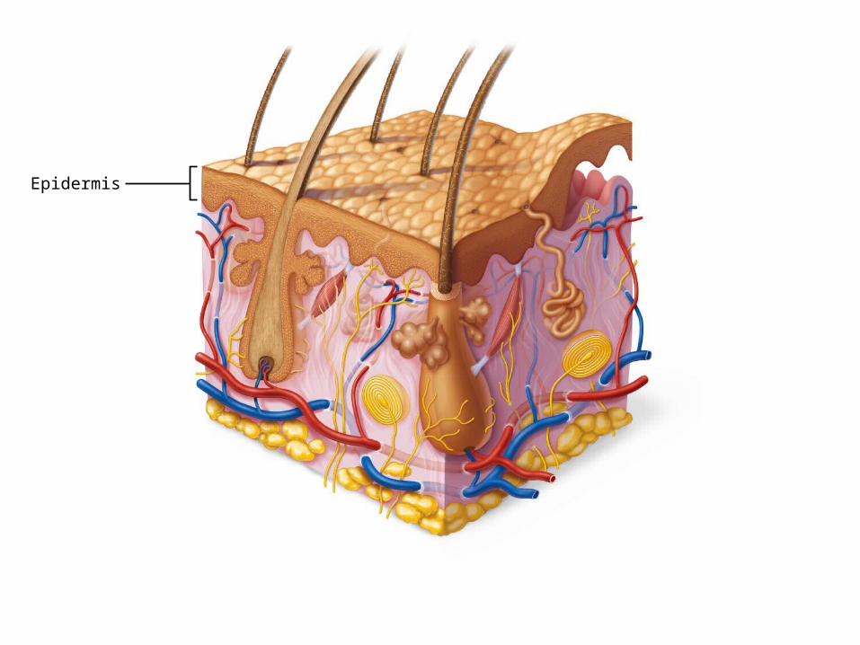
Hair shaft
Epidermis
Papillarylayer
Dermis Reticularlayer
Hypodermis
Dermal papillae
Sweat pore
Cutaneous plexus
Adipose tissue
Eccrine sweat glandArrector pili muscle
Sebaceous gland
Hair follicleHair root

Skin (Integument)
3 distinct regions:
– Epidermis—superficial region
• Keratinized stratified squamous epithelial tissue
– Dermis—underlies epidermis
• Fibrous connective tissue
– Hypodermis• Adipose tissue that absorbs
shock & insulates• Anchors skin to underlying
structures – muscles
Epidermis

Epidermis
Keratinized stratified squamous epithelium•Five distinct layers
– Stratum basale– Stratum spinosum– Stratum granulosum– Stratum lucidum – Stratum corneum
•With four cell types– Keratinocytes– Melanocytes– Dendritic (Langerhans) cells– Tactile (Merkel) cells
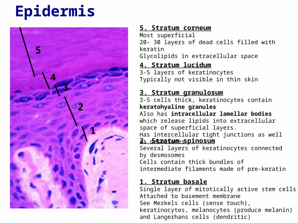
Epidermis
1
2
3 4
5
5. Stratum corneumMost superficial 20- 30 layers of dead cells filled with keratin Glycolipids in extracellular space
4. Stratum lucidum 3-5 layers of keratinocytesTypically not visible in thin skin
3. Stratum granulosum3-5 cells thick, keratinocytes contain keratohyaline granules Also has intracellular lamellar bodies which release lipids into extracellular space of superficial layers. Has intercellular tight junctions as well as desmosomes
2. Stratum spinosumSeveral layers of keratinocytes connected by desmosomesCells contain thick bundles of intermediate filaments made of pre-keratin
1. Stratum basaleSingle layer of mitotically active stem cellsAttached to basement membrane See Merkels cells (sense touch), keratinocytes, melanocytes (produce melanin) and Langerhans cells (dendritic)
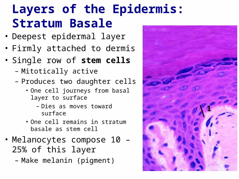
Layers of the Epidermis: Stratum Basale
• Deepest epidermal layer • Firmly attached to dermis• Single row of stem cells
– Mitotically active– Produces two daughter cells
• One cell journeys from basal layer to surface
– Dies as moves toward surface• One cell remains in stratum basale
as stem cell
• Melanocytes compose 10 – 25% of this layer– Make melanin (pigment)
1

Layers of the Epidermis:Stratum Spinosum
• Several layers thick
• Cells contain web-like system of intermediate prekeratin filaments attached to desmosomes
• Abundant melanosomes and dendritic cells
2

Layers of the Epidermis: Stratum Granulosum
• Thin - four to six cell layers • Cell appearance changes
– Cells flatten– Nuclei and organelles disintegrate– Keratinization begins
• Cells accumulate keratohyaline granules
– Help form keratin in upper layers
– Cell accumulate lamellar granules• Their water-resistant glycolipid
slows water loss
• Cells above this layer die– Too far from dermal capillaries
3

Layers of the Epidermis: Stratum Lucidum
• Only in thick skin
• Thin, translucent band superficial to the stratum granulosum
• A few rows of flat, dead keratinocytes
4

Layers of the Epidermis: Stratum Corneum
• 20–30 rows of dead, flat, anucleate keratinized membranous sacs
• Three-quarters of epidermal thickness
• Though dead, its cells have functions– Protect deeper cells from
environment and water loss– Protect from abrasion and
penetration– Physical, biological, and
chemical barrier
5

Cells of the Epidermis
Keratinocytes– Produce fibrous protein keratin– Most cells of epidermis– Tightly connected by desmosomes
Melanocytes– 10–25% of cells in deepest epidermis– Produce melanin – packaged into
melanosomes• Protect apical surface of keratinocyte
nucleus from UV damage
Dendritic (Langerhans) cells– Macrophages – key activators of
immune system
Merkel cells– Sensory touch receptors
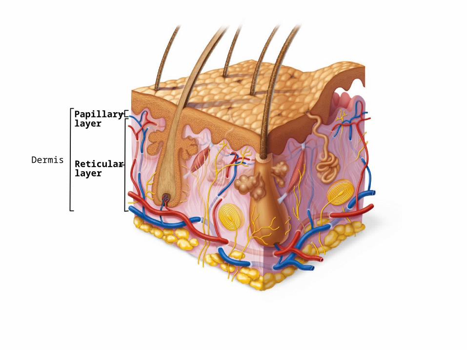
Papillarylayer
Dermis Reticularlayer

Dermis
• Strong, flexible connective tissue• Cells
– Fibroblasts, macrophages, and occasionally mast cells and white blood cells
• Fibers in matrix bind body together– "Hide" used to make leather
• Contains nerve fibers; blood and lymphatic vessels
• Contains epidermal hair follicles; oil and sweat glands
• Two layers: – Papillary– Reticular

Layers of the Dermis: Papillary Layer
• Loose tissue– Phagocytes can
patrol for microorganisms
• Dermal papillae– Superficial
peglike projections
• Areolar connective tissue with collagen and elastic fibers and blood vessels
Papillary layer

Dermal Papillae
• Some contain Meissner's corpuscles (touch receptors)
• Some contain free nerve endings (pain receptors)
• In thick skin lie atop dermal ridges that cause epidermal ridges– Collectively ridges called
friction ridges• Enhance gripping ability• Contribute to sense of touch• Pattern is fingerprints

© 2013 Pearson Education, Inc.

© 2013 Pearson Education, Inc.
Subpapillary plexus and Receptors

© 2013 Pearson Education, Inc.
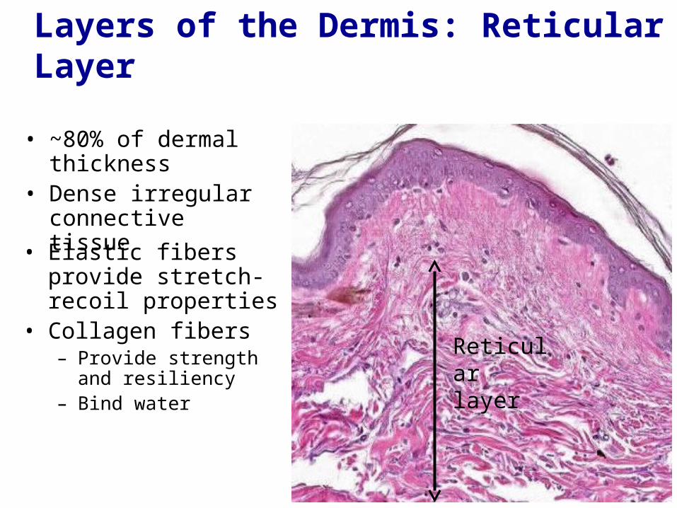
Layers of the Dermis: Reticular Layer
• Elastic fibers provide stretch-recoil properties
• Collagen fibers – Provide strength and
resiliency– Bind water
Reticular layer
• ~80% of dermal thickness
• Dense irregular connective tissue

Hair Follicles• Hair bulb
– Expanded deep end– Hair follicle receptor
(root hair plexus)– Sensory nerve endings -
touch receptors– Hair matrix
• Actively dividing area
• Arrector pili– Smooth muscle attached
to follicle– Responsible for "goose
bumps"
• Hair papilla– Dermal tissue - blood supply

Eccrine Sweat Glands
• Most numerous• Abundant on palms, soles, and forehead• Ducts connect to pores
• Function in thermoregulation
• Their secretion is sweat– 99% water, salts,
vitamin c, antibodies, dermcidin, metabolic wastes

Apocrine Sweat Glands
• Confined to axillary and anogenital areas
• Sweat + fatty substances + proteins– Odorless until bacterial
interaction body odor
• Ducts empty into hair follicles• Hormonally controlled
– Begin functioning at puberty
• Modified apocrine glands– Ceruminous glands—lining of
external ear canal; secrete cerumen (earwax)
– Mammary glands – secrete milk

Sebaceous (Oil) Glands
• Widely distributed– Not in thick skin of palms
and soles
• Most develop from hair follicles and secrete into hair follicles
• Relatively inactive until puberty– Stimulated by hormones,
especially androgens
• Secrete sebum– Oily holocrine secretion– Bactericidal