lab management criteria 1199seiu funds...400 buckwalter place boulevard, bluffton, sc 29910 dear...
TRANSCRIPT

LAB MANAGEMENT CRITERIA
1199SEIU Funds
Effective July 16, 2015
Clinical criteria for medical necessity review of lab management services.
© 2015 CareCore | MedSolutions. All rights reserved.

Please note the following: CPT Copyright 2015 American Medical Association. All rights reserved. CPT is a registered trademark of the American Medical Association.
CareCore | MedSolutions Laboratory Laboratory Management Criteria V2.0.2015
© 2015 CareCore | MedSolutions. All rights reserved. 400 Buckwalter Place Boulevard, Bluffton, SC 29910 • (800) 918-8924 www.carecorenational.com
2 of 415
Please note the following: All information provided by the NCCN is “Referenced with permission from the NCCN Clinical Practice Guidelines in Oncology (NCCN Guidelines™) © 2015 National Comprehensive Cancer Network. The NCCN Guidelines™ and illustrations herein may not be reproduced in any form for any purpose without the express written permission of the NCCN. To view the most recent and complete version of the NCCN Guidelines, go online to NCCN.org.”
CareCore | MedSolutions Laboratory Laboratory Management Criteria V2.0.2015
© 2015 CareCore | MedSolutions. All rights reserved. 400 Buckwalter Place Boulevard, Bluffton, SC 29910 • (800) 918-8924 www.carecorenational.com
3 of 415

Dear Provider,
This document provides detailed descriptions of CareCore National’s basic criteria for laboratory services. These criteria are used for the certification of requests and administration of laboratory benefits for our clients for a range of laboratory tests some of which are represented by one CPT or HCPCS code and others represented by several codes. They have been carefully researched and are continually updated in order to be consistent with the most current evidence-based guidelines and recommendations for laboratory testing from national and international medical societies and evidence-based medicine research centers. In addition, the criteria are supplemented by information published in peer reviewed literature. If you believe that our criteria require modification, please send suggested changes with supporting references to the Laboratory Management Program at the address listed below.
Our health plan clients review the development and application of these criteria. Every CareCore National, LLC health plan client develops a unique list of CPT codes that are part of their utilization management programs. Health Plan medical policy supersedes CareCore National, LLC when there is conflict with the CareCore criteria and the health plan medical policy. If you are unsure of whether or not a specific health plan has made modifications to these basic criteria in their medical policy please contact the plan or access the plan’s website for additional information.
CareCore National works hard to make your clinical review experience a pleasant one. For that reason, we have peer reviewers available to assist you should you have specific questions about a procedure. For your convenience, CareCore National’s Customer Service support is available from 7 a.m. to 7 p.m. Our toll free number is (800) 918-8924.
Gregg P. Allen, M. D. FAAFP EVP and Chief Medical Officer
CareCore | MedSolutions Laboratory Laboratory Management Criteria V2.0.2015
© 2015 CareCore | MedSolutions. All rights reserved. 400 Buckwalter Place Boulevard, Bluffton, SC 29910 • (800) 918-8924 www.carecorenational.com
4 of 415

General Information about This Policy Manual Description The CareCore National policy manual contains medical and reimbursement policies that are created and approved by CareCore National’s Laboratory Management Program personnel and policy advisors, internal Medical Advisory Committee, and external Medical Advisory Board. CareCore National’s polices are created using evidence-based medicine including, but not limited to, professional society guidelines, consensus statements, and peer-reviewed literature. CareCore National’s policies are intended to provide a library for adoption or a basis for development of tailored coverage criteria for a Health Plan.
Purpose To establish evidence-based definitions, decision support, medical necessity criteria, coverage limitations, and payment rules for molecular and genetic testing.
This manual is organized into the following sections: Molecular and Genetic Clinical Use Policies The policies in this section are intended to provide general policy guidance for the common settings and scenarios in which genetic testing is used (e.g. prenatal, diagnostic, cancer). These policies address the overarching coverage principles that broadly apply based on the purpose of the test. They also address specific use situations that may apply to many different tests (e.g. predictive testing for a known familial mutation). Each of these overarching policies includes an inventory of all available test-specific policies that apply to that use. For example, the Pharmacogenomic Testing policy includes a list of all policies for tests that may be used to assess drug response or toxicity risk. Because tests can be used for multiple purposes, the same test-specific policy may be referenced by more than one Clinical Use Policy. However, when a test specific policy is not available, the overarching coverage principles found in these Clinical Use Policies may be applied. Molecular and Genetic Test Specific Policies The policies in this section address a test or group of tests that are used to assess some health condition. The purpose of these policies is to provide a framework for determining medical necessity and coverage determinations for a specific test, including where more limited testing may be supported by the medical evidence when broader testing is not. These policies provide background about each condition, the available tests, the scenarios in which the test may be used, and the evidence used to determine medical necessity criteria. Glossary This glossary contains definitions for common genetics, medical and laboratory terminology Administrative Policies If applicable for this plan, administrative policies are included that define coding and reimbursement criteria and requirements.
CareCore | MedSolutions Laboratory Laboratory Management Criteria V2.0.2015
© 2015 CareCore | MedSolutions. All rights reserved. 400 Buckwalter Place Boulevard, Bluffton, SC 29910 • (800) 918-8924 www.carecorenational.com
5 of 415

Limitations and Restrictions When using this manual in electronic or printed form, the following restrictions apply:
• Evidence-based genetic testing is defined as the identification of targeted genetic sequences within the genome of an individual with clinically-identified risk factors or traits suspected of being specific to the genetic disorder, condition, or trait under investigation.
• The medical policies contained in this manual are the proprietary property of CareCore National, LLC, for use by its clients only. These medical policies may not be posted, shared, altered, cited or reproduced without the express written consent of CCN. Commercial use of these policies is prohibited.
• Medical policies are not to be considered medical advice for a specific patient. Policies are used in the process of determining whether a service may be medically necessary and eligible for coverage.
• Medical Policies are interpreted and applied at the sole discretion of the Health Plan. • Current Procedural Terminology (CPT®) codes and descriptions are the property of the American
Medical Association with all rights reserved.
CareCore | MedSolutions Laboratory Laboratory Management Criteria V2.0.2015
© 2015 CareCore | MedSolutions. All rights reserved. 400 Buckwalter Place Boulevard, Bluffton, SC 29910 • (800) 918-8924 www.carecorenational.com
6 of 415

Table of Contents
Policy Page Molecular and Genetic Clinical Use Policies .............................................................................................................................. 10 Genetic Testing for Cancer Susceptibility and Hereditary Cancer Syndromes ............................................................................... 11 Genetic Testing for Carrier Status .................................................................................................................................................. 13 Genetic Testing for Non-Medical Purposes .................................................................................................................................... 16 Genetic Testing for Prenatal Screening and Diagnostic Testing .................................................................................................... 17 Genetic Testing for the Screening, Diagnosis, and Monitoring of Cancer ...................................................................................... 20 Genetic Testing to Diagnose Non-Cancer Conditions .................................................................................................................... 22 Genetic Testing to Predict Disease Risk ......................................................................................................................................... 25 Investigational and Experimental Molecular/Genomic Testing ....................................................................................................... 27 Pharmacogenomic Testing for Drug Toxicity and Response .......................................................................................................... 33 Preimplantation Genetic Screening and Diagnosis ......................................................................................................................... 36 Molecular and Genetic Test-Specific Policies ............................................................................................................................ 37 ABL Tyrosine Kinase Sequencing for Chronic Myeloid Leukemia .................................................................................................. 38 Afirma Gene Expression Classifier for Thyroid Cancer .................................................................................................................. 41 Alpha-1-Antitrypsin Deficiency Testing ........................................................................................................................................... 44 Amyotrophic Lateral Sclerosis Known Familial Mutation Analysis .................................................................................................. 47 Angelman Syndrome Testing .......................................................................................................................................................... 51 APOE Variant Analysis for Alzheimer Disease ............................................................................................................................... 57 Ashkenazi Jewish Carrier Screening .............................................................................................................................................. 60 Ataxia-Telangiectasia ...................................................................................................................................................................... 64 BCR-ABL Testing for Chronic Myeloid Leukemia ........................................................................................................................... 67 Bloom Syndrome Testing ................................................................................................................................................................ 69 BRAF Testing for Colorectal Cancer Anti-EGFR Response ........................................................................................................... 74 BRAF V600E Testing for Melanoma Kinase Inhibitor Response .................................................................................................... 76 BRCA Ashkenazi Jewish Founder Mutation Testing ...................................................................................................................... 79 BRCA Known Familial Mutation Analysis ....................................................................................................................................... 83 BRCA1/2 Deletion/Duplication Analysis .......................................................................................................................................... 86 BRCA Sequencing .......................................................................................................................................................................... 89 BRCA Sequencing for Olaparib Response ..................................................................................................................................... 95 Brugada Syndrome Known Familial Mutation Analysis .................................................................................................................. 98 Brugada Syndrome Multigene Panels .......................................................................................................................................... 102 Brugada Syndrome Sequencing ................................................................................................................................................... 106 CADASIL Known Familial Mutation Analysis ................................................................................................................................ 109 CADASIL Testing .......................................................................................................................................................................... 112 Canavan Disease Testing ............................................................................................................................................................. 115 Celiac Disease Testing ................................................................................................................................................................. 119 CellSearch Circulating Tumor Cell Count for Breast Cancer Prognosis ....................................................................................... 121 Charcot-Marie-Tooth Neuropathy Testing Panel .......................................................................................................................... 123 Chromosome Microarray Testing For Developmental Disorders .................................................................................................. 126 Chromosomal Microarray for Prenatal Diagnosis ......................................................................................................................... 130 Chromosome Analysis for Blood, Bone Marrow, and Solid Tumor Cancers ................................................................................ 134 Cologuard Screening for Colorectal Cancer ................................................................................................................................. 136 ConfirmMDx for Prostate Cancer Risk Assessment ..................................................................................................................... 139 Corus CAD for Obstructive Coronary Artery Disease ................................................................................................................... 141 CYP2C9 and VKORC1 Testing for Warfarin Response ............................................................................................................... 146 CYP2C19 Variant Analysis for Clopidogrel Response .................................................................................................................. 149 CYP2D6 Variant Analysis for Tamoxifen Response ..................................................................................................................... 152 Cystic Fibrosis Testing .................................................................................................................................................................. 155 Dentatorubral-Pallidoluysian Atrophy Testing ............................................................................................................................... 162 DPYD Variant Analysis for 5-FU Toxicity ...................................................................................................................................... 165
CareCore | MedSolutions Laboratory Laboratory Management Criteria V2.0.2015
© 2015 CareCore | MedSolutions. All rights reserved. 400 Buckwalter Place Boulevard, Bluffton, SC 29910 • (800) 918-8924 www.carecorenational.com
7 of 415

Duchenne & Becker Muscular Dystrophy Testing ........................................................................................................................ 167 EGFR Testing for Non-Small Cell Lung Cancer TKI Response .................................................................................................... 171 Expanded Carrier Screening Panels ............................................................................................................................................. 174 Factor II/Prothrombin Testing for Thrombophilia .......................................................................................................................... 178 Factor V Leiden Testing for Thrombophilia ................................................................................................................................... 182 Familial Adenomatous Polyposis Testing ..................................................................................................................................... 188 Familial Malignant Melanoma Testing .......................................................................................................................................... 192 Flow Cytometry ............................................................................................................................................................................. 195 Fragile X Syndrome Testing ......................................................................................................................................................... 204 Fragile X Associated Tremor/Ataxia Syndrome Testing ............................................................................................................... 207 Gaucher Disease Testing ............................................................................................................................................................. 210 Hereditary Hemochromatosis Testing ........................................................................................................................................... 215 Hereditary Cancer Syndrome Multigene Panels ........................................................................................................................... 218 HIV Tropism Testing for Maraviroc Response .............................................................................................................................. 227 HLA-B*1502 Variant Analysis for Carbamazepine Response ...................................................................................................... 230 HLA-B*5701 Genotyping for Abacavir Hypersensitivity ................................................................................................................ 232 Huntington Disease Testing .......................................................................................................................................................... 235 Hypertrophic Cardiomyopathy Testing .......................................................................................................................................... 238 KRAS Testing for Anti-EGFR Response in Metastatic Colorectal Cancer .................................................................................... 243 Li-Fraumeni Syndrome Testing ..................................................................................................................................................... 246 Long QT Syndrome Testing .......................................................................................................................................................... 249 Lynch Syndrome Genetic Testing ................................................................................................................................................. 253 Lynch Syndrome Tumor Screening - First-Tier ............................................................................................................................. 262 Lynch Syndrome Tumor Screening - Second-Tier ........................................................................................................................ 266 Mammaprint 70-Gene Breast Cancer Recurrence Assay ............................................................................................................. 270 Mammostrat Breast Cancer Recurrence Assay ............................................................................................................................ 273 MGMT Testing for Malignant Glioma Alkylating Agent Response ................................................................................................ 275 MTHFR Variant Analysis for Hyperhomocysteinemia ................................................................................................................... 277 MUTYH Associated Polyposis Testing ......................................................................................................................................... 280 Niemann Pick Disease Types A & B Testing ................................................................................................................................ 285 Niemann Pick, Type C Testing ..................................................................................................................................................... 290 Non-Invasive Prenatal Testing ...................................................................................................................................................... 295 OncotypeDX for Breast Cancer Prognosis ................................................................................................................................... 300 OncotypeDX for Colorectal Cancer Recurrence Risk ................................................................................................................... 304 PCA3 Testing for Prostate Cancer ................................................................................................................................................ 307 Peutz-Jeghers Syndrome Testing ................................................................................................................................................. 309 PTEN Hamartoma Tumor Syndromes Testing ............................................................................................................................. 313 Prader-Willi Syndrome Testing ..................................................................................................................................................... 319 Prenatal Aneuploidy FISH Testing ................................................................................................................................................ 324 Prenatal Chromosome Analysis .................................................................................................................................................... 327 Prenatal Maternal Serum Screening ............................................................................................................................................. 330 Prolaris for Prostate Cancer Prognosis......................................................................................................................................... 332 Rett Syndrome Testing ................................................................................................................................................................. 335 Sexually Transmitted Infections: Molecular .................................................................................................................................. 339 Spinal Muscular Atrophy Testing .................................................................................................................................................. 354 Tay-Sachs Disease Testing .......................................................................................................................................................... 360 Tissue of Origin Testing for Cancer of Unknown Primary ............................................................................................................. 366 TPMT Testing for Thiopurine Drug Response .............................................................................................................................. 368 UGT1A1 Mutation Analysis for Irinotecan Response .................................................................................................................... 371 UroVysion FISH for Bladder Cancer ............................................................................................................................................. 373 VeriStrat Testing for NSCLC TKI Response ................................................................................................................................. 375 Von Hippel-Lindau Disease Testing .............................................................................................................................................. 378 Glossary ...................................................................................................................................................................................... 382 Administrative Policies .............................................................................................................................................................. 393 Laboratory Claim Reimbursement ................................................................................................................................................ 394
CareCore | MedSolutions Laboratory Laboratory Management Criteria V2.0.2015
© 2015 CareCore | MedSolutions. All rights reserved. 400 Buckwalter Place Boulevard, Bluffton, SC 29910 • (800) 918-8924 www.carecorenational.com
8 of 415

Molecular Pathology Tier 2 Molecular CPT Codes ....................................................................................................................... 403 Molecular Testing S Codes ........................................................................................................................................................... 407 Retired Molecular Pathology CPT Codes ..................................................................................................................................... 411 Unlisted Molecular Pathology CPT Code 81479 ........................................................................................................................... 413
CareCore | MedSolutions Laboratory Laboratory Management Criteria V2.0.2015
© 2015 CareCore | MedSolutions. All rights reserved. 400 Buckwalter Place Boulevard, Bluffton, SC 29910 • (800) 918-8924 www.carecorenational.com
9 of 415

Molecular and Genetic Clinical Use Policies
CareCore | MedSolutions Laboratory Laboratory Management Criteria V2.0.2015
© 2015 CareCore | MedSolutions. All rights reserved. 400 Buckwalter Place Boulevard, Bluffton, SC 29910 • (800) 918-8924 www.carecorenational.com
10 of 415

Genetic Testing for Cancer Susceptibility and Hereditary Cancer Syndromes
Description Genetic testing for cancer susceptibility and hereditary cancer syndromes is performed in people with known risk factors for an inherited form of cancer. Testing may be used in people diagnosed with cancer when there are “red flags” in the person’s personal medical and/or family history for a hereditary form. Predictive genetic testing may also be performed for this group of conditions, in people known to be at increased risk of developing an inherited condition based on their family history. A positive genetic test result increases the risk for cancer (types vary by the gene involved) and, therefore, impacts medical management decisions around screening, prevention, and treatment.
• Tests used to screen for or make a diagnosis of cancer are covered separately as Genetic Testing for the Screening, Diagnosis, and Monitoring of Cancer.
• This policy does not address diagnostic or predictive testing for conditions other than hereditary cancer. Refer to Genetic Testing to Diagnose Non-Cancer Conditions and Genetic Testing to Predict Disease Risk for those purposes.
Criteria: General Coverage Guidance Individuals may be considered for genetic testing for hereditary cancer syndromes when ALL of the following conditions are met:
• Technical and clinical validity: The test must be accurate, sensitive and specific, based on sufficient, quality scientific evidence to support the claims of the test.
• Clinical utility: Healthcare providers can use the test results to provide significantly better medical care for the individual.
• Reasonable use: The usefulness of the test is not significantly offset by negative factors, such as expense, clinical risk, or social or ethical challenges.
Limits:
• Testing will be considered only for the number of genes or tests necessary to establish carrier status. A tiered approach to testing, with reflex to more detailed testing and/or different genes, will be required when clinically possible.
• Genetic testing is indicated once per lifetime per condition. Exceptions may be considered if technical advances in testing demonstrate significant advantages that would support a medical need to retest.
Criteria: Special Circumstances The following policies address a group of tests that are used for similar purposes. Because a variety of tests may be used, but the circumstances that justify testing are the same, individual test-specific policies are not necessary.
CareCore | MedSolutions Laboratory Laboratory Management Criteria V2.0.2015
© 2015 CareCore | MedSolutions. All rights reserved. 400 Buckwalter Place Boulevard, Bluffton, SC 29910 • (800) 918-8924 www.carecorenational.com
11 of 415

Her
edita
ry C
ance
r
Predictive testing for at-risk people with known familial mutations
The genetic mutation(s) associated with a hereditary cancer syndrome can often be defined in an affected family member, allowing for testing of at-risk relatives for those specific mutations. Testing for known familial mutations is reasonable when ALL of the following conditions are met:
• The mutation(s) in the family have been clearly defined by previous genetic testing and information about those mutations can be provided to the testing lab.
• Technical and clinical validity: The test must be accurate, sensitive and specific to the familial mutations.
• Clinical utility: Healthcare providers can use the test results to provide significantly better medical care for the individual.
• Reasonable use: The usefulness of the test is not significantly offset by negative factors, such as expense, clinical risk, or social or ethical challenges.
Limits:
• Testing will be considered only for the known familial mutations when clinically possible. • Predictive genetic testing is indicated once per lifetime per condition. • Predictive genetic testing will be considered only for adult individuals (age 18 and over).
Exceptions may be considered if there are medical management and/or significant psychosocial benefits to testing prior to adulthood.1,2
References 1. American Society of Human Genetics/American College of Medical Genetics Report. Points to consider: ethical,
legal, and psychosocial implications of genetic testing in children and adolescents. Am J Hum Genet. 1995; 57:1233-41.
2. National Society of Genetic Counselors Position Statement. Prenatal and childhood testing for adult-onset disorders. Adopted 1995. Available at: http://www.nsgc.org/about/position.cfm.
Criteria: Test-specific Policies Policies are available for the following hereditary cancer syndrome tests. See the individual policy documents arranged alphabetically by policy name in the Test-specific Policies section. For tests without a specific policy, use the General Coverage Guidance provided in Section 1.
• BRACAnalysis® Rearrangement Test (BART) • Cowden syndrome (PTEN gene) • Familial adenomatous polyposis (FAP) and attenuated FAP • Familial Malignant Melanoma • Fanconi Anemia , Group D1 • Hereditary Breast Ovarian Cancer Syndrome (HBOC) • Li-Fraumeni syndrome (TP53 gene) • Lynch Syndrome (Hereditary non-polyposis colorectal cancer, HNPCC) • Microsatellite instability (MSI)/immunohistochemistry (IHC) tumor screening for hereditary non-
polyposis colorectal cancer • MUTYH-associated polyposis (MAP) • Peutz-Jeghers syndrome (PJS)
CareCore | MedSolutions Laboratory Laboratory Management Criteria V2.0.2015
© 2015 CareCore | MedSolutions. All rights reserved. 400 Buckwalter Place Boulevard, Bluffton, SC 29910 • (800) 918-8924 www.carecorenational.com
12 of 415
Genetic Testing for Carrier Status Description Carrier screening is performed to identify genetic risks that could impact reproductive decision-making for parents or prospective parents. Carriers are generally not affected but have an increased risk to have a child with a genetic condition. Carrier screening may be available for autosomal recessive conditions, X-linked conditions, and certain chromosome abnormalities. Ideally, carrier screening is performed prior to pregnancy so that a full range of reproductive options are available to an at-risk couple. However, in practice, it is often performed early in pregnancy when prenatal care is established.
• This policy does not include prenatal or preimplantation genetic testing. Refer to policies on Genetic Testing for Prenatal Screening and Diagnostic Testing and Preimplantation Genetic Screening and Diagnosis for those purposes.
• In addition, testing that may identify carriers who have clinical signs and symptoms (e.g., cystic fibrosis testing for men with congenital absence of the vas deferens, fragile X genetic testing for women with premature ovarian failure) is addressed as Genetic Testing to Diagnose Non-Cancer Conditions.
Criteria: General Coverage Guidance Individuals may be considered for genetic testing for carrier screening when ALL of the following conditions are met:
• Technical and clinical validity: The test must be accurate, sensitive and specific, based on sufficient, quality scientific evidence to support the claims of the test.
• Clinical utility: Healthcare providers can use the test results to provide significantly better medical care and/or assist individuals with reproductive planning.
• Reasonable use: The usefulness of the test is not significantly offset by negative factors, such as expense, clinical risk, or social or ethical challenges.
Limits:
• Testing will only be considered for the number of genes or tests necessary to establish carrier status. A tiered approach to testing, with reflex to more detailed testing and/or different genes, will be required when clinically possible.
• Carrier testing will be allowed once per lifetime. Exceptions may be considered if technical advances in testing demonstrate significant advantages that would support a medical need to retest.
• Carrier testing is indicated only in adults. Carrier screening in minor children is not indicated, except in the case of a pregnancy of the minor child.
Routine Carrier Screening
Individuals may be considered for routine carrier screening when testing is supported by evidence-based guidelines from governmental organizations and/or well-recognized professional societies in the United States.
CareCore | MedSolutions Laboratory Laboratory Management Criteria V2.0.2015
© 2015 CareCore | MedSolutions. All rights reserved. 400 Buckwalter Place Boulevard, Bluffton, SC 29910 • (800) 918-8924 www.carecorenational.com
13 of 415

Car
rier S
tatu
s
Carrier Screening Based on Family History
Individuals may be considered for carrier screening based on a family history of a genetic condition when ALL of the following conditions are met in addition to the general criteria above:
• The diagnosis of a genetic condition in a family member is known. • The parent(s) or prospective parent(s) are at-risk to be carriers of that condition based on the
pattern of inheritance. • The genetic condition is associated with potentially severe disability or has a lethal natural history.
Partner Testing of Known Carrier or Affected Individuals
Individuals may be considered for carrier screening if their partners are known carrier or affected individuals when all of the following conditions are met in addition to the general criteria above:
• The diagnosis of a genetic condition or carrier status in the partner is known. • The genetic condition is associated with potentially severe disability or has a lethal natural history.
Criteria: Special Situations Exclusions Multiplex Carrier Screening
Multiplex carrier screening tests are designed to identify carrier status or predict risk for many genetic diseases (70 or more) in a single test. Several multiplex carrier screening tests are available now. Others are known to be in development and will come to market in the next few years. Each test has a unique set of diseases included in novel and proprietary genetic testing platforms. Of the genetic conditions included in the currently available multiplex carrier screening tests, 12 of them are recommended for at least some people based on ethnicity by either the American College of Obstetrics and Gynecology (ACOG) and/or the American College of Medical Genetics (ACMG). However, mutation analysis is not the preferred initial screening test for some.
These tests do not meet the criteria above for technical and clinical validity and clinical utility: • The technologies used by the multiplex carrier screening tests are novel. Information about the
test's performance, if available, is often provided completely by the laboratory marketing the test, which could be subject to bias.
• Some of the commonly included tests, such as beta-thalassemia and Tay-Sachs disease, have inexpensive and reliable screening tests available (CBC with RBC indices and hexosaminidase A enzyme activity, respectively) that are superior to genetic testing.
• Multiplex carrier screening tests typically include carrier screening for many diseases that have not been identified as appropriate for population-based carrier screening. They may also include disorders, such as hereditary hemochromatosis and factor V Leiden, which affect primarily adults and are generally manageable. These kinds of conditions do not meet the requirements for reproductive carrier screening programs.
CareCore | MedSolutions Laboratory Laboratory Management Criteria V2.0.2015
© 2015 CareCore | MedSolutions. All rights reserved. 400 Buckwalter Place Boulevard, Bluffton, SC 29910 • (800) 918-8924 www.carecorenational.com
14 of 415

Car
rier S
tatu
s
Criteria: Test-specific Policies Policies are available for the following tests designed to predict carrier status. See the individual policy documents arranged alphabetically by policy name in the Test-specific Policies section. For tests without a specific policy, use the General Coverage Guidance provided in Section 1. Carrier screening for:
• Ashkenazi Jewish Diseases • Ataxia Telangiectasia • Bloom Syndrome • Canavan Disease • Cystic Fibrosis • Duchenne/Becker Muscular Dystrophy • Fragile X syndrome • Gaucher Disease • Hemoglobinopathies (alpha-thalassemia, beta-thalassemia, and sickle cell disease) • Niemann Pick Disease, Types A and B • Tay-Sachs Disease
CareCore | MedSolutions Laboratory Laboratory Management Criteria V2.0.2015
© 2015 CareCore | MedSolutions. All rights reserved. 400 Buckwalter Place Boulevard, Bluffton, SC 29910 • (800) 918-8924 www.carecorenational.com
15 of 415

Genetic Testing for Non-Medical Purposes Description While most traditional genetic tests are used to for clear medical purposes, advances in gene discovery and genetic testing technology allow laboratories to offer genetic testing for other uses. Testing for paternity, ancestry, and non-disease traits such as baldness and eye color may be highly accurate and interesting. However, because these kinds of tests are not useful for medical management in the vast majority of cases, they are typically excluded from consideration.
Criteria: General Coverage Guidance Any genetic test that DOES NOT meet the following criteria is excluded from consideration:
• Technical and clinical validity: The test must be accurate, sensitive and specific, based on sufficient, quality scientific evidence to support the claims of the test.
• Clinical utility: Healthcare providers can use the test results to provide significantly better medical care for the individual.
• Reasonable use: The usefulness of the test is not significantly offset by negative factors, such as expense, clinical risk, or social or ethical challenges.
Criteria: Test-specific Policies The following types of testing are specifically excluded from consideration:
• Genome-wide association studies (GWAs): testing a large number of genetic variations spread across the whole genome for disease associations, generally done for information outside of a specific clinical need or context
o Common trade names: 23andMe, Navigenics, Pathway Genomics, deCODEme • Paternity testing: testing to establish biological relationships, often between a father and
child(ren) but sometimes to determine other kinds of relationships (siblings, grandparents, etc.) • Ancestry testing: testing that helps people discover more about the genetic make-up of their
ancestors, generally used by genealogists and those interested in family history o Common trade names: Ancestry.com, 23andMe, Pathway Genomics, Family Tree DNA,
deCODEme • Non-disease trait testing: testing for physical traits (e.g., eye color, hair color, male pattern
baldness, cellulite) that do not have associated health problems, or can be deemed cosmetic in nature.
• Nutritional: testing for variations in metabolism pathways that may suggest vitamin or other nutritional supplements.
o Common trade names: MyDNAVitamins, GeneWise • Athletic ability: Testing to predict athletic performance types.
o Common trade names: SportGene, Athleticode • Genetic testing related to dating services
o Common trade names: Scientific Match
CareCore | MedSolutions Laboratory Laboratory Management Criteria V2.0.2015
© 2015 CareCore | MedSolutions. All rights reserved. 400 Buckwalter Place Boulevard, Bluffton, SC 29910 • (800) 918-8924 www.carecorenational.com
16 of 415

Genetic Testing for Prenatal Screening and Diagnostic Testing
Description Prenatal screening and diagnostic testing is performed during pregnancy to identify fetuses at increased risk for or affected with genetic conditions and birth defects. Screening with ultrasound and maternal serum markers is routinely offered. Prenatal diagnosis by chorionic villus sampling or amniocentesis for chromosome abnormalities is available to all women. However, it is usually offered specifically to those at higher risk because of maternal age, a positive screen result, abnormal ultrasound findings, or known risk of a genetic condition based on family history. Investigations for fetal infection and blood antigen incompatibility may also be performed in the prenatal period. Results of testing are used to guide reproductive decision-making, pregnancy management and anticipatory management of the infant at birth. Note: This policy does not include prenatal or preconception carrier screening or preimplantation genetic testing. Please refer to Genetic Testing for Carrier Status and Preimplantation Genetic Screening and Diagnosis for those purposes.
Criteria: General Coverage Guidance Individuals may be considered for genetic testing for prenatal screening and diagnostic testing when ALL of the following conditions are met:
• Technical and clinical validity: The test must be accurate, sensitive and specific, based on sufficient, quality scientific evidence to support the claims of the test.
• Clinical utility: Healthcare providers can use the test results to provide significantly better medical care and/or assist patients with reproductive planning.
• Reasonable use: The usefulness of the test is not significantly offset by negative factors, such as expense, clinical risk, or social or ethical challenges.
Limits:
• Testing will only be covered for the number of genes or tests necessary to establish a prenatal diagnosis. A tiered approach to testing, with reflex to more detailed testing and/or different genes, will be required when clinically possible.
• Prenatal diagnostic testing will be allowed once per pregnancy. Exceptions may be considered if ambiguous results require retesting for clarification.
Criteria: Special Prenatal Diagnosis Circumstances Each of the following policies addresses a group of tests that are used for similar purposes in pregnancy. Because a variety of tests may be used, but the circumstances that justify testing are the same, individual test-specific policies are not necessary.
CareCore | MedSolutions Laboratory Laboratory Management Criteria V2.0.2015
© 2015 CareCore | MedSolutions. All rights reserved. 400 Buckwalter Place Boulevard, Bluffton, SC 29910 • (800) 918-8924 www.carecorenational.com
17 of 415

Pren
atal
Prenatal Diagnostic Testing Based on Family History
Prenatal genetic testing, generally by amniocentesis or CVS, for the diagnosis of a genetic condition is reasonable when the following conditions are met:
• The pregnancy is at an increased risk for a genetic disease because of ANY of the following: o At least one parent is known or suspected to be a carrier of a genetic condition based on
the family history and/or previous carrier testing results; or o One or both parent(s) are affected with a genetic condition; or o A sibling is affected with a genetic condition; AND
• The genetic condition is associated with potentially severe disability or has a lethal natural history. Fetal Infectious Disease Testing
Genetic testing may be used for the diagnosis of an infectious disease (e.g., cytomegalovirus, toxoplasmosis, parvovirus B19, and varicella zoster) in a fetus according to current guidelines from the American College of Obstetricians and Gynecologists (ACOG). Prenatal testing, generally by amniocentesis or CVS, is reasonable when ANY of the following conditions are met:
• Clinical signs and symptoms of a current infection in the mother; OR • Serologic evidence of a current or recent infection in the mother (with or without clinical signs); OR • Fetal abnormalities identified on ultrasound indicating an increased risk for a congenital infection
References 1. ACOG practice bulletin. Perinatal viral and parasitic infections. Number 20, September 2000 (reaffirmed 2008). Int
J Gynaecol Obstet. 2002 Jan;76(1):95-107. Blood Antigen Incompatibility Testing
Prenatal genetic testing, generally by amniocentesis, for the determination of blood antigen genotype is supported by current evidence-based recommendations from the American College of Obstetricians and Gynecologists.1 Fetal antigen genotyping is reasonable when the following conditions are met:
• A positive erythrocyte antibody screen in the mother; AND EITHER o The father’s blood antigen genotype is known and indicates a risk for the fetus to be
positive; OR o The father’s blood antigen genotype is not known and unavailable
References 2. ACOG Practice Bulletin, Number 75, August 2006. Management of alloimmunization during pregnancy. Obstet
Gynecol. 2006;108(2):457-64.
Criteria: Test-specific Policies Policies are available for the following prenatal diagnostic tests. See the individual policy documents arranged alphabetically by policy name in the Test-specific Policies section. Note that prenatal diagnosis may be just one of several test uses addressed in the same policy (e.g., a policy such as Canavan Disease Testing may address diagnostic, carrier, and prenatal diagnostic testing). For tests without a specific policy, use the General Coverage Guidance provided in Section 1 above.
CareCore | MedSolutions Laboratory Laboratory Management Criteria V2.0.2015
© 2015 CareCore | MedSolutions. All rights reserved. 400 Buckwalter Place Boulevard, Bluffton, SC 29910 • (800) 918-8924 www.carecorenational.com
18 of 415

Pren
atal
• Aneuploidy FISH • Ataxia Telangiectasia • Bloom Syndrome • Canavan Disease • Cystic Fibrosis • Fragile X Syndrome • Gaucher Disease • Niemann Pick Disease, Types A and B • Niemann-Pick Disease, Type C • Prenatal Diagnosis, Chromosome Abnormalities • Prenatal Maternal Serum Screening • Rett Syndrome • Spinocerebellar Ataxia, Types 1, 2, 3, 6, 7, 12, and 17 • Tay-Sachs Disease
CareCore | MedSolutions Laboratory Laboratory Management Criteria V2.0.2015
© 2015 CareCore | MedSolutions. All rights reserved. 400 Buckwalter Place Boulevard, Bluffton, SC 29910 • (800) 918-8924 www.carecorenational.com
19 of 415

Genetic Testing for the Screening, Diagnosis, and Monitoring of Cancer
Description Genetic testing for screening, diagnosis and monitoring of cancer refers to molecular diagnostic tests whose purposes include identifying the possible presence of cancer in asymptomatic, average risk individuals; confirming the absence or presence of cancer; and monitoring the absence or presence of cancer after a prior diagnosis and treatment.
Screening The goal of cancer screening is to identify the possible presence of cancer before symptoms appear. Screening tests cannot diagnose cancer, but typically determine if there is an increased chance cancer is present, and triages individuals for more invasive, diagnostic testing. Most cancer screening does not include genetic testing, but instead relies on physical exam, radiological exams, or non-genetic laboratory tests. Advances in human genetics, however, have identified several molecular diagnostic tests that may provide clues for early cancer detection. Diagnosis When cancer is suspected because of an abnormal screening test or symptoms, blood tests for tumor markers or molecular testing on tissue samples can aid in confirming a diagnosis of cancer. These tests may contribute information to helping the clinician understand prognosis and treatment options. Monitoring During treatment, or after an apparently successful treatment, active monitoring is often recommended to identify if the cancer is responding to treatment or has returned or spread, before any symptoms appear. Monitoring may include increased surveillance or routine blood tests for tumor markers, and increasingly, molecular genetic tests.
• Tests used to determine hereditary cancer risk are covered separately as Genetic Testing for Cancer Susceptibility and Hereditary Cancer Syndromes.
• This policy does not address drug response to cancer, or testing to determine which therapies to use. Please refer to Pharmacogenomic Testing for Drug Toxicity and Response for that purpose.
• This policy does not address diagnostic or predictive testing for conditions other than non-inherited cancer. Refer to Genetic Testing to Diagnose Non-Cancer Conditions and Genetic Testing to Predict Disease Risk for those purposes.
Criteria: General Coverage Guidance Individuals may be considered for genetic testing for screening, diagnosing, or monitoring cancer when ALL of the following conditions are met:
• Technical and clinical validity: The test must be accurate, sensitive and specific, based on sufficient, quality scientific evidence to support the claims of the test.
CareCore | MedSolutions Laboratory Laboratory Management Criteria V2.0.2015
© 2015 CareCore | MedSolutions. All rights reserved. 400 Buckwalter Place Boulevard, Bluffton, SC 29910 • (800) 918-8924 www.carecorenational.com
20 of 415

Can
cer
• Clinical utility: Healthcare providers can use the test results to provide significantly better medical care for the individual.
• Reasonable use: The usefulness of the test is not significantly offset by negative factors, such as expense, clinical risk, or social or ethical challenges.
Limits:
• Testing will be considered only for the number of genes or tests necessary. A tiered approach to testing, with reflex to more detailed testing and/or different genes, will be required when clinically possible.
• For tests that look for changes in germline DNA (i.e., not tumor DNA or viral DNA), testing will be allowed once per lifetime per gene. Exceptions may be considered if technical advances in testing demonstrate significant advantages that would support a medical need to retest.
Criteria: Test-specific Policies Policies are available for the following tests designed to screen for, diagnose, or monitor cancer. See the individual policy documents arranged alphabetically by policy name in the Test-specific Policies section. For tests without a specific policy, use the General Coverage Guidance provided in Section 1.
• Bone Marrow Biopsy Chromosome Analysis • CellSearch™ • Gene Expression Profiling for Cancers of Unknown Primary (CUP) • GSTP1 Testing, Prostate Cancer • PCA3 Testing, Prostate Cancer • Prolaris® • RET/PTC Rearrangement, Thyroid Cancer • UroVysion®
CareCore | MedSolutions Laboratory Laboratory Management Criteria V2.0.2015
© 2015 CareCore | MedSolutions. All rights reserved. 400 Buckwalter Place Boulevard, Bluffton, SC 29910 • (800) 918-8924 www.carecorenational.com
21 of 415
Genetic Testing to Diagnose Non-Cancer Conditions
Description Diagnostic testing is performed in patients with clinical signs or symptoms of a non-cancer genetic condition. The genetic test may confirm or rule out a clinical diagnosis. In some cases, genetic testing is the gold standard for making a diagnosis based on evidence- or consensus-based guidelines. In others, it may be used to confirm a clinical diagnosis, offer prognostic information that impacts management, or rule out a diagnosis in the differential. Often, diagnostic testing of an affected individual will offer results that are relevant to the testing of other family members.
• This policy does not include risk assessment or predictive testing for at-risk, asymptomatic individuals. Please refer to Genetic Testing to Predict Disease Risk for that purpose.
• Diagnostic testing of a pregnancy or an embryo is covered by policies on Genetic Testing for Prenatal Screening and Diagnostic Testing and Preimplantation Genetic Screening and Diagnosis, respectively.
• In addition, testing for hereditary cancer syndromes is addressed separately under Genetic Testing for Cancer Susceptibility and Hereditary Cancer Syndromes.
Criteria: General Coverage Guidance Individuals may be considered for diagnostic genetic testing when ALL of the following conditions are met:
• Clinical signs and symptoms in the individual are consistent with the diagnosis in question. • Technical and clinical validity: The test must be accurate, sensitive and specific, based on
sufficient, quality scientific evidence to support the claims of the test. • Clinical utility: Healthcare providers can use the test results to provide significantly better medical
care for the individual. • Reasonable use: The usefulness of the test is not significantly offset by negative factors, such as
expense, clinical risk, or social or ethical challenges. Limits:
• Testing will be considered only for the number of genes or tests necessary to establish carrier status. A tiered approach to testing, with reflex to more detailed testing and/or different genes, will be required when clinically possible.
• Diagnostic genetic testing will be allowed once per lifetime per condition. Exceptions may be considered if technical advances in testing demonstrate significant advantages that would support a medical need to retest.
Criteria: Special Circumstances: Diagnostic testing of a child to inform reproductive planning and testing for parents or testing for siblings
CareCore | MedSolutions Laboratory Laboratory Management Criteria V2.0.2015
© 2015 CareCore | MedSolutions. All rights reserved. 400 Buckwalter Place Boulevard, Bluffton, SC 29910 • (800) 918-8924 www.carecorenational.com
22 of 415

Dia
gnos
tic
Diagnostic genetic testing may be requested in a symptomatic child with a known genetic condition. While diagnostic testing may not impact management of the affected child, the information gained from genetic testing may be needed to perform accurate carrier testing in the child’s parent(s) and/or genetic diagnosis in a sibling. In these circumstances, diagnostic genetic testing in the child may be considered when ALL of the following conditions are met:
• The diagnosis of the disease in the affected child is certain or highly probable based on clinical signs and symptoms, history, imaging, and/or results of other laboratory testing.
• The results of the genetic test in the symptomatic child must be required in order to perform accurate carrier testing in the child’s parent(s) and/or genetic diagnosis in a sibling.
• Technical and clinical validity: The test must be accurate, sensitive and specific, based on sufficient, quality scientific evidence to support the claims of the test.
• Clinical utility: Healthcare providers can use the test results to provide informative genetic testing for the child’s parents and/or for a current or future at-risk pregnancy.
• Reasonable use: The usefulness of the test is not significantly offset by negative factors, such as expense, clinical risk, or social or ethical challenges.
Limits:
• Testing will be indicated only for the number of genes or tests necessary to establish the familial mutation(s). A tiered approach to testing, with reflex to more detailed testing and/or different genes, will be required when clinically possible.
• Diagnostic genetic testing will be allowed once per lifetime per condition. Exceptions may be considered if technical advances in testing demonstrate significant advantages that would support a medical need to retest.
Criteria: Test-specific Policies Policies are available for the following tests designed predict disease risk. See the individual policy documents arranged alphabetically by policy name in the Test-specific Policies section. For tests without a specific policy, use the General Coverage Guidance provided in Section 1.
• Alzheimer’s disease • Alpha-1-Antitrypsin • Angelman Syndrome • Array CGH • Ataxia Telangiectasia • Bloom Syndrome • Brugada Syndrome • CADASIL • Canavan • Celiac Disease • Cystic Fibrosis (includes Congenital Absence of the Vas Deferens) • Charcot-Marie-Tooth • Duchenne/Becker Muscular Dystrophy
CareCore | MedSolutions Laboratory Laboratory Management Criteria V2.0.2015
© 2015 CareCore | MedSolutions. All rights reserved. 400 Buckwalter Place Boulevard, Bluffton, SC 29910 • (800) 918-8924 www.carecorenational.com
23 of 415

Dia
gnos
tic
• Dentatorubral-Pallidoluysian Atrophy (DRPLA) • Factor II (Prothrombin) • Factor V Leiden • Fragile X Syndrome • Gaucher Disease • Hemochromatosis • Hypertrophic cardiomyopathy • Huntington Disease • Long QT syndrome • Niemann Pick Disease, Type C • Rett Syndrome • Spinocerebellar Ataxia • Tay-Sachs Disease
CareCore | MedSolutions Laboratory Laboratory Management Criteria V2.0.2015
© 2015 CareCore | MedSolutions. All rights reserved. 400 Buckwalter Place Boulevard, Bluffton, SC 29910 • (800) 918-8924 www.carecorenational.com
24 of 415

Genetic Testing to Predict Disease Risk Description Predictive genetic testing is performed in people known to be at increased risk of developing an inherited non-cancer condition (for the purposes of this policy) based on their family history. For some conditions, a positive genetic test predicts with certainty that the person will eventually develop signs and symptoms of a condition. For other conditions, a positive genetic test result indicates an increased risk (susceptibility) for a condition. A negative result may rule out a condition, or lower the risk significantly. Having test results may improve medical management through improved screening, preventive measures, prophylactic medication, and other means.
• This policy does not include testing of a symptomatic individual. Please refer to Genetic Testing to Diagnose Non-Cancer Conditions for that purpose.
• Predictive testing for hereditary cancer syndromes is addressed separately under Genetic Testing for Cancer Susceptibility and Hereditary Cancer Syndromes.
Criteria: General Coverage Guidance Individuals may be considered for predictive genetic testing when ALL of the following conditions are met:
• The individual is known to be at-risk for developing inherited condition because a parent, sibling, or child is affected by or known to be a carrier of a genetic disease.
• Technical and clinical validity: The test must be accurate, sensitive and specific, based on sufficient, quality scientific evidence to support the claims of the test.
• Clinical utility: Healthcare providers can use the test results to provide significantly better medical care for the individual.
• Reasonable use: The usefulness of the test is not significantly offset by negative factors, such as expense, clinical risk, or social or ethical challenges.
Limits:
• Testing will be considered only for the number of genes or tests necessary to establish carrier status. A tiered approach to testing, with reflex to more detailed testing and/or different genes, will be required when clinically possible.
• Predictive genetic testing will be allowed once per lifetime per condition. Exceptions may be considered if technical advances in testing demonstrate significant advantages that would support a medical need to retest.
• Predictive testing will be considered only for adult individuals (age 18 and over). Exceptions may be considered if there are medical management and/or significant psychosocial benefits to testing prior to adulthood.1,2
CareCore | MedSolutions Laboratory Laboratory Management Criteria V2.0.2015
© 2015 CareCore | MedSolutions. All rights reserved. 400 Buckwalter Place Boulevard, Bluffton, SC 29910 • (800) 918-8924 www.carecorenational.com
25 of 415

Pred
ictiv
e
Criteria: Special circumstances Testing for Known Familial Mutations
The genetic mutation(s) associated with a genetic disease can often be defined in an affected family member, allowing for testing of at-risk relatives for those specific mutations. Testing for known familial mutations may be considered when ALL of the following conditions are met:
• The mutations in the family have been clearly defined by previous genetic testing and information about those mutations can be provided to the testing lab.
• Technical and clinical validity: The test must be accurate, sensitive and specific to the familial mutations.
• Clinical utility: Healthcare providers can use the test results to provide significantly better medical care for the individual.
• Reasonable use: The usefulness of the test is not significantly offset by negative factors, such as expense, clinical risk, or social or ethical challenges.
Limits:
• Testing will be considered only for the known familial mutations when clinically possible. • Predictive genetic testing will be allowed once per lifetime per condition. • Predictive testing will be considered only for adult individuals (age 18 and over). Exceptions may
be considered if there are medical management and/or significant psychosocial benefits to testing prior to adulthood.1,2
References 1. American Society of Human Genetics/American College of Medical Genetics Report. Points to consider: ethical,
legal, and psychosocial implications of genetic testing in children and adolescents. Am J Hum Genet. 1995;57:1233-41.
2. National Society of Genetic Counselors Position Statement. Prenatal and childhood testing for adult-onset disorders. Adopted 1995. Available at: http://www.nsgc.org/about/position.cfm.
Criteria: Test-specific Policies Policies are available for the following tests designed to predict disease risk. See the individual policy documents arranged alphabetically by policy name in the Test-specific Policies section. For tests without a specific policy, use the General Coverage Guidance.
• Alzheimer’s disease • Amyotrophic lateral sclerosis (ALS) • CADASIL • Charcot-Marie-Tooth neuropathy • Duchenne/Becker Muscular Dystrophy • Dentatorubral-Pallidoluysian Atrophy (DRPLA) • Huntington Disease • Hypertrophic cardiomyopathy • Long QT syndrome • Factor II (Prothrombin) • Factor V LeidenSpinocerebellar Ataxia
CareCore | MedSolutions Laboratory Laboratory Management Criteria V2.0.2015
© 2015 CareCore | MedSolutions. All rights reserved. 400 Buckwalter Place Boulevard, Bluffton, SC 29910 • (800) 918-8924 www.carecorenational.com
26 of 415

Investigational and Experimental Molecular/Genomic Testing
Procedure covered by this policy: Procedure Code(s)
Requires: Prior-
authorization* Claims Review and/or Payment Rules Apply†
Investigational and experimental tests that make use of molecular and/or genomic technologies
81479, 84999, 81599, Others Yes Yes
* - Clinical Review necessary prior to authorization for this procedure. † - Additional information may be required upon claim receipt.
Description Molecular and genomic (MolGen) tests are routinely released to market that make use of novel technologies or have a novel clinical application. These tests are often available on a clinical basis long before the evidence base required to support clinical validity and utility is established. Because these tests are often proprietary, there may be no independent test evaluation data available in the early stages to support the laboratory's claims regarding test performance and utility. An experimental or investigational procedure is generally defined as the use of a service, supply, drug or device that is not recognized as standard medical care for the condition, disease, illness or injury being treated as determined by the health plan based on independent review of peer reviewed literature and scientific data. Investigational and experimental (I&E) MolGen tests refer to assays involving chromosomes, DNA, RNA, or gene products that have insufficient data to determine the net health impact, which typically means there is insufficient data to support that a test accurately assesses the outcome of interest (analytical and clinical validity), significantly improves health outcomes (clinical utility), and/or performs better than an existing standard of care medical management option. Such tests are also not generally accepted as standard of care in the evaluation or management of a particular condition. In the case of MolGen testing, FDA clearance is not a reliable standard given the number of laboratory developed tests that currently fall outside of FDA oversight and FDA clearance often does not assess clinical utility. As new MolGen tests become commercially available, the evidence base is reviewed. Tests determined to be investigational/experimental by the Health Plan are catalogued in this policy. When the evidence base for any test becomes significant enough, a separate, test-specific policy will be created. MolGen tests determined to be investigational and/or experimental are excluded from coverage. Note that a single CPT/HCPCS code may describe more than one MolGen test. Some tests under a single code may be covered while others are determined to be I&E.
Criteria This section catalogues some, but not all, molecular and genomic tests that have been determined to be investigational or experimental. I&E tests may also be addressed in test-specific policies and the reader is referred to those documents for additional information. Given the rate of new tests becoming clinically available, tests that will be I&E may not yet be addressed in this policy but such decisions will be made upon individual case review.
CareCore | MedSolutions Laboratory Laboratory Management Criteria V2.0.2015
© 2015 CareCore | MedSolutions. All rights reserved. 400 Buckwalter Place Boulevard, Bluffton, SC 29910 • (800) 918-8924 www.carecorenational.com
27 of 415

Inve
stig
atio
nal/E
xper
imen
tal
Novel Oncology Molecular/Genomic Tests
The following tests used in the screening, diagnosis, prognostication, and/or treatment decision-making for various neoplasms are not covered. Gene Expression Assays:
• BluePrint Molecular Subtyping Profile [Proprietary 80-gene expression signature to classify Basal-type, Luminal-type and ERBB2-type breast cancers by Agendia]
• Breast Cancer Index(SM) (BCI) [Proprietary biomarker profile to assess distant breast cancer recurrence from BioTheranostics]
• ColonSentry [Proprietary 7-gene signature to detect colorectal cancer from GeneNews] • ColoPrint [Proprietary 18-gene signature to assess colon cancer recurrence risk from Agendia] • Decipher assay [proprietary 22 RNA biomarker assay to assess prostate cancer risk post surgery
from GenomeDx Biosciences] • DecisionDx-GBM assay [Proprietary metagene expression assay to predict glioblastoma
response to the first-line standard of care treatment from Castle Biosciences] • DecisionDx-Melanoma assay [Proprietary 31-gene signature to assess melanoma metastatic risk
from Castle Biosciences] • DecisionDx-Thymoma assay [Proprietary 9-gene signature to assess thymoma metastatic risk
from Castle Biosciences] • DecisionDx-UM assay [Proprietary 15-gene signature to assess uveal/ocular melanoma
metastatic risk from Castle Biosciences] • MammaPrint+ [Proprietary 70-gene signature to assess breast cancer distant recurrence risk from
Agendia] • Mammostrat+ [Proprietary 5-gene biomarker panel that estimates recurrence risk for some breast
cancers from Clarient] • miRInform Thyroid [Proprietary 17-gene expression assay to identify thyroid nodule malignancy
from Interpace Diagnostics] • MyPRS Testing [Proprietary 70 gene expression profile designed to predict prognosis of myeloma
from Signal Genetics] • OncotypeDX Breast Cancer Assay DCIS [Proprietary 12-gene expression assay to predict the
risk of DCIS local recurrence from Genomic Health] • OncotypeDX Colon Cancer Assay+ [Proprietary 12-gene expression assay to assess colon
cancer recurrence risk from Genomic Health] • OncotypeDX Prostate Cancer Assay [Proprietary 17-gene expression assay to predict more or
less favorable prostate cancer pathology from Genomic Health] • Pervenio Lung RS Test [Proprietary 14-gene expression assay for risk stratification of early stage
NSCLC from Life Technologies] • Prolaris [Proprietary 46-gene expression signature to predict prostate cancer prognosis from
Myriad Genetics] • Symphony Profile [Combination of four proprietary Agendia breast cancer tests] • TargetPrint [Proprietary gene expression test to quantify ER, PR, and HER2 from Agendia] • TheraPrint [Proprietary 56-gene panel to identify potential breast cancer targets from Agendia]
CareCore | MedSolutions Laboratory Laboratory Management Criteria V2.0.2015
© 2015 CareCore | MedSolutions. All rights reserved. 400 Buckwalter Place Boulevard, Bluffton, SC 29910 • (800) 918-8924 www.carecorenational.com
28 of 415

Inve
stig
atio
nal/E
xper
imen
tal
Other Novel Assays: • ArgusCyte Breast Health Test [Proprietary test to detect circulating breast cancer tumor cells
(CTC) and molecular treatment target expression in nipple aspirate fluid from Atossa Genetics] • CellSearch Circulating Tumor Cell Test [FDA-cleared system to capture and enumerate CTCs] • CertNDx Hematuria Testing [Proprietary test from Predictive Biosciences assessing FGFR3,
MMP2, TWIST1 and NID2] • CertNDx Molecular Grading [Proprietary test from Predictive Biosciences assessing FGFR3 and
Ki-67 IHC] • CertNDx Recurrence Testing [Proprietary test from Predictive Biosciences assessing FGFR3,
MMP2, Vimentin and NID2] • ConfirmMDx for Prostate Cancer [Proprietary DNA methylation assay to distinguish true negative
biopsies by MDxHealth] • DecisionDx-G-CIMP assay [Proprietary DNA methylation assay of nine CpG islands in eight
genes to predict survival based on standard of care management of glioma from Castle Biosciences]
• ForeCyte Breast Health Test [Proprietary test to detect small numbers of abnormal cells in nipple aspirate fluid as an adjunct to mammography from Atossa Genetics]
• Knowerror [Proprietary test for DNA based specimen provenance confirmation by Strand Diagnostics]miRInform Pancreas Test [Proprietary score based on expression levels of seven microRNAs to differentiate pancreatic ductal adenocarcinoma from chronic pancreatitis provided by Asuragen]
• NADiA ProsVue [Proprietary nucleic acid detection immunoassay designed to determine the rate of change of serum total prostate specific antigen over time to predict prostate cancer recurrence risk from Iris Personalized Medicine]
• Ova1 [Proprietary five biomarker panel to predict malignancy risk of gynecological mass from Vermillion] CPT code 81503
• PathFinderTG [Proprietary topographic genotyping assay to be used when a definitive pathologic diagnosis cannot be made from RedPath Integrated Pathology]
• PAULA [Proprietary panel of six biomarkers designed to detect lung cancer in asymptomatic individuals at high risk from Genesys Biolabs]
• Previstage GCC Colorectal Cancer Staging Test [Proprietary GCC/GUCY2C gene expression test to detect metastatic colorectal cancer from DagnoCure]
• Prezeon [Proprietary PTEN loss of function test to predict more aggressive disease with several cancers from Myriad Genetics]
• Prostate Core Mitomic Test [Proprietary test using mitochondrial DNA to detect prostate cancer not identified by standard biopsy pathology from Mitomics]
• ProstaVysion [Proprietary panel of two biomarkers designed to predict prostate cancer prognosis from Bostwick Laboratories]
• ROMA Risk of Ovarian Malignancy Algorithm [Proprietary test using the combination of CA125 + HE4 antigens to assess the likelihood of malignancy before surgery; test kit from Fujirebio Diagnostics, Inc. and offered by several reference laboratories] CPT code 81500
• Rosetta Kidney Cancer Test [Proprietary microRNA-based assay that differentiates 4 main histological types of primary kidney tumors from Rosetta Genomics]
• Rosetta Lung Cancer Test [Proprietary microRNA-based assay that identifies four main subtypes of lung cancer from Rosetta Genomics]
CareCore | MedSolutions Laboratory Laboratory Management Criteria V2.0.2015
© 2015 CareCore | MedSolutions. All rights reserved. 400 Buckwalter Place Boulevard, Bluffton, SC 29910 • (800) 918-8924 www.carecorenational.com
29 of 415

Inve
stig
atio
nal/E
xper
imen
tal
• Rosetta Mesothelioma [Proprietary microRNA-based assay that differentiates malignant pleural mesothelioma from carcinomas in the lung and pleura from Rosetta Genomics]
• Skin DNA Mitomic Test [Proprietary test using mitochondrial DNA to detect prostate cancer not identified by standard biopsy pathology from Mitomics]
• Sun Exposure Mitomic test [Proprietary test to screen for level of sun-related DNA damage from Mitomics]
Cancer of Unknown Primary Testing+, Including:
• CancerTYPE ID [Proprietary 92-gene molecular classifer by BioTheranostics] • ResponseDX Tissue of Origin Test [Proprietary microarray based gene expression diagnostic
from Response Genetics] • Rosetta Cancer Origin Test [Proprietary microRNA-based test for 49 identifiable origins of
metastatic tumors from Rosetta Genomics] Cardiovascular Molecular/Genomic Tests
The following tests used to predict cardiovascular disease and/or direct therapy are not covered.
• 4q25-AF Risk Genotype Test (rs2200733 allele) • 9p21 Genotype Test (rs10757278 and rs1333049 alleles) • Apolipoprotein E Genotype • C-GAAP (Clopidogrel Genetic Absorption Activation Panel) [Proprietary test from Transgenomic
Lab, includes ABCB1 and CYP2C19 gene variants] • KIF6 Genotype Test • LPA-Aspirin Genotype Test (4399Met allele) • LPA-Intron 25 Genotype Test • Methylenetetrahydrofolate Reductase (MTHFR) (677C>T and 1298A>C gene variants) – CPT code
81291 • Statin Induced Myopathy Genotype (SLCO1B1)
Gene Variant or Marker Risk Assessment Tests
The following tests that make use of inherited genomic information to assess disease risk, prognosis, or subtyping are not covered.
• ARISk Autism Risk Assessment Test [Proprietary test from IntegraGen] • BREVAGen [Proprietary sporadic breast cancer risk based on genetic markers by Phenogen
Sciences] • Cardiac Health Insight [Proprietary test from Pathway Genomics that assesses genetic markers
for a cardiac-related conditions] • Crohn's prognostic test [NOD2/CARD15 gene variant testing] • Eyedox genetic test [Proprietary test to type/subtype and determine severity of color vision
deficiency from Genevolve]
CareCore | MedSolutions Laboratory Laboratory Management Criteria V2.0.2015
© 2015 CareCore | MedSolutions. All rights reserved. 400 Buckwalter Place Boulevard, Bluffton, SC 29910 • (800) 918-8924 www.carecorenational.com
30 of 415

Inve
stig
atio
nal/E
xper
imen
tal
• Health Conditions Insight [Proprietary test from Pathway Genomics that assesses genetic markers for a variety of health conditions]
• IBD sgi Diagnostic [Proprietary test from Prometheus with genomic components including ATG16L1, STAT3, NKX2-3, and ECM1 gene variants.]
• LactoTYPE [Proprietary test from Prometheus that assesses the hypolactasia C/T genetic variant] • Macula Risk [Proprietary test from ArcticDx to predict risk of age-related macular degeneration
progression] • Methylenetetrahydrofolate Reductase (MTHFR) (677C>T and 1298A>C gene variants) • Pathway Fit [Proprietary test from Pathway Genomics that focuses on metabolism, diet, and
exercise traits] • RetnaGene AMD [Proprietary test from Sequenom CMM to predict risk of wet AMD progression] • ScoliScore [Proprietary test for progressive and protective genes designed to estimate the risk for
adolescent idiopathic scoliosis progression from Transgenomic] – CPT code 0004M • Skin DNA Mitomic Test [Proprietary test for MC1R gene variants to predict increased
susceptibility to UV radiation from Mitomics] Whole Exome/Whole Genome Sequencing
CPT codes 81415, 81416, 81417, 81425, 81426, 81427
• ClariView Exome [Claritas Genomics] • EmExome [Emory Genetics Laboratory] • Exome Sequencing with Symptom-Guided Analysis [ARUP] • First-Tier Exome [Ambry Genetics] • Whole Exome Sequencing [Baylor Medical Genetics Laboratories] • XomeDx/XomeDxPlus: Whole Exome Sequencing [GeneDX]
Pharmacogenomic Panels
• Drug Response (Medication) Insight [Proprietary test from Pathway Genomics ] • Genecept Assay [Proprietary panel of biomarker tests to predict response to different psychiatric
treatments from Genomind] • GeneSightRx ADHD [Proprietary test from AssureRx assessing three genes] • GeneSightRx Analgesic [Proprietary test from AssureRx assessing two genes] • GeneSightRx Psychotropic [Proprietary test from AssureRx assessing six genes] • TheraGuide 5-FU [Proprietary panel of DPYD and TYMS gene variants to assess risk of 5-
fluorouracil toxicity from Myriad Genetics] Non-cancer Gene Expression Assays
• Corus CAD [Coronary artery disease risk proprietary test from XDx Expression Diagnostics] • Renal Transplant Monitoring (FOXP3, Granzyme B, Perforin, IP10) [Gene expression panel
that is an indirect indicator of immune response designed to detect or monitor renal transplant rejection from Quest Diagnostics]
CareCore | MedSolutions Laboratory Laboratory Management Criteria V2.0.2015
© 2015 CareCore | MedSolutions. All rights reserved. 400 Buckwalter Place Boulevard, Bluffton, SC 29910 • (800) 918-8924 www.carecorenational.com
31 of 415

Inve
stig
atio
nal/E
xper
imen
tal
• VectraDA [Proprietary panel of 12 biomarkers that yields a rheumatoid arthritis disease activity score from Crescendo Bioscience]
+ Addressed in test-specific policy
CareCore | MedSolutions Laboratory Laboratory Management Criteria V2.0.2015
© 2015 CareCore | MedSolutions. All rights reserved. 400 Buckwalter Place Boulevard, Bluffton, SC 29910 • (800) 918-8924 www.carecorenational.com
32 of 415

Pharmacogenomic Testing for Drug Toxicity and Response
Description For the purposes of this policy, pharmacogenomic tests are performed to assess a person’s response to therapy or risk for toxicity from drug treatment. Testing may be performed prior to treatment, in order to determine if the individual has genetic differences that could affect drug response and/or increase the risk for adverse drug reactions. Testing may also be performed during treatment, to assess whether an individual is having an adequate response or to investigate the cause of an unusual or adverse reaction.
Criteria: General Coverage Guidance Pharmacogenomic tests may be indicated when ALL of the following conditions are met:
• The individual is currently taking or considering treatment with a drug that has an associated pharmacogenomic test.
• Technical and clinical validity: The test must be accurate, sensitive, and specific, based on sufficient, quality scientific evidence to support the claims of the test.
• Clinical utility: Healthcare providers can use the test results to guide changes in drug therapy management.
• Reasonable use: The usefulness of the test is not significantly offset by negative factors, such as expense, clinical risk, or social, or ethical challenges.
Limits:
• Testing will be covered only for the number of genes or tests necessary to establish drug response. When available and cost-efficient, a tiered approach to testing, with reflex to more detailed testing and/or different genes, is recommended.
• For pharmacogenomic tests that look for changes in germline DNA (i.e., not tumor DNA or viral DNA), testing will be allowed once per lifetime per gene. Exceptions may be considered if technical advances in testing demonstrate significant advantages that would support a medical need to retest.
Criteria: Special Circumstances Exclusions Coverage for some tests may be excluded from the plan’s benefit. These tests may be considered investigational or are not supported by existing evidence, professional guidelines and/or the FDA, or their use in medical management is deemed to be still evolving. The following pharmacogenomic tests are typically not a covered benefit.
• 5HT2C (Serotonin Receptor) gene variants • Ankyrin G gene variants • COMT (Catechol Methyl Transferase) gene variants
CareCore | MedSolutions Laboratory Laboratory Management Criteria V2.0.2015
© 2015 CareCore | MedSolutions. All rights reserved. 400 Buckwalter Place Boulevard, Bluffton, SC 29910 • (800) 918-8924 www.carecorenational.com
33 of 415

Phar
mac
ogen
omic
• CYP450 gene variants (including, but not limited to CYP1A2, CYP2D6, CYP2C9, CYP2C19, CYP3A4, CYP3A5) for psychotherapeutic, cardiovascular, or general drug response
• DRD2 (Dopamine Receptor) gene variants • KIF6 gene variants • MTHFR gene variants • NAT2 gene variants • OPRM1 gene variants • SLC6A4 (5-HTTLPR) serotonin transporter variants
Criteria: Test-specific Policies Policies are available for the following pharmacogenomic tests. See the individual policy documents arranged alphabetically by policy name in the Test-specific Policies section. For tests without a specific policy, use the General Coverage Guidance provided in Section 1.
• BCR-ABL Transcript Level Testing • BRAF Testing, Anti-EGFR Treatment Response • CCR5 Tropism Testing • CYP2C9 & VKORC1 for Warfarin Metabolism • CYP2C19 for Clopidogrel Response • CYP2D6 for Tamoxifen Metabolism • DPD Deficiency Testing • EGFR Mutation Analysis, NSCLC • HLA-B*1502 • HLA-B*5701 • KRAS Testing, Anti-EGFR Treatment Response • MammaPrint® Breast Cancer Assay • Mammostrat® Breast Cancer Assay • OncotypeDX Breast Cancer Assay • OncotypeDX Colon Cancer Assay • TPMT Testing • UGT1A1 Testing
References 1. Sachse C, Brockmöller J, Bauer S, Roots I. Cytochrome P450 2D6 variants in a Caucasian population: allele
frequencies and phenotypic consequences. Am J Hum Genetics. 1997;60:284-95. 2. Schroth W, Antoniadou L, Fritz P, et al. Breast cancer treatment outcome with adjuvant tamoxifen relative to
patient CYP2D6 and CYP2C19 genotypes. J Clin Oncol. 2007;25(33):5187-93. 3. Goetz MP, Knox SK, Suman VJ, et al. The impact of cytochrome P450 2D6 metabolism in women receiving
adjuvant tamoxifen. Breast Cancer Res Treat. 2007;101:113-21. 4. Newman WG, Hadfield KD, Latif A, et al. Impaired tamoxifen metabolism reduces survival in familial breast cancer
patients. Clin Cancer Res. 2008;14(18):5913-8. 5. National Comprehensive Cancer Network. NCCN Clinical Practice Guidelines in Oncology: Breast Cancer,
Version 2.2011. Available at: http://www.nccn.org/professionals/physician_gls/PDF/breast.pdf.
CareCore | MedSolutions Laboratory Laboratory Management Criteria V2.0.2015
© 2015 CareCore | MedSolutions. All rights reserved. 400 Buckwalter Place Boulevard, Bluffton, SC 29910 • (800) 918-8924 www.carecorenational.com
34 of 415

Phar
mac
ogen
omic
6. Visvanathan K, Lippman SM, Hurley P, Temin S. American Society of Clinical Oncology clinical practice guideline update on the use of pharmacologic interventions including tamoxifen, raloxifene, and aromatase inhibition for breast cancer risk reduction. Gynecol Oncol. 2009;115(1):132-4.
7. Schroth W, et al. Association between CYP2D6 polymorphisms and outcomes among women with early stage breast cancer treated with tamoxifen. JAMA. 2009;302(13):1429-36. Available at: http://jama.ama-assn.org/cgi/content/full/302/13/1429.
8. Seruga B,Amir E. Cytochrome P450 2D6 and outcomes of adjuvant tamoxifen therapy: results of a meta-analysis. Breast Cancer Res Treat. 2010 Aug; 122(3):609-17
CareCore | MedSolutions Laboratory Laboratory Management Criteria V2.0.2015
© 2015 CareCore | MedSolutions. All rights reserved. 400 Buckwalter Place Boulevard, Bluffton, SC 29910 • (800) 918-8924 www.carecorenational.com
35 of 415

Preimplantation Genetic Screening and Diagnosis Description Preimplantation genetic diagnosis (PGD) and Preimplantation Genetic Screening (PGS) are used to detect genetic conditions, chromosome abnormalities, and fetal sex during assisted reproduction with in vitro fertilization (IVF). Genetic testing is performed on cells from the developing embryo prior to implantation. Only those embryos not affected with a genetic condition are implanted. PGD or PGS may allow at-risk couples to avoid a pregnancy affected with a genetic condition. The use of PGS aneuploidy testing has been reviewed by the American College of Obstetricians and Gynecologists (ACOG).1 The Society for Assisted Reproductive Technology and the American Society for Reproductive Medicine published a joint practice committee opinion to address the safety, accuracy, and overall efficacy of PGD and Preimplantation Genetic Screening (PGS).2
• This policy does not include prenatal or preconception carrier screening. Please refer to Genetic
Genetic Testing for Carrier Status for that purpose. • This policy does not cover prenatal genetic testing. Please see Genetic Testing for Prenatal
Screening and Diagnostic Testing for genetic testing done during pregnancy. • Note that this policy ONLY addresses the genetic testing component of PGS or PGD. Coverage of
any procedures, services, and/or tests related to assisted reproduction is subject to any applicable plan benefit limitations.
Criteria The Health Plan does not cover any services related to assisted reproduction, including any genetic testing done for the purpose of preimplantation genetic screening or diagnosis.
Reference 1. ACOG Committee Opinion, Number 430, March 2009. Preimplantation genetic screening for aneuploidy. Obstet
Gynecol. 2009;113(3):766-7. 2. American Society for Reproductive Medicine and Society for Assisted Reproductive Technology. Preimplantation
genetic testing: a Practice Committee opinion. Fert Steril. 2008; 90(5 Suppl):S136-143.
CareCore | MedSolutions Laboratory Laboratory Management Criteria V2.0.2015
© 2015 CareCore | MedSolutions. All rights reserved. 400 Buckwalter Place Boulevard, Bluffton, SC 29910 • (800) 918-8924 www.carecorenational.com
36 of 415

Molecular and Genetic Test-Specific Policies
CareCore | MedSolutions Laboratory Laboratory Management Criteria V2.0.2015
© 2015 CareCore | MedSolutions. All rights reserved. 400 Buckwalter Place Boulevard, Bluffton, SC 29910 • (800) 918-8924 www.carecorenational.com
37 of 415

ABL Tyrosine Kinase Sequencing for Chronic Myeloid Leukemia
Procedure(s) covered by this policy: Procedure Code(s)
Requires:
Prior-authorization* Lab Procedure Restrictions†
ABL1 Targeted Mutation Analysis 81401 No No ABL1 Tyrosine Kinase Domain Sequencing 81403 No No
* - Clinical Review necessary prior to authorization for this procedure. † - Lab procedures require specified sequence to be followed and additional information is required to be supplied by lab performing procedure(s).
What Are CML and BCR-ABL? • Chronic myelogenous leukemia (CML) is a hematopoietic stem cell disease that results in
overgrowth of white blood cells in the bone marrow. It is defined by the presence of the Philadelphia chromosome (Ph), a translocation between chromosomes 9 and 22 that results in the fusion of two genes known as BCR and ABL.1,2
• Acute lymphoblastic leukemia (ALL) is a different form of leukemia, but may also be positive for the Philadelphia chromosome (Ph+). About 3% of pediatric ALL and 25% of adult ALL is Ph+.3
• Detection of the BCR-ABL fusion gene is diagnostic for CML and Ph+ ALL and can be established by fluorescent in situ hybridization (FISH) or quantitative real-time polymerase chain reaction (qPCR).2
• The three phases of CML are chronic, accelerated and blastic. In the chronic phase, there are few symptoms and most people are diagnosed after a routine blood test reveals the characteristic blood count and differential. If not treated, the disease will progress to the accelerated and blastic phases, symptoms of which include fever, bone pain, splenomegaly, fatigue and weakness.1
• First line treatment for CML and some Ph+ ALL is with a class of drugs called tyrosine kinase inhibitors (TKIs), which block the production of the BCR-ABL fusion gene protein product. Three TKI therapies are available as first-line therapies: imatinib (Gleevec®), nilotinib (Tasigna®), and dasatinib (Sprycel®). These TKI therapies have all demonstrated proven benefit, and median survival is expected to approach normal life expectancy for most patients with CML.1,2
• Monitoring of patients for treatment response to TKIs includes routine measurement of the BCR-ABL fusion gene protein product via qPCR prior to initiation of treatment and during treatment every 3 months.2
• For individuals who display apparent treatment resistance, consideration of alternative treatment options may be appropriate.2 Treatment resistance in both CML and ALL can be caused by mutations in the BCR-ABL kinase domain.2,3
Test Information • ABL1 tyrosine kinase domain mutation analysis is performed on a blood or bone marrow aspirate
sample. • Testing is performed by either:
o Targeted mutation analysis for specific resistance variants, such as T315I o Sequencing of the entire ABL1 tyrosine kinase domain
CareCore | MedSolutions Laboratory Laboratory Management Criteria V2.0.2015
© 2015 CareCore | MedSolutions. All rights reserved. 400 Buckwalter Place Boulevard, Bluffton, SC 29910 • (800) 918-8924 www.carecorenational.com
38 of 415

AB
L Ty
rosi
ne K
inas
e Se
quen
cing
Guidelines and Evidence • The National Comprehensive Cancer Network (NCCN, 2015)2 for CML states:
o BCR-ABL kinase domain analysis should be performed when: "Chronic phase:
• Inadequate initial response to TKI therapy (lack of PCyR or BCR-ABL1/ABL1 > 10% (IS) at 3 and 6 months or less than a CCyR at 12 and 18 months).
• Any sign of loss of response (defined as hematologic or cytogenetic relapse)
• 1-log increase in BCR-ABL1 transcript levels and loss of MMR Disease progression to accelerated or blast phase."
o "Mutational analysis is helpful in the selection of subsequent TKI therapy for patients with inadequate initial response to first-line or second-line TKI therapy. Mutational analysis would also be helpful to identify a subgroup of patients who demand careful monitoring (as these patients are at a higher risk of progression) and the subset of patients who will be eligible for allogeneic HSCT."
o These recommendations are category 2A: "based on lower-level evidence and there is non-uniform NCCN consensus (but no major disagreement)"
• The National Comprehensive Cancer Network (NCCN, 2014)3 for ALL states: o ABL gene mutation testing should be considered for all Ph+ ALL in adolescents, young
adults, and adults (AYA). o These recommendations are category 2A: "based on lower-level evidence and there is
non-uniform NCCN consensus (but no major disagreement)"
Criteria BCR-ABL kinase domain mutation analysis is indicated in: • Individuals with CML who have:
o An inadequate initial response to TKI therapy (lack of PCyR or BCR-ABL1/ABL1 > 10% (IS) at 3 and 6 months or less than a complete cytogenetic response at 12 and 18 months), or
o Any sign of loss of response (hematologic or cytogenetic relapse), or o A 1-log increase in BCR-ABL1 transcript levels and loss of MMR, or o Disease progression to accelerated or blast phase, or
• Individuals with Ph+ ALL. • Note that BCR-ABL kinase domain mutation analysis is not indicated in other cancer types for which
tyrosine kinase inhibitor therapy may be considered
References 1. National Cancer Institute. Chronic Myelogenous Leukemia Treatment (PDQ®). Last modified May 23, 2014. At:
http://www.cancer.gov/cancertopics/pdq/treatment/CML/HealthProfessional/page1. 2. National Comprehensive Cancer Network. NCCN Clinical Practice Guidelines in Oncology: Chronic Myelogenous
Leukemia. V.1.2015: http://www.nccn.org/professionals/physician_gls/PDF/cml.pdf. 3. National Comprehensive Cancer Network. NCCN Clinical Practice Guidelines in Oncology: Acute Lymphoblastic
Leukemia. V.1.2014: http://www.nccn.org/professionals/physician_gls/pdf/all.pdf.
CareCore | MedSolutions Laboratory Laboratory Management Criteria V2.0.2015
© 2015 CareCore | MedSolutions. All rights reserved. 400 Buckwalter Place Boulevard, Bluffton, SC 29910 • (800) 918-8924 www.carecorenational.com
39 of 415

AB
L Ty
rosi
ne K
inas
e Se
quen
cing
4. Association for Molecular Pathology, Clinical Practice Committee. Laboratory practice guidelines for detecting and reporting BCR-ABL drug resistance mutations in chronic myelogenous leukemia and acute lymphoblastic leukemia. J Mol Diagn. 2009;11(1):4-11.
CareCore | MedSolutions Laboratory Laboratory Management Criteria V2.0.2015
© 2015 CareCore | MedSolutions. All rights reserved. 400 Buckwalter Place Boulevard, Bluffton, SC 29910 • (800) 918-8924 www.carecorenational.com
40 of 415

Afirma Gene Expression Classifier for Thyroid Cancer
Procedure(s) covered by this policy:
Procedure Code(s)
Requires:
Prior-authorization* Lab Procedure Restrictions†
Afirma Gene Expression Classifier 84999 Yes No * - Clinical Review necessary prior to authorization for this procedure. † - Lab procedures require specified sequence to be followed and additional information is required to be supplied by lab performing procedure(s).
What Is the Afirma Gene Expression Classifier for Thyroid Cancer? • The Afirma test is based on a gene expression classifier that uses FNA samples for determining
the risk of malignancy in thyroid nodules previously diagnosed as cytologically indeterminate (i.e. not clearly benign or malignant) that would otherwise be recommended for diagnostic thyroid surgery.1
• Palpable thyroid nodules are often evaluated using fine needle aspiration (FNA) to rule out malignancy. In 15-30% of cases, the result is indeterminate.2 Cytologically indeterminate nodules may then be referred for diagnostic surgery; however, 70-80% have benign results. 3, 4
• In order to help avoid unnecessary diagnostic surgeries, gene expression testing may be used to further characterize these nodules as benign or suspicious for cancer.
• The Afirma test is intended for cytologically indeterminate FNA biopsy samples including atypia of undetermined significance/follicular lesion of undetermined significance (AUS/FLUS), and follicular or Hürthle Cell Neoplasms.5
• When indicated, the Afirma test must be used in conjunction with cytopathology, ultrasound assessment, and other clinical factors to determine a patient’s risk of thyroid cancer and the necessity of thyroid surgery.5
• Afirma test results correlate with the postoperative surgical pathology,5 which may guide the decision to observe the patient’s nodule in lieu of surgical resection.6
Test Information • Full Afirma testing may include a combination of cytopathology and gene expression testing. This
policy addresses only the gene expression testing component. o An FNA sample can be submitted for cytopathology assessment. o If the cytopathology diagnosis is benign or malignant, the analysis is complete. o If the cytopathology diagnosis is indeterminate, the Gene Expression Classifier is
performed. • The Afirma Gene Expression Classifier test measures the gene expression levels of 142 genes
from FNA biopsy specimens.1 These 142 genes are correlated with histologically benign thyroid nodules that were previously diagnosed as cytologically indeterminate in two prospective multicenter clinical validation studies.5,7 A retrospective multicenter study confirmed originally published Afirma Gene Expression Classifier test performance.8
• The Afirma test result is reported as benign or suspicious for malignancy.1
CareCore | MedSolutions Laboratory Laboratory Management Criteria V2.0.2015
© 2015 CareCore | MedSolutions. All rights reserved. 400 Buckwalter Place Boulevard, Bluffton, SC 29910 • (800) 918-8924 www.carecorenational.com
41 of 415

Afir
ma
o An Afirma benign result has a negative predictive value of 95% (i.e. a risk of malignancy of 5% or less).
o Afirma Suspicious for Malignancy results have a positive predictive value for malignancy of 38%.
Guidelines and Evidence • The National Comprehensive Cancer Network (NCCN, 2014) Thyroid Carcinoma Guidelines
incorporate the use of molecular tests in the evaluation of indeterminate thyroid nodules. For FNA results consistent with Follicular or Hürthle Cell Neoplasms, or Atypia of undetermined significance/Follicular lesion of undetermined significance (AUS/FLUS) with a "High clinical suspicion of malignancy", they state:6
o "Molecular diagnostics may be useful to allow reclassification of follicular lesions (ie, follicular neoplasm, Hürthle cell neoplasm, atypia of undetermined significance (AUS), follicular lesions of undetermined significance (FLUS)) as they are more likely to be benign or more likely to be malignant...If molecular testing predicts a risk of malignancy comparable to the risk of malignancy seen with a benign FNA cytology (approximately 5% or less), consider observation."
Criteria • Testing Multiple Samples:
o The Afirma test is reimbursed only once per date of service, and o The Afirma test is indicated only once per thyroid nodule per lifetime.
• Required Clinical Characteristics: o Afirma Gene Expression Classifier testing is indicated for thyroid nodules with
indeterminate FNA results that are included in the following cytopathology categories*: Atypia of undetermined significance/follicular lesion of undetermined significance
(AUS/FLUS), or Follicular or Hürthle Cell Neoplasm, and
o The patient is not undergoing thyroid surgery for diagnostic confirmation. • Required Testing Process:
o If FNA of a nodule is indicated to evaluate for malignancy, and the sample is sent to Veracyte for cytopathology, the gene expression classifier is only indicated when the result is indeterminate, and
o Supporting documentation of an appropriate indeterminate cytology result will be required for payment.
References 1. Afirma® Gene Expression Classifier: health professionals’ website. Available at http://Afirma.com 2. Cooper DS, Doherty GM, Haugen BR, Kloos RT, Lee SL, Mandel SJ, et al. Revised American Thyroid Association
management guidelines for patients with thyroid nodules and differentiated thyroid cancer. Thyroid. 2009; 19: 1167-1214.
3. Wang CC, Friedman L, Kennedy GC, Wang H, Kebebew E, Steward DL, et al. A Large Multicenter Correlation Study of Thyroid Nodule Cytopathology and Histopathology. Thyroid. 2011; 21: 243-251.
4. Lewis CM, Chang K-P, Pitman M, Faquin WC, Randolph GW. Thyroid Fine-Needle Aspiration Biopsy: Variability in Reporting. Thyroid. 2009; 19(7): 717-723.
CareCore | MedSolutions Laboratory Laboratory Management Criteria V2.0.2015
© 2015 CareCore | MedSolutions. All rights reserved. 400 Buckwalter Place Boulevard, Bluffton, SC 29910 • (800) 918-8924 www.carecorenational.com
42 of 415

Afir
ma
5. Alexander EK, Kennedy GC, Baloch ZW, Cibas ES, Chudova D, Diggans J, Friedman L, Kloos RT, LiVolsi VA, Mandel SJ, Raab SS, Rosai J, Steward DL, Walsh PS, Wilde JI, Zeiger MA, Lanman RB, Haugen BR. Preoperative Diagnosis of Benign Thyroid Nodules with Indeterminate Cytology. New Engl J Med. 2012; 367: 705-15.
6. National Comprehensive Cancer Network. NCCN Clinical Practice Guidelines in Oncology: Thyroid Carcinoma. V2.2014. Available at: http://www.nccn.org/professionals/physician_gls/pdf/thyroid.pdf
7. Chudova D, Wilde JI, Wang ET, et al. Molecular Classification of Thyroid Nodules Using High-Dimensionality Genomic Data. J Clin Endocr Metab. 2010; 95: 5296-5304.
8. Alexander EK, Schorr M, Klopper J, Kim C, Sipos J, Nabhan F, Parker C, Steward DL, Mandel SJ, Haugen BR: Multicenter Clinical Experience with the Afirma® Gene Expression Classifier. J Clin Endocr Metab. 2014; 99: 119-25.
CareCore | MedSolutions Laboratory Laboratory Management Criteria V2.0.2015
© 2015 CareCore | MedSolutions. All rights reserved. 400 Buckwalter Place Boulevard, Bluffton, SC 29910 • (800) 918-8924 www.carecorenational.com
43 of 415

Alpha-1-Antitrypsin Deficiency Testing
Procedure(s) covered by this policy: Procedure
Code(s)
Requires:
Prior-authorization* Lab Procedure Restrictions†
Pi Typing 82104 No No SERPINA1 Targeted Mutation Analysis 81332 No No SERPINA1 Sequencing 81479 Yes Yes
* - Clinical Review necessary prior to authorization for this procedure. † - Lab procedures require specified sequence to be followed and additional information is required to be supplied by lab performing procedure(s).
What is Alpha-1 Antitrypsin Deficiency? • Alpha-1 antitrypsin deficiency (AATD) is inherited in an autosomal recessive manner. It results from
mutations in the SERPINA1 gene, which codes for the enzyme alpha-1 antitrypsin (AAT).1 • It is estimated that 1 in 5000 to 1 in 7000 people in North America have AATD.1 However, AATD is
an under recognized condition, with estimates that only about 10% of those affected are actually diagnosed.3
• The most common clinical manifestation is chronic obstructive pulmonary disease (COPD), particularly emphysema.1-3 Smoking is a major environmental risk factor for lung disease in AATD, increasing the risk for emphysema by 1000-fold.2
• AATD also increases the risk for neonatal/childhood liver disease (manifested by obstructive jaundice and hyperbilirubinemia) and early onset adult liver disease (usually cirrhosis and fibrosis).1
• AATD may first be suspected based on reduced serum levels of alpha-1 antitrypsin. Confirmatory testing includes either protease inhibitor typing or genetic testing for common mutations.1
Test Information • Protease Inhibitor (PI) typing by isoelectric focusing to determine phenotype (PI*Z, PI*S).1 PI
typing is considered the "gold standard" for diagnosing AATD, as it can detect normal as well as variant alleles, but cannot detect null alleles1,3 If PI typing is ambiguous, mutation testing should be performed.1
• SERPINA1 targeted mutation analysis tests for the two common mutations in the gene (Z and S), which make up greater than 95% of the mutations.1 The Z allele is by far the most common and more severe variant.2
• SERPINA1 sequencing is available, but only appropriate in limited situations. Sequencing will identify 99% of mutations associated with AATD.1
Guidelines and Evidence • The American Thoracic Society and the European Respiratory Society (2003) statement on
the Standards for the Diagnosis and Management of Individuals with Alpha-1 Antitrypsin Deficiency states that testing for AATD is recommended for the following indications (quoted directly):2
CareCore | MedSolutions Laboratory Laboratory Management Criteria V2.0.2015
© 2015 CareCore | MedSolutions. All rights reserved. 400 Buckwalter Place Boulevard, Bluffton, SC 29910 • (800) 918-8924 www.carecorenational.com
44 of 415

Alp
ha-1
-Ant
itryp
sin
o Symptomatic adults with emphysema, chronic obstructive pulmonary disease (COPD), or asthma with airflow obstruction that is incompletely reversible after aggressive treatment with bronchodilators
o Individuals with unexplained liver disease, including neonates, children, and adults, particularly the elderly
o Asymptomatic individuals with persistent obstruction on pulmonary function tests with identifiable risk factors (e.g., cigarette smoking, occupational exposure)
o Adults with necrotizing panniculitis o Siblings of an individual with AATD
• However, these guidelines do not specifically comment on the use of SERPINA1 sequencing in the diagnostic work-up. When ambiguous results are obtained between quantification, genotype or phenotype assays, gene sequencing can identify rare variants or null alleles that would otherwise be missed.
o Prins et al. (2008) sequenced exons 2, 3, and 5 of the SERPINA1 gene from 66 patients with AAT concentration less than or equal to 1.0 g/L, and predicted that up to 22% of the disease-associated AAT deficiency alleles could be missed by S and Z genotyping or by phenotyping. They identified rare alleles Mprocida, Mpalermo, M6passau, Mwurzburg, Mheerlen and the previously undescribed null alleles Q0soest and Q0amersfoort. The authors recommend direct sequencing of the coding regions of the SERPINA1 gene for patients with suspected AATD based on a serum AAT concentration ≤1.0 g/L.4
o Ferrarotti et al. (2007) described a protocol they developed to optimize AAT deficiency diagnosis from dried blood spot samples. The protocol has an initial screen using quantification of AAT and genotyping for the S and Z deficiency alleles. Discordant samples are then reflexed to PI typing. Sequencing is used for any samples in which the plasma AAT level is low (<70 mg/dL), and the genotype/phenotype results are PI*MS or PI*MZ. Specific testing for the Q0isola di procida allele is also performed, which results from a deletion and therefore cannot be detected by sequencing. While this report described the protocol used, it did not comment on the sensitivity or specificity of this approach.5
Criteria Consideration for alpha-1 antitrypsin deficiency (AATD) testing is determined according to diagnostic guidelines from the American Thoracic Society.2 Protease inhibitor (PI) typing or SERPINA1 common mutation analysis (S, Z) may be considered in individuals who meet the following criteria:1,2
• Abnormally low (<120mg/dL) or borderline (90-140mg/dL) alpha-1 antitrypsin (AAT) levels; AND • At least one of the following:
o Symptomatic adults with emphysema, chronic obstructive pulmonary disease (COPD), or asthma with airflow obstruction that is incompletely reversible after aggressive treatment with bronchodilators; OR
o Individuals of any age with unexplained liver disease; OR o Asymptomatic individuals with persistent obstruction on pulmonary function tests who have
identifiable risk factors (e.g., cigarette smoking, occupational exposure); OR o Adults with necrotizing panniculitis; OR o Siblings of an individual with AATD
CareCore | MedSolutions Laboratory Laboratory Management Criteria V2.0.2015
© 2015 CareCore | MedSolutions. All rights reserved. 400 Buckwalter Place Boulevard, Bluffton, SC 29910 • (800) 918-8924 www.carecorenational.com
45 of 415

Alp
ha-1
-Ant
itryp
sin
Sequencing of the SERPINA1 gene may be considered in individuals who meet the following criteria:1 • There are discrepancies between clinical presentation, serum alpha-1 antitrypsin
quantification, targeted mutation analysis, and/or PI typing; OR • The presence of rare variants or null alleles (which cannot be identified by other methods) is
suspected.
References 1. Stoller JK, Lacbawan FL, Aboussouan LS. Alpha-1 Antitrypsin Deficiency. (Last updated 2014) Copyright,
University of Washington, Seattle. 1997-2014. Available at http://www.ncbi.nlm.nih.gov/bookshelf/br.fcgi?book=gene&part=alpha1-a.
2. Stoller JK, Snider GL, Brantly ML, et al. American Thoracic Society/European Respiratory Society statement: standards for the diagnosis and management of individuals with alpha-1 antitrypsin deficiency. Am J Respir Crit Care Med. 2003;168:818-900.
3. Silverman EK, Sandhaus RA. Alpha1-Antitrypsin Deficiency. N Engl J Med. 2009:360(26):2749-57. 4. Prins J, van der Meijden BB, Kraaijenhagen RJ, Wielders JP. Inherited chronic obstructive pulmonary disease:
new selective-sequencing workup for α1-antitrypsin deficiency identifies 2 previously unidentified null alleles. Clin Chem. 2008;54:101-7.
CareCore | MedSolutions Laboratory Laboratory Management Criteria V2.0.2015
© 2015 CareCore | MedSolutions. All rights reserved. 400 Buckwalter Place Boulevard, Bluffton, SC 29910 • (800) 918-8924 www.carecorenational.com
46 of 415

Amyotrophic Lateral Sclerosis Known Familial Mutation Analysis
Procedure(s) covered by this policy:
Procedure Code(s)
Requires: Prior-authorization* Lab Procedure Restrictions†
SOD1 Known Familial Mutation Analysis 81403 Yes No
C9orf72 Known Familial Mutation Analysis 81479 Yes No
FUS Known Familial Mutation Analysis 81403 Yes No
TARDBP Known Familial Mutation Analysis 81403 Yes No
* - Clinical Review necessary prior to authorization for this procedure. † - Lab procedures require specified sequence to be followed and additional information is required to be supplied by lab performing procedure(s).
What Is Amyotrophic Lateral Sclerosis? • Amyotrophic lateral sclerosis (ALS) is a disease caused by the progressive degradation of motor
neurons (nerve cells that control muscle movement).1 ALS may initially present with muscle weakness, twitching, cramping, or slurred speech.1 Symptoms worsen over time and include muscle atrophy and difficulty swallowing.1
• Most cases of suspected ALS are diagnosed based on a unique combination of symptoms and exclusion of similar disorders. The Escorial Criteria were developed in 2000 to standardize the clinical diagnosis of ALS.3 These include:
o The presence of upper and lower motor neuron deterioration. o The progressive spread of symptoms. o No clinical evidence of other diseases with similar symptoms.
• The average age of ALS onset is 56 years if the affected individual has no family history, and 46 years old if there is a family history of ALS.1,3 However, there are infantile and juvenile onset forms that should also prompt consideration of a genetic etiology.1
• ALS is fatal. The average survival after diagnosis is 3 years, but can vary widely. Treatment focuses on slowing progression with medication and therapy.1
• Between 4 and 8 out every 100,000 people develop ALS. About 90% of ALS cases are sporadic, and the remaining 10% of individuals have familial ALS (FALS).1
• There are more than 15 genes known to cause FALS, many of which have clinically available genetic testing (summarized in the table below1). FALS subtypes are named based on the causative gene (e.g., ALS1 subtype is caused by SOD1 gene mutations).
CareCore | MedSolutions Laboratory Laboratory Management Criteria V2.0.2015
© 2015 CareCore | MedSolutions. All rights reserved. 400 Buckwalter Place Boulevard, Bluffton, SC 29910 • (800) 918-8924 www.carecorenational.com
47 of 415

Am
yotr
ophi
c La
tera
l Scl
eros
is
• Most people with FALS have an autosomal dominant form, meaning only one mutation is required
to cause disease. In this case, children of an affected person have a 50% chance of inheriting the disease-causing mutation. There are, however, rare autosomal recessive forms of ALS. Two mutations are required to cause recessive types and usually only siblings are affected (no parent-to-child transmission).
Gene Symbol FALS Subtype % of Individuals with FALS
Inheritance Clinical Testing Available
SOD1 ALS1 20% Autosomal dominant Yes
C9orf72 ALS/FTD 23%-30% Autosomal dominant Yes
FUS/TLS ALS6 ~4% Autosomal dominant Yes
TARDBP ALS10 1%-4% Autosomal dominant Yes
18q21 ALS3 Rare Autosomal dominant No
SETX ALS4 (Motor neuropathy with pyramidal features) Rare Autosomal
dominant Yes
20p13 ALS7 Rare Autosomal dominant No
VAPB ALS8 (Finkel type SMA or SMA IV) Rare Autosomal
dominant Yes
FIG4 ALS11 Rare Autosomal dominant Yes
17q ALS/FTD Rare Autosomal dominant No
ANG ALS9 Rare Autosomal dominant Yes
VCP ALS14 Unknown Autosomal dominant Yes
ALS2 ALS2 Rare Autosomal recessive Yes
15q15.1-q21.1 ALS5 Rare Autosomal recessive No
OPTN ALS12 Rare Autosomal recessive Yes
SPG20 SPG20 Rare Autosomal recessive Yes
CareCore | MedSolutions Laboratory Laboratory Management Criteria V2.0.2015
© 2015 CareCore | MedSolutions. All rights reserved. 400 Buckwalter Place Boulevard, Bluffton, SC 29910 • (800) 918-8924 www.carecorenational.com
48 of 415

Am
yotr
ophi
c La
tera
l Scl
eros
is
Test Information • Known familial mutation analysis can provide predictive information about the risk to develop
ALS. It can also be used to diagnose ALS when the patient does not yet meet the full ALS diagnostic criteria.2
o Once a mutation has been identified through sequencing in an affected family member, it is straightforward to test at-risk relatives for that one mutation. The involved gene and precise mutation name/location must be known.
o The detection rate for a known familial mutation is greater than 99%.
Guidelines and Evidence No U.S. evidence-based guidelines have been identified.
• Guidelines from the European Federation of Neurological Societies (EFNS, 2011) address the molecular diagnosis of ALS and other neurogenetic disorders. They state:4
o "Currently, molecular diagnosis mainly has implications for genetic counseling rather than for therapy. However, when more directed causal therapies become available in the future, establishing a correct genetic diagnosis in a given patient will be essential. Despite the rather low prevalence sequencing of the small SOD1 gene should be considered in patients with ALS with dominant inheritance to offer presymptomatic or prenatal diagnosis, if this is requested by the family (Level B)."
For Diagnostic Purposes:
• Consensus guidelines from the World Federation of Neurology Research Group on Motor Neuron Diseases (2000) revise the El Escorial criteria to improve ALS diagnostic sensitivity.3 This group doesn't specify when genetic testing should be done, but they do state "The demonstration of the presence of a pathogenetically relevant gene mutation can assist in the diagnosis of ALS (such as SOD1)." These criteria set a lower threshold for diagnosis when an ALS-causing mutation is known in the family. For example, a patient may be diagnosed as "Clinically Definite Familial ALS — Laboratory-supported" with evidence of only upper or lower motor neuron disease in one region; whereas a definite diagnosis without genetic test results requires upper and lower motor neuron disease in three regions.
For Predictive Purpose:
• A European Federation of Neurological Societies Task Force (EFNS, 2005) addressed presymptomatic testing in its diagnosis and management guidelines:5
o "Pre-symptomatic genetic testing should only be performed in first degree adult blood-relatives of patients with a known SOD1 gene mutation. Testing should only be performed on a strictly volunteer basis."4
• Identifying a SOD1 mutation in a pre-symptomatic individual can impact future management and overall prognosis of ALS, but is considered controversial because of reduced penetrance (not everyone with a mutation will necessarily develop symptoms), lack of overall intervention or prevention strategies, and inability to predict the age of onset.1,2
• A 2009 expert-authored review states: "Presymptomatic testing for a TARDBP mutation is complicated because the penetrance is unknown, there is an inability to predict the age of onset, and there is a lack of preventive measures. Because of the individualized nature of predictive testing, consultation with a genetic counselor and a psychologist to obtain informed consent is
CareCore | MedSolutions Laboratory Laboratory Management Criteria V2.0.2015
© 2015 CareCore | MedSolutions. All rights reserved. 400 Buckwalter Place Boulevard, Bluffton, SC 29910 • (800) 918-8924 www.carecorenational.com
49 of 415

Am
yotr
ophi
c La
tera
l Scl
eros
is
recommended. At this time, no established testing protocol (as in, e.g., Huntington disease) exists, although establishment of such protocols has been suggested. However, to err on the side of caution, testing centers often follow a similar protocol."6
Criteria • Genetic Counseling
o Pre- and post-test genetic counseling by an appropriate provider (as deemed by the Health Plan policy), AND
• Previous Genetic Testing: o No previous full gene sequencing and/or large rearrangement testing of the gene with the
known familial mutation, AND • Known familial mutation in a gene that causes amyotrophic lateral sclerosis (e.g., SOD1, C9orf72,
FUS, TARDBP) identified in a 1st, 2nd, or 3rd degree relative(s), AND • Age 18 years or older, AND • Rendering laboratory is a qualified provider of service per the Health Plan policy.
Predictive genetic testing for ALS in the absence of a known familial mutation is specifically excluded by policy.
References 1. Kinsley L, Siddique T. Amyotrophic Lateral Sclerosis Overview. (Updated May 31, 2012). In: GeneReviews at
GeneTests: Medical Genetics Information Resource (database online). Copyright, University of Washington, Seattle. 1997-2014. Available at http://www.ncbi.nlm.nih.gov/books/NBK1450/.
2. Battistini S, Giannini F, Greco G, et al. SOD1 mutations in amyotrophic lateral sclerosis. Results from a multicenter Italian study. J Neurol. 2005 Jul;252(7):782-788.
3. Brooks BR, Miller RG, Swash M, et al. El Escorial revisited: revised criteria for the diagnosis of amyotrophic lateral sclerosis. Amyotroph Lateral Scler Other Motor Neuron Disord. 2000 Dec;1(5):293-299.
4. Burgunder JM, Schols L, Baets J, et al. EFNS guidelines for the molecular diagnosis of neurogenetic disorders: motoneuron, peripheral nerve and muscle disorders. Eur J Neurol. 2011 Feb;18(2):207-17.
5. Andersen PM, Borasio GD, Dengler R, et al.; EFNS Task Force on Diagnosis and Management of Amyotrophic Lateral Sclerosis. EFNS task force on management of amyotrophic lateral sclerosis: guidelines for diagnosing and clinical care of patients and relatives. Eur J Neurol. 2005 Dec;12(12):921-928.
6. Harms MH, Miller TM, Baloh RH. TARDBP-Related Amyotrophic Lateral Sclerosis. (Updated May 28, 2009). In: GeneReviews at GeneTests: Medical Genetics Information Resource (database online). Copyright, University of Washington, Seattle. 1997-2014. Available at http://www.ncbi.nlm.nih.gov/books/NBK5942/.
CareCore | MedSolutions Laboratory Laboratory Management Criteria V2.0.2015
© 2015 CareCore | MedSolutions. All rights reserved. 400 Buckwalter Place Boulevard, Bluffton, SC 29910 • (800) 918-8924 www.carecorenational.com
50 of 415

Angelman Syndrome Testing Procedure(s) covered by this policy: Procedure Code(s)
Requires: Use Management
Prior-authorization*
Lab Procedure Restrictions†
SNRPN/UBE3A Methylation Analysis 81331 No No Diagnostic Covered with
Criteria FISH Analysis for 15q11-q13 Deletion 88271 No No Diagnostic Covered with
Criteria Chromosome 15 Uniparental Disomy 81402 Yes Yes Diagnostic Covered with
Criteria Imprinting Center Defect Analysis 81479 Yes Yes Diagnostic Covered with
Criteria
UBE3A Sequencing 81406 Yes Yes Diagnostic Covered with Criteria
UBE3A Known Familial Mutation Analysis
81403 Yes No Carrier Testing Prenatal
Diagnosis
Covered with Criteria
Imprinting Center Known Familial Mutation Analysis
81479 Yes No Carrier Testing Prenatal
Diagnosis
Covered with Criteria
* - Clinical Review necessary prior to authorization for this procedure. † - Lab procedures require specified sequence to be followed and additional information is required to be supplied by lab performing procedure(s).
What Is Angelman Syndrome? • Angelman syndrome (AS) is characterized by:1
o Developmental delay with intellectual disability o Severe speech impairment — usually with minimal or no word use o Gait ataxia and limb tremors o Seizures and microcephaly o Happy demeanor with hand flapping o Decreased need for sleep
• Features of Angelman syndrome are caused by a missing or defective UBE3A gene inherited from the patient’s mother.2
• A missing or defective UBE3A gene can be caused by a gene deletion, gene mutation, uniparental disomy (two copies of paternal chromosome), imprinting defect, or a chromosome rearrangement.2,3
Test Information • SNRPN/UBE3A Methylation Analysis: This test is typically the first test in the evaluation of both
Angelman syndrome (AS) and Prader-Willi syndrome (PWS). It will detect about 80% of patients with AS and >99% of patients with PWS. However, DNA methylation analysis does not identify the
CareCore | MedSolutions Laboratory Laboratory Management Criteria V2.0.2015
© 2015 CareCore | MedSolutions. All rights reserved. 400 Buckwalter Place Boulevard, Bluffton, SC 29910 • (800) 918-8924 www.carecorenational.com
51 of 415
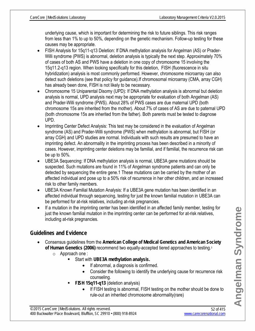
Ang
elm
an S
yndr
ome
underlying cause, which is important for determining the risk to future siblings. This risk ranges from less than 1% to up to 50%, depending on the genetic mechanism. Follow-up testing for these causes may be appropriate.
• FISH Analysis for 15q11-q13 Deletion: If DNA methylation analysis for Angelman (AS) or Prader-Willi syndrome (PWS) is abnormal, deletion analysis is typically the next step. Approximately 70% of cases of both AS and PWS have a deletion in one copy of chromosome 15 involving the 15q11.2-q13 region. When looking specifically for this deletion, FISH (fluorescence in situ hybridization) analysis is most commonly performed. However, chromosome microarray can also detect such deletions (see that policy for guidance).If chromosomal microarray (CMA, array CGH) has already been done, FISH is not likely to be necessary.
• Chromosome 15 Uniparental Disomy (UPD): If DNA methylation analysis is abnormal but deletion analysis is normal, UPD analysis next may be appropriate for evaluation of both Angelman (AS) and Prader-Willi syndrome (PWS). About 28% of PWS cases are due maternal UPD (both chromosome 15s are inherited from the mother). About 7% of cases of AS are due to paternal UPD (both chromosome 15s are inherited from the father). Both parents must be tested to diagnose UPD.
• Imprinting Center Defect Analysis: This test may be considered in the evaluation of Angelman syndrome (AS) and Prader-Willi syndrome (PWS) when methylation is abnormal, but FISH (or array CGH) and UPD studies are normal. Individuals with such results are presumed to have an imprinting defect. An abnormality in the imprinting process has been described in a minority of cases. However, imprinting center deletions may be familial, and if familial, the recurrence risk can be up to 50%.
• UBE3A Sequencing: If DNA methylation analysis is normal, UBE3A gene mutations should be suspected. Such mutations are found in 11% of Angelman syndrome patients and can only be detected by sequencing the entire gene.1 These mutations can be carried by the mother of an affected individual and pose up to a 50% risk of recurrence in her other children, and an increased risk to other family members.
• UBE3A Known Familial Mutation Analysis: If a UBE3A gene mutation has been identified in an affected individual through sequencing, testing for just the known familial mutation in UBE3A can be performed for at-risk relatives, including at-risk pregnancies.
• If a mutation in the imprinting center has been identified in an affected family member, testing for just the known familial mutation in the imprinting center can be performed for at-risk relatives, including at-risk pregnancies.
Guidelines and Evidence • Consensus guidelines from the American College of Medical Genetics and American Society
of Human Genetics (2006) recommend two equally-accepted tiered approaches to testing.3 o Approach one :
Start with UBE3A methylation analysis. • If abnormal, a diagnosis is confirmed. • Consider the following to identify the underlying cause for recurrence risk
counseling. FISH 15q11-q13 (deletion analysis)
• If FISH testing is abnormal, FISH testing on the mother should be done to rule-out an inherited chromosome abnormality(rare)
CareCore | MedSolutions Laboratory Laboratory Management Criteria V2.0.2015
© 2015 CareCore | MedSolutions. All rights reserved. 400 Buckwalter Place Boulevard, Bluffton, SC 29910 • (800) 918-8924 www.carecorenational.com
52 of 415
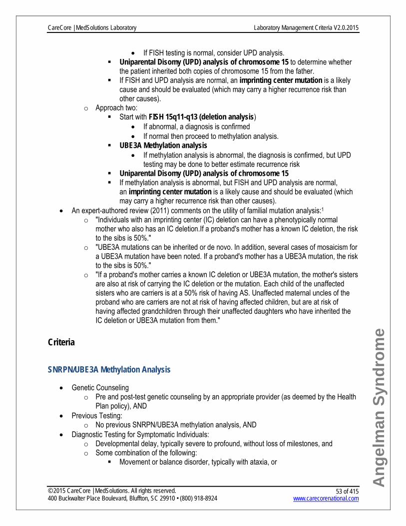
Ang
elm
an S
yndr
ome
• If FISH testing is normal, consider UPD analysis. Uniparental Disomy (UPD) analysis of chromosome 15 to determine whether
the patient inherited both copies of chromosome 15 from the father. If FISH and UPD analysis are normal, an imprinting center mutation is a likely
cause and should be evaluated (which may carry a higher recurrence risk than other causes).
o Approach two: Start with FISH 15q11-q13 (deletion analysis)
• If abnormal, a diagnosis is confirmed • If normal then proceed to methylation analysis.
UBE3A Methylation analysis • If methylation analysis is abnormal, the diagnosis is confirmed, but UPD
testing may be done to better estimate recurrence risk Uniparental Disomy (UPD) analysis of chromosome 15 If methylation analysis is abnormal, but FISH and UPD analysis are normal,
an imprinting center mutation is a likely cause and should be evaluated (which may carry a higher recurrence risk than other causes).
• An expert-authored review (2011) comments on the utility of familial mutation analysis:1 o "Individuals with an imprinting center (IC) deletion can have a phenotypically normal
mother who also has an IC deletion.If a proband's mother has a known IC deletion, the risk to the sibs is 50%."
o "UBE3A mutations can be inherited or de novo. In addition, several cases of mosaicism for a UBE3A mutation have been noted. If a proband's mother has a UBE3A mutation, the risk to the sibs is 50%."
o "If a proband's mother carries a known IC deletion or UBE3A mutation, the mother's sisters are also at risk of carrying the IC deletion or the mutation. Each child of the unaffected sisters who are carriers is at a 50% risk of having AS. Unaffected maternal uncles of the proband who are carriers are not at risk of having affected children, but are at risk of having affected grandchildren through their unaffected daughters who have inherited the IC deletion or UBE3A mutation from them."
Criteria SNRPN/UBE3A Methylation Analysis
• Genetic Counseling o Pre and post-test genetic counseling by an appropriate provider (as deemed by the Health
Plan policy), AND • Previous Testing:
o No previous SNRPN/UBE3A methylation analysis, AND • Diagnostic Testing for Symptomatic Individuals:
o Developmental delay, typically severe to profound, without loss of milestones, and o Some combination of the following:
Movement or balance disorder, typically with ataxia, or
CareCore | MedSolutions Laboratory Laboratory Management Criteria V2.0.2015
© 2015 CareCore | MedSolutions. All rights reserved. 400 Buckwalter Place Boulevard, Bluffton, SC 29910 • (800) 918-8924 www.carecorenational.com
53 of 415

Ang
elm
an S
yndr
ome
Frequent laughter/smiling, apparent happy demeanor; easily excitable personality (often with uplifted hand-flapping, or waving movements),or hypermotoric behavior, or
Speech impairment with no or minimal number of words, AND • Rendering laboratory is a qualified provider of service per the Health Plan policy.
FISH Analysis for 15q11-q13 Deletion
• Genetic Counseling o Pre and post-test counseling by a medical geneticist or genetic counselor, AND
• Previous Testing: o No previous chromosomal microarray o No previous 15q11-q13 deletion analysis, AND
• Diagnostic Testing for Symptomatic Individuals: o Developmental delay, typically severe to profound, without loss of milestones, and o Some combination of the following:
Movement or balance disorder, typically with ataxia, or Frequent laughter/smiling, apparent happy demeanor; easily excitable personality
(often with uplifted hand-flapping, or waving movements),or hypermotoric behavior, or
Speech impairment with no or minimal number of words, AND • Rendering laboratory is a qualified provider of service per the Health Plan policy.
Chromosome 15 Uniparental Disomy
• Genetic Counseling o Pre and post-test counseling by a medical geneticist or genetic counselor, AND
• Previous Testing: o SNRPN/UBE3A methylation analysis results are abnormal, and o 15q11-q13 deletion analysis is negative, and o No previous chromosome 15 UPD studies, AND
• Diagnostic Testing for Symptomatic Individuals: o Developmental delay, typically severe to profound, without loss of milestones, and o Some combination of the following:
Movement or balance disorder, typically with ataxia, or Frequent laughter/smiling, apparent happy demeanor; easily excitable personality
(often with uplifted hand-flapping, or waving movements),or hypermotoric behavior, or
Speech impairment with no or minimal number of words, AND • Rendering laboratory is a qualified provider of service per the Health Plan policy.
Imprinting Center Defect Analysis
• Genetic Counseling o Pre and post-test counseling by a medical geneticist or genetic counselor, AND
• Previous Testing:
CareCore | MedSolutions Laboratory Laboratory Management Criteria V2.0.2015
© 2015 CareCore | MedSolutions. All rights reserved. 400 Buckwalter Place Boulevard, Bluffton, SC 29910 • (800) 918-8924 www.carecorenational.com
54 of 415

Ang
elm
an S
yndr
ome
o SNRPN/UBE3A methylation analysis results are abnormal, and o 15q11-q13 deletion analysis is negative, and o Previous chromosome 15 UPD testing is negative, and o No previous imprinting center (IC) analysis, AND
• Diagnostic Testing for Symptomatic Individuals: o Developmental delay, typically severe to profound, without loss of milestones, and o Some combination of the following:
Movement or balance disorder, typically with ataxia, or Frequent laughter/smiling, apparent happy demeanor; easily excitable personality
(often with uplifted hand-flapping, or waving movements), or hypermotoric behavior, or
Speech impairment with no or minimal number of words, AND • Rendering laboratory is a qualified provider of service per the Health Plan policy.
UBE3A Sequencing
• Genetic Counseling o Pre and post-test counseling by a medical geneticist or genetic counselor, AND
• Previous Testing: o SNRPN/UBE3A methylation analysis results are normal, and o No previous sequencing of UBE3A, AND
• Personal History: o Developmental delay, typically severe to profound, without loss of milestones, and o Movement or balance disorder, typically with ataxia, and o Frequent laughter/smiling, apparent happy demeanor; easily excitable personality (often
with uplifted hand-flapping, or waving movements),or hypermotoric behavior, and o Speech impairment with no or minimal number of words, AND
• Rendering laboratory is a qualified provider of service per the Health Plan policy. UBE3A Known Familial Mutation Analysis or Imprinting Center Known Familial Mutation Analysis
• Genetic Counseling o Pre and post-test counseling by a medical geneticist or genetic counselor, AND_
• Previous Testing: o No previous UBE3A sequencing or imprinting center defect analysis testing, AND
• Family History: o Familial UBE3A or imprinting center defect mutation known in blood relative, AND
• Rendering laboratory is a qualified provider of service per the Health Plan policy.
References 1. Williams CA, Dagli AI, Driscoll DJ. (Updated June 2011). Angelman Syndrome. In: GeneReviews at GeneTests:
Medical Genetics Information Resource (database online). Available at: http://www.ncbi.nlm.nih.gov/bookshelf/br.fcgi?book=gene&part=angelman.
2. Jiang Y, Lev-Lehman E, Bressler J, Tsai TF, Beaudet AL. Genetics of Angelman syndrome. Am J Hum Genet. 1999;65:1-6.
CareCore | MedSolutions Laboratory Laboratory Management Criteria V2.0.2015
© 2015 CareCore | MedSolutions. All rights reserved. 400 Buckwalter Place Boulevard, Bluffton, SC 29910 • (800) 918-8924 www.carecorenational.com
55 of 415

Ang
elm
an S
yndr
ome
3. Cassidy S, Beaudet A, Knoll J, et al. Diagnostic Testing for Prader-Willi and Angelman Syndromes: Report of the American Society of Human Genetics/American College of Medical Genetics Test and Technology Transfer Committee. Available at: http://www.acmg.net/StaticContent/StaticPages/Prader_Willi.pdf.
CareCore | MedSolutions Laboratory Laboratory Management Criteria V2.0.2015
© 2015 CareCore | MedSolutions. All rights reserved. 400 Buckwalter Place Boulevard, Bluffton, SC 29910 • (800) 918-8924 www.carecorenational.com
56 of 415

APOE Variant Analysis for Alzheimer Disease
Procedure(s) covered by this policy: Procedure
Code(s)
Requires: Non-covered
APOE Genotyping 81401
Investigational and Experimental S3852
What Is Alzheimer Disease? • Alzheimer disease (AD) is characterized by adult-onset, progressive dementia with cerebral cortical
atrophy and beta amyloid plaque formation.1 Common findings include memory loss, confusion, speech issues, hallucinations, and personality and behavioral changes such as poor judgment, agitation, and withdrawal.2,3 Symptoms of Alzheimer disease usually start after 60-65 years old.2
• The general population lifetime risk of Alzheimer disease is about 10%. First-degree relatives (siblings, offspring) of a single person in the family with Alzheimer disease have a 20-25% lifetime risk.2
• Of all people with Alzheimer disease, about 25% have at least two affected people in the family (called "familial Alzheimer disease").2
o Most people (95%) with familial Alzheimer disease develop symptoms after 65.2 This is called "late-onset familial Alzheimer disease." Late-onset familial Alzheimer disease is believed to have complex inheritance with multiple susceptibility genes and environmental factors playing a role.2
o In about 5% of familial cases, symptoms consistently start before 65.2 This is called "early onset familial Alzheimer disease" (EOFAD). EOFAD is an autosomal dominant inherited disorder caused by different genes than those that may predispose to late-onset Alzheimer disease.1
• There are three common versions of the APOE gene — e2, e3, and e4. The e4 variant is significantly associated with Alzheimer disease.2
o People with Alzheimer disease, and especially late-onset familial Alzheimer disease, are more likely to have one or two copies of APOE e4. For example, less than 1% of unaffected people have two copies of e4 (e4/e4), but nearly 19% of people with familial Alzheimer disease have two copies of e4.2
o However, e4 is not necessary to develop Alzheimer disease and having no copies of e4 does not rule out the disease.2,4
o APOE e4 appears to cause susceptibility to Alzheimer disease, but the reason is unclear.2,5
Test Information • Clinical testing is available to determine which two APOE gene versions a person has inherited.
o One laboratory in the U.S. directly tests for these three variants (e2, e3, e4) to assist diagnosis or predict risk of Alzheimer disease.6 Because the assay uses a well-established methodology (RFLP), it is expected to accurately identify which variants are present but this data is not documented. Other laboratories do essentially the same test but limit the
CareCore | MedSolutions Laboratory Laboratory Management Criteria V2.0.2015
© 2015 CareCore | MedSolutions. All rights reserved. 400 Buckwalter Place Boulevard, Bluffton, SC 29910 • (800) 918-8924 www.carecorenational.com
57 of 415

APO
E- A
lzhe
imer
Dis
ease
interpretation to cardiovascular disease risk (which may also be associated with APOE variants but clinical utility is unclear).
o Other laboratories may infer the presence of APOE e4 based on SNP (single nucleotide polymorphisms) analysis. SNPs are normal DNA variations that occur throughout a person’s DNA. Certain SNPs, that do not directly assess APOE e4, occur more frequently in people with APOE e4. 7 Depending on the methodology used, these results may not be as reliable.
Guidelines and Evidence • Diagnostic: No current U.S. evidence-based guidelines have been identified. • A clinical diagnosis relies on symptoms and neuroimaging, which is correct about 80-90% of the
time. When two copies of e4 are identified in someone with a clinical diagnosis, the accuracy of that diagnosis increases to 97%.2 The diagnosis can only be confirmed by neuropathology at autopsy. Because finding APOE e4 neither confirms nor rules out Alzheimer disease on its own, its clinical utility is unclear.4
• European Federation of Neurological Societies (2010): "The ApoE e4 allele is the only genetic factor consistently implicated in late-onset AD, but it is neither necessary nor sufficient for development of the disease. Hence, there is no evidence to suggest ApoE testing is useful in a diagnostic setting."4
• Older, consensus-based guidelines include: o American College of Medical Genetics (ACMG, 1995): "Studies to date indicate that the
APOE genotype alone does not provide sufficient sensitivity or specificity to allow genotyping to be used as a diagnostic test. Because AD develops in the absence of APOE epsilon-4 and because many with APOE epsilon-4 seem to escape disease, genotyping is also not recommended for use as a predictive genetic test."5
o National Institute of Aging/Alzheimer's Association Working (1997):8 "Insofar as patients with AD are more likely to have an APOE-e4 allele than are
patients with other forms of dementia or individuals without dementia, physicians may choose to use APOE genotyping as an adjunct to other diagnostic tests for AD."
"Since genotyping cannot provide certainty about the presence or absence of AD, it should not be used as the sole diagnostic test."
• Predictive: APOE results have been used to predict a person’s lifetime risk for Alzheimer disease. For example, a person with two copies of APOE e4 has an estimated 30% lifetime risk to develop the disease.2 Predicted risks vary based on gender and number of e4 variants. While these risk estimates may be of personal interest, they do not currently have significant clinical utility.2,5
o National Institute of Aging/Alzheimer's Association Working (1997):8 "The use of APOE genotyping to predict future risk of AD in symptom-free
individuals is not recommended at this time."
Criteria • This test is considered investigational and/or experimental.
o Investigational and experimental (I&E) molecular and genomic (MolGen) tests refer to assays involving chromosomes, DNA, RNA, or gene products that have insufficient data to determine the net health impact, which typically means there is insufficient data to support that a test accurately assesses the outcome of interest (analytical and clinical validity),
CareCore | MedSolutions Laboratory Laboratory Management Criteria V2.0.2015
© 2015 CareCore | MedSolutions. All rights reserved. 400 Buckwalter Place Boulevard, Bluffton, SC 29910 • (800) 918-8924 www.carecorenational.com
58 of 415

APO
E- A
lzhe
imer
Dis
ease
significantly improves health outcomes (clinical utility), and/or performs better than an existing standard of care medical management option. Such tests are also not generally accepted as standard of care in the evaluation or management of a particular condition.
o In the case of MolGen testing, FDA clearance is not a reliable standard given the number of laboratory developed tests that currently fall outside of FDA oversight and FDA clearance often does not assess clinical utility.
References 1. Bird T. (Updated October 2012). Early-Onset Familial Alzheimer Disease. In GeneReviews at GeneTests: Medical
Genetics Information Resource (database online). Copyright, University of Washington, Seattle. Available at http://www.ncbi.nlm.nih.gov/bookshelf/br.fcgi?book=gene&part=alzheimer-early.
2. Bird T. (Updated April 2014). Alzheimer Disease Overview. In GeneReviews at GeneTests: Medical Genetics Information Resource (database online). Copyright, University of Washington, Seattle. Available at:http://www.ncbi.nlm.nih.gov/bookshelf/br.fcgi?book=gene&part=alzheimer.
3. Genetics Home Reference. Alzheimer disease. Published September 19, 2010. Available at http://ghr.nlm.nih.gov/condition/alzheimer-disease.
4. Hort J, O'Brien JT, Gainotti G, et al. EFNS guidelines for the diagnosis and management of Alzheimer's disease.Eur J Neurol. 2010 Oct;17(10):1236-48.
5. ACMG Consensus Statement on Use of Apolipoprotein E Testing for Alzheimer Disease. JAMA. 1995;274:1627-1629.
6. Athena Diagnostics. Test Catalog. Available at: http://www.athenadiagnostics.com/content/test-catalog/. 7. Nyholt DR, Yu CE, Visscher PM. On Jim Watson's APOE status: genetic information is hard to hide. Eur J Hum
Genet. 2009 Feb;17(2):147-9. Epub 2008 Oct 22. 8. National Institute on Aging-Alzheimer's Association Working Group; Apolipoprotein E genotyping in Alzheimer's
disease. National Institute on Aging/Alzheimer’s Association Working Group. Lancet. 1996;347:1091-5.
CareCore | MedSolutions Laboratory Laboratory Management Criteria V2.0.2015
© 2015 CareCore | MedSolutions. All rights reserved. 400 Buckwalter Place Boulevard, Bluffton, SC 29910 • (800) 918-8924 www.carecorenational.com
59 of 415

Ashkenazi Jewish Carrier Screening
Procedure(s) covered by this policy: Procedure
Code(s)
Requires: Prior-authorization* Lab Procedure
Restrictions†
Bloom Syndrome: BLM Targeted Mutation Analysis 81209 No No
Cystic Fibrosis: CFTR Targeted Mutation Analysis 81220 No No Canavan Disease: ASPA Targeted Mutation Analysis 81200 No No
Familial Dysautonomia: IKBKAP Targeted Mutation Analysis 81260 No No
Fanconi Anemia, Type C: FANCC Targeted Mutation Analysis 81242 No No
Gaucher Disease: GBA Targeted Mutation Analysis 81251 No No
Glycogen Storage Disease, Type 1a: G6PC Targeted Mutation Analysis 81250 No No
Maple Syrup Urine Disease Type 1A/B: BCKDHB Targeted Mutation Analysis 81205 No No
Mucolipidosis IV: MCOLN1 Targeted Mutation Analysis 81290 No No
Niemann-Pick Disease, Type A/B: SMPD1 Targeted Mutation Analysis 81330 No No
Tay-Sachs Disease: HEXA Targeted Mutation Analysis 81255 No No
Unlisted molecular pathology procedure (Used for testing various less common disorders) 81479 Yes No
* - Clinical Review necessary prior to authorization for this procedure. † - Lab procedures require specified sequence to be followed and additional information is required to be supplied by lab performing procedure(s).
What Is Ashkenazi Jewish Carrier Screening? • Ashkenazi Jewish carrier screening is available for certain genetic conditions that are more
common and/or have superior mutation detection rates in the Ashkenazi Jewish population.1-3 "Ashkenazi" refers to someone whose Jewish ancestors originally came from Central or Eastern Europe (e.g., Russia, Poland, Germany, Hungary, Lithuania, etc). Most Jewish people in the US are of Ashkenazi descent.
• Ashkenazi Jewish carrier screening is available for a large number of conditions (see the table below for most conditions). There are regional differences in the number and types of tests commonly offered. Patients and providers may choose all or a subset of these conditions.
• These Jewish genetic diseases are inherited in an autosomal recessive manner. An affected individual must inherit a gene mutation from both parents.1,2
o Individuals who inherit only one mutation are called carriers. Carriers do not show symptoms of the disease, but have a 50% chance of passing on the mutation to their children.
CareCore | MedSolutions Laboratory Laboratory Management Criteria V2.0.2015
© 2015 CareCore | MedSolutions. All rights reserved. 400 Buckwalter Place Boulevard, Bluffton, SC 29910 • (800) 918-8924 www.carecorenational.com
60 of 415

Ash
kena
zi J
ewis
h C
arrie
r Scr
eeni
ng
o Two carriers of the same disease have a 25% chance of having a child with the disorder. • While these genetic diseases are individually rare, the overall chance for an individual of Ashkenazi
Jewish descent to be a carrier for one of these genetic diseases is 1 in 4 to 1 in 5.2,3 An individual can also be a carrier of more than one condition.
• People from other ethnic backgrounds can be carriers of these conditions, but it is generally less common or the test is not as effective at identifying carrier status.
Test Information • Ashkenazi Jewish carrier screening can be offered to couples or individuals of Ashkenazi Jewish
descent when they are planning a pregnancy (preconceptional) or during a pregnancy (prenatal).1-3 If only one member of the couple is Jewish, carrier screening should start with the Jewish partner. Both parents must be carriers to have an affected child, so reproductive partners of known carriers should also be offered testing even if not Jewish.
• Carrier screening generally looks for a small number of gene mutations that are particularly common in the Ashkenazi Jewish population. In addition, enzyme analysis is particularly effective for Tay-Sachs disease and is generally preferred to mutation testing. The carrier detection rate is >95% in the Ashkenazi Jewish population for most diseases.3 See the table below for more details.
• The detection rate for these tests in the non-Ashkenazi population is unknown for most conditions, but generally low. Exceptions include cystic fibrosis and Tay-Sachs enzyme analysis, which each have good detection rates in non-Jewish populations.
• A negative test result in one or both partners significantly lowers the chance of an affected child, but does not eliminate it.2
Follow the disease name to see a complete discussion of carrier testing for that condition.
Ashkenazi Jewish Genetic Disease
Ashkenazi Carrier Frequency
What the Test Usually Looks For* (Mutation Names)
Chance the Test Will Correctly Find an Ashkenazi Jewish Carrier
Bloom syndrome3 1/107 1 mutation (2281del6ins7)
More than 99%
Canavan disease3 1/41 2 mutations (E285A, Y231X)
97.4%
Cystic fibrosis2 1/29 23 most common mutations in several ethnic groups
97%
Dihydrolipoamide dehydrogenase deficiency4
1/107 2 mutations (G229C and Y35X)
More than 95%
Familial dysautonomia3
1/31 2 mutations (2507+6TtoC, R696P)
More than 99%
Familial 1/68 2 mutations (c.3989- 90%
CareCore | MedSolutions Laboratory Laboratory Management Criteria V2.0.2015
© 2015 CareCore | MedSolutions. All rights reserved. 400 Buckwalter Place Boulevard, Bluffton, SC 29910 • (800) 918-8924 www.carecorenational.com
61 of 415

Ash
kena
zi J
ewis
h C
arrie
r Scr
eeni
ng
hyperinsulinism4 9G>A and Phel1387del)
Fanconi anemia group C3
1/89 1 mutation (IVS4+4AtoT)
More than 99%
Gaucher disease3 1/18 4 mutations (N370S, 84GG, L444P, IVS2+1GtoA)
Up to 94.6%
Glycogen storage disease type 1A (GSD1A)5
1/71 1 mutation (R83C) 93% to 100%
Joubert syndrome 26 1/92 1 mutation (R12L) 99% Maple syrup urine disease (MSUD)7
1/80 3 mutations (R183P, G278S, E372X)
About 99%
Mucolipidosis IV3 1/127 2 mutations (IVS3–2AtoG, Del6.4kb)
95%
Nemaline myopathy4 1/168 1 mutation (R2478_D2512del)
More than 95%
Niemann-Pick disease type A3
1/90 3 mutations (R496L, L302P, fsP330)
97%
Tay-Sachs disease3 1/90 Mutation analysis: 3 mutations (1278insTATC, 1421+1GtoC, G269S) OR
92-94%
Hexosaminidase A enzyme analysis
About 98%
Usher syndrome III4 1/120 1 mutation (N48K) More than 95%
Guidelines and Evidence • The American College of Obstetrics and Gynecology (ACOG, 2009)2 and the American
College of Medical Genetics (ACMG, 2008)3 recommend carrier screening for a group of disorders when at least one member of a couple is Ashkenazi Jewish and that couple is pregnant or planning pregnancy.
o Both organizations agree that testing should be offered for cystic fibrosis, Canavan disease, familialdysautonomia, and Tay-Sachs.
o ACMG also recommends routine testing for Fanconi anemia, Niemann-Pick, Bloom syndrome, mucolipidosisIV, and Gaucher disease;3 while ACOG states "individuals of Ashkenazi Jewish descent may inquire about the availability of carrier screening for other disorders" and educational materials may be provided to assist informed decision making about additional tests.2
• Carrier screening for common Ashkenazi Jewish mutations for maple syrup urine disease, glycogen storage disease 1A, dihydrolipoamide dehydrogenase deficiency, familial hyperinsulinemia, Joubert syndrome 2, nemaline myopathy, and Usher syndrome type III is now clinically available, but these tests are not specifically addressed in
CareCore | MedSolutions Laboratory Laboratory Management Criteria V2.0.2015
© 2015 CareCore | MedSolutions. All rights reserved. 400 Buckwalter Place Boulevard, Bluffton, SC 29910 • (800) 918-8924 www.carecorenational.com
62 of 415

Ash
kena
zi J
ewis
h C
arrie
r Scr
eeni
ng
current carrier screening guidelines. However, the 2008 ACMG guidelines outline the criteria for recommending additional carrier screening in the Ashkenazi Jewish population as new tests become available. These include:3
o The natural history must be well understood, o People affected with the disorder must have significant morbidity and mortality, and o The test must have greater than 90% detection OR the allele frequency must be at least
1%. • Dilipoamide dehydrogenase deficiency4, familial hyperinsulinism4, GSD1a5, Joubert syndrome
26, MSUD7,8, nemaline myopathy4, and Usher syndrome type III4 meet these criteria.
Criteria Testing may be considered for carrier screening for all or any desired subset of the Ashkenazi Jewish genetic diseases when BOTH of the following are met:
• The individual is planning a pregnancy or currently pregnant; AND • At least one partner of a couple is Ashkenazi Jewish
o NOTE: Detection rates for testing are higher in people with Ashkenazi Jewish ancestry. If only one partner of a couple is Ashkenazi Jewish, testing should start in that person when possible.
Testing may be considered for carrier screening of a single Ashkenazi Jewish disease, regardless of ethnicity or reproductive plans, if EITHER of the following are met:
• The individual has a family history of one of these conditions; OR • The individual’s partner is a known carrier or affected with any of these conditions
References 1. Monaghan KG, Feldman GL, Palomaki GE, Spector EB; Ashkenazi Jewish Reproductive Screening Working
Group; Molecular Subcommittee of the ACMG Laboratory Quality Assurance Committee. Technical standards and guidelines for reproductive screening in the Ashkenazi Jewish population. Genet Med. 2008;10(1):57-72.
2. ACOG Committee on Genetics. ACOG committee opinion. Number 442, Prenatal and preconceptional carrier screening for genetic diseases in individuals of Eastern European Jewish descent. Obstet Gynecol. 2009;114:950
3. Gross SJ, Pletcher BA, Monaghan KG; Professional Practice and Guidelines Committee. Carrier screening in individuals of Ashkenazi Jewish descent. Genet Med. 2008;10(1):54-56.
4. Scott SA, Edelmann L, Liu L, Luo M, Desnick RJ, Kornreich R. Experience with carrier screening and prenatal diagnosis for 16 Ashkenazi Jewish genetic diseases. Hum Mutat. 2010 Nov;31(11):1240-50.
5. Bali DS, Chen YT. (Updated September 2013). Glycogen Storage Disease 1a. In GeneReviews at GeneTests: Medical Genetics Information Resource (database online). Copyright, University of Washington, Seattle. 1997-2014. Available at http://www.ncbi.nlm.nih.gov/books/NBK1312.
6. Mt. Sinai Genetic Testing Laboratory website. Available at: http://icahn.mssm.edu/research/labs/genetic-testing-laboratory/testing-services/molecular-genetics-laboratory/joubert-syndrome.
7. Schrijver, I Kulm M, Gardner PI, Pergament, EP, Fiddler MB. Comprehensive arrayed primer extention array for the detection of 59 sequence variants in 15 conditions prevalent among the Ashkenazi Jewish population. J Mol Diagn. 2007 Apr;9(2):228-36.
8. Strauss KA, Puffenberger EG, Morton DH. Maple Syrup Urine Disease (Updated May 2013). In: GeneReviews at GeneTests: Medical Genetics Information Resource (database online). Copyright, University of Washington, Seattle. 1997-2014. Available at http://www.ncbi.nlm.nih.gov/books/NBK1319.
CareCore | MedSolutions Laboratory Laboratory Management Criteria V2.0.2015
© 2015 CareCore | MedSolutions. All rights reserved. 400 Buckwalter Place Boulevard, Bluffton, SC 29910 • (800) 918-8924 www.carecorenational.com
63 of 415

Ataxia-Telangiectasia Procedure(s) covered by this policy:
Procedure Code(s)
Requires: Prior-
authorization* Lab Procedure Restrictions†
ATM Known Familial Mutation Analysis 81403 No No ATM Sequencing 81408 Yes No ATM Deletion/Duplication Analysis 81479 Yes Yes * - Clinical Review necessary prior to authorization for this procedure. † - Lab procedures require specific sequence to be followed or additional information is required and must be supplied by the lab performing procedure(s) for full claim payment.
What is Ataxia- Telangiectasia? • Ataxia-telangiectasia (A-T) is a progressive neurological disorder caused by mutations in the ATM
gene. Onset is typically between the ages of 3 and 5 years. Signs and symptoms include:1 o Truncal and gait ataxia o Ocular apraxia o Slurred speech o Head tilting (after the age of 6 months) o Conjunctival telangiectasias o Immunodeficiencies o Malignancies (especially leukemias and lymphomas) o Radiation sensitivity
• Although individuals with A-T live to adulthood, they are at an increased risk for early death. Currently, most individuals live beyond 25 years, with some surviving into their 50s. Cause of death is associated with A-T associated cancers, infection, and pulmonary failure.1
• The prevalence of A-T is approximately 1 in 40,000 to 1 in 100,000 live US births. 1,2 It is the most common cause of childhood progressive cerebellar ataxia in most countries.3
• A-T is inherited in an autosomal recessive inheritance pattern. Males and females are equally likely to be affected. If both parents are carriers of A-T, the risk for a pregnancy to be affected is 1 in 4 (25%). Preimplantation and prenatal diagnosis are available for couples known to be at-risk.
• ATM has been implicated as a candidate gene for an increased risk for breast cancer, especially in women with a strong family history of breast cancer.4-7 Epidemiological data has also suggested an increased risk for cardiovascular disease in carriers as well.6,7 Therefore, carriers of ATM mutant alleles may need to be screened for breast cancer and cardiovascular disease.
Test Information • Sequence analysis of the ATM gene can identify ~90-95% of A-T mutations in affected
individuals.1 • Deletion/duplication analysis of the ATM gene can identify another 1-2% of mutations.1 • Testing for known ATM familial mutations: Once a deleterious mutation has been identified,
relatives of affected individuals can be tested. o Detection of at-risk individuals affects medical management in the case of breast cancer
screening and cardiovascular disease screening.
CareCore | MedSolutions Laboratory Laboratory Management Criteria V2.0.2015
© 2015 CareCore | MedSolutions. All rights reserved. 400 Buckwalter Place Boulevard, Bluffton, SC 29910 • (800) 918-8924 www.carecorenational.com
64 of 415

Ata
xia-
Tela
ngie
ctas
ia
o Prenatal testing is available to individuals with a known family mutation. Genetic testing can be performed on amniocytes obtained through amniocentesis or chorionic villi obtained through a chorionic villus sampling.
Guidelines and Evidence • The Eighth International Workshop on Ataxia-Telangiectasia was convened in 1999. The workshop
described ATM mutations and cancer risk in heterozygotes, and potential therapeutic approaches. Genetic testing strategies were not described.8
• Genetic testing is approved to confirm a diagnosis in anyone who meets clinical criteria for A-T. Individuals meeting clinical criteria for A-T testing will undergo sequence analysis. Deletion/duplication testing is offered to those meeting the criteria and have tested negative through sequence analysis. Additionally, genetic testing is approved to determine the carrier status in an at risk relative with a known family mutation. Individuals with a family member with a known A-T mutation(s) should be tested for that/those mutation(s).
Criteria Known ATM Family Mutation Testing
• Genetic Counseling: o Pre and post-test counseling by a medical geneticist, genetic counselor or neurologist,
AND • Previous Genetic Testing:
o No previous genetic testing of ATM, AND • Carrier Screening Individuals:
o Known family mutation in ATM in 1st, 2nd, or 3rd degree biologic relative(s), OR • Prenatal Testing for At-Risk Pregnancies:
o ATM mutations identified in both biologic parents. ATM Full Sequence Analysis
• Clinical Consultation & Genetic Counseling: o Examination by a geneticist, oncologist, or neurologist family with hereditary ataxias, and o Pre and post-test counseling by a medical geneticist or genetic counselor, AND
• Previous Genetic Testing: o No previous ATM gene sequencing, and o No known ATM mutation in family, AND
• Diagnostic Testing for Symptomatic Individuals: o Elevated Alpha-fetoprotein (AFP) levels, or o Decreased ATM protein detected by immunoblotting, and o Progressive cerebellar ataxia, or o Truncal and gait ataxia, or o Oculomotor apraxia, OR
CareCore | MedSolutions Laboratory Laboratory Management Criteria V2.0.2015
© 2015 CareCore | MedSolutions. All rights reserved. 400 Buckwalter Place Boulevard, Bluffton, SC 29910 • (800) 918-8924 www.carecorenational.com
65 of 415

Ata
xia-
Tela
ngie
ctas
ia
• Diagnostic Testing for Carriers: o One mutation detected by targeted mutation analysis, and o Elevated Alpha-fetoprotein (AFP) levels, or o Decreased ATM protein detected by immunoblotting, OR
• Testing for Individuals with Family History or Partners of Carriers: o 1st, 2nd, or 3rd, degree relative diagnosed with Ataxia-Telangiectasia clinical diagnosis,
family mutation unknown, and testing unavailable, or o Partner is monoallelic or biallelic for ATM mutation, and o Has living children with this partner, or o Has the potential and intention to reproduce
ATM Duplication/Deletion Analysis†
• Clinical Consultation & Genetic Counseling: o Examination by a geneticist, oncologist, or neurologist family with hereditary ataxias, and o Pre and post-test counseling by a medical geneticist or genetic counselor, AND
• Previous Genetic Testing: o No previous deletion/duplication analysis of ATM, and o No mutations detected in full sequencing, or o Heterozygous for mutation and elevated alpha-fetoprotein levels or decreased ATM protein
detected by immunoblotting †Lab Testing Restrictions: Testing is authorized after no mutations detected with full sequence analysis.
References 1. Gatti R. (Updated 2010) Ataxia-Telangiectasia. In: GeneReviews: Medical Genetics Information Resource
(database online). Copyright, University of Washington, Seattle. 1997-2014. Available at: www.ncbi.nlm.nih.gov/books/NBK26468.
2. National Organization for Rare Disorders. Ataxia Telangiectasia. Available at: http://www.rarediseases.org/rare-disease-information/rare-diseases/byID/406/printFullReport
3. Perlman S, Becker-Catania S, Gatti RA. Ataxia-telangiectasia: diagnosis and treatment. Semin Pediatr Neurol. 2003;10:173-82.
4. Ahmed M, Rahman N. ATM and breast cancer susceptibility. Oncogene. 2006;25:5906–11. 5. Ripperger T. Breast cancer susceptibility: Current knowledge and implications for genetic counselling. Euro J Hum
Genet. 2009;17:722-31. 6. Swift M et al. Malignant neoplasms in the families of patients with ataxia-telangiectasia. Cancer Res. 1976;36:209. 7. Swift, Morrell, Massey et al. Incidence of cancer in 161 families affected by ataxia-telangiectasia. NEJM
1991;325:831-836. 8. Lavin MF, Concannon P, Gatti RA. Eighth International Workshop on Ataxia-Telangiectasia (ATW8). Cancer Res.
1999;59:3845-9.
CareCore | MedSolutions Laboratory Laboratory Management Criteria V2.0.2015
© 2015 CareCore | MedSolutions. All rights reserved. 400 Buckwalter Place Boulevard, Bluffton, SC 29910 • (800) 918-8924 www.carecorenational.com
66 of 415

BCR-ABL Testing for Chronic Myeloid Leukemia
Procedure(s) covered by this policy: Procedure
Code(s)
Requires:
Prior-authorization* Lab Procedure Restrictions†
BCR-ABL1 Detection, Major Breakpoint 81206 No No BCR-ABL1 Detection, Minor Breakpoint 81207 No No BCR-ABL1 Detection, Other Breakpoint 81208 No No FISH Analysis for t(9;22) BCR-ABL 88271 No No
* - Clinical Review necessary prior to authorization for this procedure. † - Lab procedures require specified sequence to be followed and additional information is required to be supplied by lab performing procedure(s).
What Are CML and BCR-ABL? • Chronic myelogenous leukemia (CML) is a hematopoietic stem cell disease that results in
overgrowth of white blood cells in the bone marrow. It is defined by the presence of the Philadelphia chromosome (Ph), a translocation between chromosomes 9 and 22 that results in the fusion of two genes known as BCR and ABL.1,2
• Acute lymphoblastic leukemia (ALL) is a different form of leukemia, but may also be positive for the Philadelphia chromosome (Ph+). About 3% of pediatric ALL and 25% of adult ALL is Ph+.3
• Detection of the BCR-ABL fusion gene is diagnostic for CML and Ph+ ALL and can be established by fluorescent in situ hybridization (FISH) or quantitative real-time polymerase chain reaction (QPCR).2
• The three phases of CML are chronic, accelerated and blastic. In the chronic phase, there are few symptoms and most people are diagnosed after a routine blood test reveals the characteristic blood count and differential. If not treated, the disease will progress to the accelerated and blastic phases, symptoms of which include fever, bone pain, splenomegaly, fatigue and weakness.1
• First line treatment for CML and some Ph+ ALL is with a class of drugs called tyrosine kinase inhibitors (TKIs), which block the production of the BCR-ABL fusion gene protein product. Three TKI therapies are available as first-line therapies: imatinib(Gleevec®), nilotinib (Tasigna®), and dasatinib (Sprycel®). These TKI therapies have all demonstrated proven benefit, and median survival is expected to approach normal life expectancy for most patients with CML.12
• Monitoring of patients for treatment response to TKIs includes routine measurement of the BCR-ABL fusion gene protein product via QPCR prior to initiation of treatment and during treatment every 3 months.2
• For individuals who display apparent treatment resistance, consideration of alternative treatment options may be appropriate.2 Treatment resistance in both CML and ALL can be caused by mutations in the BCR-ABL kinase domain.2,3
Test Information • qPCR for BCR-ABL transcript levels: Bone marrow cytogenetics and measurement of BCR-ABL
transcript levels by quantitative polymerase chain reaction is recommended before initiation of treatment as well as for assessing response to therapy.
CareCore | MedSolutions Laboratory Laboratory Management Criteria V2.0.2015
© 2015 CareCore | MedSolutions. All rights reserved. 400 Buckwalter Place Boulevard, Bluffton, SC 29910 • (800) 918-8924 www.carecorenational.com
67 of 415

BC
R-A
BL
Tran
scrip
t Lev
el T
estin
g
• FISH for t(9;22) BCR-ABL: If collection of bone marrow is not feasible, fluorescence in situ hybridization (FISH) on peripheral blood specimen using dual probes for the BCR and ABL genes is an acceptable method of confirming the diagnosis of CML.4
Guidelines and Evidence • The National Comprehensive Cancer Network (NCCN, 2014) recommends bone marrow
cytogenetics to confirm a diagnosis of CML. If bone marrow is not available, FISH on a peripheral blood specimen using probes for both BCR and ABL can confirm the diagnosis.2
o NCCN recommends BCR-ABL transcript levels be obtained by quantitative RT-PCR: At diagnosis Every three months when a patient is responding to TKI therapy After a patient
reaches complete cytogenetic response, every 3 months for three years and every 3-6 months thereafter
If a patient has a rising level of BCR-ABL transcripts (1 log increase). Evaluate patient compliance and repeat testing in 1 – 3 months and/or perform ABL kinase mutation analysis.
These recommendations are category 2A: "based upon lower-level evidence, there is uniform NCCN consensus that the intervention is appropriate."
Criteria BCR-ABL transcript level testing is indicated in individuals at the initiation of treatment and at regular intervals (ranges from every month to once every 3-6 months) during treatment with ANY of the following drug therapies:
• Imatinib (Gleevec®) • Nilotinib (Tasigna®) • Dasatinib (Sprycel®)
References 1. National Cancer Institute. Chronic Myelogenous Leukemia Treatment (PDQ®). Last modified May 23, 2014. At:
http://www.cancer.gov/cancertopics/pdq/treatment/CML/HealthProfessional/page1. 2. National Comprehensive Cancer Network. NCCN Clinical Practice Guidelines in Oncology: Chronic Myelogenous
Leukemia. V.3.2014: http://www.nccn.org/professionals/physician_gls/PDF/cml.pdf. 3. National Comprehensive Cancer Network. NCCN Clinical Practice Guidelines in Oncology: Acute Lymphoblastic
Leukemia. V.1.2014: http://www.nccn.org/professionals/physician_gls/pdf/all.pdf. 4. Association for Molecular Pathology, Clinical Practice Committee. Laboratory practice guidelines for detecting and
reporting BCR-ABL drug resistance mutations in chronic myelogenous leukemia and acute lymphoblastic leukemia. J Mol Diagn. 2009;11(1):4-11.
CareCore | MedSolutions Laboratory Laboratory Management Criteria V2.0.2015
© 2015 CareCore | MedSolutions. All rights reserved. 400 Buckwalter Place Boulevard, Bluffton, SC 29910 • (800) 918-8924 www.carecorenational.com
68 of 415

Bloom Syndrome Testing
Procedure(s) covered by this policy: Procedure
Code(s)
Requires:
Prior-authorization* Lab Procedure Restrictions†
Sister Chromatid Exchange 88245 No No BLM Known Familial Mutation Analysis 81403 No No BLM Targeted Mutation Analysis 81209 No No
BLM Sequencing 81479 Yes Yes BLM Deletion/Duplication Analysis 81479 Yes Yes
* - Clinical Review necessary prior to authorization for this procedure. † - Lab procedures require specific sequence to be followed or additional information is required and must be supplied by the lab performing procedure(s) for full claim payment.
What Is Bloom Syndrome? • Bloom syndrome is a genetic disorder in which an individual’s chromosomes contain large breaks,
gaps, or rearrangements.1 • Affected individuals are usually smaller than average and suffer from a variety of symptoms:1-2
o Intrauterine growth retardation that persists into childhood. o Long, narrow face, small lower jaw, and prominent nose and ears o Sensitivity to sunlight. Exposure to sunlight causes a characteristic butterfly-shaped rash
on the face. o Chronic lung problems, diabetes, and immune deficiencies. o Cancer diagnosis at an early age. o Learning disabilities.
• Fewer than 300 cases of Bloom syndrome have been reported since the disease was first described over 50 years ago.2,3
o About 1 in 48,000 Ashkenazi Jews have Bloom syndrome, and 25% of all affected individuals have Ashkenazi Jewish ancestry.2
• There is no cure for Bloom syndrome. Treatment involves continuous monitoring by multiple physicians and specialists.1,3
• Bloom syndrome is caused by a genetic mutation in the BLM gene.1,3,4 o BLM is essential to maintaining the stability of chromosomes during DNA replication and
cell division.3,4 o Mutations in BLM lead to mistakes during cellular replication.3,4 o Individuals with Bloom syndrome have multiple breaks, gaps, and genetic rearrangements
in their chromosomes, leading to a unique combination of signs and symptoms.3,4 • A diagnosis of Bloom syndrome is suspected when the patient presents with the characteristic
suite of signs and symptoms. This diagnosis can be confirmed by genetic testing and is needed to differentiate between other disorders with overlapping symptoms. There are several types of tests available for diagnostic purposes.
• Bloom syndrome is an autosomal recessive disorder, meaning that an affected individual must inherit BLM gene mutations from each parent.1,4
CareCore | MedSolutions Laboratory Laboratory Management Criteria V2.0.2015
© 2015 CareCore | MedSolutions. All rights reserved. 400 Buckwalter Place Boulevard, Bluffton, SC 29910 • (800) 918-8924 www.carecorenational.com
69 of 415

Blo
om S
yndr
ome
o Individuals who inherit only one mutation are called carriers. Carriers do not show symptoms of Bloom syndrome, but have a 50% chance of passing on the mutation to their children.
o Two carriers of Bloom syndrome have a 1 in 4 (25%) chance for each pregnancy to be affected with Bloom syndrome.
Test Information • Sister Chromatid Exchange: SCE is the standard analysis for diagnosis of Bloom syndrome. The
method involves exposing an individual’s cells to bromodeoxyuridine (BrdU), a compound that helps identify which cells contain chromosomes with unusually large numbers of rearrangements, or ‘exchanges.’ Individuals with Bloom syndrome will have a substantially higher number of these exchanges compared with unaffected individuals.5 This test can be used for prenatal diagnosis of at-risk pregnancies on chorionic villi or amniocytes.4
• BLM Known Familial Mutation Analysis: Once a deleterious mutation has been identified in an affected person, relatives and at-risk pregnancies can be tested.
• BLM Targeted Mutation Analysis: This test looks for the BLM gene mutation most often found in Ashkenazi Jewish patients, called BLMAsh.4 The detection rate of this mutation in Ashkenazi Jewish individuals is >95%.4 BLM Sequencing: This test looks for mutations across the entire gene, and can identify at least 90% of disease-causing mutations.4 It is typically used only for diagnosis of an affected individual or carrier testing of a non-Ashkenazi Jewish individual when the partner is a known carrier.
• BLM Deletion/Duplication Analysis: This test looks for deletions and duplication in the gene that would not be detected by sequencing analysis.4 It is typically performed in reflex to sequencing analysis when there is a high suspicion for disease.4
Guidelines and Evidence • No evidence-based US guidelines have been identified for diagnostic testing. • A 2013 expert-authored review suggests the following diagnostic testing strategy:4
o "To confirm/establish the diagnosis Bloom’s syndrome: Cytogenetic demonstration of a characteristically greatly increased SCE
frequency, OR Molecular demonstration either of homozygosity for a Bloom syndrome-causing
mutation in BLM or of compound heterozygosity for two different Bloom syndrome-causing mutations. Sequence analysis should be performed first. If neither or only one mutation in BLM is identified, deletion/duplication analysis should be considered."
• The American College of Medical Genetics (ACMG, 2008)6 and the American College of Obstetrics and Gynecologists (ACOG, 2009)7 support offering carrier testing for Bloom syndrome to individuals of Ashkenazi Jewish descent for the common blmAsh mutation.
o Guidelines support the testing of individuals of Ashkenazi Jewish descent, even when their partner is non-Ashkenazi Jewish. In this situation, testing would start with the individual who is Jewish and if blmAsh mutation is detected, sequencing of BLM in the non-Ashkenazi Jewish partner would follow.6 If the woman is pregnant, testing may need to be conducted on both partners simultaneously in order to receive results in a timely fashion.7
CareCore | MedSolutions Laboratory Laboratory Management Criteria V2.0.2015
© 2015 CareCore | MedSolutions. All rights reserved. 400 Buckwalter Place Boulevard, Bluffton, SC 29910 • (800) 918-8924 www.carecorenational.com
70 of 415

Blo
om S
yndr
ome
o If one or both partners are found to be carriers of Bloom syndrome, genetic counseling should be provided and prenatal testing offered, if appropriate.
• A 2013 expert-authored review states:4 o "Carrier testing for at-risk relatives requires prior identification of the BLM disease-causing
mutations in the family." • A 2013 expert-authored review states:4
o "Prenatal diagnosis of at-risk pregnancies is possible by cytogenetic analysis, specifically by an SCE analysis."
o "Prenatal diagnosis by molecular genetic testing and preimplantation genetic diagnosis (PGD) for at-risk pregnancies require prior identification of the BLM disease-causing mutations in the family."
Criteria Sister Chromatid Exchange (Chromosome Analysis for Breakage Syndromes)
• Genetic Counseling: o Pre and post-test counseling by an appropriate provider (as deemed by the Health Plan
policy), AND • Previous Genetic Testing:
o No previous sister chromatid exchange analysis performed, and o No previous BLM full sequencing, or BLM sequencing performed and only one mutation
identified, and o No known BLM mutation in biologic relative, and o If Ashkenazi Jewish, targeted mutation analysis performed and no mutation detected or
one mutation detected, AND • Diagnostic Testing for Symptomatic Individuals:
o Unexplained severe intrauterine growth retardation that persists throughout infancy and childhood (< 5th percentile), or
o An unusually small individual (<5th percentile) who develops erythematous skin lesions in the ”butterfly area” of the face after sun exposure, or
o An unusually small individual (<5th percentile) who develops a malignancy OR • Prenatal Testing for At-Risk Pregnancies:
o Known increased risk due to affected first-degree relative , AND • Rendering laboratory is a qualified provider of service per the Health Plan policy.
BLM Known Familial Mutation Analysis
• Genetic Counseling: o Pre and post-test counseling by an appropriate provider (as deemed by the Health Plan
policy), AND • Previous Genetic Testing:
o No previous genetic testing of BLM, AND • Carrier Screening:
o Known family mutation in BLM identified in 1st, 2nd, or 3rd degree biologic relative(s), OR • Prenatal Testing for At-Risk Pregnancies:
o BLM mutation identified in both biologic parents, AND • Rendering laboratory is a qualified provider of service per the Health Plan policy.
CareCore | MedSolutions Laboratory Laboratory Management Criteria V2.0.2015
© 2015 CareCore | MedSolutions. All rights reserved. 400 Buckwalter Place Boulevard, Bluffton, SC 29910 • (800) 918-8924 www.carecorenational.com
71 of 415

Blo
om S
yndr
ome
BLM Targeted Mutation Analysis†
• Genetic Counseling: o Pre and post-test counseling by an appropriate provider (as deemed by the Health Plan
policy), AND • Previous Genetic Testing:
o No previous BLM genetic testing, including AJ screening panels containing targeted mutation analysis for blmAsh, AND
• Carrier Screening: o Ashkenazi Jewish descent, and o Have the potential and intention to reproduce, AND
• Rendering laboratory is a qualified provider of service per the Health Plan policy. BLM Sequencing
• Genetic Counseling: o Pre and post-test counseling by an appropriate provider (as deemed by the Health Plan
policy), AND • Previous Genetic Testing:
o No previous BLM full sequencing, and o No known BLM mutation in biologic relative, and o If Ashkenazi Jewish, targeted mutation analysis performed and no mutation detected or
one mutation detected, AND • Diagnostic Testing for Symptomatic Individuals:
o Unexplained severe intrauterine growth retardation that persists throughout infancy and childhood (< 5th percentile), or
o An unusually small individual (<5th percentile) who develops erythematous skin lesions in the ”butterfly area” of the face after sun exposure, or
o An unusually small individual (<5th percentile) who develops a malignancy, OR • Testing for Individuals with Family History or Partners of Carriers:
o 1st, 2nd, or 3rd degree biologic relative with Bloom syndrome clinical diagnosis, family mutation unknown, and testing unavailable, or
o Partner is monoallelic or biallelic for BLM mutation, and o Have the potential and intention to reproduce, AND
• Rendering laboratory is a qualified provider of service per the Health Plan policy. BLM Deletion/Duplication Analysis
• Genetic Counseling: o Pre and post-test counseling by an appropriate provider (as deemed by the Health Plan
policy), AND • Previous Genetic Testing:
o Previous BLM full sequencing, and no mutations or only one mutation detected, AND • Diagnostic Testing for Symptomatic Individuals:
o Unexplained severe intrauterine growth retardation that persists throughout infancy and childhood (< 5th percentile), or
o An unusually small individual (<5th percentile) who develops erythematous skin lesions in the ”butterfly area” of the face after sun exposure, or
CareCore | MedSolutions Laboratory Laboratory Management Criteria V2.0.2015
© 2015 CareCore | MedSolutions. All rights reserved. 400 Buckwalter Place Boulevard, Bluffton, SC 29910 • (800) 918-8924 www.carecorenational.com
72 of 415

Blo
om S
yndr
ome
o An unusually small individual (<5th percentile) who develops a malignancy, OR • Testing for Individuals with Family History or Partners of Carriers:
o 1st, 2nd, or 3rd degree biologic relative with Bloom syndrome clinical diagnosis, family mutation unknown, and testing unavailable, or
o Partner is monoallelic or biallelic for BLM mutation, and o Have the potential and intention to reproduce, AND
Rendering laboratory is a qualified provider of service per the Health Plan policy.
References 1. Bloom Syndrome. (Updated 2010). In: Genetics Home References: Your Guide to Understanding Genetic
Conditions (database online). A Service of the US National Library of Medicine. Available at http://ghr.nlm.nih.gov/condition/bloom-syndrome.
2. Bloom D. Congenital telangiectatic erythema resembling lupus erythematosus in dwarfs; probably a syndrome entity. AMA Am J Dis Child. 1954 Dec;88(6):754-8.
3. Hickson ID, Davies SL, Li JL, et al. Role of the Bloom's syndrome helicase in maintenance of genome stability.Biochem Soc Trans. 2001 May;29(Pt 2):201-4.
4. Sanz MM, German J. (Updated March 2013). Bloom’s Syndrome. In: GeneReviews at GeneTests: Medical Genetics Information Resource (database online). Copyright, University of Washington, Seattle. 1997-2014. Available at: http:// www.ncbi.nlm.nih.gov/bookshelf/br.fcgi?book=gene&part=bloom.
5. Howell RT, Davies T. Diagnosis of Bloom's syndrome by sister chromatid exchange evaluation in chorionic villus cultures. Prenat Diagn. 1994 Nov;14(11):1071-3.
6. ACOG Committee Opinion No. 442: Preconception and Prenatal Carrier Screening for Genetic Diseases in Individuals of Eastern European Jewish Descent. Obstet Gynecol. 2009;114(4):950 910.1097/AOG.1090b1013e3181bd1012f1094.
7. Gross SJ, Pletcher BA, Monaghan KG. Carrier screening in individuals of Ashkenazi Jewish descent. Genet Med. Jan 2008;10(1):54-6.
CareCore | MedSolutions Laboratory Laboratory Management Criteria V2.0.2015
© 2015 CareCore | MedSolutions. All rights reserved. 400 Buckwalter Place Boulevard, Bluffton, SC 29910 • (800) 918-8924 www.carecorenational.com
73 of 415

BRAF Testing for Colorectal Cancer Anti-EGFR Response
Procedure(s) covered by this policy: Procedure
Code(s)
Requires:
Non-covered
BRAF V600E Targeted Mutation Analysis 81210 Investigational and Experimental
BRAF Sequencing 81406 Investigational and Experimental
What Is BRAF? • BRAF is part of a cell signaling pathway that helps control cell growth. Changes, or mutations, in
the BRAF gene can cause out of control cell growth, which may lead to cancer.1 The most common BRAF mutation is called V600E (previously known as V599E).
• About 5-9% of colorectal cancer tumors have a V600E BRAF mutation.1 • Patients with a V600E BRAF mutation appear to have a poorer prognosis. Tumors with BRAF
mutations may have less response to anti-EGFR therapies like cetuximab (Erbitux®) and panitumumab (Vectibix®).1
Test Information • Targeted mutation analysis: Laboratories most commonly test for the BRAF V600E mutation,
which accounts for about 90% of activating BRAF mutations.4 Mutation analysis requires relatively little tumor material for testing and has high sensitivity. It is also relatively inexpensive.2,3 BRAF mutation analysis is done on fresh, frozen, or paraffin-embedded tissue from either a primary tumor or metastasis.1-3 Some molecular diagnostic laboratories perform BRAF mutation analysis by laboratory-developed methods, while others use FDA-approved test kits. Laboratory-developed tests may vary in the specimen type required, methodology used, mutations tested, sensitivity, and other test-specific data.
• Sequencing: Some laboratories sequence all or part of the BRAF gene, which will find a broader spectrum of mutations than targeted mutation analysis. Laboratories that offer sequencing generally do so for a subset of exons where most BRAF activating mutations have been identified. Sequence analysis requires more and higher quality tumor material for testing than targeted mutation analysis. This method is typically less efficient and more expensive than targeted mutation analysis.2,3,5
• Note that BRAF mutation analysis has several other test applications with different criteria (such as melanoma therapeutic response, Lynch syndrome tumor screening, and Noonan syndrome diagnosis). Ensure you are reviewing the correct use of the test.
CareCore | MedSolutions Laboratory Laboratory Management Criteria V2.0.2015
© 2015 CareCore | MedSolutions. All rights reserved. 400 Buckwalter Place Boulevard, Bluffton, SC 29910 • (800) 918-8924 www.carecorenational.com
74 of 415

BR
AF-
Col
orec
tal C
ance
r
Guidelines and Evidence National Comprehensive Cancer Network (NCCN, 2015) guidelines state:1
• "Patients with a V600E BRAF mutation appear to have a poorer prognosis. Limited available data suggest a lack of antitumor activity from anti-EGFR monoclonal antibodies in the presence of a V600E mutation when used after a patient has progressed on first-line therapy. "
• "Although BRAF genotyping can be considered for patients with tumors characterized by the wild-type KRAS/NRAS, this testing is currently optional and not a necessary part of decision-making regarding use of anti-EGFR agents."
Criteria BRAF mutation testing is considered investigational and experimental due to NCCN guidelines stating that this testing is optional and not of clear clinical benefit in decision-making regarding the use of anti-EGFR agents.
References 1. NCCN Updates Guidelines for Colorectal Cancer. NCCN Clinical Practice Guidelines in Oncology. v.1.2015.
Available at: http://www.nccn.org/professionals/physician_gls/PDF/colon.pdf. 2. Jakubauskas A, Griskevicius L. KRAS and BRAF mutational status analysis from formalin-fixed, paraffin-
embedded tissues using multiplex polymerase chain reaction-based assay. Arch Pathol Lab Med. 2010; 134:620-4.
3. Packham D, Ward RL, Lin VA, et al. Implementation of novel pyrosequencing assays to screen for common mutations of BRAF and KRAS in a cohort of sporadic colorectal cancers. Diagn Mol Pathol. 2009; 18(2):62-71.
4. Pichler M, Balic M, Stadelmeyer E, et al. Evaluation of high-resolution melting analysis as a diagnostic tool to detect the BRAF V600E mutation in colorectal tumors. JMD. 2009; 11(2):140-7.
5. Fakih M. KRAS mutation screening in colorectal cancer: From paper to practice. Clin Colorectal Cancer. 2010;9(1):22-30.
CareCore | MedSolutions Laboratory Laboratory Management Criteria V2.0.2015
© 2015 CareCore | MedSolutions. All rights reserved. 400 Buckwalter Place Boulevard, Bluffton, SC 29910 • (800) 918-8924 www.carecorenational.com
75 of 415

BRAF V600E Testing for Melanoma Kinase Inhibitor Response
Procedure(s) covered by this policy: Procedure Code(s)
Requires:
Prior-authorization* Lab Procedure Restrictions†
BRAF V600E Targeted Mutation Analysis 81210 Yes No * - Clinical Review necessary prior to authorization for this procedure. † - Lab procedures require specified sequence to be followed and additional information is required to be supplied by lab performing procedure(s).
What Is BRAF Mutation Analysis? • BRAF is part of a cell signaling pathway that helps control cell growth. Mutations in the BRAF gene
can cause out of control cell growth, which may lead to cancer.1,2 The most common BRAF mutation is called V600E (previously known as V599E), which accounts for about 70-90% of mutations in this gene.1,3
• About 40-60% of cutaneous melanomas have a V600E BRAF mutation.1 • Vemurafenib (Zelboraf®), dabrafenib (Tafinlar®), and trametinib (Mekinist®) are orally-administered
kinase inhibitors that is able to block the function of the mutated BRAF protein.1,2 They are specifically indicated for the treatment of patients with metastatic or unresectable melanoma whose tumors have a BRAF V600E mutation. 1,2 They are not recommended for use in patients with wild type BRAF melanoma.2,4-6
Test Information • Targeted mutation analysis: Laboratories most commonly test for the BRAF V600E mutation,
which accounts for about 90% of activating BRAF mutations.4 Mutation analysis requires relatively little tumor material for testing and has high sensitivity. It is also relatively inexpensive.2,3 BRAF mutation analysis is done on fresh, frozen, or paraffin-embedded tissue from either a primary tumor or metastasis.1-3 Some molecular diagnostic laboratories perform BRAF mutation analysis by laboratory-developed methods, while others use FDA-approved test kits. Laboratory-developed tests may vary in the specimen type required, methodology used, mutations tested, sensitivity, and other test-specific data.
• Vemurafenib was approved in 2011 for use along with an FDA approved companion diagnostic developed by Roche molecular diagnostics called the cobas® 4800 BRAF V600 Mutation Test. The cobas 4800 BRAF V600 mutation test was clinically validated in the trials conducted for approval of vemurafenib. This testing specifically checks for the V600E mutation in formalin-fixed, paraffin-embedded melanoma tumor tissue.2
• In 2013, dabrafenib and trametinib were approved for use along with an FDA approved companion diagnostic developed by Roche molecular diagnostics called the THxID BRAF test. The THxID BRAF test was clinically validated in the clinical studies supporting the approval of dabrafenib and trametinib.
CareCore | MedSolutions Laboratory Laboratory Management Criteria V2.0.2015
© 2015 CareCore | MedSolutions. All rights reserved. 400 Buckwalter Place Boulevard, Bluffton, SC 29910 • (800) 918-8924 www.carecorenational.com
76 of 415

BR
AF
V600
E
Guidelines and Evidence • The National Comprehensive Cancer Network (NCCN, 2014) includes vemurafenib, dabrafenib
and trametinib as options for treatment of advanced or metastatic melanoma which require companion diagnostic testing. The guidelines state “Vemurafenib, dabrafenib, and trametinib are recommended only for patients with a V600 mutation of the BRAF gene documented by an FDA-approved or Clinical Laboratory Improvement Amendments (CLIA)-approved facility.”
• The US Food and Drug Administration (FDA) approved each of these drugs with a companion diagnostic:
o Zelboraf: "Confirmation of BRAF V600E mutation using an FDA approved test is required for selection of patients appropriate for ZELBORAF® therapy. The efficacy and safety of ZELBORAF® have not been studied in patients with wild-type BRAF melanoma."2
o Tafinlar: " The U. S. Food and Drug Administration (FDA) approved dabrafenib (TAFINLAR capsule, GlaxoSmithKline, LLC), for the treatment of patients with unresectable or metastatic melanoma with BRAF V600E mutation as detected by an FDA-approved test. Dabrafenib is not indicated for the treatment of patients with wild-type BRAF melanoma because of the potential risk of tumor promotion. Concurrent with this action, FDA approved the THxID BRAF assay (bioMerieux, Inc.) for detection of BRAF V600E mutations."5
o Trametinib: "The U. S. Food and Drug Administration approved trametinib (MEKINIST tablet, GlaxoSmithKline, LLC), for the treatment of patients with unresectable or metastatic melanoma with BRAF V600E or V600K mutation as detected by an FDA-approved test. Concurrent with this approval, FDA approved the THxID BRAF assay (bioMerieux, Inc.) for detection of BRAF V600E and V600K mutations. Trametinib is not indicated for treatment of patients who have received prior BRAF inhibitor therapy."6
Criteria Requests for testing will be authorized if the following criteria are met. Requests not meeting the criteria will result with a medical review of the case. Testing may be considered in individuals who meet the following criteria:
• Individual has been diagnosed with metastatic or unresectable melanoma, and • At least one of the following treatment is being considered: Zelboraf® (vemurafenib), Tafinlar®
(dabrafenib), or Mekinist® (trametinib), and • BRAF V600 testing has not been performed previously
Exclusions BRAF V600E tumor marker testing is not currently indicated as a companion diagnostic or for therapy selection for any other tumor types and is therefore not covered for these uses.
References 1. Chapman PB, Hauschild A, Robert C, et al; BRIM-3 Study Group. Improved survival with vemurafenib in
melanoma with BRAF V600E mutation. N Engl J Med. 2011 Jun 30;364(26):2507-16. Available at: http://www.nejm.org/doi/pdf/10.1056/NEJMoa1103782
2. Vemurafenib (ZELBORAF™) prescribing information. August 2011. Available at: http://www.gene.com/gene/products/information/zelboraf/pdf/pi.pdf
CareCore | MedSolutions Laboratory Laboratory Management Criteria V2.0.2015
© 2015 CareCore | MedSolutions. All rights reserved. 400 Buckwalter Place Boulevard, Bluffton, SC 29910 • (800) 918-8924 www.carecorenational.com
77 of 415

BR
AF
V600
E
3. Long G, Menzie AM , Nagrial AM, et al. Prognostic and clinicopathic associations of oncogenic BRAF in metastatic melanoma. J Clin Oncol. 2011;29:1239-46.
4. National Comprehensive Cancer Network: Practice Guidelines in Oncology. Melanoma (v.2.2014). Available at: http://www.nccn.org/professionals/physician_gls/pdf/melanoma.pdf
5. Dabrafenib (Tafinlar®) prescribing information. May 2013. Available at: us.gsk.com/products/assets/us_tafinlar.pdf
6. Trametinib (Mekinist®) prescribing information. May 2013. Available at: https://www.gsksource.com/gskprm/htdocs/documents/MEKINIST-PI-PIL.PDF
CareCore | MedSolutions Laboratory Laboratory Management Criteria V2.0.2015
© 2015 CareCore | MedSolutions. All rights reserved. 400 Buckwalter Place Boulevard, Bluffton, SC 29910 • (800) 918-8924 www.carecorenational.com
78 of 415

BRCA Ashkenazi Jewish Founder Mutation Testing
Procedure(s) covered by this policy: Procedure
Code(s)
Requires:
Prior-authorization* Lab Procedure Restrictions†
BRCA 1/2 Ashkenazi Jewish Founder Mutations 81212 Yes No * - Clinical Review necessary prior to authorization for this procedure. † - Lab procedures require specified sequence to be followed and additional information is required to be supplied by lab performing procedure(s).
What Is Hereditary Breast and Ovarian Cancer? • Hereditary breast and ovarian cancer (HBOC) is an inherited form of cancer characterized by:1
o Personal history of breast cancer at a young age (typically under age 50) o Personal history of two primary breast cancers o Personal history of both breast and ovarian cancer o Personal history of a triple negative breast cancer (ER-, PR-, HER2-) o Personal history of ovarian/fallopian tube/primary peritoneal cancer o Multiple cases of breast and/or ovarian cancer in a family o Personal or family history of male breast cancer o Personal or family history of pancreatic cancer with breast or ovarian cancer o Previously identified BRCA1/2 mutation in the family o Any of the above with Ashkenazi Jewish ancestry
• Up to 10% of all breast cancer is associated with an inherited gene mutation, with BRCA1/2 accounting for about 20-25% of all hereditary cases.1-4
• About 1 in 400 people in the general population have a BRCA1/2 mutation. The prevalence of mutations is higher in people of Norwegian, Dutch or Icelandic ethnicity.1 The prevalence of BRCA mutations varies among African Americans, Hispanics, Asian Americans, and non-Hispanic whites. 2
• About 1 in 40 people of Ashkenazi Jewish ancestry have a BRCA1/2 mutation. The majority of the risk in the Ashkenazi Jewish population is associated with three common founder mutations, two of which are in the BRCA1 gene and one in the BRCA2 gene. These three mutations account for 98-99% of identified mutations in the Ashkenazi Jewish population.1,5
• People with a BRCA mutation have an increased risk of breast cancer (40-80%), ovarian cancer (11-40%), male breast cancer (1-10%), prostate cancer (up to 39%), pancreatic cancer (1-7%), and several other types of cancer.1 Screening and prevention options are available to specifically address the increased risk of these cancers in a person with a BRCA mutation.1
• BRCA mutations are inherited in an autosomal dominant manner. Children of an affected individual have a 50% risk to inherit the susceptibility gene.1 The risk for breast and ovarian cancer varies among family members and between families.
• Other inherited cancer syndromes that can include breast cancer are Li-Fraumeni syndrome , Cowden syndrome, Hereditary Diffuse Gastric Cancer syndrome, and Peutz Jeghers syndrome.1
CareCore | MedSolutions Laboratory Laboratory Management Criteria V2.0.2015
© 2015 CareCore | MedSolutions. All rights reserved. 400 Buckwalter Place Boulevard, Bluffton, SC 29910 • (800) 918-8924 www.carecorenational.com
79 of 415
BR
CA
Ash
kena
zi J
ewis
h Fo
unde
r
Test Information • Four types of BRCA testing are available. Each may be appropriate for different clinical situations. • Ashkenazi Jewish founder mutation testing includes the three mutations most commonly found
in the Ashkenazi Jewish population: 187delAG and 5385insC in BRCA1 and 6174delT in BRCA2.1 o Testing for these three most common mutations detects about 98% of mutations in those
with Ashkenazi Jewish ancestry.1,6 o This test is appropriate for those who meet criteria (see Guidelines below) AND have
Ashkenazi Jewish ancestry.5-7 • Other testing options (see related summaries for details):
o Full sequence testing o Deletion/duplication analysis o Known familial mutation
Guidelines and Evidence • The National Comprehensive Cancer Network (NCCN, 2014)6 evidence and consensus-based
guidelines include unaffected women with a family history of cancer, those with a known mutation in the family, those with a personal history of breast cancer and/or ovarian cancer, those with a personal history of pancreatic and/or prostate (Gleason score at least 7) cancer, and men.
o Based on these guidelines, and the recommendations of the National Society of Genetic Counselors (2013)7 the founder mutation analysis is appropriate for any individual with Ashkenazi Jewish ancestry with a personal history of breast, epithelial ovarian, fallopian tube, or primary peritoneal cancer. When there is a personal history of pancreatic or prostate cancer (Gleason score at least 7), additional family history of hereditary breast ovarian cancer syndrome related cancers is required.
o These recommendations are Category 2A, defined as "lower-level evidence with uniform NCCN consensus."
• The U.S. Preventive Services Task Force (USPSTF, 2013) recommendations address women who do not have a personal history of breast and/or ovarian cancer, but rather have a family history of these cancer types.8
• The USPSTF guideline recommends that primary care providers identify women who have a family history of breast, ovarian, fallopian tube, or peritoneal cancer with one of several screening tools. These tools are designed to identify woman who may be at an increased risk to carry a BRCA mutation. Women identified as high risk should then be referred for genetic counseling and, if indicated after counseling, BRCA testing.
• Women identified as high risk by these screening tools typically have one or more of the following characteristics:
o A first or second degree relative with breast cancer before 50 years old o A first or second degree relative with ovarian cancer o A first or second degree relative with bilateral/multifocal breast cancer o A first or second degree male relative with breast cancer o A first or second degree relative with both breast and ovarian cancers o Two or more relatives (first, second, third degree) with breast and/or ovarian cancer o Two or more relatives (first, second, third degree) with breast and/or prostate/pancreatic
cancer o Presence of Ashkenazi Jewish ancestry with any of the above
CareCore | MedSolutions Laboratory Laboratory Management Criteria V2.0.2015
© 2015 CareCore | MedSolutions. All rights reserved. 400 Buckwalter Place Boulevard, Bluffton, SC 29910 • (800) 918-8924 www.carecorenational.com
80 of 415
BR
CA
Ash
kena
zi J
ewis
h Fo
unde
r
o The USPSTF considers this a level B recommendation: "The USPSTF found at least fair evidence that [the service] improves important health outcomes and concludes that benefits outweigh harms."
Criteria • Genetic Counseling
o Pre- and post-test genetic counseling by an appropriate provider (as deemed by the Health Plan policy)6,8,9 AND
• Previous Genetic Testing: o No previous full sequence testing, and o No previous deletion/duplication analysis, and o No previous Ashkenazi Jewish founder mutation testing, AND
• Age 18 years or older, AND • Diagnostic Testing for Symptomatic Individuals:6
o Ashkenazi Jewish descent, and Epithelial ovarian, fallopian tube, or primary peritoneal cancer diagnosis at any
age, and/or Male or female breast cancer diagnosis at any age, OR Personal history of pancreatic cancer or prostate cancer (Gleason score at least 7)
at any age with at least one close blood relative* with breast and/or ovarian and/or pancreatic and/or prostate cancer (Gleason score at least 7) at any age
• Predisposition Testing for Presymptomatic/Asymptomatic Individuals: o Ashkenazi Jewish descent and a first or second degree relative meeting the following:6
Epithelial ovarian, fallopian tube, or primary peritoneal cancer diagnosis at any age, and/or
Male or female breast cancer diagnosis at any age, or Personal history of pancreatic cancer or prostate cancer (Gleason score at least 7)
at any age with at least one close blood relative* with breast and/or ovarian and/or pancreatic and/or prostate cancer (Gleason score at least 7) at any age, and
The affected relative is deceased, unable, or unwilling to be tested†, or Close blood relative (1st, 2nd, or 3rd degree) with a known founder mutation in a
BRCA1/2 gene, AND • Rendering laboratory is a qualified provider of service per the Health Plan policy.
*First-degree relatives (parents, siblings, children); second-degree relatives (aunts, uncles, grandparents, grandchildren, nieces, nephews and half-siblings); and third-degree relatives (great-grandparents, great-aunts, great-uncles, and first cousins) on the same side of the family. ** Note: Full gene sequencing of BRCA1/2 is authorized if no founder mutations are detected by 81212 and the individual meets the criteria above. 6, 7 †Testing of unaffected individuals should only be considered when an affected family member is unavailable for testing due to the significant limitations in interpreting a negative result.
References 1. Petrucelli N, Daly MB, Bars Culver JO, and Feldman GL. BRCA1 and BRCA2 hereditary breast/ovarian cancer.
(Updated September 26, 2013). In GeneReviews at GeneTests: Medical Genetics Information Resource
CareCore | MedSolutions Laboratory Laboratory Management Criteria V2.0.2015
© 2015 CareCore | MedSolutions. All rights reserved. 400 Buckwalter Place Boulevard, Bluffton, SC 29910 • (800) 918-8924 www.carecorenational.com
81 of 415

BR
CA
Ash
kena
zi J
ewis
h Fo
unde
r
(database online). Copyright, University of Washington, Seattle. 1997-2014. Available at http://www.ncbi.nlm.nih.gov/books/NBK1247/
2. NCI Fact Sheet for BRCA1 and BRCA2: Cancer Risk and Genetic Testing (Reviewed 01/22/2014): http://www.cancer.gov/cancertopics/factsheet/Risk/BRCA#r1
3. van der Groep P, van der Wall E, van Diest, P. Pathology of hereditary breast cancer. Cell Oncol. 2011; 34:71-88.
4. Walsh, T and King, MC. Ten genes for inherited breast cancer. Cancer Cell. 2007:11; 103-5. 5. Rubinstein WS. Hereditary breast cancer in Jews. Fam Cancer. 2004; 3(3-4):249-57. 6. National Comprehensive Cancer Network. NCCN Clinical Practice Guidelines in Oncology: Genetic/Familial High-
Risk Assessment: Breast and Ovarian. V.1.2014. Available at http://www.nccn.org/professionals/physician_gls/PDF/genetics_screening.pdf
7. Berliner JL, Fay AM, Cummings SA, Burnett B, Tillmanns T. NSGC Practice Guideline: Risk assessment and genetic counseling for hereditary breast and ovarian cancer. J Genet Counsel. 2013; 22:155-63.
8. U.S. Preventive Services Task Force (USPSTF). Risk assessment, Genetic Counseling, and Genetic Testing for BRCA-Related Cancer in Women: recommendation statement. Available at http://www.uspreventiveservicestaskforce.org/uspstf12/brcatest/brcatestfact.pdf
9. Riley BD, Culver JO, Skrzynia C et al. Essential Elements of Genetic Cancer Risk assessment, counseling, and testing: updated recommendations of the National Society of Genetic Counselors. J Genet Couns. 2012; 21(2):151-61
CareCore | MedSolutions Laboratory Laboratory Management Criteria V2.0.2015
© 2015 CareCore | MedSolutions. All rights reserved. 400 Buckwalter Place Boulevard, Bluffton, SC 29910 • (800) 918-8924 www.carecorenational.com
82 of 415

BRCA Known Familial Mutation Analysis
Procedure(s) covered by this policy: Procedure
Code(s)
Requires:
Prior-authorization* Lab Procedure Restrictions†
BRCA1 Known Familial Mutation Analysis 81215 Yes No BRCA2 Known Familial Mutation Analysis 81217 Yes No * - Clinical Review necessary prior to authorization for this procedure. † - Lab procedures require specified sequence to be followed and additional information is required to be supplied by lab performing procedure(s).
What Is Hereditary Breast and Ovarian Cancer? • Hereditary breast and ovarian cancer (HBOC) is an inherited form of cancer characterized by:1
o Personal history of breast cancer at a young age (typically under age 50) o Personal history of two primary breast cancers o Personal history of both breast and ovarian cancer o Personal history of a triple negative breast cancer (ER-, PR-, HER2-) o Personal history of ovarian/fallopian tube/primary peritoneal cancer o Multiple cases of breast and/or ovarian cancer in a family o Personal or family history of male breast cancer o Personal or family history of pancreatic cancer with breast or ovarian cancer o Previously identified BRCA1/2 mutation in the family o Any of the above with Ashkenazi Jewish ancestry
• Up to 10% of all breast cancer is associated with an inherited gene mutation, with BRCA1/2 accounting for about 20-25% of all hereditary cases.1-4
• About 1 in 400 people in the general population has a BRCA1/2 mutation. The prevalence of mutations is higher in people of Norwegian, Dutch or Icelandic ethnicity.1 The prevalence of BRCA mutations varies among African Americans, Hispanics, Asian Americans, and non-Hispanic whites.2
• About 1 in 40 people of Ashkenazi Jewish ancestry has a BRCA1/2 mutation. The majority of the risk in the Ashkenazi Jewish population is associated with three common founder mutations, two of which are in the BRCA1 gene and one in the BRCA2 gene. These three mutations account for 98-99% of identified mutations in the Ashkenazi Jewish population.1,6
• People with a BRCA mutation have an increased risk of breast cancer (40-80%), ovarian cancer (11-40%), male breast cancer (1-10%), prostate cancer (up to 39%), pancreatic cancer (1-7%), and several other types of cancer.1 Screening and prevention options are available to specifically address the increased risk of these cancers in a person with a BRCA mutation.1
• BRCA mutations are inherited in an autosomal dominant manner. Children of an affected individual have a 50% risk to inherit the susceptibility gene.1 The risk for breast and ovarian cancer varies among family members and between families.
• Other inherited cancer syndromes that can include breast cancer are Li-Fraumeni syndrome , Cowden syndrome, Hereditary Diffuse Gastric Cancer syndrome, and Peutz Jeghers syndrome.1
Test Information • Four types of BRCA testing are available. Each may be appropriate for different clinical situations.
CareCore | MedSolutions Laboratory Laboratory Management Criteria V2.0.2015
© 2015 CareCore | MedSolutions. All rights reserved. 400 Buckwalter Place Boulevard, Bluffton, SC 29910 • (800) 918-8924 www.carecorenational.com
83 of 415

BR
CA
Kno
wn
Fam
ilial
Mut
atio
n
• Known familial mutation analysis looks for a specific mutation in either the BRCA1/2 gene previously identified in a family member.
o This test is appropriate for those who have a known BRCA mutation in the family AND are not Ashkenazi Jewish.
o It is important to note that founder mutation testing may be appropriate for those with Ashkenazi Jewish ancestry, even with a known familial mutation, since these mutations are common enough that multiple mutations can be found in the same Ashkenazi Jewish individual or family. If the familial mutation is not one of the three Ashkenazi Jewish mutations, then known familial mutation analysis for that mutation should be performed in addition to the founder mutation panel.1.6
• Other testing options (see related summaries for details): o Ashkenazi Jewish founder mutations o Full sequence testing o Deletion/duplication analysis
Guidelines and Evidence The National Comprehensive Cancer Network (2014)6 evidence and consensus-based guidelines include recommendations for those with a known mutation in the family. These recommendations are category 2A, defined as "lower-level evidence with uniform NCCN consensus."
• Based on these guidelines, and the recommendations of the National Society of Genetic Counselors (2013) 7, known familial mutation analysis is appropriate for non-Ashkenazi Jewish individuals from a family with a known BRCA1/2 mutation.
Criteria • Genetic Counseling
o Pre- and post-test genetic counseling by an appropriate provider (as deemed by the Health Plan policy),6, 8, 9 AND
• Previous Genetic Testing: o No previous full sequence testing or deletion/duplication analysis, AND
• Known family mutation in BRCA1/2 identified in 1st , 2nd , or 3rd degree relative(s), AND • Age 18 years or older, AND • Rendering laboratory is a qualified provider of service per the Health Plan policy.
References 1. Petrucelli N, Daly MB, Bars Culver JO, and Feldman GL. BRCA1 and BRCA2 hereditary breast/ovarian cancer.
(Updated September 26, 2013). In GeneReviews at GeneTests: Medical Genetics Information Resource (database online). Copyright, University of Washington, Seattle. 1997-2011. Available at http://www.ncbi.nlm.nih.gov/books/NBK1247/.
2. NCI Fact Sheet for BRCA1 and BRCA2: Cancer Risk and Genetic Testing (Reviewed 01/22/2014): http://www.cancer.gov/cancertopics/factsheet/Risk/BRCA#r1.
3. van der Groep P, van der Wall E, van Diest, P. Pathology of hereditary breast cancer. Cell Oncol. 2011;34:71-88.
4. Walsh, T and King, MC. Ten genes for inherited breast cancer. Cancer Cell. 2007:11;103-5. 5. Rubinstein WS. Hereditary breast cancer in Jews. Fam Cancer. 2004;3(3-4):249-257.
CareCore | MedSolutions Laboratory Laboratory Management Criteria V2.0.2015
© 2015 CareCore | MedSolutions. All rights reserved. 400 Buckwalter Place Boulevard, Bluffton, SC 29910 • (800) 918-8924 www.carecorenational.com
84 of 415

BR
CA
Kno
wn
Fam
ilial
Mut
atio
n
6. National Comprehensive Cancer Network. NCCN Clinical Practice Guidelines in Oncology: Genetic/Familial High-Risk Assessment: Breast and Ovarian. V.1.2014. Available at http://www.nccn.org/professionals/physician_gls/PDF/genetics screening.pdf.
7. Berliner JL, Fay AM, Cummings SA, Burnett B, Tillmanns T. NSGC Practice Guideline: Risk assessment and genetic counseling for hereditary breast and ovarian cancer. J Genet Counsel. 2013;22:155-163.
8. U.S. Preventive Services Task Force (USPSTF) (2013). Risk assessment, Genetic Counseling, and Genetic Testing for BRCA-Related Cancer in Women: recommendation statement. Available at http://www.uspreventiveservicestaskforce.org/uspstf12/brcatest/brcatestfinalrs.pdf.
9. Riley BD, Culver JO, Skrzynia C et al. Essential Elements of Genetic Cancer Risk assessment, counseling, and testing: updated recommendations of the National Society of Genetic Counselors. J Genet Couns. 2012;21(2):151-61
CareCore | MedSolutions Laboratory Laboratory Management Criteria V2.0.2015
© 2015 CareCore | MedSolutions. All rights reserved. 400 Buckwalter Place Boulevard, Bluffton, SC 29910 • (800) 918-8924 www.carecorenational.com
85 of 415

BRCA1/2 Deletion/Duplication Analysis
Procedure(s) covered by this policy: Procedure
Code(s)
Requires:
Prior-authorization* Lab Procedure Restrictions†
BRCA1/2 Deletion/Duplication Analysis 81213 Yes Yes * - Clinical Review necessary prior to authorization for this procedure. † - Lab procedures require specified sequence to be followed and additional information is required to be supplied by lab performing procedure(s).
What Is Hereditary Breast and Ovarian Cancer? • Hereditary breast and ovarian cancer (HBOC) is an inherited form of cancer characterized by:1
o Personal history of breast cancer at a young age (typically under age 50) o Personal history of two primary breast cancers o Personal history of both breast and ovarian cancer o Personal history of a triple negative breast cancer (ER-, PR-, HER2-) o Personal history of ovarian/fallopian tube/primary peritoneal cancer o Multiple cases of breast and/or ovarian cancer in a family o Personal or family history of male breast cancer o Personal or family history of pancreatic cancer with breast or ovarian cancer o Previously identified BRCA1/2 mutation in the family o Any of the above with Ashkenazi Jewish ancestry
• Up to 10% of all breast cancer is associated with an inherited gene mutation, with BRCA1/2 accounting for about 20-25% of all hereditary cases.1-4
• About 1 in 400 people in the general population has a BRCA1/2 mutation. The prevalence of mutations is higher in people of Norwegian, Dutch or Icelandic ethnicity.1 The prevalence of BRCA mutations varies among African Americans, Hispanics, Asian Americans, and non-Hispanic whites.2
• About 1 in 40 people of Ashkenazi Jewish ancestry have a BRCA1/2 mutation. The majority of the risk in the Ashkenazi Jewish population is associated with three common founder mutations, two of which are in the BRCA1 gene and one in the BRCA2 gene. These three mutations account for 98-99% of identified mutations in the Ashkenazi Jewish population.1,5,6
• People with a BRCA mutation have an increased risk of breast cancer (40-80%), ovarian cancer (11-40%), male breast cancer (1-10%), prostate cancer (up to 39%), pancreatic cancer (1-7%), and several other types of cancer.1 Screening and prevention options are available to specifically address the increased risk of these cancers in a person with a BRCA mutation.1
• BRCA mutations are inherited in an autosomal dominant manner. Children of an affected individual have a 50% risk to inherit the susceptibility gene.1 The risk for breast and ovarian cancer varies among family members and between families.
• Other inherited cancer syndromes that can include breast cancer are Li-Fraumeni syndrome , Cowden syndrome, Hereditary Diffuse Gastric Cancer syndrome, and Peutz Jeghers syndrome.1
Test Information • Four types of BRCA testing are available. Each may be appropriate for different clinical situations.
CareCore | MedSolutions Laboratory Laboratory Management Criteria V2.0.2015
© 2015 CareCore | MedSolutions. All rights reserved. 400 Buckwalter Place Boulevard, Bluffton, SC 29910 • (800) 918-8924 www.carecorenational.com
86 of 415

BR
CA
1/2
Del
etio
n/D
uplic
atio
n
o Deletion/duplication analysis looks for large rearrangements, duplications, and deletions in the BRCA1/2 genes. When a full sequence analysis is negative, reflex deletion/duplication analysis may be covered. Attestation of a negative full sequence result is required.
• Other testing options (see related summaries for details): o Ashkenazi Jewish founder mutation testing o Known familial mutation o Full sequence testing
Guidelines and Evidence • The National Comprehensive Cancer Network (2014)6 guidelines state that: "Comprehensive
genetic testing includes full sequencing of BRCA1/2 and detection of large genomic rearrangements."
• The National Society of Genetic Counselors (2013) 7 guidelines also state that: "[For patients with negative sequencing results], it may be appropriate to request additional analysis to detect large genomic rearrangements in both BRCA1 and BRCA2 genes."
• In non-Ashkenazi Jewish individuals: If no mutation or inconclusive results are reported after sequence analysis, testing for large deletions/duplications in BRCA1/2 should be considered. 1,6,7
• Frequency of gene rearrangements is reviewed in a 2010 study by Stadler et al:7 o "Genomic rearrangements in the BRCA1 gene are found in 1.3-5.1% of families with
histories highly suggestive of an inherited predisposition, accounting for 8-19% of all BRCA1 mutations."
o "The prevalence of genomic rearrangements in the BRCA2 gene appears to be lower, with such alterations accounting for 0-11% of all BRCA2 mutations."
o In their series of 108 patients with a qualifying history and negative results from BRCA1/2 sequencing, none had mutations found by rearrangement testing. The authors conclude: "Major gene rearrangements involving the BRCA1/2 genes appear to contribute little to the burden of inherited predisposition to breast and ovarian cancer in the Ashkenazim."
• Jackson et al 20148 addresses the characteristics of individuals who are more likely to have a large rearrangements in BRCA1/2:
o Latin American/Caribbean ancestry o Number of first-degree relatives with breast cancer (1 or more) o Younger age at first breast cancer diagnosis (average age of 39.8 years) o More likely to have ER- and PR- breast cancers o More likely to have more two breast cancers as well as ovarian cancer o More likely to have infiltrating ductal carcinoma with ductal carcinoma in situ features
Criteria • Genetic Counseling
o Pre- and post-test genetic counseling by an appropriate provider (as deemed by the Health Plan policy), 6, 9,10 AND
• Previous Genetic Testing: o No previous BRCA deletion/duplication analysis, and o Meets criteria for full sequence analysis of BRCA1/2, and
CareCore | MedSolutions Laboratory Laboratory Management Criteria V2.0.2015
© 2015 CareCore | MedSolutions. All rights reserved. 400 Buckwalter Place Boulevard, Bluffton, SC 29910 • (800) 918-8924 www.carecorenational.com
87 of 415

BR
CA
1/2
Del
etio
n/D
uplic
atio
n
o Previous BRCA1/2 sequencing, and no mutations found†, AND • Rendering laboratory is a qualified provider of service per the Health Plan policy.
References 1. Petrucelli N, Daly MB, Bars Culver JO, and Feldman GL. BRCA1 and BRCA2 hereditary breast/ovarian cancer.
(Updated September 26, 2013). In GeneReviews at GeneTests: Medical Genetics Information Resource (database online). Copyright, University of Washington, Seattle. 1997-2014. Available at http://www.ncbi.nlm.nih.gov/books/NBK1247/.
2. NCI Fact Sheet for BRCA1 and BRCA2: Cancer Risk and Genetic Testing (Reviewed 01/22/2014): http://www.cancer.gov/cancertopics/factsheet/Risk/BRCA#r1.
3. van der Groep P, van der Wall E, van Diest, P. Pathology of hereditary breast cancer. Cell Oncol. 2011;34:71-88.
4. Walsh, T and King, MC. Ten genes for inherited breast cancer. Cancer Cell. 2007:11;103-5. 5. Rubinstein WS. Hereditary breast cancer in Jews. Fam Cancer. 2004;3(3-4):249-257. 6. National Comprehensive Cancer Network. NCCN Clinical Practice Guidelines in Oncology: Genetic/Familial High-
Risk Assessment: Breast and Ovarian. V.1.2014. Available at http://www.nccn.org/professionals/physician gls/PDF/genetics screening.pdf.
7. Berliner JL, Fay AM, Cummings SA, Burnett B, Tillmanns T. NSGC Practice Guideline: Risk assessment and genetic counseling for hereditary breast and ovarian cancer. J Genet Counsel. 2013;22:155-163.
8. Jackson SA et al. Characteristics of individuals with breast cancer rearrangements in BRCA1 and BRCA2. Cancer. 2014 Feb 12.
9. U.S. Preventive Services Task Force (USPSTF). Risk assessment, Genetic Counseling, and Genetic Testing for BRCA-Related Cancer in Women: recommendation statement. Available at http://www.uspreventiveservicestaskforce.org/uspstf12/brcatest/brcatestfact.pdf.
10. Riley BD, Culver JO, Skrzynia C et al. Essential Elements of Genetic Cancer Risk assessment, counseling, and testing: updated recommendations of the National Society of Genetic Counselors. J Genet Couns. 2012;21(2):151-6
CareCore | MedSolutions Laboratory Laboratory Management Criteria V2.0.2015
© 2015 CareCore | MedSolutions. All rights reserved. 400 Buckwalter Place Boulevard, Bluffton, SC 29910 • (800) 918-8924 www.carecorenational.com
88 of 415

BRCA Sequencing
Procedure(s) covered by this policy: Procedure
Code(s)
Requires:
Prior-authorization* Lab Procedure Restrictions†
BRCA1/2 Sequencing 81211 Yes Yes BRCA1 Sequencing 81214 Yes Yes BRCA2 Sequencing 81216 Yes Yes * - Clinical Review necessary prior to authorization for this procedure. † - Lab procedures require specified sequence to be followed and additional information is required to be supplied by lab performing procedure(s).
What Is Hereditary Breast and Ovarian Cancer? • Hereditary breast and ovarian cancer (HBOC) is an inherited form of cancer characterized by:1
o Personal history of breast cancer at a young age (typically under age 50) o Personal history of two primary breast cancers o Personal history of both breast and ovarian cancer o Personal history of a triple negative breast cancer (ER-, PR-, HER2-) o Personal history of ovarian/fallopian tube/primary peritoneal cancer o Multiple cases of breast and/or ovarian cancer in a family o Personal or family history of male breast cancer o Personal or family history of pancreatic cancer with breast or ovarian cancer o Previously identified BRCA1/2 mutation in the family o Any of the above with Ashkenazi Jewish ancestry
• Up to 10% of all breast cancer is associated with an inherited gene mutation, with BRCA1/2 accounting for about 20-25% of all hereditary cases.1-4
• About 1 in 400 people in the general population has a BRCA1/2 mutation. The prevalence of mutations is higher in people of Norwegian, Dutch or Icelandic ethnicity.1 The prevalence of BRCA mutations varies among African Americans, Hispanics, Asian Americans, and non-Hispanic whites.2
• About 1 in 40 people of Ashkenazi Jewish ancestry has a BRCA1/2 mutation. The majority of the risk in the Ashkenazi Jewish population is associated with three common founder mutations, two of which are in the BRCA1 gene and one in the BRCA2 gene. These three mutations account for 98-99% of identified mutations in the Ashkenazi Jewish population.1,6
• People with a BRCA mutation have an increased risk of breast cancer (40-80%), ovarian cancer (11-40%), male breast cancer(1-10%), prostate cancer(up to 39%), pancreatic cancer(1-7%), and several other types of cancer.1 Screening and prevention options are available to specifically address the increased risk of these cancers in a person with a BRCA mutation.1
• BRCA mutations are inherited in an autosomal dominant manner. Children of an affected individual have a 50% risk to inherit the susceptibility gene.1 The risk for breast and ovarian cancer varies among family members and between families.
• Other inherited cancer syndromes that can include breast cancer are Li-Fraumeni syndrome , Cowden syndrome, Hereditary Diffuse Gastric Cancer syndrome, and Peutz Jeghers syndrome.1
CareCore | MedSolutions Laboratory Laboratory Management Criteria V2.0.2015
© 2015 CareCore | MedSolutions. All rights reserved. 400 Buckwalter Place Boulevard, Bluffton, SC 29910 • (800) 918-8924 www.carecorenational.com
89 of 415

BR
CA
Seq
uenc
ing
Test Information • Four types of BRCA testing are available. Each may be appropriate for different clinical situations. • Full sequence analysis of BRCA1/2 genes looks at all of the coding regions of the BRCA1/2
genes, and often includes analysis of five common BRCA1/2 gene duplications and deletions. o When full sequence analysis is negative, reflex deletion/duplication analysis may be
appropriate. Attestation of a negative full sequence result is required. o Full sequence testing is typically appropriate as an initial test for people who meet criteria
(see Guidelines below) and do NOT have Ashkenazi Jewish ancestry.6, 7 • Other testing options (see related summaries for details):
o Ashkenazi Jewish founder mutation testing o Known familial mutation o Deletion/duplication analysis o Cancer Multigene Panels- BRCA1/2 gene testing is also available in the form of multigene
panels for individuals with a personal and/or family history of cancer suggestive of more than one hereditary cancer syndrome.
Guidelines and Evidence • The National Comprehensive Cancer Network (NCCN, 2014)6 evidence and consensus-based
guidelines address test indications for those with a personal history of HBOC-related cancers, those with a known mutation in the family, and unaffected individuals with a family history of HBOC-related cancer.
o Based on these guidelines, and the recommendations of the National Society of Genetic Counselors (2013),7 BRCA sequence analysis is appropriate in individuals with a personal and/or family history of cancer when any of the following criteria are met: Personal history of breast cancer plus one or more of the following in non-
Ashkenazi Jewish individuals: • Diagnosed at age 45 years or younger; OR • Diagnosed at age 50 or younger with at least one close blood relative with
breast cancer diagnosed at any age, OR • Diagnosed at age 60 years or younger with a triple negative (ER-, PR-,
HER2-) breast cancer; OR • Two breast primaries when the first breast cancer diagnosis occurred at
age 50 or younger; OR • Diagnosed at any age with at least one close blood relative* with breast
cancer diagnosed at age 50 years or younger and/or at least one close blood relative* diagnosed with epithelial ovarian, fallopian tube or primary peritoneal cancer at any age; OR
• Diagnosed at any age with two or more close blood relatives* with breast cancer at any age; OR
• Diagnosed at any age with two or more close blood relatives* with pancreatic cancer or prostate cancer (Gleason score at least 7) at any age; OR
• Diagnosed at age 50 years or younger with an unknown or limited** family history; OR
CareCore | MedSolutions Laboratory Laboratory Management Criteria V2.0.2015
© 2015 CareCore | MedSolutions. All rights reserved. 400 Buckwalter Place Boulevard, Bluffton, SC 29910 • (800) 918-8924 www.carecorenational.com
90 of 415

BR
CA
Seq
uenc
ing
• Close male relative* with breast cancer Personal history of epithelial ovarian/fallopian tube/primary peritoneal cancer
(without history of breast cancer). Personal history of male breast cancer Personal history of pancreatic cancer or prostate cancer (Gleason score at least 7)
at any age with at least two close blood relatives* with breast and/or ovarian and/or pancreatic or prostate cancer (Gleason score at least 7) at any age
Family history only, no personal diagnosis of cancer plus either one of the following:
• First- or second- degree blood relative meeting any of the above criteria OR
• Third-degree blood relative with breast and/or ovarian cancer AND 2 or more close blood relatives* with breast cancer (at least one diagnosed at or before age 50) and/or ovarian, primary peritoneal, or fallopian tube cancer.
• NCCN states "Testing of unaffected individuals should only be considered when an appropriate affected family member is unavailable for testing." They caution that the significant limitations in interpreting results from unaffected relatives must be discussed.
Ashkenazi Jewish women who are negative for founder mutation testing, and have a high pre-test probability of carrying a BRCA mutation.(1,6)
o *Close blood relatives include: first-degree relatives (parents, siblings, children); second-degree relatives (aunts, uncles, grandparents, grandchildren, nieces, nephews and half-siblings); and third-degree relatives (great-grandparents, great-aunts, great-uncles, and first cousins) on the same side of the family.
o **Limited family history is defined as “fewer than two first- or second-degree female relatives having lived beyond age 45 in either lineage.”5
o These recommendations are Category 2A, defined as "lower-level evidence with uniform NCCN consensus."
• The U.S. Preventive Services Task Force (USPSTF, 2013) recommendations address women who do not have a personal history of breast and/or ovarian cancer, but rather have a family history of these cancer types.8 The USPSTF guideline recommends:
o "That primary care providers screen women who have family members with breast, ovarian, tubal, or peritoneal cancer with 1 of several screening tools designed to identify a family history that may be associated with an increased risk for potentially harmful mutations in breast cancer susceptibility genes (BRCA1/2). Women with positive screening results should receive genetic counseling and, if indicated after counseling, BRCA testing."
o The USPSTF considers this a level B recommendation: "The USPSTF found at least fair evidence that [the service] improves important health outcomes and concludes that benefits outweigh harms."
o The USPSTF guidelines no longer make explicit recommendations as to who should have BRCA1/2 gene testing -- only genetic counseling. In general, women identified as high risk by these screening tools have one or more of the following characteristics: A first or second degree relative with breast cancer before 50 years old A first or second degree relative with ovarian cancer A first or second degree relative with bilateral/multifocal breast cancer
CareCore | MedSolutions Laboratory Laboratory Management Criteria V2.0.2015
© 2015 CareCore | MedSolutions. All rights reserved. 400 Buckwalter Place Boulevard, Bluffton, SC 29910 • (800) 918-8924 www.carecorenational.com
91 of 415

BR
CA
Seq
uenc
ing
A first or second degree male relative with breast cancer A first or second degree relative with both breast and ovarian cancers Two or more relatives (first, second, third degree) with breast and/or ovarian
cancer Two or more relatives (first, second, third degree) with breast and/or
prostate/pancreatic cancer Presence of Ashkenazi Jewish ancestry with any of the above
Criteria • Genetic Counseling
o Pre and post-test genetic counseling by an appropriate provider (as deemed by the Health Plan policy), 6, 8, 9 AND
• Previous Genetic Testing: o No previous full sequencing of BRCA1/2, and o No known mutation identified by previous BRCA analysis, AND
• Age 18 years or older, AND • Diagnostic Testing for Symptomatic Individuals: 6
o Personal History: Female with breast cancer diagnosis ≤45 years of age, and/or Two breast primary tumors with first diagnosis ≤50 years of age and second
diagnosis at any age (ipsilateral or bilateral), and/or Diagnosed ≤60 years of age with estrogen receptor negative, progesterone
receptor negative, and HER2 negative (triple negative) breast cancer and/or Diagnosed ≤50 years of age with at least one close blood relative with breast
cancer diagnosed at any age Diagnosed ≤50 years of age with a limited family history (NCCN provides this
guidance regarding limited family history: “individuals with limited family history, such as fewer than two first- or second- degree female relatives having lived beyond 45 in either lineage, may have an underestimated probability of a familial mutation”), and/or
Male with breast cancer at any age, and/or Epithelial ovarian, fallopian tube, or primary peritoneal cancer diagnosis at any
age, OR o Personal & Family History Combination:6
Initial breast cancer diagnosis at any age and one or more of the following: • Breast cancer in at least 1 close blood relative (first-, second-, or third-
degree) ≤50 years of age, and/or • Epithelial ovarian, fallopian tube, or primary peritoneal cancer in at least 1
close blood relative (first-, second-, or third- degree) at any age, and/or • At least 2 close blood relatives (first-, second-, or third- degree on same
side of family) with breast cancer at any age, or • At least 2 close blood relatives (first-, second-, or third- degree on same
side of family) with pancreatic cancer or prostate cancer (Gleason score at least 7) at any age, and/or
CareCore | MedSolutions Laboratory Laboratory Management Criteria V2.0.2015
© 2015 CareCore | MedSolutions. All rights reserved. 400 Buckwalter Place Boulevard, Bluffton, SC 29910 • (800) 918-8924 www.carecorenational.com
92 of 415

BR
CA
Seq
uenc
ing
• Male close blood relative (first-, second-, or third- degree) with breast cancer, and/or
Personal history of pancreatic cancer or prostate cancer (Gleason score at least 7) at any age with ≥2 close blood relatives (on the same side of the family) with breast and/or ovarian and/or pancreatic and/or prostate cancer (Gleason score at least 7) at any age, OR
• Predisposition Testing for Presymptomatic/Asymptomatic Individuals o Non-Ashkenazi Jewish descent, and one or more of the following: 1, 6
A first or second degree relative with breast cancer at age 45 or younger, or A first or second degree relative with two primary breast cancers, with the first
diagnosis occurring at age 50 or younger, or A first or second degree relative with a triple negative breast cancer (ER-, PR-,
her2-) occurring at age 60 or younger, or A first or second degree relative with ovarian/fallopian tube/primary peritoneal
cancer at any age, or A first or second degree relative with male breast cancer at any age, or A combination of two or more first or second degree relatives on the same side of
the family with breast cancer, one of whom was diagnosed at age 50 or younger, or
A combination of three or more first or second degree relatives on the same side of the family with breast cancer regardless of age at diagnosis, or
A combination of both breast and ovarian/fallopian tube/primary peritoneal cancer among two or more first or second degree relatives on the same side of the family, or
A first or second degree relative with both breast and ovarian/fallopian tube/primary peritoneal cancer at any age, or
A first, second, or third degree relative with a known BRCA1/2 mutation, or A combination of three or more first or second degree relatives on the same side
of the family with breast or ovarian/fallopian tube/primary peritoneal cancer AND pancreatic or prostate (Gleason score ≥7) cancer at any age, or
The probability of a BRCA mutation in the patient is greater than or equal to 10% as calculated by BRCAPRO, or
Ashkenazi Jewish woman who is negative for founder mutation testing, and has a high pre-test probability of carrying a BRCA mutation† 1, 6 AND
o Unaffected member is the most informative person to test. All affected family members are deceased, or all affected family members have been contacted and are unwilling to be tested, AND
• Rendering laboratory is a qualified provider of service per the Health Plan policy. *First-degree relatives (parents, siblings, children); second-degree relatives (aunts, uncles, grandparents, grandchildren, nieces, nephews and half-siblings); and third-degree relatives (great-grandparents, great-aunts, great-uncles, and first cousins) on the same side of the family.
†Lab Testing Restrictions: Testing is authorized after no mutations detected with founder mutation testing
CareCore | MedSolutions Laboratory Laboratory Management Criteria V2.0.2015
© 2015 CareCore | MedSolutions. All rights reserved. 400 Buckwalter Place Boulevard, Bluffton, SC 29910 • (800) 918-8924 www.carecorenational.com
93 of 415

BR
CA
Seq
uenc
ing
References 1. Petrucelli N, Daly MB, Bars Culver JO, and Feldman GL. BRCA1 and BRCA2 hereditary breast/ovarian cancer.
(Updated September 26, 2013). In GeneReviews at GeneTests: Medical Genetics Information Resource (database online). Copyright, University of Washington, Seattle. 1997-2014. Available at http://www.ncbi.nlm.nih.gov/books/NBK1247/.
2. NCI Fact Sheet for BRCA1 and BRCA2: Cancer Risk and Genetic Testing (Reviewed 01/22/2014): http://www.cancer.gov/cancertopics/factsheet/Risk/BRCA#r1.
3. van der Groep P, van der Wall E, van Diest, P. Pathology of hereditary breast cancer. Cell Oncol. 2011; 34:71-88.
4. Walsh, T and King, MC. Ten genes for inherited breast cancer. Cancer Cell. 2007:11; 103-5.Rubinstein WS. Hereditary breast cancer in Jews. Fam Cancer. 2004; 3(3-4):249-257.
5. National Comprehensive Cancer Network. NCCN Clinical Practice Guidelines in Oncology: Genetic/Familial High-Risk Assessment: Breast and Ovarian. V.1.2014. Available at http://www.nccn.org/professionals/physician_gls/PDF/genetics screening.pdf.
6. Berliner JL, Fay AM, Cummings SA, Burnett B, Tillmanns T. NSGC Practice Guideline: Risk assessment and genetic counseling for hereditary breast and ovarian cancer. J Genet Counsel. 2013; 22:155-163.
7. U.S. Preventive Services Task Force (USPSTF). Risk assessment, Genetic Counseling, and Genetic Testing for BRCA-Related Cancer in Women: recommendation statement. Available at http://www.uspreventiveservicestaskforce.org/uspstf12/brcatest/brcatestfact.pdf.
8. Riley BD, Culver JO, Skrzynia C et al. Essential Elements of Genetic Cancer Risk assessment, counseling, and testing: updated recommendations of the National Society of Genetic Counselors. J Genet Couns. 2012; 21(2):151-61.
CareCore | MedSolutions Laboratory Laboratory Management Criteria V2.0.2015
© 2015 CareCore | MedSolutions. All rights reserved. 400 Buckwalter Place Boulevard, Bluffton, SC 29910 • (800) 918-8924 www.carecorenational.com
94 of 415

BRCA Sequencing for Olaparib Response
Procedure covered by this policy: Procedure
Code(s)
Requires:
Prior-authorization* Lab Procedure Restrictions†
BRCA1 Sequencing 81214 Yes No BRCA1/2 SequencingŦ 81211 Yes No BRCA 2 Sequencing 81216 Yes No BRCA1/2 Ashkenazi Jewish Founder Mutation Analysis 81212 Yes No
BRCA1 Known Familial Mutation Analysis 81215 Yes No BRCA2 Known Familial Mutation Analysis 81217 Yes No BRCA1/2 Deletion/Duplication AnalysisŦ 81213 Yes Yes * - Clinical Review necessary prior to authorization for this procedure. † - Lab procedures require specific sequence to be followed or additional information is required and must be supplied by the lab performing procedure(s) for full claim payment. Ŧ- Includes BRACAnalysis CDx™
What Is BRCA Testing for Olaparib Response? • The National Cancer Institute estimates approximately 22,000 women were diagnosed with ovarian
cancer and more than 50% were expected to die from this cancer in 2014.1 • It is estimated that approximately 15% of all ovarian cancer is associated with hereditary BRCA
mutations.2 About 1 in 400 people in the general population has a BRCA mutation.3 • A female who has an inherited (germline) mutation in a BRCA gene, has approximately an 8.46 to
30-fold increased risk to develop ovarian cancer before the age of 70. The baseline risk for ovarian cancer in the general population is 1.3%.2
• BRCA genes are tumor suppressor genes, which means, that they help prevent cells from growing and dividing too rapidly or in an uncontrolled way. They encode proteins involved in repairing damaged DNA, thereby helping to maintain the stability of the genetic information contained in the cells. If both copies of one, or both, of these genes are mutated, the DNA repair process does not occur properly and the damaged DNA can allow cells to grow and divide uncontrollably, leading to the development of a tumor.4
• Poly (ADP-ribose) polymerase (PARP) is a family of proteins involved in a number of cellular processes including DNA repair and programmed cell death. In the case of DNA repair, a PARP inhibitor will block the PARP from performing its normal function of repairing damaged single-stranded DNA. If the damaged single-stranded DNA persists through cell replication and cell division, double stranded breaks in the DNA can occur.
• Olaparib (Lynparza™) is an oral PARP inhibitor manufactured by AstraZeneca. This product is thought to act by prohibiting PARP from starting the DNA repair process.5
• When PARP is inhibited in individuals who have BRCA mutations, the proteins encoded for by these genes will not be able to repair the double-stranded breaks, so the cell will be overcome with damaged DNA and die. The cells that do not contain a BRCA mutation will still be able to effectively repair any DNA damage using other DNA repair methods and persevere even though PARP is inhibited.5
CareCore | MedSolutions Laboratory Laboratory Management Criteria V2.0.2015
© 2015 CareCore | MedSolutions. All rights reserved. 400 Buckwalter Place Boulevard, Bluffton, SC 29910 • (800) 918-8924 www.carecorenational.com
95 of 415

BR
CA
-Ola
parib
Res
pons
e
Test Information
• Four types of BRCA testing are available. Each may be appropriate depending on the clinical situations.
o Full sequence analysis of BRCA1/2 genes, looks at all of the coding regions of the BRCA1 and BRCA2 genes, and often includes analysis of five common BRCA1 gene duplications and deletions. This test is appropriate for those who meet criteria.
o Deletion/duplication analysis looks for large rearrangements, duplications, and deletions in the BRCA1/2 genes. When a full sequence analysis fails to identify a mutation, reflex
deletion/duplication analysis may be covered. Attestation of a negative full sequence result is required for coverage.
o Ashkenazi Jewish founder mutation testing includes the three mutations most commonly found in the Ashkenazi Jewish population: 187delAG and 5385insC in BRCA1 and 6174delT in BRCA2.1 Testing for these three most common mutations detects about 98% of mutations in
those with Ashkenazi Jewish ancestry.3 This test is appropriate for those who meet criteria AND have Ashkenazi Jewish
ancestry. o Known familial mutation analysis looks for a specific mutation in either the BRCA1/2
gene previously identified in a family member. This test is appropriate for those who have a known BRCA mutation in the family
AND are not Ashkenazi Jewish. It is important to note that founder mutation testing may be appropriate for those
with Ashkenazi Jewish ancestry, even with a known familial mutation. Founder mutations are common enough that multiple mutations can be found in the same Ashkenazi Jewish individual or family.
If the individual is Ashkenazi Jewish and the familial mutation is not one of the three Ashkenazi Jewish mutations, then known familial mutation analysis for that mutation is appropriate in combination with the founder mutation panel.3
Guidelines and Evidence • The U.S. Food and Drug Administration (FDA) approved Lynparza (olaparib) with a companion
diagnostic in December 2014: "Lynparza is a poly (ADP-ribose) polymerase (PARP) inhibitor indicated as monotherapy in patients with deleterious or suspected deleterious germline BRCA mutated (as detected by an FDA-approved test) advanced ovarian cancer who have been treated with three or more prior lines of chemotherapy."6
• BRACAnalysis CDx, developed by Myriad Genetic Laboratories, was approved by the FDA in 2014 as an in vitro diagnostic device: "BRACAnalysis CDx is an in vitro diagnostic device intended for the qualitative detection and classification of variants in the protein coding regions and intron/exon boundaries of the BRCA1 and BRCA2 genes using genomic DNA obtained from whole blood specimens collected in EDTA. Single nucleotide variants and small insertions and deletions (indels) are identified by polymerase chain reaction (PCR) and Sanger sequencing. Large deletions
CareCore | MedSolutions Laboratory Laboratory Management Criteria V2.0.2015
© 2015 CareCore | MedSolutions. All rights reserved. 400 Buckwalter Place Boulevard, Bluffton, SC 29910 • (800) 918-8924 www.carecorenational.com
96 of 415

BR
CA
-Ola
parib
Res
pons
e
and duplications in BRCA1 and BRCA2 are detected using multiplex PCR. Results of the test are used as an aid in identifying ovarian cancer patients with deleterious or suspected deleterious germline BRCA variants eligible for treatment with Lynparza (olaparib)."7
• The National Comprehensive Cancer Network (NCCN, 2015) guidelines for ovarian cancer provide the following direction in regards to selection of olaparib treatment: "For patients with deleterious germline BRCA-mutated (as detected by an FDA-approved test or other validated test performed in a CLIA-approved facility) advanced ovarian cancer who have been treated with three or more lines of chemotherapy."8
Criteria • Previous Testing:
o Full gene sequencing of BRCA1 and BRCA2 has not been previously performed, AND • Personal History:
o Advanced ovarian cancer ,and o At least three prior lines of chemotherapy, AND
• Rendering laboratory is a qualified provider of service per the Health Plan policy.
References 1. National Cancer Institute: Surveillance, Epidemiology and End Results Program (SEER)--Fact Sheets: Ovary
Cancer. Available at http://seer.cancer.gov/statfacts/html/ovary.html. 2. National Cancer Institute: BRCA1 and BRCA2: Cancer Risk and Genetic Testing. Available at
http://www.cancer.gov/cancertopics/genetics/brca-fact-sheet. 3. Petrucelli N, Daly MB, Bars Culver JO, and Feldman GL. BRCA1 and BRCA2 hereditary breast/ovarian cancer.
(Updated September 26, 2013). In GeneReviews at GeneTests: Medical Genetics Information Resource (database online). Copyright, University of Washington, Seattle. 1997-2015. Available at http://www.ncbi.nlm.nih.gov/books/NBK1247/.
4. Genetics Home Reference: BRCA1/2. Available at http://ghr.nlm.nih.gov/gene/BRCA1 and http://ghr.nlm.nih.gov/gene/BRCA2.
5. Weil M, Chen A. PARP inhibitor treatment in ovarian and breast cancer. Curr Probl Cancer. 2011;35(1):7-50. 6. Lynparza (olaparib) prescribing information. Available at
http://www.accessdata.fda.gov/drugsatfda_docs/label/2014/206162lbl.pdf. 7. BRACAnalysis CDx labeling information. Available at http://www.accessdata.fda.gov/cdrh
docs/pdf14/P140020c.pdf. 8. National Comprehensive Cancer Network. NCCN Clinical Practice Guidelines in Oncology: Ovarian Cancer.
V.1.2015. Available at http://www.nccn.org/professionals/physician gls/pdf/ovarian.pdf.
CareCore | MedSolutions Laboratory Laboratory Management Criteria V2.0.2015
© 2015 CareCore | MedSolutions. All rights reserved. 400 Buckwalter Place Boulevard, Bluffton, SC 29910 • (800) 918-8924 www.carecorenational.com
97 of 415

Brugada Syndrome Known Familial Mutation Analysis
Procedure(s) covered by this policy: Procedure
Code(s)
Requires:
Prior-authorization* Lab Procedure Restrictions†
SCN5A Known Familial Mutation Analysis 81403 Yes No
SCN1B Known Familial Mutation Analysis 81403 Yes No
CACNB2 Known Familial Mutation Analysis 81403 Yes No
CACNA1C Known Familial Mutation Analysis 81479 Yes No
GPD1L Known Familial Mutation Analysis 81479 Yes No
* - Clinical Review necessary prior to authorization for this procedure. † - Lab procedures require specified sequence to be followed and additional information is required to be supplied by lab performing procedure(s).
What Is Brugada Syndrome? • Brugada syndrome (BrS) is an inherited channelopathy characterized by right precordial ST
elevation. This can result in cardiac conduction delays at different levels, syncope and/or a lethal arrhythmia resulting in sudden cardiac death.
• Although the typical presentation of BrS is sudden death in a male in his 40s with a previous history of syncope, BrS has been seen in individuals between the ages of 2 days and 85 years1, as well as females.2
• The diagnosis of BrS is based on ECG results, clinical presentation and family history. A diagnosis of either type 1, 2 or 3 ECG results with a personal history of fainting spells, ventricular fibrillation, self-terminating polymorphic ventricular tachycardia, or electrophysiologic inducibility can help identify those at risk for BrS. A family history of syncope, coved-type ECGs, or sudden cardiac death, especially in an autosomal dominant inheritance pattern, can help aid in the diagnosis.3,4
• BrS has been associated with up to 13 different genes and >400 mutations3,5,6, and is estimated to be seen in about 1 in 2000 individuals. Approximately 65-75% of families with a clinical diagnosis of BrS do not test positive for a mutation in one of the known genes, suggesting that there are other genes that have not been identified.3,5
o SCN5A is responsible for the majority of BrS cases (15-30%). o There are reports that CACNA1C and CACNB2B may account for up to 11% of cases of
BrS. 6,7 o Each of the other genes comprise <5% of mutations in each case.
• BrS has variable expression and incomplete penetrance. Approximately 25% of gene positive individuals have an ECG diagnostic of BrS.3,5 Additionally, 80% individuals with a disease-causing mutation only present with symptoms when challenged with a sodium channel blocker.2,8
CareCore | MedSolutions Laboratory Laboratory Management Criteria V2.0.2015
© 2015 CareCore | MedSolutions. All rights reserved. 400 Buckwalter Place Boulevard, Bluffton, SC 29910 • (800) 918-8924 www.carecorenational.com
98 of 415

Bru
gada
Syn
drom
e K
now
n Fa
mili
al
• BrS is found worldwide, but seems to have a higher incidence in Southeast Asia. In countries such as Japan, the Philippines, Laos, and Thailand, a condition called Sudden Unexplained Nocturnal Death syndrome (SUNDs) has been associated with mutations in the SCN5A, suggesting that this condition is actually Brugada Syndrome.9,10 In these countries, SUNDS is the second most common cause of death of men under age 40 years.3
• BrS is inherited in an autosomal dominant inheritance pattern. This means that an individual has a 50% chance of passing on a mutation to their children. Additionally, parents and siblings of known carriers have a 50% chance of being carriers of the same mutation. When a mutation in a child is not found in the parents, it is assumed that there is a de novo mutation in the child. De novo mutations are estimated to occur in approximately 1% of cases. 3 Siblings would still need to be tested to rule out germline mutations. A DNA test for BrS should be offered to the person who has the most obvious disease, as that individual will more likely test positive than someone without disease. At this time, population wide carrier screening for BrS is not recommended.5
Test Information • Genetic confirmation of BrS can occur through sequence analysis and deletion analysis of the
commonly affected genes. Testing typically begins in an individual in the family who has a clinical diagnosis of BrS. See the Brugada Syndrome Sequencing summary for more information.
• Once a deleterious mutation is identified in a family member, at-risk relatives can be tested for only that specific mutation. Testing by single site analysis is greater than 99% accurate. 3
Guidelines and Evidence • A 2011 expert consensus statement from the Heart Rhythm Society (HRS) and the European
Heart Rhythm Association (EHRA) recommends:5 o “Comprehensive or BrS1 (SCN5A) targeted BrS genetic testing can be useful for any
patient in whom a cardiologist has established a clinical index of suspicion for BrS based on examination of the patient's clinical history, family history, and expressed electrocardiographic (resting 12-lead ECGs and/or provocative drug challenge testing) phenotype.”
o “Genetic testing is not indicated in the setting of an isolated type 2 or type 3 Brugada ECG pattern.”
o “Mutation-specific genetic testing is recommended for family members and appropriate relatives following the identification of the BrS-causative mutation in an index case.”
Criteria • Genetic Counseling
o Pre and post-test counseling by a medical geneticist, genetic counselor, or cardiologist, AND
• Previous Genetic Testing: o No previous genetic testing for Brugada Syndrome, AND
• Diagnostic and Predisposition Testing: o Brugada Syndrome family mutation identified in biologic relative(s), OR
• Prenatal Testing:
CareCore | MedSolutions Laboratory Laboratory Management Criteria V2.0.2015
© 2015 CareCore | MedSolutions. All rights reserved. 400 Buckwalter Place Boulevard, Bluffton, SC 29910 • (800) 918-8924 www.carecorenational.com
99 of 415

Bru
gada
Syn
drom
e K
now
n Fa
mili
al
o Brugada syndrome identified in biologic relative(s), AND • Rendering laboratory is a qualified provider of service per the Health Plan policy.
References 1. Huang MH, Marcus FI. Idiopathic Brugada-type electrocardiographic pattern in an octogenarian. J Electrocardiol.
2004;37(2):109-11. 2. Hong K, Brugada J, Oliva A, et al. Value of electrocardiographic parameters and ajmaline test in the diagnosis of
Brugada syndrome caused by SCN5A mutations. Circulation. Nov 9 2004;110(19):3023-7. 3. Brugada R, Compuzano O, Brugada P, Brugada J, Hong K. Brugada Syndrome. (Updated April 10, 2014.) In
GeneReviews at GeneTests: Medical Genetics Information Resource (database online). Copyright, University of Washington, Seattle, 1993-2014: Available at: http://www.ncbi.nlm.nih.gov/books/NBK1517.
4. Antzelevitch C, Brugada P, Borggrefe M, et al. Brugada syndrome: report of the second consensus conference: endorsed by the Heart Rhythm Society and the European Heart Rhythm Association. Circulation. 2005;111(5):659-70.
5. Ackerman MJ, Priori SG, Willems S, et al. HRS/EHRA expert consensus statement on the state of genetic testing for the channelopathies and cardiomyopathies: this document was developed as a partnership between the Heart Rhythm Society (HRS) and the European Heart Rhythm Association (EHRA). Europace. 2011;13(8):1077-1109.
6. Schwartz PJ, Ackerman MJ, George AL, Wilde AAM. Impact of Genetics on Clinical Management of Channelopathies. J Amer Coll Cardiol. 2013;62:169–80.
7. Kapplinger JD, Tester DJ, Alders M, et al. An international compendium of mutations in the SCN5A encoded cardiac sodium channel in patients referred for Brugada syndrome genetic testing. Heart Rhythm. 2010;7(1):33-46.
8. Benito B, Brugada J, Brugada R, Brugada P. Brugada syndrome. Revista espanola de cardiologia. 2009;62(11):1297-1315.
9. Vatta M, Dumaine R, Varghese G, et al. Genetic and biophysical basis of sudden unexplained nocturnal death syndrome (SUNDS), a disease allelic to Brugada syndrome. Hum Mol Genet. 2002;11(3):337-45.
10. Hong K, Berruezo-Sanchez A, Poungvarin N, et al. Phenotypic characterization of a large European family with Brugada syndrome displaying a sudden unexpected death syndrome mutation in SCN5A. J Cardiovasc Electrophysiol. 2004;15(1):64-9.
CareCore | MedSolutions Laboratory Laboratory Management Criteria V2.0.2015
© 2015 CareCore | MedSolutions. All rights reserved. 400 Buckwalter Place Boulevard, Bluffton, SC 29910 • (800) 918-8924 www.carecorenational.com
100 of 415

CareCore | MedSolutions Laboratory Laboratory Management Criteria V2.0.2015
© 2015 CareCore | MedSolutions. All rights reserved. 400 Buckwalter Place Boulevard, Bluffton, SC 29910 • (800) 918-8924 www.carecorenational.com
101 of 415

Brugada Syndrome Multigene Panels
Procedure(s) covered by this policy: Procedure Code(s)
Requires: Prior-authorization* Non-covered
Brugada Syndrome Genetic Testing (SCN5A and Variants) S3861 Yes N/A
SCN5A Sequencing 81407 Yes N/A
SCN1B Sequencing 81404 Yes N/A
CACNB2 Sequencing 81406 Yes N/A
CACNA1C Sequencing 81479 Yes N/A
Long QT Syndrome Gene Sequencing Panel 81280 Yes N/A
Long QT Syndrome Gene Deletion/Duplication Panel 81282 Yes N/A
* - Clinical Review necessary prior to authorization for this procedure
What Is Brugada Syndrome? • Brugada syndrome (BrS) is an inherited channelopathy characterized by right precordial ST
elevation. This can result in cardiac conduction delays at different levels, syncope and/or a lethal arrhythmia resulting in sudden cardiac death.
• Although the typical presentation of BrS is sudden death in a male in his 40s with a previous history of syncope, BrS has been seen in individuals between the ages of 2 days and 85 years1, as well as females.2
• The diagnosis of BrS is based on ECG results, clinical presentation and family history. A diagnosis of either type 1, 2 or 3 ECG results with a personal history of fainting spells, ventricular fibrillation, self-terminating polymorphic ventricular tachycardia, or electrophysiologic inducibility can help identify those at risk for BrS. A family history of syncope, coved-type ECGs, or sudden cardiac death, especially in an autosomal dominant inheritance pattern, can help aid in the diagnosis.3,4
• BrS has been associated with up to 13 different genes and >400 mutations3,5,6, and is estimated to be seen in about 1 in 2000 individuals. Approximately 65-75% of families with a clinical diagnosis of BrS do not test positive for a mutation in one of the known genes, suggesting that there are other genes that have not been identified.3,5
o SCN5A is responsible for the majority of BrS cases (15-30%). o There are reports that CACNA1C and CACNB2B may account for up to 11% of cases of
BrS. 6,7 o Each of the other genes comprise <5% of mutations in each case.
CareCore | MedSolutions Laboratory Laboratory Management Criteria V2.0.2015
© 2015 CareCore | MedSolutions. All rights reserved. 400 Buckwalter Place Boulevard, Bluffton, SC 29910 • (800) 918-8924 www.carecorenational.com
102 of 415

Bru
gada
Syn
drom
e K
now
n Fa
mili
al
• BrS has variable expression and incomplete penetrance. Approximately 25% of gene positive individuals have an ECG diagnostic of BrS.3,5 Additionally, 80% individuals with a disease-causing mutation only present with symptoms when challenged with a sodium channel blocker.2,8
• BrS is found worldwide, but seems to have a higher incidence in Southeast Asia. In countries such as Japan, the Philippines, Laos, and Thailand, a condition called Sudden Unexplained Nocturnal Death syndrome (SUNDs) has been associated with mutations in the SCN5A, suggesting that this condition is actually Brugada Syndrome.9,10 In these countries, SUNDS is the second most common cause of death of men under age 40 years.3
• BrS is inherited in an autosomal dominant inheritance pattern. This means that an individual has a 50% chance of passing on a mutation to their children. Additionally, parents and siblings of known carriers have a 50% chance of being carriers of the same mutation. When a mutation in a child is not found in the parents, it is assumed that there is a de novo mutation in the child. De novo mutations are estimated to occur in approximately 1% of cases. 3 Siblings would still need to be tested to rule out germline mutations. A DNA test for BrS should be offered to the person who has the most obvious disease, as that individual will more likely test positive than someone without disease. At this time, population wide carrier screening for BrS is not recommended.5
Test Information • Commercial genetic testing is available for a number of genes shown to cause Brugada syndrome.
The composition of multigene panels will vary by laboratory. • Testing will find a mutation in approximately 24-41% of individuals with clinical diagnosis of
Brugada syndrome.11,20 • Other testing for Brugada Syndrome is available:
o Known Familial Mutation Analysis can be considered for individuals with a known mutation in the family.
o Sequencing for SCN5A may be appropriate. SCN5A accounts for the majority of Brugada Syndrome cases.
Guidelines and Evidence • A 2011 expert consensus statement from the Heart Rhythm Society (HRS) and the European
Heart Rhythm Association (EHRA) recommends:5 o “Comprehensive or BrS1 (SCN5A) targeted BrS genetic testing can be useful for any
patient in whom a cardiologist has established a clinical index of suspicion for BrS based on examination of the patient's clinical history, family history, and expressed electrocardiographic (resting 12-lead ECGs and/or provocative drug challenge testing) phenotype.”
o “Genetic testing is not indicated in the setting of an isolated type 2 or type 3 Brugada ECG pattern.”
o “Mutation-specific genetic testing is recommended for family members and appropriate relatives following the identification of the BrS-causative mutation in an index case.”
CareCore | MedSolutions Laboratory Laboratory Management Criteria V2.0.2015
© 2015 CareCore | MedSolutions. All rights reserved. 400 Buckwalter Place Boulevard, Bluffton, SC 29910 • (800) 918-8924 www.carecorenational.com
103 of 415

Bru
gada
Syn
drom
e K
now
n Fa
mili
al
Criteria The clinical utility of Brugada multigene panels has not been well established. Mutations in SCN5A are responsible for 15-30% of cases of Brugada Syndrome, making it the most common known genetic cause of BrS. There are 7 other genes associated with BrS, but mutations in each gene account for <5% of cases of BrS, therefore mutation yield on a multi-gene panel is expected to be very low.5 For these reasons, Brugada multigene panels are not covered.
References 1. Khan IA. Long QT syndrome: diagnosis and management. Am Heart J. Jan 2002;143(1):7-14. 2. Ackerman MJ, Priori SG, Willems S, et al. HRS/EHRA expert consensus statement on the state of genetic testing
for the channelopathies and cardiomyopathies: this document was developed as a partnership between the Heart Rhythm Society (HRS) and the European Heart Rhythm Association (EHRA). Europace. 2011;13(8):1077-109.
3. Huang MH, Marcus FI. Idiopathic Brugada-type electrocardiographic pattern in an octogenarian. J Electrocardiol. 2004;37(2):109-11.
4. Hong K, Brugada J, Oliva A, et al. Value of electrocardiographic parameters and ajmaline test in the diagnosis of Brugada syndrome caused by SCN5A mutations. Circulation. Nov 9 2004;110(19):3023-3027.
5. Brugada R, Compuzano O, Brugada P, Brugada J, Hong K. Brugada Syndrome. In GeneReviews at GeneTests: Medical Genetics Information Resource (database online). Copyright, University of Washington, Seattle. 1993-2014. Available at: http://www.ncbi.nlm.nih.gov/books/NBK1517.
6. Antzelevitch C, Brugada P, Borggrefe M, et al. Brugada syndrome: report of the second consensus conference: endorsed by the Heart Rhythm Society and the European Heart Rhythm Association. Circulation. 2005;111(5):659-70.
7. Modell SM, Bradley DJ, Lehmann MH. Genetic testing for long QT syndrome and the category of cardiac ion channelopathies. PLOS Currents Evidence on Genomic Tests. http://currents.plos.org/genomictests/article/genetic-testing-for-long-qt-syndrome-and-thecategory-of-cardiac-ion-channelopathies.
8. Kapplinger JD, Tester DJ, Alders M, et al. An international compendium of mutations in the SCN5Aencoded cardiac sodium channel in patients referred for Brugada syndrome genetic testing. Heart rhythm : the official journal of the Heart Rhythm Society. 2010;7(1):33-46.
9. Benito B, Brugada J, Brugada R, Brugada P. Brugada syndrome. Revista espanola de cardiologia. Nov 2009;62(11):1297-1315.
10. Gallagher MM, Forleo GB, Behr ER, et al. Prevalence and significance of Brugada-type ECG in 12,012 apparently healthy European subjects. International journal of cardiology. 2008;130(1):44-8.
11. Transgenomic. Familion: Brugada Syndrome. http://labs.transgenomic.com/cardiology-familion. 12. Miyasaka Y, Tsuji H, Yamada K, et al. Prevalence and mortality of the Brugada-type electrocardiogram in one city
in Japan. Journal of the American College of Cardiology. Sep 2001;38(3):771-4. 13. Vatta M, Dumaine R, Varghese G, et al. Genetic and biophysical basis of sudden unexplained nocturnal death
syndrome (SUNDS), a disease allelic to Brugada syndrome. Hum Mol Genet. Feb 1 2002;11(3):337-45. 14. Hong K, Berruezo-Sanchez A, Poungvarin N, et al. Phenotypic characterization of a large European family with
Brugada syndrome displaying a sudden unexpected death syndrome mutation in SCN5A. J Cardiovasc Electrophysiol. Jan 2004;15(1):64-9.
15. Priori SG, Napolitano C. Role of genetic analyses in cardiology: part I: mendelian diseases: cardiac channelopathies. Circulation. Feb 28 2006;113(8):1130-5.
16. Lehnart SE, Terrenoire C, Reiken S, et al. Stabilization of cardiac ryanodine receptor prevents intracellular calcium leak and arrhythmias. P Natl Acad Sci USA. May 16 2006;103(20):7906-10.
17. Garratt CJ, Elliott P, Behr E, et al. Heart Rhythm UK position statement on clinical indications for implantable cardioverter defibrillators in adult patients with familial sudden cardiac death syndromes. Europace. Aug 2010;12(8):1156-75.
18. Haddow J, Palomaki GE. ACCE: a model process for evaluating data on emerging genetic tests. In: Khoury MJ, Little J, Burke W, eds. Human Genome Epidemiology: a Scientific Foundation for using genetic information to improve health and prevent disease. New York: Oxford University Press;2003:217-33.
19. John Welsh Cardiovascular Diagnostic Laboratory, Baylor College of Medicine. Panel N: Brugada syndrome. http://www.bcm.edu/pediatrics/cardiology/.
CareCore | MedSolutions Laboratory Laboratory Management Criteria V2.0.2015
© 2015 CareCore | MedSolutions. All rights reserved. 400 Buckwalter Place Boulevard, Bluffton, SC 29910 • (800) 918-8924 www.carecorenational.com
104 of 415

Bru
gada
Syn
drom
e K
now
n Fa
mili
al
20. GeneDx. Brugada syndrome (BrS) sequencing panel (LQTS). 2014; http://www.genedx.com/. 21. Schwartz PJ, Ackerman MJ, George AL, Wilde AAM. Impact of Genetics on Clinical Management of
Channelopathies. Journal of the American College of Cardiology. 2013;62:169–80.
CareCore | MedSolutions Laboratory Laboratory Management Criteria V2.0.2015
© 2015 CareCore | MedSolutions. All rights reserved. 400 Buckwalter Place Boulevard, Bluffton, SC 29910 • (800) 918-8924 www.carecorenational.com
105 of 415

Brugada Syndrome Sequencing
Procedure(s) covered by this policy: Procedure
Code(s)
Requires:
Prior-authorization* Lab Procedure Restrictions†
SCN5A Sequencing 81407 Yes No
SCN5A Deletion/Duplication Analysis 81479 Yes Yes
* - Clinical Review necessary prior to authorization for this procedure. † - Lab procedures require specified sequence to be followed and additional information is required to be supplied by lab performing procedure(s).
What Is Brugada Syndrome? • Brugada syndrome (BrS) is an inherited channelopathy characterized by right precordial ST
elevation. This can result in cardiac conduction delays at different levels, syncope and/or a lethal arrhythmia resulting in sudden cardiac death.
• Although the typical presentation of BrS is sudden death in a male in his 40s with a previous history of syncope, BrS has been seen in individuals between the ages of 2 days and 85 years1, as well as females.2
• The diagnosis of BrS is based on ECG results, clinical presentation and family history. A diagnosis of either type 1, 2 or 3 ECG results with a personal history of fainting spells, ventricular fibrillation, self-terminating polymorphic ventricular tachycardia, or electrophysiologic inducibility can help identify those at risk for BrS. A family history of syncope, coved-type ECGs, or sudden cardiac death, especially in an autosomal dominant inheritance pattern, can help aid in the diagnosis.3,4
• BrS has been associated with up to 13 different genes and >400 mutations3,5,6, and is estimated to be seen in about 1 in 2000 individuals. Approximately 65-75% of families with a clinical diagnosis of BrS do not test positive for a mutation in one of the known genes, suggesting that there are other genes that have not been identified.3,5
o SCN5A is responsible for the majority of BrS cases (15-30%). o There are reports that CACNA1C and CACNB2B may account for up to 11% of cases of
BrS. 6,7 o Each of the other genes comprise <5% of mutations in each case.
• BrS has variable expression and incomplete penetrance. Approximately 25% of gene positive individuals have an ECG diagnostic of BrS.3,5 Additionally, 80% individuals with a disease-causing mutation only present with symptoms when challenged with a sodium channel blocker.2,8
• BrS is found worldwide, but seems to have a higher incidence in Southeast Asia. In countries such as Japan, the Philippines, Laos, and Thailand, a condition called Sudden Unexplained Nocturnal Death syndrome (SUNDs) has been associated with mutations in the SCN5A, suggesting that this condition is actually Brugada Syndrome.9,10 In these countries, SUNDS is the second most common cause of death of men under age 40 years.3
• BrS is inherited in an autosomal dominant inheritance pattern. This means that an individual has a 50% chance of passing on a mutation to their children. Additionally, parents and siblings of known carriers have a 50% chance of being carriers of the same mutation. When a mutation in a child is not found in the parents, it is assumed that there is a de novo mutation in the child. De novo mutations are estimated to occur in approximately 1% of cases. 3 Siblings would still need to be
CareCore | MedSolutions Laboratory Laboratory Management Criteria V2.0.2015
© 2015 CareCore | MedSolutions. All rights reserved. 400 Buckwalter Place Boulevard, Bluffton, SC 29910 • (800) 918-8924 www.carecorenational.com
106 of 415
Bru
gada
Syn
drom
e Se
quen
cing
tested to rule out germline mutations. A DNA test for BrS should be offered to the person who has the most obvious disease, as that individual will more likely test positive than someone without disease. At this time, population wide carrier screening for BrS is not recommended.5
Test Information • Full sequence analysis of the SCN5A gene is available through a number of commercial
laboratories. • Deletion/duplication testing for SCN5A is also available, and is typically done in reflex to a negative
result from full sequence analysis. • Testing will find a mutation in approximately 15-30% of individuals with clinical diagnosis of
Brugada syndrome.7 • Other testing for Brugada Syndrome is available:
o Known Familial Mutation Analysis can be considered for individuals with a known mutation in the family.
o Multigene Panels can be considered, but is typically not recommended.
Guidelines and Evidence • A 2011 expert consensus statement from the Heart Rhythm Society (HRS) and the European
Heart Rhythm Association (EHRA) recommends:5 o “Comprehensive or BrS1 (SCN5A) targeted BrS genetic testing can be useful for any
patient in whom a cardiologist has established a clinical index of suspicion for BrS based on examination of the patient's clinical history, family history, and expressed electrocardiographic (resting 12-lead ECGs and/or provocative drug challenge testing) phenotype.”
o “Genetic testing is not indicated in the setting of an isolated type 2 or type 3 Brugada ECG pattern.”
o “Mutation-specific genetic testing is recommended for family members and appropriate relatives following the identification of the BrS-causative mutation in an index case.”
Criteria Brugada Syndrome Full Sequence Analysis of SCN5A
• Genetic Counseling o Pre and post-test counseling by a medical geneticist, genetic counselor, or cardiologist,
AND • Previous Genetic Testing:
o No previous genetic testing for Brugada Syndrome, AND • Diagnostic Testing for Symptomatic Individuals:
o Type 1, 2, or 3 ECG results, and o Documented ventricular fibrillation, or o Self-terminating polymorphic ventricular tachycardia, or o A family history of sudden cardiac death, or o Coved-type ECGs in family members, or
CareCore | MedSolutions Laboratory Laboratory Management Criteria V2.0.2015
© 2015 CareCore | MedSolutions. All rights reserved. 400 Buckwalter Place Boulevard, Bluffton, SC 29910 • (800) 918-8924 www.carecorenational.com
107 of 415

Bru
gada
Syn
drom
e Se
quen
cing
o Electrophysiologic inducibility, or o Syncope , or o Nocturnal agonal respiration (breaths that persist after cessation of heartbeat),
OR • Predisposition Testing for Presymptomatic/Asymptomatic Individuals:
o Biologic relative(s) (1st, 2nd, or 3rd degree) diagnosed with BrS clinically, and no family mutation identified, or
o Sudden death in biologic relative(1st, 2nd, or 3rd degree), and o Type 1 ECG changes, AND
• Rendering laboratory is a qualified provider of service per the Health Plan policy. Brugada Deletion/Duplication Analysis of SCN5A
• Genetic Counseling o Pre and post-test counseling by a medical geneticist, genetic counselor, or cardiologist,
AND • Previous Genetic Testing:(a)
o No mutation identified with Brugada Syndrome sequence analysis of SCN5A, AND • Rendering laboratory is a qualified provider of service per the Health Plan policy.
References 1. Huang MH, Marcus FI. Idiopathic Brugada-type electrocardiographic pattern in an octogenarian. J Electrocardiol.
2004;37(2):109-11. 2. Hong K, Brugada J, Oliva A, et al. Value of electrocardiographic parameters and ajmaline test in the diagnosis of
Brugada syndrome caused by SCN5A mutations. Circulation. Nov 9 2004;110(19):3023-7. 3. Brugada R, Compuzano O, Brugada P, Brugada J, Hong K. Brugada Syndrome. (Updated April 10, 2014.) In
GeneReviews at GeneTests: Medical Genetics Information Resource (database online). Copyright, University of Washington, Seattle, 1993-2014: Available at: http://www.ncbi.nlm.nih.gov/books/NBK1517.
4. Antzelevitch C, Brugada P, Borggrefe M, et al. Brugada syndrome: report of the second consensus conference: endorsed by the Heart Rhythm Society and the European Heart Rhythm Association. Circulation. 2005;111(5):659-70.
5. Ackerman MJ, Priori SG, Willems S, et al. HRS/EHRA expert consensus statement on the state of genetic testing for the channelopathies and cardiomyopathies: this document was developed as a partnership between the Heart Rhythm Society (HRS) and the European Heart Rhythm Association (EHRA). Europace. 2011;13(8):1077-1109.
6. Schwartz PJ, Ackerman MJ, George AL, Wilde AAM. Impact of Genetics on Clinical Management of Channelopathies. J Amer Coll Cardiol. 2013;62:169–80.
7. Kapplinger JD, Tester DJ, Alders M, et al. An international compendium of mutations in the SCN5A encoded cardiac sodium channel in patients referred for Brugada syndrome genetic testing. Heart Rhythm. 2010;7(1):33-46.
8. Benito B, Brugada J, Brugada R, Brugada P. Brugada syndrome. Revista espanola de cardiologia. 2009;62(11):1297-1315.
9. Vatta M, Dumaine R, Varghese G, et al. Genetic and biophysical basis of sudden unexplained nocturnal death syndrome (SUNDS), a disease allelic to Brugada syndrome. Hum Mol Genet. 2002;11(3):337-45.
10. Hong K, Berruezo-Sanchez A, Poungvarin N, et al. Phenotypic characterization of a large European family with Brugada syndrome displaying a sudden unexpected death syndrome mutation in SCN5A. J Cardiovasc Electrophysiol. 2004;15(1):64-9.
CareCore | MedSolutions Laboratory Laboratory Management Criteria V2.0.2015
© 2015 CareCore | MedSolutions. All rights reserved. 400 Buckwalter Place Boulevard, Bluffton, SC 29910 • (800) 918-8924 www.carecorenational.com
108 of 415

CADASIL Known Familial Mutation Analysis
Procedure(s) covered by this policy: Procedure
Code(s)
Requires:
Prior-authorization* Lab Procedure Restrictions†
NOTCH3 Known Familial Mutation Analysis 81403 Yes No * - Clinical Review necessary prior to authorization for this procedure. † - Lab procedures require specified sequence to be followed and additional information is required to be supplied by lab performing procedure(s).
What Is CADASIL? • CADASIL (Cerebral Autosomal Dominant Arteriopathy with Subcortical Infarcts and
Leukoencephalopathy) is an adult-onset form of cerebrovascular disease There are no generally accepted clinical diagnostic criteria for CADASIL and symptoms vary among affected individuals, however typical signs and symptoms include:1,2
o Stroke-like episodes before age 60 years o Cognitive disturbance o Psychiatric/behavioral abnormalities o Migraine with aura o Recurrent seizures
• Brain Magnetic Resonance Imaging (MRI) findings include T2-signal-abnormalities in the white matter of the temporal pole and T2-signal-abnormalities in the external capsule.1-2
• CADASIL is a rare disease. Cases have been reported worldwide with a prevalence of 1 in 50,000 to 1 in 121,000 individuals, though this may be an underestimate.1-3
• CADASIL is thought to be the most common form of hereditary stroke and vascular dementia in adults.
• CADASIL is an autosomal dominant disease caused by mutations in the NOTCH3 gene. Each offspring of an individual with CADASIL has a 50% chance of inheriting the disease-causing mutation.
• To date, NOTCH3 is the only gene in which mutations are known to cause CADASIL. NOTCH3 encodes a transmembrane receptor that is primarily expressed in vascular smooth-muscle cells, preferentially in small arteries. Mutations in NOTCH3 generally increase or decrease the number of cysteine residues in the extracellular domain of the protein, which then accumulate in small arteries of affected individuals.1 These accumulations are seen as granular osmophilic material (GOM) deposits in the walls of affected vessels seen on biopsy and are a pathologic hallmark of CADASIL.1
• Management and treatment of individuals is generally symptomatic and supportive.1-3
Test Information
• CADASIL is suspected in an individual with the clinical signs and MRI findings as described above. A positive family history for stroke or dementia is also indicative of disease in symptomatic individuals. However, a negative family history should not exclude the diagnosis, as de novo mutations may occur.1,3
• In order to firmly establish a diagnosis of CADASIL, one or both of the following is required:
CareCore | MedSolutions Laboratory Laboratory Management Criteria V2.0.2015
© 2015 CareCore | MedSolutions. All rights reserved. 400 Buckwalter Place Boulevard, Bluffton, SC 29910 • (800) 918-8924 www.carecorenational.com
109 of 415

CA
DA
SIL
Kno
wn
Fam
ilial
Mut
atio
n
o Documentation of characteristic deposits within small blood vessels by skin biopsy.1-3 Specificity of skin biopsy findings is high as the characteristic deposits have not
been documented in any other disorder.3 Specificity has been reported to range from 45%-100%.3 Sensitivity and specificity can be maximized by to >90% by immunostaining for NOTCH3 protein.
o Documentation of a typical NOTCH3 mutation by genetic gene sequencing.1-3 Mutation detection may reach >95% in individuals with strong clinical suspicion of
CADASIL1. To date, all mutations in NOTCH3 causing CADASIL have been in exons 2-24.1 Some laboratories outside of the US offer tiered testing beginning with sequence analysis of select exons followed by sequence analysis of the remaining exons if a mutation is not identified. Other laboratories offer only sequence analysis of the entire coding region. In the United States, a limited number of laboratories offer CADASIL testing and all perform full gene sequencing at the time of this review.
There is evidence of founder mutation in individuals from the islands of Taiwan and Jeju as well as Finland and middle Italy.3-5
• A correct diagnosis of CADASIL is important because the clinical course of disease is different from individuals with other types of cerebral small-vessel disease and proven therapies for stroke have not been validated in individuals with CADASIL .3 However, no specific treatments for CADASIL exist.1-3
• No clear genotype-phenotype correlations exist for individuals with CADASIL and symptoms can vary considerably even within families.3,4
• Once a mutation in an affected individual has been identified, testing at risk individuals in the family is possible (see CADASIL Testing- NOTCH3 Sequencing policy).
Guidelines and Evidence
• No evidence-based U.S. testing guidelines have been identified. • Evidence from one 2009 retrospective cohort study suggests that an adequate skin biopsy for
analysis of granular osmophilic material is a cost effective way to determine a diagnosis of CADASIL in symptomatic individuals.5 The authors suggest that biopsy results can be used to guide the decision for who should have genetic testing, particularly in individuals with no known familial mutation or from ethnic populations with no evidence of founder mutations.5
• Patients with CADASIL should avoid anticoagulants, angiography, and smoking to avoid disease-related complications, so clinical utility is represented.1,3 Because of the risk for intracerebral hemorrhage, use of antiplatelets rather than anticoagulants is considered for prevention of ischemic attacks. Statins are used for treatment of hypercholesterolemia and antihypertensive drugs are used for hypertension.6
• A two-center cohort study found that blood pressure and hemoglobin A1c levels were associated with cerebral mini bleeds in CADASIL patients.3 Therefore, controlling blood pressure and glucose levels may improve the clinical course of the disease. It is also reasonable to control for high cholesterol and high blood pressure given the high rate of ischemic stroke seen in CADASIL.3
• Pescini et al (2012) published a scale to help guide clinicians in selecting patients for NOTCH3 genetic analysis due to a high probability of a CADASIL genetic diagnosis. This scale assigns weighted scores to common features of CADASIL. The authors state that their scale is “accurate
CareCore | MedSolutions Laboratory Laboratory Management Criteria V2.0.2015
© 2015 CareCore | MedSolutions. All rights reserved. 400 Buckwalter Place Boulevard, Bluffton, SC 29910 • (800) 918-8924 www.carecorenational.com
110 of 415

CA
DA
SIL
Kno
wn
Fam
ilial
Mut
atio
n
with optimal sensitivity and specificity values (96.7% and 74.2%, respectively); however, our results need to be confirmed and further validated.”
Criteria
• Genetic Counseling: o Pre and post-test counseling by a medical geneticist, genetic counselor, or other specialist
as deemed by Health Plan policy, AND • Previous Genetic Testing:
o No previous genetic testing for NOTCH3 mutations, AND • Predictive Testing:
o Member has a first-degree relative (i.e. parent, sibling, child) with an identified NOTCH3 gene mutation, AND
• Rendering laboratory is a qualified provider of service per the Health Plan policy.
References 1. Lesnick Oberstein SAJ, Boon EMJ, Dichgans M. (Updated June 2012). CADASIL. In: GeneReviews at
GeneTests: Medical Genetics Information Resource (database online). Copyright University of Washington, Seattle. 1993-2014. Available at http://www.ncbi.nlm.nih.gov/bookshelf/br.fcgi?book=gene&part=cadasil.
2. Dichgans M. (Updated June 8, 2009). Cerebral Autosomal Dominant Arteriopathy with Subcortical Infarcts and Leukoencephalopathy (CADASIL). [e-literature review]. UpToDate®, 2009. Available by subscription online: http://www.uptodate.com/home/index.html.
3. Choi JC. Cerebral Autosomal Dominant Arteriopathy with Subcortical Infarcts and Leukoencephalopathy: A Genetic Cause of Cerebral Small Vessel Disease. J Clin Neurol. 2010;6:1-9.
4. Adib-Samii P, Brice G, Martin R, Markus H. Clinical Spectrum of CADASIL and the Effect of Cardiovascular Risk Factors on Phenotype: Study in 200 Consecutively Recruited Individuals. Stroke. 2010;41:630-634.
5. Tikka S, Mykkanen K, Ruchoux MM, Bergholm R, Juanna M, Poyhonen M, Yki-Jarvinen H, Joutel A, Viitanen M, Baumann M, Kalimo H. Congruence Between NOTCH3 Mutations and GOM in 131 CADASIL Patients. Brain.2009 Apr;132(Pt4):933-9.
6. Chabriat H, Joutel A, Dichgans M, Tournier-Lasserve E, Bousser MG. Cadasil. Lancet Neurol. Jul 2009;8(7):643-653.
7. Pescini F, Nannucci S, Bertaccini B, et al., The Cerebral Autosomal-Dominant Arteriopathy With Subcortical Infarcts and Leukoencepalopaty (CADASIL) Scale: a screening tool to select patients for NOTCH3 gene analysis. Stroke. 2012 Nov;43(11):2871-6
CareCore | MedSolutions Laboratory Laboratory Management Criteria V2.0.2015
© 2015 CareCore | MedSolutions. All rights reserved. 400 Buckwalter Place Boulevard, Bluffton, SC 29910 • (800) 918-8924 www.carecorenational.com
111 of 415

CADASIL Testing
Procedure(s) covered by this policy: Procedure
Code(s)
Requires:
Prior-authorization* Lab Procedure Restrictions†
NOTCH3 Sequencing 81406 Yes No * - Clinical Review necessary prior to authorization for this procedure. † - Lab procedures require specified sequence to be followed and additional information is required to be supplied by lab performing procedure(s).
What Is CADASIL? • CADASIL (Cerebral Autosomal Dominant Arteriopathy with Subcortical Infarcts and
Leukoencephalopathy) is an adult-onset form of cerebrovascular disease There are no generally accepted clinical diagnostic criteria for CADASIL and symptoms vary among affected individuals, however typical signs and symptoms include:1,2
o Stroke-like episodes before age 60 years o Cognitive disturbance o Psychiatric/behavioral abnormalities o Migraine with aura o Recurrent seizures
• Brain Magnetic Resonance Imaging (MRI) findings include T2-signal-abnormalities in the white matter of the temporal pole and T2-signal-abnormalities in the external capsule.1-2
• CADASIL is a rare disease. Cases have been reported worldwide with a prevalence of 1 in 50,000 to 1 in 121,000 individuals, though this may be an underestimate.1-3
• CADASIL is thought to be the most common form of hereditary stroke and vascular dementia in adults.
• CADASIL is an autosomal dominant disease caused by mutations in the NOTCH3 gene. Each offspring of an individual with CADASIL has a 50% chance of inheriting the disease-causing mutation.
• To date, NOTCH3 is the only gene in which mutations are known to cause CADASIL. NOTCH3 encodes a transmembrane receptor that is primarily expressed in vascular smooth-muscle cells, preferentially in small arteries. Mutations in NOTCH3 generally increase or decrease the number of cysteine residues in the extracellular domain of the protein, which then accumulate in small arteries of affected individuals.1 These accumulations are seen as granular osmophilic material (GOM) deposits in the walls of affected vessels seen on biopsy and are a pathologic hallmark of CADASIL.1
• Management and treatment of individuals is generally symptomatic and supportive.1-3
Test Information • CADASIL is suspected in an individual with the clinical signs and MRI findings as described above.
A positive family history for stroke or dementia is also indicative of disease in symptomatic individuals. However, a negative family history should not exclude the diagnosis, as de novo mutations may occur.1,3
• In order to firmly establish a diagnosis of CADASIL, one or both of the following is required: o Documentation of characteristic deposits within small blood vessels by skin biopsy.1-3
CareCore | MedSolutions Laboratory Laboratory Management Criteria V2.0.2015
© 2015 CareCore | MedSolutions. All rights reserved. 400 Buckwalter Place Boulevard, Bluffton, SC 29910 • (800) 918-8924 www.carecorenational.com
112 of 415
CA
DA
SIL
Test
ing
Specificity of skin biopsy findings is high as the characteristic deposits have not been documented in any other disorder.3 Specificity has been reported to range from 45%-100%.3 Sensitivity and specificity can be maximized by to >90% by immunostaining for NOTCH3 protein.
o Documentation of a typical NOTCH3 mutation by genetic gene sequencing.1-3 Mutation detection may reach >95% in individuals with strong clinical suspicion of
CADASIL1. To date, all mutations in NOTCH3 causing CADASIL have been in exons 2-24.1 Some laboratories outside of the US offer tiered testing beginning with sequence analysis of select exons followed by sequence analysis of the remaining exons if a mutation is not identified. Other laboratories offer only sequence analysis of the entire coding region. In the United States, a limited number of laboratories offer CADASIL testing and all perform full gene sequencing at the time of this review.
There is evidence of founder mutation in individuals from the islands of Taiwan and Jeju as well as Finland and middle Italy.3-5
• A correct diagnosis of CADASIL is important because the clinical course of disease is different from individuals with other types of cerebral small-vessel disease and proven therapies for stroke have not been validated in individuals with CADASIL .3 However, no specific treatments for CADASIL exist.1-3
• No clear genotype-phenotype correlations exist for individuals with CADASIL and symptoms can vary considerably even within families.3,4
• Once a mutation in an affected individual has been identified, testing at risk individuals in the family is possible (see CADASIL- NOTCH3 Known Familial Mutation Analysis policy).
Guidelines and Evidence • No evidence-based U.S. testing guidelines have been identified. • Evidence from one 2009 retrospective cohort study suggests that an adequate skin biopsy for
analysis of granular osmophilic material is a cost effective way to determine a diagnosis of CADASIL in symptomatic individuals.5 The authors suggest that biopsy results can be used to guide the decision for who should have genetic testing, particularly in individuals with no known familial mutation or from ethnic populations with no evidence of founder mutations.5
• Patients with CADASIL should avoid anticoagulants, angiography, and smoking to avoid disease-related complications, so clinical utility is represented.1,3 Because of the risk for intracerebral hemorrhage, use of antiplatelets rather than anticoagulants is considered for prevention of ischemic attacks. Statins are used for treatment of hypercholesterolemia and antihypertensive drugs are used for hypertension.6
• A two-center cohort study found that blood pressure and hemoglobin A1c levels were associated with cerebral mini bleeds in CADASIL patients.3 Therefore, controlling blood pressure and glucose levels may improve the clinical course of the disease. It is also reasonable to control for high cholesterol and high blood pressure given the high rate of ischemic stroke seen in CADASIL.3
• Pescini et al (2012) published a scale to help guide clinicians in selecting patients for NOTCH3 genetic analysis due to a high probability of a CADASIL genetic diagnosis. This scale assigns weighted scores to common features of CADASIL. The authors state that their scale is “accurate with optimal sensitivity and specificity values (96.7% and 74.2%, respectively); however, our results need to be confirmed and further validated.”
CareCore | MedSolutions Laboratory Laboratory Management Criteria V2.0.2015
© 2015 CareCore | MedSolutions. All rights reserved. 400 Buckwalter Place Boulevard, Bluffton, SC 29910 • (800) 918-8924 www.carecorenational.com
113 of 415

CA
DA
SIL
Test
ing
Criteria • Genetic Counseling:
o Pre and post-test counseling by a medical geneticist, genetic counselor, or other specialist as deemed by Health Plan policy, AND
• Previous Genetic Testing: o No previous genetic testing for NOTCH3 mutations, AND
• Diagnostic Testing: o Member has ambiguous or indeterminate results from both MRI and skin biopsy, and o A high index of suspicion remains for CADASIL diagnosis based on clinical findings, AND
• Rendering laboratory is a qualified provider of service per the Health Plan policy.
References 1. Lesnick Oberstein SAJ, Boon EMJ, Dichgans M. (Updated June 2012). CADASIL. In: GeneReviews at
GeneTests: Medical Genetics Information Resource (database online). Copyright University of Washington, Seattle. 1993-2014. Available at http://www.ncbi.nlm.nih.gov/bookshelf/br.fcgi?book=gene&part=cadasil.
2. Dichgans M. (Updated June 8, 2009). Cerebral Autosomal Dominant Arteriopathy with Subcortical Infarcts and Leukoencephalopathy (CADASIL). [e-literature review]. UpToDate®, 2009. Available by subscription online: http://www.uptodate.com/home/index.html.
3. Choi JC. Cerebral Autosomal Dominant Arteriopathy with Subcortical Infarcts and Leukoencephalopathy: A Genetic Cause of Cerebral Small Vessel Disease. J Clin Neurol. 2010;6:1-9.
4. Adib-Samii P, Brice G, Martin R, Markus H. Clinical Spectrum of CADASIL and the Effect of Cardiovascular Risk Factors on Phenotype: Study in 200 Consecutively Recruited Individuals. Stroke. 2010;41:630-634.
5. Tikka S, Mykkanen K, Ruchoux MM, Bergholm R, Juanna M, Poyhonen M, Yki-Jarvinen H, Joutel A, Viitanen M, Baumann M, Kalimo H. Congruence Between NOTCH3 Mutations and GOM in 131 CADASIL Patients. Brain.2009 Apr;132(Pt4):933-9.
6. Chabriat H, Joutel A, Dichgans M, Tournier-Lasserve E, Bousser MG. Cadasil. Lancet Neurol. Jul 2009;8(7):643-653.
7. Pescini F, Nannucci S, Bertaccini B, et al., The Cerebral Autosomal-Dominant Arteriopathy With Subcortical Infarcts and Leukoencepalopaty (CADASIL) Scale: a screening tool to select patients for NOTCH3 gene analysis. Stroke. 2012 Nov;43(11):2871-6
CareCore | MedSolutions Laboratory Laboratory Management Criteria V2.0.2015
© 2015 CareCore | MedSolutions. All rights reserved. 400 Buckwalter Place Boulevard, Bluffton, SC 29910 • (800) 918-8924 www.carecorenational.com
114 of 415

Canavan Disease Testing
Procedure(s) covered by this policy: Procedure
Code(s)
Requires:
Prior-authorization* Lab Procedure Restrictions†
ASPA Known Familial Mutation Analysis 81403 Yes No ASPA Targeted Mutation Analysis 81200 Yes No ASPA Sequencing 81479 Yes Yes ASPA Deletion/Duplication Analysis 81479 Yes Yes * - Clinical Review necessary prior to authorization for this procedure. † - Lab procedures require specific sequence to be followed or additional information is required and must be supplied by the lab performing procedure(s) for full claim payment.
What Is Canavan Disease? • Canavan disease is a genetic disorder leading to progressive damage to the brain’s nerve cells.1-2 • Signs and symptoms of Canavan disease usually begin in infancy and include:1
o Developmental delays (motor skills, learning disabilities, problems sleeping) o Weak muscle tone (hypotonia) o Large head size (macrocephaly) o Abnormal posture o Seizures
• Canavan disease is caused by changes, or mutations, to the ASPA gene.1 o ASPA helps make an enzyme called aspartoacylase.1 o This enzyme is essential to maintain the health of myelin, the nerve cells’ protective
covering, by breaking down harmful compounds that would otherwise degrade myelin.1 The most significant of these compounds that break down myelin is called N-acetylaspartic acid (NAA).
o In the absence of aspartoacylase, the myelin protective covering of the nerve is eventually destroyed. Without this protective covering, nerve cells malfunction and die.1
• Canavan disease is suspected when a patient presents with classic signs and symptoms. Diagnosis is confirmed by biochemical and/or genetic testing.2 Biochemical tests analyze either NAA levels or aspartoacylase enzyme activity in someone with suspected Canavan disease.
o Affected individuals cannot break down NAA, so it accumulates in the blood or urine. o Affected individuals will have severely reduced or nonexistent aspartoacylase enzyme
activity. • Canavan disease is most often found in Ashkenazi Jewish populations.1,2
o About 1 in 40 people of Ashkenazi Jewish descent are carriers for Canavan disease.2 Because of this relatively high carrier rate, population based screening in the Ashkenazi Jewish population is available. (see Ashkenazi Jewish Carrier Screening).
o Between 1 in 6,400 and 1 in 13,500 Ashkenazi Jews have the disease.1 o The prevalence among the general population is significantly lower.2
• Canavan disease does not usually allow survival beyond childhood.1 • Canavan disease is an autosomal recessive disorder, meaning that an affected individual must
inherit two ASPA gene mutations - one from each parent.1,2
CareCore | MedSolutions Laboratory Laboratory Management Criteria V2.0.2015
© 2015 CareCore | MedSolutions. All rights reserved. 400 Buckwalter Place Boulevard, Bluffton, SC 29910 • (800) 918-8924 www.carecorenational.com
115 of 415

Can
avan
Dis
ease
o Individuals with only one mutation are called carriers. Carriers do not show symptoms of Canavan disease, but have a 50% chance of passing on the mutation to their children who will also be carriers.
o If two unaffected carriers have children, each of their pregnancies has a 1 in 4 (25%) chance of being affected with Canavan disease.
Test Information • Targeted mutation analysis is the most common genetic test for Canavan disease. The panel
looks for up to four of the most common mutations in the ASPA gene linked to Canavan disease, including the Glu285Ala and Tyr231X mutations, which account for 98% of all Ashkenazi Jewish cases.2,3 The panel also includes the p.Ala305Glu mutation, which accounts for between 40% and 60% of all non-Ashkenazi Jewish cases.2,3
• Sequence analysis looks for mutations across the entire coding region of the ASPA gene. In addition to the more common mutations found in the Ashkenazi Jewish population, sequencing is also able to find less common mutations found in non-Ashkenazi Jews.2,3 Sequence analysis has a detection rate of 87% in all populations.2
• Deletion/duplication analysis will find gene rearrangements that are too large to be detected by sequencing. Large deletions in the ASPA gene have been reported but are believed to be uncommon.2 Therefore, deletion/duplication analysis is unlikely indicated in most cases.
• Once mutations have been identified in a symptomatic individual, carrier testing can be performed on at-risk relatives using this same targeted mutation panel or perhaps known familial mutation analysis for the specific mutation identified in the affected individual.
• If both members of a couple are carriers with identified mutations, prenatal diagnosis of an at-risk pregnancy is possible using this same targeted mutation panel or known familial mutation analysis for the specific mutation(s) identified in the parents.
Guidelines and Evidence • No US guidelines addressing the role of genetic testing in diagnosing Canavan disease have been
identified. • Diagnosis relies upon demonstrated increased levels of N-acetylaspartic acid (NAA) in the urine.
Molecular genetic testing can be used for confirmation of the diagnosis and to help family planning by identifying individuals at risk of being carriers.2
• A 2010 expert-authored review states the following regarding molecular genetic testing for diagnostic purposes:2
o The targeted mutation panel may be used to confirm a clinical and/or biochemical diagnosis, especially if the patient has Ashkenazi Jewish ancestry.
o "Sequence analysis of the ASPA coding region is available on a clinical basis for individuals in whom mutations were not identified by targeted mutation analysis."
o "Deletion/duplication analysis. Exonic or whole-gene deletions are rare in individuals with Canavan disease. The authors encountered two individuals with complete deletion of the ASPA gene and two with partial deletions. Deleted segments of various sizes of cDNA have been reported."
• The American College of Medical Genetics (ACMG, 2008) supports offering carrier testing for Canavan disease to individuals of Ashkenazi Jewish descent for the two common mutations. It is
CareCore | MedSolutions Laboratory Laboratory Management Criteria V2.0.2015
© 2015 CareCore | MedSolutions. All rights reserved. 400 Buckwalter Place Boulevard, Bluffton, SC 29910 • (800) 918-8924 www.carecorenational.com
116 of 415

Can
avan
Dis
ease
anticipated that the detection rate will be ~97%. This test should be offered to individuals of reproductive age, preferentially prior to pregnancy, with genetic counseling performed by a geneticist or genetic counselor. ACMG supports the testing of individuals of Ashkenazi Jewish descent, even when their partner is non-Ashkenazi Jewish. In this situation, testing would start with the individual who is Ashkenazi and reflex back to the partner if necessary.4
• The American College of Obstetrics and Gynecologists (ACOG, 2009) recommends that individuals who are considering a pregnancy or are pregnant should consider testing if at least one member of the couple is Ashkenazi Jewish or has a relative with Canavan disease. If the woman is pregnant, testing may need to be conducted on both partners simultaneously in order to receive results in a timely fashion. If one or both partners are found to be carriers of Canavan disease, genetic counseling should be provided, and prenatal testing offered, if appropriate.5
• Once a mutation is identified in the family, at-risk family members may be tested for that mutation to determine whether they are carriers.2
• Mutation analysis can be used to test at-risk pregnancies when both parents have a known mutation.2
Criteria Known ASPA Family Mutation Testing
• Genetic Counseling: o Pre and post-test counseling by a medical geneticist or genetic counselor, AND
• Previous Genetic Testing: o No previous genetic testing of ASPA, AND
• Carrier Screening for Asymptomatic Individuals: o Known family mutation in ASPA in 1st, 2nd, or 3rd degree biologic relative, OR
• Prenatal Testing for At-Risk Pregnancies: o ASPA mutations identified in both biologic parents
ASPA Targeted Mutation Analysis for Common Mutations
• Genetic Counseling: o Pre and post-test counseling by a medical geneticist or genetic counselor, AND
• Previous Genetic Testing: o No previous ASPA genetic testing, including AJ screening panels containing targeted
mutation analysis for Canavan disease, AND • Diagnostic Testing or Carrier Screening:
o Ashkenazi Jewish descent, regardless of disease status and N-acetylaspartic acid (NAA) levels, OR
• Prenatal Testing for At-Risk Pregnancies: o ASPA Ashkenazi mutations identified in both biologic parents.
ASPA Full Sequence Analysis†
• Genetic Counseling: o Pre and post-test counseling by a medical geneticist or genetic counselor, AND
• Previous Genetic Testing:
CareCore | MedSolutions Laboratory Laboratory Management Criteria V2.0.2015
© 2015 CareCore | MedSolutions. All rights reserved. 400 Buckwalter Place Boulevard, Bluffton, SC 29910 • (800) 918-8924 www.carecorenational.com
117 of 415

Can
avan
Dis
ease
o No previous ASPA gene sequencing and o No known ASPA mutation in family, and o No mutations or one mutation detected by common mutation panel,
AND • Diagnostic Testing for Symptomatic Individuals:
o Increased levels of N-acetylaspartic acid (NAA) in urine, and o An individual age three to five months of age with a triad of hypotonia, macrocephaly and
head lag, or o Failure to attain independent sitting, walking or speech, OR
• Testing for Individuals with Family History or Partners of Carriers: o 1st, 2nd, or 3rd degree biologic relative with Canavan disease clinical diagnosis, family
mutation unknown, and testing unavailable, or o Partner is monoallelic or biallelic for ASPA mutation, and
Have the potential and intention to reproduce †Lab Testing Restrictions: Full Sequence Analysis is authorized if no known ASPA mutation in family, or no mutations or one mutation detected by common mutation panel.
References 1. Canavan Disease. In: Genetics Home References: Your Guide to Understanding Genetic Conditions (database
online). A Service of the US National Library of Medicine. Available at http://ghr.nlm.nih.gov/condition/canavan-disease.
2. Matalon R, Bhatia G. (Updated August 2011). Canavan Disease. In: GeneReviews at GeneTests: Medical Genetics Information Resource (database online). Copyright, University of Washington, Seattle. 1997-2014. Available at http://www.ncbi.nlm.nih.gov/bookshelf/br.fcgi?book=gene&part=canavan.
3. Elpeleg ON, Shaag A. The spectrum of mutations of the aspartoacylase gene in Canavan disease in non-Jewish patients. J Inher Metab Dis. 1999;22(4):531-4.
4. Gross SJ, Pletcher BA, Monaghan KG. Carrier screening in individuals of Ashkenazi Jewish descent. Genet Med. Jan 2008;10(1):54-56.
5. ACOG Committee Opinion No. 442: Preconception and Prenatal Carrier Screening for Genetic Diseases in Individuals of Eastern European Jewish Descent. Obstet Gynecol. 2009;114(4):950.
CareCore | MedSolutions Laboratory Laboratory Management Criteria V2.0.2015
© 2015 CareCore | MedSolutions. All rights reserved. 400 Buckwalter Place Boulevard, Bluffton, SC 29910 • (800) 918-8924 www.carecorenational.com
118 of 415

Celiac Disease Testing Procedure(s) covered by this policy: Procedure
Code(s)
Requires: Prior-authorization* Lab Procedure
Restrictions†
HLA Typing 81376 No No HLA Typing 81377 No No HLA Typing 81382 No No HLA Typing 81383 No No
* - Clinical Review necessary prior to authorization for this procedure. † - Lab procedures require specified sequence to be followed and additional information is required to be supplied by lab performing procedure(s).
What Is Celiac Disease? • Celiac disease is an immune-mediated disorder that mainly affects the digestive tract.1-4 • Symptoms include diarrhea, constipation, vomiting, abdominal pain and bloating, growth problems,
iron deficiency anemia, osteoporosis and other complications of malabsorption.1-4 • Celiac disease affects infants, children, and adults and can present at any age. It affects about 1 in
every 100 people in the U.S.2,3 • Celiac is caused by exposure to dietary gluten (a protein molecule found in wheat, barley and rye)
in people who are predisposed based on their genetic makeup.1-4 • An initial diagnosis of celiac disease is highly suspected based on serologic testing and is
confirmed by finding characteristic changes on intestinal biopsy. Intestinal biopsy remains the gold standard for making a diagnosis of celiac disease.1-4
• Patients with certain medical conditions and relatives of people with celiac disease are known to have an increased risk of developing the condition.2,3
Test Information • Two genetic markers are associated with celiac disease — HLA-DQ2 and HLA-DQ8. These
variants are present in about 30-40% of the general population, but more than 99% of patients with celiac disease have one or more of these variants1. If a person suspected of having celiac disease is found not to have one of these markers, the diagnosis can be essentially excluded.2-4
Guidelines and Evidence • Consensus-based guidelines from the American Gastroenterological Association (2006)2, the
National Institutes of Health (2005)3 and the North American Society for Pediatric Gastroenterology, Hepatology and Nutrition (2005)4 support the use of testing as follows.
• HLA typing for celiac disease is appropriate for ruling out celiac disease in people who: o Have ambiguous or indeterminate results from serology and biopsy2-4 o Started a gluten-free diet without appropriate diagnostic testing and refuse or are unable to
undergo a gluten challenge2,3 o Have an increased risk for celiac disease because of their family or medical history2,4
• There is strong evidence supporting the role of HLA typing for excluding a diagnosis of celiac disease in symptomatic and at-risk patients who have negative test results. However, positive test
CareCore | MedSolutions Laboratory Laboratory Management Criteria V2.0.2015
© 2015 CareCore | MedSolutions. All rights reserved. 400 Buckwalter Place Boulevard, Bluffton, SC 29910 • (800) 918-8924 www.carecorenational.com
119 of 415

Cel
iac
results cannot confirm a diagnosis, because the HLA-DQ2 and -DQ8 markers are very common in the general population.1-4
Criteria Consideration for genetic testing for celiac-associated HLA variants DQ2 and DQ8 is determined according to diagnostic guidelines from the American Gastroenterological Association, NIH Consensus Development Conference Statement on Celiac Disease, and the North American Society for Pediatric Gastroenterology, Hepatology and Nutrition.2-4 Testing may be considered in individuals who meet the following criterion:
• Celiac disease is in the differential diagnosis, but the individual has had ambiguous or indeterminate results from serology and biopsy
References 1. Snyder C, Young D, Green P, Taylor A. (Updated July 3, 2008). Celiac disease. In: GeneReviews at GeneTests:
Medical Genetics Information Resource (database online). Copyright University of Washington, Seattle. 1993-2014. Available at: http://www.ncbi.nlm.nih.gov/bookshelf/br.fcgi?book=gene&part=celiac
2. American Gastroenterological Association (AGA) Institute. Medical Position Statement on the Diagnosis and Management of Celiac Disease. Gastroenterology. 2006;131:1977-1980.
3. National Institutes of Health. NIH Consensus Development Conference Statement on Celiac Disease, June 28-30, 2004. Gastroenterology. 2005;128:S1-S9.
4. North American Society for Pediatric Gastroenterology, Hepatology and Nutrition (NASPGHN). Clinical Guideline for the Diagnosis and Treatment of Celiac Disease in Children. J Pediatr Gas
CareCore | MedSolutions Laboratory Laboratory Management Criteria V2.0.2015
© 2015 CareCore | MedSolutions. All rights reserved. 400 Buckwalter Place Boulevard, Bluffton, SC 29910 • (800) 918-8924 www.carecorenational.com
120 of 415

CellSearch Circulating Tumor Cell Count for Breast Cancer Prognosis
Procedure(s) covered by this policy: Procedure Code(s)
Requires:
Non-covered
CELLSEARCH CTC Circulating Tumor Cell Test 86152 Investigational and Experimental
CTC Physician Interpretation and Report 86153 Investigational and Experimental
What Are Circulating Tumor Cells? • Circulating tumor cells (CTCs) are cells whose source is unknown, but may have broken away from
tumor tissue and are circulating in the blood stream.1-3 o They are rare in healthy individuals, but often present in people with metastatic cancer.1
• The presence of CTCs in breast cancer patients may predict metastasis of an aggressive primary tumor.1,2
• A 2004 study found that patients undergoing treatment for metastatic breast cancer with >5 CTCs/7.5 mL had shorter progression-free survival (PFS) and shorter overall survival (OS) than patients with <5 CTCs/7.5 mL.2
• The results of these and other studies suggest that measuring CTCs could be a useful prognostic tool for patients with metastatic breast cancer.
• CTCs may be measured before the start of therapy, and then after each therapy cycle (usually 4-5 weeks).3
Test Information • The CellSearch® Circulating Tumor Cells Test measures CTC levels in the blood of breast cancer
patients to identify risk for distant metastasis.3 • The purpose of CellSearch is to distinguish normal cells from CTCs with fluorescent nucleic acid
dye.3 o Results are generally reported at number of CTCs per 7.5 ml of whole blood.2,4
• It has been reported that CellSearch correctly measures the levels of CTCs in 99.7% of breast cancer patients.1
• CellSearch was cleared by the FDA in 2004.4 Guidelines and Evidence
• No US evidence-based guidelines for CellSearch are currently available or in development. • The American Society of Clinical Oncology (ASCO, 2007) has discussed the utility of
CellSearch:3 o “The measurement of circulating tumor cells (CTCs) should not be used to make the
diagnosis of breast cancer or to influence any treatment decisions in patients with breast cancer. Similarly, the use of the recently FDA-cleared test for CTC (CellSearch Assay) in
CareCore | MedSolutions Laboratory Laboratory Management Criteria V2.0.2015
© 2015 CareCore | MedSolutions. All rights reserved. 400 Buckwalter Place Boulevard, Bluffton, SC 29910 • (800) 918-8924 www.carecorenational.com
121 of 415

Cel
lSea
rch
patients with metastatic breast cancer cannot be recommended until further validation confirms the clinical value of this test.”
Criteria • This test is considered investigational and/or experimental.
o Investigational and experimental (I&E) molecular and genomic (MolGen) tests refer to assays involving chromosomes, DNA, RNA, or gene products that have insufficient data to determine the net health impact, which typically means there is insufficient data to support that a test accurately assesses the outcome of interest (analytical and clinical validity), significantly improves health outcomes (clinical utility), and/or performs better than an existing standard of care medical management option. Such tests are also not generally accepted as standard of care in the evaluation or management of a particular condition.
o In the case of MolGen testing, FDA clearance is not a reliable standard given the number of laboratory developed tests that currently fall outside of FDA oversight and FDA clearance often does not assess clinical utility.
References 1. Allard WJ, Matera J, Miller MC, et al. Tumor cells circulate in the peripheral blood of all major carcinomas but not
in healthy subjects or patients with nonmalignant diseases. Clin Cancer Res. 2004;10:6897-6904. 2. Cristofanilli M, Budd T, Ellis M, et al. Circulating tumor cells, disease progression, and survival in metastatic breast
cancer. N Engl J Med. 2004;351:781-791. 3. Harris L, Fritsche H, Mennel R, et al. American Society of Clinical Oncology 2007 Update of Recommendations
for the Use of Tumor Markers in Breast Cancer. J Clin Oncol.2007 Nov 20;25(33):5287-5312. 4. Food and Drug Administration. CellSearch™ Epithelial Cell Kit / CellSpotter™ Analyzer. Available at:
http://www.fda.gov/MedicalDevices/ProductsandMedicalProcedures/DeviceApprovalsandClearances/Recently-ApprovedDevices/ucm081239.htm
CareCore | MedSolutions Laboratory Laboratory Management Criteria V2.0.2015
© 2015 CareCore | MedSolutions. All rights reserved. 400 Buckwalter Place Boulevard, Bluffton, SC 29910 • (800) 918-8924 www.carecorenational.com
122 of 415

Charcot-Marie-Tooth Neuropathy Testing Panel
Procedure(s) covered by this policy: Procedure
Code(s)
Requires:
Prior-authorization Lab Procedure
Restrictions
DNM2 Sequencing 81479 Non-covered N/A FGD4 Sequencing 81479 Non-covered N/A HSPB8 Sequencing 81479 Non-covered N/A MTMR2 Sequencing 81479 Non-covered N/A NDRG1 Sequencing 81479 Non-covered N/A PMP22 Deletion/Duplication Analysis 81324 Non-covered N/A SBF2 Sequencing 81479 Non-covered N/A TRPV4 Sequencing 81479 Non-covered N/A YARS Sequencing 81479 Non-covered N/A PMP22 Sequencing 81325 Non-covered N/A GJB1 Sequencing 81403 Non-covered N/A EGR2 Sequencing 81404 Non-covered N/A LITAF Sequencing 81404 Non-covered N/A HSPB1 Sequencing 81404 Non-covered N/A MPZ Sequencing 81405 Non-covered N/A PRX Sequencing 81405 Non-covered N/A GDAP1 Sequencing 81405 Non-covered N/A RAB7A Sequencing 81405 Non-covered N/A NEFL Sequencing 81405 Non-covered N/A MFN2 Sequencing 81406 Non-covered N/A GARS Sequencing 81406 Non-covered N/A LMNA Sequencing 81406 Non-covered N/A FIG4 Sequencing 81406 Non-covered N/A SH3TC2 Sequencing 81406 Non-covered N/A GJB1 Deletion/Duplication Analysis 81479 Non-covered N/A
What Is Charcot-Marie-Tooth Neuropathy? • Charcot-Marie-Tooth neuropathy (CMT) is a group of inherited genetic conditions characterized by
chronic motor and sensory polyneuropathy.1 Onset is typically before 30 years of age.1 The key finding in CMT is symmetric, slowly progressive distal motor neuropathy (feet and/or hands). This is expressed as distal muscle weakness/wasting and atrophy with sensory loss, depressed reflexes and foot deformities (including pes cavus and hammer toes).1
• The diagnosis of CMT is suspected based on clinical and family history, neurological exam, and nerve conduction and EMG studies.1 Acquired causes of neuropathy including alcoholism, vitamin B12 deficiency, thyroid disease, diabetes mellitus, HIV infection, and others, should be ruled out.1
CareCore | MedSolutions Laboratory Laboratory Management Criteria V2.0.2015
© 2015 CareCore | MedSolutions. All rights reserved. 400 Buckwalter Place Boulevard, Bluffton, SC 29910 • (800) 918-8924 www.carecorenational.com
123 of 415

Cha
rcot
-Mar
ie-T
ooth
Pan
el
• Molecular genetic testing can be used to establish a specific diagnosis, which aids in understanding the prognosis and risk assessment for family members.1
• CMT is the most common inherited neurological disorder. The prevalence of all CMT types is 1 in 3,300.1
• CMT is divided into five types based on EMG findings and mode of inheritance: CMT1, CMT2, Dominant Intermediate CMT, CMT4, and CMT X.1 Within each type, there are a number of subtypes, distinguished by causative gene. Mutations in over 30 genes have been linked to CMT.1 Within a CMT type, specific subtypes are often distinguishable only by genetic testing. For most types, there is one gene that accounts for a large proportion of affected patients.1
• CMT can be inherited in an autosomal dominant, autosomal recessive, or an X-linked manner.1
Test Information • The CMT Advanced Evaluation - Comprehensive is currently offered only by Athena Diagnostics.
The panel includes testing for mutations in 15 genes related to CMT:2 o Duplications/deletion analysis of PMP22 o Deletion analysis of CX32 (GJB1) o Sequencing of PMP22, MPZ (P0), EGR2, CX32 (GJB1), NFL, GDAP1, LITAF/SIMPLE,
MFN2, Periaxin, SH3TC2, FIG4, RAB7, GARS, LMNA, and HSPB1 • Detection rate for this panel is unknown.2 • Identifying CMT subtype in a sporadic patient (no known family history) can be difficult due to high
new mutation rates.
Guidelines and Evidence • Evidence-based guidelines from the American Academy of Neurology (2009) recommend testing
for CMT, but with a tiered approach:3 o "Genetic testing should be conducted for the accurate diagnosis and classification of
hereditary neuropathies." (level A recommendation = "established as effective, ineffective or harmful (or established as useful/predictive or not useful/predictive) for the given condition in the specified population")
o "Genetic testing may be considered in patients with cryptogenic polyneuropathy who exhibit a hereditary neuropathy phenotype. Initial genetic testing should be guided by the clinical phenotype, inheritance pattern, and electrodiagnostic features and should focus on the most common abnormalities which are CMT1A duplication/HNPP deletion, Cx32 (GJB1), and MFN2 mutation screening." (level C recommendation = "possibly effective, ineffective or harmful (or possibly useful/predictive or not useful/predictive) for the given condition in the specified population")
o "There is insufficient evidence to determine the usefulness of routine genetic testing in patients with cryptogenic polyneuropathy who do not exhibit a hereditary neuropathy phenotype." (level U recommendation = "data inadequate or conflicting; given current knowledge, treatment (test, predictor) is unproven")
• Comprehensive CMT panels test almost known genes related to CMT simultaneously, but this is not usually necessary or cost-effective, and therefore not recommended.1,3
• DiVincenzo et al. [2014] described their experience testing more than 17,000 patients for CMT using a commercially available comprehensive panel of 14 genes. Overall, they identified a
CareCore | MedSolutions Laboratory Laboratory Management Criteria V2.0.2015
© 2015 CareCore | MedSolutions. All rights reserved. 400 Buckwalter Place Boulevard, Bluffton, SC 29910 • (800) 918-8924 www.carecorenational.com
124 of 415

Cha
rcot
-Mar
ie-T
ooth
Pan
el
mutation in 18.5% of patients. Notably they state that “Among patients with a positive genetic finding in a CMT-related gene, 94.9% were positive in one of four genes ( PMP22 , GJB1 , MPZ , or MFN2 ). The results of our study in a population in over 17,000 individuals support the initial genetic testing of four genes ( PMP22 , GJB1 , MPZ , and MFN2 ) followed by an evaluation of rarer genetic causes in the diagnostic evaluation of CMT.”
Criteria CMT Advanced Evaluation - Comprehensive (Athena Diagnostics)
American Academy of Neurology guidelines recommend genetic testing that is “guided by the clinical phenotype, inheritance pattern (if available), and electrodiagnostic features (demyelinating and axonal).” The AAN does not support complete panels of all known CMT genes, but rather recommends a stepwise evaluation method to improve genetic screening efficiency.3 Therefore, small panels of testing based on inheritance pattern or electrodiagnostic features may be appropriate, but complete panels of all known CMT genes are not covered.
• This test is considered investigational and/or experimental. o Investigational and experimental (I&E) molecular and genomic (MolGen) tests refer to
assays involving chromosomes, DNA, RNA, or gene products that have insufficient data to determine the net health impact, which typically means there is insufficient data to support that a test accurately assesses the outcome of interest (analytical and clinical validity), significantly improves health outcomes (clinical utility), and/or performs better than an existing standard of care medical management option. Such tests are also not generally accepted as standard of care in the evaluation or management of a particular condition.
o In the case of MolGen testing, FDA clearance is not a reliable standard given the number of laboratory developed tests that currently fall outside of FDA oversight and FDA clearance often does not assess clinical utility.
References 1. Bird TD. Charcot-Marie-Tooth Hereditary Neuropathy Overview. (Updated March 2014). In GeneReviews at
GeneTests: Medical Genetics Information Resource (database online). Copyright, University of Washington, Seattle. 1997-2014. Available at http://www.ncbi.nlm.nih.gov/books/NBK1358.
2. Athena Diagnostics website. Available at http://athenadiagnostics.com. 3. England JD, Gronseth GS, Franklin G, et al. AAN Practice Parameter: Evaluation of distal symmetric
polyneuropathy: Role of laboratory and genetic testing (an evidence-based review). Neurology. 2009;72:185-92. 4. Szigeti K, Nelis E, Lupski JR. Molecular diagnostics of Charcot-Marie-Tooth disease and related peripheral
neuropathies. Neuromolecular Med. 2006;8(1-2):243-54. 5. DiVincenzo C1, Elzinga CD1, Medeiros AC, et al. The allelic spectrum of Charcot-Marie-Tooth disease in over
17,000 individuals with neuropathy. Mol Genet Genomic Med. 2014 Nov;2(6):522-9.
CareCore | MedSolutions Laboratory Laboratory Management Criteria V2.0.2015
© 2015 CareCore | MedSolutions. All rights reserved. 400 Buckwalter Place Boulevard, Bluffton, SC 29910 • (800) 918-8924 www.carecorenational.com
125 of 415

Chromosome Microarray Testing For Developmental Disorders
Procedure(s) covered by this policy: Procedure
Code(s)
Requires: Prior-
authorization* Lab Procedure Restrictions†
Chromosomal Microarray [BAC], Constitutional 81228 Yes No
Chromosomal Microarray [SNP], Constitutional
81229 Yes No
Chromosomal Microarray [CGH], Constitutional S3870 Yes No
Chromosomal Microarray 88271 Not Covered Not Covered
* - Clinical Review necessary prior to authorization for this procedure. † - Lab procedures require specified sequence to be followed and additional information is required to be supplied by lab performing procedure(s).
What Are Copy Number Variants in Developmental Disorders? • Intellectual disability (ID) and congenital birth defects affect approximately 3-4% of the general
population.1 Autism spectrum disorders (ASD), including pervasive developmental delay (PDD), are also of increasing concern, with recent CDC incidence figures estimating 1 in 155 affected children.2
• The etiology of these developmental disorders is complex. Some developmental problems may be caused by environmental factors, such as injury and infection. However, genetic causes also play a significant role.1,2
• A causative explanation can be determined in about 40-60% of patients with ID3 and at over 30% of patients with ASD.2 Identifying an underlying genetic cause in these patients may:2,3
• Provide diagnostic and prognostic information o Improve health screening and prevention for some conditions o Allow for testing of family members and accurate recurrence risk counseling o Empower the patient and family to acquire needed services and support
• Small deletions and duplications of genetic material account for a significant proportion of developmental disorders without a clear etiology based on clinical findings. These changes are called "copy number variants" (CNVs). CNVs are detected using chromosomal microarray (CMA) testing. CMA is known by several names including array-comparative genomic hybridization (aCGH) and single-nucleotide polymorphism arrays (SNP-array).
• Diagnostic yield differs based on clinical presentation: o Approximately 10-19% of people with unexplained ID or developmental delay (DD) will
have CNVs.4-7 o A similar diagnostic yield for ASD is estimated at 7-10%.2,8 o About 13% of spontaneous pregnancy losses had CNVs identified in one small prospective
study.9 • If a unique CNV is detected in a child, it is usually necessary to test both parents to determine
whether the CNV is inherited or a new (de novo) genetic change. This information along with
CareCore | MedSolutions Laboratory Laboratory Management Criteria V2.0.2015
© 2015 CareCore | MedSolutions. All rights reserved. 400 Buckwalter Place Boulevard, Bluffton, SC 29910 • (800) 918-8924 www.carecorenational.com
126 of 415

Chr
omos
omal
Mic
roar
ray-
Dev
elop
men
tal
parental findings can be used to weigh the possibilities of a benign vs. pathogenic variant. However, even with parental studies, the clinical outcome may remain unclear.5 A de novo variant is more likely to represent a pathologic abnormality.5,6
Test Information • Chromosomal microarray (CMA) testing generally works by fluorescently tagging DNA from a
patient test sample with one color and combining it with a control sample tagged in a different color. The two samples are mixed and then added to the array chip, where they compete to hybridize with the DNA fragments on the chip. By comparing the test sample versus the control, computer analysis can determine where genetic material has been deleted or duplicated in the patient.
• There are a growing number of CMA testing platforms, including non-chip based applications, which differ in approach and resolution. Testing guidelines do not endorse one CMA over another. However, international consensus guidelines do suggest that CMAs should have coverage better than that offered by a standard karyotype (~5 Mb), and resolution of ≥400 kb throughout the genome.4
• CMAs include the subtelomeric regions and all known chromosome microdeletion syndrome regions, such as those for 22q11.2 (DiGeorge) syndrome, Williams syndrome (7p11.2), and Smith-Magenis syndrome (17p11.2). Therefore, subtelomeric and disease-specific FISH tests are not needed in parallel with CMA, or as follow-up to normal CMA results.
• In contrast to typical chromosome analysis, CMA testing does not require dividing cells in culture. This makes testing possible in samples that may be difficult to culture, such as those from perinatal losses.
• While there are significant advantages of CMA over conventional karyotyping with regard to resolution and yield, there are disadvantages as well. Limitations of CMA include the inability to detect 1) balanced translocations or inversions, 2) certain forms of polyploidy, 3) low level mosaicism, and 4) some marker chromosomes. Additional disadvantages of CMA include the detection of CNVs of uncertain clinical significance, the inability to differentiate free trisomies from unbalanced Robertsonian translocations, and the high cost of testing as compared to traditional karyotyping.2
Guidelines and Evidence • The American College of Medical Genetics (ACMG, 2010) Professional Practice and Guidelines
Committee recommends CMA as a first-tier test for the evaluation of "multiple anomalies not specific to a well-defined genetic syndrome, apparently non-syndromic developmental delay/intellectual disability, and autism spectrum disorders."4
• The International Standard Cytogenomic Array Consortium (ISCA, 2010) recommends offering CMA as a first-tier genetic test, in place of karyotype, for patients with unexplained developmental delay/intellectual disability, autism spectrum disorders, or birth defects.5
• The American College of Obstetricians and Gynecologists (ACOG, 2013) and Society for Maternal Fetal Medicine (SMFM, 2013) joint committee opinion on chromosomal microarray states that: "In cases of intrauterine fetal demise or stillbirth when further cytogenetic analysis is desired, chromosomal microarray analysis on fetal tissue (i.e. amniotic fluid, placenta, or products
CareCore | MedSolutions Laboratory Laboratory Management Criteria V2.0.2015
© 2015 CareCore | MedSolutions. All rights reserved. 400 Buckwalter Place Boulevard, Bluffton, SC 29910 • (800) 918-8924 www.carecorenational.com
127 of 415

Chr
omos
omal
Mic
roar
ray-
Dev
elop
men
tal
of conception) is recommended because of the increased likelihood of obtaining results and improved detection of causative abnormalities."10
• Although CMA is more costly than traditional chromosome testing, a cost-benefit analysis model suggests that CMA "provides good value for money" when used as a first-tier test.11
Criteria • Genetic Counseling
o Pre- and post-test genetic counseling by an appropriate provider (as deemed by the Health Plan policy), AND
• Previous Genetic Testing:* o No previous chromosomal microarray (CMA) testing, AND
• Diagnostic Testing for Symptomatic Individuals: o Testing performed on living child or adult, intrauterine fetal demise, or stillbirth, and o Diagnosis cannot be made on clinical evaluation alone, and o Common aneuploidy (trisomy 13, 18, 21, or sex chromosome) is not a suspected
diagnosis, and o One of the following presentations:
Apparently nonsyndromic DD/ID, or Autism spectrum disorder, or Multiple congenital anomalies† not specific to a well-delineated genetic syndrome
†Multiple congenital anomalies defined as 1) two or more major anomalies affecting different organ systems or 2) one major and two or more minor anomalies affecting different organ systems. [Major structural abnormalities are generally serious enough as to require medical treatment on their own (such as surgery) and are not minor developmental variations that may or may not suggest an underlying disorder.] *NOTE: Microarray is considered a first tier test in the evaluation of postnatal developmental disorders. Therefore, it often is not necessary to do chromosome analysis or FISH in conjunction with microarray. Microarray requests following such testing will require review. Exclusions and Other Considerations
• CMA should not be used in cases of family history of chromosome rearrangement in phenotypically normal individuals
• CMA should not be used in individuals experiencing multiple miscarriages. • If routine karyotype and CMA are ordered simultaneously, only the most appropriate test based on
clinical history will be considered for coverage. • If CMA has been performed, the following tests are often excessive and are not considered
medically necessary. Each test may require prior authorization or medical necessity review during the claims process:
o Routine karyotype o FISH analysis o Telomere analysis o More than one type of microarray analysis (i.e. if 81228 performed, 81229 is not medically
necessary)
CareCore | MedSolutions Laboratory Laboratory Management Criteria V2.0.2015
© 2015 CareCore | MedSolutions. All rights reserved. 400 Buckwalter Place Boulevard, Bluffton, SC 29910 • (800) 918-8924 www.carecorenational.com
128 of 415

Chr
omos
omal
Mic
roar
ray-
Dev
elop
men
tal
References 1. Manning M, Hudgins L; for the Professional Practice and Guidelines Committee. American College of Medical
Genetics Practice Guidelines: Use of array-based technology in the practice of medical genetics. Genet Med. 2007;9(9):650-3.
2. Schaefer GB, Mendelsohn NJ; Professional Practice and Guidelines Committee. Clinical genetics evaluation in identifying the etiology of autism spectrum disorders: 2013 guideline revisions. Genet Med. 2013 May;15(5):399-407. doi: 10.1038/gim.2013.32. Epub 2013 Mar 21. Available at https://www.acmg.net/docs/pp-g-ASD-schaffer-aop-gim201332a.pdf.
3. Shaffer LG; American College of Medical Genetics Professional Practice and Guidelines Committee. American College of Medical Genetics guideline on the cytogenetic evaluation of the individual with developmental delay or mental retardation. Genet Med. 2005;7:650-4.
4. Manning M, Hudgins L; for the Professional Practice and Guidelines Committee. American College of Medical Genetics Practice Guidelines: Array-based technology and recommendations for utilization in medical genetics practice for detection of chromosomal abnormalities. Genet Med. 2010;12(11);742-5. Available at http://www.acmg.net/StaticContent/PPG/Array based technology and recommendations for.13.pdf.
5. Miller DT, Adam MP, Aradhya S, et al. Consensus statement: chromosomal microarray is a first-tier clinical diagnostic test for individuals with developmental disabilities or congenital anomalies. Am J Hum Genet. 2010;86(5):749-64.
6. Sagoo GS, Butterworth AS, Sanderson S, Shaw-Smith C, Higgins JP, Burton H. Array CGH in patients with learning disability (mental retardation) and congenital anomalies: updated systematic review and meta-analysis of 19 studies and 13,926 subjects. Genet Med. 2009 Mar;11(3):139-46.
7. Hochstenbach R, van Binsbergen E, Engelen J, et al. Array analysis and karyotyping: workflow consequences based on a retrospective study of 36,325 patients with idiopathic developmental delay in the Netherlands. Eur J Med Genet. 2009 Jul-Aug;52(4):161-9.
8. Miles JH, McCathren RB, Stichter J, Shinawi M. Autism Spectrum Disorders (Updated April 13, 2010). In: GeneReviews at GeneTests: Medical Genetics Information Resource (database online). Copyright, University of Washington, Seattle. 1997-2014. Available at http://www.ncbi.nlm.nih.gov/books/NBK1442/.
9. Warren JE, Turok DK, Maxwell TM, Brothman AR, Silver RM. Array comparative genomic hybridization for genetic evaluation of fetal loss between 10 and 20 weeks of gestation. Obstet Gynecol. 2009 Nov;114(5):1093-102.
10. American College of Obstetricians and Gynecologists and Society for Maternal-Fetal Medicine Committee Opinion No. 581: the use of chromosomal microarray analysis in prenatal diagnosis. Obstet Gynecol. 2013 Dec;122(6):1374-7. doi: 10.1097/01.AOG.0000438962.16108.d1.
11. Regier DA, Friedman JM, Marra CA. Value for money? Array genomic hybridization for diagnostic testing for genetic causes of intellectual disability. Am J Hum Genet. 2010;86:765-72.
CareCore | MedSolutions Laboratory Laboratory Management Criteria V2.0.2015
© 2015 CareCore | MedSolutions. All rights reserved. 400 Buckwalter Place Boulevard, Bluffton, SC 29910 • (800) 918-8924 www.carecorenational.com
129 of 415

Chromosomal Microarray for Prenatal Diagnosis
Procedure(s) covered by this policy: Procedure
Code(s)
Requires: Prior-
authorization* Lab Procedure Restrictions†
Chromosomal Microarray [BAC], Constitutional 81228 Yes No
Chromosomal Microarray [SNP], Constitutional 81229 Yes No
Chromosomal Microarray [FISH], Constitutional 88271 Not Covered Not Covered
* - Clinical Review necessary prior to authorization for this procedure. † - Lab procedures require specified sequence to be followed and additional information is required to be supplied by lab performing procedure(s).
What Are Copy Number Variants in Developmental Disorders? • Intellectual disability (ID) and congenital birth defects affect approximately 3-4% of the general
population.1 Major structural birth defects can often be identified prenatally by ultrasound evaluation, while some minor anomalies and ID cannot.2
• The etiology of congenital anomalies is complex. Some developmental problems may be caused by environmental factors, such as injury and infection. However, genetic causes also play a significant role.1,2
• Routine chromosome analysis (karyotyping) by chorionic villus sampling (CVS) or amniocentesis has historically been a first-line test in the evaluation of a pregnancy identified with congenital birth defects.2
• However, small deletions and duplications of genetic material account for a significant proportion of developmental disorders without a clear etiology based on clinical findings.1 These changes are called "copy number variants" (CNVs). CNVs are detected using chromosomal microarray (CMA) testing. CMA is known by several names including array-comparative genomic hybridization (aCGH) and single-nucleotide polymorphism arrays (SNP-array).1
• Chromosomal microarray on chorionic villi or amniocytes is indicated in a pregnancy identified with one or more major structural abnormalities. Identifying an underlying genetic cause in these patients may:2
o Provide diagnostic and prognostic information o Guide prenatal management and decision-making o Allow for testing of family members and accurate recurrence risk counseling
• If a unique CNV is detected in a fetus, it is usually necessary to test both parents to determine whether the CNV is inherited or a new (de novo) genetic change. This information along with parental findings can be used to weigh the possibilities of a benign vs. pathogenic variant. However, even with parental studies, the clinical outcome may remain unclear.5 A de novo variant is more likely to represent a pathologic abnormality.5,6
CareCore | MedSolutions Laboratory Laboratory Management Criteria V2.0.2015
© 2015 CareCore | MedSolutions. All rights reserved. 400 Buckwalter Place Boulevard, Bluffton, SC 29910 • (800) 918-8924 www.carecorenational.com
130 of 415
Chr
omos
omal
Mic
roar
ray
Pren
atal
Test Information • Chromosomal microarray (CMA) testing generally works by fluorescently tagging DNA from a
patient test sample with one color and combining it with a control sample tagged in a different color. The two samples are mixed and then added to the array chip, where they compete to hybridize with the DNA fragments on the chip. By comparing the test sample versus the control, computer analysis can determine where genetic material has been deleted or duplicated in the patient.
• There are a growing number of CMA testing platforms, including non-chip based applications, which differ in approach and resolution. Testing guidelines do not endorse one CMA over another.2,4 However, international consensus guidelines do suggest that CMAs should have coverage better than that offered by a standard karyotype (~5 Mb), and resolution of ≥400 kb throughout the genome.4
• CMAs include the subtelomeric regions and all known chromosome microdeletion syndrome regions, such as those for 22q11.2 (DiGeorge) syndrome, Williams syndrome (7p11.2), and Smith-Magenis syndrome (17p11.2). Therefore, subtelomeric and disease-specific FISH tests are not needed in parallel with CMA, or as follow-up to normal CMA results.
• In contrast to typical chromosome analysis, CMA testing does not require dividing cells in culture. This makes testing possible in samples that may be difficult to culture, such as those from perinatal losses. 5,6
• While there are significant advantages of CMA over conventional karyotyping with regard to resolution and yield, there are disadvantages as well. Limitations of CMA include the inability to detect 1) balanced translocations or inversions, 2) certain forms of polyploidy, 3) low level mosaicism, and 4) some marker chromosomes. Additional disadvantages of CMA include the detection of CNVs of uncertain clinical significance, the inability to differentiate free trisomies from unbalanced Robertsonian translocations, and the high cost of testing as compared to traditional karyotyping.2
Guidelines and Evidence • The American College of Obstetricians and Gynecologists Committee on Genetics and the Society
for Maternal-Fetal Medicine (2013) published a joint committee opinion regarding the application of chromosomal microarray in the prenatal setting. This opinion recommended that CMA replaces fetal kayotyping for “patients with a fetus with one or more major structural anomalies identified on ultrasonographic examination and who are undergoing invasive prenatal diagnosis”.5
• Diagnostic yield of CMA testing differs based on clinical presentation. The results of one recent multicenter trial of CMA in the prenatal setting were published in 2012. This study reported that CMA identified a clinically relevant deletion or duplication in 6% of prenatal cases with a structural anomaly and normal karyotype. In addition, 1.7% of prenatal cases with an indication of advanced maternal age or positive screening results and normal karyotype had a clinically relevant deletion or duplication identified by CMA.3
• In a large series of fetuses with ultrasound anomalies and normal conventional karyotype, CMA detected chromosome abnormalities in 5% of fetuses and up to 10% in those with 3 or more anatomic abnormalities.2
CareCore | MedSolutions Laboratory Laboratory Management Criteria V2.0.2015
© 2015 CareCore | MedSolutions. All rights reserved. 400 Buckwalter Place Boulevard, Bluffton, SC 29910 • (800) 918-8924 www.carecorenational.com
131 of 415

Chr
omos
omal
Mic
roar
ray
Pren
atal
Criteria • Genetic Counseling
o Pre- and post-test genetic counseling by an appropriate provider (as deemed by the Health Plan policy), AND
• Previous Genetic Testing: o No previous chromosomal microarray testing in the same pregnancy, AND
• Diagnostic Prenatal Testing:‡ o The member has sufficient risk of fetal CNV to justify invasive prenatal diagnosis. [It is
important to note that invasive diagnostic procedures such as chorionic villus sampling and amniocentesis are associated with risks; the provider and patient must have determined that the associated benefits outweigh the risks.]
‡Microarray may also be used in association with in utero fetal demise, stillbirth, or neonatal death. If microarray will be performed on fetal tissue after delivery, reference the developmental disorders policy.
Exclusions and Other Considerations:
• If routine karyotype and CMA are ordered simultaneously, only the most appropriate test based on clinical history will be considered for coverage.
• Full karyotype in addition to CMA is considered excessive in the prenatal diagnosis setting. However, a limited 5 cell analysis will be approved in addition to CMA if criteria for CMA are met. This approval will be subject to claims review to ensure that the appropriate number of units for a limited 5 cell analysis is billed.
• If CMA has been performed, the following tests are often excessive and are not considered medically necessary. Each test may require prior authorization or medical necessity review during the claims process.
o Routine karyotype o FISH analysis o Telomere analysis o More than one type of microarray analysis (i.e. if 81228 performed, 81229 is not medically
necessary)
References 1. Manning M, Hudgins L; for the Professional Practice and Guidelines Committee. American College of Medical
Genetics Practice Guidelines: Use of array-based technology in the practice of medical genetics. Genet Med. 2007;9(9):650-3.
2. Schaefer GB, Mendelsohn NJ; Professional Practice and Guidelines Committee. Clinical genetics evaluation in identifying the etiology of autism spectrum disorders: 2013 guideline revisions. Genet Med. 2013 May;15(5):399-407. doi: 10.1038/gim.2013.32. Epub 2013 Mar 21. Available at https://www.acmg.net/docs/pp-g-ASD-schaffer-aop-gim201332a.pdf.
3. Shaffer LG; American College of Medical Genetics Professional Practice and Guidelines Committee. American College of Medical Genetics guideline on the cytogenetic evaluation of the individual with developmental delay or mental retardation. Genet Med. 2005;7:650-4.
4. Manning M, Hudgins L; for the Professional Practice and Guidelines Committee. American College of Medical Genetics Practice Guidelines: Array-based technology and recommendations for utilization in medical genetics practice for detection of chromosomal abnormalities. Genet Med. 2010;12(11);742-5. Available at http://www.acmg.net/StaticContent/PPG/Array based technology and recommendations for.13.pdf.
CareCore | MedSolutions Laboratory Laboratory Management Criteria V2.0.2015
© 2015 CareCore | MedSolutions. All rights reserved. 400 Buckwalter Place Boulevard, Bluffton, SC 29910 • (800) 918-8924 www.carecorenational.com
132 of 415

Chr
omos
omal
Mic
roar
ray
Pren
atal
5. Miller DT, Adam MP, Aradhya S, et al. Consensus statement: chromosomal microarray is a first-tier clinical diagnostic test for individuals with developmental disabilities or congenital anomalies. Am J Hum Genet. 2010;86(5):749-64.
6. Sagoo GS, Butterworth AS, Sanderson S, Shaw-Smith C, Higgins JP, Burton H. Array CGH in patients with learning disability (mental retardation) and congenital anomalies: updated systematic review and meta-analysis of 19 studies and 13,926 subjects. Genet Med. 2009 Mar;11(3):139-46.
7. Hochstenbach R, van Binsbergen E, Engelen J, et al. Array analysis and karyotyping: workflow consequences based on a retrospective study of 36,325 patients with idiopathic developmental delay in the Netherlands. Eur J Med Genet. 2009 Jul-Aug;52(4):161-9.
8. Miles JH, McCathren RB, Stichter J, Shinawi M. Autism Spectrum Disorders (Updated April 13, 2010). In: GeneReviews at GeneTests: Medical Genetics Information Resource (database online). Copyright, University of Washington, Seattle. 1997-2014. Available at http://www.ncbi.nlm.nih.gov/books/NBK1442/.
9. Warren JE, Turok DK, Maxwell TM, Brothman AR, Silver RM. Array comparative genomic hybridization for genetic evaluation of fetal loss between 10 and 20 weeks of gestation. Obstet Gynecol. 2009 Nov;114(5):1093-102.
10. American College of Obstetricians and Gynecologists and Society for Maternal-Fetal Medicine Committee Opinion No. 581: the use of chromosomal microarray analysis in prenatal diagnosis. Obstet Gynecol. 2013 Dec;122(6):1374-7. doi: 10.1097/01.AOG.0000438962.16108.d1.
11. Regier DA, Friedman JM, Marra CA. Value for money? Array genomic hybridization for diagnostic testing for genetic causes of intellectual disability. Am J Hum Genet. 2010;86:765-72.
CareCore | MedSolutions Laboratory Laboratory Management Criteria V2.0.2015
© 2015 CareCore | MedSolutions. All rights reserved. 400 Buckwalter Place Boulevard, Bluffton, SC 29910 • (800) 918-8924 www.carecorenational.com
133 of 415

Chromosome Analysis for Blood, Bone Marrow, and Solid Tumor Cancers
Procedure(s) covered by this policy:
Procedure Code(s)
Requires:
Prior-authorization* Lab Procedure Restrictions†
Chromosome Analysis, Blood or Bone Marrow 88237 No No
Chromosome Analysis, Solid Tumor 88239 No No * - Clinical Review necessary prior to authorization for this procedure. † - Lab procedures require specified sequence to be followed and additional information is required to be supplied by lab performing procedure(s).
What Are Chromosome Abnormalities in Cancer? • A chromosome abnormality is any difference in the structure, arrangement, or amount of genetic
material packaged into the chromosomes. Chromosome abnormalities have been identified in many types of cancer, including leukemias, lymphomas, and solid tumors.1
• Chromosome abnormalities can include deletions, duplications, balanced or unbalanced rearrangements, and gain or loss of whole or partial chromosomes. These abnormalities can play a key role in the development, diagnosis, and monitoring cancer.1
• Some chromosome abnormalities are characteristic of certain types of malignancy, and can be used to classify a type or subtype of cancer. For example, the "Philadelphia chromosome" is defined by a common translocation between chromosomes 9 and 22, and indicates chronic myelogenous leukemia in most cases.1
• The cytogenetics of a cancer can also change over time or in response to treatment. Therefore, chromosome analysis can be used to monitor disease progression and treatment response.1
Test Information • Chromosome analysis — also called karyotyping — requires stimulating cells to divide, arresting
cell division at metaphase when the chromosomes can be seen microscopically, and staining to visualize the banding patterns.2
• Chromosome analysis is routinely performed on bone marrow biopsy for the diagnosis of leukemia, lymphoma, and other hematological disorders.
• Chromosome analysis will identify any differences from normal that can be seen under the microscope. This includes entire missing or extra chromosomes, deletions or duplications within a chromosome that are large enough to be seen by microscope, and rearrangements including translocations and inversions. Smaller copy number changes can be identified using chromosome microarray, although that testing isn't routine for cancer.
Guidelines and Evidence • The National Comprehensive Care Network (2011) considers chromosome analysis of a bone
marrow biopsy to be routine standard of care in the evaluation of acute myeloid leukemia (AML), chronic myelogenous leukemia (CML), multiple myeloma (MM), myelodysplastic syndromes (MDS), and Burkitt’s lymphoma (BL).3
• The American College of Medical Genetics (2010) provides technical laboratory guidelines for chromosome studies for acquired abnormalities:4
CareCore | MedSolutions Laboratory Laboratory Management Criteria V2.0.2015
© 2015 CareCore | MedSolutions. All rights reserved. 400 Buckwalter Place Boulevard, Bluffton, SC 29910 • (800) 918-8924 www.carecorenational.com
134 of 415

Chr
omos
ome
Ana
lysi
s- C
ance
r
o "A patient with an acquired clonal chromosomal abnormality or one who is at high risk for developing such an abnormality may have multiple cytogenetic studies during the course of his or her disease."
o "Bone marrow/blood: In most cases, bone marrow is the tissue of choice for analysis of suspected premalignant or malignant hematologic disorders."
o "Lymph nodes: Common diagnoses include Hodgkin and non-Hodgkin lymphomas, including follicular, diffuse large B-cell, marginal zone, mantle cell, T-cell, and anaplastic large cell lymphoma."
o "Solid tumors: Cytogenetic analysis of tumor tissue is performed to detect and characterize chromosomal abnormalities for purposes of diagnosis, prognosis, and patient management."
Criteria Chromosome analysis on a bone marrow biopsy meets criteria without further review when performed in the evaluation of leukemia, lymphoma, and other hematological disorders.
References 1. Heim S, Mitelman F. Nonrandom chromosome abnormalities in cancer &mdash an overview. In: Heim S,
Mitelman F, eds. Cancer Cytogenetics. Hoboken, NJ: John Wilkey and Sons, Inc.; 2009:25-44. 2. Gisselsson D. Cytogenetic methods. In: Heim S, Mitelman F, eds. Cancer Cytogenetics. Hoboken, NJ: John
Wilkey and Sons, Inc.; 2009:9-16. 3. National Comprehensive Cancer Network. Guidelines for treatment of cancer by site: acute myeloid leukemia
(v.2.2011), chronic myelogenous leukemia (v.2.2011), multiple myeloma (v.1.2011), myelodysplastic syndromes (v.2.2011), and non-Hodgkin’s lymphoma (v.3.2011). Available at:http://www.nccn.org/professionals/physician gls/f guidelines.asp#site.
4. American College of Medical Genetics. Standards and guidelines for clinical genetics laboratories: clinical cytogenetics. 2009 edition, revised January 2010. Available at http://www.acmg.net/.
CareCore | MedSolutions Laboratory Laboratory Management Criteria V2.0.2015
© 2015 CareCore | MedSolutions. All rights reserved. 400 Buckwalter Place Boulevard, Bluffton, SC 29910 • (800) 918-8924 www.carecorenational.com
135 of 415

Cologuard Screening for Colorectal Cancer
Procedure covered by this policy: Procedure Code(s):
Requires: Prior-
authorization Non-covered Stool-based DNA Colorectal Cancer Screening
G0464 S3890 Yes Investigational and
Experimental
What Is Cologuard® Screening for Colorectal Cancer? • Colorectal cancer (CRC) is one of the most common types of cancers, with over 136,000 new
cases identified each year in the United States.1 It typically affects adults over 55 years old, with a median age at diagnosis of 68 years.1
• Screening programs for CRC allow for its early detection. The earlier CRC is caught, the better chance a person has of surviving. Five year survival rates are 89.8% for localized cancer, 70.5% for cancer that has spread regionally, and 12.9% for CRC with distant metastasis.1
• Standard recommended screening for CRC includes fecal occult blood testing, sigmoidoscopy, CT colonography, or colonoscopy. Screening begins at age 50 years (age 45 years for African Americans) and continues until at least age 75 for people at average risk for CRC.2
• Although several screening tests have been endorsed and found to be cost-effective, compliance with CRC screening recommendations is limited. According to 2010 data from the Centers for Disease Control and Prevention (CDC), the percentage of adults over 50 years who reported their CRC screening was up to date ranged from 54.1% to 75.2%, depending on the state. The CDC estimates that 28 million Americans are not up-to-date on CRC screening.3
• The Cologuard screening test (Exact Sciences) is a proprietary multiple molecular marker assay that measures the presence of certain markers in a stool sample. It is intended to identify people at increased risk for CRC.4 It offers an alternative to current screening options.
Test Information • Cologuard is performed on a stool sample collected at home and sent to the laboratory for
analysis. No bowel preparation or dietary or medication restrictions are required to complete the test.4
• Cologuard analyzes 11 molecular markers, including hemoglobin and DNA markers, in the stool sample. Three categories of markers are targeted for testing:4
o Hypermethylation of the promoter regions of the NDRG4 and BMP3 genes o Point mutations in the KRAS gene o Hemoglobin assays, which can be associated with the presence of blood in the colon.
• The non-DNA immunochemical assay component used to detect blood is similar to other available Fecal Immunochemical Test (FIT) assays.
• Cologuard provides a single, combined result: positive or negative. People who receive positive results should be referred for a diagnostic colonoscopy. Those with negative results can continue with standard CRC screening recommendations.4
• Performance characteristics of the Cologuard assay were determined by a large, prospective multicenter trial (DeeP-C Study) and published by Imperiale and colleagues:5
CareCore | MedSolutions Laboratory Laboratory Management Criteria V2.0.2015
© 2015 CareCore | MedSolutions. All rights reserved. 400 Buckwalter Place Boulevard, Bluffton, SC 29910 • (800) 918-8924 www.carecorenational.com
136 of 415

Col
ogua
rd
o 9989 participants completed testing and were aged 50-84 years, asymptomatic, and at average risk for CRC. All participants provided a stool sample and underwent diagnostic colonoscopy. The primary outcome was the ability of the Cologuard test to detect CRC.
o Sensitivity: 65 subjects had CRC. 60 of these people had positive Cologuard results, giving a
sensitivity of 92.3% for identifying cancer [95%CI: 83.0-97.5]. 757 had advanced precancerous lesions. 321 of these people had positive
Cologuard results, giving a sensitivity of 42.4% for identifying precancerous lesions [95%CI: 38.9-46.0].
Comparable sensitivities of fecal immunochemical testing were 73.8% and 23.8%, respectively, in this trial.
o Specificity: 9167 subjects had non-advanced adenomas, non-neoplastic findings, and
negative results on colonoscopy. 7936 of these people had negative Cologuard results, giving a specificity of 86.6% [95%CI 85.9-87.2].
If only those with “true negative” colonoscopies are considered, the specificity was 89.8% [95%CI 88.9-90.7].
Comparable specificities of fecal immunochemical testing were 94.9% and 96.4%, respectively, in this trial.
Guidelines and Evidence • Current CRC cancer screening guidelines from the U.S. Preventative Services Task Force
(USPSTF, 2008; under revision for 2015) recommend the use of fecal occult blood testing, sigmoidoscopy, and colonoscopy. Fecal DNA tests are not recommended, but the guidelines provide this commentary:4
o “Potential Preventable Burden: Fecal DNA has potential as a highly specific test, and it could reduce harms associated with follow-up of false-positive test results.
o Current Practice: Fecal DNA tests are evolving, and no test is widely used. o Costs: Fecal DNA is likely to have a high monetary cost per test.”
• CRC screening guidelines from the National Comprehensive Cancer Network (NCCN, 2014) do not recommend stool DNA testing as a routine screening modality.6 They state: “Emerging technologies such as stool DNA have shown increasing evidence as a reasonably accurate screening modality, but there are limited data to determine an interval between screening. At present, stool DNA is not considered a primary screening modality.”
• The U.S. Food and Drug Administration approved Cologuard in August 2014 as an in vitro diagnostic.7
• A Blue Cross Blue Shield TEC Special Report (2014) reviewed the evidence for Cologuard testing as a CRC screening modality.8 They conclude:
o “The recent study [Imperiale et al, 2014] evaluating a fecal DNA test showed higher sensitivity but lower specificity than FIT. The diagnostic characteristics of the test are consistent with reduced colorectal cancer mortality if used in a longitudinal screening program. However, it remains to be determined how effective the test would be when used at a particular frequency within a screening program, and thus its efficacy and impact on resource use compared with established methods are unknown.”
CareCore | MedSolutions Laboratory Laboratory Management Criteria V2.0.2015
© 2015 CareCore | MedSolutions. All rights reserved. 400 Buckwalter Place Boulevard, Bluffton, SC 29910 • (800) 918-8924 www.carecorenational.com
137 of 415

Col
ogua
rd
o “The results of a recent large-scale study of a fecal DNA test (Cologuard) in a screening population have shown that the test has higher sensitivity and lower specificity than FIT, an established screening method. Study quality was good, though the exclusion of some indeterminate fecal DNA test results from the analysis may have biased results slightly. In addition, this study of the diagnostic characteristics of a test for detecting cancer and cancer precursors does not establish efficacy for prevention of colorectal cancer. Effective screening for colorectal cancer requires a program with established screening intervals and appropriate follow-up for positive tests.”
• A prospective, longitudinal study (ClinicalTrials.gov identifier NCT02419716) is currently underway to evaluate the impact of repeat Cologuard testing in an average-risk population at three-year intervals.9
Criteria • This test is considered investigational and/or experimental.
o Investigational and experimental (I&E) molecular and genomic (MolGen) tests refer to assays involving chromosomes, DNA, RNA, or gene products that have insufficient data to determine the net health impact, which typically means there is insufficient data to support that a test accurately assesses the outcome of interest (analytical and clinical validity), significantly improves health outcomes (clinical utility), and/or performs better than an existing standard of care medical management option. Such tests are also not generally accepted as standard of care in the evaluation or management of a particular condition.
o In the case of MolGen testing, FDA clearance is not a reliable standard given the number of laboratory-developed tests that currently fall outside of FDA oversight and FDA clearance often does not assess clinical utility.
References 1. SEER Cancer Statistics Factsheets: Colon and Rectum Cancer. National Cancer Institute. Bethesda, MD.
Available at http://seer.cancer.gov/statfacts/html/colorect.html. 2. U.S. Preventive Services Task Force. Final Recommendation Statement: Colorectal Cancer: Screening. October
2008. Available at http://www.uspreventiveservicestaskforce.org/Page/Document/RecommendationStatementFinal/colorectal-cancer-screening.
3. Centers for Disease Control and Prevention. Colorectal Cancer. Available at http://www.cdc.gov/cancer/colorectal/index.htm.
4. Cologuard website. Available at http://www.cologuardtest.com. 5. Imperiale TF, Ransohoff DF, Itzkowitz SH, et al. Multitarget stool DNA testing for colorectal-cancer screening. N
Engl J Med. 2014 Apr 3;370(14):1287-97. doi: 10.1056/NEJMoa1311194. Epub 2014 Mar 19. 6. National Comprehensive Cancer Network (NCCN). Practice Guidelines in Oncology. Colorectal Cancer
Screening. V.1.2014. Available at http://www.nccn.org/professionals/physician_gls/pdf/colorectal_screening.pdf. 7. FDA News Release: FDA approves first non-invasive DNA screening test for colorectal cancer. August 11, 2014.
Available at http://www.fda.gov/NewsEvents/Newsroom/PressAnnouncements/ucm409021.htm. 8. Blue Cross Blue Shield Association TEC Special Report. Fecal DNA Analysis for Colorectal Cancer Screening.
Available at http://www.bcbs.com/blueresources/tec/vols/29/29_8.pdf. 9. ClinicalTrials.gov. Available at https://clinicaltrials.gov/ct2/show/NCT02419716.
CareCore | MedSolutions Laboratory Laboratory Management Criteria V2.0.2015
© 2015 CareCore | MedSolutions. All rights reserved. 400 Buckwalter Place Boulevard, Bluffton, SC 29910 • (800) 918-8924 www.carecorenational.com
138 of 415

ConfirmMDx for Prostate Cancer Risk Assessment
Procedure(s) covered by this policy: Procedure Code(s)
Requires:
Non-covered
ConfirmMDx for Prostate Cancer 81479 Investigational and Experimental
What Is ConfirmMDx Testing for Prostate Cancer? • Prostate cancer is the most common cancer among men, with over 200,000 new cases identified
each year in the United States.1,2 The median age at diagnosis is 66 years.3 Older men are more likely to be affected than younger men, and African American men have higher rates compared to men of other ethnic backgrounds.3
• Screening programs for prostate cancer allow for its early detection. Screening is typically performed by prostate-specific antigen (PSA) test and digital rectal examination (DRE).2,6
• Diagnosis is confirmed by prostate biopsy.4-6 Biopsy is typically performed by a collection approximately 12 needle biopsy cores.6
• Initial biopsies only detect 65-77% of prostate cancers, and repeat biopsies are frequently performed.7,8 The false negative rate of biopsy may be as high as 25%.9
• The ConfirmMDx™ test (MDx Health) is a proprietary epigenetic assay that measures gene methylation associated with the presence of cancer. Results are intended to assist in determining which patients likely have a true negative biopsy, and which patients are at increased risk for occult cancer. Results may prevent unnecessary repeat biopsies in unaffected men, and triage higher risk patients for repeat biopsies and treatment, as needed.10
Test Information • ConfirmMDxTM measures the methylation levels (using quantitative methylation PCR) of 3 genes
(GSTP1, APC and RASSF1) associated with prostate cancer. The test is performed on formalin-fixed, paraffin-embedded prostate specimens from a 12-core biopsy.
• Results are reported with methylation positive/negative for each biopsy core, along with a map of the regions where methylation is distributed.10
• Negative predictive value of the test is approximately 90%, based on results of a large, blinded clinical evaluation study.11
Guidelines and Evidence • Prostate cancer screening (2014) and treatment (2015) guidelines from the National
Comprehensive Cancer Network (NCCN) do not address or recommend ConfirmMDx testing.5,6
CareCore | MedSolutions Laboratory Laboratory Management Criteria V2.0.2015
© 2015 CareCore | MedSolutions. All rights reserved. 400 Buckwalter Place Boulevard, Bluffton, SC 29910 • (800) 918-8924 www.carecorenational.com
139 of 415

Con
firm
MD
x
• While the clinical validity of the test has been established,11 data regarding the clinical utility of the ConfirmMDx test is still emerging. There is no clear evidence at this time that use of the ConfirmMDx assay reduces repeat prostate biopsies.
• The Prostate Assay Specific Clinical Utility at Launch study (PASCUAL; ClinicalTrials.gov identifier NCT02250313) is a large, prospective clinical trial to evaluate the impact of ConfirmMDx on clinical decision-making and procedure costs. The study started in September 2014 and is expected to continue for 2 years.12
Criteria • This test is considered investigational and/or experimental.
o Investigational and experimental (I&E) molecular and genomic (MolGen) tests refer to assays involving chromosomes, DNA, RNA, or gene products that have insufficient data to determine the net health impact, which typically means there is insufficient data to support that a test accurately assesses the outcome of interest (analytical and clinical validity), significantly improves health outcomes (clinical utility), and/or performs better than an existing standard of care medical management option. Such tests are also not generally accepted as standard of care in the evaluation or management of a particular condition.
o In the case of MolGen testing, FDA clearance is not a reliable standard given the number of laboratory developed tests that currently fall outside of FDA oversight and FDA clearance often does not assess clinical utility.
References 1. Centers for Disease Control and Prevention. Cancer Among Men. Available at:
http://www.cdc.gov/cancer/dcpc/data/men.htm. 2. National Cancer Institute. Prostate cancer treatment (PDQ). Available at:
http://www.cancer.gov/cancertopics/pdq/treatment/prostate/HealthProfessional#_1679_toc. 3. National Cancer Institute. SEER Stat Fact Sheet: Prostate Cancer. Available at:
http://seer.cancer.gov/statfacts/html/prost.html. 4. Centers for Disease Control and Prevention. Prostate Cancer. Available at:
http://www.cdc.gov/cancer/prostate/index.htm. 5. National Comprehensive Cancer Network (NCCN). Practice Guidelines in Oncology. Prostate Cancer. v.1.2015.
Available at: http://www.nccn.org/professionals/physician gls/pdf/prostate.pdf. 6. National Comprehensive Cancer Network (NCCN). Practice Guidelines in Oncology. Prostate Cancer Early
Detection. v.1.2014. Available at: http://www.nccn.org/professionals/physician gls/pdf/prostate detection.pdf. 7. Djavan B, Ravery V, Zlotta A, et al. Prospective evaluation of prostate cancer detected on biopsies 1, 2, 3 and 4:
when should we stop? J Urol. 2001;166(5):1679-83. doi: 10.1016/S0022-5347(05)65652-2. 8. Roehl KA, Antenor JA, Catalona WJ. Serial biopsy results in prostate cancer screening study. J Urol.
2002;167(6):2435-9. doi: 10.1016/S0022-5347(05)64999-3. 9. Kronz JD, Allan CH, Shaikh AA, Epstein JI. Predicting cancer following a diagnosis of high-grade prostatic
intraepithelial neoplasia on needle biopsy: data on men with more than one follow-up biopsy. Am J Surg Pathol. 2001 Aug;25(8):1079-85.
10. ConfirmMDx website. Available at: http://mdxhealth.com/products-and-technology/products/confirmmdx-for-prostate-cancer.
11. Stewart GD, Van Neste L, Delvenne P, et al. Clinical utility of an epigenetic assay to detect occult prostate cancer in histopathologically negative biopsies: results of the MATLOC study. J Urol. 2013 Mar;189(3):1110-6. doi: 10.1016/j.juro.2012.08.219. Epub 2012 Oct 8.
12. ClinicalTrials.gov. Available at: https://clinicaltrials.gov/ct2/show/NCT02250313.
CareCore | MedSolutions Laboratory Laboratory Management Criteria V2.0.2015
© 2015 CareCore | MedSolutions. All rights reserved. 400 Buckwalter Place Boulevard, Bluffton, SC 29910 • (800) 918-8924 www.carecorenational.com
140 of 415

Corus CAD for Obstructive Coronary Artery Disease
Procedure(s) covered by this policy: Procedure
Code(s) Requires:
Prior authorization* Non-covered
Corus CAD Gene Expression Test 84999 Yes Investigational and Experimental
* - Clinical Review necessary prior to authorization for this procedure.
What is the Corus CAD test for Obstructive CAD? • Heart disease is the leading cause of death for both men and women, accounting for 1 in 7 US
deaths.1 Coronary heart disease is the most common type of heart disease.2 • Patients with signs and symptoms of obstructive CAD, the result of a chronic inflammatory process
that ultimately results in progressive luminal narrowing and acute coronary syndromes, may be evaluated with a variety of tests according to risk. Coronary angiography is the gold standard for diagnosing obstructive CAD, but it is invasive and associated with a low but finite risk of harm. Thus, coronary angiography is recommended solely for patients at high risk of CAD.3
• For patients initially assessed to be at low-to-intermediate risk, observation and noninvasive diagnostic methods, which may include imaging methods such as coronary computed tomography angiography (CCTA) or Myocardial Perfusion Imaging (MPI), may be recommended.
• Even noninvasive imaging methods, however, have potential risks of exposure to radiation and contrast material. Despite efforts to risk stratify patients with noninvasive testing, the subsequent yield at coronary angiography remains low. In one study of nearly 400,000 patients without known CAD undergoing elective coronary angiography, only approximately 38% were found to have obstructive CAD.4
• Corus CAD is a blood-based test designed to exclude the presence of obstructive CAD in symptomatic patients.
o It is suggested as a first-line diagnostic modality in the ambulatory care setting ahead of noninvasive imaging to rule out obstructive CAD as the cause of a patient’s symptoms.
o Corus CAD is intended for use in adult patients with stable, non-acute presentation of symptoms suggestive of obstructive CAD who:5 are not diabetic have not been diagnosed with prior myocardial infarction (MI) nor have had a
previous revascularization procedure are not currently taking steroids, immunosuppressive agents or chemotherapeutic
agents
Test Information • Corus CAD is a gene expression test that integrates the mRNA activity of 23 genes known to be
involved in the development of and/or response to atherosclerosis into a single score, which can identify patients without obstructive CAD.6
o Obstructive CAD is defined as:7
CareCore | MedSolutions Laboratory Laboratory Management Criteria V2.0.2015
© 2015 CareCore | MedSolutions. All rights reserved. 400 Buckwalter Place Boulevard, Bluffton, SC 29910 • (800) 918-8924 www.carecorenational.com
141 of 415

Cor
us C
AD
>50% stenosis in at least one coronary artery by Quantitative Coronary Angiography (QCA) core lab.
> 50% QCA stenosis corresponds to 65 – 75% stenosis on clinical angiography. • Some of these genes are sex-specific, accounting for key biological differences between men and
women in the development of CAD.6 • A proprietary algorithm converts gene expression changes to a score that ranges from 1 to 40.
The specific numeric value is translated into a percentage likelihood of the patient having obstructive CAD.7,8
o Patients with scores <15 (“low score”) have a low likelihood (<8%) of having obstructive CAD.8
• The test potentially eliminates 46% of patients (those with scores <15) from further cardiac workup due to the low likelihood of their symptoms being caused by obstructive CAD.8
• Test performance in the intended use population (disease prevalence of about15%):8 o Sensitivity = 89% o Specificity = 52% o Negative predictive value (NPV) = 96%
Guidelines and Evidence • Corus CAD is not mentioned in any of the current applicable American College of Cardiology (ACC)
or American Heart Association (AHA) guidelines, policy statements or scientific statements.9,10,11,12 • Clinical validity studies:
o PREDICT7 Prospective, multi-center, blinded study in 39 U.S. sites. 1569 non-diabetic patients undergoing cardiac catheterization. The predictive accuracy of the Corus CAD score was good, with AUC = 0.70 + 0.02. Corus CAD significantly improved the ability to detect underlying obstructive CAD
compared with clinical assessment (based on the Diamond-Forrester [D-F] clinical risk score).
Test significantly improved MPI accuracy in identifying underlying obstructive CAD. o COMPASS8
Prospective, multi-center study in 19 U.S. sites. 431 non-diabetic symptomatic patients scheduled for MPI. Primary end point: Receiver-operating characteristics (ROC) analysis to discriminate
≥50% stenosis by QCA. Corus CAD significantly improved the ability to detect underlying obstructive CAD
compared to MPI. Corus CAD outperformed clinical factors as assessed by D-F criteria and Morise
score. Six-month follow-up on 97% of patients showed that 27 of 28 patients with major
adverse cardiovascular events (MACE) or revascularization had scores >15. • Clinical utility studies:
o IMPACT-CARD13 Prospective, single-center study at Vanderbilt University Medical Center. 83 prospective non-diabetic symptomatic patients presenting to the cardiologist’s
office with 83 matched historical controls.
CareCore | MedSolutions Laboratory Laboratory Management Criteria V2.0.2015
© 2015 CareCore | MedSolutions. All rights reserved. 400 Buckwalter Place Boulevard, Bluffton, SC 29910 • (800) 918-8924 www.carecorenational.com
142 of 415

Cor
us C
AD
A change in the diagnostic testing pattern pre/post Corus CAD testing was noted in 48/83 patients (58% observed vs. 10% expected change, p<0.001).
• Low Score (<15): 56% decreased intensity of testing; 44% had no change. • High Score (>15): 52% increased intensity of testing; 39% had no change.
71% reduced testing rate in prospective group vs. historical cohort (p<0.001). Follow-up (chart review/phone call) in 180 d to ensure plan was followed & get
MACE. • 0 patients of 161 (0.0%; 97% Follow-up) had MACE.
o IMPACT-PCP14 Prospective, multi-center study of 4 practice sites. 251 non-diabetic symptomatic patients presenting to the primary care physician’s
(PCP) office. 51% of patients had a low score (<15). A change in the diagnostic testing pattern pre/post Corus CAD testing was noted in
145/251 patients (58% observed vs. 10% expected change, p<0.001). • Low Score (<15): 60% decreased intensity of testing; 38% had no change. • High Score (>15): 40% increased intensity of testing; 47% had no change.
Follow-up (chart review/phone call) in 30 d to ensure plan was followed & record MACE.
• 1 patient of 247 (0.4%) had “MACE” (hemorrhagic CVA 5 d after testing, later determined not to meet criteria for MACE).
o REGISTRY-115 Prospective, multi-center chart review of non-diabetic patients with typical and/or
atypical symptoms suggestive of obstructive CAD at 7 sites. 342 patients presenting to PCP office.
• Study designed for 670 patients with an interim look at 335. • Study stopped early due to meeting primary endpoint.
49% of patients had a low score (<15). Patients with low Corus CAD score (<15) had 94% decreased odds of referral
versus patients with high score (> 15) (p < 0.0001). • For every 10 point decrease in score, had 14x decreased likelihood of
referral to cardiology or advanced cardiac testing (p<0.0001). Referral rate: 6% for low scores, 70% for high scores. Followed for minimum of 180 days (Avg. F/U = 267 days).
• 21 cardiac caths, 2 from patients with low scores; 19 from patients with high scores.
• MACE rate = 1.5% (5/342); 1 in low score group (percutaneous coronary intervention [PCI]), 3 in high score group (PCI x 2 and myocardial infarction [MI]) plus another not judged to be related to CV disease.
• Ongoing trials o The PRESET Registry: A Registry to Evaluate Patterns of Care Associated With the Use of
Corus® CAD in Real World Clinical Care Settings. ClinicalTrials.gov Identifier: NCT01677156.16
o Effect of Exercise Stress Testing on Peripheral Gene Expression Using CORUS™ CAD Diagnostic Test. ClinicalTrials.gov Identifier: NCT01486030.17
CareCore | MedSolutions Laboratory Laboratory Management Criteria V2.0.2015
© 2015 CareCore | MedSolutions. All rights reserved. 400 Buckwalter Place Boulevard, Bluffton, SC 29910 • (800) 918-8924 www.carecorenational.com
143 of 415

Cor
us C
AD
o PROspective Multicenter Imaging Study for Evaluation of Chest Pain - The PROMISE Trial. ClinicalTrials.gov Identifier: NCT01174550.18
Criteria • Based on the current evidence review, Corus CAD is considered Investigational and Experimental.
o While clinical utility studies have demonstrated that Corus CAD results can influence clinical decision making, there is insufficient data to demonstrate that these decisions improve health outcomes as measured by the presence of major adverse cardiovascular events (MACE).
o The relatively small number of patients in the clinical utility trials (total n = 676) and the distribution of these patients (across less than a dozen practice sites) also raises questions about whether these results are generalizable to the entire US.
• This test is considered investigational and/or experimental. o Investigational and experimental (I&E) molecular and genomic (MolGen) tests refer to
assays involving chromosomes, DNA, RNA, or gene products that have insufficient data to determine the net health impact, which typically means there is insufficient data to support that a test accurately assesses the outcome of interest (analytical and clinical validity), significantly improves health outcomes (clinical utility), and/or performs better than an existing standard of care medical management option. Such tests are also not generally accepted as standard of care in the evaluation or management of a particular condition.
o In the case of MolGen testing, FDA clearance is not a reliable standard given the number of laboratory developed tests that currently fall outside of FDA oversight and FDA clearance often does not assess clinical utility.
References 1. Go AS, Mozaffarian D, Roger VL, et al. American Heart Association Statistics Committee and Stroke Statistics
Subcommittee. Heart Disease and Stroke Statistics—2014 Update: A Report from the American Heart Association. Circulation. 2014;129:1–267.
2. Murphy SL, Xu JQ, Kochanek KD. Deaths: Final data for 2010. Natl Vital Stat Rep. 2013;61(4). Available at: http://www.cdc.gov/nchs/data/nvsr/nvsr61/nvsr61_04.pdf.
3. Jneid H, Anderson JL, Wright RS, et al. 2012 ACCF/AHA Focused Update of the Guideline for the Management of Patients with Unstable Angina/Non–ST-Elevation Myocardial Infarction (Updating the 2007 Guideline and Replacing the 2011 Focused Update): A Report of the American College of Cardiology Foundation/American Heart Association Task Force on Practice Guidelines. Circulation. 2012;126:875–910.
4. Patel MR, Peterson ED, Dai D, et al. Low Diagnostic Yield of Elective Coronary Angiography. N Engl J Med. 2010;362(10):886-95.
5. CardioDx Corus CAD Clinicians’ website: Patient Selection. Available at http://www.cardiodx.com/corus-cad/patient-selection.
6. CardioDx Corus CAD Product Overview website: What is Corus CAD? Available at: http://www.cardiodx.com/corus-cad/product-overview.
7. Rosenberg S, Elashoff MR, Beineke, P, et al. Multicenter Validation of the Diagnostic Accuracy of a Blood-Based Gene Expression Test for Assessing Obstructive Coronary Artery Disease in Nondiabetic Patients. Ann Intern Med. 2010;153:425-434.
8. Thomas GS, Voros S, McPherson JA, et al. A Blood Based Gene Expression Test for Obstructive Coronary Artery Disease Tested in Symptomatic Non-Diabetic Patients Referred for Myocardial Perfusion Imaging: The COMPASS Study. Circ Cardiovasc Genet. 2013;6(2):154-162.
9. Fihn SD, Gardin JM, Abrams J, et al. 2012 ACCF/AHA/ACP/AATS/PCNA/SCAI/STS Guideline for the Diagnosis and Management of Patients with Stable Ischemic Heart Disease. A Report of the American College of Cardiology Foundation/American Heart Association Task Force on Practice Guidelines, and the American College of Physicians, American Association for Thoracic Surgery, Preventive Cardiovascular Nurses Association, Society for
CareCore | MedSolutions Laboratory Laboratory Management Criteria V2.0.2015
© 2015 CareCore | MedSolutions. All rights reserved. 400 Buckwalter Place Boulevard, Bluffton, SC 29910 • (800) 918-8924 www.carecorenational.com
144 of 415

Cor
us C
AD
Cardiovascular Angiography and Interventions, and Society of Thoracic Surgeons. J Am Coll Cardiol. 2012;60(24):e44-e164.
10. Fihn SD, Blankenship JC, Alexander KP, et al. 2014 ACC/AHA/AATS/PCNA/SCAI/STS Focused Update of the Guideline for the Diagnosis and Management of Patients with Stable Ischemic Heart Disease: A Report of the American College of Cardiology/American Heart Association Task Force on Practice Guidelines, and the American Association for Thoracic Surgery, Preventive Cardiovascular Nurses Association, Society for Cardiovascular Angiography and Interventions, and Society of Thoracic Surgeons. J Am Coll Cardiol. 2014;64(18):1929-1949.
11. Ashley EA, Hershberger RE, Caleshu C, et al. American Heart Association Advocacy Coordinating Committee. Genetics and Cardiovascular Disease: A Policy Statement from the American Heart Association. Circulation. 2012;126:142-157.
12. Ganesh SK, Arnett DK, Assimes TL, et al. Genetics and Genomics for the Prevention and Treatment of Cardiovascular Disease: Update: A Scientific Statement from the American Heart Association Council on Functional Genomics and Translational Biology, Council on Epidemiology and Prevention, Council on Basic Cardiovascular Sciences, Council on Cardiovascular Disease in the Young, Council on Cardiovascular Surgery and Anesthesia, Council on Clinical Cardiology, Council on Cardiovascular and Stroke Nursing, and Stroke Council. Circulation. 2013;128:1-39.
13. McPherson JA, Davis K, Yau M, et al. The Clinical Utility of Gene Expression Testing on the Diagnostic Evaluation of Patients Presenting to the Cardiologist with Symptoms of Suspected Obstructive Coronary Artery Disease: Results From the IMPACT (Investigation of a Molecular Personalized Coronary Gene Expression Test on Cardiology Practice Pattern) Trial. Crit Pathw Cardiol. 2013;12(2):37-42.
14. Herman L, Froelich J, Kanelos D, et al. Utility of a Genomic-Based, Personalized Medicine Test in Patients Presenting With Symptoms Suggesting Coronary Artery Disease. J Am Board Fam Med. 2014; 27:258-267.
15. Ladapo JA Lyons H, Yau M, et al. Enhanced Assessment of Chest Pain and Related Symptoms in the Primary Care Setting Through the Use of a Novel Personalized Medicine Genomic Test: Results From a Prospective Registry Study. Am J Med Qual. 2014 May 5. [Epub ahead of print].
16. CardioDx. The PRESET Registry: A Registry to Evaluate Patterns of Care Associated With the Use of Corus® CAD in Real World Clinical Care Settings. In: ClinicalTrials.gov [Internet]. Bethesda (MD): National Library of Medicine (US). 2000 – [cited 2015 Feb 6]. Available from: https://clinicaltrials.gov/ct2/show/NCT01677156. NLM Identifier: NCT01677156.
17. CardioDx. Mayo Clinic. Effect of Exercise Stress Testing on Peripheral Gene Expression Using CORUS™ CAD Diagnostic Test. In: ClinicalTrials.gov [Internet]. Bethesda (MD): National Library of Medicine (US). 2000 – [cited 2015 Feb 6]. Available from: https://clinicaltrials.gov/ct2/show/NCT01486030?term=cardiodx&rank=2. NLM Identifier: NCT01486030.
18. Duke University; National Heart, Lung, and Blood Institute (NHLBI). PROspective Multicenter Imaging Study for Evaluation of Chest Pain - The PROMISE Trial. In: ClinicalTrials.gov [Internet]. Bethesda (MD): National Library of Medicine (US). 2000 - [cited 2015 Feb 6]. Available from: https://www.clinicaltrials.gov/ct2/show/NCT01174550?term=PROMISE&rank=11. NLM Identifier: NCT01174550.
CareCore | MedSolutions Laboratory Laboratory Management Criteria V2.0.2015
© 2015 CareCore | MedSolutions. All rights reserved. 400 Buckwalter Place Boulevard, Bluffton, SC 29910 • (800) 918-8924 www.carecorenational.com
145 of 415

CYP2C9 and VKORC1 Testing for Warfarin Response
Procedure(s) covered by this policy:
Procedure Code(s)
Requires: Prior-
authorization* Non-covered
CYP2C9 Genotyping 81227 Yes Investigational and
Experimental G9143
VKORC1 Genotyping 81355 Yes Investigational and Experimental G9143
What Is Warfarin Sensitivity Testing? • Warfarin (Coumadin®) is a commonly prescribed anticoagulant with a narrow therapeutic range and
a 20-fold inter-individual variation in dose requirements.1 Incorrect dosage, especially during the initial dosing phase, is associated with either severe bleeding or failure to prevent thromboembolism.2
• Approximately 21% of patients who receive anticoagulant therapy will experience a major or minor bleeding event.2 Environmental and genetic factors combined influence 55% of warfarin dose variability and include: age, height, body mass index (BMI), gender, diet, genetic variations in CYP2C9 and VKORC1, use of concomitant medications and indication for warfarin.3,5
• The activity of two genes [cytochrome P450 2C9 (CYP2C9) and vitamin K epoxide reductase complex subunit-1 (VKORC1)] impact the rate of warfarin metabolism and account for up to 40% of the inter-individual dose requirements for warfarin.5
• CYP2C9 is a p450 enzyme that influences warfarin pharmacokinetics by impacting the rate of metabolism. Poor or intermediate metabolizing 2C9 variants are seen in between 2% to 20% of the population depending on ethnicity.2 Carriers of alleles *2 and *3 have decreased warfarin metabolism and may require lower warfarin doses.5
• Vitamin K activity is important to the blood’s ability to clot. VKORC1 influences the pharmacodynamics and sensitivity of warfarin on the vitamin K cycle. Approximately 14% to 89% of the population display VKORC1 enzyme inhibition making them more sensitive to warfarin.2 Carriers of VKORC1 AA genotype (high warfarin sensitivity) require a significantly lower warfarin dose compared to individuals with genotype GA or GG.5
• Testing these two genes predicts variability in warfarin dosage requirements. The presence of gene variants in CYP2C9 and VKORC1 indicate that more careful dosing and monitoring is required to achieve therapeutic anticoagulation and to decrease risk of bleeding or clotting during warfarin dose titration.
CareCore | MedSolutions Laboratory Laboratory Management Criteria V2.0.2015
© 2015 CareCore | MedSolutions. All rights reserved. 400 Buckwalter Place Boulevard, Bluffton, SC 29910 • (800) 918-8924 www.carecorenational.com
146 of 415

CYP
2C9
& V
KO
RC
1 - W
arfa
rin
Test Information
Guidelines and Evidence • There has been a mixed response to genotyping from professional associations, payors, and other
organizations, largely because data supporting the utility of genetic testing to improve clinical endpoints is still evolving.
• The Clinical Pharmacogenetics Implementation Consortium (CPIC, 2011) guidelines state "The recommendations for dosing based on genotype contained herein are rated as level A, or strong, and are derived from numerous observational studies and some prospective studies that suggest the ability to more accurately identify stable therapeutic warfarin dose requirements through use of both genetic and clinical information. However, there are limited prospective data from randomized trials on the use of genetic information to guide warfarin dosing (summarized in Supplementary Note S4), and the impact on clinical outcomes is unknown, although several such studies are currently ongoing, the largest of which are described in Supplementary Note S5."9
• The American College of Medical Genetics (ACMG, 2008) and the American College of Chest Physicians (ACCP, 2008) both suggest against routine genotyping to guide warfarin dosing until better evidence is available to support a policy decision, but the ACMG does say that testing might be useful to explain unexpected warfarin responses.4,6
• An FDA Advisory Committee convened in November of 2005 voted unanimously that "sufficient mechanistic and clinical evidence exists to support the recommendation to use lower doses of warfarin for individuals with genetic variations in CYP2C9 and VKORC1 that lead to reduced activities." Furthermore, their report states "genotyping people in the induction phase of warfarin therapy would reduce adverse events and improve achievement" of a stable dose for anticoagulation.9
• Product labeling for Coumadin (warfarin) has been updated based on FDA recommendation to explain the impact of genetic variations and response to warfarin. Labeling also includes the range of expected therapeutic warfarin doses based on CYP2C9 and VKORC1 genotypes.5
Criteria This test is considered investigational and/or experimental.
o Investigational and experimental (I&E) molecular and genomic (MolGen) tests refer to assays involving chromosomes, DNA, RNA, or gene products that have insufficient data to determine the net health impact, which typically means there is insufficient data to support that a test accurately assesses the outcome of interest (analytical and clinical validity), significantly improves health outcomes (clinical utility), and/or performs better than an existing standard of care medical management option. Such tests are also not generally accepted as standard of care in the evaluation or management of a particular condition.
o In the case of MolGen testing, FDA clearance is not a reliable standard given the number of laboratory developed tests that currently fall outside of FDA oversight and FDA clearance often does not assess clinical utility.
References 1. Kangelaris KD, Bent S, Nussbaum RL.Genetic Testing Before Anticoagulation? A Systematic Review of
Pharmacogenetic Dosing of Warfarin. J Gen Intern Med. 2009 May;24(5):656-64.
CareCore | MedSolutions Laboratory Laboratory Management Criteria V2.0.2015
© 2015 CareCore | MedSolutions. All rights reserved. 400 Buckwalter Place Boulevard, Bluffton, SC 29910 • (800) 918-8924 www.carecorenational.com
147 of 415
CYP
2C9
& V
KO
RC
1 - W
arfa
rin
2. American Medical Association (AMA) Molecular medicine. Brochure: Personalized Healthcare Report 2008: Warfarin and genetic testing.
3. Kamali F. Genetic influences on the response to warfarin. Curr Opin Hematol. 2006;13(5):357-361. 4. Flockhart DA, O'Kane D, Williams MS, et al; ACMG Working Group on Pharmacogenetic Testing of CYP2C9,
VKORC1 Alleles for Warfarin Use. Pharmacogenetic testing of CYP2C9 and VKORC1 alleles for warfarin. Genet Med. 2008 Feb;10(2):139-50.
5. U.S. Food and Drug Administration. Coumadin Labeling. Updated January 22, 2010. Available at:http://www.accessdata.fda.gov/drugsatfda docs/label/2010/009218s108lbl.pdf.
6. Ansell J, Hirsh J, Hylek E, Jacobson A, Crowther M, Palareti G; American College of Chest Physicians. Pharmacology and management of the vitamin K antagonists: American College of Chest Physicians Evidence-Based Clinical Practice Guidelines (8th Edition). Chest. 2008;133(6 Suppl):160S-198S.
7. Baudhuin L. Clinical Laboratory News: Warfarin Pharmacogenetics. February 2008;34(2). 8. Epstein RS, Moyer TP, Aubert RE, et al. Warfarin genotyping reduces hospitalization rates: results from the MM-
WES (Medco-Mayo warfarin effectiveness study). J Am Coll Cardiol. 2010;55(25):2804-12. 9. Johnson A, Gong L, Whirl-Carrillo, et al. Clinical Pharmacogenetics Implementation Consortium Guidelines for
CYP2C9 and VKORC1 Genotypes and Warfarin Dosing. Clin Pharmacol Ther. 2011;90(4):625-629. 10. Clinical Pharmacology Subcommittee Advisory Committee Meeting, November 14, 2005. Available
at:http://www.fda.gov/ohrms/dockets/ac/05/transcripts/2005-4194T1.pdf.
CareCore | MedSolutions Laboratory Laboratory Management Criteria V2.0.2015
© 2015 CareCore | MedSolutions. All rights reserved. 400 Buckwalter Place Boulevard, Bluffton, SC 29910 • (800) 918-8924 www.carecorenational.com
148 of 415

CYP2C19 Variant Analysis for Clopidogrel Response
Procedure covered by this policy: Procedure
Code(s)
Requires:
Prior-authorization* Lab Procedure Restrictions†
CYP2C19 Genotyping 81225 Yes No * - Clinical Review necessary prior to authorization for this procedure. † - Lab procedures require specific sequence to be followed or additional information is required and must be supplied by the lab performing procedure(s) for full claim payment.
What Is CYP2C19 Testing for Clopidogrel Response? • Clopidogrel (Plavix®) is a prodrug that must be converted by CYP2C19 to an active form to inhibit
clot formation. Variants in the CYP2C19 gene can result in reduced or enhanced enzyme function, which in turn affects clopidogrel activity.1,2 Genetic variants account for about 12% of the variability in clopidogrel response.3
• CYP2C19 variant testing can be used to predict response to clopidogrel and modify the therapeutic strategy when necessary.1,2 CYP2C19 variant testing determines if a person is a poor, intermediate, extensive, or ultrarapid metabolizer.
o A person with two nonfunctional alleles is classified as a poor metabolizer.1,2 About 2-3% of Caucasians and blacks and up to 20% East Asians are poor metabolizers.
o People with one loss-of-function allele (*1 and any combination of *2-*8) are intermediate metabolizers and represent 30-50% and 40-45% of these populations, respectively.3,4
o The CYP2C19*17 variant is associated with increased enzyme function or gain of function carriers. Prevalence of the CYP2C19*17 allele is typically <5% in Asians and about four times higher in Caucasian and African populations.5
• Several studies have demonstrated a reduced effectiveness of clopidogrel in people with reduced CYP2C19metabolism. Poor metabolizers may be at increased risk of nonfatal stroke, MI, or death from any cause in patients with poor metabolism.6-11 In contrast, an analysis of the CURE trial and ACTIVE trial, involving 5059 genotyped patients with acute coronary syndromes, did not find an effect of CYP2C19 genotype on outcome in homozygous, heterozygous or in those who were not carriers of the loss of function alleles.12
• CYP2C19 ultrarapid metabolizers (*17 carriers) may be at increased risk for clopidogrel related bleeding.5However, a recent study showed ultrarapid metabolizers had a greater benefit from clopidogrel therapy than noncarriers, without increased bleeding events.12
Test Information • CYP2C19 testing identifies the most common gene variants and is performed on buccal or blood
samples. o CYP2C19*1 is the normal functioning allele. o The most common loss of function alleles are *2 and *3. o CYP2C19*4, *5, *6, *7, and *8 alleles are much less common and are associated with
absent or reduced CYP2C19 enzyme function.2
CareCore | MedSolutions Laboratory Laboratory Management Criteria V2.0.2015
© 2015 CareCore | MedSolutions. All rights reserved. 400 Buckwalter Place Boulevard, Bluffton, SC 29910 • (800) 918-8924 www.carecorenational.com
149 of 415

CYP
2C19
-Clo
pido
grel
o CYP2C19*17 allele is associated with increased enzyme function or gain-of-function carriers.
Guidelines and Evidence • U.S. Food and Drug Adminstration (FDA) approved product labeling for Plavix® (clopidogrel)
was revised in March 2010 to include a boxed warning of the diminished effectiveness in patients with poor CYP2C19 metabolism. The following summarizes the boxed warning:1,2
o Effectiveness of Plavix® depends on activation to an active metabolite by the cytochrome P450 (CYP) system, principally CYP2C19.
o Poor metabolizers treated with Plavix at recommended doses have higher cardiovascular event rates following acute coronary syndrome (ACS) or percutaneous coronary intervention (PCI) than people with normal CYP2C19 function.
o Tests are available to identify a person’s CYP2C19 genotype and can be considered as a factor in therapeutic strategy. Consider alternative treatment or treatment strategies in people identified as CYP2C19 poor metabolizers.
• In July 2010, the American College of Cardiology Foundation (ACCF) and the American Heart Association (AHA) issued a Clopidogrel Clinical Alert for approaches to the FDA black box warning which include the following points:3,4
o An emphasis on adherence to the existing ACCF/AHA guidelines for the use of antiplatelet therapy.
o Clinicians should be aware that genetic variability in CYP enzymes alter clopidogrel metabolism and that diminished responsiveness to clopidogrel has been associated with adverse patient outcomes in registry experiences and clinical trials.
o The predictive value of pharmacogenomic testing is very limited at this time, but studies are ongoing.
o Evidence is insufficient to recommend routine genetic testing or platelet function testing but may be considered for people at moderate to high risk for poor outcomes. If a person is tested and found to be a poor metabolizer, other therapies should be considered: For coronary patients — consider Prasugrel (Effient®) For TIA/stroke patients — consider aspirin or aspirin plus extended release
dipyridamole. Prasugrel is contraindicated in TIA/stroke. o For people who experience adverse reactions (i.e. adverse CV event or thrombosis, not
bleeding) on clopidogrel several options exist: Clopidogrel can be switched to prasugrel. Clopidogrel dose can be increased (though little data exists). Platelet function testing may be performed to determine if patients are clopidogrel
non-responders. For stroke patients, aspirin alone or combination of aspirin plus extended-release
dipyridamole can be considered. o Higher loading doses and maintenance doses of clopidogrel have been found to improve
platelet inhibition and might be considered alternatives for high-risk patients who respond poorly to clopidogrel. New antiplatelet drugs such as prasugrel and if approved, ticagrelor, are additional alternatives. Other possibilities are adding cilostazol (Pletal®) to standard doses of aspirin and clopidogrel, though data with this combination is still accruing. Follow up platelet function testing might be considered to ensure adequate platelet inhibition.
CareCore | MedSolutions Laboratory Laboratory Management Criteria V2.0.2015
© 2015 CareCore | MedSolutions. All rights reserved. 400 Buckwalter Place Boulevard, Bluffton, SC 29910 • (800) 918-8924 www.carecorenational.com
150 of 415

CYP
2C19
-Clo
pido
grel
Criteria • Previous Testing:
o No previous genetic testing of CYP2C19, AND • Personal History:
o Currently on clopidogrel therapy, or o Use of clopidogrel therapy is being proposed for patient at moderate to high risk for poor
outcome.
References 1. Plavix (clopidogrel) Prescribing Information. Bristol-Myers Squibb/Sanofi Pharmaceuticals Partnership,
Bridgewater, NJ: March 2010. Available at http://www.accessdata.fda.gov/drugsatfda docs/label/2010/020839s042lbl.pdf.
2. FDA Drug Safety Communication: Reduced effectiveness of Plavix (clopidogrel) in patients who are poor metabolizers of the drug. Available at http://www.fda.gov/Drugs/DrugSafety/PostmarketDrugSafetyInformationforPatientsandProviders/ucm203888.htm.
3. Roden DM, Shuldiner AR. Responding to the clopidogrel warning by the US food and Drug Administration. Real life is complicated. Circulation. 2010;122:445-8.
4. Holmes DR. Dehmer GJ, Kaul S, et al. ACCF/AHA clopidogrel clinical alert: Approaches to the FDA "boxed Warning." A report of the American College of Cardiology Foundation Task Force on Clinical Expert on Clinical Expert Consensus Documents and the American Heart Association. Circulation. 2010;122:537-57.
5. Sibbing D, Koch W, Gebhard D, et al. Cytochrome 2C19*17 allelic variant, platelet aggregation, bleeding events, and stent thrombosis in clopidogrel-treated patients with coronary stent placement. Circulation. 2010;121:512–8.
6. Shuldiner AR, O'Connell JR, Bliden KP, et al. Association of cytochrome P450 2C19 genotype with the antiplatelet effect and clinical efficacy of clopidogrel therapy. JAMA. 2009;302(8):849-57.
7. Mega JL, Close SL, Wiviott SD, et al. Cytochrome P-450 polymorphisms and response to clopidogrel. N Engl J Med. 2009;360(4):354-62.
8. Simon T, Verstuyft C, Mary-Krause M, et al. Genetic determinants of response to clopidogrel and cardiovascular events.N Engl J Med. 2009;360(4):363-75.
9. Trenk D, Hochholzer W, Fromm MF, et al. Cytochrome P450 2C19 681G>A polymorphism and high on-clopidogrel platelet reactivity associated with adverse 1-year clinical outcome of elective percutaneous coronary intervention with drug-eluting or bare-metal stents. J Am Coll Cardiol. 2008;51:1925-34.
10. Aleil B, Jacquemin L, De Poli F, et al. Clopidogrel 150 mg/day to overcome low responsiveness in patients undergoing elective percutaneous coronary intervention: results from the VASP-02 (Vasodilator-Stimulated Phosphoprotein-02) randomized study. JACC Cardiovasc Interv. 2008;1(6):631-8.
11. Gladding P, Webster M, Zeng I, et al. The pharmacogenetics and pharmacodynamics of clopidogrel response: an analysis from the PRINC (Plavix Response in Coronary Intervention) trial. JACC Cardiovasc Interv. 2008;1:620-7.
12. Pare G, Mehta SR, Yusuf S, et al. Effects of CYP2C19 genotype on outcomes of clopidogrel treatment. N Engl J Med. 2010 Oct 28;363(18):1704-14.
CareCore | MedSolutions Laboratory Laboratory Management Criteria V2.0.2015
© 2015 CareCore | MedSolutions. All rights reserved. 400 Buckwalter Place Boulevard, Bluffton, SC 29910 • (800) 918-8924 www.carecorenational.com
151 of 415

CYP2D6 Variant Analysis for Tamoxifen Response
Procedure covered by this policy: Procedure
Code Requires:
Prior-authorization* Non-covered
CYP2D6 Genotyping 81226 Yes Investigational and Experimental
What Is CYP2D6 Testing for Tamoxifen Response? • The cytochrome P450 2D6 (CYP2D6) enzyme is involved in metabolizing tamoxifen into endoxifen,
which is 30-100 times more effective than tamoxifen and considered to be primarily responsible for the pharmacologic effects of tamoxifen.1
• Studies suggest that certain variations (polymorphisms) in the CYP2D6 gene result in reduced or absent enzyme function, which may lead to lower levels of active tamoxifen metabolites and reduced treatment efficacy.2-4
• CYP2D6 testing has, therefore, been proposed to guide adjuvant therapy decisions in some circumstances.
o Postmenopausal women considering tamoxifen have a choice between tamoxifen and aromatase inhibitors.5 Results of CYP2D6 testing could influence that decision, although data about the utility of testing has been mixed (see Guidelines/Evidence below for details).
o Testing is not indicated for perimenopausal and premenopausal women with hormone-positive breast cancer. Tamoxifen is the current standard of care for these patients,5 and no alternative treatment plans have been approved.
o Testing is not recommended for patients considering tamoxifen in the preventative setting.6
Test Information • CYP2D6 testing is usually performed on a buccal swab or blood sample using polymerase chain
reaction (PCR) to look for certain common variants. • More than 75 variants of the CYP2D6 gene have been noted to date.7 Variants most commonly
included in available test panels are: *2, *3, *4. *5, *6, *7, *8, *9, *10, *11, *12, *14, *15, *17, *41, *1XN, *2XN, *4XN, *10XN, *17XN, *41XN.
• Genotype results are generally assigned a metabolizer phenotype: Phenotype Genotype(s)8
Poor Metabolizer (PM) Two CYP2D6 inactive variants
Intermediate Metabolizer (IM) One normal and one inactive variant One inactive and one reduced-activity variant Two reduced-activity variants
Extensive Metabolizer (EM) Two normal CYP2D6 alleles Ultrarapid Metabolizer (UM) More than two copies of the normal CYP2D6 allele
CareCore | MedSolutions Laboratory Laboratory Management Criteria V2.0.2015
© 2015 CareCore | MedSolutions. All rights reserved. 400 Buckwalter Place Boulevard, Bluffton, SC 29910 • (800) 918-8924 www.carecorenational.com
152 of 415

CYP
2D6-
Tam
oxife
n
• The frequency of the CYP2D6 metabolizer phenotypes varies with ethnicity. About 5-10% of Caucasians are poor metabolizers, while the frequency is much lower in Africans and Asians.7
Guidelines and Evidence
• Evidence-based guidelines from the National Comprehensive Cancer Network (NCCN, 2012) state: "At this time, based on current data the [NCCN Breast Cancer] panel recommends against CYP2D6 testing for women being considered for tamoxifen therapy."5 (category 2A: The recommendation is based on lower level evidence and there is uniform NCCN consensus)
• Practice guidelines from the American Society of Clinical Oncologists (ASCO, 2009) state: "Given the limited evidence, CYP2D6 testing is currently not recommended in the preventive setting."6
• Two important large clinical trials have most directly addressed clinical utility of CYP2D6 testing for tamoxifen response.9,10 Both found that CYP2D6 genotype did not predict long-term outcome among tamoxifen users.
o Regan et al. performed CYP2D6 variant testing on tumor tissue from 4393 patients enrolled in the BIG 1-98 trial and evaluated the association with breast cancer recurrence. BIG 1-98 was an international, randomized double-blind trial that compared tamoxifen monotherapy, letrozole (an aromatase inhibitor) monotherapy, and sequential therapy (2 years of one and 3 years of another). Patients were mostly Caucasian and all had postmenopausal, hormone receptor-positive, operable breast cancer. Results found a non-statistically significant association between metabolizer phenotype and recurrence (poor metabolizer vs. extensive metabolizer HR = 0.58, 95% CI = 0.28 to 1.21). The authors concluded "The results of this study do not support using the presence or absence of hot flushes or the pharmacogenetic testing of CYP2D6 to determine whether to treat postmenopausal breast cancer patients with tamoxifen."9
o Similarly, Rae et al. found no association between CYP2D6 genotype and breast cancer recurrence in people treated with tamoxifen from the randomized double-blind Arimidex, Tamoxifen, Alone or in Combination (ATAC) trial (n=1203; poor metabolizer vs. extensive metabolizer HR = 1.25, 95% CI = 0.55 to 3.15). The authors conclude "The results do not support the hypothesis that CYP2D6 genotype predicts clinical benefit of adjuvant tamoxifen treatment among postmenopausal breast cancer patients."10
Criteria This test is considered investigational and/or experimental.
o Investigational and experimental (I&E) molecular and genomic (MolGen) tests refer to assays involving chromosomes, DNA, RNA, or gene products that have insufficient data to determine the net health impact, which typically means there is insufficient data to support that a test accurately assesses the outcome of interest (analytical and clinical validity), significantly improves health outcomes (clinical utility), and/or performs better than an existing standard of care medical management option. Such tests are also not generally accepted as standard of care in the evaluation or management of a particular condition.
o In the case of MolGen testing, FDA clearance is not a reliable standard given the number of laboratory developed tests that currently fall outside of FDA oversight and FDA clearance often does not assess clinical utility.
CareCore | MedSolutions Laboratory Laboratory Management Criteria V2.0.2015
© 2015 CareCore | MedSolutions. All rights reserved. 400 Buckwalter Place Boulevard, Bluffton, SC 29910 • (800) 918-8924 www.carecorenational.com
153 of 415

CYP
2D6-
Tam
oxife
n
References 1. Fleeman N, Martin Saborido C, Payne K, et al. The clinical effectiveness and cost-effectiveness of genotyping for
CYP2D6 for the management of women with breast cancer treated with tamoxifen: a systematic review. Health Technol Assess. 2011 Sep;15(33):1-102.
2. Schroth W, Antoniadou L, Fritz P, et al. Breast cancer treatment outcome with adjuvant tamoxifen relative to patient CYP2D6 and CYP2C19 genotypes. J Clin Oncol. 2007;25(33):5187-93.
3. Goetz MP, Knox SK, Suman VJ, et al. The impact of cytochrome P450 2D6 metabolism in women receiving adjuvant tamoxifen. Breast Cancer Res Treat. 2007;101:113-21.
4. Newman WG, Hadfield KD, Latif A, et al. Impaired tamoxifen metabolism reduces survival in familial breast cancer patients. Clin Cancer Res. 2008;14(18):5913-8.
5. National Comprehensive Cancer Network. NCCN Clinical Practice Guidelines in Oncology: Breast Cancer, Version 1.2012. Available at http://www.nccn.org/professionals/physician gls/PDF/breast.pdf.
6. Visvanathan K, Lippman SM, Hurley P, Temin S. American Society of Clinical Oncology clinical practice guideline update on the use of pharmacologic interventions including tamoxifen, raloxifene, and aromatase inhibition for breast cancer risk reduction. Gynecol Oncol. 2009;115(1):132-4.
7. Zhou S. Polymorphism of human CYP450 2D6 and its clinical significance: Part I. Clin Pharamcokinet. 2009;48(11):689-723.
8. Higgins MC, Rae JM, Flockhart DA, et al. Pharmacogenetics of tamoxifen: who should undergo CYP2D6 genetic testing? J NCCN. 2009;7(2):203-13.
9. Regan MM, Leyland-Jones B, Bouzyk M, et al.; Breast International Group (BIG) 1-98 Collaborative Group. CYP2D6 genotype and tamoxifen response in postmenopausal women with endocrine-responsive breast cancer: the breast international group 1-98 trial. J Natl Cancer Inst. 2012 Mar 21;104(6):441-51.
10. Rae JM, Drury S, Hayes DF, et al.; ATAC trialists. CYP2D6 and UGT2B7 genotype and risk of recurrence in tamoxifen-treated breast cancer patients. J Natl Cancer Inst. 2012 Mar 21;104(6):452-60.
CareCore | MedSolutions Laboratory Laboratory Management Criteria V2.0.2015
© 2015 CareCore | MedSolutions. All rights reserved. 400 Buckwalter Place Boulevard, Bluffton, SC 29910 • (800) 918-8924 www.carecorenational.com
154 of 415

Cystic Fibrosis Testing Procedure(s) covered by this policy:
Procedure Code(s)
Requires: Prior-
authorization* Lab Procedure Restrictions†
CFTR Targeted Mutation Analysis 81220 No No CFTR Known Familial Mutation Analysis 81221 No No CFTR Sequencing 81223 Yes Yes CFTR Deletion/Duplication Analysis 81222 Yes Yes CFTR Poly T Tract (5T) Genotyping 81224 No Yes * - Clinical Review necessary prior to authorization for this procedure. † - Lab procedures require specific sequence to be followed or additional information is required and must be supplied by the lab performing procedure(s) for full claim payment.
What Is Cystic Fibrosis? • Classic cystic fibrosis (CF) is a genetic disorder that causes chronic lung
disease, pancreatic insufficiency, and male infertility.1,2 It is caused by mutations in the CFTR gene.1
• CF affects approximately 1 in 3200 Caucasian newborns.1 While CF is most common in Caucasians, it can occur in any ethnic group.2
• Patient registry data from 2010 indicate that the median lifespan for people with classic CF is about 38 years.3 Treatment advances continue to extend the life of patients with CF. Several therapies in development or currently available target specific CFTR gene mutations, such as the FDA-approved Kalydeco™ for people with the G551D mutation.4
• CF is an autosomal recessive condition. Males and females are equally likely to be affected.1 If both parents are carriers of CF, the risk for a pregnancy to be affected is 1 in 4 (25%).1 If one partner is affected with CF and the other partner is a carrier, the risk for a pregnancy to be affected is 1 in 2 (50%). Preimplantation and prenatal diagnosis are available for couples known to be at-risk.
• Most signs of CF can't be identified on prenatal ultrasound examination. However, pregnancies in which fetal echogenic bowel is identified on ultrasound are at an increased risk to be affected with CF.1
• Prenatal diagnosis for CF can be performed on a sample from chorionic villus sampling (CVS) or amniocentesis:1
o If both parents are known carriers, a mutation panel that includes both parental mutations is typically the test of choice.
o If only one parent is a carrier, or if testing is indicated because of echogenic bowel, testing with a large mutation panel or sequencing offers greater sensitivity.
• Newborn screening (NBS) programs include screening for CF, though the screening protocol may vary by state.5
• Several other conditions share some clinical similarities to CF, are also caused by mutations in the CFTR gene, but do not meet the diagnostic criteria for classic CF. These are called "CFTR-related disorders" and include congenital bilateral absence of vas deferens (CBAVD/CAVD), acute recurrent or chronic pancreatitis, and some respiratory tract conditions such as bronchiectasis, sinusitis, and nasal polyps.6 Such conditions may also be called non-classic CF.1
CareCore | MedSolutions Laboratory Laboratory Management Criteria V2.0.2015
© 2015 CareCore | MedSolutions. All rights reserved. 400 Buckwalter Place Boulevard, Bluffton, SC 29910 • (800) 918-8924 www.carecorenational.com
155 of 415

Cys
tic F
ibro
sis
o CBAVD is frequently identified after semen analysis shows absent sperm (azoospermia). CBAVD is often caused by one severe CFTR mutation and one mild mutation (including the 5T allele). At least one CFTR mutation can be found in up to 80% of men with CAVD.1 Because of this association, CFTR analysis is routinely performed for men with azoospermia.
Test Information • CFTR mutation panels: The American College of Medical Genetics has defined a panel of 23
common, pan-ethnic mutations that occur at a frequency of at least 0.1% in patients with cystic fibrosis.7 While this panel was created for carrier screening purposes, the CF diagnostic guidelines also endorse its use in that setting for most patients.2 Laboratories performing mutation panel testing routinely include all of these mutations. Many laboratories expand their panels with more mutations intended to increase the detection rate, particularly in non-Caucasian populations. Expanded mutation panels generally test for 70 or more CFTR mutations. The detection rates of expanded panels vary by laboratory and depend on the mutations included and the patient's ethnicity.1
• CFTR sequencing detects more than 98% of mutations.1 Sequencing is generally performed in reflex to normal mutation panel results, and reserved for specific situations in which a mutation panel is insufficient.
• CFTR deletion/duplication analysis identifies mutations that sequencing would not find. This test is performed in reflex to normal sequencing results.1
• Intron 8 poly-T analysis identifies the number of thymidine bases in intron 8 of the CFTR gene. The three common variants are 5T, 7T, and 9T.1 The 5T variant is considered a mild mutation with reduced penetrance, while 7T and 9T are considered normal variants.1
o Testing is typically done in reflex to the identification of an R117H mutation by CFTR mutation panel testing.1,7-9 The 5T variant also modifies the effect of the R117H mutation if the two mutations are located on the same chromosome.1 R117H is a mild CFTR mutation included in the standard panel recommended by the American College of Medical Genetics.7-9 If R117H is identified by CF testing, reflex testing for the 5T variant is indicated to provide information relevant to genetic counseling.1,7-9
o 5T variant analysis may also be performed alone or included in CFTR testing panels when the testing is done specifically to evaluate a man with CAVD.1,8 The 5T variant is more commonly found in men with CAVD in the absence of other symptoms of CF. In one large study, 25% of men with CAVD who had CFTR mutations identified had at least one copy of the 5T variant identified.10
o CFTR known familial mutation analysis: Once the mutations in affected or carrier family members have been identified, other relatives and at-risk pregnancies can be tested for those mutations.1 Mutation panels are often used in this situation, as long as they include the family mutation. If the family mutation is rare or unique, testing for just that mutation may be needed.
Guidelines and Evidence • Evidence-based guidelines from the American College of Obstetrics and Gynecology (20059,
limited update 201111) and the American College of Medical Genetics (2004)7 recommend that CF
CareCore | MedSolutions Laboratory Laboratory Management Criteria V2.0.2015
© 2015 CareCore | MedSolutions. All rights reserved. 400 Buckwalter Place Boulevard, Bluffton, SC 29910 • (800) 918-8924 www.carecorenational.com
156 of 415

Cys
tic F
ibro
sis
carrier screening using a mutation panel be offered to all couples who are pregnant or planning a pregnancy or those with a family history of CF.
o ACOG adds "It is becoming increasingly difficult to assign a single ethnicity to individuals. It is reasonable, therefore, to offer CF carrier screening to all patients. Screening is most efficacious in the non-Hispanic white and Ashkenazi Jewish populations."9
o These guidelines state that expanded mutation screening or sequencing may be beneficial in:
An individual with a family history of CF with an unknown mutation7,9 An individual whose reproductive partner is a known CF carrier, has CF, or has
CAVD7,9 • Consensus-based guidelines from the American Society for Reproductive Medicine in
partnership with the Society for Male Reproduction and Urology (2008) recommend cystic fibrosis testing for men with CAVD and their partners, stating:12
o "A man with CBAVD should be assumed to harbor a CFTR mutation. Therefore, before any treatments using his sperm, testing should be offered to the female partner to exclude the possibility (approximately 4%) that she too may be a carrier. All such couples should be offered genetic counseling." These guidelines do not specify a preferred testing methodology.
• Consensus-based guidelines from the Cystic Fibrosis Foundation (2008)2 outlines the ways in
which a CF diagnosis can be established (summarized in the table below). Characteristic features of CF include chronic sinopulmonary disease (such as persistent infection with characteristic CF pathogens, chronic productive cough, bronchiectasis, airway obstruction, nasal polyps, and digital clubbing), gastrointestinal/nutritional abnormalities (including meconium ileus, pancreatic insufficiency, chronic pancreatitis, liver disease, and failure to thrive), salt loss syndromes, and obstructive azoospermia in males (due to CAVD).
When at least one characteristic feature is present, a diagnosis of CF can be confirmed by:
In the absence of symptoms, a CF diagnosis can be established in:
• Two abnormal sweat chloride values • Identification of two CFTR gene mutations • Characteristic transepithelial nasal potential
difference (NPD)
• A newborn with two CFTR gene mutations identified via newborn screening
• A pregnancy found to have two CFTR mutations on prenatal testing
• These guidelines state that "Individuals with sweat chloride values in the intermediate range (30 to 59
mmol/L for infants under age 6 months; 40 to 59 mmol/L for older individuals) should undergo extensive CFTR mutation analysis (ie, expanded panel of CFTR mutations, evaluation for deletions, or gene sequencing)."
• Consensus-based diagnostic guidelines from the Cystic Fibrosis Foundation (2008)2 state that a CF diagnosis can be established in a pregnancy found to have two CF disease-causing mutations on prenatal testing.
• Evidence-based guidelines from the American College of Obstetrics and Gynecology (2011)11 recommend: "For couples in which both partners are carriers, genetic counseling is recommended to review prenatal testing and reproductive options." In the discussion, ACOG adds that for "A woman [who] is a carrier of a CF mutation and her partner is unavailable for testing or paternity
CareCore | MedSolutions Laboratory Laboratory Management Criteria V2.0.2015
© 2015 CareCore | MedSolutions. All rights reserved. 400 Buckwalter Place Boulevard, Bluffton, SC 29910 • (800) 918-8924 www.carecorenational.com
157 of 415
Cys
tic F
ibro
sis
is unknown. Genetic counseling to review the risk of having an affected child and prenatal testing options and limitations may be helpful."
• No US evidence-based guidelines have been identified that specifically address CF prenatal diagnosis for echogenic bowel. However, it is standard practice and evidence-based guidelines from the Society of Obstetricians and Gynaecologists of Canada (SOGC, 2005)13 state: "Grade 2 and 3 echogenic bowel is associated with both chromosomal and nonchromosomal abnormalities. Expert review is recommended to initiate the following:...laboratory investigations that should be offered, including fetal karyotype, maternal serum screening, DNA testing for cystic fibrosis (if appropriate), and testing for congenital infection (II-2 A)." [Evidence level II-2: "Evidence from well-designed cohort (prospective or retrospective) or case-control studies, preferably from more than one centre or research group." Recommendation classification A: "There is good evidence to support the recommendation for use of a diagnostic test, treatment, or intervention."]
Criteria CFTR Standard Panel Testing
• Genetic Counseling: o Pre and post-test genetic counseling by an appropriate provider (as deemed by the Health
Plan policy)14, AND • Previous Genetic Testing:
o No previous genetic testing for CFTR mutation(s), AND • Diagnostic Testing for Symptomatic Individuals:
o Individuals with a intermediate range/equivocal sweat chloride test (30-55mmol/L in infants, or 40-59mmol/L after 6 months of age), or
o Individuals with a negative sweat chloride test when o Symptoms of CF are present, or o Idiopathic chronic (acute recurrent) pancreatitis present with non-focal workup, or o Infants with meconium ileus or other symptoms indicative of CF and are too young to
produce adequate volumes of sweat for sweat chloride test, or o Infants with an elevated IRT value on newborn screening, or o Males with oligospermia/azoospermia/congenital absence of vas deferens (CAVD)8,15,16,
OR • Carrier Screening:
o Be of reproductive age, and o Have potential and intention to reproduce, or o Have reproductive partner with family history of CF, or o Have reproductive partner with CAVD, or o Currently pregnant, OR
• Embryos or At-Risk Fetuses: o Either biological parent has a diagnosis of CF, or o Family history of CF is present, or o Both parents are carriers of CF mutations, or o Echogenic bowel has been identified on ultrasound in a fetus, OR
• Rendering laboratory is a qualified provider of service per the Health Plan policy.
CareCore | MedSolutions Laboratory Laboratory Management Criteria V2.0.2015
© 2015 CareCore | MedSolutions. All rights reserved. 400 Buckwalter Place Boulevard, Bluffton, SC 29910 • (800) 918-8924 www.carecorenational.com
158 of 415
Cys
tic F
ibro
sis
CFTR Family Mutation(s) Testing
• Genetic Counseling: o Pre and post-test genetic counseling by an appropriate provider (as deemed by the Health
Plan policy)14, AND • Previous Testing:
o No previous genetic testing for known CFTR family mutation(s), or o Previous CFTR panel testing was not inclusive of known family mutation, AND
• Carrier Screening: o Family CFTR mutation(s) in known biologic relative, OR
• Embryos or At-Risk Fetuses: o Either biological parent is a known carrier of a CFTR mutation, AND
• Rendering laboratory is a qualified provider of service per the Health Plan policy. CFTR Complete Gene Sequencing†
• Genetic Counseling: o Pre and post-test genetic counseling by an appropriate provider (as deemed by the Health
Plan policy)14, AND • Previous Genetic Testing:
o Previous CFTR Standard Panel was negative (no mutation found) or only one mutation was found, AND
• Diagnostic Testing for Symptomatic Individuals: o Individuals with a negative or equivocal sweat chloride test, and
Unexplained COPD or bronchiectasis with unexplained chronic or recurrent sinusitis and abnormal pulmonary function tests (PFTs), or
Idiopathic chronic (acute recurrent) pancreatitis is present, or o Infants with meconium ileus or other symptoms indicative of CF and are too young to
produce adequate volumes of sweat for sweat chloride test, or o Infants with an elevated IRT value on newborn screening and a negative 23 mutation
panel, OR • Carrier Screening
o An individual with a family history of CF with an unknown mutation, or o An individual whose reproductive partner is a known CF carrier, has a diagnosis of CF, or
has a diagnosis of CAVD, AND • Rendering laboratory is a qualified provider of service per the Health Plan policy.
†Lab Testing Restrictions: Previous CFTR Standard Panel was negative CFTR Deletion/Duplication Testing†
• Genetic Counseling
CareCore | MedSolutions Laboratory Laboratory Management Criteria V2.0.2015
© 2015 CareCore | MedSolutions. All rights reserved. 400 Buckwalter Place Boulevard, Bluffton, SC 29910 • (800) 918-8924 www.carecorenational.com
159 of 415

Cys
tic F
ibro
sis
o Pre and post-test genetic counseling by an appropriate provider (as deemed by the Health Plan policy), AND
• Previous Genetic Testing: o No previous CFTR deletion/duplication testing, and o Previous CFTR Gene Sequencing was negative (no mutation found)or only one mutation
was found, and o No known familial mutation, AND
• Rendering laboratory is a qualified provider of service per the Health Plan policy †Lab Testing Restrictions: Previous CFTR Gene Sequencing was negative CFTR Intron 8 Poly T Analysis†
• Genetic Counseling o Pre and post-test genetic counseling by an appropriate provider (as deemed by theHealth
Plan policy), AND • Previous Genetic Testing:
o No previous CFTR intron 8 poly T testing, AND • Diagnostic testing:
o Diagnosis of male infertility (congenital absence of vas deferens [CAVD], obstructive azoospermia), or
o Diagnosis of non-classic CF, OR • Carrier testing†:
o CFTR mutation analysis performed and R117H mutation detected • Rendering laboratory is a qualified provider of service per the Health Plan policy
†Lab Testing Restrictions: R117H mutation previously detected by CFTR analysis
References 1. Moskowitz SM, Chmiel JF, Sternen DL, Cheng E, Cutting GR. (Updated February 19, 2008). CFTR-Related
Disorders. In: GeneReviews: Medical Genetics Information Resource (database online). Copyright, University of Washington, Seattle. 1997-2014. Available at: http://www.ncbi.nlm.nih.gov/books/NBK1250/.
2. Farrell PM, Rosenstein BJ, White TB, et al; Cystic Fibrosis Foundation. Guidelines for diagnosis of cystic fibrosis in newborns through older adults: Cystic Fibrosis Foundation consensus report. J Pediatr. 2008;153(2):S4-S14.
3. Cystic Fibrosis Foundation Patient Registry. 2010 Annual Data Report. Bethesda, MD. Available at: http://www.cff.org/UploadedFiles/LivingWithCF/CareCenterNetwork/PatientRegistry/2010-Patient-Registry-Report.pdf.
4. Cystic Fibrosis Foundation: Drug Development Pipeline. Available at: http://www.cff.org/research/DrugDevelopmentPipeline/.
5. Baby's First Test: Cystic Fibrosis. Available at: http://www.babysfirsttest.org/newborn-screening/conditions/cystic-fibrosis-cf.
6. Bombieri C, Claustres M, De Boeck K, et al. Recommendations for the classification of diseases as CFTR-related disorders. J Cyst Fibros. 2011 Jun;10 Suppl 2:S86-102.
7. Watson MS, Cutting GR, Desnick RJ, et al. Cystic fibrosis population carrier screening: 2004 revision of American College of Medical Genetics mutation panel. Genet Med. 2004;6:387-91.
8. American College of Medical Genetics. Standards and guidelines for clinical genetics laboratories: CFTR mutation testing. 2008 version, updated March 2011. Available at
CareCore | MedSolutions Laboratory Laboratory Management Criteria V2.0.2015
© 2015 CareCore | MedSolutions. All rights reserved. 400 Buckwalter Place Boulevard, Bluffton, SC 29910 • (800) 918-8924 www.carecorenational.com
160 of 415

Cys
tic F
ibro
sis
https://www.acmg.net/ACMG/Publications/Laboratory_Standards_Guidelines/ACMG/Publications/Laboratory_Standards_Guidelines.aspx?hkey=8d2a38c5-97f9-4c3e-9f41-38ee683bcc84.
9. Committee on Genetics, American College of Obstetricians and Gynecologists. ACOG Committee Opinion. Number 325, December 2005. Update on carrier screening for cystic fibrosis. Obstet Gynecol. 2005;106(6):1465-8.
10. Claustres M, Guittard C, Bozon D, et al. Spectrum of CFTR mutations in cystic fibrosis and in congenital absence of the vas deferens in France. Hum Mutat. 2000;16(2):143-56.
11. American College of Obstetricians and Gynecologists Committee on Genetics. ACOG Committee Opinion No. 486: Update on carrier screening for cystic fibrosis. Obstet Gynecol. 2011 Apr;117(4):1028-31.
12. Practice Committee of the American Society for Reproductive Medicine in collaboration with the Society for Male Reproduction and Urology. Evaluation of the azoospermic male. Fertil Steril. 2008:90;S74-7.
13. Van den Hof MC, Wilson RD; Diagnostic Imaging Committee, Society of Obstetricians and Gynaecologists of Canada; Genetics Committee, Society of Obstetricians and Gynaecologists of Canada. Fetal soft markers in obstetric ultrasound. J Obstet Gynaecol Can. 2005 Jun;27(6):592-636.
CareCore | MedSolutions Laboratory Laboratory Management Criteria V2.0.2015
© 2015 CareCore | MedSolutions. All rights reserved. 400 Buckwalter Place Boulevard, Bluffton, SC 29910 • (800) 918-8924 www.carecorenational.com
161 of 415

Dentatorubral-Pallidoluysian Atrophy Testing
Procedure(s) covered by this policy: Procedure
Code
Requires:
Prior-authorization* Lab Procedure Restrictions†
ATN1 Expansion Analysis 81401 Yes No * - Clinical Review necessary prior to authorization for this procedure. † - Lab procedures require specific sequence to be followed or additional information is required and must be supplied by the lab performing procedure(s) for full claim payment.
What is Dentatorubral-Pallidoluysian Atrophy? • Dentatorubral-pallidoluysian atrophy (DRPLA) is a progressive neurologic disorder. Age of onset
ranges from one year of age to 62 years of age; the mean age of onset is 30 years of age.1 o In adults (over ~age 20), DRPLA presents as ataxia, choreoathetosis, and dementia or
character changes. This is known as the non-progressive myoclonic epilepsy phenotype or (non-PME) phenotype.
o In people under ~age 20, DRPLA typically manifests with progressive intellectual deterioration, behavior changes, ataxia, myoclonus, and seizures. This is known as the progressive myoclonic epilepsy phenotype (PME) phenotype.
o Neuropathology demonstrates degeneration of the dentatorubral and pallidoluysian systems.2 In addition, white matter lesions have been described.1
• DRPLA is also known as Myoclonic Epilepsy with Choreoathetosis; Naito-Oyanagi Disease; Haw River Syndrome; Ataxia, Chorea, Seizures, and Dementia.1
• Although initially thought to be a disorder of the Japanese population, DRPLA has been diagnosed in people from a variety of other ethnic backgrounds. Its prevalence is estimated to be about 0.48 in 100,000 in the Japanese population based on a study conducted by Tsjuji et al in 2008.3
• The diagnosis of DRPLA is based on presenting findings, family history, and the results of molecular genetic testing demonstrating an expansion of the CAG trinucleotide/polyglutamine tract in ATN1.1
o Normal alleles typically have a repeat length of 6 to 35. o Individuals with DRPLA have a full penetrance allele with repeat length > 48 repeats,
usually 48-93.1 o So-called ‘mutable normal’ alleles may exist, i.e., alleles with repeats between 36 and 47.
Mutable normal alleles do not result in symptoms for the individual, but they are unstable and may increase in size when transmitted to offspring.1
• The age of onset and clinical presentation is indirectly correlated with the size of the expansion. On average, people with large expansions have earlier onset (and the PME phenotype) than those with a smaller number of repeats.1,2
o Although the size of the trinucleotide repeat is inversely correlated with the age of onset, the number of repeats cannot be used for specific prediction of symptoms or age of onset in an asymptomatic person. Repeat length is estimated to account for 50-68% of the variability in age of onset, the other contributing factors are not known.4
• DRPLA is inherited in an autosomal dominant manner. Males and females are equally likely to be affected. A person with DRPLA has a 50% chance of passing the ATN1 mutation to each of his/her children.
CareCore | MedSolutions Laboratory Laboratory Management Criteria V2.0.2015
© 2015 CareCore | MedSolutions. All rights reserved. 400 Buckwalter Place Boulevard, Bluffton, SC 29910 • (800) 918-8924 www.carecorenational.com
162 of 415

DR
PLA
o Most individuals with DRPLA have inherited the mutation from a parent. The parent may not have had signs of DRPLA because the number of repeats he or she had were below the ‘threshold’ for manifesting symptoms (‘mutable normal’ or ‘intermediate’ alleles) or the number of repeats was within the disease-causing range, but small in number thus the parent with the abnormal allele has not yet developed symptoms.
o Unaffected persons with mutable normal or intermediate alleles may pass this allele to offspring and the allele may undergo intergenerational expansion to a disease-causing range. The amount that of expansion depends upon the size of the repeat and gender of the transmitting parent. When the expansion is inherited from the father, increase in size of the expansion tends to be larger than when the disease-causing allele is inherited from the mother.1 As a result, individuals who inherit the mutation from their father tend to have onset of disease 26-29 years earlier than their affected parent; when inheritance is from the mother, the onset of disease is about 14-15 years earlier.1
Test Information • DRPLA molecular genetic testing identifies the number of CAG trinucleotide/polyglutamine repeats
in ATN1. A repeat length of >48 confirms the diagnosis of disease. Testing is >99% accurate. Once the diagnosis is confirmed in an affected relative, pre-symptomatic/predictive testing, prenatal diagnosis, and preimplantation genetic diagnosis are available to at-risk family members.
Guidelines and Evidence • No U.S. guidelines exist for genetic testing for DRPLA. • A 2010 expert-authored review states:1
o "The diagnosis of dentatorubral-pallidoluysian atrophy (DRPLA) is established in individuals with disease-causing CAG trinucleotide expansions in ATN1 (DRPLA) who are: Under age 20 years and have ataxia, myoclonus, seizures, and progressive
intellectual deterioration; Over age 20 years and have ataxia, choreoathetosis, dementia, and psychiatric
disturbance." o "Most individuals diagnosed with DRPLA have an affected parent. It is appropriate to
evaluate both parents of an affected individual with molecular genetic testing even if they are asymptomatic."
o "Testing of asymptomatic at-risk adults for DRPLA in the presence of nonspecific or equivocal symptoms is predictive testing, not diagnostic testing. When testing at-risk individuals for DRPLA, it is helpful to test for the CAG expansion in an affected family member to confirm the molecular diagnosis in the family."
o "At-risk asymptomatic adult family members may seek testing in order to make personal decisions regarding reproduction, financial matters, and career planning. Others may have different motivations including simply the "need to know." Testing of asymptomatic at-risk adult family members usually involves pre-test interviews in which the motives for requesting the test, the individual's knowledge of DRPLA, the possible impact of positive and negative test results, and neurologic status are assessed."
o "Requests from parents for testing of asymptomatic at-risk individuals during childhood require sensitive and understanding counseling. Consensus holds that individuals under
CareCore | MedSolutions Laboratory Laboratory Management Criteria V2.0.2015
© 2015 CareCore | MedSolutions. All rights reserved. 400 Buckwalter Place Boulevard, Bluffton, SC 29910 • (800) 918-8924 www.carecorenational.com
163 of 415

DR
PLA
age 18 at risk for adult-onset disorders should not have testing in the absence of symptoms."
o "If the disease-causing mutation has been identified in the family, prenatal diagnosis for pregnancies at increased risk is possible by analysis of DNA extracted from fetal cells obtained by amniocentesis (usually performed at ~15-18 weeks’ gestation) or chorionic villus sampling (usually performed at ~10-12 weeks’ gestation)."
Criteria
• Clinical Consultation & Genetic Counseling: o Examination by a geneticist or physician familiar with hereditary neurological disease and o Pre and post-test counseling by a medical geneticist, and/or genetic counselor, or
specialist familiar with hereditary neurological disease, AND • Previous Testing:
o No previous ATN1 testing for DRPLA, AND • Diagnostic Testing for Symptomatic Individuals:
o < 20 years of age and 2 or more of the following: Ataxia Myoclonus Seizures Progressive intellectual deterioration/behavior changes Affected 1st degree biologic relative or Japanese/Haw River descent, OR
o ≥ 20 years of age and 2 or more of the following: Ataxia Choreoathetosis Affected 1st degree biologic relative or Japanese/Haw River descent, OR
• Predisposition Testing for Presymptomatic/Asymptomatic Individuals: o ATN1 CAG trinucleotide expansion detected in 1st degree biologic relative, or o Suspected DRPLA in a deceased 1st, 2nd, or 3rd degree biologic relative who was not
genetically diagnosed
References 1. Tsuji S. (Updated June 2010). DRPLA. In: GeneReviews at GeneTests: Medical Genetics Information Resource
(database online). Available at http://www.ncbi.nlm.nih.gov/books/NBK1491/. 2. Wardle M, Majounie E, Williams NM, Rosser AE, Morris HR, Robertson NP. Dentatorubral pallidoluysian atrophy
in South Wales. J Neurol Neurosurg Psychiatry. 2008;79(7):804-7. 3. Tsuji S, Onodera O, Goto J, Nishizawa M. Sporadic ataxias in Japan--a population-based epidemiological study.
Cerebellum. 2008;7(2):189-97. 4. Potter NT. The relationship between (CAG)n repeat number and age of onset in a family with dentatorubral-
pallidoluysian atrophy (DRPLA): diagnostic implications of confirmatory and predictive testing. J Med Genet. Feb 1996;33(2):168-70.
CareCore | MedSolutions Laboratory Laboratory Management Criteria V2.0.2015
© 2015 CareCore | MedSolutions. All rights reserved. 400 Buckwalter Place Boulevard, Bluffton, SC 29910 • (800) 918-8924 www.carecorenational.com
164 of 415

DPYD Variant Analysis for 5-FU Toxicity
Procedure(s) covered by this policy: Procedure
Code(s)
Requires:
Prior-authorization* Lab Procedure Restrictions†
DPYD Genotyping 81400 No No * - Clinical Review necessary prior to authorization for this procedure. † - Lab procedures require specified sequence to be followed and additional information is required to be supplied by lab performing procedure(s).
What Is Dihydropyrimidine Dehydrogenase Testing for 5-FU Toxicity? • 5-fluorouracil (5-FU) is a common, broad-spectrum chemotherapeutic agent.1,2 • Dihydropyrimidine dehydrogenase (DPD) is the enzyme involved in the first step of the breakdown
of 5-fluorouracil (5-FU), to 5-fluoro-5, 6-dihydro-fluorouracil (FUH2).3-5 • More than 80% of a dose of 5-fluorouracil is metabolized by DPD to FUH2. This metabolite has
much lower toxicity than 5-FU.4 • A small percentage (≤10%) of 5-FU patients develop grade III-IV toxicity (neutropenia, nausea,
vomiting, severe diarrhea, stomatitis, mucosistitis, and neuropathy)6,7, which can be life-threatening.
• One primary cause for toxicity is DPD deficiency.4,6,7 An estimated 0.1-3% of the population has DPD deficiency, caused by variants in the dihydropyrimidine dehydrogenase (DPYD) gene.5,8 In particular, about 1% of the population has the DPYD IVS14 +1G>A variant (also called DPYD*2A) that is found to be associated with a seven-fold increased risk for grade III/IV 5-FU toxicity.9-11
• Individuals found to have a DPYD genetic variant require lowered drug doses or alternative therapies.7,9
• Testing may also be used to investigate a possible cause of toxicity if a person experiences adverse effects while on a 5-FU based therapy.6
Test Information • Testing for the DPYD variant IVS14+1G>A should be considered prior to initiating treatment with 5-
fluorouracil and capecitabine for most patients. • Testing is widely available and highly accurate for this variant (>99% detection rate). Testing does
not look for any other variants in the DPYD gene.
Guidelines and Evidence • The FDA has acknowledged DPD deficiency as a risk factor for 5-FU related toxicity on multiple
drug inserts. However, testing is not explicitly recommended or required prior to treatment initiation. o FDA updated the drug insert for Xeloda®12 in 2003, listing DPD deficiency as a
contraindication. o Carac® Cream13 and Efudex® topical solutions and cream14 also carry a warning for
patients with known or suspected DPD deficiency. o DPYD variant testing is listed by the FDA as a valid biomarker in the context of approved
drug labeling.15
CareCore | MedSolutions Laboratory Laboratory Management Criteria V2.0.2015
© 2015 CareCore | MedSolutions. All rights reserved. 400 Buckwalter Place Boulevard, Bluffton, SC 29910 • (800) 918-8924 www.carecorenational.com
165 of 415

DPY
D- 5
-FU
Tox
icity
• Though not specified in professional guidelines or otherwise, there is general consensus that given the large number of patients treated each year with 5-FU, and the human and economical cost of severe toxic side effects, pre-therapeutic detection of DPD deficiency should be considered.7,16,17
Criteria DPD deficiency testing by DPYD IVS14+1G>A variant analysis is indicated in individuals considering or currently on therapy with any 5-FU containing drug:
• 5-fluorouracil (Fluorouracil®, Adrucil®) • Capecitabine (Xeloda®) • Fluorouracil topical formulations (Carac®, Efudex®, Fluoroplex®)
References 1. van Kuilenburg AB, Maring JG, Schalhorn A, et al. Pharmacokinetics of 5-fluorouracil in patients heterozygous for
the IVS14+1G > A mutation in the dihydropyrimidine dehydrogenase gene. Nucleosides Nucleotides Nucleic Acids. 2008;27(6):692-8.
2. Lim W-T, McLeod HL. Should screening for DPD deficiency be mandatory before 5-FU exposure? [Editorial]Onkologie. 2004;27:531-3.
3. Capecitabine (Xeloda) Prescribing Information. Nutley, NJ: Roche Laboratories, November 2000. 4. Schwab M, Zanger UM, Marx C, et al. Role of genetic and non-genetic factors for fluorouracil treatment-related
severe toxicity: A prospective clinical trial by German 5-FU Toxicity Study Group. J Clin Oncol. 2008;26:2131-8. 5. Yen JL, McLeod HL. Should DPD analysis be required prior to prescribing fluoropyrimidines? Eur J Cancer.
1007;43:1011-16. 6. Halmos B, Krishnamurthi SS. Enterotoxicity of chemotherapeutic agents. UpToDate, database online v. 17.3. 7. Lazar A, Jetter A. [Pharmacogenetics in oncology: 5-fluorouracil and the dihydropyrimidine
dehydrogenase]. Dtsch Med Wochenschr. 2008;133(28-29):1501-4. 8. Genetics Home Reference. Genes: DPYD. Available at http://ghr.nlm.nih.gov/gene/DPYD. 9. Raida M, Schwabe W, Häusler P, et al. Prevalence of a common point mutation in the dihydropyrimidine
dehydrogenase (DPD) gene within the 5'-splice donor site of intron 14 in patients with severe 5-fluorouracil (5-FU)- related toxicity compared with controls. Clin Cancer Res. 2001;7(9):2832-9.
10. van Kuilenburg AB, Muller EW, Haasjes J, et al. Lethal outcome of a patient with a complete dihydropyrimidine dehydrogenase (DPD) deficiency after administration of 5-fluorouracil: frequency of the common IVS14+1G>A mutation causing DPD ceficiency. Clin Cancer Res. 2001;7(5):1149-53.
11. Van Kuilenburg AB. Dihydropyrimidine dehydrogenase and the efficacy and toxicity of 5-fluorouracil. Eur J Cancer. May 2004;40(7):939-50.
12. Xeloda® (capecitabine tablet) package insert. Roche Pharmaceuticals. Available at:http://dailymed.nlm.nih.gov/dailymed/drugInfo.cfm?id=17339.
13. CARAC® (fluorouracil cream) package insert. Dermik Laboratories. Available at:http://dailymed.nlm.nih.gov/dailymed/drugInfo.cfm?id=8871.
14. Efudex® (fluorouracil) Topical Solutions and Cream package insert. ICN Pharmaceuticals, Inc. Available at:http://www.fda.gov/ohrms/dockets/dockets/04p0557/04p-0557-cp00001-02-Tab-A-vol1.pdf.
15. FDA Table of valid genomic biomarkers in the context of approved drug labels. Available at http://www.fda.gov/Drugs/ScienceResearch/ResearchAreas/Pharmacogenetics/ucm083378.htm.
16. Morel A, Boisdron-Celle M, Fey L, et al. Clinical relevance of different dihydropyrimidine dehydrogenase gene single nucleotide polymorphisms on 5-fluorouracil tolerance. Mol Cancer Ther. 2006;5:2895-2904.
17. Van Kuilenburg AB, De Abreu RA, van Gennip AH. Pharmacogenetic and clinical aspects of dihydropyrimidine dehydrogenase deficiency. Ann Clin Biochem. 2003;40(pt1):41-5.
CareCore | MedSolutions Laboratory Laboratory Management Criteria V2.0.2015
© 2015 CareCore | MedSolutions. All rights reserved. 400 Buckwalter Place Boulevard, Bluffton, SC 29910 • (800) 918-8924 www.carecorenational.com
166 of 415

Duchenne & Becker Muscular Dystrophy Testing Procedure(s) covered by this policy: Procedure Code
Requires: Prior-authorization* Lab Procedure Restrictions†
DMD Known Familial Mutation Analysis 81479 Yes No DMD Deletion/Duplication Analysis 81161 Yes No DMD Sequencing 81408 Yes Yes * - Clinical Review necessary prior to authorization for this procedure. † - Lab procedures require specific sequence to be followed or additional information is required and must be supplied by the lab performing procedure(s) for full claim payment.
What Is Duchenne/Becker Muscular Dystrophy? • Duchenne muscular dystrophy (DMD) is an X-linked inherited neuromuscular disorder affecting 1 in
3500 boys.1 It is typically diagnosed by age 4. • The main clinical findings of DMD include:1
o rapidly progressive skeletal muscle weakness and wasting that is more proximal than distal
o a delay in motor milestones (such as walking at 18 months) o calf pseudohypertrophy o wheelchair dependency by 12 years o dilated cardiomyopathy o reduced life expectancy o greatly elevated serum creatine kinase (CK) concentration
• Genetic testing confirms a clinical diagnosis in affected males. Muscle biopsy may be used for diagnosis when molecular testing does not find a mutation.2
• Although this is an X-linked disorder, some females may exhibit symptoms, and some carriers may develop related symptoms later in life, including muscle weakness and cardiomyopathy.1
• Becker muscular dystrophy (BMD) is a similar disorder caused by the same gene that has a later age of onset and is less common than Duchenne. It is typically diagnosed by age 10, and people with BMD are often still able to walk into their 20s. The typical features include:1
o progressive skeletal muscle weakness o wheelchair dependence after age 16 years if at all o flexion contractures of the elbows o dilated cardiomyopathy o greatly elevated serum CK concentration
Test Information • DMD deletion/duplication testing is the best first test, which detects genetic changes in about
65-80% of males with DMD and up to 95% of males with BMD.1 DMD deletion/duplication testing can also be used to identify a mutation in a known or suspected carrier female, if an affected male is not available for molecular analysis.1
• DMD sequence analysis will identify. About 30-35% of DMD genetic changes are the kind that can only be found by sequencing.1 DMD sequencing analysis can also be used to identify a
CareCore | MedSolutions Laboratory Laboratory Management Criteria V2.0.2015
© 2015 CareCore | MedSolutions. All rights reserved. 400 Buckwalter Place Boulevard, Bluffton, SC 29910 • (800) 918-8924 www.carecorenational.com
167 of 415

Duc
henn
e M
uscu
lar D
ystr
ophy
mutation in a known or suspected carrier female, if an affected male is not available for molecular analysis.1
• Once the familial mutation is identified, at-risk family members can have reliable and accurate testing for just that mutation. 1
Guidelines and Evidence • The Centers for Disease Control and Prevention (CDC) selected the Care Considerations
Working Group (2010) to create guidelines for diagnosis and management of DMD:2 o "Testing for a DMD mutation in a blood sample is always necessary even if DMD is first
confirmed by the absence of dystrophin protein expression on muscle biopsy. The results of genetic testing provide the clinical information required for genetic counseling, prenatal diagnosis, and consideration for future mutation-specific therapies."2
o "If deletion/duplication testing is negative, then dystrophin gene sequencing should be done to look for point mutations or small deletions/insertions."2
o "Full characterization of the mutation (deletion endpoints or exact position of any point mutation) is required to allow correlation of the predicted effect of the mutation on the reading frame of the gene, which is the major determinant of the phenotypic variability seen in dystrophinopathy, as well as to determine eligibility for the mutation-specific treatments currently in trials."2
• American Academy of Pediatrics (AAP, 2005) guidelines on cardiac care address screening for DMD/BMD carriers.3
o "Carriers of DMD or BMD should be made aware of the risk of developing cardiomyopathy and educated about the signs and symptoms of heart failure."3
o "Carriers of DMD or BMD should be referred for evaluation by a cardiac specialist with experience in the treatment of heart failure and/or neuromuscular disorders. Patients should undergo initial complete cardiac evaluation in late adolescence or early adulthood or at the onset of cardiac signs and symptoms, if these signs or symptoms appear earlier."3
o "Carriers should be screened with a complete cardiac evaluation at a minimum of every 5 years starting at 25 to 30 years of age."3
o "Treatment of cardiac disease is similar to that outlined for boys with DMD or BMD."3
Criteria Known DMD Family Mutation Testing
• Clinical Consultation &Genetic Counseling: o Examination by a geneticist or physician familiar with hereditary neurological disease and o Pre and post-test counseling by a medical geneticist and/or genetic counselor, or a
specialist familiar with neurological disease, AND • Previous Genetic Testing:
o No previous genetic testing of DMD,AND • Diagnostic or Predictive Testing and Carrier Screening:
CareCore | MedSolutions Laboratory Laboratory Management Criteria V2.0.2015
© 2015 CareCore | MedSolutions. All rights reserved. 400 Buckwalter Place Boulevard, Bluffton, SC 29910 • (800) 918-8924 www.carecorenational.com
168 of 415

Duc
henn
e M
uscu
lar D
ystr
ophy
o DMD mutation identified in 1st, 2nd, or 3rd degree biologic relative(s). DMD Deletion/Duplication Analysis
• Clinical Consultation &Genetic Counseling: o Examination by a geneticist or physician familiar with hereditary neurological disease and o Pre and post-test counseling by a medical geneticist and/or genetic counselor, or specialist
familiar with hereditary disease, AND • Previous Testing:
o No previous DMD genetic testing, AND • Diagnostic Testing for Symptomatic Individuals:
o Progressive symmetric muscle weakness (proximal greater than distal), i.e., leg, pelvic and shoulder girdle muscles, and calf hypertrophy, and positive Gower maneuver, or
o Elevated serum CK concentration, and o Progressive symmetric muscle weakness (proximal greater than distal), i.e., leg, pelvic and
shoulder girdle muscles, or o Calf hypertrophy, or o Positive Gower maneuver, or o Male gender, or o Onset of symptoms by early adulthood (usually by adolescence), or o Delayed motor milestones, or o Gait problems; waddling gait or o Learning difficulties, or o Quadriceps weakness; activity-induced cramping, or o Family history consistent with X-linked inheritance, OR
• Carrier Screening and Predictive Testing for Presymptomatic/Asymptomatic at Risk Individuals: o DMD or BMD diagnosed in 1st or 2nd degree family member and no known mutation at this
time, and o Family history consistent with X-linked inheritance, OR
DMD Reflex Full Sequence Analysis†
• Clinical Consultation & Genetic Counseling: o Examination by a geneticist or physician familiar with hereditary neurological disease and o Pre and post-test counseling by a medical geneticist and/or genetic counselor, or a
specialist familiar with neurological disease, AND • Previous testing:
o No mutations detected by deletion/duplication analysis in DMD, and o No previous full sequencing analysis of DMD
†Lab Testing Restrictions: Testing is authorized after no mutations detected with deletion/duplication analysis.
CareCore | MedSolutions Laboratory Laboratory Management Criteria V2.0.2015
© 2015 CareCore | MedSolutions. All rights reserved. 400 Buckwalter Place Boulevard, Bluffton, SC 29910 • (800) 918-8924 www.carecorenational.com
169 of 415

Duc
henn
e M
uscu
lar D
ystr
ophy
References 1. Darras BT, Korf BR, Urion DK. Dystrophinopathies. (Updated November 2011). In GeneReviews at GeneTests:
Medical Genetics Information Resource (database online). Copyright, University of Washington, Seattle. 1997-2014. Available at http://www.ncbi.nlm.nih.gov/bookshelf/br.fcgi?book=gene&part=dbmd
2. Bushby K, Finkel R, Birnkrant DJ et al. Diagnosis and management of Duchenne muscular dystrophy, part 1: diagnosis, and pharmacological and psychosocial management. Lancet Neurol. 2010 Jan;9(1):77-93.
3. American Academy of Pediatrics. Cardiovascular Health Supervision for Individuals Affected by Duchenne or Becker Muscular Dystrophy. Pediatrics. 2005;116(6);1569-73.
CareCore | MedSolutions Laboratory Laboratory Management Criteria V2.0.2015
© 2015 CareCore | MedSolutions. All rights reserved. 400 Buckwalter Place Boulevard, Bluffton, SC 29910 • (800) 918-8924 www.carecorenational.com
170 of 415

EGFR Testing for Non-Small Cell Lung Cancer TKI Response
Procedure(s) covered by this policy: Procedure
Code(s)
Requires:
Prior-authorization* Lab Procedure Restrictions†
EGFR Targeted Mutation Analysis 81235 No No * - Clinical Review necessary prior to authorization for this procedure. † - Lab procedures require specified sequence to be followed and additional information is required to be supplied by lab performing procedure(s).
What Is EGFR Testing in Non-Small Cell Lung Cancer? • Non-small cell lung cancer (NSCLC) is the most common type of lung cancer, and is associated
with exposure to cigarette smoking.1 • About 80-85% of NSCLC tumors express the epidermal growth factor receptor (EGFR).1 EGFR is a
cell surface receptor that causes activation of the intracellular tyrosine kinase domain. Overexpression of EGFR results in increased proliferation and survival of cells, leading to the growth of tumors.1
• The drugs erlotinib (Tarceva®) and gefitinib (Iressa®) are used in the treatment of people with advanced NSCLC.1These drugs are tyrosine kinase inhibitors (TKIs). They directly inhibit the EGFR pathway by binding to the epidermal growth factor receptor and blocking downstream signaling resulting in reduced tumor growth.1,2 Unlike erlotinib, gefitinib has not demonstrated an improvement in tumor response or survival in NSCLC. As a result, gefitinib is only available through a manufacturer-sponsored access program (Iressa® Access Program) for patients who are benefitting or have benefitted from treatment with gefitinib or for patients participating in a clinical trial.6
• The presence of a mutation in a specific region of the EGFR gene is associated with positive response to TKIs. About 10-15% of Caucasian and up to 40% of Asian NSCLC patients have mutations in EGFR. Mutations occur more often in patients with adenocarcinoma, women, and patients who never smoked.1-3
• Testing an NSCLC patient for EGFR mutations can be helpful to select patients who are more likely to respond to TKI therapy.1
• EGFR is upstream from another gene, KRAS, in the signaling pathway. EGFR mutations and KRAS mutations are mutually exclusive: patients with NSCLC may have an EGFR mutation or a KRAS mutation, but not both.1
Test Information • Targeted analysis of the EGFR gene can be performed by two different methods:
o Mutation panels check specifically for the two most common EGFR mutations, E19del and L858R. These mutations account for up to 79% of all EGFR mutations.1
o Sequencing of specific exons (18-21) will find any mutation in the region (tyrosine kinase domain).1
• Testing by either method is sensitive and accurate,1 and both methods are commonly used by commercial laboratories doing testing.
CareCore | MedSolutions Laboratory Laboratory Management Criteria V2.0.2015
© 2015 CareCore | MedSolutions. All rights reserved. 400 Buckwalter Place Boulevard, Bluffton, SC 29910 • (800) 918-8924 www.carecorenational.com
171 of 415

EGFR
- NSC
LC
• EGFR activity can also be measured by fluorescence in situ hybridization (FISH) and immunohistochemistry (IHC) though targeted mutation analysis is the more routinely recommended test.1,2
Guidelines and Evidence • The American Society of Clinical Oncology (ASCO, 2011) provisional clinical opinion states
that:2 o "On the basis of the results of five phase III RCTs, patients with advanced NSCLC of the
lung who are being considered for first-line therapy with an EGFR TKI (patients who have not previously received chemotherapy or an EGFR TKI) should have their tumor tested for EGFR mutations to determine whether an EGFR TKI or chemotherapy is the appropriate first-line therapy."
• The National Comprehensive Cancer Network (NCCN, 2011) guidelines recommend:1 o EGFR mutation analysis should be performed on tumors of people with recurrent
or metastatic disease, if the tumor pathology is consistent with adenocarcinoma, large cell, or non-small cell lung cancer-not otherwise specified. Testing is not indicated in those with squamous cell carcinoma. (category 1: "based on high-level evidence (e.g. randomized controlled trials) and there is uniform NCCN consensus.")
o Patients who are EGFR-mutation positive should receive erlotinib alone or in combination with their current chemotherapy. (category 2A: "based on lower-level evidence and there is uniform NCCN consensus.")
o "DNA mutational analysis is the preferred method to assess for EGFR status, although fluorescence in situ hybridization ([FISH] to determine gene copy number) and immunohistochemistry (to determine level of expression) have been used. Various DNA mutation detection assays can be used to determine the EGFR mutation status in tumor cells."
• EGFR is listed as an FDA-approved biomarker for both erlotinib and gefitinib.4 However, the product labeling for these drugs does not currently contain recommendations for EGFR mutation testing.5,6
Criteria EGFR mutation testing is indicated in individuals with non-small cell lung cancer prior to initiation of treatment with erlotinib therapy.
References 1. National Comprehensive Cancer Network: Practice Guidelines in Oncology v.3.2011. Non-Small Cell Lung
Cancer. Available at http://www.nccn.org/professionals/physician_gls/PDF/nscl.pdf. 2. Keedy VL, Temin S, Somerfield MR, et al. American Society of Clinical Oncology Provisional Clinical Opinion:
Epidermal growth factor receptor (EGFR) mutation testing for patients with advanced non–small-cell lung cancer considering first-line EGFR tyrosine kinase inhibitor therapy. J Clin Oncol. Published online ahead of print, April 11, 2011. Available at http://jco.ascopubs.org/cgi/doi/10.1200/JCO.2010.31.8923.
3. Tsao MS, Sakurada A, Cutz JC, et al. Erlotinib in lung cancer-molecular and clinical predictors of outcome. N Eng J Med. 2005;353:133-44.
4. FDA. Table of valid genomic biomarkers in the context of approved drug labels. Available at http://www.fda.gov/Drugs/ScienceResearch/ResearchAreas/Pharmacogenetics/ucm083378.htm.
CareCore | MedSolutions Laboratory Laboratory Management Criteria V2.0.2015
© 2015 CareCore | MedSolutions. All rights reserved. 400 Buckwalter Place Boulevard, Bluffton, SC 29910 • (800) 918-8924 www.carecorenational.com
172 of 415

EGFR
- NSC
LC
5. Erlotinib (Tarceva®) prescribing Information. San Francisco, CA 94080-4990: Genetech Oncology. Available at http://www.gene.com/gene/products/information/pdf/tarceva-prescribing.pdf. Revised April 2010.
6. Gefitinib (Iressa®) prescribing Information. Wilmington, DE 19850: Astra Zeneca Pharmaceuticals. Available at http://www1.astrazeneca-us.com/pi/iressa.pdf.
CareCore | MedSolutions Laboratory Laboratory Management Criteria V2.0.2015
© 2015 CareCore | MedSolutions. All rights reserved. 400 Buckwalter Place Boulevard, Bluffton, SC 29910 • (800) 918-8924 www.carecorenational.com
173 of 415

Expanded Carrier Screening Panels
Procedure(s) covered by this policy: Procedure
Code(s)
Requires:
Prior-authorization* Lab Procedure Restrictions†
CFTR Targeted Mutation Analysis 81220 No No CFTR Sequencing 81223 Yes No SMN1 Deletion/Duplication Analysis 81400 No No SMN1/SMN2 Gene Dosage Analysis 81401 Yes No FMR1 Targeted Mutation Analysis 81243 No No HBA1/HBA2 Targeted Mutation Analysis 81257 No No HBB Deletion/Duplication Analysis 81403 No No HBA1/HBA2 Deletion/Duplication Analysis 81404 No No Hemoglobin Electrophoresis 83020 No No ASPA Targeted Mutation Analysis 81200 No No BCKDHB Targeted Mutation Analysis 81205 No No BLM Targeted Mutation Analysis 81209 No No FANCC Targeted Mutation Analysis 81242 No No G6PC Targeted Mutation Analysis 81250 No No GBA Targeted Mutation Analysis 81251 No No HEXA Targeted Mutation Analysis 81255 No No IKBKAP Targeted Mutation Analysis 81260 No No MCOLN1 Targeted Mutation Analysis 81290 No No SMPD1 Targeted Mutation Analysis 81330 No No ABCC8 Targeted Mutation Analysis 81401 No No NEB Targeted Mutation Analysis 81400 No No CLRN1 Targeted Mutation Analysis 81400 No No SERPINA1 Targeted Mutation Analysis 81332 No No Unlisted molecular pathology procedure 81479 Yes No * - Clinical Review necessary prior to authorization for this procedure. † - Lab procedures require specified sequence to be followed and additional information is required to be supplied by lab performing procedure(s).
CareCore | MedSolutions Laboratory Laboratory Management Criteria V2.0.2015
© 2015 CareCore | MedSolutions. All rights reserved. 400 Buckwalter Place Boulevard, Bluffton, SC 29910 • (800) 918-8924 www.carecorenational.com
174 of 415

Expa
nded
Car
rier S
cree
ning
Pan
els
What Are Expanded Carrier Screening Panels? • Expanded carrier screening panels are designed to identify carrier status or predict risk for many
genetic diseases (70 or more) in a single test. It is typically offered to patients planning a pregnancy or currently pregnant. The genetic diseases that are tested for range in severity from lethal in infancy to so mild an affected individual may never develop symptoms. Some conditions are quite common, especially in certain ethnic groups, while others are rare.
• A carrier has a single recessive gene mutation that does not cause symptoms for the person with the mutation. Most commonly, both parents have to be carriers of the same genetic condition to have an affected child. In this case, each pregnancy has a 25% risk to be affected when both parents are carriers of mutations in the same gene.
• Expanded carrier screening panels may include mutations for some X-linked conditions as well. In this case, a mother can be an unaffected carrier but is at risk to have a son with the genetic disease if she passes on that mutation. The father does not need to be a carrier to have an affected child in this situation.
• It is generally believed that all people carry several recessive gene mutations. An estimated 1 in 580 births has an autosomal recessive condition and 1 in 2000 have an X-linked condition.1
• Carrier screening is most commonly done for reproductive planning, to identify couples at risk for having a child with a recessive inherited disorder. Carrier screening for a specific disorder may be done when there is a positive family history, in adult adoptees with limited family history, and for couples who are consanguineous.
Test Information • Several expanded carrier screening panels are available. Each test has a unique set of diseases
included in novel and proprietary genetic testing platforms. The number of mutations tested varies considerably by condition, ranging from a single mutation for rare conditions to over 100 mutations for cystic fibrosis.
• Complete testing information, including a list of all conditions screened, can be found at the laboratory websites. Examples of expanded carrier screening panels include:
o Carrier Status DNA Insight (Pathway Genomics) o Counsyl Universal Carrier Screening o Good Start o Inherigen (GenPath) o InheriTest Carrier Screen (Integrated Genetics) o Natera One o nxtPanel (Progenity)
Guidelines and Evidence • No evidence-based guidelines have addressed simultaneous carrier screening for a large number
of disorders. • The American College of Medical Genetics and Genomics (ACMG; 2013) published a position
statement on prenatal/preconception carrier screening. This statement did not provide evidence-based guidance for specific tests or conditions. Rather, it provides general considerations for disease inclusion, clinical relevance, laboratory performance, reporting, and genetic counseling.2
CareCore | MedSolutions Laboratory Laboratory Management Criteria V2.0.2015
© 2015 CareCore | MedSolutions. All rights reserved. 400 Buckwalter Place Boulevard, Bluffton, SC 29910 • (800) 918-8924 www.carecorenational.com
175 of 415

Expa
nded
Car
rier S
cree
ning
Pan
els
• Current guidelines from the American College of Obstetrics and Gynecology (ACOG) and/or the American College of Medical Genetics (ACMG) only address 12 of the genetic conditions included in available expanded carrier screening panels:3-8
o Ashkenazi Jewish Genetic Diseases:6 Bloom syndrome Canavan disease Cystic fibrosis Familial dysautonomia Fanconi anemia type C Gaucher disease Mucolipidosis IV Niemann-Pick disease type A Tay-Sachs disease
o Beta-thalassemia4 o Cystic fibrosis7 o Sickle cell disease4 o Spinal muscular atrophy8
Although such large panels are usually significantly less expensive than doing each carrier screening test individually, most of the included tests are rarely indicated for such reasons as: • Mutation analysis is not the preferred initial screening test for some. For example, a CBC with RBC
indices is the initial screening test for beta-thalassemia followed by hemoglobin analysis for individuals with microcytic anemia.3 Measuring hexosaminidase A activity may be preferable to mutation analysis for Tay-Sachs carrier screening, especially in non-Jewish populations.4
• Depending on ethnicity, currently expanded carrier screening panels are expected to identify up to 40% of people tested as carriers of a recessive gene mutations. Therefore, if this screening is routinely offered, many patients will require counseling for a positive result, and partner testing must be offered. The most complete partner testing is often by full gene sequencing. Availability of partner testing, cost, turnaround time, and the possibility of identifying a variant of unknown significance by sequencing make this a complex clinical scenario to manage in the routine reproductive setting.
• Some conditions included in expanded carrier screens are exceedingly rare except in certain ethnicities.
• Some expanded carrier screens include testing for conditions that are relatively mild, treatable, or have onset in adulthood.
• Some tests included in expanded carrier screens have not been recommended for population-based carrier screening.
Criteria Individual gene(s) included in expanded carrier screening panels should be covered based on the medical necessity criteria for each gene. Any genes that are included in a expanded panel but do NOT meet medical necessity criteria are not a covered service. It will be at the laboratory, provider, and patient’s discretion to determine if a multi-gene panel remains the preferred testing option. See the gene-specific policies for guidance:
CareCore | MedSolutions Laboratory Laboratory Management Criteria V2.0.2015
© 2015 CareCore | MedSolutions. All rights reserved. 400 Buckwalter Place Boulevard, Bluffton, SC 29910 • (800) 918-8924 www.carecorenational.com
176 of 415

Expa
nded
Car
rier S
cree
ning
Pan
els
• Alpha-1 Antitrypsin Deficiency • Alpha-Thalassemia • Ashkenazi Jewish Diseases
o Bloom Syndrome o Canavan Disease o Familial Dysautonomia o Familial Hyperinsulinism o Fanconi Anemia o Gaucher Disease o Glycogen Storage Disease Ia o Maple Syrup Urine Disease o Mucolipidosis IV o Nemaline Myopathy o Niemann Pick Disease, Types A and B o Tay-Sachs Disease o Usher Syndrome, Type III
• Beta-Thalassemia • Cystic Fibrosis • Fragile X Syndrome • Sickle Cell Disease • Spinal Muscular Atrophy
For tests without a specific policy, use the Genetic Testing for Carrier Status Policy.
References 1. Thompson MW, McInnes RR, Willard HF. Thompson & Thompson Genetics in Medicine. 5th ed. Philadelphia:
Saunders; 1991. 2. Grody WW, Thompson BH, Gregg AR, et al. ACMG position statement on prenatal/preconception expanded
carrier screening.Genet Med. 2013;15(6):482-3. 3. ACOG Practice Bulletin. Number 78, January 2007. Hemoglobinopathies in pregnancy. Obstet Gynecol. 2007
Jan;109(1):229-37. 4. ACOG Committee Opinion. Number 318, October 2005. Screening for Tay-Sachs disease. Obstet Gynecol. 2005
Oct;106(4):893-4. 5. ACOG Committee Opinion. Number 442, October 2009. Preconception and prenatal carrier screening for genetic
diseases in individuals of Eastern European Jewish descent. Obstet Gynecol. 2009 Oct;114(4):950-3. 6. ACOG Committee Opinion. Number 325, December 2005. Update on carrier screening for cystic fibrosis. Obstet
Gynecol 2005;106(6):1465-8. 7. Prior TW. Carrier screening for spinal muscular atrophy. Genet Med. 2008 Nov;10(11):840-2. 8. ACOG Committee Opinion. Number 432, May 2009. Spinal muscular atrophy. Obstet Gynecol. 2009
May;113(5):1194-96
CareCore | MedSolutions Laboratory Laboratory Management Criteria V2.0.2015
© 2015 CareCore | MedSolutions. All rights reserved. 400 Buckwalter Place Boulevard, Bluffton, SC 29910 • (800) 918-8924 www.carecorenational.com
177 of 415

Factor II/Prothrombin Testing for Thrombophilia
Procedure(s) covered by this policy: Procedure
Code(s)
Requires:
Prior-authorization* Lab Procedure Restrictions†
F2 Targeted Mutation Analysis 81240 No No * - Clinical Review necessary prior to authorization for this procedure. † - Lab procedures require specified sequence to be followed and additional information is required to be supplied by lab performing procedure(s).
What Is Prothrombin Thrombophilia? • Prothrombin thrombophilia is a genetic disorder that increases one’s risk for developing abnormal
blood clots (venous thromboembolism or VTE).1 • Prothrombin thrombophilia is caused by a genetic change, or mutation, in the F2 gene called
G20210A.1-3 o The F2 gene produces a protein that helps to initiate the formation of blood clots.1 o The prothrombin mutation shifts the F2 gene into overdrive, increasing one’s risk of VTE.1 o The prothrombin mutation is one of several mutations linked to an increase risk for blood
clotting.2,3 • The formation of abnormal blood clots can lead to conditions like deep vein thrombosis (DVT) and
pulmonary embolism.1,2 • Prothrombin thrombophilia is also linked to an increased risk of miscarriage or other pregnancy
complications like preeclampsia, slow fetal growth, and placental abruption.1,2 • About 2% of Caucasians have at least one prothrombin mutation.1,2
o Inheriting one prothrombin mutation increases one’s risk for developing VTE threefold.1 o Inheriting two prothrombin mutations increases one’s risk twentyfold.1 o Inheriting a prothrombin mutation with other genetic risk factors such as Factor V Leiden
also significantly increases the risk for developing VTE.1 • Definitive diagnosis of prothrombin thrombophilia relies on both clinical and genetic testing.2,3
Test Information • Factor II mutation analysis looks for the G20210A mutation, and determines how many copies of
that mutation are present.2,3 Understanding the number of prothrombin mutations in a suspected case is essential for proper diagnosis, management, and screening. The detection rate for prothrombin mutation analysis is virtually 100%.2,4
• Individuals with the prothrombin mutation often have mildly elevated prothrombin levels. These levels can be measured directly in suspected cases of prothrombin thrombophilia.2 However, levels vary among individuals and even overlap significantly with the normal range.2 Prothrombin levels are therefore not reliable for the diagnosis of prothrombin thrombophilia, and mutation analysis remains the best choice for definitive diagnosis.2
Guidelines and Evidence • No evidence-based U.S. guidelines for prothrombin G20210A mutation analysis have been
identified.
CareCore | MedSolutions Laboratory Laboratory Management Criteria V2.0.2015
© 2015 CareCore | MedSolutions. All rights reserved. 400 Buckwalter Place Boulevard, Bluffton, SC 29910 • (800) 918-8924 www.carecorenational.com
178 of 415

Fact
or II
• Consensus guidelines from the College of American Pathologists (CAP, 2002) related to diagnostic issues in thrombophilia have been issued. These guidelines were obtained by evaluating the literature since 1996 and were accepted if 70% consensus were reached. The guidelines are summarized below:3
o Prothrombin G20210A testing should be performed in the following individuals: A first VTE before age 50 years A first unprovoked VTE at any age A history of recurrent VTE Venous thrombosis at unusual sites such as the cerebral, mesenteric, portal, or
hepatic veins VTE during pregnancy or the puerperium VTE associated with the use of oral contraceptives or hormone replacement
therapy (HRT) A first VTE at any age in an individual with a first-degree family member with a
VTE before age 50 years Women with unexplained fetal loss after the first trimester
o Prothrombin G20210A testing may be considered in the following individuals/circumstances, but is more controversial: Selected women with unexplained early-onset severe preeclampsia, placental
abruption, or significant intrauterine growth retardation A first VTE related to tamoxifen or other selective estrogen receptor modulators
(SERM) Female smokers under age 50 years with a myocardial infarction Individuals older than age 50 years with a first provoked VTE in the absence of
malignancy or an intravascular device Asymptomatic adult family members of people with one or two known prothrombin
G20210A alleles, especially those with a strong family history of VTE at a young age
Asymptomatic female family members of people with known prothrombin thrombophilia who are pregnant or considering oral contraception or pregnancy
o Prothrombin G20210A testing is not recommended for the following: General population screening Routine initial testing during pregnancy Routine initial testing prior to the use of oral contraceptives, HRT, or SERMs Prenatal or newborn testing Routine testing in asymptomatic children Routine initial testing in adults with arterial thrombosis
• A consensus statement from the American College of Medical Genetics (ACMG, 2001) on factor V Leiden mutation analysis also provided guidance about prothrombin testing. These older guidelines generally agree with the CAP guidelines of 2002.4
• An Agency for Health Care Research and Quality supported systematic review (AHRQ, 2009) found that, while mutation analysis is effective at identifying prothrombin mutations, "the incremental value of testing individuals with VTE for these mutations is uncertain. The literature does not conclusively show that testing individuals with VTE or their family members for FVL or prothrombin G20210A confers other harms or benefits. If testing is done in conjunction with education, it may increase knowledge about risk factors for VTE."5
CareCore | MedSolutions Laboratory Laboratory Management Criteria V2.0.2015
© 2015 CareCore | MedSolutions. All rights reserved. 400 Buckwalter Place Boulevard, Bluffton, SC 29910 • (800) 918-8924 www.carecorenational.com
179 of 415

Fact
or II
Criteria Consideration for Factor II (prothrombin) G20210A genetic testing for thrombophilia is determined according to guidelines from the American College of Medical Genetics, the College of American Pathology, the National Society of Genetic Counselors, and the American College of Obstetricians and Gynecologists.3,6-9 Testing is indicated in individuals who meet ANY of the following criteria:
• Venous thromboembolism (VTE) at a young age (<50 years); OR • Recurrent VTE; OR • Unusual VTE site, such as those involving the hepatic, portal, mesenteric, or cerebral veins; OR • VTE associated with pregnancy or oral contraceptive use; OR • VTE associated with hormone replacement therapy, selective estrogen receptor modulators
(SERMs), or tamoxifen; OR • Personal and close family history of VTE; OR • Unprovoked VTE at any age; OR • Family history of venous thrombosis at a young age (<50 years); OR • Women experiencing recurrent pregnancy loss; OR • Women with a history of other unexplained poor pregnancy outcomes, including severe
preeclampsia, placental abruption, fetal growth retardation, and stillbirth; OR • Family history of either mutation, particularly when results may impact oral contraceptive use or
pregnancy management; OR • Myocardial infarction before age 50, particularly in female smokers
References 1. Prothrombin Thrombophilia. (Updated May 2010). In: Genetics Home Reference US National Library of Medicine
(database online). Copyright, National Institutes of Health. 1993-2014. Available at: http://ghr.nlm.nih.gov/condition/prothrombin-thrombophilia
2. Kujovich J. Prothrombin Thrombophilia. In: GeneReviews at GeneTests: Medical Genetics Information Resource (database online). Copyright, University of Washington, Seattle. 1997-2014. Available at: http://www.ncbi.nlm.nih.gov/bookshelf/br.fcgi?book=gene&part=ptt.
3. McGlennen RC, Key NS. Clinical and Laboratory Management of the Prothrombin G20210A Mutation. Arch Pathol Lab Med. Nov 2002;126:1319-1325.
4. American College of Medical Genetics (ACMG). Technical Standards and Guidelines: Venous Thromboembolism (Factor V Leiden and Prothrombin 20210G>A Testing): A Disease-Specific Supplement to the Standards and Guidelines for Clinical Genetics Laboratories. 2004. Available at http://www.acmg.net/Pages/ACMG Activities/stds-2002/fv-pt.htm.
5. Segal JB, Brotman DJ, Necochea AJ, et al. Predictive Value of Factor V Leiden and Prothrombin G20210A in Adults With Venous Thromboembolism and in Family Members of Those With a Mutation: A Systematic Review. JAMA. 2009 Jun;301(23):2472-85.
6. Grody WW, Griffin JH, Taylor AK, et al. American College of Medical Genetics consensus statement on factor V Leiden mutation testing. Genet Med. 2001;3(2):139-148.
7. Press RD, Bauer KA, Kujovich JL, Heit JA. Clinical utility of factor V leiden (R506Q) testing for the diagnosis and management of thromboembolic disorders. Arch Pathol Lab Med. 2002 Nov;126(11):1304-18.
8. Laurino MY, Bennett RL, Saraiya DS, et al. Genetic evaluation and counseling of couples with recurrent miscarriage: recommendations of the National Society of Genetic Counselors. J Genet Couns. 2005 Jun;14(3):165-81.
CareCore | MedSolutions Laboratory Laboratory Management Criteria V2.0.2015
© 2015 CareCore | MedSolutions. All rights reserved. 400 Buckwalter Place Boulevard, Bluffton, SC 29910 • (800) 918-8924 www.carecorenational.com
180 of 415

Fact
or II
9. Committee on Practice Bulletins – Gynecology, American College of Obstetricians and Gynecologists. ACOG Practice Bulletin No. 84: Prevention of deep vein thrombosis and pulmonary embolism. Obstet Gynecol. 2007 Aug;110(2 Pt 1):429-40.
CareCore | MedSolutions Laboratory Laboratory Management Criteria V2.0.2015
© 2015 CareCore | MedSolutions. All rights reserved. 400 Buckwalter Place Boulevard, Bluffton, SC 29910 • (800) 918-8924 www.carecorenational.com
181 of 415

Factor V Leiden Testing for Thrombophilia
Procedure covered by this policy: Procedure
Code(s)
Requires:
Prior-authorization* Lab Procedure Restrictions†
F5 Leiden Genotyping 81241 No No * - Clinical Review necessary prior to authorization for this procedure. † - Lab procedures require specific sequence to be followed or additional information is required and must be supplied by the lab performing procedure(s) for full
What Is Factor V Leiden Thrombophilia? • About 1 in 1000 people in the U.S. experiences a first venous thromboembolism (VTE) each year,
and about one-third of symptomatic patients will develop pulmonary embolism (PE).1 VTE is a multifactorial condition, usually arising from a combination of genetic, acquired and circumstantial events and risk factors.
• A variant in the factor V gene (F5), called factor V Leiden (FVL), is the most common genetic risk factor for thrombophilia (hypercoagulability) among Caucasians.
o F5 plays a critical role in forming blood clots.2 o A molecule called activated protein C (APC) keeps the size of clots in check by turning off
F5 when clots have formed completely.2 o The FVL variant prevents APC from inactivating F5, increasing the chance of developing
abnormal blood clots.2 o The FVL variant is one of several changes in the F5 gene that are reportedly linked to an
increase risk of blood clotting.3 • The risk for FVL-related thrombosis depends on whether one or two FVL variants are present and
additional risk factors, such as prothrombin gene variants. o A single FVL variant increases the risk for initial VTE up to 3-8 fold. Two FVL variants
increases the risk more dramatically at 18-80 fold.3,4 While the risk of subsequent VTE is significantly increased in anyone with a history of VTE, the risk for recurrent VTE attributable to a FVL variant after a first event is much more modest with a pooled odds ratio of 1.56 for single variant and 2.65 for two variants.4
o The increased risk for pregnancy-related VTE is estimated at 8 fold with a single FVL variant and 20-40 fold with two variants.3
o The risk for oral contraceptive-related VTE is estimated at 16 fold with a single FVL variant and over 100 fold with two variants.3
o These mutations have also appeared to have a small but significant association with some poor pregnancy outcomes in retrospective studies. However, more recent prospective data does not support an increased incidence of pregnancy loss among those with an FVL variant.5 There has been conflicting evidence about the association of these variants with other pregnancy complications, such as severe preeclampsia, intrauterine growth restriction, and placental abruption.3,5
o Inheriting an FVL variant with other genetic risk factors also significantly increases the risk for developing VTE. For example, inheriting both a single FVL variant and a single prothrombin variant appears to increase the risk for VTE 20 fold.3
CareCore | MedSolutions Laboratory Laboratory Management Criteria V2.0.2015
© 2015 CareCore | MedSolutions. All rights reserved. 400 Buckwalter Place Boulevard, Bluffton, SC 29910 • (800) 918-8924 www.carecorenational.com
182 of 415

Fact
or V
Lei
den
• The frequency of FVL varies by ethnicity with about 5% of Caucasians, 2% of Hispanics, and 1% of African Americans in the US having one FVL variant.4 About 1 in 1500 Caucasian people have two variants.4
Test Information • Factor V Leiden genotyping looks specifically for the Leiden variant (1691G>A;R506Q) in the F5
gene. The detection rate for genotyping is virtually 100%.3 Genotyping can determine how many Leiden variants a person has and therefore can provide information about relative risk of clotting. Understanding the number of Leiden variants in a suspected case is essential for proper diagnosis and management.
• In addition to favor V Leiden genotyping, the modified APC resistance assay is available to detect factor V Leiden thrombophilia. This assay makes use of the fact that the Leiden variant creates a protein that resists inactivation by activated protein C (APC). The APC resistance assay is effective, but does not determine how many copies of the Leiden variant are present. Therefore, if positive, factor V Leiden genotyping is recommended to confirm the findings and quantify the number of variants present.3
• Proposed uses for a positive test result include: o Treatment decisions for preventing recurrent VTE in an affected person o Primary prevention of VTE in at-risk relatives o Decisions about use of oral contraceptives, hormone replacement therapy, or other
estrogen-containing therapies o Management decisions for preventing VTE or other possibly associated complications in
pregnancy
Guidelines and Evidence • Early consensus statements from the American College of Medical Genetics (ACMG, 2001)6 and
the College of American Pathologists (CAP, 2002)7 recommended factor V Leiden (FVL) variant testing in the populations most likely to have a mutation. These included:
o VTE at a young age (<50 years) o Recurrent VTE o Unusual VTE site, such as those involving the hepatic, portal, mesenteric, or cerebral
veins o VTE associated with pregnancy or oral contraceptive use o VTE associated with hormone replacement therapy, selective estrogen receptor
modulators (SERMs), or tamoxifen o Personal and close family history of VTE o Unprovoked VTE at any age o Family history of VTE at a young age (<50 years)
• An Agency for Health Care Research and Quality (AHRQ, 2009) supported systematic review found that, while variant analysis is effective at identifying FVL variants, "the incremental value of testing individuals with VTE for these mutations is uncertain. The literature does not conclusively show that testing individuals with VTE or their family members for FVL or prothrombin G20210A confers other harms or benefits. If testing is done in conjunction with education, it may increase knowledge about risk factors for VTE."8
CareCore | MedSolutions Laboratory Laboratory Management Criteria V2.0.2015
© 2015 CareCore | MedSolutions. All rights reserved. 400 Buckwalter Place Boulevard, Bluffton, SC 29910 • (800) 918-8924 www.carecorenational.com
183 of 415

Fact
or V
Lei
den
• The Evaluation of Genomic Applications in Practice and Prevention (EGAPP, 2011), an initiative of the CDC Office of Public Health Genomics, evaluated the clinical utility evidence for two limited scenarios: 1) anticoagulation duration to prevent recurrence in people with idiopathic VTE and 2) primary VTE prevention in their at-risk relatives. They specifically exclude individuals with other risk factors for VTE, such as estrogen-containing therapy use. EGAPP makes the following recommendations:4
o “[EGAPP] found adequate evidence to recommend against routine testing for Factor V Leiden (FVL) and/or prothrombin 20210G>A (PT) in the following circumstances: (1) adults with idiopathic venous thromboembolism (VTE). In such cases, longer term secondary prophylaxis to avoid recurrence offers similar benefits to patients with and without one or more of these mutations. (2) Asymptomatic adult family members of patients with VTE and an FVL or PT mutation, for the purpose of considering primary prophylactic anticoagulation. Potential benefits are unlikely to exceed potential harms.”
o Because anticoagulation is associated with significant risks and these mutations are associated with relatively low absolute VTE risk, the potential harms of overtreatment in these scenarios appears to outweigh the benefits of testing. However, test results may be used for other treatment decisions, such as anticoagulation in high-risk situations (e.g., surgery, pregnancy, long-distance travel), avoidance of estrogen-containing therapies, or the use of low-risk preventive measures (e.g., compression hose, activity counseling, smoking cessation). The authors noted that the evidence was insufficient to determine if testing might have utility in some situations, such as for influencing patient behavior or identifying those with homozygous mutations or combined thrombophilias. Therefore, these findings have limited application to the broader decision about who should be tested.
• Several other organizations have issued guidelines that help inform a decision about clinical utility by defining the change, or lack of change, in management of patients with known FVL thrombophilia in specific clinical circumstances.
o VTE management: The American College of Chest Physicians (ACCP, 2008) recommends the
same management for unprovoked VTE or VTE associated with a transient (reversible) risk factor (such as estrogen-containing therapies) irrespective of FVL results.9 These guidelines add “The presence of hereditary thrombophilia has not been used as a major factor to guide duration of anticoagulation for VTE in these guidelines because evidence from prospective studies suggests that these factors are not major determinants of the risk of recurrence.”9
Also note that the above referenced EGAPP (2011) study specifically addresses this test use and finds “There is no evidence that knowledge of FVL/PT mutation status in patients with VTE affects anticoagulation treatment to avoid recurrence.” “There is convincing evidence that anticoagulation beyond 3 months reduces recurrence of VTE, regardless of mutation status.”4
o Pregnancy management: The American College of Chest Physicians (ACCP, 2008) recommends the
same management for VTE in a current pregnancy or for those with a prior VTE history during or outside of pregnancy irrespective of FVL results. However, if a higher risk thrombophilia is present, such as two Leiden variants or a combination of a Leiden and prothrombin variant, ACCP recommends some form of treatment and not simply surveillance.10
CareCore | MedSolutions Laboratory Laboratory Management Criteria V2.0.2015
© 2015 CareCore | MedSolutions. All rights reserved. 400 Buckwalter Place Boulevard, Bluffton, SC 29910 • (800) 918-8924 www.carecorenational.com
184 of 415

Fact
or V
Lei
den
Thrombophilia in pregnancy guidelines from the American College of Obstetricians and Gynecologists (ACOG, 2013) state:
• Testing is controversial and is “is useful only when results will affect management decisions, and is not useful in situations where treatment is indicated for other risk factors.” However, they add that screening “may be considered” for those with “A personal history of venous thromboembolism that was associated with a nonrecurrent risk factor (eg, fractures, surgery, and prolonged immobilization). The recurrence risk among untreated pregnant women with such a history and a thrombophilia was 16% (odds ratio, 6.5; 95% confidence interval, 0.8–56.3).”5
• They add “Testing for inherited thrombophilias in women who have experienced recurrent fetal loss or placental abruption is not recommended because it is unclear if anticoagulation therapy reduces recurrence. Although there may be an association in these cases, there is insufficient clinical evidence that antepartum prophylaxis with unfractionated heparin or low molecular weight heparin (LMWH) prevents recurrence in these patients”
o Estrogen-containing therapy decisions: American College of Obstetricians and Gynecologists (ACOG, 2006)
contraceptive use guidelines state "Combination contraceptives are not recommended for women with a documented history of unexplained venous thromboembolism or venous thromboembolism associated with pregnancy or exogenous estrogen use, unless they are taking anticoagulants."11 Therefore, estrogen-containing drugs are contraindicated based on a history of VTE alone irrespective of FVL results.
American Association of Clinical Endocrinologists (AACE, 2011) menopause guidelines says only the following about menopausal hormone therapy (MHT): "Estrogen therapy has been associated with an increased risk of venous thromboembolic disease within 1 to 2 years after initiation of therapy. The increased relative risk (RR) is high, but the increased absolute risk is quite small…The incidence was greater with increasing age, obesity, and factor V Leiden mutations (45 [EL 1; RCT]). Women with a history of venous thromboembolic disease should be carefully advised about this risk when MHT is being considered.”12
o Family history of a Leiden variant: The above referenced EGAPP (2011) statement specifically addresses this test
use for VTE prophylaxis and found “There is no evidence that knowledge of FVL/PT mutation status among asymptomatic family members of patients with VTE leads to anticoagulation aimed at avoiding initial episodes of VTE.”4
American College of Obstetricians and Gynecologists (ACOG, 2010) states that testing is controversial and should only be done when the results will change management. However, they add that screening “may be considered” for those with “A first-degree relative (eg, parent or sibling) with a history of high-risk thrombophilia.”5
Generally, estrogen-containing drugs must be approached with caution in anyone with a significant family history of VTE or known FVL and/or PT mutations, but no
CareCore | MedSolutions Laboratory Laboratory Management Criteria V2.0.2015
© 2015 CareCore | MedSolutions. All rights reserved. 400 Buckwalter Place Boulevard, Bluffton, SC 29910 • (800) 918-8924 www.carecorenational.com
185 of 415

Fact
or V
Lei
den
US evidence-based guidelines were identified that addressed testing in this scenario. Guidelines from the British Society for Haematology (BSH, 2010) most directly address FVL and PT testing in at-risk relatives for the purposes of deciding about estrogen-containing therapies. They recommend considering “alternative contraceptive or transdermal HRT [hormone replacement therapy]” when a first-degree relative: “has not been tested or is negative… Testing for heritable thrombophilia will provide an uncertain estimate of risk and is not recommended (1C).” or “has been tested and the result is positive… Offer alternative contraception, counsel that negative result would not exclude increased risk. However, testing may assist in counseling of selected women particularly if a high risk thrombophilia has been identified in the symptomatic relative (C).”13
• The evidence supporting an association between FVL variants and thrombosis is adequate (clinical validity). However, there are no clinical situations in which FVL testing is either mandatory or specifically recommended in guidelines due to generally insufficient clinical utility data. Factor V Leiden genotyping may have some utility in limited circumstances where there is a recognized increased risk to have at least one mutation based on established risk factors, where the results will be used to direct management beyond the current VTE, and particularly when individuals are found to have a combination of more than one factor V Leiden mutation or additional genetic thrombophilias (despite the absence of reliable indicators). If testing is performed, there should be a specific plan for how the results will impact management.
Criteria • Genetic Counseling
o Pre and/or post-test counseling by a qualified provider as deemed appropriate by Health Plan policy, AND
• Previous Genetic Testing: o No previous genetic testing for Factor V Leiden mutation, AND
• Individual has at least one of the following risk factors suggesting a higher likelihood of having one or more factor V Leiden variants:
o Unprovoked/idiopathic venous thromboembolism at any age, or o History of recurrent venous thromboembolism, or o Venous thrombosis at an unusual site (e.g., cerebral, mesenteric, hepatic, and portal
veins), or o Venous thromboembolism during pregnancy or the puerperium, or o Venous thromboembolism associated with the use of estrogen-containing therapies (e.g.,
oral contraceptives or hormone replacement therapy), or o A personal history of any venous thromboembolism combined with a first-degree family
member with venous thromboembolism before the age of 50 years, or o Known factor V Leiden variant(s) identified in at least one 1st degree relative (parent,
sibling, child). (Note: 2nd or 3rd degree relatives may be considered when 1st degree relatives are unavailable or unwilling to be tested), AND
• Test results will be used for guiding management decisions beyond simply therapy of a current first venous thrombosis event or related future prophylaxis decisions, AND
• Rendering laboratory is a qualified provider of service per the Health Plan policy.
CareCore | MedSolutions Laboratory Laboratory Management Criteria V2.0.2015
© 2015 CareCore | MedSolutions. All rights reserved. 400 Buckwalter Place Boulevard, Bluffton, SC 29910 • (800) 918-8924 www.carecorenational.com
186 of 415

Fact
or V
Lei
den
The following factor V Leiden genotyping test applications are specifically considered non-covered indications:
• Testing without clear evidence of an increased likelihood of having at least one factor V Leiden variant. This includes but is not limited to:
o Testing performed as part of expanded cardiovascular disease screening o Testing based on the presence of conditions with unclear evidence including stroke,
myocardial infarction, pregnancy loss, and pregnancy complications
References 1. White RH. The epidemiology of venous thromboembolism. Circulation. 2003;107(23 Suppl 1):I4-8. 2. Factor V Leiden Thrombophilia. (Reviewed August 2010). In: Genetics Home Reference: US National Library of
Medicine (database online). Copyright, National Institutes of Health. 1993-2015. Available at: http://ghr.nlm.nih.gov/condition/factor-v-leiden-thrombophilia.
3. Kujovich J. Factor V Leiden Thrombophilia. In: GeneReviews at GeneTests: Medical Genetics Information Resource (database online). Copyright, University of Washington, Seattle. 1997-2015. Available at: http://www.ncbi.nlm.nih.gov/bookshelf/br.fcgi?book=gene&part=factor-v-leiden.
4. Evaluation of Genomic Applications in Practice and Prevention (EGAPP) Working Group. Recommendations from the EGAPP Working Group: routine testing for Factor V Leiden (R506Q) and prothrombin (20210G>A) mutations in adults with a history of idiopathic venous thromboembolism and their adult family members. Genet Med. 2011 Jan;13(1):67-76.
5. American College of Obstetricians and Gynecologists Women's Health Care Physicians. ACOG Practice Bulletin No. 138: Inherited thrombophilias in pregnancy. Obstet Gynecol. 2013 Sep;122(3):706-17.
6. Grody WW, Griffin JH, Taylor AK, et al. American College of Medical Genetics Consensus Statement on Factor V Leiden Mutation Testing. Genet Med.2001 Mar-Apr;3(2):139-148. Available at http://www.acmg.net/StaticContent/StaticPages/Factor_V.pdf.
7. Press RD, Bauer KA, Kujovich JL, Heit JA. Clinical utility of factor V leiden (R506Q) testing for the diagnosis and management of thromboembolic disorders. Arch Pathol Lab Med. 2002 Nov;126(11):1304-18.
8. Segal JB, Brotman DJ, Necochea AJ, et al. Predictive Value of Factor V Leiden and Prothrombin G20210A in Adults With Venous Thromboembolism and in Family Members of Those With a Mutation: A Systematic Review. JAMA. 2009 Jun;301(23):2472-2485. Available at http://jama.ama-assn.org/cgi/reprint/301/23/2472.
9. Kearon C1, Kahn SR, Agnelli G, Goldhaber S, Raskob GE, Comerota AJ; American College of Chest Physicians. Antithrombotic therapy for venous thromboembolic disease: American College of Chest Physicians Evidence-Based Clinical Practice Guidelines (8th Edition). Chest. 2008 Jun;133(6 Suppl):454S-545S.
10. Bates SM, Greer IA, Pabinger I, Sofaer S, Hirsh J; American College of Chest Physicians. Venous thromboembolism, thrombophilia, antithrombotic therapy, and pregnancy: American College of Chest Physicians Evidence-Based Clinical Practice Guidelines (8th Edition). Chest. 2008 Jun;133(6 Suppl):844S-886S.
11. ACOG Committee on Practice Bulletins-Gynecology. ACOG practice bulletin. No. 73: Use of hormonal contraception in women with coexisting medical conditions. Obstet Gynecol. 2006 Jun;107(6):1453-72.
12. Goodman NF, Cobin RH, Ginzburg SB, Katz IA, Woode DE; American Association of Clinical Endocrinologists. American Association of Clinical Endocrinologists Medical Guidelines for Clinical Practice for the diagnosis and treatment of menopause: executive summary of recommendations. Endocr Pract. 2011 Nov-Dec;17(6):949-54.
13. Baglin T, Gray E, Greaves M, et al; British Committee for Standards in Haematology. Clinical guidelines for testing for heritable thrombophilia. Br J Haematol. 2010 Apr;149(2):209-20.
CareCore | MedSolutions Laboratory Laboratory Management Criteria V2.0.2015
© 2015 CareCore | MedSolutions. All rights reserved. 400 Buckwalter Place Boulevard, Bluffton, SC 29910 • (800) 918-8924 www.carecorenational.com
187 of 415

Familial Adenomatous Polyposis Testing Procedure(s) covered by this policy:
Procedure Code(s)
Requires: Prior-
authorization* Lab Procedure Restrictions†
APC Known Familial Mutation Analysis 81202 No No
APC Sequencing 81201 Yes No
APC Duplication/Deletion Analysis 81203 Yes Yes * - Clinical Review necessary prior to authorization for this procedure. † - Lab procedures require specific sequence to be followed or additional information is required and must be supplied by the lab performing procedure(s) for full claim payment.
What Is Familial Adenomatous Polyposis (FAP)? • FAP is an inherited colorectal cancer syndrome that accounts for up to 1 in 200 colorectal
cancers.1 • FAP is clinically diagnosed when a person has 100 or more colorectal adenomatous polyps or
fewer than 100 polyps and a family member with FAP. Polyposis typically begins before age 40. Virtually all people with classic FAP will develop colorectal cancer without intervention. Other clinical manifestations include:1
o Modestly increased risk for other malignancies including cancers of the thyroid, small bowel, stomach, liver (hepatoblastoma, typically seen in children under 5), pancreas, brain (medulloblastoma), and bile duct.
o Additional gastrointestinal manifestations including duodenal adenomas and gastric polyps.
o Non-gastrointestinal manifestations including osteomas (often of the mandible or skull), dental abnormalities (supernumerary teeth, odontomas), desmoid tumors, soft tissue tumors (epidermoid cysts, fibromas), nasopharyngeal angiofibromas, and congenital hypertrophy of retinal epithelium (CHRPE).1 Isolated CHRPE may be found in the general population, but multiple CHRPE in an at-risk family member may be suspicious for FAP.
o FAP with osteomas or soft tissue tumors suggests the Gardner syndrome variant. FAP with medulloblastoma suggests the Turcot syndrome variant.
• Attenuated FAP (AFAP) is a milder form characterized by the presence of 10-99 polyps. Colon cancer generally presents at a later age than classic FAP. Individuals with 100 or more polyps occurring at later ages (35 to 40 years or older) may be found to have AFAP. A personal history of colorectal cancer before age 60 (without polyposis) and a family history of multiple adenomatous polyps may also be seen with AFAP. Currently, there is no consensus regarding precise diagnostic criteria for AFAP.1,2
• Almost all cases of FAP and some cases of AFAP are due to mutations in the adenomatous polyposis coli (APC) gene, a tumor suppressor gene. Most people inherit an APC mutation from an affected parent, but up to 1 in 4 people with FAP have a new mutation with no known affected family members. Parents of someone with FAP may also be unaffected due to germline mosaicism (a mix of normal and mutated copies of the APC gene are confined to the parent's eggs or sperm).1
CareCore | MedSolutions Laboratory Laboratory Management Criteria V2.0.2015
© 2015 CareCore | MedSolutions. All rights reserved. 400 Buckwalter Place Boulevard, Bluffton, SC 29910 • (800) 918-8924 www.carecorenational.com
188 of 415

Fam
ilial
Ade
nom
atou
s Po
lypo
sis
• Management and prevention strategies for those affected with or at-risk for FAP/AFAP include regular flexible sigmoidoscopy or colonoscopy screening beginning at 10-12 years for FAP and 18-20 for AFAP.1 Prophylactic colectomy is generally recommended when sufficient polyps emerge.1
Test Information • APC sequence analysis is used to identify disease-causing mutations in those clinically diagnosed
with FAP/AFAP.3-6 Testing may be considered for close relatives of someone with FAP when an affected relative is unavailable for testing.5
o Sequence analysis detects a mutation in up to 90% of individuals clinically diagnosed with FAP.1 The mutation detection rate is lower for those with AFAP than classic FAP.2
• APC deletion/duplication testing is typically performed in reflex to negative analysis. Deletion/duplication testing detects an additional 8-12% of mutations in those with clinical suspicion of FAP.1
• Once a disease-causing mutation has been identified, at-risk family members can be tested for that known familial mutation. This may be called single site mutation analysis. Those proven not to have inherited a known family mutation through genetic testing can avoid the additional screening required for those at-risk for FAP.1
• A common variant in the APC gene, called I1307K, may mildly increase the risk for colorectal cancer, but does not cause FAP. Testing for this variant is not widely accepted.
Guidelines and Evidence • Consensus guidelines from the American Gastroenterological Association (AGA, 2001)
recommend:3 o APC gene testing in individuals age 10 or older to confirm the diagnosis of FAP or AFAP,
or to provide presymptomatic screening in individuals age 10 or older with a first-degree relative with FAP or AFAP.
o First testing an affected family member to establish if a detectable mutation is present in the family.
• Evidence- and consensus-based guidelines from the National Comprehensive Cancer Network (NCCN, 2010)state:5
o "APC genetic testing is recommended in a proband to confirm a diagnosis of FAP and allow for mutation specific testing in family members. Additionally knowing the location of the mutation in the APC gene can be helpful for predicting severity of polyposis, rectal involvement and desmoid tumors."
o When the family mutation is known, APC gene testing is recommended for at-risk family members (defined as first-degree relatives or more distant relatives if closer relatives are unavailable or unwilling to be tested).
o When the family mutation is not known, APC gene testing may be considered for first-degree relatives when an affected family member is not available or not willing to test first.
o These recommendations are Category 2A, defined as "lower-level evidence with uniform NCCN consensus."
• Evidence-based guidelines from the American College of Gastroenterology (ACG, 2009) recommend:6
CareCore | MedSolutions Laboratory Laboratory Management Criteria V2.0.2015
© 2015 CareCore | MedSolutions. All rights reserved. 400 Buckwalter Place Boulevard, Bluffton, SC 29910 • (800) 918-8924 www.carecorenational.com
189 of 415

Fam
ilial
Ade
nom
atou
s Po
lypo
sis
o "patients with classic FAP (>100 adenomas) should be advised to pursue genetic counseling and genetic testing, if they have siblings or children who could potentially benefit from this testing". [Grade 2B: "weak recommendation, moderate-quality evidence"].
• Note that NCCN excluded I1307K variant testing from the guideline "because there is very little evidence to date indicating what kind of screening should be offered to individuals with this mutation."5
Criteria Known APC Family Mutation(s) Testing
• Genetic Counseling o Pre and post-test genetic counseling by an appropriate provider (as deemed by the Health
Plan policy), AND • Previous Genetic Testing:
o No previous genetic APC mutation testing, AND o Diagnostic or Predisposition Testing: o Family History: o Known family mutation in APC identified in 1stdegree relative(s).(Note: 2nd or 3rd degree
relatives may be considered when 1st degree relatives are unavailable or unwilling to be tested), AND
• Rendering laboratory is a qualified provider of service per the Health Plan policy. Full Sequence Analysis of APC
• Genetic Counseling o Pre and post-test genetic counseling by an appropriate provider (as deemed by the Health
Plan policy), AND • Previous Genetic Testing:
o No previous APC mutation testing, and o No known familial mutation, AND
• Diagnostic Testing for Symptomatic Individuals**: o Personal history
Known or suspected diagnosis of FAP (greater than 100 adenomatous polyps), or Differential/suspected diagnosis of AFAP (10-100 adenomatous polyps), OR
• Predisposition Testing for Presymptomatic/Asymptomatic Individuals: o Family history:
First degree relative of an individual with a diagnosis of FAP or AFAP. (Note: Whenever possible, an affected family member should be tested first), OR
• Rendering laboratory is a qualified provider of service per the Health Plan policy **NCCN and AGA guidelines recommend APC genetic testing to confirm a diagnosis of FAP and to provide information about mutation location which may be helpful in predicting severity of polyposis, rectal involvement, and desmoid tumors. Additionally, this testing allows for mutation-specific testing in family members.3,5
CareCore | MedSolutions Laboratory Laboratory Management Criteria V2.0.2015
© 2015 CareCore | MedSolutions. All rights reserved. 400 Buckwalter Place Boulevard, Bluffton, SC 29910 • (800) 918-8924 www.carecorenational.com
190 of 415
Fam
ilial
Ade
nom
atou
s Po
lypo
sis
Duplication/Deletion Analysis of APC†
• Genetic Counseling o Pre and post-test genetic counseling by an appropriate provider (as deemed by the Health
Plan policy), AND • Previous Genetic Testing:
o No previous large rearrangement testing, and o Previous APC sequencing performed and no mutations found, and o No known familial mutation, AND
• Rendering laboratory is a qualified provider of service per the Health Plan policy †Lab Test Restrictions: Previous APC sequencing performed and no mutations found
References 1. Burt RW, Jasperson KW. (Updated March 2014). APC-Associated Polyposis Conditions. In: GeneReviews at
GeneTests: Medical Genetics Information Resource (database online). Copyright University of Washington, Seattle. 1993-2014. Available at http://www.ncbi.nlm.nih.gov/books/NBK1345/
2. Knudsen AL, Bülow S, Tomlinson I, Möslein G, Heinimann K, Christensen IJ. Attenuated Familial Adenomatous Polyposis (AFAP) Results from an international collaborative study. Colorectal Dis. 2010 Oct;12(10 Online):e243-9.
3. American Gastroenterological Association. American Gastroenterological Association medical position statement: Hereditary colorectal cancer and genetic testing. Gastroenterology. 2001;121(1):195-7.
4. American Gastroenterological Association Technical Review on Hereditary Colorectal Cancer and Genetic Testing. Gastroenterology. 2001;121(1):198-213.
5. National Comprehensive Cancer Network Colorectal Cancer Screening Panel. NCCN Clinical Practice Guidelines in Oncology: Colorectal cancer screening, Version 2.2011. Available at http://www.nccn.org/professionals/physician_gls/PDF/colorectal_screening.pdf.
6. Rex DK, Johnson DA, Anderson JC, et al. American College of Gastroenterology Guidelines for Colorectal Cancer Screening 2009. Am J Gastroenterol. 2009 Mar;104(3):739-50.
CareCore | MedSolutions Laboratory Laboratory Management Criteria V2.0.2015
© 2015 CareCore | MedSolutions. All rights reserved. 400 Buckwalter Place Boulevard, Bluffton, SC 29910 • (800) 918-8924 www.carecorenational.com
191 of 415

Familial Malignant Melanoma Testing Procedure(s) covered by this policy:
Procedure Code
Prior-authorization* Requires
Non-covered
CDKN2A Known Familial Mutation Analysis 81403 Yes Investigational and
Experimental CDK4 Known Familial Mutation Analysis 81479 Yes Investigational and
Experimental
CDKN2A Sequencing 81404 Yes Investigational and Experimental
CDKN2A Deletion/Duplication Analysis 81479 Yes Investigational and
Experimental
CDK4 Sequencing 81479 Yes Investigational and Experimental
What Is Familial Malignant Melanoma? • The lifetime risk of melanoma for someone born in the U.S may reach 1 in 55.1 The incidence
continues to rise dramatically.1 • Most melanoma is sporadic. It usually is the result of a combination of genetic susceptibility
(probably from several relatively low risk gene variants such as those involved with pigment) and environmental risk factors such as sun exposure.1-4
• About 4-8% of people with melanoma have a family history of at least one first-degree relative (parent, child, sibling) with melanoma.3,5 Less than 1% to 2% have multiple affected relatives, which suggests a stronger genetic susceptibility.2,5
• Familial malignant melanoma (FMM) is a strongly inherited form of melanoma. FMM is most likely in a family when there are three or more close relatives diagnosed with melanoma.2 Other factors that may also suggest FMM include:2,4,5
o Melanoma diagnosed younger than usual (average diagnosis age 30s versus 50s in people without FMM)
o More than one melanoma primary in the same individual o Melanoma and pancreatic cancer in the same family o Multiple, atypical moles, called dysplastic nevi that are often larger than 5mm in diameter
with irregular borders. Melanoma with multiple nevi has also been called familial atypical mole-malignant melanoma syndrome. However, the presence or absence of such moles is no longer viewed as a reliable predictor of FMM in a family.
• Several genes have been linked to a higher risk of melanoma in families. CDNK2A gene mutations account for most of the currently identifiable FMM mutations, followed by CDK4 mutations.6
• FMM is an autosomal dominant condition, meaning that only one gene mutation is needed to increase susceptibility to melanoma. A person with FMM has a 50% chance to pass the mutation to each child.
• People who inherit an FMM mutation do not always develop melanoma. Data for CDKN2A mutations suggest that in Europe the melanoma risk is 5% by age 40 and 60% by age 80.4 The likelihood may vary with geographic location and sun exposure.5
CareCore | MedSolutions Laboratory Laboratory Management Criteria V2.0.2015
© 2015 CareCore | MedSolutions. All rights reserved. 400 Buckwalter Place Boulevard, Bluffton, SC 29910 • (800) 918-8924 www.carecorenational.com
192 of 415

Fam
ilial
Mal
igna
nt M
elan
oma
• Familial melanoma is also associated with some other inherited cancer syndromes, like Li Fraumeni syndrome, inherited retinoblastoma, and xeroderma pigmentosum.2
Test Information • CDKN2A Sequencing: Identifies the majority of FMM-causing mutations, and is usually the first
step in testing. The likelihood that genetic testing will identify an FMM mutation varies with the personal and family history. The chance of finding a CDKN2A mutation is:
o 20-40% of people with melanoma from a family with at least 3 affected first-degree relatives.2,6
o Less than 5% of those with only 2 affected first-degree relatives2 o 15% in someone with multiple melanoma primaries and no known family history2 o 25-40% in people diagnosed with familial atypical mole-malignant melanoma syndrome —
a subset of FMM characterized by >50 atypical nevi with characteristic microscopy features7
o 74% of families with FMM and pancreatic cancer6 • CDKN2A Deletion/Duplication Analysis: Tests for large deletions that cannot be identified
by sequencing. • CDK4 Sequencing: Sequencing, sometimes of only exon 2, is also available, but mutations are
uncommon, accounting for only 2-3% of FMM cases.6 • CDKN2A Known Familial Mutation Analysis: When the family mutation is known, testing for only
the family mutation can be performed in at-risk relatives. Test accuracy approaches 100%.2 • CDK4 Known Familial Mutation Analysis: When the family mutation is known, testing for only
the family mutation can be performed in at-risk relatives. Test accuracy approaches 100%.2
Guidelines and Evidence • No evidence-based U.S. guidelines were identified. • FMM genetic testing outside of the research setting is not currently recommended for several
reasons, including: o Currently available testing does not detect a mutation in a significant number of people
who appear to have FMM. Therefore, a negative result cannot rule out FMM and should not change the prevention and screening plan for at-risk people.2
o Individuals with FMM mutations need essentially the same prevention and screening as anyone at high risk for melanoma (family history, pigmentation, multiple moles, history of blistering sunburn).2 Therefore, identifying an FMM-causing mutation is also not expected to change screening or treatment.5
o When a family FMM mutation has been found, other relatives who test negative for that mutation at best only return to the background risk for melanoma (which may be as high as 1 in 25) and still need regular skin screening.2
o A significant percentage of people with recognized FMM mutations do not develop melanoma, which is especially true when sun exposure is limited by geography or prevention.4
• The Melanoma Genetics Consortium (GenoMEL), an international research collaborative group, published a consensus statement in 1999 stating, "DNA testing for mutations in known melanoma susceptibility genes should only rarely be performed outside of defined research programs. With
CareCore | MedSolutions Laboratory Laboratory Management Criteria V2.0.2015
© 2015 CareCore | MedSolutions. All rights reserved. 400 Buckwalter Place Boulevard, Bluffton, SC 29910 • (800) 918-8924 www.carecorenational.com
193 of 415

Fam
ilial
Mal
igna
nt M
elan
oma
this general proviso, two distinct clinical situations need further consideration: families in which a CDKN2A mutation has been identified in a proband as part of a research study and families for which no prior testing of affected individuals has been conducted."2
o "Individuals who choose to undergo genetic testing [in a research setting] should have a second independent diagnostic (as distinct from research) DNA test performed in an accredited genetic testing laboratory."2
o For at-risk relatives with a known familial mutation, test sensitivity is virtually 100%. However, the likelihood of developing melanoma in mutation-positive individuals is largely unknown and there is "lack of proved efficacy of prevention and surveillance strategies based on DNA testing, even for mutation carriers." They do acknowledge potential benefits could include enhanced motivation to adhere to prevention and screening guidelines, earlier melanoma diagnosis if the biopsy threshold is lower, and lower anxiety for those who learn they are negative for a known family mutation.2
• The National Comprehensive Cancer Network (NCCN) Melanoma Guideline (updated 2015) includes family history as a melanoma risk factor and alters management based on this risk. However, these guidelines do not address genetic testing for FMM.1
Criteria • This test is considered investigational and/or experimental.
o Investigational and experimental (I&E) molecular and genomic (MolGen) tests refer to assays involving chromosomes, DNA, RNA, or gene products that have insufficient data to determine the net health impact, which typically means there is insufficient data to support that a test accurately assesses the outcome of interest (analytical and clinical validity), significantly improves health outcomes (clinical utility), and/or performs better than an existing standard of care medical management option. Such tests are also not generally accepted as standard of care in the evaluation or management of a particular condition.
o In the case of MolGen testing, FDA clearance is not a reliable standard given the number of laboratory developed tests that currently fall outside of FDA oversight and FDA clearance often does not assess clinical utility.
References 1. National Comprehensive Cancer Network. Melanoma Clinical Practice Guideline V3.2015. Available at
http://www.nccn.org/professionals/physician gls/PDF/melanoma.pdf. 2. Kefford R, Newton Bishop J, Bergman W, Tucker M, on Behalf of the Melanoma Genetics Consortium.
Counseling and DNA Testing for Individuals Perceived to Be Genetically Predisposed to Melanoma: A consensus Statement of the Melanoma Genetics Consortium.J Clin Oncol.1999;17(10):3245-51.
3. Debniak T. Familial Malignant Melanoma — Overview. Hered Cancer Clin Pract. 2004;2(3):123-129. 4. Melanoma Genetics Consortium (GenoMEL). Physician Information: Genetic counselling and testing for hereditary
melanoma. Available at www.genomel.org/physician information.php#js counselling. 5. American Society of Clinical Oncology. Cancer.Net Guide Familial Malignant Melanoma. Available at
http://www.cancer.net/patient/Cancer+Types/Familial+Malignant+Melanoma. 6. Goldstein AM, Chan M, Harland M, et al.; Melanoma Genetics Consortium (GenoMEL). High-risk melanoma
susceptibility genes and pancreatic cancer, neural system tumors, and uveal melanoma across GenoMEL. Cancer Res. 2006;66(20):9818-28.
7. Mize DE, Bishop M, Resse E, Sluzevich J. Familial Atypical Multiple Mole Melanoma Syndrome. In Riegert-Johnson D, Boardman L, Hefferon T, Spurck L, eds. Familial Cancer Syndromes. National Library of Medicine (US), NCBI; 2009. Available at: http://www.ncbi.nlm.nih.gov/bookshelf/br.fcgi?book=famcan.
CareCore | MedSolutions Laboratory Laboratory Management Criteria V2.0.2015
© 2015 CareCore | MedSolutions. All rights reserved. 400 Buckwalter Place Boulevard, Bluffton, SC 29910 • (800) 918-8924 www.carecorenational.com
194 of 415

Flow Cytometry Procedure(s) included in this policy:
Procedure Code(s)
Requires: Prior-authorization* Claims Rules Applied†
Flow cytometry, cell cycle or DNA analysis 88182 None All codes subject to
review Flow cytometry, cell surface, cytoplasmic, or nuclear marker, technical component only; first marker
88184 None All codes subject to review
Flow cytometry, cell surface, cytoplasmic, or nuclear marker, technical component only; each additional marker (List separately in addition to code for first marker)
88185 None All codes subject to review
Flow cytometry, interpretation; 2 to 8 markers 88187 None All codes subject to
review Flow cytometry, interpretation; 9 to 15 markers 88188 None All codes subject to
review Flow cytometry, interpretation; 16 or more markers 88189 None All codes subject to
review * - Clinical Review necessary prior to authorization for this procedure. † See Claims Reimbursement Policy
Description Flow cytometry is a method that counts the number and proportion of cells with a variety of characteristics, including cell surface or cytoplasmic stains/antibodies for specific biomarkers, as they move single file through a laser beam. Specimens are most commonly fluids such as blood or bone marrow, but it is also possible to test solid samples. A variety of disorders are associated with distinct biomarker patterns, which can be used to diagnose or subtype these disorders.1 Examples of common uses of flow cytometry in medicine include:
• Hematopoietic neoplasm evaluation and monitoring is the most common use, which includes leukemia and lymphoma phenotyping and minimal residual disease detection (when individuals with acute leukemia appear to be in remission having levels of disease below detection on bone marrow samples but detectable through flow cytometry).Initial evaluation is generally limited and extensive flow cytometry evaluation should be reserved for those cases with a higher likelihood of neoplasia based on initial evaluation.2
• HIV infection monitoring to accurately and reliably evaluate the number of CD4 positive T cells.3 • Organ transplantation matching and monitoring, which can identify problematic antibodies and
evaluate biomarkers and activation antigens that can distinguish transplant rejection from other issues.1
CareCore | MedSolutions Laboratory Laboratory Management Criteria V2.0.2015
© 2015 CareCore | MedSolutions. All rights reserved. 400 Buckwalter Place Boulevard, Bluffton, SC 29910 • (800) 918-8924 www.carecorenational.com
195 of 415

Flow
Cyt
omet
ry
• Immunodeficiencies, which may be associated with absent or impaired cell proteins (primary disease), leukocyte dysfunction, and markers of immune status in lymphocytes (secondary disease).1
• Autoimmune conditions may be associated with certain characteristic, detectable auto-antibodies.1 • Paroxysmal nocturnal hemoglobinuria (PNH) is a rare stem cell disorder diagnosed through the
detection of deficient proteins by flow cytometry.1 The following combination(s) of CPT codes may be used unless more specific CPT codes exist (e.g., 86355-86367, 86828-86835). Any deviation from these CPT coding standards is subject to review and denial if not properly coded.
• 88184 is used to describe the technical component of the first marker applied (maximum one unit). • 88185 is used for each additional marker applied and billed with the applicable number of units.
Therefore, 88185 should not be billed without 88184. • Because these two codes describe only the technical component, there are three other
interpretation codes that may be applied based on the number of markers assessed (each billed with a maximum of one unit):
o 88187 for evaluating 2 to 8 markers o 88188 for evaluating 9 to 15 markers o 88189 for evaluating 16 or more markers
Criteria This policy addresses common clinical applications of flow cytometry-based tests that are billed using CPT codes 88184-88189. It is not intended to encompass flow cytometry-based tests billed using more specific CPT codes (e.g., 86355-86367, 86828-86835). Hematopoietic Neoplasm Evaluation and Monitoring
Clinical Indications: Because the flow cytometry markers used to evaluate a sample are necessarily different based on clinical indication, information from other evaluations (e.g., morphology), sample type, and the laboratory setting, this policy addresses general principles of marker panel selection.2
• In the initial evaluation of suspected hematopoietic neoplasm: o Common non-neoplastic causes of the clinical presentation (e.g., infection or asplenia with
leukocytosis, etc.) should be reasonably ruled out before flow cytometry is employed. o A limited but sufficient number of markers should be used in the initial evaluation that
allows identification of all major categories of neoplasia (B, T, myeloid, or plasma cell lineages) under consideration based on the clinical indication.
o Testing with additional markers is indicated to further characterize disease when the initial evaluation is suggestive.
• For staging or evaluating residual disease in patients with a known diagnosis of hematopoietic neoplasm, a limited panel of markers characteristic of that neoplasm should be used.
Testing Policy:
CareCore | MedSolutions Laboratory Laboratory Management Criteria V2.0.2015
© 2015 CareCore | MedSolutions. All rights reserved. 400 Buckwalter Place Boulevard, Bluffton, SC 29910 • (800) 918-8924 www.carecorenational.com
196 of 415

Flow
Cyt
omet
ry
Most presentations, even non-specific indications that require evaluation of several lineages (e.g., anemia, thrombocytopenia, etc.), should rarely require more than 23 flow cytometry markers and monitoring of a known hematopoietic neoplasia diagnosis requires fewer. Therefore:
• In addition to the one marker represented by CPT 88184, reimbursement will routinely be limited to 22units of CPT 88185.
• ICD code information may be compared with units billed to identify cases with possible excess units that will require post-service medical necessity review. Expected unit number is based on the required cell lineage evaluation by medical indication outlined in the 2006 Bethesda flow cytometry guidelines.2
• When a laboratory routinely bills more than an average of 14 markers, claims from that laboratory will be subject to post-service medical necessity review
HIV Monitoring
Clinical Indications: Flow cytometry is an important method for determining the percentage of lymphocytes that express antigens used to identify CD4+ T cells, and to directly measure absolute T cell counts in the case of single-platform technology (SPT).3
• Four antibodies are routinely required (CD45, CD3, CD4, CD8), which may be applied in three- or four-color antibody panels.
• For pediatric patients, additional antibodies may be required to determine CD19+ B-cell values, which is an indicator of immune status in this population.
Testing Policy: • The most commonly required flow cytometry studies for HIV are represented by marker-specific
CPT codes (e.g., 86355-86367). The non-specific flow cytometry codes should not be used when a more specific code exists.
• Therefore, the non-specific CPT codes addressed in this policy should not routinely be required for HIV monitoring. Post-service medical necessity review may be employed when such codes are used for HIV monitoring as indicated by the following ICD codes:
• ICD9 Codes: o Table 1: ICD9 Indicating HIV Positive Status
• ICD10 Codes: o Table 1: ICD10 Codes Indicating HIV Positive Status
Non-Covered Clinical Indications
Clinical Indications: Flow cytometry procedures are not covered for the evaluation of the following indications:
• Detection of sexually transmitted organisms, such as human papillomavirus • Hypertension or cardiovascular disease risk
Testing Policy: Flow cytometry will not be reimbursed when billed with any of the following ICD codes:
• ICD9 Codes: o Table 2: ICD9 Codes Indicating Testing for STIs
CareCore | MedSolutions Laboratory Laboratory Management Criteria V2.0.2015
© 2015 CareCore | MedSolutions. All rights reserved. 400 Buckwalter Place Boulevard, Bluffton, SC 29910 • (800) 918-8924 www.carecorenational.com
197 of 415

Flow
Cyt
omet
ry
o Table 3: ICD9 Codes Indicating Testing for Hypertension or Cardiovascular Disease Screening
• ICD10 Codes: o Table 2: ICD10 Codes Indicating Testing for STIs o Table 3: ICD10 Codes Indicating Testing for Hypertension or Cardiovascular Disease
Screening Other Clinical Indications
Clinical Indications: Flow cytometry has a variety of applications that cannot all be adequately addressed by policy. All flow cytomtery studies must be performed for well validated and medically necessary indications. Testing Policy: When flow cytometry is billed with ICD codes that do not suggest one of the other clinical indications addressed in this policy, post-service medical necessity review may be employed. See the Reimbursement Policy that addresses Post-Service Medical Necessity Review for more information.
ICD9 Codes
ICD9 codes in this section may be used to support or refute medical necessity as described in the above policies.
Table 1: ICD9 Indicating HIV Positive Status ICD9 Code or Range
Description
042 Human immunodeficiency virus [HIV] disease 079.53 Human immunodeficiency virus, type 2 [HIV-2] 795.71 Nonspecific serologic evidence of human immunodeficiency virus [HIV] V08 Asymptomatic human immunodeficiency virus [HIV] infection status Table 2: ICD9 Codes Indicating Testing for STIs ICD9 Code or Range Description 054.1X Genital herpes 078.88 Other specified diseases due to chlamydiae 079.4 Human papillomavirus in conditions classified elsewhere and of unspecified site 079.88 Other specified chlamydial infection 079.98 Unspecified chlamydial infection 090.0-099.9 Syphilis and other venereal diseases 112.1 Candidiasis of vulva and vagina 112.2 Candidiasis of other urogenital sites 131.X Trichomoniasis 597.8X Other urethritis 598.0X Urethral stricture due to infection
CareCore | MedSolutions Laboratory Laboratory Management Criteria V2.0.2015
© 2015 CareCore | MedSolutions. All rights reserved. 400 Buckwalter Place Boulevard, Bluffton, SC 29910 • (800) 918-8924 www.carecorenational.com
198 of 415

Flow
Cyt
omet
ry
Table 2: ICD9 Codes Indicating Testing for STIs 599.0 Urinary tract infection, site not specified 599.9 Unspecified disorder of urethra and urinary tract 614.0 - 616.9 Inflammatory disease of female pelvic organs 617.0 - 629.9 Other disorders of female genital tract 646.6X Infections of genitourinary tract in pregnancy 698.1 Pruritus of genital organs 795.0X Abnormal Papanicolaou smear of cervix and cervical HPV 795.1X Abnormal Papanicolaou smear of vagina and vaginal HPV V01.6 Contact with or exposure to venereal diseases V01.79 Contact with or exposure to other viral diseases V01.89 Contact with or exposure to other communicable diseases V01.9 Contact with or exposure to unspecified communicable disease V15.89 Other specified personal history presenting hazards to health V22.X Normal pregnancy V23.X Supervision of high-risk pregnancy V24.X Postpartum care and examination V25.X Encounter for contraceptive management V26.X Procreative management V28.6 Antenatal screening for Streptococcus B V28.9 Unspecified antenatal screening V45.5X Presence of contraceptive device V61.5 Multiparity V61.6 Illegitimacy or illegitimate pregnancy V61.7 Other unwanted pregnancy V65.44 Human immunodeficiency virus (HIV) counseling V65.45 Counseling on other sexually transmitted diseases V69.2 High-risk sexual behavior V70.0 Routine general medical examination at a health care facility V70.3 Other general medical examination for administrative purposes V70.5 Health examination of defined subpopulations V70.8 Other specified general medical examinations V70.9 Unspecified general medical examination V72.3X Gynecological examination V72.4X Pregnancy examination or test V72.60 Laboratory examination, unspecified V72.62 Laboratory examination ordered as part of a routine general medical examination V73.8X Other specified viral and chlamydial diseases V73.9X Unspecified viral and chlamydial disease V74.5 Screening examination for venereal disease
CareCore | MedSolutions Laboratory Laboratory Management Criteria V2.0.2015
© 2015 CareCore | MedSolutions. All rights reserved. 400 Buckwalter Place Boulevard, Bluffton, SC 29910 • (800) 918-8924 www.carecorenational.com
199 of 415

Flow
Cyt
omet
ry
Table 2: ICD9 Codes Indicating Testing for STIs V75.9 Screening examination for unspecified infectious disease V76.2 Screening for malignant neoplasms of cervix Table 3: ICD9 Codes Indicating Testing for Hypertension or Cardiovascular Disease Screening ICD9 Code or Range Description 401.X-405.X Hypertensive Disease 410.X-414.X Ischemic Heart Disease 785.9 Other symptoms involving cardiovascular system 786.5X Chest pain 794.3X Nonspecific abnormal results of function studies: Cardiovascular V17.49 Family history of other cardiovascular diseases V47.2 Other cardiorespiratory problems V71.7 Observation for suspected cardiovascular disease V81.2 Screening for other and unspecified cardiovascular conditions
ICD10 Codes
ICD10 codes in this section may be used to support or refute medical necessity as described in the above policies.
Table 1: ICD10 Codes Indicating HIV Positive Status ICD10 Code or Range
Description
B20 Human immunodeficiency virus [HIV] disease B97.35 Human immunodeficiency virus, type 2 [HIV-2] O98.7X Human immunodeficiency virus [HIV] disease complicating pregnancy, childbirth
and the puerperium R75 Inconclusive laboratory evidence of human immunodeficiency virus [HIV] Z21 Asymptomatic human immunodeficiency virus [HIV] infection status Table 2: ICD10 Codes Indicating Testing for STIs ICD10 Code or Range
Description
A50.X Congenital syphilis A51.X Early syphilis A52.X Late syphilis A53.X Other and unspecified syphilis A54.X Gonococcal infection A55 Chlamydial lymphogranuloma (venereum) A56.X Other sexually transmitted chlamydial diseases
CareCore | MedSolutions Laboratory Laboratory Management Criteria V2.0.2015
© 2015 CareCore | MedSolutions. All rights reserved. 400 Buckwalter Place Boulevard, Bluffton, SC 29910 • (800) 918-8924 www.carecorenational.com
200 of 415

Flow
Cyt
omet
ry
Table 2: ICD10 Codes Indicating Testing for STIs A57 Chancroid A58 Granuloma inguinale A59.X Trichomoniasis A60.X Anogenital herpesviral [herpes simplex] infections A63.X Other predominantly sexually transmitted diseases, not elsewhere classified A64 Unspecified sexually transmitted disease A74.89 Other chlamydial diseases A74.9 Chlamydial infection, unspecified (includes childbirth and postpartum) B37.3 Candidiasis of vulva and vagina B37.4X Candidiasis of other urogenital sites B97.7 Papillomavirus as the cause of diseases classified elsewhere L29.3 Anogenital pruritus, unspecified M02.30 Reiter's disease, unspecified site N34.X Urethritis and urethral syndrome N35.111 Postinfective urethral stricture, not elsewhere classified, male, meatal N37 Urethral disorders in diseases classified elsewhere N39.0 Urinary tract infection, site not specified N39.9 Disorder of urinary system, unspecified N70.X Salpingitis and oophoritis N71.X Inflammatory disease of uterus, except cervix N72 Inflammatory disease of cervix uteri N73.X Other female pelvic inflammatory diseases N74 Female pelvic inflammatory disorders in diseases classified elsewhere N75.X Diseases of Bartholin's gland N76.X Other inflammation of vagina and vulva N77.X Vulvovaginal ulceration and inflammation in diseases classified elsewhere N94.1 Dyspareunia O09.X Supervision of high risk pregnancy O23.X Infections of genitourinary tract in pregnancy O86.1X Other infection of genital tract following delivery O86.2X Urinary tract infection following delivery R87.5 Abnormal microbiological findings in specimens from female genital organs R87.6X Abnormal cytological findings in specimens from female genital organs R87.8X Other abnormal findings in specimens from female genital organs Z00.00 Encounter for general adult medical examination without abnormal findings Z00.8 Encounter for other general examination Z01.4X Encounter for gynecological examination Z11.3 Encounter for screening for infections with a predominantly sexual mode of
transmission
CareCore | MedSolutions Laboratory Laboratory Management Criteria V2.0.2015
© 2015 CareCore | MedSolutions. All rights reserved. 400 Buckwalter Place Boulevard, Bluffton, SC 29910 • (800) 918-8924 www.carecorenational.com
201 of 415

Flow
Cyt
omet
ry
Table 2: ICD10 Codes Indicating Testing for STIs Z11.51 Encounter for screening for human papillomavirus (HPV) Z11.59 Encounter for screening for other viral diseases Z11.8 Encounter for screening for other infectious and parasitic diseases Z11.9 Encounter for screening for infectious and parasitic diseases, unspecified Z12.4 Encounter for screening for malignant neoplasm of cervix Z20.2 Contact with and (suspected) exposure to infections with a predominantly sexual
mode of transmission Z20.6 Contact with and (suspected) exposure to human immunodeficiency virus [HIV] Z20.818 Contact with and (suspected) exposure to other bacterial communicable diseases Z20.828 Contact with and (suspected) exposure to other viral communicable diseases Z20.89 Contact with and (suspected) exposure to other communicable diseases Z20.9 Contact with and (suspected) exposure to unspecified communicable disease Z30.X Encounter for contraceptive management Z31.X Encounter for procreative management Z32.X Encounter for pregnancy test and childbirth and childcare instruction Z33.X Pregnant state Z34.X Encounter for supervision of normal pregnancy Z36 Encounter for antenatal screening of mother Z39.X Encounter for maternal postpartum care and examination Z64.0 Problems related to unwanted pregnancy Z64.1 Problems related to multiparity Z71.7 Human immunodeficiency virus [HIV] counseling Z72.5X High risk sexual behavior Z77.9 Other contact with and (suspected) exposures hazardous to health Z97.5 Presence of (intrauterine) contraceptive device Table 3: ICD10 Codes Indicating Testing for Hypertension or Cardiovascular Disease Screening ICD10 Code or Range Description I10 Essential (primary) hypertension I11.X Hypertensive heart disease I12.X Hypertensive chronic kidney disease I13.X Hypertensive heart and chronic kidney disease I15.X Secondary hypertension I20.X Angina pectoris I21.X ST elevation (STEMI) and non-ST elevation (NSTEMI) myocardial infarction I22.X Subsequent ST elevation (STEMI) and non-ST elevation (NSTEMI) myocardial
infarction I23.X Certain current complications following ST elevation (STEMI) and non-ST
elevation (NSTEMI) myocardial infarction (within the 28 day period) I24.X Other acute ischemic heart diseases
CareCore | MedSolutions Laboratory Laboratory Management Criteria V2.0.2015
© 2015 CareCore | MedSolutions. All rights reserved. 400 Buckwalter Place Boulevard, Bluffton, SC 29910 • (800) 918-8924 www.carecorenational.com
202 of 415

Flow
Cyt
omet
ry
Table 3: ICD10 Codes Indicating Testing for Hypertension or Cardiovascular Disease Screening I25.X Chronic ischemic heart disease R07.2 Precordial pain R07.8X Other chest pain R07.9 Chest pain, unspecified R09.89 Other specified symptoms and signs involving the circulatory and respiratory
systems R94.3X Abnormal results of cardiovascular function studies Z13.6 Encounter for screening for cardiovascular disorders Z82.4X Family history of ischemic heart disease and other diseases of the circulatory
system
References 1. Ormerod M. Flow Cytometry - A Basic Introduction. Available at: http://flowbook.denovosoftware.com/. Last
updated 2010. 2. Wood BL, Arroz M, Barnett D, et al. 2006 Bethesda International Consensus recommendations on the
immunophenotypic analysis of hematolymphoidneoplasia by flow cytometry: optimal reagents and reporting for the flow cytometric diagnosis of hematopoietic neoplasia. Cytometry B ClinCytom. 2007; 72(suppl 1):S14-S22.
3. Mandy FF, Nicholson JK, McDougal JS; CDC. Guidelines for performing single-platform absolute CD4+ T-cell determinations with CD45 gating for persons infected with human immunodeficiency virus. Centers for Disease Control and Prevention. MMWR Recomm Rep. 2003 Jan 31; 52(RR-2):1-13. Available at: http://www.cdc.gov/mmwr/preview/mmwrhtml/rr5202a1.htm
CareCore | MedSolutions Laboratory Laboratory Management Criteria V2.0.2015
© 2015 CareCore | MedSolutions. All rights reserved. 400 Buckwalter Place Boulevard, Bluffton, SC 29910 • (800) 918-8924 www.carecorenational.com
203 of 415

Fragile X Syndrome Testing
Procedure(s) covered by this policy: Procedure
Code(s)
Requires:
Prior-authorization* Lab Procedure Restrictions†
FMR1 Expansion Analysis 81243 No No
FMR1 Methylation Analysis 81244 No No * - Clinical Review necessary prior to authorization for this procedure. † - Lab procedures require specific sequence to be followed or additional information is required and must be supplied by the lab performing procedure(s) for full claim payment.
What Is Fragile X Syndrome? • Fragile X syndrome is the most common cause of inherited mental retardation affecting
approximately 1 in 4,000 males and 1 in 8,000 females.1,2 Due to a mutation on the X-chromosome, males tend to be more often and more severely affected than females.
• Symptoms vary widely and may include the following:1,2 o Mental retardation o Autism o Large head o Long face o Prominent forehead and chin o Protruding ears o Loose joints o Large testes o Motor and language delays o Behavioral differences
• Fragile X syndrome is caused by a type of genetic mutation called a triplet repeat. A triplet repeat is a sequence of three nucleotide building blocks (CGG) that is variably repeated within the FMR1 gene. A full mutation (>200 repeats) usually causes the gene to be abnormally methylated, turning it off. The number of CGG repeat copies within the FMR1 gene can expand from one generation to the next, a property known as anticipation.2,3
• Predictive (carrier) testing can be performed for at-risk relatives when there is a family history of fragile X, mental retardation of unknown etiology, or some other characteristic conditions.3
• A woman carrying a premutation or full mutation is at risk to have a child affected with fragile X. The actual risk depends on the number of repeats in her FMR1 gene.1 Prenatal testing is available for pregnancies at-risk.
Test Information • FMR1 CGG expansion analysis measures the number of CGG repeat copies within the FMR1
gene. Repeat number classifies results as normal, intermediate, premutation, or full mutation.2,3 The same analysis can be used for diagnostic, carrier, and prenatal testing.
CareCore | MedSolutions Laboratory Laboratory Management Criteria V2.0.2015
© 2015 CareCore | MedSolutions. All rights reserved. 400 Buckwalter Place Boulevard, Bluffton, SC 29910 • (800) 918-8924 www.carecorenational.com
204 of 415

Frag
ile X
• FMR1 CGG methylation analysis is typically assessed in those with a full mutation.1 A full mutation (>200 repeats) usually causes the gene to be abnormally methylated, turning it off. Measuring the number of repeats is equally accurate on fetal samples from amniocentesis and CVS. However, methylation status is not established early in pregnancy when CVS is usually performed. Follow-up amniocentesis may be needed to resolve unclear CVS results.1,4
Guidelines and Evidence • Consensus guidelines from the American Academy of Pediatrics (AAP, 2011) that address
health supervision of fragile X state: o "Because children with fragile X syndrome may not have apparent physical features, any
child who presents with developmental delay, borderline intellectual abilities, or mental retardation or has a diagnosis of autism without a specific etiology should undergo molecular testing for fragile X syndrome to determine the number of CGG repeats (Fig 1)...Fragile X testing should also be considered in patients in whom there is suspected, but not molecularly proven, Sotos syndrome or Prader-Willi syndrome. On the other hand, fragile X testing, is not routinely warranted for children with isolated attention-deficit/hyperactivity disorder."5
• Practice guidelines from the American College of Medical Genetics (ACMG, 2005) recommend diagnostic testing for fragile X syndrome for "Individuals of either sex with mental retardation, developmental delay, or autism, especially if they have (a) any physical or behavioral characteristics of fragile X syndrome, (b) a family history of fragile X syndrome, or (c) male or female relatives with undiagnosed mental retardation." 2
• Practice guidelines from the American College of Medical Genetics (ACMG, 2005)2 and the American College of Obstetricians and Gynecologists (ACOG, 2010)4 support carrier screening for fragile X syndrome:
o ACMG: "Individuals seeking reproductive counseling who have (a) a family history of fragile X syndrome or (b) a family history of undiagnosed mental retardation."
o ACOG: "Women with a family history of fragile X-related disorders, unexplained mental retardation or developmental delay, autism, or premature ovarian insufficiency are candidates for genetic counseling and fragile X premutation carrier screening."
• Practice guidelines from the American College of Medical Genetics (ACMG, 2005)2 and the American College of Obstetricians and Gynecologists (ACOG, 2010)4 support prenatal screening for fragile X syndrome:
o ACMG states that fragile X testing is appropriate in "Fetuses of known carrier mothers."
o ACOG: "Prenatal testing for fragile X syndrome by amniocentesis or CVS should be offered to known carriers of the fragile X premutation or full mutation. Although amniocentesis and CVS are reliable for determining the number of triplet repeats, CVS may not adequately determine the methylation status of the FMR1 gene."
Criteria Targeted Mutation Analysis for CGG Trinucleotide Repeat Expansion in FMR1
• Genetic Counseling:
CareCore | MedSolutions Laboratory Laboratory Management Criteria V2.0.2015
© 2015 CareCore | MedSolutions. All rights reserved. 400 Buckwalter Place Boulevard, Bluffton, SC 29910 • (800) 918-8924 www.carecorenational.com
205 of 415

Frag
ile X
o Medical evaluation by a physician familiar with Fragile X, and o Pre and post-test counseling by a medical geneticist or genetic counselor, AND
• Previous Genetic Testing: o No previous molecular genetic testing of FMR1, AND
• Diagnostic Testing for Symptomatic Individuals: o Males and females with speech and/or language delay, motor development delay, mental
retardation (MR), or autism, and the following have been ruled out: Fragile XE syndrome, OR
o Female with premature ovarian failure (cessation of menses before age of 40 years) , OR o Males and females ≥50 years with progressive intention tremor and cerebellar ataxia of
unknown origin, OR • Prenatal Testing for At-Risk Pregnancies:
o CGG trinucleotide repeat expansion in FMR1 identified in biologic mother*, OR • Carrier Screening and Predictive Testing for Presymptomatic/Asymptomatic At Risk Individuals:
o Known CGG trinucleotide repeat expansion in FMR1 in 1st, 2nd, or 3rd degree biologic relative, or
o Family history of premature ovarian failure (cessation of menses before age of 40 years), or
o Family history of movement disorder and Cerebellar ataxia has been ruled out Other movement disorders have been ruled out, or
o Family history of undiagnosed MR, or o Prior cytogenetic test suspicious for fragile X
* Note: CVS must be interpreted with caution. The number of CGG repeats in the fetus can be accurately determined; however, often the methylation status of FMR1 is not yet established in chorionic villi at the time of sampling. CVS results may lead to a situation in which follow-up amniocentesis is necessary to resolve an ambiguous result.
References 1. McConkie-Rosell A, Finucane B, Cronister A, Abrams L, Bennett RL, Pettersen BJ. Genetic counseling for fragile
X syndrome: updated recommendations of the national society of genetic counselors. J Genet Couns. 2005;14(4):249-270. Available at http://www.springerlink.com/content/r54356r13r0740u7/fulltext.pdf.
2. Sherman S, Pletcher BA, Driscoll DA. Fragile X syndrome: diagnostic and carrier testing. Genet Med. 2005;7(8):584-7.
3. Technical Standards and Guidelines for Fragile X Testing: A Revision to the Disease-Specific Supplements to the Standards and Guidelines for Clinical Genetics Laboratories of the American College of Medical Genetics 2006 edition. Available at http://www.acmg.net/Pages/ACMG_Activities/stds-2002/fx.htm.
4. Carrier screening for fragile X syndrome. Committee Opinion No. 469. American College of Obstetricians and Gynecologists. Obstet Gynecol 2010;116:1008-10.
5. Hersh JH, Saul RA; Committee on Genetics. Health supervision for children with fragile X syndrome. Pediatrics. 2011 May;127(5):994-1006.
CareCore | MedSolutions Laboratory Laboratory Management Criteria V2.0.2015
© 2015 CareCore | MedSolutions. All rights reserved. 400 Buckwalter Place Boulevard, Bluffton, SC 29910 • (800) 918-8924 www.carecorenational.com
206 of 415

Fragile X Associated Tremor/Ataxia Syndrome Testing
Procedure(s) covered by this policy: Procedure
Code(s)
Requires:
Prior-authorization* Lab Procedure Restrictions†
FMR1 Expansion Analysis 81243 No No * - Clinical Review necessary prior to authorization for this procedure. † - Lab procedures require specific sequence to be followed or additional information is required and must be supplied by the lab performing procedure(s) for full claim payment.
What Is Fragile X-Associated Tremor/Ataxia Syndrome? • Fragile X-associated tremor/ataxia syndrome (FXTAS) is a neurodegenerative disorder
characterized by progressive cerebellar ataxia and/or intention tremor usually presenting after age 50 in individuals with a premutation allele in the gene for fragile X (FMR1).1
• Fragile X syndrome, FXTAS, and other related disorders are caused by a type of genetic mutation called a triplet repeat. A triplet repeat is a sequence of three nucleotide building blocks (CGG) that is variably repeated within the FMR1 gene. The number of triplet repeats determines whether the gene is normal, intermediate, or has a premutation or full mutation.3,4 Premutation carriers — the group at risk for FXTAS — have 55 to 200 CGG repeats.1
• Both male and female premutation carriers are at risk for FXTAS. Approximately 40% of males over the age of 50, with a premutation allele, will develop FXTAS. The risk to female premutation carriers appears to be lower.1,2
• Other neurologic findings of FXTAS include:1 o Short term memory loss o Executive function deficits o Cognitive decline o Dementia o Parkinsonism o Peripheral neuropathy o Lower limb proximal weakness
• A diagnosis is confirmed by the presence of a FMR1 premutation and white matter lesions on MRI in the middle cerebellar peduncles and/or brain stem, with attention tremor and/or gait ataxia.1
Test Information • FMR1 CGG expansion analysis measures the number of CGG repeat copies within the FMR1
gene. Repeat number classifies results as normal, intermediate, premutation, or full mutation.2,3 The same analysis can be used for diagnostic, carrier, and prenatal testing.
Guidelines and Evidence • Consensus guidelines from the American College of Medical Genetics (ACMG,2005)
recommend FXTAS testing for the following people:
CareCore | MedSolutions Laboratory Laboratory Management Criteria V2.0.2015
© 2015 CareCore | MedSolutions. All rights reserved. 400 Buckwalter Place Boulevard, Bluffton, SC 29910 • (800) 918-8924 www.carecorenational.com
207 of 415

Frag
ile X
Ass
ocia
ted
Trem
or/A
taxi
a
o "Men and women who are experiencing late onset intention tremor and cerebellar ataxia of unknown origin, especially if they have (a) a family history of movement disorders, (b) a family history of fragile X syndrome, or (c) male or female relatives with undiagnosed mental retardation."3
• Evidence-based guidelines from the European Federation of Neurological Societies (EFNS,2010) state:
o "Recommendations for FXTAS genetic testing: Genetic testing for the X-linked FXTAS is recommended when there is a clinical suspicion, and it is readily available in many laboratories (Class B)."4 [Class B rating = "(probably effective, ineffective, or harmful) requires at least one convincing class II study or overwhelming class III evidence"5]
Criteria Targeted Mutation Analysis for CGG Trinucleotide Repeat Expansion in FMR1
• Genetic Counseling: o Medical evaluation by a physician familiar with Fragile X, and o Pre and post-test counseling by a medical geneticist or genetic counselor, AND
• Previous Genetic Testing: o No previous molecular genetic testing of FMR1, AND
• Diagnostic Testing for Symptomatic Individuals: o Males and females with speech and/or language delay, motor development delay, mental
retardation (MR), or autism, and the following have been ruled out: Fragile XE syndrome, OR
o Female with premature ovarian failure (cessation of menses before age of 40 years) , OR o Males and females ≥50 years with progressive intention tremor and cerebellar ataxia of
unknown origin, OR • Prenatal Testing for At-Risk Pregnancies:
o CGG trinucleotide repeat expansion in FMR1 identified in biologic mother*, OR • Carrier Screening and Predictive Testing for Presymptomatic/Asymptomatic At Risk Individuals:
o Known CGG trinucleotide repeat expansion in FMR1 in 1st, 2nd, or 3rd degree biologic relative, or
o Family history of premature ovarian failure (cessation of menses before age of 40 years), or
o Family history of movement disorder and Cerebellar ataxia has been ruled out Other movement disorders have been ruled out, or
o Family history of undiagnosed MR, or o Prior cytogenetic test suspicious for fragile X
* Note: CVS must be interpreted with caution. The number of CGG repeats in the fetus can be accurately determined; however, often the methylation status of FMR1 is not yet established in chorionic villi at the time of sampling. CVS results may lead to a situation in which follow-up amniocentesis is necessary to resolve an ambiguous result.
CareCore | MedSolutions Laboratory Laboratory Management Criteria V2.0.2015
© 2015 CareCore | MedSolutions. All rights reserved. 400 Buckwalter Place Boulevard, Bluffton, SC 29910 • (800) 918-8924 www.carecorenational.com
208 of 415

Frag
ile X
Ass
ocia
ted
Trem
or/A
taxi
a
References 1. Saul R, Tarleton J. FMR1-Related Disorders. (Updated April 2012). In: GeneReviews at GeneTests: Medical
Genetics Information Resource (database online). Copyright, University of Washington, Seattle. 1993-2014. Available at http://www.ncbi.nlm.nih.gov/bookshelf/br.fcgi?book=gene&part=fragilex.
2. McConkie-Rosell A, Finucane B, Cronister A, Abrams L, Bennett RL, Pettersen BJ. Genetic counseling for fragile X syndrome: updated recommendations of the national society of genetic counselors. J Genet Couns. 2005;14(4):249-270. Available at http://www.springerlink.com/content/r54356r13r0740u7/fulltext.pdf.
3. Sherman S, Pletcher BA, Driscoll DA. Fragile X syndrome: diagnostic and carrier testing. Genet Med. 2005;7(8):584-587.
4. Gasser T, Finsterer J, Baets J, et al.; EFNS. EFNS Guidelines on the Molecular Diagnosis of Ataxias and Spastic Paraplegias. Eur J Neurol. 2010 Feb;17(2):179-88.
5. Brainin M, Barnes M, Baron JC, et al.; Guideline Standards Subcommittee of the EFNS Scientific Committee. Guidance for the preparation of neurological management guidelines by EFNS scientific task forces--revised recommendations 2004. Eur J Neurol. 2004 Sep;11(9):577-81.
CareCore | MedSolutions Laboratory Laboratory Management Criteria V2.0.2015
© 2015 CareCore | MedSolutions. All rights reserved. 400 Buckwalter Place Boulevard, Bluffton, SC 29910 • (800) 918-8924 www.carecorenational.com
209 of 415

Gaucher Disease Testing Procedure(s) covered by this policy: Procedure Code(s)
Requires: Prior-authorization* Lab Procedure Restrictions†
GBA Known Familial Mutation Analysis 81403 No No
GBA Targeted Mutation Analysis 81251 No No GBA Sequencing 81479 Yes Yes * - Clinical Review necessary prior to authorization for this procedure. † - Lab procedures require specific sequence to be followed or additional information is required and must be supplied by the lab performing procedure(s) for full claim payment.
What Is Gaucher Disease? • Gaucher disease is a genetic disease that affects multiple organs and tissues. There are several
types of Gaucher disease, each with varying signs and symptoms: 1,2 o Type 1 is the most common type of Gaucher Disease. Unlike other types, type 1 does not
affect the central nervous system (CNS). Symptoms include enlargement of the liver and spleen (hepatosplenomegaly), anemia, low blood platelets, lung disease, and bone abnormalities.
o Type 2/Type 3. These types are rarer, usually more severe, and affect the brain and CNS. Common symptoms include seizures, hyperextension of the spine, and lockjaw, in addition to the symptoms listed above for type 1. Type 2 is more severe, and affected individuals usually do not survive past childhood. Type 3 affected individuals have more slowly progressing symptoms and can survive into adulthood.
o Perinatal lethal. The most severe form of Gaucher disease has symptoms that begin during pregnancy or in early infancy, including swelling, dry/scaly skin (ichthyosis), and serious neurological problems. Affected infants usually survive only a few days after birth.
o Cardiovascular. This type has mainly heart manifestations. Symptoms include the hardening of heart valves, eye abnormalities, bone disease, and enlarged spleen.
o These subtypes are identified through clinical symptoms and do not correlate well with the different mutations that cause Gaucher disease. 2
• Gaucher disease is relatively common in Ashkenazi Jewish populations, affecting about 1 in 500 to 1 in 1,000 people.1 It is much less common in the general population, affecting about 1 in 50,000 to 1 in 100,000 people.1
• Gaucher disease is caused by changes, or mutations to the GBA gene.1-3 The GBA gene makes the enzyme beta-glucosylceramidase, also called acid beta-glucocerebrosidase. This enzyme helps break down fatty substances in cells. Mutations in GBA lead to a buildup of these fatty substances to toxic levels. This buildup damages tissues and organs, leading to symptoms of Gaucher disease.1-3
• Gaucher disease is an autosomal recessive disorder. An affected person inherits two GBA gene mutations -- one from each parent.1,2
o People who have only one GBA mutation are called carriers. Carriers do not show symptoms of Gaucher disease, but have a 50% chance of passing the mutation on to their children.
CareCore | MedSolutions Laboratory Laboratory Management Criteria V2.0.2015
© 2015 CareCore | MedSolutions. All rights reserved. 400 Buckwalter Place Boulevard, Bluffton, SC 29910 • (800) 918-8924 www.carecorenational.com
210 of 415

Gau
cher
o Two carriers of Gaucher disease have a 25% chance of having a child affected with the disease.
• Clinical findings alone are insufficient for a definitive diagnosis of Gaucher disease.2 • If Gaucher disease is suspected in a symptomatic person, beta-glucosylceramidase enzyme
testing should be performed first. People affected with Gaucher disease have 0-15% the normal level of beta-glucosylceramidase compared to healthy individuals. Measuring beta-glucosylceramidase levels is a reliable way to confirm a suspected case of Gaucher disease.2,4,5 beta-glucosylceramidase levels within the normal range rule out Gaucher disease.
• Genetic testing can be used to identify the disease-causing mutations in an affected person diagnosed by enzyme analysis.1 This is done primarily for reproductive purposes when parents of an affected child need to know the mutations for preimplantation genetic diagnosis or prenatal diagnosis. Mutation analysis can also confirm disease-causing mutations when a diagnosis by enzyme analysis is inconclusive.1 Enzyme testing is not appropriate to identify unaffected carriers.2
Test Information • GBA Mutation Panel. Clinically-available testing panels look for four or more of most common
mutations in the GBA gene. o Four mutations (N370S, L444P, 84GG, IVS2+1) account for about 90% of mutations in the
Ashkenazi Jewish population and about 50%-60% of mutations in the non-Ashkenazi Jewish population.1
o Some laboratories include several other common mutations in their panels. o Carrier screening by GBA mutation panel for Gaucher disease is widely available as part
of an "Ashkenazi Jewish Panel" that includes several other genetic disease that are more common in this population. (See Ashkenazi Jewish Carrier Screening for more information.)
• GBA Sequence Analysis. This test analyzes the entire coding region of the GBA gene and will find mutations that the GBA mutation panel could not.1
o This test is indicated only in people with Gaucher disease who have one or no mutations identified by mutation panel testing.
o The detection rate of sequencing is about 99%. • When there is a family history of Gaucher disease, the family mutations should be identified prior to
carrier screening when possible. A mutation panel can be used if the family mutations are included in the panel. If the family mutations are not included in the panel and were identified through sequencing, then GBA known familial mutation testing is necessary.2
• Prenatal or preimplantation genetic diagnosis is possible in at-risk pregnancies if the parental mutations are known.
Guidelines and Evidence • No US evidence- based diagnostic guidelines have been identified. • A 2008 expert-authored review recommends the following testing strategy for diagnosis of an
affected person:1 o "Assay of glucosylceramidase enzyme activity in leukocytes or other nucleated cells is the
confirmatory diagnostic test."
CareCore | MedSolutions Laboratory Laboratory Management Criteria V2.0.2015
© 2015 CareCore | MedSolutions. All rights reserved. 400 Buckwalter Place Boulevard, Bluffton, SC 29910 • (800) 918-8924 www.carecorenational.com
211 of 415

Gau
cher
o "Molecular genetic testing and the identification of two disease-causing alleles provides additional confirmation of the diagnosis but should not be used in place of biochemical testing."
o "Molecular genetic testing of a proband originally diagnosed by biochemical testing may be considered for genetic counseling purposes, primarily to identify the disease-causing mutations to permit carrier detection among at-risk relatives."
• Reviews published in peer-reviewed medical literature support this and offer some considerations for genotyping:
o Archives of Internal Medicine (1998):4 "The most efficient and reliable method of establishing the diagnosis of Gaucher
disease is the assay of ß-glucocerebrosidase activity." "Knowledge of the genotype may be helpful in predicting the severity and rate of
progression of clinical symptoms in patients. For example, the homozygous N370S allele is usually associated with a generally less severe phenotype, although with wide clinical variability; the heterozygous state for N370S is protective against central nervous system involvement; and the L444P allele in the homozygous state is associated with early neurologic symptoms common in the types 2 and 3 clinical classifications."
o The Brazilian Study Group on Gaucher Disease (2009):5 "Definitive diagnosis of [Gaucher disease] requires confirmation by the acid ß-
glucosidase enzyme assay in leukocytes or fibroblasts." "N370S homozygotes generally present with a less severe phenotype, whereas
L444P and D409Hhomozygosity confers neurologic involvement. Despite these general genotype-phenotype correlations, disease severity, and clinical outcomes cannot be predicted on the basis of genotype."
• Professional guidelines generally support Gaucher disease carrier screening for those at increased risk.6.7
• Consensus guidelines from the American College of Obstetricians and Gynecologists (ACOG, 2009) address carrier screening and prenatal diagnosis for Gaucher disease:6
o "Individuals with a positive family history of one of these disorders [including Gaucher disease] should be offered carrier screening for the specific disorder and may benefit from genetic counseling."
o Carrier screening for Ashkenazi Jewish people is routinely recommended for some disorders (i.e., Tay-Sachs, Canavan, cystic fibrosis, familial dysautonomia). However, for testing of a group of other disorders more common in this population (including Gaucher disease), ACOG simply states: "Individuals of Ashkenazi Jewish descent may inquire about the availability of carrier screening for other disorders."
o "If it is determined that this individual [an Ashkenazi Jewish descent partner] is a carrier, the other partner should be offered screening."
o "When both partners are carriers of one of these disorders, they should be referred for genetic counseling and offered prenatal diagnosis."
• Consensus guidelines from the American College of Medical Genetics (2008) recommend routine carrier screening for a group of disorders that includes Gaucher disease when at least one member of the couple is Ashkenazi Jewish and that couple is pregnant or planning pregnancy.7
CareCore | MedSolutions Laboratory Laboratory Management Criteria V2.0.2015
© 2015 CareCore | MedSolutions. All rights reserved. 400 Buckwalter Place Boulevard, Bluffton, SC 29910 • (800) 918-8924 www.carecorenational.com
212 of 415

Gau
cher
Criteria Carrier Testing
Known GBA Family Mutation(s) Testing
• Genetic Counseling: o Pre and post-test counseling by an appropriate provider (as deemed by the Health Plan
policy), AND • Previous Genetic Testing:
o No previous molecular genetic testing of GBA, AND • Carrier Screening:
o GBA mutation(s) identified in 1st, 2nd, or 3rd degree biologic relative(s), OR • Prenatal Testing for At-Risk Pregnancies:
o GBA mutation(s) identified in both biologic parents. GBA Targeted Mutation Analysis for Ashkenazi Mutations (Four Mutations)
• Genetic Counseling: Pre and post-test counseling by an appropriate provider (as deemed by the Health Plan policy), AND
• Previous Genetic Testing: o No previous GBA genetic testing, including Ashkenazi Jewish screening panels containing
targeted mutation analysis for Gaucher disease, AND • Carrier Screening:
o Ashkenazi Jewish descent, regardless of disease status and results of glucosylceramidase assay.*, and
o Intention to reproduce Diagnostic and Expanded Carrier Testing
GBA Full Sequence Analysis† • Genetic Counseling:
o Pre and post-test counseling by an appropriate provider (as deemed by the Health Plan policy), AND
• Previous Genetic Testing: o No previous GBA full sequencing analysis, and o If Ashkenazi Jewish, testing for 4 common mutations is negative, AND
• Diagnostic Testing for Symptomatic Individuals: o Glucosylceramidase enzyme activity in peripheral blood leukocytes is 0-15% of normal
activity, and o Characteristic bone changes including osteopenia, focal lytic or sclerotic bone lesions or
osteonecrosis, or o Hepatosplenomegaly and hematologic changes including anemia or thrombocytopenia, or
CareCore | MedSolutions Laboratory Laboratory Management Criteria V2.0.2015
© 2015 CareCore | MedSolutions. All rights reserved. 400 Buckwalter Place Boulevard, Bluffton, SC 29910 • (800) 918-8924 www.carecorenational.com
213 of 415

Gau
cher
o Primary neurologic disease which could include one or more of the following: cognitive impairment, bulbar signs, pyramidal signs, oculomotor apraxia, or seizures (progressive myoclonic epilepsy), OR
• Diagnostic Testing for Asymptomatic Carriers: o One mutation detected by targeted mutation analysis, and o Glucosylceramidase enzyme activity in peripheral blood leukocytes is 0-15% of normal
activity, OR • Testing for Individuals with Family History or Partners of Carriers:
o 1st, 2nd, or 3rd degree biologic relative with Gaucher disease clinical diagnosis, family mutation unknown and testing unavailable, or
o Partner is monoallelic or biallelic for GBA mutation, and has the potential and intention to reproduce with this partner.
References 1. Gaucher Disease. In: Genetics Home References: Your Guide to Understanding Genetic Conditions (database
online). A Service of the US National Library of Medicine. Available at http://ghr.nlm.nih.gov/condition/gaucher-disease.
2. Pastore GM, Hughes DA. (Updated September 2013). Gaucher Disease. In: GeneReviews at GeneTests: Medical Genetics Information Resource (database online). Copyright, University of Washington, Seattle. 1997-2014. Available at: http://www.ncbi.nlm.nih.gov/bookshelf/br.fcgi?book=gene&part=gaucher.
3. Tayebi N, Stubblefield BK, Park JK, et al. Reciprocal and nonreciprocal recombination at the glucocerebrosidase gene region: implications for complexity in Gaucher disease. Am J Hum Genet. 2003 Mar;72(3):519-34.
4. Charrow J, Esplin JA, Gribble TJ. Gaucher disease: recommendations on diagnosis, evaluation, and monitoring.Arch Intern Med. 1998 Sept;158:1754-60.
5. Martins AM, Valadares ER, Porta G, et al. Recommendations on diagnosis, treatment, and monitoring of Gaucher Disease. J Pediatrics. 2009 Oct;155(4):S10-18.
6. ACOG Committee on Genetics. ACOG committee opinion. Number 442, Prenatal and preconceptional carrier screening for genetic diseases in individuals of Eastern European Jewish descent. Obstet Gynecol. 2009;114:950-3.
7. Gross SJ, Pletcher BA, Monaghan KG; Professional Practice and Guidelines Committee. Carrier screening in individuals of Ashkenazi Jewish descent. Genet Med. 2008;10(1):54-6.
CareCore | MedSolutions Laboratory Laboratory Management Criteria V2.0.2015
© 2015 CareCore | MedSolutions. All rights reserved. 400 Buckwalter Place Boulevard, Bluffton, SC 29910 • (800) 918-8924 www.carecorenational.com
214 of 415

Hereditary Hemochromatosis Testing
Procedure(s) covered by this policy: Procedure
Code(s)
Requires:
Prior-authorization* Lab Procedure Restrictions†
HFE Targeted Mutation Analysis 81256 No No * - Clinical Review necessary prior to authorization for this procedure. † - Lab procedures require specified sequence to be followed and additional information is required to be supplied by lab performing procedure(s).
What Is Hereditary Hemochromatosis? • Hereditary hemochromatosis (HH) is an autosomal recessive genetic disorder that leads to excess
iron absorption and storage in the liver, heart, pancreas, and other organs.1 • Symptoms of hemochromatosis may include:1,2
o Hepatomegaly, liver disease, jaundice, cirrhosis, liver cancer o Heart disease, arrhythmia, cardiomyopathy o Unexplained weakness, chronic fatigue, apathy o Arthritis, arthralgia o Increased skin pigmentation (bronze color) o Weight loss, hair loss o Hypothyroidism, hypopituitarism o Amenorrhea, early menopause o Loss of libido, impotence o Adult-onset diabetes
• HH is caused by mutations in the HFE gene.1 About 1 in 200 to 1 in 400 people in the U.S. are affected with HH.1
• HH is most common in Caucasians, with up to 11% of the population being carriers. The disorder is less common in African Americans and Hispanics, with the carrier prevalence being 2.3% and 3% respectively. HH is very rare in Asians, with less than 1 in 1000 being carriers.1
• HH can be effectively treated in most people. Phlebotomy therapy can alleviate almost all symptoms of iron overload if initiated before organ damage occurs.3
• Current guidelines support HFE genetic testing in people with:2,4 o Serologic evidence of iron overload, considered to be a transferrin saturation >45% and
elevated ferritin o A known family history of hemochromatosis o A known familial mutation in the HFE gene in a first degree relative
Test Information • HFE Mutation Analysis checks for common changes in the HFE gene associated with HH:
C282Y, H63D, and S65C.1 C282Y and H63D are the most common and account for 87% of hereditary hemochromatosis in European populations.1 Many labs do not test for S65C because it accounts for <1% of hereditary hemochromatosis.1 The combination of these mutations determines both the chances of symptoms occurring and their severity.
CareCore | MedSolutions Laboratory Laboratory Management Criteria V2.0.2015
© 2015 CareCore | MedSolutions. All rights reserved. 400 Buckwalter Place Boulevard, Bluffton, SC 29910 • (800) 918-8924 www.carecorenational.com
215 of 415

Hem
ochr
omat
osis
Guidelines and Evidence • The American Association for the Study of Liver Diseases (AASLD) Practice Guidelines
(2001):4 o "Genotyping to detect HFE mutations should be performed for all individuals who have
abnormal iron studies and on those who are first-degree relatives of identified homozygotes."
• Screening for Hereditary Hemochromatosis: A Clinical Practice Guideline from the American College of Physicians (2005):2
o "Physicians should discuss the risks, benefits, and limitations of genetic testing in patients with a positive family history of hereditary hemochromatosis or those with elevated serum ferritin level or transferrin saturation. Before genetic testing, individuals should be made aware of the benefits and risks of genetic testing. This should include discussing available treatment and its efficacy; costs involved; and social issues, such as impact of disease labeling, insurability and psychological well-being, and the possibility of as-yet-unknown genotypes associated with hereditary hemochromatosis."
Criteria Consideration for HFE genetic testing for hereditary hemochromatosis is determined according to diagnostic guidelines from the American Association for the study of Liver Disease and the American College of Physicians.2,4 Current guidelines support HFE genetic testing in people with:
• Serologic evidence of iron overload, defined as transferrin saturation >45% and elevated ferritin • A known family history of hemochromatosis • A known familial mutation in the HFE gene in a first degree relative
Iron studies are the FIRST step in the diagnostic process and should be considered in individuals with some combination of the following signs and symptoms:
• Hepatomegaly, cirrhosis, and hepatocellular carcinoma • Cardiomyopathy and arrhythmias • Diabetes mellitus type I and II • Impotence and loss of libido • Amenorrhea, Infertility • Arthritis and arthralgia (particularly in metacarpophalangeal joints) • Progressively increased skin pigmentation (“bronzing” of the skin)
References 1. Kowdley KV, Tait JF, Bennett RL, Motulsky AG. (Updated April 2012). HFE-Associated Hereditary
Hemochromatosis. In: GeneReviews at GeneTests: Medical Genetics Information Resource (database online). Copyright, University of Washington, Seattle. 1997-2014. Available at http://www.ncbi.nlm.nih.gov/bookshelf/br.fcgi?book=gene&part=hemochromatosis
2. Qaseem A, Aronson M, Fitterman N, Snow V, Weiss KB, Owens DK; Clinical Efficacy Assessment Subcommittee of the American College of Physicians. Screening for hereditary hemochromatosis: A clinical practice guideline from the American College of Physicians. Ann Intern Med. 2005;143(7):517-21. Available at http://www.annals.org/content/143/7/517.full.pdf
CareCore | MedSolutions Laboratory Laboratory Management Criteria V2.0.2015
© 2015 CareCore | MedSolutions. All rights reserved. 400 Buckwalter Place Boulevard, Bluffton, SC 29910 • (800) 918-8924 www.carecorenational.com
216 of 415

Hem
ochr
omat
osis
3. Barton JC, McDonnell SM, Adams PC, et al. Management of hemochromatosis. Hemochromatosis Management Working Group. Ann Intern Med. 1998;129(11):932-9.
4. Tavill, AS; American Association for the Study of Liver Diseases (AASLD); American College of Gastroenterology; American Gastroenterological Association. Diagnosis and management of hemochromatosis. Hepatology. 2001: 33(5):1321-8. Available at http://publish.aasld.org/practiceguidelines/Documents/Practice%20Guidelines/hemochratosis.pdf.
CareCore | MedSolutions Laboratory Laboratory Management Criteria V2.0.2015
© 2015 CareCore | MedSolutions. All rights reserved. 400 Buckwalter Place Boulevard, Bluffton, SC 29910 • (800) 918-8924 www.carecorenational.com
217 of 415

Hereditary Cancer Syndrome Multigene Panels Procedure(s) covered by this policy: Procedure Code(s)
Requires: Prior-
authorization* Lab Procedure Restrictions†
APC Sequencing 81201 Yes No
APC Deletion/Duplication Analysis 81203 Yes Yes
ATM Sequencing 81408 Yes Yes
BRCA1/2 Sequencing 81211 Yes No
BRCA1/2 Deletion/Duplication Analysis
81213 Yes Yes
BRCA1 Sequencing 81214 Yes No
BRCA2 Sequencing 81216 Yes No Chromosomal Microarray [BAC], Constitutional 81228 Yes No
Chromosomal Microarray [SNP], Constitutional 81229 Yes No
MLH1 Sequencing 81292 Yes No MLH1 Deletion/Duplication Analysis 81294 Yes Yes
MSH2 Sequencing 81295 Yes No MSH2 Deletion/Duplication Analysis 81297 Yes Yes
MSH6 Sequencing 81298 Yes No MSH6 Deletion/Duplication Analysis 81300 Yes Yes
PMS2 Sequencing 81317 Yes No PMS2 Deletion/Duplication Analysis 81319 Yes Yes
PTEN Sequencing 81321 Yes No PTEN Deletion/Duplication Analysis 81323 Yes Yes
CareCore | MedSolutions Laboratory Laboratory Management Criteria V2.0.2015
© 2015 CareCore | MedSolutions. All rights reserved. 400 Buckwalter Place Boulevard, Bluffton, SC 29910 • (800) 918-8924 www.carecorenational.com
218 of 415

Her
edita
ry C
ance
r Syn
drom
e
Molecular pathology procedure, Level 1 (eg, identification of single germline variant [eg, SNP] by techniques such as restriction enzyme digestion or melt curve analysis)
81400 Yes Yes
Molecular pathology procedure, Level 2 (eg, 2-10 SNPs, 1 methylated variant, or 1 somatic variant [typically using nonsequencing target variant analysis], or detection of a dynamic mutation disorder/triplet repeat)
81401 Yes Yes
Molecular pathology procedure, Level 3 (eg, >10 SNPs, 2-10 methylated variants, or 2-10 somatic variants [typically using non-sequencing target variant analysis], immunoglobulin and T-cell receptor gene rearrangements, duplication/deletion variants of 1 exon, loss of heterozygosity [LOH], uniparental disomy [UPD]) Chromosome 1p-/19q- (eg, glial tumors), deletion analysis Chromosome 18q- (eg, D18S55, D18S58, D18S61, D18S64, and D18S69) (eg, colon cancer), allelic imbalance assessment (ie, loss of heterozygosity) COL1A1/PDGFB (t(17;22)) (eg, dermatofibrosarcoma protuberans), translocation analysis, multiple breakpoints, qualitative, and quantitative, if performed
81402 Yes Yes
EPCAM Deletion/Duplication 81403 Yes Yes
CareCore | MedSolutions Laboratory Laboratory Management Criteria V2.0.2015
© 2015 CareCore | MedSolutions. All rights reserved. 400 Buckwalter Place Boulevard, Bluffton, SC 29910 • (800) 918-8924 www.carecorenational.com
219 of 415

Her
edita
ry C
ance
r Syn
drom
e
Analysis VHL Deletion/Duplication Analysis 81403 Yes Yes
CDKN2A Sequencing 81404 Yes Yes PRSS1 Sequencing 81404 Yes Yes STK11 Deletion/Duplication Analysis 81404 Yes Yes
VHL Sequencing 81404 Yes Yes SMAD4 Deletion/Duplication Analysis 81405 Yes Yes
STK11 Sequencing 81405 Yes Yes TP53 Sequencing 81405 Yes Yes CDH1 Sequencing 81406 Yes Yes MUTYH Sequencing 81406 Yes Yes PALB2 Sequencing 81406 Yes Yes RET Sequencing 81406 Yes Yes SMAD4 Sequencing 81406 Yes Yes Molecular pathology procedure, Level 8 (eg, analysis of 26-50 exons by DNA sequence analysis, mutation scanning or duplication/deletion variants of >50 exons, sequence analysis of multiple genes on one platform)
81407 Yes Yes
Molecular pathology procedure, Level 9 (eg, analysis of >50 exons in a single gene by DNA sequence analysis)
81408 Yes Yes
Hereditary colon cancer syndromes (eg, Lynch syndrome, familial adenomatosis polyposis); genomic sequence analysis panel, must include analysis
81435 Yes Yes
CareCore | MedSolutions Laboratory Laboratory Management Criteria V2.0.2015
© 2015 CareCore | MedSolutions. All rights reserved. 400 Buckwalter Place Boulevard, Bluffton, SC 29910 • (800) 918-8924 www.carecorenational.com
220 of 415

Her
edita
ry C
ance
r Syn
drom
e
of at least 7 genes, including APC, CHEK2, MLH1, MSH2, MSH6, MUTYH, and PMS2 Hereditary colon cancer syndromes (eg, Lynch syndrome, familial adenomatosis polyposis); duplication/deletion gene analysis panel, must include analysis of at least 8 genes, including APC, MLH1, MSH2, MSH6, PMS2, EPCAM, CHEK2, and MUTYH
81436 Yes Yes
Unlisted molecular pathology procedure 81479 Yes Yes
What are Hereditary Cancer Syndromes? • Most cancer is sporadic and believed to be caused by a mix of environmental and inherited risk
factors. However, about 5-10% of cancers are believed to have a major inherited component.1 • When a mutation in a single gene causes a significantly increased risk for certain cancers, it is
called a hereditary cancer syndrome. Hereditary cancer syndromes are usually characterized by a pattern of specific cancer types occurring together in the same family, younger cancer diagnosis ages than usual, and other co-existing non-cancer conditions.
• There are at least 50 hereditary cancer syndromes.1 Some of the most common are listed below with example cancers:
o Hereditary breast and ovarian cancer syndrome (HBOC): breast, ovarian/fallopian tube/primary peritoneal cancer, pancreatic, prostate cancers
o Lynch syndrome: colorectal, endometrial, small bowel, stomach, ovarian, pancreatic, ureteral and renal pelvis, biliary tract, brain, sebaceous adenoma, and keratoacanthoma tumors
o Familial adenomatous polyposis: colorectal cancer, gastrointestinal tract polyps, osteomas, thyroid cancer and hepatoblastoma
o MUTYH-associated polyposis: colorectal cancer, adenomas, hyperplastic polyps o Cowden syndrome: benign and malignant tumors of the breast, endometrium, and thyroid o Li Fraumeni syndrome: soft tissue sarcoma, osteosarcoma, leukemia, melanoma, and
cancer of the breast, pancreas, colon, adrenal cortex, stomach, esophagus and brain o Peutz-Jeghers syndrome: polyps (hamartomas) in the stomach, small intestine and colon,
and pancreas, lung, breast, uterine and ovarian cancer • Many hereditary cancer syndromes can include the same types of cancer and therefore have
overlapping clinical findings (e.g., breast cancer is a feature of HBOC caused by BRCA mutations, Li Fraumeni syndrome, Cowden syndrome, and others). Sometimes, the pattern of cancers in the family or pathognomonic features makes the underlying syndrome clear. However, in many cases it can be difficult to reliably diagnose hereditary cancer syndromes based on clinical and family history alone.
CareCore | MedSolutions Laboratory Laboratory Management Criteria V2.0.2015
© 2015 CareCore | MedSolutions. All rights reserved. 400 Buckwalter Place Boulevard, Bluffton, SC 29910 • (800) 918-8924 www.carecorenational.com
221 of 415

Her
edita
ry C
ance
r Syn
drom
e
Test Information • Until recently, most sequencing tests used the Sanger sequencing methodology that was originally
developed in the 1970s. Sanger sequencing is labor intensive and did not lend itself to high-throughput applications.
• Next generation sequencing (NGS), which is also sometimes called massively parallel sequencing, has been developing since about 2005 to allow larger scale and more efficient gene sequencing. NGS relies on sequencing many copies of small pieces of DNA simultaneously and using bioinformatics to assemble the sequence.
• The efficiency of NGS has led to an increasing number of large, multi-gene testing panels. NGS panels that test several genes at once are particularly well-suited to conditions caused by more than one gene or where there is considerable clinical overlap between conditions making it difficult to reliably narrow down likely causes. As a result, several laboratories have begun to combine genes involved in causing various hereditary cancer syndromes, which often have both of those characteristics.
• Hereditary cancer syndrome multi-gene panels include a wide variety of genes and may be focused on the genetic causes of a particular cancer type or broad detection of common hereditary cancer syndromes. The following are example panels (not intended to be a complete list):
o Ambry Genetics: BRCAplus, GYNplus, BreastNext, CancerNext, ColoNext, OvaNext, PancNext, PGLNext, RenalNext
o Emory Genetics Laboratory: Hereditary Cancer Syndrome: Sequencing Panel o GeneDx: Comprehensive Cancer Panel, Breast /Ovarian Cancer Panel, Colorectal Cancer
Panel, Pancreatic Cancer Panel, Endometrial Cancer Panel o Invitae: Hereditary breast cancer, extended panel, Women's hereditary cancer, Hereditary
cancer syndromes o Mayo Medical Laboratories: Hereditary Colon Cancer Multi-Gene Panel (HCCP) o Myriad Genetic Laboratories: MyRisk o Pathway Genomics: BreastTrueTM High Risk Panel o University of Washington: BROCA, ColoSeq
• Panels may also include genes believed to be associated with cancer, but with a more modest impact on risk than recognized hereditary cancer syndromes. Results for such genes are of less clear value because there often are not clear management recommendation for mutation-positive individuals.
• NGS may not perform as well as Sanger sequencing in some applications. Results may also be obtained that cannot be adequately interpreted based on the current knowledgebase. When a sequence variation is identified that has not been previously characterized or shown to cause the disorder in question, it is called a variant of uncertain significance (VUS). VUSs are relatively common findings when sequencing large amounts of DNA with NGS.
Guidelines and Evidence • The National Comprehensive Cancer Network (NCCN) makes the following general
recommendations for using multi-gene panels in evaluating risk for breast and ovarian cancer and now includes this option in some management algorithms:2
o “Because of their complexity hereditary cancer multigene tests should be ordered in consultation with a cancer genetics professional.
CareCore | MedSolutions Laboratory Laboratory Management Criteria V2.0.2015
© 2015 CareCore | MedSolutions. All rights reserved. 400 Buckwalter Place Boulevard, Bluffton, SC 29910 • (800) 918-8924 www.carecorenational.com
222 of 415

Her
edita
ry C
ance
r Syn
drom
e
o As in other genetic testing, an affected family member should be tested first, whenever possible.
o Multi-gene testing may be more cost- and time-effective in certain cases than sequentially testing more than 2-3 single genes associated with a phenotype.
o Since genes can be easily added or removed from multi-gene tests over time by a given lab, medical records must document which genes were included in the specific multi-gene test used from each patient, and in which labs they were performed.
o Multi-gene tests vary in technical specifications (eg, depth of coverage, extent of intron/exon boundary analysis, methodology of large deletion/duplication analysis). Tests should be chosen that maximize the likelihood of identifying mutations in the genes of interest and that will alter patient management.
o Under certain circumstances, technologies used in multi-gene testing may fail to identify mutations that might be identifiable through single-gene testing. If high clinical suspicion remains for a particular syndrome after negative multi-gene test results, consultation with the testing lab and/or additional targeted genetic testing may be warranted.”
• No other cancer-specific NCCN guidelines address the use of multi-gene panels currently. • The American College of Medical Genetics has a policy statement that offers general guidance on
the clinical application of large-scale sequencing focusing primarily on whole exome and whole genome testing. However, some of the recommendations regarding counseling around unexpected results and variants of unknown significance and minimum requirements for reporting apply to many applications of NGS sequencing applications.3
Criteria This policy applies to all hereditary cancer syndrome panels, which are defined as assays that simultaneously test for more than one hereditary cancer syndrome. This policy does not apply when testing more than one gene related to the same hereditary cancer syndrome (e.g., Lynch syndrome). Medical necessity coverage generally relies on criteria established for testing individual hereditary cancer syndromes. See Table 1 for examples of genes known to be included in currently available hereditary cancer syndrome multi-gene panels with coverage guidance. This is not intended to be a complete list of available genes as these panels are evolving rapidly. However, this policy takes into account the efficiency gains from simultaneously testing multiple candidate genes. Therefore, coverage requirements rely to some degree on how the panel will be billed. Panels may be billed in a variety of ways:
• Gene sequencing portion: o A separate CPT code for sequencing each gene studied or a subset (e.g., 81201, 81294,
81297, etc.) o A single CPT code developed specifically for a particular type of panel (e.g., 81435) o A single unlisted CPT code (e.g., 81479)
• Deletion/duplication analysis portion: o A separate CPT code for deletion/duplication analysis of each gene studied or a subset
(e.g., 81203, 81292, 81294, 81404, 81479, etc.) o A single CPT code developed specifically for a particular type of panel (e.g., 81436) o Microarray analysis (e.g., 81228 or 81229)
CareCore | MedSolutions Laboratory Laboratory Management Criteria V2.0.2015
© 2015 CareCore | MedSolutions. All rights reserved. 400 Buckwalter Place Boulevard, Bluffton, SC 29910 • (800) 918-8924 www.carecorenational.com
223 of 415

Her
edita
ry C
ance
r Syn
drom
e
o Part of a single unlisted CPT code for the sequencing and deletion/duplication portions of the panel (e.g., 81479)
Hereditary cancer syndrome multi-gene panels will be covered when the following criteria are met:
• Panel will be billed with separate procedure codes for each gene analyzed (however, please note that the billed amount should not exceed the list price of the test).
o The medical necessity of each billed procedure will be assessed independently. When a patient meets medical necessity criteria for any hereditary cancer
syndrome gene(s) included in a multi-gene panel, genetic testing for the clinically indicated gene(s) will be covered. This includes the sequencing and deletion/duplication† components.
Any genes that are included in a multi-gene panel but do NOT meet medical necessity criteria will NOT be a covered service. It will be at the laboratory, provider, and patient’s discretion to determine if a multi-gene panel remains the preferred testing option.
o Sequencing and/or deletion/duplication analysis† of any hereditary cancer syndrome gene(s) should only be performed once per lifetime and will therefore only be covered once per lifetime. If gene testing was previously performed, and is now being included in a panel, such testing will not be separately reimbursable regardless of whether clinical coverage criteria are met, OR
• Panel will be billed with a single procedure code to represent all genes being sequenced, with or without another single procedure code representing the deletion/duplication analysis† portion. Code(s) may be specific to that panel or an unlisted code, such as 81479.
o No previous hereditary cancer syndrome testing has been performed Medical necessity must be established for at least two conditions included in the
panel (e.g., hereditary breast and ovarian cancer and Li Fraumeni syndrome). Note that this is two conditions and not two genes (i.e., meeting criteria for only Lynch syndrome, which is caused by mutations in at least 5 genes, would not fulfill criteria alone).
o Testing for one condition was performed and billed separately. A multi-gene panel is now being considered as a reflex and will be billed at a rate comparable to single syndrome pricing (e.g., myRisk update). Medical necessity must be established for at least one condition included in the
panel in addition to the already tested condition (e.g., hereditary breast and ovarian cancer was already performed, but Lynch syndrome criteria are also met).
† When deletion/duplication testing is not part of a single panel CPT code being billed, deletion/duplication testing should be billed in only one of the following ways:
• A separate CPT code for deletion/duplication analysis of each individual gene (may include non-specific molecular pathology tier 2 codes or unlisted code 81479), or
• A single CPT code specific to the performed deletion/duplication analysis panel, or • A single microarray procedure
Procedure codes representing multiple methods for deletion/duplication testing will not be reimbursable for the same panel (e.g., test-specific deletion/duplication procedure codes and microarray will not both be reimbursable for the same panel).
CareCore | MedSolutions Laboratory Laboratory Management Criteria V2.0.2015
© 2015 CareCore | MedSolutions. All rights reserved. 400 Buckwalter Place Boulevard, Bluffton, SC 29910 • (800) 918-8924 www.carecorenational.com
224 of 415

Her
edita
ry C
ance
r Syn
drom
e
Table 1: Coverage Guidance for Genes Included in Hereditary Cancer Syndrome Multi-Gene Panels
Gene Individual Gene CPT Code(s)
Applicable Criteria Gene Individual Gene CPT Code(s)
Applicable Criteria
APC Seq: 81201; Del/Dup: 81203
Test-Specific MSH2 Seq: 81295; Del/Dup: 81297 Test-Specific
ATM Seq: 81408; Del/Dup: 81479
Not Covered MSH6 Seq: 81298; Del/Dup: 81300 Test-Specific
ATR Seq: 81479; Del/Dup: 81479
Not Covered MUTYH Seq: 81406; Del/Dup: 81479 Test-Specific
AXIN2 Seq: 81479; Del/Dup: 81479
Not Covered NBN Seq: 81479; Del/Dup: 81479 Not Covered
BAP1 Seq: 81479; Del/Dup: 81479
Not Covered PALB2 Seq: 81406; Del/Dup: 81479 Not Covered
BARD1 Seq: 81479; Del/Dup: 81479
Not Covered PMS2 Seq: 81317; Del/Dup: 81319 Test-Specific
BMPR1A Seq: 81479; Del/Dup: 81479
General HCS Policy PRSS1 Seq: 81404; Del/Dup: 81479 Not Covered
BRIP1 Seq: 81479; Del/Dup: 81479
Not Covered PTEN Seq: 81321; Del/Dup: 81323 Test-Specific
CDH1 Seq: 81406; Del/Dup: 81479
General HCS Policy RAD50 Seq: 81479; Del/Dup: 81479 Not Covered
CDK4 Seq: 81479 Del/Dup: 81479
Test-Specific RAD51 Seq: 81479; Del/Dup: 81479 Not Covered
CDKN2A Seq: 81404; Del/Dup: 81479
Test-Specific RAD51C Seq: 81479; Del/Dup: 81479 Not Covered
CHEK1 Seq: 81479; Del/Dup: 81479
Not Covered RAD51D Seq: 81479; Del/Dup: 81479 Not Covered
CHEK2 Seq: 81479; Del/Dup: 81479
Not Covered RBBP8 Seq: 81479; Del/Dup: 81479 Not Covered
EPCAM Del/Dup: 81403 Test-Specific RET Seq: 81406; Del/Dup: 81479 General HCS Policy FAM175A Seq: 81479; Del/Dup:
81479 Not Covered SMAD4 Seq: 81406; Del/Dup: 81405 General HCS Policy
GALNT12 Seq: 81479; Del/Dup: 81479
Not Covered STK11 Seq: 81405; Del/Dup: 81404 Test-Specific
GEN1 Seq: 81479; Del/Dup: 81479
Not Covered TP53 Seq: 81405; Del/Dup: 81479 Test-Specific
GREM1 Seq: 81479; Del/Dup: 81479
Not Covered TP53BP1 Seq: 81479; Del/Dup: 81479 Not Covered
HOXB13 Seq: 81479; Del/Dup: 81479
Not Covered VHL Seq: 81404; Del/Dup: 81403 General HCS Policy
MLH1 Seq: 81292; Del/Dup: 81294
Test-Specific XRCC2 Seq: 81479; Del/Dup: 81479 Not Covered
MLH3 Seq: 81479; Del/Dup: 81479
Not Covered XRCC3 Seq: 81479; Del/Dup: 81479 Not Covered
MRE11A Seq: 81479; Del/Dup: 81479
Not Covered
• Test-Specific: Refer to the policy with coverage criteria specific to this gene. • General HCS Policy: Refer to the general “Genetic Testing for Cancer Susceptibility” and “Hereditary
Cancer Syndromes” policies. There is no test-specific policy. • Not Covered: Gene testing is not covered strictly for hereditary cancer indication. In general, this category
applies to genes that have only a low to moderate impact on cancer risk (compared to high penetrance cancer syndrome-causing genes) and no clear management guidelines associated with identifying a mutation.
CareCore | MedSolutions Laboratory Laboratory Management Criteria V2.0.2015
© 2015 CareCore | MedSolutions. All rights reserved. 400 Buckwalter Place Boulevard, Bluffton, SC 29910 • (800) 918-8924 www.carecorenational.com
225 of 415

Her
edita
ry C
ance
r Syn
drom
e
References 1. National Cancer Institute. Fact Sheets: Genetic Testing for Hereditary Cancer Syndromes. Available at:
http://www.cancer.gov/cancertopics/factsheet/Risk/genetic-testing. 2. National Comprehensive Cancer Network. Genetic/Familial High-Risk Assessment: Breast and Ovarian. Version
2.2014. Available at: http://www.nccn.org/professionals/physician_gls/pdf/genetics_screening.pdf 3. ACMG Board of Directors. Points to consider in the clinical application of genomic sequencing. Genet Med. 2012
Aug;14(8):759-61.
CareCore | MedSolutions Laboratory Laboratory Management Criteria V2.0.2015
© 2015 CareCore | MedSolutions. All rights reserved. 400 Buckwalter Place Boulevard, Bluffton, SC 29910 • (800) 918-8924 www.carecorenational.com
226 of 415

HIV Tropism Testing for Maraviroc Response
Procedure(s) covered by this policy: Procedure
Code(s)
Requires:
Prior-authorization* Lab Procedure Restrictions†
HIV Tropism Phenotyping 87999 No No HIV Tropism Genotyping 81400 No No
* - Clinical Review necessary prior to authorization for this procedure. † - Lab procedures require specified sequence to be followed and additional information is required to be supplied by lab performing procedure(s).
What Is HIV Tropism Testing for Maraviroc Response? • The human immunodeficiency virus (HIV) replicates itself in humans by infecting T-cells with CD4
receptors (often called CD4 cells). HIV enters the CD4 cell by binding one of two cell surface co-receptors: CCR5 or CXCR4.1,2
• Tropism refers to the ability of HIV virus to use one or both of these co-receptors. There are three main tropism classifications:3
o CCR5 tropism (also called R5-tropic): HIV virus that only infects cells with the CCR5 co-receptor.
o CXCR4 tropism (also called X4-tropic): HIV virus that only infects cells with the CXCR4 co-receptor.
o Dual or mixed tropism: HIV virus populations that can use either co-receptor to infect cells. • The tropism classification frequently changes over the course of the disease.
o CCR5-tropic virus predominates in early infection and treatment naïve patients.1-3 o CXCR4 tropism increases both as the disease progresses and with treatment.1 o In later infection, CXCR4 tropism emerges in about 20% of treatment naïve patients.3 o Treatment experienced patients have up to a 50% chance for the presence of CXCR4-
tropic virus.1 • Maraviroc (Selzentry®) is an antiretroviral drug that selectively binds to the CCR5 co-receptor. This
blocks CCR5-tropic HIV from binding to the co-receptor and entering the cell.4 • Maraviroc is effective only against CCR5-tropic HIV. Patients with viruses using both the CXCR4
and CCR5 receptors (dual/mixed tropic) do not respond virologically to Maraviroc.4,5 Therefore, maraviroc is not indicated for CXCR4-tropic or dual/mixed HIV.4
• HIV tropism testing: o Should be performed before maraviroc therapy is initiated. Maraviroc should only be used
in adults with CCR5-tropic HIV based on those results.2,4 o May also be considered for patients with maraviroc treatment failure. Treatment failure is
often associated with a switch to CXCR4 tropism.6 • Virologic failure on maraviroc can result from outgrowth of undetected CXCR4 virus present before
maraviroc treatment is initiated.4
Test Information • Phenotype testing (Trofile®) was the first method available and is most widely
recommended.2,7 Phenotyping works by exposing cell lines with CCR5 or CXCR4 co-receptors to virus made with a patient's HIV genes that control tropism. The virus’ ability to infect each cell line
CareCore | MedSolutions Laboratory Laboratory Management Criteria V2.0.2015
© 2015 CareCore | MedSolutions. All rights reserved. 400 Buckwalter Place Boulevard, Bluffton, SC 29910 • (800) 918-8924 www.carecorenational.com
227 of 415

HIV
Tro
pism
- M
arav
iroc
is assessed based on the expression of a reporter gene.2,8 The Trofile website states the assay is "100% sensitive at detecting 0.3% CXR4-using minor variant."8 Patients enrolled in maraviroc clinical trials were screened using the Trofile phenotype assay.8,9 A newer, more sensitive version of the assay was subsequently released.2
• The genotyping assay assesses part of the HIV1 envelope gene (the third variable loop, V3) that is the primary determinant of tropism. The website states that sensitivity is 5% at a viral load of 10,000 HIV1 copies/mL.10 Advantages of genotyping over phenotyping are a shorter turnaround time (1-2 weeks compared to 2-3 weeks) and reduced cost (approximately $1000 versus $2500).8-11
Guidelines and Evidence • A Department of Health and Human Services Panel on Antiretroviral Guidelines for Adults and
Adolescents (2009) recommends:2 o "Coreceptor tropism assay should be performed whenever the use of a CCR5 inhibitor is
being considered." [Evidence level AII] o "Coreceptor tropism testing might also be considered for patients who exhibit virologic
failure on a CCR5 inhibitor." [Evidence level BIII] • Infectious Diseases Society of America (IDSA, 2009) guidelines agree that tropism testing
should be done before starting any CCR5 antagonist. These guidelines do not address testing based on treatment failure. IDSA also states "the test currently recommended is the Trofile ES assay (Monogram Biosciences)."7
• Maraviroc (Selzentry®) has been approved for use in treatment-experienced adults with only CCR5-tropic HIV-1 virus and evidence of replication despite the use of several other antiretroviral therapies.4 Regarding tropism testing, maraviroc product labeling states that:4
o "Tropism testing must be conducted with a highly sensitive tropism assay that has demonstrated the ability to identify patients appropriate for SELZENTRY use."
o "Use of SELZENTRY is not recommended in subjects with dual/mixed or CXCR4-tropic HIV-1 as efficacy was not demonstrated in a phase 2 study of this patient group."
Criteria CCR5 tropism testing is indicated in individuals with HIV-1 considering maraviroc therapy or taking a CCR5 inhibitor with evidence of failure therapy.
References 1. Yost R, Pasquale TR, Sahloff EG. Maraviroc: a coreceptor CCR5 antagonist for management of HIV infection. Am
J Health Syst Pharm. Apr 15 2009;66(8):715-726. 2. Panel on Antiretroviral Guidelines for Adults and Adolescents. Guidelines for the use of antiretroviral agents in
HIV-1-infected adults and adolescents. Department of Health and Human Services. December 1, 2009; 1-161. Available at: http://www.aidsinfo.nih.gov/ContentFiles/AdultandAdolescentGL.pdf.
3. Simon B, Grabmeier-Pfistershammer K, Rieger A, Sarcletti M, Schmied B, Puchhammer-Stockl E. HIV coreceptor tropism in antiretroviral treatment-naive patients newly diagnosed at a late stage of HIV infection. AIDS. Aug 24 2010;24(13):2051-2058.
4. US Food and Drug Administration. Selzentry (maraviroc) tablets label. 2010;http://www.accessdata.fda.gov/drugsatfda_docs/label/2010/022128s004lbl.pdf.
5. Pfizer Inc. Maraviroc Tablets NDA 22-128: Antiviral drugs advisory committee briefing document. 2007;http://www.fda.gov/ohrms/dockets/ac/07/briefing/2007-4283b1-01-Pfizer.pdf.F
CareCore | MedSolutions Laboratory Laboratory Management Criteria V2.0.2015
© 2015 CareCore | MedSolutions. All rights reserved. 400 Buckwalter Place Boulevard, Bluffton, SC 29910 • (800) 918-8924 www.carecorenational.com
228 of 415

HIV
Tro
pism
- M
arav
iroc
6. Armand-Ugon M, Moncunill G, Gonzalez E, et al. Different selection patterns of resistance and cross-resistance to HIV-1 agents targeting CCR5. J Antimicrob Chemother. Mar 2010;65(3):417-424.
7. Aberg JA, Kaplan JE, Libman H, et al. Primary care guidelines for the management of persons infected with human immunodeficiency virus: 2009 update by the HIV medicine Association of the Infectious Diseases Society of America. Clin Infect Dis. Sep 1 2009;49(5):651-681.
8. Monogram Biosciences Inc. What Is Trofile. 2009; http://www.monogrambio.com/hiv-tests/tropism. 9. Recordon-Pinson P, Soulie C, Flandre P, et al. Evaluation of the genotypic prediction of HIV-1 coreceptor use
versus a phenotypic assay and correlation with the virological response to maraviroc: the ANRS GenoTropism study. Antimicrob Agents Chemother. Aug 2010;54(8):3335-3340.
10. Quest Diagnostics. HIV-1 Coreceptor Tropism Test Summary. Available athttp://questdiagnostics.com/hcp/testmenu/jsp/showTestMenu.jsp?fn=16710.html&labCode=AMD.
11. McCarthy MC, Vilchez R, Quiroz J, Strizki J, Liberi J, Dunkle LM. Comparison of Phenotypic (Trofile®) and Genotypic (SensiTrop II) Assays to Determine HIV Tropism in Treatment-Naïve Subjects. 16th Conference on Retroviruses and Opportunistic Infections. Montreal, Canada. 2009.
CareCore | MedSolutions Laboratory Laboratory Management Criteria V2.0.2015
© 2015 CareCore | MedSolutions. All rights reserved. 400 Buckwalter Place Boulevard, Bluffton, SC 29910 • (800) 918-8924 www.carecorenational.com
229 of 415

HLA-B*1502 Variant Analysis for Carbamazepine Response
Procedure(s) covered by this policy: Procedure
Code(s)
Requires:
Prior-authorization* Lab Procedure Restrictions†
HLA-B*1502 Genotyping 81381 No No * - Clinical Review necessary prior to authorization for this procedure. † - Lab procedures require specified sequence to be followed and additional information is required to be supplied by lab performing procedure(s).
What is HLA-B*1502? • Variation in the HLA-B gene is associated with increased risk for adverse reactions to certain
drugs. Testing positive for either one or two HLA-B*1502 alleles increases a person’s risk for a serious adverse skin reaction to carbamazepine.1,2 Carbamazepine (Tegretol®, Tegretol XR®, Equetro®, Carbatrol®) is an antiepileptic agent used in the treatment of seizure disorders, psychiatric disorders, and pain from trigeminal neuralgia.
• A strong association between the risk of developing Stevens-Johnsons syndrome (SJS) and/ or toxic epidermal necrolysis (TEN) with carbamazepine treatment and the presence of the inherited variant of the HLA-B gene, HLA-B*1502, has been demonstrated in studies involving patients of Chinese ancestry. For this population, the risk of having a serious reaction is 10 times higher than the risk in Caucasians for which 1 to 6 per 10,000 new users of carbamazepine have a serious reaction to the drug.1-6
• Across Asian populations, notable variation exists in the prevalence of HLA-B*1502. Greater than 15% of the population is reported positive in Hong Kong, Thailand, Malaysia, and parts of the Philippines, compared to about 10% in Taiwan and 4% in North China. South Asians, including Indians, appear to have intermediate prevalence of HLA-B*1502, averaging 2 to 4%, but higher in some groups. HLA-B*1502 is present in <1% of the population in Japan and Korea. HLA-B*1502 is largely absent in individuals not of Asian origin (e.g., Caucasians, African-Americans, Hispanics, and Native Americans).1
• Testing for HLA-B*1502 should be performed prior to initiating carbamazepine treatment for most patients of Asian ancestry. Over 90% of carbamazepine treated patients who will experience SJS/TEN have this reaction within the first few months of treatment and providers should consider this in determining the need for screening at-risk patients who are currently on therapy.1
• Having HLA-B*1502 is not abnormal, and there is no other known risk from having it.7
Test Information • HLA-B*1502 testing is performed using DNA extracted from whole blood or cheek cells. The test is
positive if either one or two HLA-B*1502 alleles are detected and negative if no HLA-B*1502 alleles are detected.2,7
CareCore | MedSolutions Laboratory Laboratory Management Criteria V2.0.2015
© 2015 CareCore | MedSolutions. All rights reserved. 400 Buckwalter Place Boulevard, Bluffton, SC 29910 • (800) 918-8924 www.carecorenational.com
230 of 415

HLA
-B*1
502
Guidelines and Evidence • Product labeling for carbamazepine (Tegretol XR®) warns for the potential of developing a serious
dermatological reaction from treatment with carbamazepine in HLA-B*1502 positive individuals.1 • Carbamazepine should not be used in patients positive for HLA-B*1502 unless the benefits clearly
outweigh the risks. Patients who test negative for the allele have a low risk of SJS/TEN, but should have routine monitoring for toxicity1
• Carbamazepine should be discontinued at the first sign of a rash, unless the rash is clearly not drug-related. If signs or symptoms suggest SJS/TEN, carbamazepine should not be resumed and alternative therapy should be considered.1
Criteria HLA-B*1502 variant testing is indicated in individuals with Asian ancestry prior to initiation of or during the first nine months of treatment with carbamazepine therapy.
References 1. Carbamazepine (Tegretol XR®). Prescribing information. East Hanover, NJ: Novartis, February 2009. 2. Alfirevic A, Jorgensen AL, Williamson PR, et al. HLA-B locus in Caucasian patients with carbamazepine
hypersensitivity. Pharmacogenomics. 2006 Sep;7(6):813-8. 3. Lonjou C, Thomas L, Borot N, et al. RegiSCAR Group. A marker for Stevens-Johnson syndrome: ethnicity
matters.Pharmacogenomics J. 2006 Jul-Aug;6(4):265-8. 4. Chung WH, Hung SI, Hong HS, et al. Medical genetics: a marker for Stevens-Johnson syndrome. Nature. 2004
Apr 1;428(6982):486. 5. Genetics Home Reference. Genes: HLA-B. July 2010. Available at: http://ghr.nlm.nih.gov/gene=hlab. 6. Man CB, Kwan P, Baum L, et al. Association between HLA-B*1502 allele and antiepileptic drug-induced
cutaneous reactions in Han Chinese. Epilepsia. 2007 May;48(5):1015-8. 7. U.S. Food and Drug Administration. Carbamazepine: Information for Healthcare Professionals. 12 December
2007. Available at http://www.fda.gov/Drugs/DrugSafety/PostmarketDrugSafetyInformationforPatientsandProviders/ucm124718.htm
CareCore | MedSolutions Laboratory Laboratory Management Criteria V2.0.2015
© 2015 CareCore | MedSolutions. All rights reserved. 400 Buckwalter Place Boulevard, Bluffton, SC 29910 • (800) 918-8924 www.carecorenational.com
231 of 415

HLA-B*5701 Genotyping for Abacavir Hypersensitivity
Procedure(s) covered by this policy: Procedure
Code(s)
Requires:
Prior-authorization* Lab Procedure Restrictions†
HLA-B*5701 Genotyping 81381 No No * - Clinical Review necessary prior to authorization for this procedure. † - Lab procedures require specified sequence to be followed and additional information is required to be supplied by lab performing procedure(s).
What Is HLA-B*5701? • Abacavir is used in the treatment of patients with human immunodeficiency virus (HIV). • The most important adverse effect limiting the use of abacavir is a hypersensitivity reaction (HSR)
which occurs in approximately 5-8% of patients.1 o The abacavir HSR includes a combination of rash, fever, GI symptoms (such as nausea,
vomiting, diarrhea, or abdominal cramping), constitutional symptoms (tachycardia, hypotension, myalgia, fatigue, pain, malaise, dizziness and headache) and respiratory symptoms.1
o Symptoms usually appear within the first six weeks of abacavir therapy, but can happen at any time.1-3
• People with a positive HLA-B*5701 test are at risk for abacavir HSR. Not all HLA-B*5701 carriers will have immunologic-confirmed HSR.2 In studies of people who have experienced an immunologically-confirmed HSR, about half (47.9%) test positive for the HLA-B*5701 allele.1
• People with a negative HLA-B*5701 are at low risk for abacavir HSR. A negative HLA-B*5701 test result does not completely rule out the possibility of an HSR. Those who test negative should be monitored carefully for signs of toxicity, especially in the first six weeks of treatment.4
• Demographic risk factors for abacavir HSR show a higher risk in white and Hispanic populations (5-8%) compared to 2-3% in the black population.4,5 The frequency in Asian populations is very low.2
• Screening HIV-1 patients for HLA-B*5701 prior to starting abacavir can reduce the rate of clinically suspected HSR by approximately 60%.1
Test Information • HLA-B*5701 testing is performed on a blood or cheek swab sample. The test can be performed in
different ways by different labs. Some labs will test for specific gene variants associated with the B*5701 haplotype, where other labs may sequence the DNA in the HLA-B region.
• In general, results can be interpreted as: o HLA-B*5701 positive – person is at high risk for developing abacavir HSR; abacavir-
containing drugs should be avoided. o HLA-B*5701 negative – person is at lower risk for developing abacavir HSR; if abacavir
treatment is used, this person should be monitored for toxicity.
CareCore | MedSolutions Laboratory Laboratory Management Criteria V2.0.2015
© 2015 CareCore | MedSolutions. All rights reserved. 400 Buckwalter Place Boulevard, Bluffton, SC 29910 • (800) 918-8924 www.carecorenational.com
232 of 415
HLA
-B*5
701
Guidelines and Evidence • The Infectious Disease Society of America (2009)6 and the Department of Health and Human
Services (2009)4 HIV guidelines recommend that: o HLA-B*5701 genotyping should be performed in all patients prior to initiating abacavir
therapy. o HLA-B*5701 positive patients should not be prescribed abacavir; however, the guidelines
state that if abacavir is used in HLA-B*5701 positive patients, careful monitoring for HSR is warranted.
o A negative test result does not rule out the possibility of an HSR but makes the chance of HSR less likely.
o Patients should be counseled about the potential for experiencing HSR before being treated with abacavir containing drugs, regardless of HLA-B*5701 test results.
o HLA-B*5701 positive status should be recorded as an abacavir allergy in the patient’s medical record.
• Product labeling for abacavir containing drugs recommends:7-9 o HLA-B*5701 testing prior to initiating treatment with abacavir and prior to reinitiating
abacavir when HLA-B*5701 status is unknown even if the patient has previously tolerated treatment with abacavir.
o For HLA-B*5701-positive patients, treatment with an abacavir-containing regimen is not recommended and should be considered only with close medical supervision and under exceptional circumstances when the potential benefit outweighs the risk.
o Abacavir is contraindicated in patients with previous hypersensitivity to abacavir. o Discontinue abacavir at the first sign of a suspected hypersensitivity reaction.
• Careful monitoring for adverse effects is recommended during the first six weeks of abacavir therapy, when an HSR is most likely to happen. However, an HSR can occur at any time during treatment with abacavir.1,2,7-9
Criteria HLA-B*5701 testing is indicated in individuals with HIV-1 prior to the initiation of any abacavir-containing therapy.
References 1. Mallal S, Phillips E, Carosi G, et al.; PREDICT-1 Study Team. HLA-B*5701 Screening for Hypersensitivity to
Abacavir. N Engl J Med. 2008;358:568-79. 2. Ingelman-Sundberg M. Pharmacogenomic biomarkers for prediction of severe adverse drug reactions. N Engl J
Med. 2008;358:637-9. 3. Information for Healthcare Professionals: Abacavir (marketed as Ziagen) and Abacavir-Containing Medications.
[Internet] Silver Spring (MD): US Food and Drug Administration. July 24, 2008. Available at http://www.fda.gov/Drugs/DrugSafety/PostmarketDrugSafetyInformationforPatientsandProviders/ucm123927.htm.
4. Panel on Antiretroviral Guidelines for Adults and Adolescents. Guidelines for the use of antiretroviral agents in HIV-1-infected adults and adolescents. Department of Health and Human Services. Dec 2009;1-161. Available at http://www.aidsinfo.nih.gov/ContentFiles/AdultandAdolescentGL.pdf.
5. Hughes AR, Mosteller M, Bansal AT, et al.; CNA30027 Study Team; CNA30032 Study Team. Association of genetic variations in HLA-B region and hypersensitivity to abacavir in some, but not all, populations. Pharmacogenomics. 2004;5:203-11.
CareCore | MedSolutions Laboratory Laboratory Management Criteria V2.0.2015
© 2015 CareCore | MedSolutions. All rights reserved. 400 Buckwalter Place Boulevard, Bluffton, SC 29910 • (800) 918-8924 www.carecorenational.com
233 of 415

HLA
-B*5
701
6. HIV Medicine Association of the Infectious Diseases Society of America. Primary Care Guidelines for the Management of Persons Infected with Human Immunodeficiency Virus: 2009 Update. Clinl Infect Dis. 2009;49:651-681.
7. Ziagen (abacavir sulfate oral solution and tablets) Prescribing Information. GlaxoSmithKline, Research Triangle Park, NC Jan 2009.
8. Epzicom (abcavir sulfate and lamivudine) Prescribing Information. GlaxoSmithKline, Research Triangle Park, NC March 2009.
9. Trizivir (abacavir sulfate, lamivudine and zidovudine) Prescribing Information. GlaxoSmithKline, Research Triangle Park, NC March 2009.
CareCore | MedSolutions Laboratory Laboratory Management Criteria V2.0.2015
© 2015 CareCore | MedSolutions. All rights reserved. 400 Buckwalter Place Boulevard, Bluffton, SC 29910 • (800) 918-8924 www.carecorenational.com
234 of 415

Huntington Disease Testing
Procedure covered by this policy: Procedure
Code(s)
Requires:
Prior-authorization* Lab Procedure Restrictions†
HTT Expansion Analysis 81401 Yes No * - Clinical Review necessary prior to authorization for this procedure. † - Lab procedures require specific sequence to be followed or additional information is required and must be supplied by the lab performing procedure(s) for full claim payment.
What Is Huntington Disease? • Huntington disease (HD) is an autosomal dominant neurodegenerative disorder causing
progressive cognitive, motor, and psychiatric disturbances.1 • The mean age of onset of symptoms is 35-44 years of age2; median survival time is 15-18 years
after onset.3 At this time, there is no cure for HD. • HD is caused by expansion of a CAG trinucelotide repeat mutation in the HTT gene. Unaffected
individuals have 26 or fewer CAG repeats. The intermediate range is 27-35 repeats; and a repeat length of 36 or more is disease-causing.1
• The prevalence of HD ranges from 5 to 10 per 100,000 people.4 • Individuals with CAG repeats in the intermediate range are not affected with HD. However, their
children are at risk for HD, because the repeat number can expand over generations.1 • Severity of HD symptoms typically increases and age of onset decreases as the disease is passed
through generations, especially when inherited through a male. This phenomenon is known as anticipation.1
• Approximately 3-10% of individuals with HD have onset of symptoms before 21 years of age (known as juvenile HD).5 Juvenile HD most commonly results from paternally inherited HD mutations with larger CAG repeats.
• Symptomatic HD testing is appropriate for individuals who have a known or suspected diagnosis of HD based on clinical symptoms.6-8
• Predictive HD testing is appropriate for adults who have a known family history of HD, and wish to know their HD mutation status. Predictive testing should be performed in the context of thorough counseling (described below in Guidelines/Evidence).6,7 Predictive HD testing is generally not recommended for minors.6-8Predictive testing for HD cannot accurately predict disease severity, type of symptoms, or rate of progression in asymptomatic individuals.1 However, an estimate of age of onset is possible based on the number of CAG repeats detected.9
Test Information • Testing for Huntington disease is performed by determining the number of CAG repeats in the HTT
gene.1 CAG repeat analysis has a >99% mutation detection rate.1
Guidelines and Evidence • The United States Huntington's Disease Genetic Testing Group (2003)6 has guidelines
regarding genetic testing for Huntington disease.
CareCore | MedSolutions Laboratory Laboratory Management Criteria V2.0.2015
© 2015 CareCore | MedSolutions. All rights reserved. 400 Buckwalter Place Boulevard, Bluffton, SC 29910 • (800) 918-8924 www.carecorenational.com
235 of 415

Hun
tingt
on D
isea
se
• Symptomatic testing: "Confirmatory testing by analysis of the HD gene may be offered at or after the time of the clinical diagnosis of HD. The presence of a CAG repeat expansion in a person with HD symptoms confirms the clinical impression and supports a diagnosis of HD."
• Asymptomatic (predictive) testing is supported in the context of a predictive testing protocol that includes optional neurological exam, psychological exam, social support, pre- and post-test counseling regarding implications of positive and negative test results, and documented informed consent.
• The predictive testing protocol is also supported by guidelines from the International Huntington Association and the World Federation of Neurology Research Group on Huntington's Chorea (1994).7
Criteria Targeted Mutation Analysis for CAG Trinucleotide Repeat Expansion in HTT Targeted Mutation Analysis for CAG Trinucleotide Repeat Expansion in HTT
• Clinical Consultation: o Genetic counseling - pre and post-test counseling by a medical geneticist or genetic
counselor, and o Examination by a geneticist or physician familiar with genetic movement disorders, AND
• Previous Genetic Testing: o No previous genetic testing of HTT, AND
• Diagnostic Testing for Symptomatic Individuals: o For individuals ≥18 years:(at least 2 of the following)
Progressive motor disability featuring involuntary movements (chorea) and gait disturbance, and/or
Behavioral disturbances including: • Personality change • Depression • Cognitive decline, and/or
Family History of Huntington’s Disease o For individuals ≤17 years: (at least 2 of the following)
Progressive motor disability featuring involuntary movements (chorea) and gait disturbance, and/or
Cognitive decline, and/or Stiffness or rigidity, and/or Epilepsy/myoclonus and tremor, and/or Family History of Huntington’s disease, OR
• Predictive Testing for Presymptomatic/Asymptomatic At-Risk Individuals*: o Known CAG trinucleotide repeat expansion in HTT in 1st, 2nd, or 3rd degree biologic
relative, or o One or more 1st degree biologic relative(s) with clinical diagnosis of HD and mutation
unknown/not yet tested, AND o 18 or older
*Includes prenatal testing for at-risk pregnancies.
CareCore | MedSolutions Laboratory Laboratory Management Criteria V2.0.2015
© 2015 CareCore | MedSolutions. All rights reserved. 400 Buckwalter Place Boulevard, Bluffton, SC 29910 • (800) 918-8924 www.carecorenational.com
236 of 415

Hun
tingt
on D
isea
se
References 1. Warby SC, Graham RK, and Hayden MR. (Updated April 22, 2010). Huntington Disease. In: GeneReviews at
Genetests: Medical Genetics Information Resource (database online). Copyright University of Washington, Seattle. 1993-2014. Available at http://www.ncbi.nlm.nih.gov/bookshelf/br.fcgi?book=gene&part=huntington.
2. Bates G, Harper P, Jones L. Huntington's Disease. New York: Oxford University Press; 2002. 3. Harper B. Huntington disease. J R Soc Med. 2005;98:550. 4. Conneally PM. Huntington's disease: genetics and epidemiology. Am J Hum Genet. 1984;36:506-26. 5. Gonzalez-Alegre P, Afifi AK. Clinical characteristics of childhood-onset (juvenile) Huntington disease: report of 12
patients and review of the literature. J Child Neurol. 2006 Mar;21(3):223-9. 6. Nance M, Myers R, Wexler A, Zanko A; United States Huntington's Disease Testing Group. Genetic testing for
Huntington's disease: revised HDSA guidelines. February 2003. Available at http://www.hdsa.org/images/content/1/1/11884.pdf.
7. International Huntington Association and the World Federation of Neurology Research Group on Huntington's Chorea; Guidelines for the molecular genetics predictive test in Huntington's disease. Neurology. 1994;44:1533-6.
8. National Society of Genetic Counselors Position Statement: Prenatal and childhood testing for adult-onset disorders. Adopted 1995. Available at http://www.nsgc.org/.
9. Langbehn DR, Brinkman RR, Falush D, Paulsen JS, Hayden MR. International Huntington's Disease Collaborative Group; A new model for prediction of the age of onset and penetrance for Huntington's disease based on CAG length. Clin Genet. 2004;65:267-77.
CareCore | MedSolutions Laboratory Laboratory Management Criteria V2.0.2015
© 2015 CareCore | MedSolutions. All rights reserved. 400 Buckwalter Place Boulevard, Bluffton, SC 29910 • (800) 918-8924 www.carecorenational.com
237 of 415

Hypertrophic Cardiomyopathy Testing
Procedure(s) covered by this policy: Procedure
Code(s)
Requires: Prior-
authorization* Lab Procedure Restrictions†
ACTC1 Known Familial Mutation Analysis 81403 No No CAV3 Known Familial Mutation Analysis 81403 No No GLA Known Familial Mutation Analysis 81403 No No LAMP2 Known Familial Mutation Analysis 81403 No No MUTYH6 Known Familial Mutation Analysis 81403 No No MUTYH7 Known Familial Mutation Analysis 81403 No No MYBPC3 Known Familial Mutation Analysis 81403 No No MYL2 Known Familial Mutation Analysis 81403 No No MYL3 Known Familial Mutation Analysis 81403 No No PRKAG2 Known Familial Mutation Analysis 81403 No No TNNC1 Known Familial Mutation Analysis 81403 No No TNNI3 Known Familial Mutation Analysis 81403 No No TNNT2 Known Familial Mutation Analysis 81403 No No TPM1 Known Familial Mutation Analysis 81403 No No TTR Known Familial Mutation Analysis 81403 No No Hypertrophic Cardiomyopathy Genetic Testing Panel S3865 Yes Yes
ACTN2 Known Familial Mutation Analysis 81479 Yes No ACTN2 Sequencing 81479 Yes No CSRP3 Known Familial Mutation Analysis 81479 Yes No CSRP3 Sequencing 81479 Yes No JPH2 Known Familial Mutation Analysis 81479 Yes No JPH2 Sequencing 81479 Yes No MYOZ2 Known Familial Mutation Analysis 81479 Yes No MYOZ2 Sequencing 81479 Yes No MYPN Known Familial Mutation Analysis 81479 Yes No MYPN Sequencing 81479 Yes No TTN Known Familial Mutation Analysis 81479 Yes No TTN Sequencing 81479 Yes No CAV3 Sequencing 81404 Yes No TTR Sequencing 81404 Yes No ACTC1 Sequencing 81405 Yes No GLA Sequencing 81405 Yes No LAMP2 Sequencing 81405 Yes No MYL2 Sequencing 81405 Yes No MYL3 Sequencing 81405 Yes No TNNC1 Sequencing 81405 Yes No TNNI3 Sequencing 81405 Yes No TPM1 Sequencing 81405 Yes No PRKAG2 Sequencing 81406 Yes No
CareCore | MedSolutions Laboratory Laboratory Management Criteria V2.0.2015
© 2015 CareCore | MedSolutions. All rights reserved. 400 Buckwalter Place Boulevard, Bluffton, SC 29910 • (800) 918-8924 www.carecorenational.com
238 of 415

Hyp
ertr
ophi
c C
ardi
omyo
path
y
TNNT2 Sequencing 81406 Yes No MYBPC3 Sequencing 81407 Yes No MUTYH6 Sequencing 81407 Yes No MUTYH7 Sequencing 81407 Yes No
* - Clinical Review necessary prior to authorization for this procedure. † - Lab procedures require specific sequence to be followed or additional information is required and must be supplied by the lab performing procedure(s) for full claim payment.
What Is Hypertrophic Cardiomyopathy? • Hypertrophic cardiomyopathy (HCM) is a genetic condition associated with unexplained thickening
of the heart wall surrounding the left ventricle (called left ventricular hypertrophy or LVH).1,2 • A clinical diagnosis is suggested by a nondilated left ventricle with a wall thickness of 13-15mm or
more in adults,3,4 or ≥2 standard deviations in children.5 However, some individuals with HCM have smaller LV measurements and variable patterns of LVH may be observed.4,5
• Other causes of LVH should be ruled out, including underlying cardiac disease (e.g., chronic hypertension, aortic stenosis), extreme physiologic hypertrophy ("athlete's heart"), and other multisystem disorders that may have LVH as a feature (e.g., Fabry disease, Friedreich's ataxia, Noonan syndrome, LEOPARD syndrome, Danon disease, Barth syndrome, Pompe syndrome).4,6
• Signs and symptoms are variable ranging from a lifelong asymptomatic course to progressive heart failure and sudden cardiac death.1,2
• HCM affects about 1 in 500 people, and is the most common cause of sudden cardiac death among young people under 35 — especially athletes.4
• HCM is an autosomal dominant condition. First-degree relatives (parents/siblings/children) of someone with HCM have up to a 50% chance of also being affected. Longitudinal clinical screening is recommended for at- risk relatives.2,5
• HCM is caused by a mutation in one of at least 14 genes.2 Genetic testing can be useful to confirm a diagnosis of inherited HCM in a person with unexplained LVH. A family history of LVH, heart failure, or sudden cardiac death supports the diagnosis of HCM but is not required to make a diagnosis. The severity and likelihood of cardiac death may be associated with the gene mutation that causes HCM.4
• Identifying a gene mutation does not significantly change management for someone diagnosed with HCM.6 However, once the disease-causing mutation is identified, at-risk relatives can have reliable genetic testing to define their risk and screening needs.
Test Information • HCM Sequencing Panels vary by laboratory but most laboratories test at least the eight genes
most commonly linked to HCM. Mutations in the MYH7 and MYBC3 genes are most common.1 About 35-70% of people clinically diagnosed with HCM will have a mutation in one of the genes commonly tested.1,5
• Once a mutation is identified in a family member, the family mutation can be specifically identified with >99% accuracy in asymptomatic family members, or those with equivocal symptoms.2
CareCore | MedSolutions Laboratory Laboratory Management Criteria V2.0.2015
© 2015 CareCore | MedSolutions. All rights reserved. 400 Buckwalter Place Boulevard, Bluffton, SC 29910 • (800) 918-8924 www.carecorenational.com
239 of 415

Hyp
ertr
ophi
c C
ardi
omyo
path
y
Guidelines and Evidence • Evidence-based guidelines from the American College of Cardiology Foundation (ACCF) and
the American Heart Association (AHA) published in 2011 address the diagnosis and treatment of HCM stating: "genetic testing for HCM and other genetic causes of unexplained cardiac hypertrophy is recommended in patients with an atypical clinical presentation of HCM or when another genetic condition is suspected to the cause." (Class 1, Level of evidence B). These guidelines also state "genetic testing is reasonable in the index patient to facilitate the identification of first-degree family members at risk for developing HCM." (Class IIa, Level of Evidence B).5
• Evidence-based practice guidelines from the Heart Failure Society of America (HFSA, 2009) address genetic evaluation of cardiomyopathy, which includes HCM and several other forms. In general, HCM has the best evidence to support genetic testing of the different cardiomyopathies. HFSA recommends that "Genetic testing should be considered for the one most clearly affected person in a family to facilitate family screening and management." (evidence level A: The specific genetic test or clinical test has a high correlation with the cardiomyopathic disease of interest in reasonably large studies from multiple centers.)6
• Evidence-based guidelines from the American College of Cardiology Foundation (ACCF) and the American Heart Association (AHA) published in 2011 recommend genetic testing to assist at-risk relatives stating "genetic testing is reasonable in the index patient to facilitate the identification of first-degree family members at risk for developing HCM. (Class IIa, Level of Evidence B)." They add "Screening (clinical, with or without genetic testing) is recommended in first-degree relatives of patients with HCM. (Level of Evidence: B)"5
• The Heart Failure Society of America (2009) also recommends genetic testing for the family member most clearly affected by the condition.6
• Testing of asymptomatic first degree relatives is predictive and can help influence cardiac screening, whether lifestyle changes are needed, and risks for sudden death. However, such testing is not useful in predicting age-of-onset, severity, type of symptoms or rate of progression in asymptomatic individuals with a mutation.2
• Since symptoms can appear in childhood, testing of children who are at-risk of having a mutation may be appropriate, but requires careful consideration of the ethics of testing a minor.2
Criteria Known Family Mutation(s) for Hypertrophic Cardiomyopathy
• Genetic Counseling o Pre and post-test counseling by a medical geneticist, genetic counselor, or cardiologist, or
other specialist as deemed by Health Plan policy, AND • Previous Genetic Testing:
o No previous HCM-associated genetic testing, AND • Diagnostic/Predisposition Testing for Presymptomatic/Asymptomatic Individuals*:
o HCMknown family mutation in 1st degree biologic relative, AND o Rendering Laboratory is a qualified provider of service per Health Plan policy.
CareCore | MedSolutions Laboratory Laboratory Management Criteria V2.0.2015
© 2015 CareCore | MedSolutions. All rights reserved. 400 Buckwalter Place Boulevard, Bluffton, SC 29910 • (800) 918-8924 www.carecorenational.com
240 of 415

Hyp
ertr
ophi
c C
ardi
omyo
path
y
*NOTE: Since symptoms may occur in childhood, testing of children who are at-risk for a pathogenic mutation may be appropriate, but requires genetic counseling and careful consideration of ethical issues related to genetic testing in minors.3 Hypertrophic Cardiomyopathy Genetic Testing Panel
Note: Gene panels specific to HCM will be paid according to the criteria outlined in this policy. Pan-cardiomyopathy panels which include genes for HCM, as well as other conditions, such as dilated cardiomyopathy (DCM), arrhythmogenic right ventricular cardiomyopathy (ARVC), left ventricular noncompaction (LVNC), and catecholaminergic polymorphic ventricular tachycardia (CPVT) are not eligible for coverage.
• Genetic Counseling &Medical Consultation: o Pre and post-test counseling by a medical geneticist, genetic counselor, or cardiologist, or
other specialist as deemed by Health Plan policy, and o Assessment by a cardiologist familiar with hereditary causes of HCM, AND
• Previous Testing: o No previous genetic testing for HCM, AND
• Diagnostic Testing for Symptomatic Individuals: o Echocardiogram demonstrating LVH without obvious cause (valvular disease,
hypertension, infiltrative or neuromuscular disorder), and o Myocardial wall thickening of greater than or equal to 15mm (1.5cm), or o Presence of pathognomonic histopathologic features of HCM
Myocyte disarray Hypertrophy Increased myocardial fibrosis, and
o The results of the test will directly impact the diagnostic and treatment options that are recommended for the patient, and
o After genetic counseling, pedigree analysis, physical exam, and conventional diagnostic studies, a definitive diagnosis of HCM remains unclear, OR
• Predisposition Testing for Presymptomatic/Asymptomatic Individuals*: o 1st degree relative with a diagnosis of hypertrophic cardiomyopathy and known mutation. o If no known mutation in an affected 1st or 2nd degree relative, diagnostic testing criteria,
AND • Rendering Laboratory is a qualified provider of service per Health Plan policy.
References 1. Bos JM, Towbin JA, Ackerman MJ. Diagnostic, prognostic, and therapeutic implications of genetic testing for
hypertrophic cardiomyopathy. J Am Coll Cardiol. 2009 Jul 14;54(3):201-11. 2. Cirino AL, Ho C. (Updated January 2014). Familial Hypertrophic Cardiomyopathy Overview. In: GeneReviews at
GeneTests: Medical Genetics Information Resource (database online). Copyright, University of Washington, Seattle. 1997-2014. Available at: http://www.ncbi.nlm.nih.gov/books/NBK1768.
3. McKenna WJ, Spirito P, Desnos M, Dubourg O, Komajda M. Experience from clinical genetics in hypertrophic cardiomyopathy: proposal for new diagnostic criteria in adult members of affected families. Heart. 1997 Feb;77(2):130-2.
4. Maron BJ, McKenna WJ, Danielson GK, et al; American College of Cardiology Foundation Task Force on Clinical Expert Consensus Documents; European Society of Cardiology Committee for Practice Guidelines. American
CareCore | MedSolutions Laboratory Laboratory Management Criteria V2.0.2015
© 2015 CareCore | MedSolutions. All rights reserved. 400 Buckwalter Place Boulevard, Bluffton, SC 29910 • (800) 918-8924 www.carecorenational.com
241 of 415

Hyp
ertr
ophi
c C
ardi
omyo
path
y
College of Cardiology/European Society of Cardiology Clinical Expert Consensus Document on Hypertrophic Cardiomyopathy. A report of the American College of Cardiology Foundation Task Force on Clinical Expert Consensus Documents and the European Society of Cardiology Committee for Practice Guidelines. Eur Heart J. 2003 Nov;24(21):1965-91.
5. American College of Cardiology Foundation/American Heart Association Task Force on Practice Guidelines; American Association for Thoracic Surgery; American Society of Echocardiography; American Society of Nuclear Cardiology; Heart Failure Society of America; Heart Rhythm Society; Society for Cardiovascular Angiography and Interventions; Society of Thoracic Surgeons, et al. 2011 ACCF/AHA guideline for the diagnosis and treatment of hypertrophic cardiomyopathy: executive summary: a report of the American College of Cardiology Foundation/American Heart Association Task Force on Practice Guidelines. J Thorac Cardiovasc Surg. 2011 Dec;142(6):1303-38.
6. Hershberger RE, Lindenfeld J, Mestroni L, Seidman CE, Taylor MR, Towbin JA; Heart Failure Society of America. Genetic evaluation of cardiomyopathy--a Heart Failure Society of America practice guideline. J Card Fail. 2009 Mar;15(2):83-97.
CareCore | MedSolutions Laboratory Laboratory Management Criteria V2.0.2015
© 2015 CareCore | MedSolutions. All rights reserved. 400 Buckwalter Place Boulevard, Bluffton, SC 29910 • (800) 918-8924 www.carecorenational.com
242 of 415

KRAS Testing for Anti-EGFR Response in Metastatic Colorectal Cancer
Procedure(s) covered by this policy: Procedure
Code(s)
Requires:
Prior-authorization* Lab Procedure Restrictions†
KRAS Codons 12 & 13 Targeted Mutation Analysis 81275 No No
KRAS Exon 3 Targeted Mutation Analysis 81403 No No
KRAS Sequencing 81405 Yes No
* - Clinical Review necessary prior to authorization for this procedure. † - Lab procedures require specified sequence to be followed and additional information is required to be supplied by lab performing procedure(s).
What Is KRAS Mutation Analysis? • KRAS mutation analysis on metastatic colorectal cancer (mCRC) tissue helps identify patients who
are most likely to respond to EGFR-targeted therapy (Erbitux® and Vectibix®).1-4 • EGFR-targeted therapies usually bind EGFR, block its signaling to KRAS, and inhibit cellular
proliferation, angiogenesis, and metastasis.3 • Approximately 40% of mCRC tumors have an activating KRAS mutation.3 • Anti-EGFR therapy is ineffective for treating mCRC tumors with an activating KRAS mutation
because EGFR no longer controls KRAS activation. • Thus, testing identifies the subset of patients who are resistant to anti-EGFR treatment, avoiding
unnecessary drug toxicity and cost.3,5,6 In addition, some patients with KRAS mutant tumors were found to have an inferior outcome when treated with EGFR-targeted therapy.3,8
Test Information • KRAS Targeted Mutation Analysis identifies specific KRAS gene mutations — usually including
at least the seven most common mutations in codons 12 and 13 that account for more than 95% of activating mutations.3,9 It requires very little tumor material for testing, and combines high sensitivity with efficiency. It is also relatively inexpensive and is designed to detect the most common mutations within the KRAS gene. Because it does not evaluate the whole KRAS gene, it will miss the less common mutations. KRAS mutation analysis uses fresh, frozen, or paraffin-embedded tissue from either a primary tumor or metastasis.3,7
• KRAS Gene Sequencing Analysis identifies most clinically significant mutations in the KRAS
gene, including both common and rare changes. It has the broadest coverage in KRAS testing, looking at most, if not all, coding areas within the gene. However, sequence analysis requires more and higher quality tumor material for testing than PCR. This typically translates into being less
CareCore | MedSolutions Laboratory Laboratory Management Criteria V2.0.2015
© 2015 CareCore | MedSolutions. All rights reserved. 400 Buckwalter Place Boulevard, Bluffton, SC 29910 • (800) 918-8924 www.carecorenational.com
243 of 415
KR
AS
Test
ing
efficient and more expensive than targeted mutation analysis. Direct sequence analysis has lower analytical sensitivity than some targeted, PCR based assays. However, the clinical relevance of a small percentage of cells with mutant KRAS has not been established.
Guidelines and Evidence • Evidence-based guidelines from the American Society of Clinical Oncology (ASCO, 2009) state: • "Based on systematic reviews of the relevant literature, all patients with metastatic colorectal
carcinoma who are candidates for anti-EGFR antibody therapy should have their tumor tested for KRAS mutations in a CLIA-accredited laboratory. If KRAS mutation in codon 12 or 13 is detected, then patients with metastatic colorectal carcinoma should not receive anti-EGFR antibody therapy as part of their treatment."1
• Consensus from the National Comprehensive Cancer Network (NCCN, 2012) "strongly recommends KRAS genotyping of tumor tissue (either primary tumor or metastasis) in all patients with metastatic colorectal cancer." "Patients with known codon 12 or 13 KRAS mutations should not be treated with either cetuximab or panitumumab, either alone or in combination with other anticancer agents, as there is virtually no chance of benefit and the exposure to toxicity and expense cannot be justified."2
• These guidelines do not recommend a specific test methodology, but do state that testing should identify at least the common mutations in codons 12 and 13.
Criteria KRAS mutation testing is indicated in individuals with metastatic colorectal cancer prior to the initiation of treatment with cetuximab (Erbitux®) or panitumumab (Vectibix®) therapy.
References 1. Allegra CJ, Jessup JM, Somerfield MR, et al. American Society of Clinical Oncology Provisional Clinical Opinion:
Testing for KRAS Gene Mutations in Patients With Metastatic Colorectal Carcinoma to Predict Response to Anti-Epidermal Growth Factor Receptor Monoclonal Antibody Therapy. J Clin Oncol. Apr 2009;27(12):2091-6. Available at: http://jco.ascopubs.org/cgi/reprint/JCO.2009.21.9170v1.
2. National Comprehensive Cancer Network. NCCN Clinical Practice Guidelines in Oncology. v.3.2012. Available at:http://www.nccn.org/professionals/physician gls/PDF/colon.pdf.
3. Heinemann V, Stintzing S, Kirchner T, et al. Clinical relevance of EGFR- and KRAS- status in colorectal cancer patients treated with monoclonal antibodies directed against the EGFR. Cancer Treat Rev. May 2009;35(3):262-71.
4. Hamilton SR. Targeted therapy of cancer: new roles for pathologists in colorectal cancer. Mod Pathol. 2008;21:S23-S30.
5. Spano JP, Milano G, Vignot S, et al. Potential predictive markers of response to EGFR-targeted therapies in colorectal cancer. Crit Rev Oncol Hematol. 2008;66:21-30.
6. Jimeno A, Messersmith W, Hirsch F, et al. KRAS Mutations and Sensitivity to Epidermal Growth Factor Receptor Inhibitors in Colorectal Cancer: Practical Application of Patient Selection. J Clin Oncol. Mar 2009;27(7):1130-1136.
7. College of American Pathologists, Technology Assessment Committee (TAC). POET Reports: KRAS Mutation Testing for Colorectal Cancer (CRC). Updated September 29, 2009. Available at:http://www.cap.org/apps/docs/committees/technology/KRAS.pdf.
8. Fakih M. The Role of Targeted Therapy in the Treatment of Advanced Colorectal Cancer. Curr Treat Options Oncol. 2008;9(4-6):357-74.
CareCore | MedSolutions Laboratory Laboratory Management Criteria V2.0.2015
© 2015 CareCore | MedSolutions. All rights reserved. 400 Buckwalter Place Boulevard, Bluffton, SC 29910 • (800) 918-8924 www.carecorenational.com
244 of 415

KR
AS
Test
ing
9. van Krieken JHJM, Jung A, Kirchner T, et al. KRAS mutation testing for predicting response to anti-EGFR therapy for colorectal carcinoma: proposal for an European quality assurance program. Virchows Arch. 2009;454:233-235.
10. Capdevila J, Elex E, Macarulla T, et al. Anti-epidermal growth factor receptor monoclonal antibodies in cancer treatment. Cancer Treat Rev. 2009;35(4):354-63.
11. Schubbert S, Shannon K, Bollag G. Hyperactive Ras in Developmental disorders and cancer. Nat Rev Cancer. Apr 2007;7(4):295-308.
CareCore | MedSolutions Laboratory Laboratory Management Criteria V2.0.2015
© 2015 CareCore | MedSolutions. All rights reserved. 400 Buckwalter Place Boulevard, Bluffton, SC 29910 • (800) 918-8924 www.carecorenational.com
245 of 415

Li-Fraumeni Syndrome Testing Procedure(s) covered by this policy:
Procedure Code(s)
Requires: Prior-
authorization* Lab Procedure Restrictions†
TP53 Sequencing 81405 Yes No TP53 Deletion/Duplication Analysis 81479 Yes Yes TP53 Known Familial Mutation Analysis 81403 Yes No * - Clinical Review necessary prior to authorization for this procedure. † - Lab procedures require specific sequence to be followed or additional information is required and must be supplied by the lab performing procedure(s) for full claim payment.
What Is Li-Fraumeni Syndrome? • Li-Fraumeni syndrome (LFS) is a hereditary cancer predisposition syndrome typically associated
with soft tissue sarcoma, osteosarcoma, premenopausal breast cancer, brain tumor, and adrenocortical carcinomas. People with LFS also have an increased risk of a variety of other cancers.1
• Historically, there are two forms of LFS: Classic LFS, and Li-Fraumeni-like syndrome (LFL).1 LFL shares some of the features for LFS, but has less strict clinical diagnostic criteria.1 LFL is not described in all testing guidelines (see guideline section below).
• About 50% of individuals with LFS/LFL will have cancer by 30 years of age, and 90% of individuals with LFS/LFL will have cancer by 60 years of age.1
• LFS/LFL are caused by mutations in the p53 gene. Prevalence of inherited p53 mutations is estimated to be 1 in 20,000.1 The likelihood of finding a TP53 mutation is about 70% in classic LFS cases and 40-50% in LFL cases.1This condition is inherited in an autosomal dominant manner.1
Children of an affected person have a 1 in 2 (50%) chance to be affected. Most p53 mutations are inherited from an affected parent.1
Test Information • Complete gene sequencing will detect about 95% of known mutations.1 • Limited sequencing of only certain regions of the p53 gene is also available. The detection
rate of the limited sequencing tests varies between 70-90% depending on which portions are screened.1
• Deletion/duplication testing may be considered as a reflex test if a mutation is not found by sequencing. This method will identify gene rearrangements in an additional 1% of cases.
• Testing ideally begins with an affected person.1,2 Once a mutation has been identified in the family, known familial mutation testing can be done for at-risk family members.1,2
Guidelines and Evidence • National Comprehensive Cancer Network (2011) guidelines outline the following Li-Fraumeni
syndrome testing criteria (quoted directly). These are considered a category 2A recommendation: "lower level evidence with uniform NCCN consensus": 2
• Individuals from a family with a known Tp53 mutation OR
CareCore | MedSolutions Laboratory Laboratory Management Criteria V2.0.2015
© 2015 CareCore | MedSolutions. All rights reserved. 400 Buckwalter Place Boulevard, Bluffton, SC 29910 • (800) 918-8924 www.carecorenational.com
246 of 415

Li-F
raum
eni
• Classic Li-Fraumeni syndrome when ALL of the following are present: o Combination of an individual diagnosed less than age 45 years of age with a sarcoma;
AND o First-degree relative diagnosed less than 45 years of age with cancer; AND o An additional first- or second-degree relative in the same lineage with cancer diagnosed
less than 45 years of age, or a sarcoma at any age OR • Chompret Criteria, when ANY of the following are present:
o Individual with sarcoma, brain tumor, breast cancer, or adrenocortical carcinoma before age 36 years, and at least one first- or second-degree relative with cancer (other than breast cancer if the proband has breast cancer) under the age of 46 years or a relative with multiple primaries at any age; OR
o Individual with multiple primary tumors, two of which are sarcoma, brain tumor, breast cancer, and/or adrenocortical carcinoma, with the initial cancer occurring before the age of 36 years, regardless of the family history; OR
o Individual with adrenocortical carcinoma or choroid plexus carcinoma at any age of onset, regardless of the family history.
• Early onset breast cancer o Individual with breast cancer diagnosed at less than 30 years with a negative BRCA 1/2
test especially if there is a family history of sarcoma, brain tumor, adrenocortical carcinoma or choriod plexus carcinoma.
Criteria Known TP53 Family Mutation Testing
• Genetic Counseling o Pre and post-test counseling by a medical geneticist or genetic counselor, AND
• Previous Testing: o No previous genetic testing of TP53, AND
• Diagnostic and Predisposition Testing for Presymptomatic/Asymptomatic Individuals*: o Known family mutation in TP53
* Includes prenatal testing for at-risk pregnancies. Full Sequence Analysis of TP53
• Genetic Counseling o Pre and post-test counseling by a medical geneticist or genetic counselor.3,42, 5, AND
• Previous Testing: o No previous sequencing of TP53,and o No previous duplication/deletion analysis, AND
• Diagnostic Testing for Symptomatic Individuals: o Li-Fraumeni Syndrome – Chompret Criteria 2009:
Multiple primary tumors, two of which must be LFS spectrum tumors (sarcoma (not Ewing sarcoma), pre-menopausal breast cancer, brain tumor (non meningioma) ,
CareCore | MedSolutions Laboratory Laboratory Management Criteria V2.0.2015
© 2015 CareCore | MedSolutions. All rights reserved. 400 Buckwalter Place Boulevard, Bluffton, SC 29910 • (800) 918-8924 www.carecorenational.com
247 of 415

Li-F
raum
eni
adrenocortical carcinoma, leukemia, or lung bronchoalveolar cancer) with the initial diagnosis ≤ 45, or
Diagnosis of adrenocortical carcinoma (ACC) or choroid plexus tumor at any age, or
LFS spectrum tumor diagnosis at ≤ 45 years of age and • 1st or 2nd degree biologic relative diagnosed with LFS spectrum tumor
before the age of 56 (except breast cancer if proband has breast cancer) or with multiple tumors as above at any age, OR
o Li-Fraumeni-Like Syndrome – Birch Criteria: LFS spectrum tumor diagnosis at ≤ 45 years of age, and 1st or 2nd degree biologic relative diagnosed with LFS spectrum tumor at any age,
or A second 1st or 2nd degree relative with any cancer diagnosed < 60 years of age,
OR o Premenopausal (<36) breast cancer if BRCA1 and 2 testing is negative (NCCN
guideline),OR • Predisposition Testing for Presymptomatic/Asymptomatic Individuals:
o One or more biologic relatives (1st, 2nd, or 3rd degree) with a clinical diagnosis of LFS/LFL (according to criteria above)and no known family mutation or no testing to date, OR
Deletion/Duplication Analysis of TP53 †
• Genetic Counseling o Pre and post-test counseling by a medical geneticist or genetic counselor. 3,4 AND
• Previous Testing: o No previous deletion analyses of TP53, and o No mutation detected on full sequencing of TP53.
† Lab Testing Restriction: Deletion/Duplication Analysis of TP53 is authorized if no mutation is detected on full sequencing of TP53.
References 1. Schneider K, Garber J. (Updated April 2013). Li-Fraumeni Syndrome. In: GeneReviews at GeneTests: Medical
Genetics Information Resource (database online). Copyright, University of Washington, Seattle. 1997-2014. Available at: http://www.ncbi.nlm.nih.gov/books/NBK1311/.
2. National Comprehensive Cancer Network (NCCN). Clinical Practice Guidelines in Oncology. Genetic/Familial High-Risk Assessment: Breast and Ovarian. v.1.2011. Available athttp://www.nccn.org/professionals/physician gls/pdf/genetics screening.pdf.
3. Hisada M, Garber JE, Fung CY, Fraumeni JF, Jr., Li FP. Multiple primary cancers in families with Li-Fraumeni syndrome. J Natl Cancer Inst. 1998;90(8):606-11.
4. Li YJ, Sanson M, Hoang-Xuan K, et al. Incidence of germ-line p53 mutations in patients with gliomas. Int J Cancer.1995;64(6):383-7.
CareCore | MedSolutions Laboratory Laboratory Management Criteria V2.0.2015
© 2015 CareCore | MedSolutions. All rights reserved. 400 Buckwalter Place Boulevard, Bluffton, SC 29910 • (800) 918-8924 www.carecorenational.com
248 of 415

Long QT Syndrome Testing Procedure(s) covered by this policy:
Procedure Code(s)
Requires: Prior-
authorization* Lab Procedure Restrictions†
LQTS Known Familial Mutation Analysis 81281 Yes No LQTS Gene Sequencing 81280 Yes No LQTS Gene Deletion/Duplication Analysis 81282 Yes Yes
* - Clinical Review necessary prior to authorization for this procedure. † - Lab procedures require specified sequence to be followed and additional information is required to be supplied by lab performing procedure(s).
What Is Long QT Syndrome? • Signs and symptoms of long QT syndrome (LQTS) are variable, but may include a prolonged QT
interval on an electrocardiogram, torsades de pointes, syncope, seizures, cardiac arrest, and sudden cardiac death.1,2
• Symptoms typically occur in young individuals who are otherwise healthy.1 Certain events — such as exercise, emotional stress, a startle, or sleep — can trigger arrhythmia in individuals with LQTS.1 Patients are recommended to avoid these activities when possible.1
• Screening for LQTS is by electrocardiography (ECG or EKG), and sometimes includes an ambulatory ECG (Holter monitor), and/or an exercise- or medication-induced stress test.1,3 In many cases, the diagnosis of LQTS can be made based on personal and family history and clinical findings.1However, approximately 10-40% of LQTS patients will not have diagnostic ECG changes.4
• LQTS is caused by mutations in a number of genes, most of which are related to the functioning of sodium or potassium ion channels in the heart.1 Testing may offer prognostic information in some cases, as specific genes and even specific mutations within those genes may have some correlation to risk for sudden death, effectiveness of beta-blocker therapy, and preventive strategies.1,3,4
• Several forms of LQTS exist. The autosomal dominant Romano-Ward syndrome is the most common form, with a prevalence of 1 in 3000 to 1 in 5000.1,2 It affects all ethnic groups.1 All forms of LQTS are estimated to affect at least 1 in 2500 people.4
• Genetic LQTS must be differentiated from acquired LQTS, which can be caused by exposure to certain medications, certain heart conditions, bradycardia, electrolyte imbalances, dietary deficiencies, or intracranial disease.1
Test Information • Genetic testing for LQTS is typically performed with a sequencing panel. Commercially available
genetic testing exists for the AKAP9, ANK2, CACNA1C, CAV3, KCNE1, KCNE2, KCNH2, KCNJ2, KCNJ5, KCNQ1, SCN4B, SCN5A,and SNTA1 genes associated with LQTS.1 Mutations in three genes (KCNQ1, KCNH2, and SCN5A) account for the majority of cases.1,2 Testing will find a mutation in approximately 75% of patients with a clinical diagnosis of LQTS.4
CareCore | MedSolutions Laboratory Laboratory Management Criteria V2.0.2015
© 2015 CareCore | MedSolutions. All rights reserved. 400 Buckwalter Place Boulevard, Bluffton, SC 29910 • (800) 918-8924 www.carecorenational.com
249 of 415

Long
QT
Synd
rom
e
• Deletion/duplication testing for the AKAP9, ANK2, CACNA1C, CAV3, KCNE1, KCNE2, KCNH2, KCNJ2, KCNQ1, SCN4B, SCN5A, SNTA1 genes is also available. Composition of test panels varies by laboratory.
• Once the causative mutation has been identified in a family member, other at-risk relatives only need to be tested for that mutation — not a panel of genes. Testing by known familial mutation analysis is greater than 99% accurate.1
Guidelines and Evidence A 2011 expert consensus statement from the Heart Rhythm Society (HRS) and the European Heart Rhythm Association (EHRA) makes the following recommendations:4
• “Comprehensive or LQT1-3 (KCNQ1, KCNH2, and SCN5A) targeted LQTS genetic testing is recommended for any patient in whom a cardiologist has established a strong clinical index of suspicion for LQTS based on examination of the patient’s clinical history, family history, and expressed electrocardiographic (resting 12-lead ECGs and/or provocative stress testing with exercise or catecholamine infusion) phenotype.” [Class I, "is recommended"]4
• “Comprehensive or LQT1-3 (KCNQ1, KCNH2, and SCN5A) targeted LQTS genetic testing is recommended for any asymptomatic patient with QT prolongation in the absence of other clinical conditions that might prolong the QT interval (such as electrolyte abnormalities, hypertrophy, bundle branch block, etc., i.e., otherwise idiopathic) on serial 12-lead ECGs defined as QTc>480ms (prepuberty) or >500ms (adults).” [Class I, "is recommended"]4
• “Comprehensive or LQT1-3 (KCNQ1, KCNH2, and SCN5A) targeted LQTS genetic testing may be considered for any asymptomatic patient with otherwise idiopathic QTc values>460ms (prepuberty) or >480ms (adults) on serial 12-lead ECGs.” [Class IIb "may be considered"]4
• Mutation specific genetic testing is recommended for family members and other appropriate relatives subsequently following the identification of the LQTS-causative mutation in an index case.” [Class I, "is recommended"]4
• Older American College of Cardiology/American Heart Association/European Society of Cardiology (2006) guidelines on the management of ventricular arrhythmias made no specific evidence-based recommendations about genetic testing for LQTS, but do state:
o "[Genetic testing is] useful for risk stratification and for making therapeutic decisions," and they highlight the benefit for identifying family members for counseling and preventative management. They conclude: "Although genetic analysis is not yet widely available, it is advisable to try to make it accessible to LQTS patients."3
Criteria Known Family Mutation for Long QT Syndrome
• Genetic Counseling o Pre and post-test genetic counseling by an appropriate provider (as deemed by the Health
Plan policy), AND • Previous Genetic Testing:
o No previous genetic testing for Long QT Syndrome, AND • Diagnostic and Predisposition Testing:
CareCore | MedSolutions Laboratory Laboratory Management Criteria V2.0.2015
© 2015 CareCore | MedSolutions. All rights reserved. 400 Buckwalter Place Boulevard, Bluffton, SC 29910 • (800) 918-8924 www.carecorenational.com
250 of 415

Long
QT
Synd
rom
e
o Long QT Syndrome family mutation identified in 1st degree relative(s). (Note: 2nd or 3rd degree relatives may be considered when 1st degree relatives are unavailable or unwilling to be tested), AND
• Rendering Laboratory is a qualified provider of service per Health Plan policy Long QT Syndrome Full Gene Sequence Analysis
• Genetic Counseling o Pre and post-test genetic counseling by an appropriate provider (as deemed by the Health
Plan policy), AND • Previous Genetic Testing:
o No previous genetic testing for Long QT Syndrome, AND • Diagnostic Testing for Symptomatic Individuals:
o Clinical signs indicating moderate to high pre-test probability of Long QT syndrome, but diagnosis cannot be made with certainty by other methods (i.e. Schwartz criteria of 2-3), or
o Confirmation of prolonged QTc or T-wave abnormalities [>460ms (prepuberty) or >480ms (adults)on serial 12-lead ECGs] on exercise or ambulatory ECG, or during pharmacologic provocation testing and acquired cause has been ruled out, or
o A prolonged or borderline prolonged QT interval on ECG or Holter monitor and acquired cause has been ruled out, or
o Profound congenital bilateral sensorineural hearing loss and prolonged QTc, OR • Predisposition Testing for Presymptomatic/Asymptomatic Individuals:
o Biologic relative(s) (1st degree) diagnosed with LQTS clinically whose genetic diagnosis is unknown, OR
• Rendering Laboratory is a qualified provider of service per Health Plan policy. Long QT Syndrome Deletion/Duplication Analysis†
• Genetic Counseling o Pre and post-test genetic counseling by an appropriate provider (as deemed by the Health
Plan policy), AND • Previous Genetic Testing:
o No mutation identified with long QT full gene sequence analysis, or o Neither or only one mutation in KCNQ1 or KCNE1 identified in an individual with profound
congenital bilateral sensorineural hearing loss and prolonged QTc, AND • Rendering Laboratory is a qualified provider of service per Health Plan policy
† Laboratory Testing Restriction: Testing is authorized after no mutation identified with long QT full gene sequence analysis OR neither or only one mutation in KCNQ1 or KCNE1 identified.
References 1. Vincent GM. (Updated May 2012). Romano-Ward Syndrome. In GeneReviews at GeneTests: Medical Genetics
Information Resource (database online). Copyright, University of Washington, Seattle. 1993-2014. Available at http://www.ncbi.nlm.nih.gov/books/NBK1129/.
CareCore | MedSolutions Laboratory Laboratory Management Criteria V2.0.2015
© 2015 CareCore | MedSolutions. All rights reserved. 400 Buckwalter Place Boulevard, Bluffton, SC 29910 • (800) 918-8924 www.carecorenational.com
251 of 415

Long
QT
Synd
rom
e
2. Goldenberg IMD, Moss AJ. Long QT Syndrome. J Am Coll Card. 2008;51:2291-300. 3. Zipes DP, Camm AJ, Borggrefe M, et al. ACC/AHA/ESC 2006 guidelines for management of patients with
ventricular arrhythmias and the prevention of sudden cardiac death — executive summary: a report of the American College of Cardiology/American Heart Association Task Force and the European Society of Cardiology Committee for Practice Guidelines (Writing Committee to Develop Guidelines for Management of Patients with Ventricular Arrhythmias and the Prevention of Sudden Cardiac Death). Circulation. 2006;114:1088-1132.
4. Ackerman MJ, Priori SG, Willems S, et al. HRS/EHRA expert consensus statement on the state of genetic testing for the channelopathies and cardiomyopathies this document was developed as a partnership between the Heart Rhythm Society (HRS) and the European Heart Rhythm Association (EHRA). Heart Rhythm. 2011 Aug;8(8):1308-39.
CareCore | MedSolutions Laboratory Laboratory Management Criteria V2.0.2015
© 2015 CareCore | MedSolutions. All rights reserved. 400 Buckwalter Place Boulevard, Bluffton, SC 29910 • (800) 918-8924 www.carecorenational.com
252 of 415

Lynch Syndrome Genetic Testing
Procedure(s) covered by this policy: Procedure
Code(s)
Requires:
Prior-authorization* Lab Procedure Restrictions†
MLH1 Known Familial Mutation Analysis 81293 No No MSH2 Known Familial Mutation Analysis 81296 No No MSH6 Known Familial Mutation Analysis 81299 No No
PMS2 Known Familial Mutation Analysis 81318 No No Known familial variant not otherwise specified, for gene listed in Tier 1 or Tier 2, DNA sequence
81403 No No
MLH1 Sequencing 81292 Yes Yes MLH1 Deletion/Duplication Analysis 81294 Yes Yes MSH2 Sequencing 81295 Yes Yes MSH2 Deletion/Duplication Analysis 81297 Yes Yes MSH6 Sequencing 81298 Yes Yes MSH6 Deletion/Duplication Analysis 81300 Yes Yes PMS2 Sequencing 81317 Yes Yes PMS2 Deletion/Duplication Analysis 81319 Yes Yes EPCAM Deletion/Duplication Analysis 81403 Yes Yes * - Clinical Review necessary prior to authorization for this procedure. † - Lab procedures require specified sequence to be followed and additional information is required to be supplied by lab performing procedure(s).
What Is Lynch Syndrome (HNPCC)? • Lynch syndrome, also called hereditary non-polyposis colorectal cancer (HNPCC), is the most
common known hereditary cause of colon cancer, accounting for 2-5% of all colorectal cancer cases.1,2
• HNPCC is associated with a high lifetime risk for colorectal cancer (up to 80%) and endometrial cancer (20-60%), diagnosed at an earlier than usual age.3 The risk is also increased for small bowel, stomach, ovarian, pancreatic, ureteral and renal pelvis, biliary tract, brain, sebaceous adenoma, and keratoacanthoma tumors.1 The average ages of diagnosis for colorectal, endometrial, and gastric cancers are 61, 46-62, and 56 years, respectively.3 Ovarian cancer diagnoses are typically earlier, with an average age of diagnosis of 42.5 years, roughly one-third of cases being diagnosed before the age of 40.3
• Lynch syndrome includes the variants Muir-Torre syndrome (one or more Lynch syndrome-associated cancers and sebaceous neoplasms of the skin) and Turcot syndrome (Lynch syndrome with glioblastoma).3
• Lynch syndrome should be suspected when the personal and/or family cancer history meets the Revised Bethesda Guidelines4 or the Amsterdam II Criteria5 (see table below).
• Lynch syndrome is an autosomal dominant syndrome that is associated with a germline mutation in one of at least five genes: MLH1, MSH2, MSH6, PMS2, and EPCAM. Children of an affected individual have a 50% risk to inherit a mutation.
CareCore | MedSolutions Laboratory Laboratory Management Criteria V2.0.2015
© 2015 CareCore | MedSolutions. All rights reserved. 400 Buckwalter Place Boulevard, Bluffton, SC 29910 • (800) 918-8924 www.carecorenational.com
253 of 415

Lync
h Sy
ndro
me
Gen
etic
Tes
ting
• Lynch syndrome mutations inherited in an autosomal recessive manner cause constitutional MMR deficiency syndrome (CMMR-D). Testing for CMMR-D is not addressed in this summary.
Test Information • Lynch syndrome is caused by mutations in any one of at least the following five genes:3,6
o MLH1 accounts for 32%-50% of HNPCC-causing mutations. Sequencing identifies most mutations. An additional 5% can only be identified by deletion/duplication analysis.
o MSH2 accounts for about 40% of HNPCC-causing mutations. Most are found by sequencing, but 20% are detectable only by deletion/duplication analysis.
o MSH6 accounts for 7%-14% of HNPCC-causing mutations. Most will be found by sequencing, but an estimated 7% are detectable only by deletion/duplication analysis.
o PMS2 accounts for 5%-15% of HNPCC-causing mutations. Most are found by sequencing, but 20% are only detectable by deletion/duplication analysis.
o EPCAM accounts for about 1%-3% of HNPCC-causing mutations. To date, all mutations have been deletions detectable by deletion/duplication analysis (not sequencing).
• Three main approaches to Lynch syndrome genetic testing are appropriate in different clinical situations:
o Testing those with a suspected Lynch syndrome-related cancer should begin with microsatellite instability and/or immunohistochemistry testing on tumor tissue, which is discussed separately. If these tumor tests suggest Lynch syndrome, that individual should be offered genetic testing to look for a mutation that causes Lynch syndrome.1, 6, 7 If immunohistochemistry studies are abnormal, those results may suggest which of four possible mismatch repair genes is likely to harbor a mutation. Otherwise, genetic testing often starts with the MLH1 and MSH2 genes1 because they account for most Lynch syndrome cases.3 If these tumor tests are normal but a strong family history of Lynch syndrome-associated cancers is present (e.g., Amsterdam criteria are met), genetic testing may still be warranted — or tumor testing in another family member with the most suspicious cancer history may be considered.6
o If tumor screening is not possible, direct genetic testing may be reasonable if the individual meets certain criteria (see Guidelines below). Genetic testing usually starts with sequencing and deletion/duplication analysis of the MLH1 and MSH2 genes1 because they account for most Lynch syndrome cases.3 The first person tested should be the relative most likely to have Lynch syndrome in the family.
o When the family Lynch syndrome mutation is known, at-risk relatives should be tested for that specific mutation only. This is often called single site mutation analysis. Detection rates approach 100%.
Guidelines and Evidence The American College of Gastroenterology (ACG, 2009)7, the National Society of Genetic Counselors and the Collaborative Group of the Americas on Inherited Colorectal Cancer (NSGC/CGA-ICC, jointly published, 2011)6 and the National Comprehensive Cancer Network (NCCN, 2014)1, have practice guidelines that address Lynch Syndrome genetic testing. Generally, these recommendations agree:
• Test colorectal tumors by microsatellite instability and/or immunohistochemistry first when tissue is available.
CareCore | MedSolutions Laboratory Laboratory Management Criteria V2.0.2015
© 2015 CareCore | MedSolutions. All rights reserved. 400 Buckwalter Place Boulevard, Bluffton, SC 29910 • (800) 918-8924 www.carecorenational.com
254 of 415

Lync
h Sy
ndro
me
Gen
etic
Tes
ting
• Individuals with abnormal microsatellite instability and/or immunohistochemistry results should be offered genetic testing to identify a Lynch syndrome disease-causing mutation. Results from tumor testing should guide the genetic testing cascade. When tumor testing is not possible or results are inconclusive, genetic testing for an inherited mutation is indicated if a patient with a suspected Lynch syndrome-related cancer meets one of the first three Bethesda Guidelines or the family meets the Amsterdam Criteria (see tables below). If no affected family member is available for testing, at-risk relatives can consider genetic testing if the family meets the Amsterdam Criteria. However, only a mutation positive result can be clearly interpreted. Mutation negative results must be interpreted with caution; the chance of inconclusive results is high because the family mutation may not be detectable. Once a Lynch syndrome disease-causing mutation has been identified, at-risk relatives should be offered genetic testing for that specific mutation.
Revised Bethesda Guidelines4 Amsterdam II Criteria5
Consider HNPCC tumor screening if ANY ONE of the following are met: • Colorectal cancer diagnosed before age 50 • Presence of synchronous or metachronous
colorectal cancer, or colorectal cancer with other HNPCC-associated tumors*, regardless of age
• Microsatellite unstable (MSI-H) tumor pathology before age 60 (e.g., tumor-infiltrating lymphocytes, Crohn’s-like lymphocytic reaction, mucinous/signet-ring differentiation, medullary growth pattern, or other reported features)
• At least one first-degree relative (parent, sibling, child) with an HNPCC-related tumor*, one of whom was diagnosed before age 50
• At least two first- or second-degree relatives with HNPCC-related tumors* at any age
HNPCC is likely when ALL of the following are met: • There are at least three relatives with
HNPCC associated tumors* • One affected relative is a first-degree
relative (parent, sibling, child) of the other two
• Affected relatives are in two or more successive generations
• At least one HNPCC-related tumor was diagnosed before age 50
• FAP has been excluded (generally on the basis of no polyposis)
• Tumors should be verified by pathology
*HNPCC-associated tumors include colorectal, endometrial, small bowel, stomach, ovarian, pancreatic, ureteral and renal pelvis, biliary tract, brain tumors (usually glioblastomas associated with Turcot syndrome variant), sebaceous adenomas, and keratoacanthomas (associated with Muir-Torre syndrome variant).
Criteria DNA Single-site Mutation (Family Mutation) Testing
• Genetic Counseling: o Pre and post-test counseling by an appropriate provider (as deemed by the Health Plan
policy), AND • Previous Testing:
o No previous testing for inherited Lynch syndrome mutations, AND
CareCore | MedSolutions Laboratory Laboratory Management Criteria V2.0.2015
© 2015 CareCore | MedSolutions. All rights reserved. 400 Buckwalter Place Boulevard, Bluffton, SC 29910 • (800) 918-8924 www.carecorenational.com
255 of 415

Lync
h Sy
ndro
me
Gen
etic
Tes
ting
• Family History: o Known MLH1, MSH2, MSH6, PMS2, or EPCAM mutation in a close blood relative (1st,
2nd, or 3rd degree) • Age- 18 years and older, AND • Rendering laboratory is a qualified provider of service per the Health Plan policy.
Gene Sequencing and/or Deletion/Duplication Analysis of MLH1, MSH2, MSH6, PMS2, or EPCAM
• Genetic Counseling: o Pre and post-test counseling by an appropriate provider (as deemed by the Health Plan
policy), AND • Previous Testing:
o Gene requested has not been tested previously by the same methodology (i.e., sequencing or deletion/duplication analysis), AND
• Age- 18 years or older, AND • Diagnostic Testing for Symptomatic Individuals:
o Personal history of colorectal cancer (or other Lynch syndrome-related tumor*), and o If colorectal cancer (see figure A):**
MSI testing of tumor tissue shows MSI-high, or IHC testing of tumor tissue detects absence of MLH1, MSH2, MSH6, and/or PMS2
encoded protein products, and BRAF mutation analysis and/or MLH1 hypermethylation analysis performed if
indicated (according to figure A) and not consistent with sporadic CRC (sporadic CRC is likely when the tumor has MLH1 promoter hypermethylation and/or the BRAF V600E mutation.), or
o If other LS-associated tumor: Endometrial cancer diagnosed before age 50, or Presence of synchronous, metachronous colorectal, or other Lynch syndrome-
associated tumors, regardless of age, or Amsterdam II criteria are met: ≥ 3 close blood relatives (1st, 2nd, or 3rd degree) with Lynch syndrome-
associated tumor (symptomatic member can be one of the three), and One should be a first-degree relative of the other two, and ≥ 2 successive generations affected, and ≥ 1 diagnosed before age 50, OR
• Predisposition Testing for Presymptomatic/Asymptomatic Individuals: o ≥ 3 close blood relatives (1st, 2nd, or 3rd degree) with Lynch syndrome-associated tumor,
where Amsterdam II criteria are met: One should be a first degree relative of the other two, and ≥ 2 successive generations affected, and ≥ 1 diagnosed before age 50, and Familial adenomatous polyposis (FAP) ruled out, and
o IHC and/or LS genetic testing results from affected family member are unavailable • Rendering laboratory is a qualified provider of service per Health Plan policy, AND
CareCore | MedSolutions Laboratory Laboratory Management Criteria V2.0.2015
© 2015 CareCore | MedSolutions. All rights reserved. 400 Buckwalter Place Boulevard, Bluffton, SC 29910 • (800) 918-8924 www.carecorenational.com
256 of 415

Lync
h Sy
ndro
me
Gen
etic
Tes
ting
• Testing algorithm as outlined in Figure A or Figure B must be followed for payment of claim *Lynch syndrome-associated tumors include colorectal, endometrial, small bowel, stomach, ovarian, pancreatic, ureteral and renal pelvis, biliary tract, brain/CNS tumors (usually glioblastomas associated with Turcot syndrome variant), sebaceous adenomas, and keratoacanthomas (associated with Muir-Torre syndrome variant). **Lynch syndrome genetic testing for those with colorectal cancer is generally not indicated in the absence of abnormal MSI and/or IHC results on the colorectal tumor. MSI and/or IHC became part of the standard NCCN recommended evaluation for all people with colorectal cancer under the age of 70 (at a minimum) in May 2013. As a result, most people affected with colorectal cancer who are appropriate candidates for Lynch syndrome testing should have access to MSI and/or IHC. Lynch syndrome genetic testing without MSI and/or IHC results will only be considered necessary in extenuating circumstances and will require medical necessity review.
CareCore | MedSolutions Laboratory Laboratory Management Criteria V2.0.2015
© 2015 CareCore | MedSolutions. All rights reserved. 400 Buckwalter Place Boulevard, Bluffton, SC 29910 • (800) 918-8924 www.carecorenational.com
257 of 415

Lync
h Sy
ndro
me
Gen
etic
Tes
ting
Abnormal IHC
MSI – Highor No Results
Loss of MLH1 Expression
Loss of MSH2Expression
Loss of MSH6 Expression
Loss of PMS2 Expression
PMS2 Sequencing and Deletion/Duplication
Lynch Syndrome/HNPCCImmunohistochemisty (IHC) Testing
and/orMicrosatellite Instability (MSI) Testing
MSI – High
Normal IHCor No Results
MSI – Low or StableIHC Normal
No Further Testing Required
MSH6 Sequencing and Deletion/Duplication
No MutationDetected
MSH2 Sequencing and Deletion/Duplication
No MutationDetected
EPCAM
BRAF V600E
If BRAF not peformed or No
Mutation Detected
MLH1 Sequencing and Deletion/Duplication
MLH1 & MSH2 Sequencing and
Deletion/Duplication
No MutationDetected
MSH2 Sequencing and Deletion/Duplication
No MutationDetected
No MutationDetected
MSH6 Sequencing and Deletion/Duplication
PMS2 Sequencing and Deletion/Duplication
MSH2 Sequencing and Deletion/Duplication
No MutationDetected
EPCAM
BRAF V600E or MLH1 Promoter Methylation
Study
MLH1 Sequencing and Deletion/Duplication
No MutationDetected
MSH6 Sequencing and Deletion/Duplication
Hypermethylation Positive with Significant Family History or Early
Age of Onset.
MLH1 Sequencing and Deletion/Duplication
No BRAF MutationDetected and
Hypermethylation Study Negative or Not
Performed
If IHC not peformed, consider IHC testing
before continuing
MLH1 Sequencing and Deletion/Duplication
Loss of MSH2 and MSH6 Expression
Loss of MLH1 and PMS2 Expression
Diagnostic Testing for Symptomatic IndividualsFigure A 3, 11
CareCore | MedSolutions Laboratory Laboratory Management Criteria V2.0.2015
© 2015 CareCore | MedSolutions. All rights reserved. 400 Buckwalter Place Boulevard, Bluffton, SC 29910 • (800) 918-8924 www.carecorenational.com
258 of 415

Lync
h Sy
ndro
me
Gen
etic
Tes
ting
MLH1 & MSH2 Sequencing and Deletion/Duplication
No MutationDetected
No MutationDetected
MSH6 Sequencing and Deletion/Duplication
PMS2 Sequencing and Deletion/Duplication
Predisposition Testing for Presymptomatic/Asymptomatic IndividualsFigure B
CareCore | MedSolutions Laboratory Laboratory Management Criteria V2.0.2015
© 2015 CareCore | MedSolutions. All rights reserved. 400 Buckwalter Place Boulevard, Bluffton, SC 29910 • (800) 918-8924 www.carecorenational.com
259 of 415

Lync
h Sy
ndro
me
Gen
etic
Tes
ting
References 1. NCCN Clinical Practice Guidelines in Oncology: Genetics/Familial High-Risk Assessment: Colorectal. Version
1.2014. Available at: http://www.nccn.org/professionals/physician gls/pdf/genetics colon.pdf 2. Evaluation of Genomic Applications in Practice and Prevention (EGAPP) Working Group. Recommendations from
the EGAPP Working Group: genetic testing strategies in newly diagnosed individuals with colorectal cancer aimed at reducing morbidity and mortality from Lynch syndrome in relatives. Genet Med. 2009 Jan; 11(1):35-41.
3. Kohlmann W, Gruber SB. (Updated September 20, 2012). Lynch Syndrome. In: GeneReviews at GeneTests: Medical Genetics Information Resource (database online). Copyright, University of Washington, Seattle. 1997-2014. Available at http://www.ncbi.nlm.nih.gov/books/NBK1211/
4. Umar A, Boland CR, Terdiman JP, et al. Revised Bethesda Guidelines for hereditary nonpolyposis colorectal cancer (Lynch syndrome) and microsatellite instability. J Natl Cancer Inst. 2004 Feb 18; 96(4):261-8.
5. Vasen HF, Watson P, Mecklin JP, Lynch HT. New clinical criteria for hereditary nonpolyposis colorectal cancer (HNPCC, Lynch syndrome) proposed by the International Collaborative group on HNPCC. Gastroenterology. 1999 Jun; 116(6):1453-6.
6. Weissman SM, Burt R, Church J, et al. Identification of Individuals at Risk for Lynch Syndrome Using Targeted Evaluations and Genetic Testing: National Society of Genetic Counselors and the Collaborative Group of the Americas on Inherited Colorectal Cancer Joint Practice Guideline. J Genet Couns. 2011 Dec 14.
7. Rex DK, Johnson DA, Anderson JC, Schoenfeld PS, Burke CA, Inadomi JM. American College of Gastroenterology guidelines for colorectal cancer screening 2009. Am J Gastroenterol. 2009 Mar; 104(3):739-50.
CareCore | MedSolutions Laboratory Laboratory Management Criteria V2.0.2015
© 2015 CareCore | MedSolutions. All rights reserved. 400 Buckwalter Place Boulevard, Bluffton, SC 29910 • (800) 918-8924 www.carecorenational.com
260 of 415

CareCore | MedSolutions Laboratory Laboratory Management Criteria V2.0.2015
© 2015 CareCore | MedSolutions. All rights reserved. 400 Buckwalter Place Boulevard, Bluffton, SC 29910 • (800) 918-8924 www.carecorenational.com
261 of 415

Lynch Syndrome Tumor Screening - First-Tier
Procedure(s) covered by this policy: Procedure
Code(s)
Requires:
Prior-authorization* Lab Procedure Restrictions†
Microsatellite Instability 81301 No No Immunohistochemistry Tumor Screening (MLH1, MSH2, MSH6, and/or PMS2) 88341 No No
Immunohistochemistry Tumor Screening (MLH1, MSH2, MSH6, and/or PMS2) 88342 No No * - Clinical Review necessary prior to authorization for this procedure. † - Lab procedures require specified sequence to be followed and additional information is required to be supplied by lab performing procedure(s).
What Is Lynch Syndrome Tumor Screening? • Lynch syndrome, also called hereditary non-polyposis colorectal cancer (HNPCC), is the most
common known hereditary cause of colon cancer, accounting for 2-5% of all colorectal cancer cases.1,2
• Lynch syndrome is associated with a high lifetime risk for colorectal cancer (up to 80%) and endometrial cancer (20-60%), diagnosed at an earlier than usual age.3 The risk for several other cancers is also increased.3
• Lynch syndrome is caused by mutations in the following mismatch repair genes: MLH1 and MSH2 (together account for 90% of HNPCC mutations), MSH6 (up to 10%), and PMS2 (<5%).3 An additional gene called EPCAM (or TACSTD1), was found to account for 1-3% of Lynch syndrome cases.3
• Lynch syndrome gene mutations are inherited in an autosomal dominant manner, but family history alone is unreliable for identifying Lynch syndrome cases.1 Lynch syndrome mutations inherited in an autosomal recessive manner cause Constitutional MMR-Deficiency syndrome (CMMR-D).
• Individuals with colorectal or endometrial cancer due to Lynch syndrome often have abnormal immunohistochemistry (IHC) and/or microsatellite instability (MSI) results on their tumors. These tests have good sensitivity and can identify individuals at sufficient risk for Lynch syndrome to warrant follow-up genetic testing.1
• Most often, tumor screening is offered to those with cancer and a family history that suggests Lynch syndrome (see guidelines below).1,4,5
• Identifying at-risk individuals is necessary for appropriate surveillance and risk reduction.1
Test Information • Both immunohistochemistry and microsatellite instability evaluate formalin-fixed, paraffin-
embedded tumor tissue for evidence of mismatch repair defects. Lynch syndrome is caused by mutations in mismatch repair genes.
o Immunohistochemistry (IHC) detects the presence or absence of MLH1, MSH2, MSH6, +/- PMS2 mismatch repair proteins.1, 3 Most Lynch syndrome-causing mutations result in protein truncation or absent protein expression6, which leads to abnormal IHC staining. As a result, IHC will detect an estimated 83%-94% of underlying Lynch syndrome mutations in
CareCore | MedSolutions Laboratory Laboratory Management Criteria V2.0.2015
© 2015 CareCore | MedSolutions. All rights reserved. 400 Buckwalter Place Boulevard, Bluffton, SC 29910 • (800) 918-8924 www.carecorenational.com
262 of 415

Lync
h Sy
ndro
me
- Firs
t-Tie
r
colorectal tumors.2, 6 IHC has the distinct benefit of identifying the gene most likely to have a mutation.3, 6 DNA testing can then be targeted to that specific gene.
o Microsatellite Instability (MSI) compares normal and tumor tissue to detect microsatellite (stretches of repetitive DNA) size changes. Lynch syndrome mutations often cause the size of microsatellites to be unstable.3 When tumor tissue shows high microsatellite instability (MSI-H), it is indirect evidence of an underlying Lynch syndrome gene mutation. Depending on the panel of MSI markers, 80-91% of MLH1 and MSH2 mutations and 55-77% of MSH6 and PMS2 mutations will be detected by MSI testing.2
• No specific tumor screening strategy has been recommended, but studies suggest that both MSI and IHC are cost-effective.1,2
• MSI and IHC together have better sensitivity for HNPCC than either test alone3, and may be used simultaneously or sequentially.
Guidelines and Evidence • The National Comprehensive Cancer Network (NCCN, 2014)1 has published practice guidelines
that address MSI and IHC tumor screening for Lynch syndrome: o Routine tumor testing for Lynch syndrome is supported either for all CRC patients or CRC
patients diagnosed at < 70 years and also those ≥70 years who meet the Bethesda guidelines.
o "IHC and/or MSI screening of all colorectal and endometrial cancers (usually from surgical resection but may be performed on biopsies) regardless of age at diagnosis or family history, has been implemented at some centers to identify individuals at risk for Lynch syndrome. This approach was recently endorsed for colorectal cancer by the Evaluation of Genomic Applications in Practice and Prevention Working Group from the CDC and shown to be cost effective.”
o “An alternative approach is to test all patients with CRC diagnosed prior to age 70 years plus patients diagnosed at older ages who meet the Bethesda guidelines. This approach gave a sensitivity of 95.1% (95%CI, 89.8-99.0%) and a specificity of 95.5% (95%CI, 94.7-96.1%). This level of sensitivity was better than that of both the revised Bethesda and Jerusalem (testing all patients diagnosed with CRC at age <70) recommendations. While this new selective strategy failed to identify 4.9% of Lynch syndrome cases, it resulted in approximately 35% fewer tumors undergoing MMR testing.”
o "Endometrial cancer <50 y is not included in the revised Bethesda guidelines; however, recent evidence suggests that these individuals should be evaluated for Lynch syndrome."
CareCore | MedSolutions Laboratory Laboratory Management Criteria V2.0.2015
© 2015 CareCore | MedSolutions. All rights reserved. 400 Buckwalter Place Boulevard, Bluffton, SC 29910 • (800) 918-8924 www.carecorenational.com
263 of 415

Lync
h Sy
ndro
me
- Firs
t-Tie
r
Revised Bethesda Guidelines7 • Consider Lynch syndrome tumor screening if any one of the following are met:
o Colorectal cancer diagnosed before age 50 o Presence of synchronous or metachronous colorectal cancer, or colorectal cancer with
other Lynch syndrome-associated tumors*, regardless of age o Microsatellite unstable (MSI-H) tumor pathology before age 60 (e.g., tumor-infiltrating
lymphocytes, Crohn’s-like lymphocytic reaction, mucinous/signet-ring differentiation, medullary growth pattern, or other reported features)
o At least one first-degree relative (parent, sibling, child) with an Lynch syndrome-related tumor*, one of whom was diagnosed before age 50
o At least two first- or second-degree relatives with Lynch syndrome-related tumors* at any age
* Lynch syndrome-associated tumors include colorectal, endometrial, small bowel, stomach, ovarian, pancreatic, ureteral and renal pelvis, biliary tract, brain tumors (usually glioblastomas associated with Turcot syndrome variant), sebaceous adenomas, and keratoacanthomas (associated with Muir-Torre syndrome variant).
• An evidence-based recommendation from the Centers for Disease Control and Prevention sponsored Evaluation of Genomic Applications in Practice and Prevention Working Group (EGAPP, 2009) found sufficient evidence to recommend Lynch syndrome tumor screening to all individuals with newly diagnosed colorectal cancer since morbidity and mortality can be significantly improved for the patient and at-risk relatives through management changes once Lynch syndrome is diagnosed.2 Although not yet standard of care, some centers have instituted screening for all newly diagnosed colorectal and endometrial cancer.1
• A National Society of Genetic Counselors and the Collaborative Group of the Americas on Inherited Colorectal Cancer (2011) Joint Practice Guideline makes the following recommendations:8
• "Microsatellite instability (MSI) and immunohistochemistry (IHC) tumor analyses should be performed on CRC or endometrial cancers as the first-line testing strategy for any patient being evaluated for Lynch syndrome (this includes individuals with CRC or endometrial cancer who meet Amsterdam I or II criteria or Bethesda guidelines)."
• "MSI testing should include, at a minimum, the five markers included in the NCI panel." • "MSI and IHC should be performed on pretreated specimens." • "MSI and IHC can be technically challenging assays and should be performed in laboratories that
have experience with these tests to minimize the possibility of false positive or false negative results."
• "MSI and IHC should be performed, when possible, on an affected relative’s tumor when an unaffected patient is being evaluated for Lynch syndrome."
• "Direct germline genetic testing (refers to both DNA sequencing and a technology that detects large rearrangements, insertions, deletions and duplications) may be considered on an affected or unaffected patient being evaluated for Lynch syndrome when MSI and IHC testing are not feasible."
• This guideline also notes that "Approximately 25% of individuals with Lynch syndrome are not going to meet Amsterdam or Bethesda criteria so limiting MSI and IHC to individuals who meet these criteria only is inadequate and will miss a large number of individuals with Lynch syndrome."
CareCore | MedSolutions Laboratory Laboratory Management Criteria V2.0.2015
© 2015 CareCore | MedSolutions. All rights reserved. 400 Buckwalter Place Boulevard, Bluffton, SC 29910 • (800) 918-8924 www.carecorenational.com
264 of 415

Lync
h Sy
ndro
me
- Firs
t-Tie
r
Criteria Lynch Syndrome tumor screening may be considered for individuals with Lynch syndrome-related cancer* according to the revised Bethesda criteria and guidelines from the National Comprehensive Care Network (NCCN).1, 2
• Testing may be considered for individuals who meet ANY of the following criteria: • Diagnosed with colorectal cancer <70 years; OR • Diagnosed with colorectal cancer ≥ 70 years AND meets at least one of the following:
o Colorectal cancer diagnosed before age 50; OR o Presence of synchronous or metachronous colorectal cancer, or colorectal cancer with
others Lynch syndrome-associated tumors*, regardless of age; OR o Microsatellite unstable (MSI-H) tumor pathology before age 60 (e.g., tumor-infiltrating
lymphocytes, Crohn’s-like lymphocytic reaction, mucinous/signet-ring differentiation, medullary growth pattern, or other reported features); OR
o Colorectal cancer diagnosed in one or more first-degree relatives with an Lynch syndrome-related tumor, with one of the cancers being diagnosed under age 50 years; OR
o Colorectal cancer diagnosed in two or more first- or second-degree relatives with Lynch syndrome-related tumors, regardless of age; OR
• Endometrial cancer diagnosed before age 50, based on evidence published after the Bethesda guidelines.
*Lynch syndrome-related cancers include colorectal, endometrial, gastric, ovarian, pancreas, ureter and renal pelvis, brain (usually glioblastoma as seen in Turcot syndrome), and small intestinal cancers, as well as sebaceous gland adenomas and keratoacanthomas in Muir-Torre syndrome.
References 1. NCCN Clinical Practice Guidelines in Oncology: Genetics/Familial High-Risk Assessment: Colorectal. Version 1.2014. Available at: http://www.nccn.org/professionals/physician_gls/pdf/genetics_colon.pdf. 2. Evaluation of Genomic Applications in Practice and Prevention (EGAPP) Working Group. Recommendations from
the EGAPP Working Group: genetic testing strategies in newly diagnosed individuals with colorectal cancer aimed at reducing morbidity and mortality from Lynch syndrome in relatives. Genet Med. 2009 Jan; 11(1):35-41.
3. Kohlmann W, Gruber SB. (Updated September 20, 2012). Lynch Syndrome. In: GeneReviews at GeneTests: Medical Genetics Information Resource (database online). Copyright, University of Washington, Seattle. 1997-2014. Available at http://www.ncbi.nlm.nih.gov/books/NBK1211/.
4. American Gastroenterological Association. American Gastroenterological Association medical position statement: Hereditary colorectal cancer and genetic testing. Gastroenterology. 2001; 121(1):195-7.
5. Rex DK, Johnson DA, Anderson JC, Schoenfeld PS, Burke CA, Inadomi JM. American College of Gastroenterology guidelines for colorectal cancer screening 2009. Am J Gastroenterol. 2009 Mar; 104(3):739-50.
6. Shia J. Immunohistochemistry versus microsatellite instability testing for screening colorectal cancer patients at risk for hereditary nonpolyposis colorectal cancer syndrome. Part I. The utility of immunohistochemistry. J Mol Diagn. Jul 2008; 10(4):293-300. Available at http://www.ncbi.nlm.nih.gov/pmc/articles/PMC2438196/pdf/JMD010000293.pdf.
7. Umar A, Boland CR, Terdiman JP, et al. Revised Bethesda Guidelines for hereditary nonpolyposis colorectal cancer (Lynch syndrome) and microsatellite instability. J Natl Cancer Inst. 2004; 96(4):261-8.
8. Weissman SM, Burt R, Church J, et al. Identification of Individuals at Risk for Lynch Syndrome Using Targeted Evaluations and Genetic Testing: National Society of Genetic Counselors and the Collaborative Group of the Americas on Inherited Colorectal Cancer Joint Practice Guideline. J Genet Couns. 2011 Dec 14.
CareCore | MedSolutions Laboratory Laboratory Management Criteria V2.0.2015
© 2015 CareCore | MedSolutions. All rights reserved. 400 Buckwalter Place Boulevard, Bluffton, SC 29910 • (800) 918-8924 www.carecorenational.com
265 of 415

Lynch Syndrome Tumor Screening - Second-Tier
Procedure(s) covered by this policy: Procedure
Code(s)
Requires:
Prior-authorization* Lab Procedure Restrictions†
BRAF V600E Targeted Mutation Analysis 81210 No Yes MLH1 Promoter Methylation Analysis 81288 No Yes * - Clinical Review necessary prior to authorization for this procedure. † - Lab procedures require specified sequence to be followed and additional information is required to be supplied by lab performing procedure(s).
What Are BRAF Mutation and MLH1 Promoter Methylation Testing for Lynch Syndrome?
• Lynch syndrome, also called hereditary non-polyposis colorectal cancer (HNPCC), is the most common known hereditary cause of colon cancer, accounting for 2-5% of all colorectal cancer cases.1-3
o Lynch Syndrome is associated with a high lifetime risk for colorectal cancer (up to 80%) and endometrial cancer (20-60%), diagnosed at an earlier than usual age.1,4 The risk is also increased for small bowel, stomach, ovarian, pancreatic, ureteral and renal pelvis, biliary tract, brain, sebaceous adenoma, and keratoacanthoma tumors.1 Lynch syndrome is an autosomal dominant syndrome that is associated with a germline mutation in one of at least five genes: MLH1, MSH2, MSH6, PMS2, and EPCAM. Children of an affected individual have a 50% risk to inherit a mutation.
• People suspected to have colorectal or endometrial cancer caused by Lynch syndrome generally have tumor screening studies first.1,7,8 Tumors caused by Lynch syndrome often show microsatellite instability (MSI) and absent protein from one or more mismatch repair genes (MLH1, MSH2, MSH6, +/- PMS2) by immunohistochemistry (IHC).1,4
• If MSI or IHC shows signs of Lynch syndrome, the next step is usually Lynch syndrome genetic testing.
• However, another step may be useful before genetic testing when IHC indicates absent MLH1 protein. Absent MLH1 may be caused by Lynch syndrome, but is also frequently a sporadic finding in colorectal cancer. Additional testing can help determine whether MLH1-negative colorectal tumors (not endometrial or other Lynch syndrome-associated tumors) are sporadic or are associated with Lynch syndrome.
o The most common cause of absent MLH1 protein is sporadic methylation of the MLH1 gene, which causes the gene to make no protein.3
o This MLH1 methylation is often associated with a sporadic mutation in the BRAF gene. BRAF is part of a cell signaling pathway that helps control cell growth. About 6-8%
of colorectal cancer tumors have a BRAF mutation.10 A single mutation, called V600E (previously called V599E), accounts for about 90% of these BRAF mutations.3
o When MLH1 protein is absent and a BRAF mutation is present, the colon cancer is rarely caused by Lynch syndrome (i.e., the cancer is usually sporadic).3
o When MLH1 protein is absent, the tumor is negative for a BRAF V600E mutation, and MLH1 promoter methylation is present, the cancer is still generally sporadic. However,
CareCore | MedSolutions Laboratory Laboratory Management Criteria V2.0.2015
© 2015 CareCore | MedSolutions. All rights reserved. 400 Buckwalter Place Boulevard, Bluffton, SC 29910 • (800) 918-8924 www.carecorenational.com
266 of 415
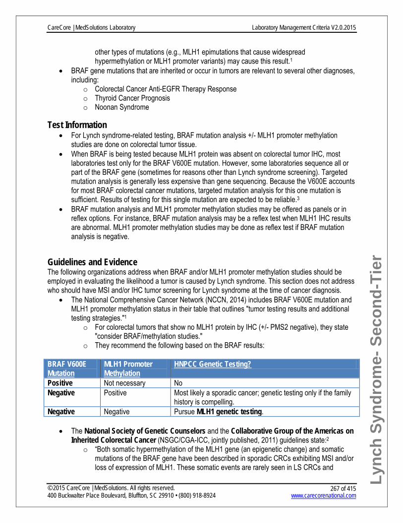
Lync
h Sy
ndro
me-
Sec
ond-
Tier
other types of mutations (e.g., MLH1 epimutations that cause widespread hypermethylation or MLH1 promoter variants) may cause this result.1
• BRAF gene mutations that are inherited or occur in tumors are relevant to several other diagnoses, including:
o Colorectal Cancer Anti-EGFR Therapy Response o Thyroid Cancer Prognosis o Noonan Syndrome
Test Information • For Lynch syndrome-related testing, BRAF mutation analysis +/- MLH1 promoter methylation
studies are done on colorectal tumor tissue. • When BRAF is being tested because MLH1 protein was absent on colorectal tumor IHC, most
laboratories test only for the BRAF V600E mutation. However, some laboratories sequence all or part of the BRAF gene (sometimes for reasons other than Lynch syndrome screening). Targeted mutation analysis is generally less expensive than gene sequencing. Because the V600E accounts for most BRAF colorectal cancer mutations, targeted mutation analysis for this one mutation is sufficient. Results of testing for this single mutation are expected to be reliable.3
• BRAF mutation analysis and MLH1 promoter methylation studies may be offered as panels or in reflex options. For instance, BRAF mutation analysis may be a reflex test when MLH1 IHC results are abnormal. MLH1 promoter methylation studies may be done as reflex test if BRAF mutation analysis is negative.
Guidelines and Evidence The following organizations address when BRAF and/or MLH1 promoter methylation studies should be employed in evaluating the likelihood a tumor is caused by Lynch syndrome. This section does not address who should have MSI and/or IHC tumor screening for Lynch syndrome at the time of cancer diagnosis.
• The National Comprehensive Cancer Network (NCCN, 2014) includes BRAF V600E mutation and MLH1 promoter methylation status in their table that outlines "tumor testing results and additional testing strategies."1
o For colorectal tumors that show no MLH1 protein by IHC (+/- PMS2 negative), they state "consider BRAF/methylation studies."
o They recommend the following based on the BRAF results:
BRAF V600E Mutation
MLH1 Promoter Methylation
HNPCC Genetic Testing?
Positive Not necessary No Negative Positive Most likely a sporadic cancer; genetic testing only if the family
history is compelling. Negative Negative Pursue MLH1 genetic testing.
• The National Society of Genetic Counselors and the Collaborative Group of the Americas on
Inherited Colorectal Cancer (NSGC/CGA-ICC, jointly published, 2011) guidelines state:2 o “Both somatic hypermethylation of the MLH1 gene (an epigenetic change) and somatic
mutations of the BRAF gene have been described in sporadic CRCs exhibiting MSI and/or loss of expression of MLH1. These somatic events are rarely seen in LS CRCs and
CareCore | MedSolutions Laboratory Laboratory Management Criteria V2.0.2015
© 2015 CareCore | MedSolutions. All rights reserved. 400 Buckwalter Place Boulevard, Bluffton, SC 29910 • (800) 918-8924 www.carecorenational.com
267 of 415

Lync
h Sy
ndro
me-
Sec
ond-
Tier
therefore may be useful in determining whether a MSI-high CRC is more likely to be sporadic.”
o “MLH1 promoter methylation and BRAF V600E mutation testing may help to reduce the number of germline genetic tests needed when IHC reveals absence of MLH1 and PMS2. However, NSGC and the CGAICC did not find enough data to recommend one test over the other or both concomitantly.”
o The likelihood of identifying a germline MLH1 with both DNA sequencing and MLPA analysis is approximately 33% when MLH1 +/- PMS2 are absent on IHC and MLH1 promoter hypermethylation is not present.
• The Centers for Disease Control and Prevention sponsored Evaluation of Genomic Applications in Practice and Prevention Working Group (EGAPP, 2009) published evidence-based recommendations focused on Lynch syndrome tumor screening by MSI and IHC. They include some information about BRAF mutation analysis and MLH1 promoter methylation, but do not make formal recommendations regarding these two tests.3
o However, the CDC website provides additional information about these guidelines. For BRAF V600E mutation analysis, they find adequate evidence of clinical validity and utility with an overall recommendation of "Sufficient evidence to recommend use for the benefit of relatives."11
o The CDC website does not address MLH1 promoter methylation, but an EGAPP supplemental evidence review (that accompanied the recommendations) states: "This supplemental evidence review did not involve a formal search or statistical summary concerning the literature on methylation testing. The literature suggests, however, that BRAF V600E mutation testing and methylation testing of the MLH1 promoter region among CRC cases with absent MLH1 protein might avoid similar numbers of sequencing tests with little loss in Lynch syndrome detection."12
Criteria BRAF V600E Mutation Analysis or MLH1 Promoter Methylation Status
• Previous Testing: o IHC testing* has been performed and indicates a loss of MLH1 protein, AND
• Diagnostic Testing for Symptomatic Individuals: o Personal history of colorectal cancer, and
• Rendering laboratory is a qualified provider of service per Health Plan policy, AND
CareCore | MedSolutions Laboratory Laboratory Management Criteria V2.0.2015
© 2015 CareCore | MedSolutions. All rights reserved. 400 Buckwalter Place Boulevard, Bluffton, SC 29910 • (800) 918-8924 www.carecorenational.com
268 of 415

Lync
h Sy
ndro
me-
Sec
ond-
Tier
References 1. NCCN Clinical Practice Guidelines in Oncology: Genetics/Familial High-Risk Assessment: Colorectal. Version
1.2014. Available at: http://www.nccn.org/professionals/physician_gls/pdf/genetics_colon.pdf 2. Weissman SM, Burt R, Church J, et al. Identification of Individuals at Risk for Lynch Syndrome Using Targeted
Evaluations and Genetic Testing: National Society of Genetic Counselors and the Collaborative Group of the Americas on Inherited Colorectal Cancer Joint Practice Guideline. J Genet Couns. 2011 Dec 14.
3. Evaluation of Genomic Applications in Practice and Prevention (EGAPP) Working Group. Recommendations from the EGAPP Working Group: genetic testing strategies in newly diagnosed individuals with colorectal cancer aimed at reducing morbidity and mortality from Lynch syndrome in relatives. Genet Med. 2009 Jan;11(1):35-41.
4. Kohlmann W, Gruber SB. (Updated September 20, 2012). Lynch Syndrome. In: GeneReviews at GeneTests: Medical Genetics Information Resource (database online). Copyright, University of Washington, Seattle. 1997-2014. Available at http://www.ncbi.nlm.nih.gov/books/NBK1211/
5. Ligtenberg MJ, Kuiper RP, Chan TL, et al. Heritable somatic methylation and inactivation of MSH2 in families with Lynch syndrome due to deletion of the 3' exons of TACSTD1. Nat Genet. 2009 Jan;41(1):112-7.
6. Kovacs ME, Papp J, Szentirmay Z, Otto S, Olah E. Deletions removing the last exon of TACSTD1 constitute a distinct class of mutations predisposing to Lynch syndrome. Hum Mutat. 2009 Feb;30(2):197-203.
7. American Gastroenterological Association. American Gastroenterological Association medical position statement: Hereditary colorectal cancer and genetic testing. Gastroenterology. 2001;121(1):195-7.
8. Rex DK, Johnson DA, Anderson JC, Schoenfeld PS, Burke CA, Inadomi JM. American College of Gastroenterology guidelines for colorectal cancer screening 2009. Am J Gastroenterol. 2009 Mar;104(3):739-50.
9. Shia J. Immunohistochemistry versus microsatellite instability testing for screening colorectal cancer patients at risk for hereditary nonpolyposis colorectal cancer syndrome. Part I. The utility of immunohistochemistry. J Mol Diagn. Jul 2008;10(4):293-300. Available at http://www.ncbi.nlm.nih.gov/pmc/articles/PMC2438196/pdf/JMD010000293.pdf
10. Roth AD, Tejpar S, Delorenzi M, et al. Prognostic role of KRAS and BRAF in stage II and III resected colon cancer: results of the translational study on the PETACC-3, EORTC 40993, SAKK 60-00 trial. J Clin Oncol. Jan 20 2010;28(3):466-74.
11. Centers for Disease Control and Prevention. Genetic Testing. Health Professionals: More About the EGAPP Lynch Syndrome Recommendation . Available at: http://www.cdc.gov/genomics/gtesting/EGAPP/recommend/lynch_provider.htm
12. Palomaki GE, McClain MR, Melillo S, Hampel HL, Thibodeau SN. EGAPP supplementary evidence review: DNA testing strategies aimed at reducing morbidity and mortality from Lynch syndrome. Genet Med. 2009 Jan;11(1):42-65
CareCore | MedSolutions Laboratory Laboratory Management Criteria V2.0.2015
© 2015 CareCore | MedSolutions. All rights reserved. 400 Buckwalter Place Boulevard, Bluffton, SC 29910 • (800) 918-8924 www.carecorenational.com
269 of 415

Mammaprint 70-Gene Breast Cancer Recurrence Assay
Procedure(s) covered by this policy: Procedure
Code(s)
Requires:
Prior-authorization* Lab Procedure Restrictions†
Mammaprint 70 Gene Signature S3854 Yes No Unlisted multianalyte assay with algorithmic analysis 81599 Yes No * - Clinical Review necessary prior to authorization for this procedure. † - Lab procedures require specified sequence to be followed and additional information is required to be supplied by lab performing procedure(s).
What Is MammaPrint? • MammaPrint is a 70-gene expression test designed to predict the chance of later-in-life recurrence
of breast cancer in women with newly diagnosed, early stage breast cancer.1 It is FDA cleared for use along with other standard prognostic methods, such as disease staging, grading and other tumor marker analyses.2
• MammaPrint is intended to assist patients and providers considering treatment with adjuvant chemotherapy. Patients assigned a "low risk" may choose hormone therapy (tamoxifen) alone and forego chemotherapy. Patients assigned a "high risk" may benefit from more aggressive treatment and choose to do chemotherapy.1
• MammaPrint is designed for women with breast cancer who have:1,2 o Stage I or II invasive carcinoma o Tumor size <5.0 cm o Node-negative (no metastasis to lymph nodes) o Estrogen receptor-positive (ER+) or -negative (ER-) disease
Test Information • MammaPrint uses a microarray platform to analyze the expression level of 70 genes in the tumor.
These 70 genes are thought to be critical in the cellular pathways to cancer metastasis.1 • Based on the test results, patients are assigned either a low risk or a high risk for a distant
recurrence. Low risk corresponds to a 10% risk of recurrence by 10 years without any additional adjuvant treatment. In contrast, those in the high risk group have a 29% risk of recurrence by 10 years without any additional adjuvant treatment.1
CareCore | MedSolutions Laboratory Laboratory Management Criteria V2.0.2015
© 2015 CareCore | MedSolutions. All rights reserved. 400 Buckwalter Place Boulevard, Bluffton, SC 29910 • (800) 918-8924 www.carecorenational.com
270 of 415

Mam
map
rint
Guidelines and Evidence
• The National Comprehensive Cancer Network (NCCN) 2014 Clinical Practice Guidelines for Breast Cancer state that:3
o The 21-gene RT-PCR assay (OncotypeDX) can be considered for patients with ER-positive, node-negative tumors measuring >0.5 cm in the management algorithm.
o "A recent comparison of simultaneous analyses of breast cancer tumors using five different gene-expression models indicated that four of these methods (including Mammaprint and Oncotype DX) provided similar predictions of clinical outcomes."[NCCN p. MS-21]
o "Currently, two prospective randomized clinical trials (TAILORx and MINDACT) are addressing the use of OncotypeDx and MammaPrint, respectively, as predictive and/or prognostic tools in populations of women with early- stage, lymph node-negative breast cancer. Pending the results of the prospective trials, the Panel considers the 21-gene RT-PCR assay as an option when evaluating patients with primary tumors characterized as 0.6-1.0 cm with unfavorable features or >1 cm, and node-negative, hormone receptor-positive, and HER2-negative (category 2A [based on lower-level evidence and there is uniform consensus])."
• The Evaluation of Genomic Applications in Practice and Prevention (EGAPP, 2009) Working Group reviewed the evidence for MammaPrint and concludes:4
o "It is unclear what population of patients would derive benefit from use of the test, and what the magnitude of that benefit would be. Prospective data from trials like MINDACT will be extremely valuable."
o "Overall, published evidence supports MammaPrint as a better predictor of the risk of distant recurrence than traditionally used tumor characteristics or algorithms, but its performance in therapeutically homogeneous populations is not yet known with precision, and it is unclear for how many women the lowest predicted risks are low enough to forgo chemotherapy."
o "No evidence is available to permit conclusions regarding the clinical utility of MammaPrint to select women who will benefit from chemotherapy."
o "To conclude, the literature on the 70-gene signature includes numerous studies that focused more on its biological underpinning and less on the clinical implications of this gene expression profile, although it has now received FDA approval for clinical use."
• Evidence-based clinical guidelines from the American Society of Clinical Oncology (ASCO, 2007) state that for multiparameter gene expression analysis for breast cancer:5
o "In newly diagnosed patients with node-negative, estrogen receptor-positive breast cancer, the Oncotype DX assay (Genomic Health Inc, Redwood City, CA) can be used to predict the risk of recurrence in patients treated with tamoxifen. Oncotype DX may be used to identify patients who are predicted to obtain the most therapeutic benefit from adjuvant tamoxifen and may not require adjuvant chemotherapy."
o "The precise clinical utility and appropriate application for other multiparameter assays, such as the MammaPrint assay (Agendia BV, Amsterdam, the Netherlands), the so-called Rotterdam Signature, and the Breast Cancer Gene Expression Ratio are under investigation."
• The US Food and Drug Administration (FDA) cleared Mammaprint for clinical use in 2007.2
CareCore | MedSolutions Laboratory Laboratory Management Criteria V2.0.2015
© 2015 CareCore | MedSolutions. All rights reserved. 400 Buckwalter Place Boulevard, Bluffton, SC 29910 • (800) 918-8924 www.carecorenational.com
271 of 415

Mam
map
rint
Criteria Genetic testing is not approved for the MammaPrint multi-gene assay on tumor tissue because it is currently considered experimental, investigational or is unproven.
References 1. MammaPrint website. Available at http://www.agendia.com/physicians/symphony-targeted-therapy-for-breast-
cancer/mammaprint-best-breast-cancer-treatment/. 2. US Food and Drug Administration. FDA clears breast cancer specific molecular prognostic test. Available at
http://www.fda.gov/NewsEvents/Newsroom/PressAnnouncements/2007/ucm108836.htm. 3. National Comprehensive Cancer Network 2014 Clinical Practice Guidelines in Oncology: Breast Cancer.
V.2.2014: Available at http://www.nccn.org/professionals/physician_gls/pdf/breast.pdf. 4. Evaluation of Genomic Applications in Practice and Prevention (EGAPP) Working Group. Recommendations from
the EGAPP Working Group: can tumor gene expression profiling improve outcomes in patients with breast cancer? Genet Med. 2009 Jan;11(1):66-73. Available at http://egappreviews.org/recommendations/breastcancer.htm
5. Harris L, Fritsche H, Mennel R, et al. American Society of Clinical Oncology 2007 Update of recommendations for the use of tumor markers in breast cancer. J Clin Oncol. 2007 Nov;25(33):5287-312.
6. Center for Medicare and Medicaid Services. Medicare Coverage Database. Available at https://www.cms.gov/medicare-coverage-database/search/advanced-search.aspx.
CareCore | MedSolutions Laboratory Laboratory Management Criteria V2.0.2015
© 2015 CareCore | MedSolutions. All rights reserved. 400 Buckwalter Place Boulevard, Bluffton, SC 29910 • (800) 918-8924 www.carecorenational.com
272 of 415

Mammostrat Breast Cancer Recurrence Assay Procedure covered by this policy:
Procedure Code
Requires: Prior-authorization* Non-covered
Mammostrat® Breast Cancer Assay S3854 Yes Investigational and Experimental
Mammostrat® Breast Cancer Assay 84999 Yes Investigational and Experimental
What Is the Mammostrat Breast Cancer Recurrence Assay? • The Mammostrat® Breast Cancer Recurrence Assay is an immunohistochemical assay that
measures levels of five proteins in tumor tissue associated with risk of breast cancer recurrence.1 • It is used in people with newly diagnosed, early stage breast cancer. • The assay looks at five proteins and determines their expression levels in the tumor. The
expression levels of these five markers are thought to influence whether the tumor will metastasize, increasing the patient’s chance of recurrence. These levels are then translated into a risk index, given as a percent chance of recurrence over 10 years.
• Physicians and patients may use the risk index as one factor in determining the course of treatment. Patients in the high risk category may benefit more from aggressive treatment, whereas patients in the low risk category may elect to forgo the aggressive chemotherapy.2
Test Information • The Mammostrat assay measures the expression level of five proteins by immunohistochemistry.
These markers are believed to be associated with breast cancer recurrence:3 o p53 plays a role in cell cycle regulation. Mutations in the p53 gene are associated with
tumor growth. o HTF9C is implicated in DNA replication and cell cycle control. o CEACAM5 is normally expressed in embryonic tissue, but is also found in some tumors. o NDRG1 may have a role in helping tumors survive aggressive treatment. o SLC7A5 can, when overexpressed, help sustain the high growth rate of cancer.
• These levels are then translated into a quantitative "risk index" via a proprietary algorithm, which divides patients into groups with low, moderate, or high risk of recurrence:1
Risk Index Risk of Breast Cancer Recurrence over 10 Years Low 7.6%
Moderate 16.3%
High 20.9%
Guidelines and Evidence • No US evidence-based guidelines exist for each gene expression profile assay currently available
or in development.
CareCore | MedSolutions Laboratory Laboratory Management Criteria V2.0.2015
© 2015 CareCore | MedSolutions. All rights reserved. 400 Buckwalter Place Boulevard, Bluffton, SC 29910 • (800) 918-8924 www.carecorenational.com
273 of 415

Mam
mos
trat
• The American Society of Clinical Oncology (ASCO, 2007)4 has not made recommendations on the use of the Mammostrat serum assay, but has made recommendations on the use of the p53 protein marker.1
o p53: "Present data are insufficient to recommend use of p53 measurements for management of patients with breast cancer. There is no change from the guideline published in 2000. Methods to define more precisely and conveniently genetic abnormalities in p53 might permit a more accurate analysis of association of p53 and clinical outcomes, either as a pure prognostic factor or as a predictor of benefit from systemic therapies. However, at present, methodologies to do so are cumbersome, expensive, and not widely available as routine clinical assays, limiting the utility of this marker in clinical practice."
• A 2010 clinical study tested the assay’s ability to accurately predict risk of breast cancer recurrence in a cohort of 1,812 women with early stage breast cancer:1
o "The Mammostrat markers are biologically independent of one another and measure aspects of physiology distinct from proliferation, HER2 status, and hormone receptor status already assessed by IHC assays that are standard of care. Collectively these data add support to a potential role for Mammostrat in management of early-stage breast cancer."
Criteria • This test is considered investigational and/or experimental.
o Investigational and experimental (I&E) molecular and genomic (MolGen) tests refer to assays involving chromosomes, DNA, RNA, or gene products that have insufficient data to determine the net health impact, which typically means there is insufficient data to support that a test accurately assesses the outcome of interest (analytical and clinical validity), significantly improves health outcomes (clinical utility), and/or performs better than an existing standard of care medical management option. Such tests are also not generally accepted as standard of care in the evaluation or management of a particular condition.
o In the case of MolGen testing, FDA clearance is not a reliable standard given the number of laboratory developed tests that currently fall outside of FDA oversight and FDA clearance often does not assess clinical utility.
References 1. Bartlett JM, Thomas J, Ross DT, et al. Mammostrat as a tool to stratify breast cancer patients at risk of recurrence
during endocrine therapy. Breast Cancer Res. 2010;12(4):R47. 2. Ross DT, Kim CY, Tang G, et al. Chemosensitivity and stratification by a five monoclonal antibody
immunohistochemistry test in the NSABP B14 and B20 trials. Clin Cancer Res. 2008 Oct 15;14(20):6602-9. 3. Ring BZ, Seitz RS, Beck R, et al. Novel prognostic immunohistochemical biomarker panel for estrogen receptor-
positive breast cancer. J Clin Oncol. 2006 Jul 1;24(19):3039-47. 4. Harris L, Fritsche H, Mennel R, et al. American Society of Clinical Oncology 2007 Update of recommendations for
the use of tumor Markers in Breast Cancer. J Clin Oncol. 2007 Nov 20;25(33):5287-312.
CareCore | MedSolutions Laboratory Laboratory Management Criteria V2.0.2015
© 2015 CareCore | MedSolutions. All rights reserved. 400 Buckwalter Place Boulevard, Bluffton, SC 29910 • (800) 918-8924 www.carecorenational.com
274 of 415

MGMT Testing for Malignant Glioma Alkylating Agent Response
Procedure(s) covered by this policy: Procedure
Code(s)
Requires:
Prior-authorization* Lab Procedure Restrictions†
MGMT Promoter Methylation Analysis 81287 Yes No * - Clinical Review necessary prior to authorization for this procedure. † - Lab procedures require specified sequence to be followed and additional information is required to be supplied by lab performing procedure(s).
What Is MGMT? • MGMT is the O6-methylguanine- DNA methyltransferase gene, which encodes an essential DNA
repair enzyme. MGMT expression in tumors causes resistance to DNA-alkylating drugs. MGMT repairs the damage produced by these DNA cross linking agents.1
• Gene methylation is a control mechanism that regulates gene expression. If the MGMT gene is hypermethylated, its expression is absent (“turned off”) or reduced (“turned down”). With less MGMT DNA repair protein present, the tumor is typically more responsive to alkylating drugs.2
• Glioblastoma is a common and aggressive brain tumor that is often treated with alkylating drugs.2 Temozolomide is a standard systemic chemotherapy shown to be effective for malignant gliomas.2
• About 40-50% of glioblastoma tumors exhibit MGMT hypermethylation, leading to increased chemosensitivity.3,4
• Treatment of gliomas often includes resection, radiation, and chemotherapy. For patients over age 70, combined treatment may not be tolerated; therefore, treatment with a single agent (radiation therapy or chemotherapy) or chemotherapy with deferred radiation therapy may be considered.1
Test Information: • MGMT promoter methylation testing is performed on paraffin embedded tumor tissue. Quantitative
methylation-sensitive PCR is used to determine MGMT gene promoter methylation levels.
Guidelines and Evidence • The National Comprehensive Cancer Network (NCCN, 2014) has recommended that patients
over age 70 with glioblastoma or gliosarcoma should have MGMT promoter methylation testing if adjuvant chemotherapy with temozolomide is being considered. Temozolomine is appropriate for those patients who are MGMT promoter methylation positive.1
Criteria • Testing criteria:
o Diagnosis of glioblastoma (or gliosarcoma), and o Good performance status (Karnofsky Performance Status, KPS, greater than or equal to
60), and o Age 70 or greater, and
CareCore | MedSolutions Laboratory Laboratory Management Criteria V2.0.2015
© 2015 CareCore | MedSolutions. All rights reserved. 400 Buckwalter Place Boulevard, Bluffton, SC 29910 • (800) 918-8924 www.carecorenational.com
275 of 415

MG
MT
o Adjuvant temozolomide chemotherapy is being considered1, AND • Rendering laboratory is a qualified provider of service per the Health Plan policy.
References 1. National Comprehensive Cancer Network. NCCN Clinical Practice Guidelines in Oncology, Central Nervous
System Cancers, Version 2.2014. Available at www.nccn.org 2. Esteller M, Garcia-Foncillas J, et al. Inactivation of the DNA-repair gene MGMT and the clinical response of
gliomas to alkylating agents. N Engl J Med. 2000 Nov 9;343(19):1350-4. 3. Gilbert, MR. Establishing the standard of care for patients with newly diagnosed and recurrent glioblastoma. Am
Soc Clin Oncol Educ Book. 2012;32:112-7. 4. Hegi ME, Liu L, Herman JG, et al. Correlation of O6-methylguanine methyltransferase (MGMT) promoter
methylation with clinical outcomes in glioblastoma and clinical strategies to modulate MGMT activity. J Clin Oncol. 2008 Sep 1;26(25):4189-99.
CareCore | MedSolutions Laboratory Laboratory Management Criteria V2.0.2015
© 2015 CareCore | MedSolutions. All rights reserved. 400 Buckwalter Place Boulevard, Bluffton, SC 29910 • (800) 918-8924 www.carecorenational.com
276 of 415

MTHFR Variant Analysis for Hyperhomocysteinemia
Procedure covered by this policy: Procedure Code(s) Requires:
Non-covered MTHFR Genotyping 81291 Investigational and Experimental * - Clinical Review necessary prior to authorization for this procedure. † - Lab procedures require specific sequence to be followed or additional information is required and must be supplied by the lab performing procedure(s) for full
What Is Hyperhomocysteinemia?
• Hyperhomocysteinemia generally refers to mild to moderate elevations of plasma homocysteine levels, which may be defined as 15 to 40 µmol/L.1
• Hyperhomocysteinemia may be caused by nutritional deficiencies, various medical conditions, certain drugs, smoking, and inherited factors — such as MTHFR gene variants.1
• The MTHFR gene encodes the 5,10-methylenetetrahydrofolate reductase (MTHFR) enzyme. MTHFR is involved in folate metabolism. The major circulating form of folate is key to converting homocysteine into methionine. Therefore, MTHFR gene variants that reduce MTHFR enzyme function may predispose one to impaired folate metabolism and ultimately mild to moderate hyperhomocysteinemia. However, homocysteine levels are usually normal if folate intake is sufficient.1
• Both hyperhomocysteinemia in general, and MTHFR variants specifically, have been reported in association with cardiovascular disease, venous thromboembolism, pregnancy complications, and certain birth defects, such as neural tube defects.1,2 However, data is inconsistent and associated risks generally small.
Test Information • MTHFR genetic testing looks for two very common gene variants: C677T and A1298C.2 • Individuals who have two variants, including at least one C677T, may have an increased risk for
hyperhomocysteinemia. However, the connection between these MTHFR variants, hyperhomocysteinemia itself, and ultimate disease risk remains unclear.3,4
• Many experts suggest that measuring homocysteine levels directly is more informative than MTHFR variant testing.5
• Note that serious mutations in the MTHFR gene (not the common variants discussed here) are rarely associated with a genetic disorder called homocystinuria.2 MTHFR variant testing will not find the mutations that cause homocystinuria.
• MTHFR gene testing may be a component of panels for thrombophilia, cardiovascular disease risk, psychiatric conditions, or preeclampsia. There is insufficient evidence in the peer-reviewed literature to establish clinical utility for any of these indications for testing.
CareCore | MedSolutions Laboratory Laboratory Management Criteria V2.0.2015
© 2015 CareCore | MedSolutions. All rights reserved. 400 Buckwalter Place Boulevard, Bluffton, SC 29910 • (800) 918-8924 www.carecorenational.com
277 of 415

MTH
FR-A
ssoc
iate
d H
yper
hom
ocys
tein
emia
Guidelines and Evidence • As part of the Choosing Wisely campaign, the Society for Maternal Fetal Medicine (2014)
released “Five Things Physicians and Patients Should Question,” which states:6 o "Don’t do an inherited thrombophilia evaluation for women with histories of pregnancy loss,
intrauterine growth restriction (IUGR), preeclampsia and abruption. Scientific data supporting a causal association between either methylenetetrahydrofolate reductase (MTHFR) polymorphisms or other common inherited thrombophilias and adverse pregnancy outcomes, such as recurrent pregnancy loss, severe preeclampsia and IUGR, are lacking."
• The American College of Medical Genetics and Genomics (ACMG, 2013) states:7 o “It was previously hypothesized that reduced enzyme activity of MTHFR led to mild
hyperhomocysteinemia which led to an increased risk for venous thromboembolism, coronary heart disease, and recurrent pregnancy loss. Recent meta-analyses have disproven an association between hyperhomocysteinemia and risk for coronary heart disease and between MTHFR polymorphism status and risk for venous thromboembolism. There is growing evidence that MTHFR polymorphism testing has minimal clinical utility and, therefore should not be ordered as a part of a routine evaluation for thrombophilia.”
• The American College of Obstetricians and Gynecologists (ACOG, 2013) states:8 o “Because of the lack of association between either heterozygosity or homozygosity for the
methylenetetrahydrofolate reductase (MTHFR) C677T polymorphism and any negative pregnancy outcomes, including any increased risk for venous thromboembolism, screening with either MTHFR mutation analyses or fasting homocysteine levels is not recommended.”
• The National Society of Genetic Counselors (NSGC, 2005) state that MTHFR variant testing is specifically not justified in the case of recurrent pregnancy loss based on available studies.9
Criteria This test is considered investigational and/or experimental.
• Investigational and experimental (I&E) molecular and genomic (MolGen) tests refer to assays involving chromosomes, DNA, RNA, or gene products that have insufficient data to determine the net health impact, which typically means there is insufficient data to support that a test accurately assesses the outcome of interest (analytical and clinical validity), significantly improves health outcomes (clinical utility), and/or performs better than an existing standard of care medical management option. Such tests are also not generally accepted as standard of care in the evaluation or management of a particular condition.
• In the case of MolGen testing, FDA clearance is not a reliable standard given the number of laboratory developed tests that currently fall outside of FDA oversight and FDA clearance often does not assess clinical utility.
References 1. Gatt A, Makris M. Hyperhomocysteinemia and venous thrombosis. Semin Hematol. 2007 Apr;44(2):70-6. 2. U.S. National Library of Medicine. Genetics Home Reference website: MTHFR. 2015. Available at
http://ghr.nlm.nih.gov/gene=mthfr.
CareCore | MedSolutions Laboratory Laboratory Management Criteria V2.0.2015
© 2015 CareCore | MedSolutions. All rights reserved. 400 Buckwalter Place Boulevard, Bluffton, SC 29910 • (800) 918-8924 www.carecorenational.com
278 of 415

MTH
FR-A
ssoc
iate
d H
yper
hom
ocys
tein
emia
3. Ray JG, Shmorgun D, Chan WS. Common C677T Polymorphism of the Methylenetetrahydrofolate Reductase Gene and the Risk of Venous Thromboembolism: Meta-Analysis of 31 Studies. Pathophysiol Haemost Thromb. 2002;32(2):51-8.
4. Clarke R, Bennett DA, Parish S, Verhoef P, Dotsch-Klerk M, et al. (2012) Homocysteine and Coronary Heart Disease: Meta-analysis of MTHFR Case-Control Studies, Avoiding Publication Bias. PLoS Med 9(2): e1001177. doi:10.1371/journal.pmed.1001177
5. Grody WW, Griffin JH, Taylor AK, Korf BR, Heit JA; ACMG Factor V. Leiden Working Group. American College of Medical Genetics consensus statement on factor V Leiden mutation testing. Genet Med. 2001 Mar-Apr;3(2):139-48.
6. Society for Maternal-Fetal Medicine Publications Committee. American Board of Internal Medicine (ABIM) Foundation Choosing Wisely Campaign. Available at: http://www.choosingwisely.org/doctor-patient-lists/society-for-maternal-fetal-medicine/
7. Hickey, SE, Curry, CJ, Toriello, HV; American College of Medical Genetics Practice Guidelines: Lack of Evidence for MTHFR Polymorphism Testing.Genetics in Medicine 2013:15(2):153-156.
8. American College of Obstetricians and Gynecologists (ACOG) Practice Bulletin no. 138: inherited thrombophilias in pregnancy. Obstet Gynecol. 2013 Sep;122(3):706-17.
9. Laurino MY, Bennett RL, Saraiya DS, et al. Genetic evaluation and counseling of couples with recurrent miscarriage: recommendations of the National Society of Genetic Counselors. J Genet Couns. 2005 Jun;14(3):165-81.
CareCore | MedSolutions Laboratory Laboratory Management Criteria V2.0.2015
© 2015 CareCore | MedSolutions. All rights reserved. 400 Buckwalter Place Boulevard, Bluffton, SC 29910 • (800) 918-8924 www.carecorenational.com
279 of 415

MUTYH Associated Polyposis Testing Procedure(s) covered by this policy:
Procedure Code(s)
Requires: Prior-
authorization* Lab Procedure Restrictions†
MUTYH Known Familial Mutation Analysis 81403 No No
MUTYH Targeted Mutation Analysis 81401 Yes No MUTYH sequencing 81406 Yes No MUTYH Deletion/Duplication Analysis 81479 Yes Yes * - Clinical Review necessary prior to authorization for this procedure. † - Lab procedures require specific sequence to be followed or additional information is required and must be supplied by the lab performing procedure(s) for full claim payment.
What Is MUTYH-Associated Polyposis? • MUTYH-associated polyposis (MAP) is an inherited colorectal cancer syndrome caused
by mutations in the MUTYH gene (also called MYH). • MAP clinical findings overlap those of familial adenomatous polyposis (FAP) and attenuated FAP
(AFAP). Affected patients most often have fewer than 100 adenomas, but cases of hundreds and occasionally over 1000 polyps have been reported.1 Hyperplastic and sessile serrated adenomatous polyps have also been seen individuals with MAP, although adenomas remain the most common polyp type in MAP.2 Duodenal adenomas occur in 18-25% of individuals with MAP and gastric polyps have been reported in about 11%.2,3
• Up to 30-40% of people who meet clinical diagnostic criteria for classic or attenuated FAP, but have normal FAP genetic test results, will have a MAP mutation.2,3
• Because MAP is not clinically distinguishable from FAP or AFAP, the identification of two MUTYH mutations is required to make a MAP diagnosis.5
• Adenomas and colorectal cancer tend to present later than FAP. The diagnosis of colorectal cancer is often after age 50.1 There is also an estimated 4% lifetime risk for duodenal cancer.2,3
• Unlike FAP, MAP is inherited in an autosomal recessive manner — both copies of the MUTYH gene must have a mutation to be affected. This means that siblings are the only relatives likely to be affected in the family history (i.e., you do not see inheritance from parent to child as with FAP).
Test Information • MUTYH Targeted Mutation Analysis: Two MUTYH mutations are particularly common (Y165C
and G382D) and account for over 80% of MUTYH mutations in Caucasians.6 Some laboratories test for only these two mutations or offer reflex options that begin with these two mutations and proceed to full gene sequencing if two mutations are not found.
• MUTYH Sequencing Analysis: MUTYH full sequencing analysis analyzes the entire gene for mutations. It is typically done in reflex to negative results from targeted mutation analysis.
• MUTYH Deletion/Duplication Analysis: If sequencing does not find two mutations, large gene deletion/duplication analysis can be performed. It remains unknown what percentage of MAP is
CareCore | MedSolutions Laboratory Laboratory Management Criteria V2.0.2015
© 2015 CareCore | MedSolutions. All rights reserved. 400 Buckwalter Place Boulevard, Bluffton, SC 29910 • (800) 918-8924 www.carecorenational.com
280 of 415

MU
TYH
Ass
ocia
ted
Poly
posi
s
due to large deletions/duplications/rearrangements in the gene and thus are detectable only with this technology. However, large deletions have been reported.7,8
• MUTYH Known Familial Mutation Analysis: Once the mutations that run in the family are known, other relatives can have testing for only those mutations. This is more accurate and cost-effective.
Guidelines and Evidence • Guidelines from the National Comprehensive Cancer Network (NCCN, 2014) on High-Risk
Colorectal Assessment states the following. o MUTYH testing criteria:1
"Personal history of >10 adenomas Individual meeting criteria for SPS [serrated polyposis syndrome, also called
hyperplastic polyposis] with at least some adenomas Known deleterious biallelic MUTYH mutations in the family"
o SPS clinical diagnostic criteria: “At least 5 serrated polyps proximal to the sigmoid colon with 2 or more of these
being >10mm Any number of serrated polyps proximal to the sigmoid colon in an individual who
has a first-degree relative with serrated polyposis Greater than 20 serrated polyps of any size, but distributed throughout the colon”
o Footnotes: "When colonic polyposis is present in a single person with a negative family
history, consider testing for a de novo APC mutation; if negative, follow with testing of MUTYH (targeted testing for the two common northern European founder mutations c.536A>G and c.1187G>A may be considered first followed by full sequencing if biallelic mutations are not found). When colonic polyposis is present only in siblings, consider recessive inheritance and test for MUTYH first. Order of testing for APC and MUTYH is at the discretion of the clinician."
“MUTYH genetic testing is not indicated based on a personal history of desmoid tumor.”
“Siblings of a patient with MAP are recommended to have site-specific genetic testing for the familial biallelic mutations. Children of an affected parent with MAP are recommended to have site-specific genetic testing for the familial mutation/s. If positive for one MUTYH mutation, full sequencing of MUTYH is recommended. Full sequencing of MUTYH also may be considered in an unaffected parent when the other parent has MAP. If the unaffected parent is found to not be heterozygous for a MUTYH mutation, genetic testing in children is not necessary. If he or she is found to have a MUTYH mutation, testing for the familial mutations in the children is recommended.”
o All recommendations are category 2A. • Evidence-based guidelines from the American College of Gastroenterology (ACG,
2009) state:4 "Patients with classic FAP, in whom genetic testing is negative, should undergo genetic testing for bi-allelic MUTYH mutations. Patients with 10 - 100 adenomas can be considered for genetic testing for attenuated FAP and if negative, MUTYH associated polyposis"[Grade 2C: Weak recommendation, low-quality or very low-quality evidence].
CareCore | MedSolutions Laboratory Laboratory Management Criteria V2.0.2015
© 2015 CareCore | MedSolutions. All rights reserved. 400 Buckwalter Place Boulevard, Bluffton, SC 29910 • (800) 918-8924 www.carecorenational.com
281 of 415

MU
TYH
Ass
ocia
ted
Poly
posi
s
Criteria Known MUTYH Family Mutation(s) Testing
• Genetic Counseling: o Pre and post-test genetic counseling by an appropriate provider (as deemed by the Health
Plan policy), AND • Previous Testing:
o No previous genetic testing for known MUTYH family mutation(s), AND • Diagnostic or Predisposition Testing:
o Two known MUTYH mutations in a sibling, or o Both parents with one or two known MUTYH mutations, AND
• Rendering laboratory is a qualified provider of service per the Health Plan policy. MUTYH Targeted Mutation Analysis for Y179C and G396D Mutations
• Genetic Counseling o Pre and post-test genetic counseling by an appropriate provider (as deemed by the Health
Plan policy), AND • Previous Testing:
o No previous MUTYH testing, AND • Diagnostic Testing for Symptomatic Individuals:
o Clinical findings: > 10 cumulative adenomas, or At least two adenomas, AND At least 5 serrated polyps proximal to the sigmoid colon (2 or more of >10mm), or Any number of serrated polyps proximal to the sigmoid colon with a first-degree
relative with serrated polyposis, or > 20 serrated polyps of any size, but distributed throughout the colon, AND
o No mutation detected on APC gene testing, or o Recessive pattern of inheritance (e.g. family history positive for only an affected sibling),
OR Testing for Presymptomatic/Asymptomatic Individuals:
o Reproductive partner of a person with MAP (to determine if children at risk), AND • Rendering laboratory is a qualified provider of service per Health Plan policy.
MUTYH Full Gene Sequencing
• Genetic Counseling o Pre and post-test genetic counseling by an appropriate provider (as deemed by the Health
Plan policy), AND • Previous Testing:
o No previous MUTYH full sequencing, and o Two mutations NOT identified through MUTYH targeted mutation analysis (Y179C and
G396D) if performed, AND
CareCore | MedSolutions Laboratory Laboratory Management Criteria V2.0.2015
© 2015 CareCore | MedSolutions. All rights reserved. 400 Buckwalter Place Boulevard, Bluffton, SC 29910 • (800) 918-8924 www.carecorenational.com
282 of 415

MU
TYH
Ass
ocia
ted
Poly
posi
s
• Diagnostic Testing for Symptomatic Individuals: o Clinical findings:
> 10 cumulative adenomas, or At least two adenomas, AND
• At least 5 serrated polyps proximal to the sigmoid colon (2 or more of >10mm), or
• Any number of serrated polyps proximal to the sigmoid colon with a first-degree relative with serrated polyposis, or
• > 20 serrated polyps of any size, but distributed throughout the colon, AND
o No mutation detected on APC gene testing, or o Recessive pattern of inheritance (e.g. family history positive for only an affected sibling),
OR • Testing for Presymptomatic/Asymptomatic Individuals:
o Reproductive partner of a person with MAP (to determine if children at risk), AND • Rendering laboratory is a qualified provider of service per Health Plan policy.
MUTYH Gene Deletion/Duplication Analysis†
• Genetic Counseling o Pre and post-test genetic counseling by an appropriate provider (as deemed by the Health
Plan policy), AND • Previous Testing:†
o MUTYH full sequencing performed, and o No mutations or only one mutation detected in MUTYH through any previous testing
(founder mutation panel or full gene sequencing), AND • Diagnostic Testing for Symptomatic Individuals:
o Clinical findings: > 10 cumulative adenomas, or At least two adenomas, AND
• At least 5 serrated polyps proximal to the sigmoid colon (2 or more of >10mm), or
• Any number of serrated polyps proximal to the sigmoid colon with a first-degree relative with serrated polyposis, or
• > 20 serrated polyps of any size, but distributed throughout the colon, AND
o No mutation detected on APC gene testing, or o Recessive pattern of inheritance (e.g. family history positive for only an affected sibling),
OR • Testing for Presymptomatic/Asymptomatic Individuals:
o Reproductive partner of a person with MAP (to determine if children at risk), AND • Rendering laboratory is a qualified provider of service per Health Plan policy.
†Lab Testing Restrictions: Targeted mutation analysis and/or full gene sequencing performed and no mutation or only one mutation identified in a patient with high clinical suspicion for MUTYH-associated
CareCore | MedSolutions Laboratory Laboratory Management Criteria V2.0.2015
© 2015 CareCore | MedSolutions. All rights reserved. 400 Buckwalter Place Boulevard, Bluffton, SC 29910 • (800) 918-8924 www.carecorenational.com
283 of 415

MU
TYH
Ass
ocia
ted
Poly
posi
s
polyposis. When MUTYH deletion/duplication analysis is performed in reflex to a negative sequencing test, the modifier 59 should be appended to the claim.
References 1. National Comprehensive Cancer Network. NCCN Clinical Practice Guidelines in Oncology: Genetic/Familial High-
Risk Assessment: Colorectal. V2.2014. Available at: www.nccn.org/professionals/physician gls/pdf/genetics colon.pdf.
2. Goodenberger M, Lindor NM. Lynch syndrome and MYH-associated polyposis: review and testing strategy. J Clin Gastroenterol. 2011 Jul;45(6):488-500.
3. Terdiman JP. MYH-associated disease: attenuated adenomatous polyposis of the colon is only part of the story.Gastroenterology 2009 Dec;137(6):1883-6.
4. Rex DK, Johnson DA, Anderson JC, Schoenfeld PS, Burke CA, Inadomi JM; American College of Gastroenterology. American College of Gastroenterology guidelines for colorectal cancer screening 2008. Am J Gastroenterol. 2009;104(3):739-50.
5. Gammon A, Kohlmann W, Burt R. Can we identify the high-risk patients to be screened? A genetic approach.Digestion. 2007;76(1):7-19.
6. Balaguer F, Castellví-Bel S, Castells A, et al; Gastrointestinal Oncology Group of the Spanish Gastroenterological Association. Identification of MYH mutation carriers in colorectal cancer: a multicenter, case-control, population-based study. Clin Gastroenterol Hepatol. 2007;5(3):379-87.
7. Torrezan GT, da Silva FC, Krepischi AC, et al. Breakpoint characterization of a novel large intragenic deletion of MUTYH detected in a MAP patient: case report. BMC Med Genet. 2011 Sep 30;12:128.
8. Rouleau E, Zattara H, Lefol C, et al. First large rearrangement in the MUTYH gene and attenuated familial adenomatous polyposis syndrome. Clin Genet. 2011 Sep;80(3):301-3.
CareCore | MedSolutions Laboratory Laboratory Management Criteria V2.0.2015
© 2015 CareCore | MedSolutions. All rights reserved. 400 Buckwalter Place Boulevard, Bluffton, SC 29910 • (800) 918-8924 www.carecorenational.com
284 of 415

Niemann Pick Disease Types A & B Testing Procedure covered by this policy: Procedure Code(s)
Requires: Prior-authorization* Lab Procedure Restrictions†
Acid Sphingomyelinase Enzyme Activity 82657 No No SMPD1 Known Familial Mutation 81403 Yes No SMPD1 Targeted Mutation Analysis 81330 No No SMPD1 Sequencing 81479 Yes No SMPD1 Deletion/Duplication Analysis 81479 Yes Yes * - Clinical Review necessary prior to authorization for this procedure. † - Lab procedures require specific sequence to be followed or additional information is required and must be supplied by the lab performing procedure(s) for full
What Is Niemann-Pick Disease (Type A and B)? • Niemann-Pick disease is a genetic disorder caused by an inability to process lipids (fats), which
results in a toxic buildup of lipids in some organs.1-3 • Two types of Niemann-Pick disease are caused by a deficiency of the acid sphingomyelinase
enzyme: o Type A, also called the "neurological" or "neuronopathic" type, causes symptoms
beginning in infancy. These include an enlarged liver and spleen (hepatosplenomegaly), psychomotor impairment with neurologic deterioration, interstitial lung disease, and eventually a classic cherry-red spot of the retina. Affected individuals usually do not survive beyond childhood.1-3
o Type B, also called the “non-neurological" or "non-neuronopathic" type, causes some symptoms similar to type A, but symptoms are usually milder and begin later. Additional symptoms include hyperlipidemia (high fat levels in blood) and thrombocytopenia (low platelets). Affected individuals can survive to adulthood.1,3
• The SMPD1 gene encodes the acid sphingomyelinase (ASM) enzyme. Gene mutations in the SMPD1 gene lead to reduced or absent sphingomyelinase enzyme activity, causing the symptoms of Niemann-Pick disease.1,3
• Niemann-Pick disease is suspected when a patient presents with hepatosplenomegaly, interstitial lung disease, and depending on the subtype, neurological symptoms in infancy or abnormal blood findings.3 However, a diagnosis cannot be made clinically.
• When Niemann-Pick disease is suspected, acid sphingomyelinase enzyme activity testing should be performed first.3 People with Niemann-Pick disease type A or B usually have less than 10% of normal ASM activity compared to healthy individuals.3 Measuring ASM enzyme activity in peripheral blood lymphocytes or cultured skin fibroblasts is a reliable way to confirm a suspected case of Niemann-Pick disease.3 However, false-negative and inconclusive results are possible.3,4 In such cases, genetic testing may be useful to resolve a diagnosis.
• About 1 in 250,000 people have Niemann-Pick disease.1,3 Type A is more common in persons of Ashkenazi Jewish descent than in the general population. In the Ashkenazi Jewish population, the frequency of Niemann-Pick disease is 1 in 40,000.1,3
• Niemann-Pick disease is an autosomal recessive disorder. An affected individual must inherit SMPD1 gene mutations from both parents.1,3
CareCore | MedSolutions Laboratory Laboratory Management Criteria V2.0.2015
© 2015 CareCore | MedSolutions. All rights reserved. 400 Buckwalter Place Boulevard, Bluffton, SC 29910 • (800) 918-8924 www.carecorenational.com
285 of 415

Nie
man
n Pi
ck D
isea
se, T
ypes
A &
B
o Individuals who inherit only one mutation are called carriers. Carriers do not show symptoms of Niemann-Pick disease, but have a 50% chance of passing on the mutation to their children.
o Two carriers of Niemann-Pick disease have a 25% chance of having a child with the disorder.
o Prenatal diagnosis for at-risk pregnancies can be performed through enzyme testing or molecular genetic testing (if the mutations in both parents are known).3
• Individuals at increased risk to have a child with Niemann-Pick disease should routinely be offered carrier screening. This includes those with:4,5
o Ashkenazi Jewish ancestry (1 in 90 carrier risk3,5) o A family history of Niemann-Pick disease (regardless of ethnicity) o A partner who is a known carrier of Niemann-Pick disease (or affected with the milder type)
Test Information • SMPD1 Mutation Analysis tests for four of the most common SMPD1 gene mutations.
o Three mutations - R496L, L302P, fsP330 - account for 97% of all cases of Niemann-Pick disease type A in Ashkenazi Jewish people.5
o The fourth mutation - deltaR608 - is a common cause of Niemann-Pick disease type B in people of North African descent.3
o Carrier screening by SMPD1 mutation panel for Niemann-Pick disease is widely available as part of an "Ashkenazi Jewish Panel" that includes several other genetic disease that are more common in this population. (See Ashkenazi Jewish Carrier Screening.)
• SMPD1 Sequencing analyzes the entire coding region of the SMPD1 is available to detect less common mutations that cannot be detected on a common mutation analysis panel. SMPD1 sequencing detects more than 95% of all SMPD1 mutations.3
• SMPD1 Deletion/Duplication Analysis is available to detect large gene rearrangements that cannot be detected by sequencing. However, the frequency of such mutations is unknown.3
• SMPD1 Known Familial Mutation Testing can be performed for at-risk relatives when the familial mutation is known and is not one of the common mutations.3
Guidelines and Evidence • Professional guidelines generally support Niemann-Pick disease carrier screening for those at
increased risk.4,5 • Consensus guidelines from the American College of Obstetricians and Gynecologists (ACOG,
2009) address carrier screening and prenatal diagnosis for Niemann-Pick disease: o "Individuals with a positive family history of one of these disorders [including Niemann-
Pick disease] should be offered carrier screening for the specific disorder and may benefit from genetic counseling."
o Carrier screening for Ashkenazi Jewish people is routinely recommended for some disorders (i.e., Tay-Sachs, Canavan, cystic fibrosis, familial dysautonomia). However, for testing of a group of other disorders more common in this population (including Niemann-Pick disease), ACOG simply states: "Individuals of Ashkenazi Jewish descent may inquire about the availability of carrier screening for other disorders."
CareCore | MedSolutions Laboratory Laboratory Management Criteria V2.0.2015
© 2015 CareCore | MedSolutions. All rights reserved. 400 Buckwalter Place Boulevard, Bluffton, SC 29910 • (800) 918-8924 www.carecorenational.com
286 of 415

Nie
man
n Pi
ck D
isea
se, T
ypes
A &
B
o "If it is determined that this individual [an Ashkenazi Jewish descent partner] is a carrier, the other partner should be offered screening."
o "When both partners are carriers of one of these disorders, they should be referred for genetic counseling and offered prenatal diagnosis."
• Consensus guidelines from the American College of Medical Genetics (2008) recommend routine carrier screening for a group of disorders that includes Niemann-Pick when at least one member of the couple is Ashkenazi Jewish and that couple is pregnant or planning pregnancy.5
• No evidence-based US diagnostic testing guidelines have been identified. • A 2009 expert-authored review recommends the following testing strategy for diagnosis of an
affected person:3 o "Assay of ASM enzyme activity in leukocytes or cultured fibroblasts." o "Molecular genetic testing to confirm the diagnosis of ASM deficiency if both disease-
causing alleles are identified, but should not be used in place of biochemical testing: For individuals of Ashkenazi Jewish background with a severe neurodegenerative
form of the disease suggestive of NPD-A and individuals of North African descent with NPD-B, targeted mutation analysis is the molecular genetic testing method of choice.
If targeted mutation analysis does not identify both mutations in individuals with enzymatically confirmed acid sphingomyelinase deficiency, sequence analysis of SMPD1 is appropriate."
Criteria Known Niemann Pick Type A or B Mutation Family Testing
• Genetic Counseling Pre and post-test genetic counseling by an appropriate provider (as deemed by the HealthPlan policy), AND
• Previous Testing: o No previous genetic testing for Niemann Pick A or B, AND
• Diagnostic and Predisposition Testing: o Niemann Pick A or B family mutation identified in biologic relative(s), OR
• Prenatal Testing: o Niemann Pick A or B mutation identified in both biologic parents, AND
• Rendering laboratory is a qualified provider of service per the Health Plan policy. Niemann Pick A or B Targeted Mutation Analysis
• Genetic Counseling o Pre and post-test genetic counseling by an appropriate provider (as deemed by the Health Plan policy), AND
• Previous Genetic Testing: o No previous genetic testing for Niemann Pick A or B
• Diagnostic Testing for Symptomatic Individuals: o Measurement of acid sphingomyelinase (ASM) enzyme activity in peripheral blood
lymphocytes or cultured skin fibroblasts (in symptomatic individuals) with negative or equivocal result where suspicion of clinical diagnosis remains high, and
CareCore | MedSolutions Laboratory Laboratory Management Criteria V2.0.2015
© 2015 CareCore | MedSolutions. All rights reserved. 400 Buckwalter Place Boulevard, Bluffton, SC 29910 • (800) 918-8924 www.carecorenational.com
287 of 415

Nie
man
n Pi
ck D
isea
se, T
ypes
A &
B
o Hepatosplenomegaly, and/or o Evidence of interstitial lung disease on chest radiograph, and/or o Developmental Delay, and/or o Cherry Red Maculae, and/or o Hyperlipidemia, and/or o Thrombocytopenia, OR
• Predisposition/Carrier Testing for Presymptomatic/Asymptomatic Individuals: o Biologic relative(s) (1st degree) diagnosed with Niemann Pick A or B clinically, and no
family mutation identified, or o Ashkenazi Jewish ancestry and intention to reproduce, AND
• Rendering laboratory is a qualified provider of service per the Health Plan policy. Niemann Pick A or B Full Sequence Analysis
• Genetic Counseling o Pre and post-test genetic counseling by an appropriate provider (as deemed by the Health o Plan policy),AND
• Previous Genetic Testing: o If Ashkenazi Jewish, common mutations have been tested and resulted negative
• Diagnostic Testing for Symptomatic Individuals: o Measurement of acid sphingomyelinase (ASM) enzyme activity in peripheral blood
lymphocytes or cultured skin fibroblasts (in symptomatic individuals) with negative or equivocal result where suspicion of clinical diagnosis remains high, and
o Hepatosplenomegaly, and/or o Evidence of interstitial lung disease on chest radiograph, and/or o Developmental Delay, and/or o Cherry Red Maculae, and/or o Hyperlipidemia, and/or o Thrombocytopenia, OR
• Predisposition Testing for Presymptomatic/Asymptomatic Individuals: o Biologic relative(s) (1st degree) diagnosed with Niemann Pick A or B clinically, and no
family mutation identified, and o If Ashkenazi Jewish, common mutations have been tested and resulted negative, AND
• Rendering laboratory is a qualified provider of service per the Health Plan policy.
Niemann Pick A or B Deletion/Duplication Analysis†
• Genetic Counseling o Pre and post-test genetic counseling by an appropriate provider (as deemed by the Health Plan policy), AND
• Previous Genetic Testing: o No previous large rearrangement testing, and o Previous SMPD1 sequencing performed and no mutations found, and o No known familial mutation, AND
• Rendering laboratory is a qualified provider of service per the Health Plan policy †Lab Test Restrictions: Previous SMPD1 sequencing performed and no mutations found
CareCore | MedSolutions Laboratory Laboratory Management Criteria V2.0.2015
© 2015 CareCore | MedSolutions. All rights reserved. 400 Buckwalter Place Boulevard, Bluffton, SC 29910 • (800) 918-8924 www.carecorenational.com
288 of 415

Nie
man
n Pi
ck D
isea
se, T
ypes
A &
B
References 1. Niemann-Pick Disease (Updated January 2008). In: Genetics Home References: Your Guide to Understanding
Genetic Conditions (database online). A Service of the US National Library of Medicine. Available at http://ghr.nlm.nih.gov/condition/niemann-pick-disease.
2. McGovern MM, Aron A, Brodie SE, et al. Natural history of Type A Niemann-Pick disease: possible endpoints for therapeutic trials. Neurology. 2006 Jan 24;66(2):228-232.
3. McGovern MM, Schuchman EH. (Updated June 2009). Acid Sphingomyelinase Deficiency. In: GeneReviews at GeneTests: Medical Genetics Information Resource (database online). Copyright, University of Washington, Seattle. 1997-2010. Available at: http://www.ncbi.nlm.nih.gov/bookshelf/br.fcgi?book=gene&part=npab.
4. ACOG Committee on Genetics. ACOG committee opinion. Number 442, Prenatal and preconceptional carrier screening for genetic diseases in individuals of Eastern European Jewish descent. Obstet Gynecol. 2009;114:950-3.
5. Gross SJ, Pletcher BA, Monaghan KG; Professional Practice and Guidelines Committee. Carrier screening in individuals of Ashkenazi Jewish descent. Genet Med. 2008;10(1):54-56.
CareCore | MedSolutions Laboratory Laboratory Management Criteria V2.0.2015
© 2015 CareCore | MedSolutions. All rights reserved. 400 Buckwalter Place Boulevard, Bluffton, SC 29910 • (800) 918-8924 www.carecorenational.com
289 of 415

Niemann Pick, Type C Testing Procedure(s) covered by this policy: Procedure Code(s)
Requires:
Prior-authorization* Lab Procedure Restrictions†
NPC1 Known Familial Mutation Analysis 81403 No No
NPC2 Known Familial Mutation Analysis 81403 No No
NPC1 Sequencing 81406 Yes No NPC2 Sequencing 81404 Yes No NPC1 Deletion/Duplication Analysis 81479 Yes Yes NPC2 Deletion/Duplication Analysis 81479 Yes Yes
* - Clinical Review necessary prior to authorization for this procedure. † - Lab procedures require specific sequence to be followed or additional information is required and must be supplied by the lab performing procedure(s) for full claim payment.
What is Niemann Pick Disease Type C? • Niemann-Pick Disease, type C (NPC) is a lipid storage condition that can present at any age,
though the classic presentation is in mid-to-late childhood. Symptoms fall into one of three categories: visceral, neurological and psychological.1
• The presentation of clinical symptoms at each stage is different: 2,3 o Infants typically present with hypotonia and developmental delay, with or without lung and
liver disease. Liver disease can be severe, resulting in the death of an infant in a few days to a few months.
o Children with NPC exhibit progressive ataxia, vertical supranuclear gaze palsy (VSGP) and dementia.
o Adults who develop NPC usually have an onset of progressive cognitive impairment or other psychiatric symptoms.
• There is wide variability with disease progression and survival rate, which can range from just a few days to, in rare circumstances, 60 years. Most individuals survive between 10-25 years.4
• Two genes have been associated with NPC: NPC1 and NPC2. The proteins of these genes are thought to work together in the cellular transport of cholesterol and other molecules. Most (90-95%) individuals with NPC have at least one identifiable gene mutation in NPC1.5,6 Only 30 families have been found to have mutations in the NPC2 gene, making mutations in this gene rare (about 4% of NPC cases).1,5,7
• There have been over 200 mutations described that cause NPC.8 Genotype-phenotype correlation is difficult to determine as most individuals are compound heterozygotes; however, there has been observation of some alleles being associated with mild or severe disease.8-10
• NPC is thought to have a prevalence of 1 in 120,000 livebirths.1 There are a few populations that have a founder effect, including French Acadians of Nova Scotia, Canada originally from Normandy France7; individuals of Hispanic descent in the Upper Rio Grande valley of the United States7; and a Bedouin group in Israel.
• NPC is inherited in an autosomal recessive inheritance pattern. Because NPC is recessive, individuals usually do not have other affected family members. Males and females are equally
CareCore | MedSolutions Laboratory Laboratory Management Criteria V2.0.2015
© 2015 CareCore | MedSolutions. All rights reserved. 400 Buckwalter Place Boulevard, Bluffton, SC 29910 • (800) 918-8924 www.carecorenational.com
290 of 415

Nie
man
n Pi
ck D
isea
se, T
ype
C
likely to be affected. When both parents are known carriers, there is a 1/4 (25%) chance for each pregnancy to be affected. Preimplantation and prenatal genetic diagnosis are available for at-risk pregnancies.
• Recently, an NPC suspicion index has been presented as a way to identify individuals with a strong suspicion of NPC, versus those who may need further evaluation and those whose suspicion is low.11 This index comprises ranked assessments of visceral, neurological and psychiatric signs and symptoms that are specific to NPC, taking family history into account, to provide an NPC risk prediction score. Patients scoring ≥70 should be referred for immediate testing. Those scoring from 40-69 should be evaluated for further signs and symptoms of a differential diagnosis. Scores below 40 have a low suspicion of NPC.1,11
• Once a diagnosis of NPC is suspected, diagnosis may include biochemical and/or genetic testing. • Healthcare management after diagnosis includes treatment for current symptoms. This generally
includes medications to prevent the onset of seizures, although treatment of liver disease, sleeping dysfunction or other symptoms should be considered as well. There is no definitive therapy available for NPC. Bone marrow transplantation (BMT), liver transplantation or the use of cholesterol lowering drugs did not prevent the progression of neurological disease.
Test Information • Filipin biochemical testing for Niemann-Pick type C involves demonstration of abnormal
intracellular cholesterol homeostasis in cultured fibroblasts.7,12 Fibroblasts are cultured in an LDL-enriched medium, and then fixed and stained with a compound called ‘filipin’. To perform biochemical testing, filipin interacts with unesterified cholesterol to make specific cholesterol-filled complexes in ~80-85% of cases.
o When this testing indicates an individual is affected, sequence/mutation analysis should be considered
o Carrier testing is not available through biochemical testing, as there is overlap of enzyme activity between carriers and non-carriers.
o The biochemical assay can be used for prenatal diagnosis if both mutations are not known.7
• NPC1 sequence analysis can identify ~80-90% of mutations in the NPC1 gene. • NPC2 sequence analysis identifies virtually 100% of mutations in the NPC2 gene. • NPC1 and NPC2 deletion/duplication analysis is available clinically for individuals who test
negative on sequence analysis. • NPC1 and NPC2 known familial mutations: Once a disease-causing mutation has been
identified, relatives of affected individuals can be tested. Because of the variability of age of onset and presenting symptoms, individuals undergoing carrier testing should be aware that they could be identified as carrying two mutant alleles, and thus affected. Individuals identified as carriers for NPC can have preimplantation or prenatal testing. Prenatal testing can be performed through mutation analysis on CVS or amniocytes if both parental mutations are known.13
Guidelines and Evidence • Consensus-based diagnostic recommendations are available from the NP-C Guidelines Working
Group (2012), an international, collaborative group of disease experts:1
CareCore | MedSolutions Laboratory Laboratory Management Criteria V2.0.2015
© 2015 CareCore | MedSolutions. All rights reserved. 400 Buckwalter Place Boulevard, Bluffton, SC 29910 • (800) 918-8924 www.carecorenational.com
291 of 415
Nie
man
n Pi
ck D
isea
se, T
ype
C
o "Laboratory diagnostic tests for NP-C are complex and can be difficult to interpret due to a variety of methodological factors. Diagnostic testing to confirm NP-C, following screening and differential diagnosis, should therefore be conducted by, or in consultation with, regional or national care centers specializing in the diagnosis of inherited metabolic disorders."
o "The demonstration of impaired intracellular cholesterol transport by filipin staining in fibroblasts cultured from patient skin biopsies remains a key diagnostic test for NP-C." "In 80–85% of cases, fluorescence microscopic examination of NP-C positive cells
typically reveals strongly fluorescent, cholesterol-filled perinuclear vesicles — the ‘classical’ cholesterol storage pattern. Most other cases with a ‘variant biochemical phenotype’ show a less pronounced, more variable cholesterol storage."
"LDL-induced cholesteryl ester formation assays are no longer systematically used as a secondary biochemical test, as they are technically challenging (particularly in variant cases), costly and time-consuming."
"Biochemical tests cannot be relied upon to identify heterozygote carriers of NP-C in whom filipin test findings may either appear normal or display mild abnormalities, with changes similar to those seen in ‘variant’ cell lines."
o Regarding genetic testing: "NP-C is caused by autosomal recessive mutations in either of two genes, NPC1
(located to chromosome 18, q11–q12) or NPC2 (located to chromosome 14; q24.3)."
"Over 95% of NP-C patients have pathological NPC1 mutations, with approximately 4% of patients expressing disease-causing mutations in NPC2; the remaining patients appear to possess as yet unidentified gene mutations."
"DNA sequencing should ideally be performed in parallel with filipin staining examinations, where possible. Significant advances have been made in genetic sequencing of NPC1 and/or NPC2 gene mutations, but it is not yet possible to replace filipin staining with DNA sequencing as the primary diagnostic method."
"Gene testing should be undertaken in all newly diagnosed patients to: • allow safe prenatal diagnosis • expedite identification of eventual affected siblings • allow detection of carriers in blood relatives • identify NPC2 patients who may be candidates for hematopoietic stem cell
transplantation."
Criteria Known Niemann-Pick Disease Type C Mutation Family Testing
• Genetic Counseling o Pre and post-test genetic counseling by an appropriate provider (as deemed by the Health
Plan policy), AND • Previous Testing:
o No previous genetic testing for Niemann-Pick C, AND • Diagnostic and Predisposition Testing:
o Niemann-Pick C family mutation identified in biologic relative(s), OR
CareCore | MedSolutions Laboratory Laboratory Management Criteria V2.0.2015
© 2015 CareCore | MedSolutions. All rights reserved. 400 Buckwalter Place Boulevard, Bluffton, SC 29910 • (800) 918-8924 www.carecorenational.com
292 of 415

Nie
man
n Pi
ck D
isea
se, T
ype
C
• Carrier Testing: o Niemann-Pick C family mutation identified in biologic relative(s), OR
• Prenatal Testing: o Niemann-Pick C mutation identified in both biologic parents AND
• Rendering laboratory is a qualified provider of service per the Health Plan policy. Niemann-Pick C Disease Full Sequence Analysis of NPC1or NPC2
• Genetic Counseling o Pre and post-test genetic counseling by an appropriate provider (as deemed by the Health
Plan policy), AND • Previous Genetic Testing:
o Biochemical testing performed on cultured skin fibroblasts showing abnormal intracellular cholesterol homeostasis, and
o No previous genetic testing for Niemann-Pick C, AND • Diagnostic Testing for Symptomatic Individuals:
o Hepatosplenomegaly and/or liver failure, or o Central hypotonia or low muscle tone characterized by frequent falls and clumsiness, or o Ocular motor abnormalities, especially saccadic eye movements (SEM) and vertical
supranuclear gaze palsy, or o Delayed or arrested speech development with or without cognitive impairment, or o Cerebellar ataxia, or o Seizures, or o Dystonia, or o Dysphagia, OR
• Predisposition Testing for Presymptomatic/Asymptomatic Individuals: o Biologic relative(s) (1st, 2nd, or 3rd degree) diagnosed with NPC clinically, and no family
mutation identified, AND • Rendering laboratory is a qualified provider of service per the Health Plan policy.
Niemann-Pick C Disease Deletion/Duplication Analysis
• Genetic Counseling o Pre and post-test genetic counseling by an appropriate provider (as deemed by the Health
Plan policy), AND • Previous Genetic Testing:
o Biochemical testing performed on cultured skin fibroblasts showing abnormal intracellular cholesterol homeostasis, and
o NPC1 and NPC2 sequencing performed and no mutations or only one mutation identified, AND
• Diagnostic Testing for Symptomatic Individuals: o Hepatosplenomegaly and/or liver failure, or o Central hypotonia or low muscle tone characterized by frequent falls and clumsiness, or o Ocular motor abnormalities, especially saccadic eye movements (SEM) and vertical
supranuclear gaze palsy, or o Delayed or arrested speech development with or without cognitive impairment, or
CareCore | MedSolutions Laboratory Laboratory Management Criteria V2.0.2015
© 2015 CareCore | MedSolutions. All rights reserved. 400 Buckwalter Place Boulevard, Bluffton, SC 29910 • (800) 918-8924 www.carecorenational.com
293 of 415
Nie
man
n Pi
ck D
isea
se, T
ype
C
o Cerebellar ataxia, or o Seizures, or o Dystonia, or o Dysphagia, OR
• Predisposition Testing for Presymptomatic/Asymptomatic Individuals: o Biologic relative(s) (1st, 2nd, or 3rd degree) diagnosed with NPC clinically, and no family
mutation identified, AND • Rendering laboratory is a qualified provider of service per the Health Plan policy.
References 1. Patterson MC, Hendriksz CJ, Walterfang M, et al. Recommendations for the diagnosis and management of
Niemann-Pick disease type C: an update. Mol Genet Metab. Jul 2012;106(3):330-44. 2. Vanier MT. Phenotypic and genetic heterogeneity in Niemann-Pick disease type C: current knowledge and
practical implications. Wien Klin Wochenschr. 1997;109(3):68-73. 3. Brady RO, Filling-Katz MR, Barton NW, Pentchev PG. Niemann-Pick disease types C and D. Neurol Clin. Feb
1989;7(1):75-88. 4. Vanier MT, Millat G. Niemann-Pick disease type C. Clin Genet.2003;64(4):269-81. 5. Park WD, O'Brien JF, Lundquist PA, et al. Identification of 58 novel mutations in Niemann-Pick disease type C:
correlation with biochemical phenotype and importance of PTC1-like domains in NPC1. Hum Mutat. 2003;22(4):313-25.
6. Greer WL, Riddell DC, Gillan TL, et al. The Nova Scotia (type D) form of Niemann-Pick disease is caused by a G3097-->T transversion in NPC1. Am J Hum Genet. Jul 1998;63(1):52-4.
7. Vanier MT. Niemann-Pick disease type C. Orphanet J Rare Dis. 2010;5:16. 8. Fernandez-Valero EM, Ballart A, Iturriaga C, et al. Identification of 25 new mutations in 40 unrelated Spanish
Niemann-Pick type C patients: genotype-phenotype correlations. Clin Genet. Sep 2005;68(3):245-54. 9. Millat G, Marçais C, Tomasetto C, et al. Niemann-Pick C1 disease: correlations between NPC1 mutations, levels
of NPC1 protein, and phenotypes emphasize the functional significance of the putative sterol-sensing domain and of the cysteine-rich luminal loop. Am J Hum Genet. Jun 2001;68(6):1373-85.
10. Klünemann HH, Elleder M, Kaminski WE, et al. Frontal lobe atrophy due to a mutation in the cholesterol binding protein HE1/NPC2. Ann Neurol. 2002;52(6):743-9.
11. Wijburg FA, Sedel F, Pineda M, et al. Development of a suspicion index to aid diagnosis of Niemann-Pick disease type C. Neurology. 2012;78(20):1560-7.
12. Pentchev PG, Comly ME, Kruth HS, et al. A defect in cholesterol esterification in Niemann-Pick disease (type C) patients. Proc Natl Acad Sci U S A. 1985;82(23):8247-51.
13. Patterson M. (Updated July 2013). Niemann-Pick Disease Type C. In: GeneReviews at GeneTests: Medical Genetics Information Resource (database online). Available at www.ncbi.nlm.nih.gov/books/NBK1296/.
CareCore | MedSolutions Laboratory Laboratory Management Criteria V2.0.2015
© 2015 CareCore | MedSolutions. All rights reserved. 400 Buckwalter Place Boulevard, Bluffton, SC 29910 • (800) 918-8924 www.carecorenational.com
294 of 415

Non-Invasive Prenatal Testing
Procedure(s) covered by this policy: Procedure
Code(s)
Requires:
Prior-authorization* Lab Procedure Restrictions†
Non-Invasive Prenatal Testing for Fetal Aneuploidy 81420 Yes No
Non-Invasive Prenatal Testing for Fetal Aneuploidy with Risk Score 81507 Yes No
Unlisted molecular pathology procedure [when specified as DNA sequencing to detect fetal aneuploidies]
81479 Not Covered Not Covered
Non-Invasive Prenatal Testing for Fetal Aneuploidy 81599 Not Covered Not Covered
Non-Invasive Prenatal Testing for Fetal Aneuploidy 84999 Not Covered Not Covered
Non-Invasive Prenatal Testing for Fetal Aneuploidy 88271 Not Covered Not Covered * - Clinical Review necessary prior to authorization for this procedure. † - Lab procedures require specified sequence to be followed and additional information is required to be supplied by lab performing procedure(s).
What Is a Chromosome Abnormality? • Humans usually have 23 pairs of chromosomes. Each chromosome has a characteristic
appearance that should be the same in each person. • A chromosome abnormality is any difference in the structure, arrangement, or amount of genetic
material packaged into the chromosomes.1 • Chromosome abnormalities can lead to a variety of developmental and reproductive disorders.
Common chromosome abnormalities that affect development include Down syndrome (trisomy 21), trisomy 18, trisomy 13, Turner syndrome, and Klinefelter syndrome.
• About 1 in 200 newborns has some type of chromosome abnormality and a higher percentage of pregnancies are affected but lost during pregnancy. About 6%-11% of stillbirths or neonatal deaths are associated with a chromosome abnormality.2,3
• The risk of having a child with an extra chromosome, notably Down syndrome, increases as a woman gets older.3 Historically, invasive prenatal diagnosis was only offered to women over the age of 35. However, many babies with Down syndrome are born to women under 35. Prenatal screening for Down syndrome and certain other chromosome abnormalities is now routinely offered to all pregnant women. As a result, prenatal diagnosis is now an option for most pregnant women.
Test Information • Non-invasive prenatal testing (NIPT) is performed on a maternal plasma sample collected after
approximately 9-10 weeks’ gestation.4 • Testing methodology relies on the presence of cell-free fetal DNA in maternal circulation.4
Approximately 10% of DNA in maternal circulation is of fetal origin.5
CareCore | MedSolutions Laboratory Laboratory Management Criteria V2.0.2015
© 2015 CareCore | MedSolutions. All rights reserved. 400 Buckwalter Place Boulevard, Bluffton, SC 29910 • (800) 918-8924 www.carecorenational.com
295 of 415

Non
Inva
sive
Pre
nata
l Tes
ting
• Next-generation sequencing analysis is performed on this DNA to identify pregnancies at high risk for chromosomal aneuploidy. Detection rates for trisomies 21, 18, and 13 are greater than 98%, with false positive rates of less than 0.5%.4
• Some laboratories also test for sex chromosome aneuploidies (such as Turner syndrome or Klinefelter syndrome) as well as rare chromosome microdeletion syndromes, with variable performance.
• Each commercial laboratory offering NIPT has a proprietary platform and bioinformatics pipeline: o The MaterniT21™ PLUS test developed by Sequenom Laboratories o The Harmony™ test developed by Ariosa Diagnostics o The verifi® test developed by Verinata Health o The Panorama™ test developed by Natera
• Chromosome analysis on invasive diagnostic testing (CVS and amniocentesis) is also routinely available for assessment of fetal chromosome abnormalities in pregnancy.
Guidelines and Evidence • In 2012, The American College of Obstetricians and Gynecologists (ACOG) and the Society
for Maternal Fetal Medicine (SMFM) jointly recommended cell free fetal DNA (cfDNA) as one option that can be used as a primary screening test in women at increased risk of aneuploidy. 4
o Indications for considering the use of cell free fetal DNA include (directly quoted): Maternal age 35 years or older at delivery Fetal ultrasonographic findings indicating an increased risk of aneuploidy History of a prior pregnancy with a trisomy Positive test result for aneuploidy, including first trimester, sequential, or integrated
screen, or a quadruple screen Parental balanced Robertsonian translocation with increased risk of fetal trisomy
13 or 21 o “Cell free fetal DNA testing should not be offered to low-risk women or women with multiple
gestations because it has not been sufficiently evaluated in these groups.” o “Cell free fetal DNA does not replace the accuracy and diagnostic precision of prenatal
diagnosis with CVS or amniocentesis, which remain an option for women.” • The International Society for Prenatal Diagnosis (ISPD) first issued a position statement on
NIPT in January 2011 and then updated its recommendations in April 2013. ISPD summarizes that:6
o NIPT for aneuploidy screening can be helpful for women determined to be high-risk by other screening methods, maternal age, or family history.
o “Analytic validity trials have been mostly focused on patients who are at high risk on the basis of maternal age or other screening tests. Efficacy in low risk populations has not yet been fully demonstrated.”
o “The tests should not be considered to be fully diagnostic and therefore are not a replacement for amniocentesis and CVS. Some affected pregnancies may not be detected and there may be false-positive results."
o “There is insufficient information to know how well the test will perform in multiple gestation pregnancies that are discordant for trisomy but, theoretically, the detection of affected pregnancies could be lower than in singletons.”
CareCore | MedSolutions Laboratory Laboratory Management Criteria V2.0.2015
© 2015 CareCore | MedSolutions. All rights reserved. 400 Buckwalter Place Boulevard, Bluffton, SC 29910 • (800) 918-8924 www.carecorenational.com
296 of 415

Non
Inva
sive
Pre
nata
l Tes
ting
• The National Society of Genetic Counselors (NSGC, 2013) practice guideline includes NIPT as an option for patients at increased risk for chromosome aneuploidy:7
o “Patients who desire screening information may be offered NIPT due to the high detection rates and low false positive rates. NIPT should only be offered in the context of informed consent, education, and counseling by a qualified provider, such as a genetic counselor. Standard confirmatory diagnostic testing should be offered as follow-up to positive NIPT results. High risk patients who decline NIPT but remain interested in screening should be made aware of alternate screening options as appropriate based on gestational age and screening availability.”
• The American College of Medical Genetics and Genomics (ACMG, 2013) published a policy statement regarding Non Invasive Prenatal Screening (NIPS), stating “although studies are promising and demonstrate high sensitivity and specificity with low false positive rates, there are limitations to NIPS.” Due to the limitations outlined in the policy statement, ACMG states that “invasive testing is recommended for confirmation of a positive screening test and should remain an option for patients seeking a definitive diagnosis. In addition to limitations of NIPS, the policy provides clear recommendations for the components of pre- and post-test genetic counseling. The policy statement does not, however, provide guidance regarding to whom NIPS should be offered. 5
• Early evidence for the performance of NIPT in the low-risk population was published by Bianchi and colleagues in the New England Journal of Medicine in February 2014.8 Following this publication, The Society for Maternal Fetal Medicine (SMFM, 2014) reiterated its position that NIPT should be limited to high-risk pregnancies, stating: “SMFM has reviewed the evidence, including this recent paper, and feels that while NIPT is a promising new technology, and this new report is important and excellent news, it is not enough to change current ACOG and SMFM recommendations. Given that just eight aneuploidies were present in the entire cohort of patients, the true test performance is difficult to determine.”9
• The Society for Maternal Fetal Medicine (SMFM, 2015) published an expert opinion on NIPT, reiterating its position regarding the use of NIPT in high-risk pregnancies, while also clearly stating that “routine screening for microdeletions with cfDNA is not recommended.”10
Criteria • Genetic Counseling:
o Pre and post-test genetic counseling by an appropriate provider (as deemed by the Health Plan policy), AND
• Previous Genetic Testing: o No previous cell free fetal DNA testing already performed during this pregnancy, and o No previous karyotyping, aneuploidy FISH, and/or array CGH already performed during
this pregnancy, AND • Diagnostic or Predisposition Testing:
o Cell-free fetal DNA-based prenatal screening for fetal aneuploidy (trisomy 13, 18, and 21) is considered medically necessary when all of the following criteria are met: Singleton pregnancy, and Gestational age within the window validated by the selected testing laboratory, and At least one of the following increased risk indications:
• Advanced maternal age defined as 35 years or older at delivery*, or
CareCore | MedSolutions Laboratory Laboratory Management Criteria V2.0.2015
© 2015 CareCore | MedSolutions. All rights reserved. 400 Buckwalter Place Boulevard, Bluffton, SC 29910 • (800) 918-8924 www.carecorenational.com
297 of 415

Non
Inva
sive
Pre
nata
l Tes
ting
• Abnormal first or second trimester screening result (nuchal translucency or maternal serum) associated with an increased risk for a chromosome abnormality detectable by NIPT, or
• Fetal ultrasound findings that suggest an increased risk for a chromosome abnormality that is detectable by NIPT**, or
• Previous pregnancy with a chromosome abnormality detectable by NIPT*, or
• Parental chromosome abnormality associated with an increased risk for a chromosome abnormality detectable by NIPT* (e.g., balanced Robertsonian translocation of chromosome 13 or 21), AND
o Rendering laboratory is a qualified provider of service per the Health Plan policy. • Additional testing:
o This policy applies to only cell-free fetal DNA-based prenatal screening for common aneuploidies of chromosomes 13, 18, and 21.
o Other screening tests that make use of cell-free fetal DNA, such as screening for aneuploidy of the X and Y chromosomes, detection of microdeletion syndromes, and detection of less common trisomies, are not separately reimbursable under these coverage guidelines. Any additional billed procedures that make use of cell-free fetal DNA must be separately reviewed.
• Additional prenatal diagnostic testing: o Prenatal diagnosis by amniocentesis or CVS following NIPT is generally only indicated
when NIPT results are abnormal or additional information becomes available throughout the pregnancy that suggests additional risk factors. Amniocentesis and/or CVS billed after NIPT are subject to medical necessity review.
*If conceived by egg and/or sperm donor, these indications must apply to the biological relationship **Prenatal diagnosis by amniocentesis or CVS is recommended when the fetus has a structural birth defect
References 1. Gardner RJM, Sutherland GR. Chromosome Abnormalities and Genetic Counseling (Oxford Monographs on
Medical Genetics, No 29). New York, NY: Oxford University Press; 2004. 2. Robinson A, Lindon MG. Clinical Genetics Handbook. 2nd ed. Cambridge, MA: Blackwell Scientific Publications;
1993. 3. ACOG Practice Bulletin No. 88, December 2007. Invasive prenatal testing for aneuploidy. Obstet Gynecol. 2007
Dec; 110(6):1459-67. 4. American College of Obstetricians and Gynecologists Committee on Genetics. Committee Opinion No. 545:
Noninvasive prenatal testing for fetal aneuploidy. Obstet Gynecol. 2012 Dec; 120(6):1532- 4. 5. Gregg A, Gross SJ, Best RG, et al. ACMG statement on noninvasive prenatal screening for fetal aneuploidy.
Genet Med. 2013; 15(5):395-8. 6. Wilson KL, Czerwinski JL, Hoskovec JM, et al. NSGC Practice Guideline: Prenatal screening and diagnostic
testing options for chromosome aneuploidy. J Genet Counsel. 2013; 22:4-15. 7. Benn P, Borell A, Chiu R, et al. Position statement from the Aneuploidy Screening Committee on behalf of the
Board of the International Society for Prenatal Diagnosis. Prenat Diagn. 2013; 33(7):622-9. 8. Bianchi DW, Parker RL, Wentworth J, Madankumar R, Saffer C, Das AF, et al; CARE Study Group. DNA
sequencing versus standard prenatal aneuploidy screening. N Engl J Med. 2014 Feb 27; 370(9):799-808.
CareCore | MedSolutions Laboratory Laboratory Management Criteria V2.0.2015
© 2015 CareCore | MedSolutions. All rights reserved. 400 Buckwalter Place Boulevard, Bluffton, SC 29910 • (800) 918-8924 www.carecorenational.com
298 of 415

Non
Inva
sive
Pre
nata
l Tes
ting
9. The Society for Maternal-Fetal Medicine press release. SMFM Physicians Recommend NIPT for High-Risk Patients. March 4, 2014. Available at: http://www.smfmnewsroom.org/2014/03/smfm-physicians-recommend-nipt-for-high-risk-patients/.
10. Society for Maternal-Fetal Medicine (SMFM), SMFM Consult Series #36 Prenatal Aneuploidy Screening using Cell Free DNA, American Journal of Obstetrics and Gynecology (2015), doi: 10.1016/j.ajog.2015.03.043
CareCore | MedSolutions Laboratory Laboratory Management Criteria V2.0.2015
© 2015 CareCore | MedSolutions. All rights reserved. 400 Buckwalter Place Boulevard, Bluffton, SC 29910 • (800) 918-8924 www.carecorenational.com
299 of 415

OncotypeDX for Breast Cancer Prognosis
Procedure(s) covered by this policy: Procedure
Code(s)
Requires:
Prior-authorization* Lab Procedure Restrictions†
Oncotype DX Breast Cancer Assay 81519 Yes No * - Clinical Review necessary prior to authorization for this procedure. † - Lab procedures require specified sequence to be followed and additional information is required to be supplied by lab performing procedure(s).
What Is Oncotype DX? • Oncotype DX® is a gene expression assay designed to determine the risk of a breast cancer
recurrence within 10 years of the original diagnosis.1 • It is intended for early stage, hormone receptor -positive, lymph node-negative breast cancer.1-3 • Oncotype DX should be used with other standard methods of breast cancer assessment such as
disease staging, grading, and other tumor markers.1,2 • Oncotype DX results appear to correlate with chemotherapy benefit, 4, 5 which may help with the
decision between tamoxifen only and adjuvant chemotherapy. Studies have demonstrated that the addition of Oncotype DX results changed treatment recommendations and decisions in 25% to 44% of patients, with the majority of recommendations changing from chemotherapy plus tamoxifen to tamoxifen only.6-8
Test Information • Oncotype DX measures the expression level of 21 genes (16 cancer and 5 reference) from
paraffin-embedded breast tumor tissue.1 These sixteen genes consistently correlated with distant recurrence-free survival in three studies that explored the expression of 250 genes in breast tumor samples.4
• The results are provided as a Recurrence Score® (RS, 0-100) with higher scores reflecting higher risk of recurrence. Three risk categories help characterize prognosis:1,2
o Low risk (RS<18), ~50% of patients tested Least aggressive tumors Metastasis unlikely 7% recurrence by 10 yrs
o Intermediate risk (RS 18-30), ~25% of patients tested More aggressive tumors Metastasis more likely 14% recurrence by 10 yrs
o High risk (RS 31 or higher), ~25% of patients tested Most aggressive tumors Metastasis most likely 31% recurrence by 10 yrs
• Patients with high scores benefit the most from chemotherapy, showing a substantial reduction in 10 year recurrence. Patients with intermediate scores show questionable benefit from chemotherapy, whereas those with low scores benefit the least from chemotherapy.2,4,5
CareCore | MedSolutions Laboratory Laboratory Management Criteria V2.0.2015
© 2015 CareCore | MedSolutions. All rights reserved. 400 Buckwalter Place Boulevard, Bluffton, SC 29910 • (800) 918-8924 www.carecorenational.com
300 of 415

Onc
otyp
eDx
Bre
ast
Guidelines and Evidence • The National Comprehensive Cancer Network (NCCN) (2015) breast cancer treatment
guidelines include Oncotype DX Breast ("21-gene RT-PCR assay") in their treatment algorithm for hormone receptor-positive, HER2-negative breast cancer.9
o They recommend considering the Oncotype DX assay in the following circumstances: Histology: Ductal, Lobular, Mixed, Metaplastic Tumor >0.5 cm (T1b-T3) pN0 or pN1mi (<2mm axillary node metastasis)
o In the discussion, NCCN guidelines state: "Pending the results of prospective trials, the Panel considers the 21-gene RT-PCR assay [Oncotype DX] as an option when evaluating patients with primary tumors characterized as 0.6-1.0cm with unfavorable features or >1cm, and node-negative, hormone receptor positive and HER2-negative (category 2A). In this circumstance, the recurrence score may be determined to assist in estimating likelihood of recurrence and benefit from chemotherapy."9 (Category 2B: The recommendation is based on lower level evidence and there is non-uniform NCCN consensus, but no major disagreement).
o In V1.2015 of the NCCN guidelines, a footnote (bb) was added that states “The 21-gene RT-PCR assay recurrence score can be considered in select patients with 1-3 involved ipsilateral axillary lymph nodes to guide the addition of combination chemotherapy to standard hormone therapy. A retrospective analysis of a prospective randomized trail suggests that this test is predictive in this group similar to its performance in node-negative disease.”
• The 12th St Gallen International Breast Cancer Conference (2011) Expert Panel proposed a new approach managing breast cancer subtypes. Regarding Oncotype DX, they state:
o “Several tests are available which define prognosis. These may indicate a prognosis so good that the doctor and patient decide that chemotherapy is not required. A strong majority of the Panel agreed that the 21-gene signature (Oncotype DX) may also be used where available to predict chemotherapy responsiveness in an endocrine responsive cohort where uncertainty remains after consideration of other tests..."11
• The Evaluation of Genomic Applications in Practice and Prevention Working Group (EGAPP, 2009) found:
o "Insufficient evidence to make a recommendation for or against the use of tumor gene expression profiles to improve outcomes in defined populations of women with breast cancer. For one test [Oncotype DX], the EWG found preliminary evidence of potential benefit of testing results to some women who face decisions about treatment options (reduced adverse events due to low risk women avoiding chemotherapy), but could not rule out the potential for harm for others (breast cancer recurrence that might have been prevented). The evidence is insufficient to assess the balance of benefits and harms of the proposed uses of the tests."12
• The 2007 evidence-based guidelines from the American Society of Clinical Oncology (ASCO) about breast cancer tumor marker use state:
o "In newly diagnosed patients with node-negative, estrogen-receptor positive breast cancer, the Oncotype DX assay can be used to predict the risk of recurrence in patients treated with tamoxifen. Oncotype DX may be used to identify patients who are predicted to obtain the most therapeutic benefit from adjuvant tamoxifen and may not require adjuvant chemotherapy. In addition, patients with high recurrence scores appear to achieve
CareCore | MedSolutions Laboratory Laboratory Management Criteria V2.0.2015
© 2015 CareCore | MedSolutions. All rights reserved. 400 Buckwalter Place Boulevard, Bluffton, SC 29910 • (800) 918-8924 www.carecorenational.com
301 of 415

Onc
otyp
eDx
Bre
ast
relatively more benefit from adjuvant chemotherapy (specifically (C)MF) than from tamoxifen. There are insufficient data at present to comment on whether these conclusions generalize to hormonal therapies other than tamoxifen, or whether this assay applies to other chemotherapy regimens."3
• Additional clinical application issues: o Male gender. No studies specific to the application of Oncotype DX in men with breast
cancer have been identified. In general, the NCCN breast cancer treatment guidelines do not differentiate treatment on the basis of gender9, which suggests Oncotype DX would not be excluded for males who meet NCCN clinical criteria for considering such testing.
o Multiple primary breast tumors. No studies specific to the application of Oncotype DX in those with multiple breast primary cancers have been identified. Guidelines do not address this issue. A single poster summarized data in a study that used the Oncotype DX test to help assess if synchronous breast cancers were independent neoplastic events or spread of a single tumor. Of 11 patients who met criteria, 5 had different risk scores by Oncotype DX testing (with 3 of these patients having tumors assigned to different risk categories). Of these 5 with significantly different scores, 4 involved bilateral tumors and the other involved tumors in different quadrants. Comparing tumors by histology, 4 of 5 had clearly different histology and 1 had equivocal histology. Of the 6 with similar risk scores, 3 had the same histology, 2 equivocal, and in only 1 case was histology clearly different between the two tumors. This very limited data suggests OncotypeDX may be useful in multiple primaries when tumors independently meet criteria.
o Positive lymph nodes. There is at least one clinical trial underway, RxPonder, to evaluate the utility of the Oncotype DX Breast Cancer assay for women with 1-3 positive lymph nodes (ER/PR-positive, HER2-negative).10 This trial aims to support chance findings from a retrospective subset analysis of the SWOG-8814 trial data that suggested Oncotype DX high and low risk scores were able to predict chemotherapy benefit regardless of node status. However, evidence to support use in node-positive disease remains limited and use in this population is controversial.9 Therefore, while NCCN has addressed this indication in a footnote, use of Oncotype DX Breast Cancer assay in this population is not yet considered standard of care.
Criteria • Testing Multiple Samples:
o No repeat Oncotype DX® testing on the same sample when a result was successfully obtained, and
o When more than one breast cancer primary is diagnosed: There should be reasonable evidence that the tumors are distinct (e.g., bilateral,
different quadrants, different histopathologic features, etc.), and There should be no evidence from either tumor that chemotherapy is indicated
with or without knowledge of the Oncotype DX test result (e.g., histopathologic features or previous Oncotype DX result of one tumor suggest chemotherapy is indicated), and
If both tumors are to be tested, both tumors must independently meet the required clinical characteristics outlined below.
• Required Clinical Characteristics:
CareCore | MedSolutions Laboratory Laboratory Management Criteria V2.0.2015
© 2015 CareCore | MedSolutions. All rights reserved. 400 Buckwalter Place Boulevard, Bluffton, SC 29910 • (800) 918-8924 www.carecorenational.com
302 of 415

Onc
otyp
eDx
Bre
ast
o Invasive breast cancer meeting all of the following criteria: Tumor size >0.5cm (5mm) in greatest dimension (T1b-T3), and Estrogen receptor positive (ER+), and HER2 negative, and
o Patient has no regional lymph node metastasis (pN0) or only micrometastases (pN1mi, malignant cells in regional lymph node(s) not greater than 2.0mm), and
o Chemotherapy is a treatment option for the patient; results from this Oncotype DX test will be used in making chemotherapy treatment decisions, and
• Rendering laboratory is a qualified provider of service per the Health Plan policy.
References 1. OncoType DX Breast Cancer Assay: Healthcare Professionals website. Available at: http://breast-
cancer.oncotypedx.com/en-US. 2. Baker J. Genomic Health, Inc. Pharmacogenomics. Apr 2007; 8(4):397-9. 3. Harris L, Fritsche H, Mennel R, et al. American Society of Clinical Oncology 2007 Update of Recommendations
for the Use of Tumor Markers in Breast Cancer. J Clin Oncol. Nov 2007; 25(33):5287-5312. 4. Paik S, Shak S, Tang G, et al. A multi-gene assay to predict recurrence of tamoxifen-treated, node-negative
breast cancer. N Engl J Med. 2004; 351(27):2817-26. 5. Paik S, Tang G, Shak S, et al. Gene expression and benefit of chemotherapy in women with node-negative,
estrogen receptor-positive breast cancer. J Clin Oncol. 2006; 24(23):3726-34. 6. Lo SS, Norton J, Mumby PB et al. Prospective multicenter study of the impact of the 21-gene recurrence score
assay on medical oncologist and patient adjuvant breast cancer treatment selection. J Clin Oncol. 2007; 25 (June 20 Supplement):577.
7. Oratz R, Paul D, Cohn AL, Sedlacek SM. Impact of a commercial reference laboratory test recurrence score on decision making in early-stage breast cancer. J Oncol Pract. 2007; 3(4):182-8.
8. Asad J, Jacobson AF, Estabrook A, et al. Does Oncotype DX recurrence score affect the management of patients with early-stage breast cancer? Am J Surg. 2008; 196:527-9
9. NCCN Clinical Practice Guidelines in Oncology: Breast Cancer. V.2.2015. Available at: http://www.nccn.org/professionals/physician_gls/pdf/breast.pdf.
10. ClinicalTrials.gov: Tamoxifen Citrate, Letrozole, Anastrozole, or Exemestane With or Without Chemotherapy in Treating Patients with Invasive RxPONDER Breast Cancer. Available at: http://www.clinicaltrials.gov/ct2/show/NCT01272037?term=rxponder&rank=1.
11. Goldhirsch A, Wood WC, Coates AS, Gelber RD, Thurlimann B, Senn HJ; Panel members. Strategies for subtypes — dealing with the diversity of breast cancer: highlights of the St. Gallen International Expert Consensus on the Primary Therapy of Early Breast Cancer 2011. Ann Oncol. 2011 Aug; 22(8):1736-47.
12. Evaluation of Genomic Applications in Practice and Prevention (EGAPP) Working Group. Recommendations from the EGAPP Working Group: can tumor gene expression profiling improve outcomes in patients with breast cancer? Genet Med. 2009 Jan; 11(1):66-73.
CareCore | MedSolutions Laboratory Laboratory Management Criteria V2.0.2015
© 2015 CareCore | MedSolutions. All rights reserved. 400 Buckwalter Place Boulevard, Bluffton, SC 29910 • (800) 918-8924 www.carecorenational.com
303 of 415

OncotypeDX for Colorectal Cancer Recurrence Risk
Procedure covered by this policy: Procedure
Code Requires:
Prior-authorization* Non-covered Oncotype DX Colon Cancer
Assay 84999 Yes Investigational and Experimental
What Is the Oncotype DX Colon Cancer Assay? • The Oncotype DX® Colon Cancer Assay measures the expression of a panel of genes in stage II
colon cancer tumors to predict the risk of future recurrence.1 • Stage II colon cancer is defined by a primary tumor that has grown into or through the outermost
layers of the colon, but has not spread to nearby lymph nodes or more distant metastasis.2 At least 12 to 13 lymph nodes should be evaluated.3,4
• Stage II colon cancer is often treated with surgery alone with good prognosis.3,4 Adjuvant chemotherapy is not routinely recommended because it does not appear to improve 5-year survival rates by more than 5% among all people with stage II disease.3,4
• However, up to 25% of people with stage II disease will have a recurrence within 5 years.3 The decision about adjuvant chemotherapy is currently influenced by factors that help predict a higher recurrence risk, including:3,4
o Inadequately sampled lymph nodes o Tumor characteristics such as T4 lesion (tumor penetrates to visceral peritoneum or
adheres/invaded other organs2), perforation, poorly differentiated histology o Microsatellite instability and/or mismatch repair expression test results (particularly if
considering 5-FU therapy only) • These prognostic markers are imperfect and the need for additional validated prognostic markers is
recognized.3 • The OncotypeDX Colon Cancer Assay proposes an additional method for stratifying recurrence
risk to assist in the adjuvant chemotherapy decision. Genomic Health, who markets the assay, suggests the optimal use may be for people with "standard risk" stage II colon cancer (T3 tumor, mismatch repair proficient/microsatellite stable) following surgery, where other accepted prognostic factors do not make the chemotherapy decision clearer.1
Test Information • The Oncotype DX Colon Cancer Assay quantifies the expression of 12 genes from paraffin-
embedded primary colon cancer tissue samples.1 o Seven cancer genes associated with recurrence-free interval: Ki-67, C-MYC, MYBL2, FAP,
BGN, INHBA, GADD45B o Five reference genes (to normalize expression levels): ATP5E, PGK1, GPX1, UBB,
VDAC2 • The results are provided as a Recurrence Score, which translates into a percent recurrence risk at
three years. Further risk information is provided based on such characteristics as T3/T4 tumor grade and mismatch repair results.1
CareCore | MedSolutions Laboratory Laboratory Management Criteria V2.0.2015
© 2015 CareCore | MedSolutions. All rights reserved. 400 Buckwalter Place Boulevard, Bluffton, SC 29910 • (800) 918-8924 www.carecorenational.com
304 of 415

Onc
otyp
eDX
Col
on
Guidelines and Evidence • The National Comprehensive Cancer Network (NCCN, 2012) colon cancer guidelines state
"There are insufficient data to recommend the use of multi-gene assay panels to determine adjuvant therapy" when outlining the adjuvant therapy and surveillance options by cancer stage.4
• The laboratory that developed this assay, Genomic Health, and collaborators published candidate gene study results that led to the selection of seven recurrence-risk genes and five reference genes for further validation.5
• In 2011, Genomic Health and collaborators published validation data that had formerly only been presented in abstract form.6 Gene expression was measured and a Recurrence Score calculated on tumor blocks from a subset of patients enrolled in the QUASAR study (a randomized study of chemotherapy versus observation) with stage II cancer after surgery (1436 patients). The Recurrence Score was shown to be significantly associated with recurrence risk (p=0.004). T stage and mismatch repair status were the best pathology predictors of recurrence, but the Recurrence Score predicted prognosis beyond those markers. A "treatment score" was also calculated, but it did not predict chemotherapy benefit. Additional validation and treatment impact data has been presented at various meetings and those abstracts can be reviewed on the OncotypeDx Scientific Publications webpage.
• While not evidence, it is worthwhile noting that Palmetto GBA Medicare instituted coverage for this test stating:7
o "Palmetto GBA has completed the Oncotype DX Colon Cancer Assay assessment and determined that the test meets criteria for analytical and clinical validity and clinical utility as a reasonable and necessary Medicare benefit. Effective September 18, 2011, Palmetto GBA will reimburse Oncotype DX Colon Cancer Assay services for patients diagnosed with Stage II colon cancer."
Criteria This test is considered investigational and/or experimental.
o Investigational and experimental (I&E) molecular and genomic (MolGen) tests refer to assays involving chromosomes, DNA, RNA, or gene products that have insufficient data to determine the net health impact, which typically means there is insufficient data to support that a test accurately assesses the outcome of interest (analytical and clinical validity), significantly improves health outcomes (clinical utility), and/or performs better than an existing standard of care medical management option. Such tests are also not generally accepted as standard of care in the evaluation or management of a particular condition.
o In the case of MolGen testing, FDA clearance is not a reliable standard given the number of laboratory developed tests that currently fall outside of FDA oversight and FDA clearance often does not assess clinical utility.
References 1. Oncotype DX Colon Cancer: Healthcare Professionals website. Available at: http://colon-
cancer.oncotypedx.com/en-US/Professional.aspx. 2. National Cancer Institute. Colon Cancer Treatment (PDQ®): Stage Information for Colon Cancer. Available at:
http://www.cancer.gov/cancertopics/pdq/treatment/colon/HealthProfessional/page3.
CareCore | MedSolutions Laboratory Laboratory Management Criteria V2.0.2015
© 2015 CareCore | MedSolutions. All rights reserved. 400 Buckwalter Place Boulevard, Bluffton, SC 29910 • (800) 918-8924 www.carecorenational.com
305 of 415

Onc
otyp
eDX
Col
on
3. Benson AB 3rd, Schrag D, Somerfield MR, et al. American Society of Clinical Oncology recommendations on adjuvant chemotherapy for stage II colon cancer. J Clin Oncol. 2004 Aug 15;22(16):3408-19.
4. National Comprehensive Cancer Network. Clinical Practice Guidelines in Oncology: Colon Cancer. Version 3.2014. Available at: http://www.nccn.org/professionals/physician gls/pdf/colon.pdf.
5. O'Connell MJ, Lavery I, Yothers G, et al. Relationship between tumor gene expression and recurrence in four independent studies of patients with stage II/III colon cancer treated with surgery alone or surgery plus adjuvant fluorouracil plus leucovorin. J Clin Oncol. 2010 Sep 1;28(25):3937-44.
6. Gray RG, Quirke P, Handley K, et al. Validation study of a quantitative multigene reverse transcriptase-polymerase chain reaction assay for assessment of recurrence risk in patients with stage II colon cancer. J Clin Oncol. 2011 Dec 10;29(35):4611-9.
7. Palmetto GBA Medicare. Jurisdiction 1 Part B: Oncotype DX Colon Cancer Assay. Updated February 27, 2014. Available at: http://www.palmettogba.com/palmetto/providers.nsf/DocsCat/Jurisdiction-11-Part-B~8M6LW70882.
CareCore | MedSolutions Laboratory Laboratory Management Criteria V2.0.2015
© 2015 CareCore | MedSolutions. All rights reserved. 400 Buckwalter Place Boulevard, Bluffton, SC 29910 • (800) 918-8924 www.carecorenational.com
306 of 415

PCA3 Testing for Prostate Cancer Procedure(s) covered by this policy: Procedure Code(s)
Requires: Prior-authorization* Lab Procedure Restrictions†
PCA3 Score 81313 Yes No
PCA3 Score S3721 Yes No
* - Clinical Review necessary prior to authorization for this procedure. † - Lab procedures require specified sequence to be followed and additional information is required to be supplied by lab performing procedure(s).
What Is Prostate Cancer Antigen 3 (PCA3)? • Prostate cancer gene 3 (PCA3) is a non-protein-coding messenger RNA (mRNA) that is highly
overexpressed in prostate cancer tissue compared with normal prostate tissue or benign prostatic hyperplasia.
• The strong association between PCA3 mRNA levels and prostate cancer led to the development of a urinary assay to measure this analyte to aid in cancer detection.1
Test Information • Following a digital rectal examination, the PCA3 gene can be quantified in urine specimens
together with the prostate-specific antigen (PSA) to generate a PCA3 score. • A high (>25) PCA3 Score indicates an increased likelihood of a positive biopsy. A low (<25) PCA3
Score is associated with a decreased likelihood of a positive biopsy.2 • It predicts the results of repeat biopsies more accurately than traditional serum PSA testing. In
previous negative biopsy, the negative predictive value of PCA3 is >90 % and positive predictive values between 20-40%.3
Guidelines and Evidence • Data from many peer-reviewed publications suggest that PCA3 gene testing, when used with other
patient information, may help address some of the well-known challenges urologists face, such as identifying prostate cancers while reducing unnecessary repeat biopsies.4-6
• The U.S Food and Drug Administration (2012) approved the Progensa PCA3 assay with the following intended use:7
o “The PROGENSA® PCA3 Assay is indicated for use in conjunction with other patient information to aid in the decision for repeat biopsy in men 50 years of age or older who have had one or more previous negative prostate biopsies and for whom a repeat biopsy would be recommended by a urologist based on current standard of care, before consideration of PROGENSA PCA3 Assay results.”
CareCore | MedSolutions Laboratory Laboratory Management Criteria V2.0.2015
© 2015 CareCore | MedSolutions. All rights reserved. 400 Buckwalter Place Boulevard, Bluffton, SC 29910 • (800) 918-8924 www.carecorenational.com
307 of 415

PCA
3 Te
stin
g
o “The Clinical Study only included men who were recommended by urologists for repeat biopsy. Therefore, the performance of the PROGENSA PCA3 Assay has not been established in men for whom a repeat biopsy was not already recommended.”
o “Black Box Warning: The PROGENSA PCA3 Assay should not be used for men with atypical small acinar proliferation (ASAP) on their most recent biopsy. Men with ASAP on their most recent biopsy should be treated in accordance with current medical guidelines.”
• The National Comprehensive Cancer Network (NCCN, 2014) guidelines for prostate cancer early detection recognize the FDA-approved use of PCA3 testing and state:8
o “PCA3 appears useful in predicting biopsy outcomes at both initial and repeat biopsies. However, it appears most useful in determining which patients should undergo a repeat biopsy.”
o “Consideration of tests which improve specificity, such as PHI, percent free PSA or PCA3, should be considered in patients thought to be at a higher risk despite a negative biopsy.”
Criteria Prostate cancer antigen testing (PCA3) may be indicated in males with ALL of the following:
• Age >50 years, and • One or more previous negative prostate biopsies, and • Continued clinical suspicion of prostate cancer based on digital rectal exam (DRE) or elevation of
prostate specific antigen (PSA) of >3 ng/mL, and for whom a repeat biopsy would be recommended by a urologist based on current standard of care, and
• Atypical small acinar proliferation (ASAP) was NOT identified on the most recent biopsy.
References 1. Freedland SJ. Screening, risk assessment, and the approach to therapy in patients with prostate cancer. Cancer.
2011;117:1123-35. 2. PCA3.org website. Patient information. Available at: http://www.pca3.org/public/. 3. Tombal B, Andriole GL, de la Taille A, et al. Clinical judgment versus biomarker prostate cancer gene 3: which is
best when determining the need for repeat prostate biopsy? Urology. 2013;81:998-1004. 4. Crawford ED, Rove KO, Trabulsi EJ, et al. Diagnostic performance of PCA3 to detect prostate cancer in men with
increased prostate specific antigen: a prospective study of 1,962 cases. The Journal of Urology. 2012;188:1726-31.
5. Luo Y, Gou X, Huang P, Mou C. The PCA3 test for guiding repeat biopsy of prostate cancer and its cut-off score: a systematic review and meta-analysis. Asian Journal of Andrology. 2014;16:487-92.
6. Shinohara K, Nguyen H, Masic S. Management of an increasing prostate-specific antigen level after negative prostate biopsy. The Urologic Clinics of North America. 2014;41:327-38.
7. U.S. Food and Drug Administration. Progensa PCA3 Assay. Approval, summary, and labeling. Available at: http://www.accessdata.fda.gov/scripts/cdrh/cfdocs/cftopic/pma/pma.cfm?num=p100033.
8. The National Comprehensive Cancer Network guidelines: Prostate cancer early detection. Version 1.2014. Available at: http://www.nccn.org/professionals/physician_gls/pdf/prostate_detection.pdf.
CareCore | MedSolutions Laboratory Laboratory Management Criteria V2.0.2015
© 2015 CareCore | MedSolutions. All rights reserved. 400 Buckwalter Place Boulevard, Bluffton, SC 29910 • (800) 918-8924 www.carecorenational.com
308 of 415

Peutz-Jeghers Syndrome Testing Procedure(s) covered by this policy:
Procedure Code(s)
Requires:
Prior-authorization* Lab Procedure Restrictions†
STK11 Sequencing 81405 Yes No
STK11 Deletion/Duplication Analysis 81404 Yes No
STK11 Known Familial Mutation Analysis 81403 No No
* - Clinical Review necessary prior to authorization for this procedure. † - Lab procedures require specified sequence to be followed and additional information is required to be supplied by lab performing procedure(s).
What Is Peutz-Jeghers Syndrome? • Peutz-Jeghers syndrome (PJS) is a genetic disorder characterized by of the development of polyps
(hamartomas) in the stomach, small intestine and colon. About a third of affected individuals present with polyps by age 10, and by age 20, about half have clinical signs and symptoms.2
• Affected people also typically have mucocutaneous pigmented lesions — lip freckling is classic, but pigmentation may also develop in the mouth, gums, nose, perianal area, and on the fingers and toes.1,2
• In addition to gastrointestinal polyps, people with PJS have an increased risk for other cancers, including those of the pancreas, lung, breast, uterus and ovaries.2
• PJS is caused by mutations in the STK11 gene. STK11 is a tumor suppressor gene. Its normal role is to control growth and development of cells in the GI tract. Mutations in STK11 cause cells to grow and divide uncontrollably, leading to the development of polyps and an increased risk for cancer.1
• PJS is inherited in an autosomal dominant pattern. Children of an affected person have a 1 in 2 (50%) chance to be affected. New mutations are common: about half of affected people have an affected parent, while the other half have a presumably new (de novo) mutation.1
• Because of the potential early onset of polyp growth, surveillance is complex and involves monitoring at-risk individuals for related cancers, starting at with baseline colonoscopy and upper GI endoscopy before age 10.2,3
Test Information • Over 100 distinct STK11 gene mutations or deletions have been identified in people with PJS.
Molecular genetic testing is performed in parallel by two methods:1 o STK11 Sequence Analysis is used to identify smaller mutations in STK11. The chance of
finding a mutation by sequencing is about 55% in those with a known family history, and about 70% in those with a negative family history.
o STK11 Deletion/Duplication Analysis is used to identify larger deletions. The chance of finding a deletion mutation is about 45% in those with a known family history, and about 21% in those without.
CareCore | MedSolutions Laboratory Laboratory Management Criteria V2.0.2015
© 2015 CareCore | MedSolutions. All rights reserved. 400 Buckwalter Place Boulevard, Bluffton, SC 29910 • (800) 918-8924 www.carecorenational.com
309 of 415

Peut
z-Je
gher
s Sy
ndro
me
• STK11 Known Familial Mutation Analysis: Once an STK11 mutation is identified in an affected person, predictive testing is available for at-risk family members, as is prenatal or preimplantation genetic diagnosis.1 Family members should be tested using the method that can accurately identify the familial mutation.
Guidelines and Evidence • Evidence-based guidelines for the diagnosis and management of PJS were published in 2010.2
These guidelines outline clinical diagnostic criteria for PJS and surveillance recommendations, but do not specifically address the utility of genetic testing.
o A clinical diagnosis of PJS may be made in an affected person when any ONE of the following is present (directly quoted): Two or more histologically confirmed PJ polyps Any number of PJ polyps detected in one individual who has a family history of
PJS in close relative(s) Characteristic mucocutaneous pigmentation in an individual who has a family
history of PJS in close relative(s) Any number of PJ polyps in an individual who also has characteristic
mucocutaneous pigmentation o "No clear genotype-phenotype correlation has been demonstrated in PJS, and no clear
differences found between cases with STK11 mutation and in those in whom no mutation has been detected."
• The National Comprehensive Cancer Network (2012)3 guidelines outline similar clinical diagnostic criteria and provide some guidance on surveillance, but do not address the use of genetic testing.
o "A clinical diagnosis of PJS can be made when an individual has two or more of the following features: Two or more Peutz-Jeghers-type hamartomatous polyps of the small intestine Mucocutaneous hyperpigmentation of the mouth, lips, nose, eyes, genitalia, or
fingers Family history of PJS"
o "The majority of cases occur due to mutations in the STK11 (LKB1) gene and clinical genetic testing is available."
o Screening procedures and intervals are outlined for breast, colon, stomach, pancreatic, small intestine, cervical, ovarian, uterine, and testicular cancers.
• Clinical diagnostic criteria have been validated by genetic testing in one series of 71 patients.4 Of 56 patients who met clinical criteria for PJS, 94% had an STK11 mutation found by a combination of sequencing and deletion/duplication analysis. Twelve patients had only a "presumptive diagnosis" of PJS based on the presence of hyperpigmentation or isolated PJS polyps, with no known family history. No STK11 mutations were found in those 12 patients.
• A 2011 expert-authored review states:1 o "Testing of at-risk asymptomatic adults for Peutz-Jeghers syndrome is available after the
disease-causing STK11 mutation has been identified in an affected family member." o "Testing for the disease-causing mutation in the absence of definite symptoms of the
disease is predictive testing. At-risk asymptomatic adult family members may seek
CareCore | MedSolutions Laboratory Laboratory Management Criteria V2.0.2015
© 2015 CareCore | MedSolutions. All rights reserved. 400 Buckwalter Place Boulevard, Bluffton, SC 29910 • (800) 918-8924 www.carecorenational.com
310 of 415

Peut
z-Je
gher
s Sy
ndro
me
molecular genetic testing in order to make personal decisions regarding medical surveillance, reproduction, financial matters, and career planning."
o "Because early detection of at-risk individuals who have an STK11 mutation affects medical management, particularly surveillance, testing of at-risk individuals during childhood is beneficial."
• The American Society of Clinical Oncologists (ASCO) position statement on genetic testing (originally published 19965; revised/affirmed in 20036 and 20107) outlines general recommendations for genetic testing for hereditary cancer syndromes and specifically addresses issues around genetic testing in at-risk children:
o "Indications for Genetic Testing: ASCO recommends that genetic testing be offered when 1) the individual has personal or family history features suggestive of a genetic cancer susceptibility condition, 2) the test can be adequately interpreted, and 3) the results will aid in diagnosis or influence the medical or surgical management of the patient or family members at hereditary risk of cancer."
o "Special Issues in Testing Children for Cancer Susceptibility: ASCO recommends that the decision to offer testing to potentially affected children should take into account the availability of evidence-based risk-reduction strategies and the probability of developing a malignancy during childhood. Where risk-reduction strategies are available or cancer predominantly develops in childhood, ASCO believes that the scope of parental authority encompasses the right to decide for or against testing."
o "Tests for high-penetrance mutations in appropriate populations have clinical utility, meaning that they inform clinical decision making and facilitate the prevention or amelioration of adverse health outcomes."
Criteria STK11 (LKB1) gene testing may be considered for individuals with a suspected or known clinical diagnosis of Peutz-Jeghers syndrome, or a known family history of a STK11 (LKB1) mutation. Testing is indicated for individuals whose medical and/or family history is consistent with ANY of these1:
• A relative with a known deleterious STK11 (LKB1) gene mutation; OR • A clinical diagnosis of PJS based on at least two of the following features:
o At least two PJS-type hamartomatous polyps of the small intestine o Mucocutaneous hyperpigmentation of the mouth, lips, nose, eyes, genitalia, or fingers o A family history of PJS
References 1. Amos C, Frazier M, McGarrity T. (Updated February 22, 2011.) Peutz-Jeghers Syndrome. In GeneReviews at
GeneTests: Medical Genetics Information Resource. Copyright, University of Washington, Seattle. 1993-2011. Available at: http://www.ncbi.nlm.nih.gov/books/NBK1266/.
2. Beggs AD, Latchford AR, Vasen HF, et al. Peutz-Jeghers syndrome: a systematic review and recommendations for management. Gut. 2010;59:975-86.
3. National Comprehensive Cancer Network. NCCN Clinical Practice Guidelines in Oncology: Colorectal cancer screening, Version 2.2012. Available at http://www.nccn.org/professionals/physician gls/pdf/colorectal screening.pdf.
4. Aretz S, Stienen D, Uhlhaas S, et al. High proportion of large genomic STK11 deletions in Peutz-Jeghers syndrome. Hum Mutat. 2005;26(6):513-9.
CareCore | MedSolutions Laboratory Laboratory Management Criteria V2.0.2015
© 2015 CareCore | MedSolutions. All rights reserved. 400 Buckwalter Place Boulevard, Bluffton, SC 29910 • (800) 918-8924 www.carecorenational.com
311 of 415

Peut
z-Je
gher
s Sy
ndro
me
5. Offit K, Biesecker B, Burt RW, et al. (Committee members). Statement of the American Society of Clinical Oncology: genetic testing for cancer susceptibility. J Clin Oncol. 1996;14:1730-6.
6. American Society of Clinical Oncology Policy Statement update: Genetic testing for cancer susceptibility. J Clin Oncol. 2003;21(12):2397-406.
7. American Society of Clinical Oncology Policy Statement update: Genetic and genomic testing for cancer susceptibility. J Clin Oncol. 2010;28(5):893-901.
CareCore | MedSolutions Laboratory Laboratory Management Criteria V2.0.2015
© 2015 CareCore | MedSolutions. All rights reserved. 400 Buckwalter Place Boulevard, Bluffton, SC 29910 • (800) 918-8924 www.carecorenational.com
312 of 415

PTEN Hamartoma Tumor Syndromes Testing Procedure(s) covered by this policy:
Procedure Code(s)
Requires: Prior-authorization* Lab Procedure Restrictions†
PTEN Known Familial Mutation Analysis 81322 No No PTEN Sequencing 81321 Yes
PTEN Deletion/Duplication Analysis 81323 Yes * - Clinical Review necessary prior to authorization for this procedure. † - Lab procedures require specified sequence to be followed and additional information is required to be supplied by lab performing procedure(s).
What Is PTEN Hamartoma Tumor Syndrome? • PTEN hamartoma tumor syndrome (PHTS) is used to describe the group of conditions caused by
PTEN mutations that include hamartomatous growths: Cowden syndrome, Bannayan-Riley-Ruvalcaba syndrome, Proteus syndrome and Proteus-like syndrome, and autism spectrum disorder with macrocephaly. Historically, these conditions have been considered clinically distinct but share an underlying genetic etiology, and show some overlap in families.1
o Cowden syndrome (CS) is characterized by an increased risk for benign and malignant tumors of the breast, endometrium, and thyroid (non-medullary).1,2 Other common features include macrocephaly and growths on the skin or mucous membranes (mucocutaneous lesions). Prevalence is estimated to be 1 in 200,000 individuals, although CS is believed to be underdiagnosed.1 Up to 85% of people with a clinical diagnosis of CS have a PTEN mutation.1
o Lhermitte-Duclos disease (LDD) is a rare, benign tumor of the cerebellum called dysplastic gangliocytoma that may present in childhood or adulthood.1,2 Most adult-onset LDD is caused by a PTEN mutation even when no other signs of CS are present.1
o Bannayan-Riley-Ruvalcaba syndrome (BRRS) is a genetic disorder characterized by macrocephaly, multiple benign intestinal polyps (hamartomatous type), lipomas, colored spots on the tip of the penis (pigmented macules of the glans penis), and hemangiomas. Some people with BRRS have mental retardation and/or birth defects. There may be an increased risk for several types of cancer, including breast, thyroid and endometrial.2 Up to 65% of people with a clinical diagnosis of BRRS have a PTEN mutation.1
o Proteus and Proteus-like syndromes are highly variable conditions characterized by overgrowth of several different tissues usually in a patchy asymmetric pattern (mosaic) that is often present from birth but gets worse over time.1 Clinical signs and symptoms include connective tissue and epidermal nevi (hamartomatous growths), ovarian cystadenomas, parotid monomorphic adenomas, lipomas, capillary/venous/lymphatic malformations, and a characteristic facial dysmorphology. Up to 50% of people with Proteus-like syndrome and 20% of people with Proteus syndrome have a PTEN mutation.1
o Autism spectrum disorder with macrocephaly, defined as >2.5 SDs above the age mean or ≥97th percentile, may be caused by a mutation in the PTEN gene.1 An estimated 3-20% of all people with ASD/macrocephaly have a PTEN mutation.1,3 The likelihood may be greater if other family members have signs and symptoms in the PHTS spectrum.
• An online tool is available to estimate the likelihood of identifying a PTEN mutation based on clinical findings: http://www.lerner.ccf.org/gmi/ccscore/.
CareCore | MedSolutions Laboratory Laboratory Management Criteria V2.0.2015
© 2015 CareCore | MedSolutions. All rights reserved. 400 Buckwalter Place Boulevard, Bluffton, SC 29910 • (800) 918-8924 www.carecorenational.com
313 of 415

PTEN
Ham
arto
ma
Tum
or
• People with CS need heightened cancer surveillance starting at age 18.2 Because of the overlap in clinical phenotypes, people with other PTEN-related conditions are advised to follow the same heightened cancer surveillance guidelines as for CS.4,5
o The lifetime risk for breast cancer is 25-50% with an average age at diagnosis of 38-46 years.1
o The lifetime risk for thyroid cancer is about 10%. Benign thyroid growths are also found in up to 75% of people with CS.1
o Endometrial cancer has an estimated 5-10% lifetime risk, although this is not well-defined.1 o The gastrointestinal polyp risk (often colonic) in patients with CS may be 80% or higher.
Early onset colorectal cancer has been reported in 13% of patients with PTEN associated CS suggesting routine colonoscopy may be warranted in this population.2,6,7
• PTEN mutations are inherited in an autosomal dominant manner, meaning that a person only needs a mutation in one copy of the gene to be affected. A child of an affected person has a 50% chance to inherit the mutation. Nearly all people with a PTEN mutation will develop symptoms (complete penetrance).1,2
Test Information • PTEN Sequencing: Evaluates each DNA nucleotide to identify mutations throughout the gene.
Such testing will detect a mutation in about 80% of people with a clinical diagnosis of CS and 60% of people with a clinical diagnosis of BRRS.1
o Sequencing of the promoter region will detect an additional 10% of PTEN mutations that cause CS.1
• PTEN Deletion/Duplication Analysis: Used in cases where a mutation is not found by sequencing. The likelihood of identifying a deletion or duplication in people with clinically diagnosed CS is unknown, but expected to be relatively low.1 About 11% of people with BRRS have large PTEN gene deletions.1
• PTEN Known Familial Mutation Analysis: Once the familial mutation is identified, testing for that one mutation can be offered to at-risk relatives. Such testing is much less expensive than complete gene testing and the results are highly reliable.
Guidelines and Evidence • Evidence-based guidelines (Category 2A) from the National Comprehensive Cancer Network
(NCCN, 2014) support the use of PTEN genetic testing in those with clinical features or a family history. They recommend PTEN genetic testing in any of the following situations:2
o Family history of a known PTEN mutation [PTEN known familial mutation testing is appropriate]
o A personal history of any of the following: Bannayan-Riley-Ruvalcaba syndrome (BRRS) Adult-onset Lhermitte Duclos disease (cerebellar dysplastic gangliocytoma) Autism spectrum disorder and macrocephaly (≥97th percentile) Two or more biopsy proven trichilemmomas Macrocephaly and at least one other major* criteria Three major* criteria without macrocephaly One major* and three or more minor** criteria
CareCore | MedSolutions Laboratory Laboratory Management Criteria V2.0.2015
© 2015 CareCore | MedSolutions. All rights reserved. 400 Buckwalter Place Boulevard, Bluffton, SC 29910 • (800) 918-8924 www.carecorenational.com
314 of 415

PTEN
Ham
arto
ma
Tum
or
Four or more minor** criteria o At-risk relative of someone clinically diagnosed with Cowden syndrome or BRRS (who has
not had genetic testing), when the at-risk relative has at least one major* or two minor** criteria. Ideally, the at-risk person is a first-degree relative (parent, sibling, child) of someone clinically diagnosed, but testing more distant relatives is acceptable if closer relatives are not available or willing to have testing.
*Major: **Minor: • Breast cancer • Endometrial cancer • Follicular thyroid cancer • Multiple GI hamartomas or ganglioneuromas • Macrocephaly (97th percentile) • Macular pigmentation of glans penis • Mucocutaneous lesions: one biopsy-proven
trichilemmoma, multiple palmoplantar keratoses, multifocal or extensive oral mucosal papillomatosis, multiple cutaneous facial papules (often verrucous)
• Autism spectrum disorder • Colon cancer • ≥ 3 esophageal glycogenic
acanthoses • Lipomas • Mental retardation (IQ≤75) • Papillary or follicular variant of
papillary thyroid cancer • Thyroid structural lesions (e.g.,
adenoma, nodule(s), goiter) • Renal cell carcinoma • Single GI hamartoma or
ganglioneuroma • Testicular lipomatosis • Vascular anomalies (including
multiple intracranial developmental venous anomalies)
o Note that these NCCN defined major and minor criteria for genetic testing do not fully align with
the major and minor criteria required for a clinical diagnosis. • The American College of Medical Genetics (ACMG, 2008) issued consensus practice guidelines on
the genetics evaluation of autism. They propose an evaluation scheme with three tiers. The first tier includes routine studies such as chromosome analysis and fragile X genetic testing. PTEN gene testing is recommended as a second tier test when the head circumference is >2.5 SDs above the mean (if no diagnosis is made via first tier testing).8
• An expert-authored review (2014) of the PTEN hamartoma syndromes states:1 o "The diagnosis of PHTS is made only when a PTEN mutation is identified." o "The appropriate order of PTEN testing to optimize yield:
1. Sequence all PTEN coding exons 1-9 and flanking intronic regions. If no pathogenic variant is identified, perform:
2. Deletion/duplication analysis. If no pathogenic variant is identified, consider: 3. Sequence analysis of the promoter region for variants that decrease gene
expression" o "The most serious consequences of PHTS relate to the increased risk of cancers
including breast, thyroid, endometrial, and to a lesser extent, renal. In this regard, the most important aspect of management of any individual with a PTEN pathogenic variant is increased cancer surveillance to detect any tumors at the earliest, most treatable stages."
CareCore | MedSolutions Laboratory Laboratory Management Criteria V2.0.2015
© 2015 CareCore | MedSolutions. All rights reserved. 400 Buckwalter Place Boulevard, Bluffton, SC 29910 • (800) 918-8924 www.carecorenational.com
315 of 415

PTEN
Ham
arto
ma
Tum
or
Criteria PTEN gene testing may be considered in individuals with a suspected or known clinical diagnosis of Cowden syndrome, Bannayan-Riley-Ruvalcaba syndrome (BRRS), or another PTEN-related hamartoma syndrome; or who have a known family history of a PTEN mutation. PTEN Known Familial Mutation Analysis
• Genetic Counseling: o Pre and post-test counseling by an appropriate provider (as deemed by the Health Plan
policy), AND • Previous Testing:
o No previous genetic testing of PTEN, AND • Diagnostic and Predisposition Testing:
o Known deleterious family mutation in PTEN identified in 1st, 2nd, or 3rd degree biologic relative(s), AND
• Rendering laboratory is a qualified provider of service per the Health Plan policy. PTEN Sequencing
• Genetic Counseling o Pre and post-test genetic counseling by an appropriate provider (as deemed by the Health
Plan policy), AND • Previous Testing:
o No previous genetic testing of PTEN, AND • Diagnostic Testing for Symptomatic Individuals
o Personal history of ANY of the following: Bannayan Riley-Ruvalcaba syndrome; OR Adult Lhermitte-Duclos disease (LDD); OR Autism spectrum disorder and macrocephaly; OR At least two biopsy-proven trichilemmomas; OR At least two major criteria* (one must be macrocephaly); OR Three major criteria* without macrocephaly; OR One major and at least three minor criteria*; OR Four or more minor criteria*, OR
• Predisposition testing for Presymptomatic/Asymptomatic Individuals: o At-risk person with a family history of:
A relative (includes first-degree relative or more distant relatives if the first-degree relative is unavailable or unwilling to be tested ) with a clinical diagnosis of Cowden syndrome or BRR (no previous genetic testing); AND
One major OR two minor criteria* in the at-risk person, AND • Rendering laboratory is a qualified provider of service per the Health Plan policy.
CareCore | MedSolutions Laboratory Laboratory Management Criteria V2.0.2015
© 2015 CareCore | MedSolutions. All rights reserved. 400 Buckwalter Place Boulevard, Bluffton, SC 29910 • (800) 918-8924 www.carecorenational.com
316 of 415

PTEN
Ham
arto
ma
Tum
or
PTEN Deletion/Duplication Analysis:
• Genetic Counseling o Pre and post-test genetic counseling by an appropriate provider (as deemed by the Health
Plan policy), AND • Previous Testing:
o Sequence analysis of PTEN has been performed and resulted negative, and o No previous deletion/duplication testing, AND
• Rendering laboratory is a qualified provider of service per the Health Plan policy. *Criteria for testing purposes are: *Major: **Minor:
• Breast cancer • Endometrial cancer • Follicular thyroid cancer • Multiple GI hamartomas or ganglioneuromas • Macrocephaly (97th percentile) • Macular pigmentation of glans penis • Mucocutaneous lesions: one biopsy-proven
trichilemmoma, multiple palmoplantar keratoses, multifocal or extensive oral mucosal papillomatosis, multiple cutaneous facial papules (often verrucous)
• Autism spectrum disorder • Colon cancer • ≥ 3 esophageal glycogenic
acanthoses • Lipomas • Mental retardation (IQ≤75) • Papillary or follicular variant of
papillary thyroid cancer • Thyroid structural lesions (e.g.,
adenoma, nodule(s), goiter) • Renal cell carcinoma • Single GI hamartoma or
ganglioneuroma • Testicular lipomatosis • Vascular anomalies (including
multiple intracranial developmental venous anomalies)
References 1. Eng C. (Updated January 2014). PTEN Hamartoma Tumor Syndrome (PHTS). In: GeneReviews at GeneTests:
Medical Genetics Information Resource (database online). Available at http://www.ncbi.nlm.nih.gov/books/NBK1488/
2. National Comprehensive Cancer Network (NCCN). Practice Guidelines in Oncology. Genetic/Familial High-Risk Assessment: Breast and Ovarian. v.1.2011. Available at http://www.nccn.org/professionals/physician gls/PDF/genetics screening.pdf
3. Varga EA, Pastore M, Prior T, Herman GE, McBride KL. The prevalence of PTEN mutations in a clinical pediatric cohort with autism spectrum disorders, developmental delay, and macrocephaly. Genet Med. 2009 Feb;11(2):111-7.
4. Gammon A, Jasperson K, Kohlmann W, Burt RW. Hamartomatous polyposis syndromes. Best Pract Res Clin Gastroenterol. 2009;23(2):219-31.
5. Heald B, Mester J, Rybicki L, Orloff MS, Burke CA, Eng C. Frequent gastrointestinal polyps and colorectal adenocarcinomas in a prospective series of PTEN mutation carriers. Gastroenterology. 2010 Dec;139(6):1927-33.
CareCore | MedSolutions Laboratory Laboratory Management Criteria V2.0.2015
© 2015 CareCore | MedSolutions. All rights reserved. 400 Buckwalter Place Boulevard, Bluffton, SC 29910 • (800) 918-8924 www.carecorenational.com
317 of 415

PTEN
Ham
arto
ma
Tum
or
6. Pilarski R, Eng C. Will the real Cowden syndrome please stand up (again)? Expanding mutational and clinical spectra of the PTEN hamartoma tumour syndrome. J Med Genet. 2004;41(5):323-6.
7. Stanich PP, Owens VL, Sweetser S, et al. Colonic polyposis and neoplasia in Cowden syndrome. Mayo Clin Proc. 2011 Jun;86(6):489-92.
8. Schaefer GB, Mendelsohn NJ; for the Professional Practice and Guidelines Committee. American College of Medical Genetics Practice Guidelines: Clinical genetics evaluation in identifying the etiology of autism spectrum disorders. Genet Med. 2008;10(4):301-5.
CareCore | MedSolutions Laboratory Laboratory Management Criteria V2.0.2015
© 2015 CareCore | MedSolutions. All rights reserved. 400 Buckwalter Place Boulevard, Bluffton, SC 29910 • (800) 918-8924 www.carecorenational.com
318 of 415

Prader-Willi Syndrome Testing
Procedure(s) covered by this policy: Procedure
Code(s)
Requires: Prior-
authorization* Lab Procedure Restrictions†
SNRPN/UBE3A Methylation Analysis 81331 No No FISH Analysis for 15q11-q13 Deletion 88271 No No Chromosome 15 Uniparental Disomy 81402 Yes Yes Imprinting Center Defect Analysis 81479 Yes Yes Imprinting Center Known Familial Mutation Analysis 81479 Yes No * - Clinical Review necessary prior to authorization for this procedure. † - Lab procedures require specified sequence to be followed and additional information is required to be supplied by lab performing procedure(s).
What Is Prader-Willi Syndrome? • Prader-Willi syndrome (PWS) is characterized by:1
o Decreased muscle tone (hypotonia) and feeding difficulties in early infancy o Insatiable appetite in childhood that often results in obesity o Developmental delay o Short stature o Behavior problems o Small hands and feet o Underdeveloped genitalia and infertility
• Features of Prader-Willi syndrome are caused when the Prader-Willi critical region (PWCR) on chromosome 15 is only inherited from the mother and there is no copy from the father. Prader-Willi syndrome can be caused by a chromosome deletion, uniparental disomy (two copies of the maternal chromosome), or imprinting defect. There are several genetic tests available that can help diagnose Prader-Willi syndrome.1,2
Test Information • SNRPN/UBE3A Methylation Analysis: This test is typically the first test in the evaluation of both
Angelman syndrome and Prader-Willi syndrome. It will detect about 80% of patients with Angelman syndrome and >99% of patients with Prader-Willi syndrome. However, DNA methylation analysis does not identify the underlying cause, which is important for determining the risk to future siblings. This risk ranges from less than 1% to up to 50%, depending on the genetic mechanism. Follow-up testing for these causes may be appropriate.
• FISH Analysis for 15q11-q13 Deletion: If DNA methylation analysis for Angelman (AS) or Prader-Willi syndrome (PWS) is abnormal, deletion analysis is typically the next step. Approximately 70% of cases of both AS and PWS have a deletion in one copy of chromosome 15 involving the 15q11.2-q13 region. When looking specifically for this deletion, FISH (fluorescence in situ hybridization) analysis is most commonly performed. However, chromosome microarray can also detect such deletions (see that policy for guidance).If chromosomal microarray (CMA, array CGH) has already been done, FISH is not likely to be necessary.
CareCore | MedSolutions Laboratory Laboratory Management Criteria V2.0.2015
© 2015 CareCore | MedSolutions. All rights reserved. 400 Buckwalter Place Boulevard, Bluffton, SC 29910 • (800) 918-8924 www.carecorenational.com
319 of 415

Prad
er-W
illi S
yndr
ome
• Chromosome 15 Uniparental Disomy (UPD): If DNA methylation analysis is abnormal but deletion analysis is normal, UPD analysis next may be appropriate for evaluation of both Angelman (AS) and Prader-Willi syndrome (PWS). About 28% of PWS cases are due maternal UPD (both chromosome 15s are inherited from the mother). Both parents must be tested to diagnose UPD.
• Imprinting Center Defect Analysis: This test may be considered in the evaluation of Angelman syndrome (AS) and Prader-Willi syndrome (PWS) when methylation is abnormal, but FISH (or array CGH) and UPD studies are normal. Individuals with such results are presumed to have an imprinting defect. An abnormality in the imprinting process has been described in a minority of cases. However, imprinting center deletions may be familial, and if familial, the recurrence risk can be up to 50%.
• Imprinting Center Known Familial Mutation Analysis: If a mutation in the imprinting center has been identified in an affected family member, testing for just the known familial mutation in the imprinting center can be performed for at-risk relatives, including at-risk pregnancies.
Guidelines and Evidence • The American College of Medical Genetics and American Society of Human Genetics
(2006) recommends two equally-accepted tiered approaches to testing for individuals exhibiting symptoms of Prader-Willi syndrome.2
o Approach one : Methylation analysis will detect >99% of individuals with PWS including those
with deletion, uniparental disomy, or imprinting defect. • If methylation testing is abnormal, it confirms the clinical diagnosis.
However, to help determine whether there are risks of PWS in other family members it may be necessary to perform FISH, UPD and/or Imprinting Center testing to determine the exact cause of the abnormal methylation.
FISH 15q11-q13 (deletion analysis) • If FISH testing is abnormal (70% of individuals with PWS will have a
deletion) chromosome analysis may be considered to rule out a familial chromosome rearrangement (rare)
• If FISH testing is normal, it is appropriate to consider UPD analysis. Uniparental Disomy (UPD) analysis of chromosome 15 determines if the
patient inherited both copies of chromosome 15 from the mother. If methylation analysis is abnormal, but FISH and UPD analysis are normal, it is
usually assumed there is an imprinting center mutation (which carries a higher recurrence risk than other causes). There is limited clinical testing available.1
o Approach two: FISH 15q11-q13 (deletion analysis)2
• If abnormal, a diagnosis of PWS is confirmed. Chromosome analysis may be considered to rule out a familial chromosome rearrangement (rare)
• If normal then proceed to methylation analysis. Methylation analysis
• If methylation analysis is abnormal, PWS diagnosis is confirmed, but UPD testing can occur to better understand recurrence risk
Uniparental Disomy (UPD) analysis of chromosome 15
CareCore | MedSolutions Laboratory Laboratory Management Criteria V2.0.2015
© 2015 CareCore | MedSolutions. All rights reserved. 400 Buckwalter Place Boulevard, Bluffton, SC 29910 • (800) 918-8924 www.carecorenational.com
320 of 415

Prad
er-W
illi S
yndr
ome
If methylation analysis is abnormal, but FISH and UPD analysis are normal, it is usually assumed there is an imprinting center mutation (which carries a higher recurrence risk than other causes). There is limited clinical testing available.3
• Some of the same authors of the ACMG guidelines separately suggested "if the individual appears to fulfill the clinical diagnostic criteria for PWS, methylation testing may be used initially. If PWS is one of several possible diagnoses, cytogenetic analysis [chromosomes] with FISH for 15q11.2-q13 deletion can be done initially."1
• "Prader-Willi syndrome (PWS) is a complex disorder whose diagnosis may be difficult to establish on clinical grounds and whose genetic basis is heterogeneous."2
Criteria SNRPN/UBE3A Methylation Analysis
• Genetic Counseling o Pre and post-test genetic counseling by an appropriate provider (as deemed by the Health
Plan policy), AND • Previous Testing:
o No previous SNRPN/UBE3A methylation analysis, AND • Diagnostic Testing for Symptomatic Individuals:
• Developmental delay or intellectual disability, and • Some combination of the following:
o Neonatal hypotonia, or o Feeding problems (i.e., poor suck) or poor growth in infancy, or o Obesity and/or food-related behavior problems (i.e., hyperphagia; obsession with food), or o Characteristic facial features, or o Hypogonadism AND
• Rendering laboratory is a qualified provider of service per the Health Plan policy. FISH Analysis for 15q11-q13 Deletion
• Genetic Counseling o Pre and post-test counseling by a medical geneticist or genetic counselor, AND
• Previous Testing: o No previous 15q11-q13 deletion analysis, and o No previous chromosomal microarray, AND
• Diagnostic Testing for Symptomatic Individuals: o Developmental delay or intellectual disability, and o Some combination of the following:
o Neonatal hypotonia, or o Feeding problems (i.e., poor suck) or poor growth in infancy, and o Obesity and/or food-related behavior problems (i.e., hyperphagia; obsession with food)or o Characteristic facial features, or o Hypogonadism, AND
CareCore | MedSolutions Laboratory Laboratory Management Criteria V2.0.2015
© 2015 CareCore | MedSolutions. All rights reserved. 400 Buckwalter Place Boulevard, Bluffton, SC 29910 • (800) 918-8924 www.carecorenational.com
321 of 415

Prad
er-W
illi S
yndr
ome
• Rendering laboratory is a qualified provider of service per the Health Plan policy. Chromosome 15 Uniparental Disomy
• Genetic Counseling o Pre and post-test counseling by a medical geneticist or genetic counselor, AND
• Previous Testing: o SNPRN/UBE3A methylation analysis results are abnormal, and o 15q11-q13 deletion analysis is negative, and o No previous chromosome 15 UPD studies, AND
• Diagnostic Testing for Symptomatic Individuals: o Developmental delay or intellectual disability, and o Some combination of the following:
Neonatal hypotonia, or Feeding problems (i.e., poor suck) or poor growth in infancy, or Obesity and/or food-related behavior problems (i.e., hyperphagia; obsession with
food), or Characteristic facial features, or Hypogonadism AND
• Rendering laboratory is a qualified provider of service per the Health Plan policy. Imprinting Center Defect Analysis
• Genetic Counseling o Pre and post-test counseling by a medical geneticist or genetic counselor, AND
• Previous Testing: o SNRPN/UBE3A methylation analysis results are abnormal, and o 15q11-q13 deletion analysis is negative, and o Previous chromosome 15 UPD studies negative, and o No previous imprinting center (IC) analysis, AND
• Diagnostic Testing for Symptomatic Individuals: o Developmental delay or intellectual disability, and o Some combination of the following:
Neonatal hypotonia, or Feeding problems (i.e., poor suck) or growth failure in infancy, or Obesity and/or food-related behavior problems (i.e., hyperphagia; obsession with
food), or Characteristic facial features, or Hypogonadism AND
• Rendering laboratory is a qualified provider of service per the Health Plan policy Imprinting Center Known Familial Mutation Analysis
• Genetic Counseling o Pre and post-test counseling by a medical geneticist or genetic counselor, AND
• Previous Testing:
CareCore | MedSolutions Laboratory Laboratory Management Criteria V2.0.2015
© 2015 CareCore | MedSolutions. All rights reserved. 400 Buckwalter Place Boulevard, Bluffton, SC 29910 • (800) 918-8924 www.carecorenational.com
322 of 415

Prad
er-W
illi S
yndr
ome
o No previous imprinting center defect analysis testing, AND • Family History:
o Familial imprinting center defect mutation known in blood relative, AND • Rendering laboratory is a qualified provider of service per the Health Plan policy.
References 1. Driscoll DJ, Miller JL, Schwartz S, Cassidy SB. Prader-Willi Syndrome. (Updated January 2014). In: GeneReviews
at GeneTests: Medical Genetics Information Resource (database online). Available at: http://www.ncbi.nlm.nih.gov/bookshelf/br.fcgi?book=gene&part=pws.
2. Cassidy S, Beaudet A, Knoll J, Ledbetter D, Nicholls R, Schwartz S, Butler M, Watson M. Diagnostic Testing for Prader-Willi and Angelman Syndromes: Report of the American Society of Human Genetics/American College of Medical Genetics Test and Technology Transfer Committee. Available at: http://www.acmg.net/StaticContent/StaticPages/Prader Willi.pdf.
3. Gunay-Aygun M, Schwartz S, Heeger S, O'Riordan MA, Cassidy SB. The changing purpose of Prader-Willi syndrome clinical diagnostic criteria and proposed revised criteria. Pediatrics. 2001;108(5):E92.
CareCore | MedSolutions Laboratory Laboratory Management Criteria V2.0.2015
© 2015 CareCore | MedSolutions. All rights reserved. 400 Buckwalter Place Boulevard, Bluffton, SC 29910 • (800) 918-8924 www.carecorenational.com
323 of 415

Prenatal Aneuploidy FISH Testing Procedure(s) covered by this policy:
Procedure Code(s)
Requires: Prior-
authorization* Lab Procedure Restrictions†
FISH Analysis for Aneuploidy 88271 88275 88291
No No
* - Clinical Review necessary prior to authorization for this procedure. † - Lab procedures require specified sequence to be followed and additional information is required to be supplied by lab performing procedure(s).
What Is a Chromosome Abnormality? • Humans usually have 23 pairs of chromosomes. Each chromosome has a characteristic
appearance that should be the same in each person. • A chromosome abnormality is any difference in the structure, arrangement, or amount of genetic
material packaged into the chromosomes.1 Aneuploidy refers to an abnormal number of chromosomes (i.e. extra or missing).1
• Chromosome abnormalities can lead to a variety of developmental and reproductive disorders. Common chromosome abnormalities that affect development include: Down syndrome (trisomy 21), trisomy 18, trisomy 13, Turner syndrome, and Klinefelter syndrome.
• About 1 in 200 newborns has some type of chromosome abnormality and a higher percentage of pregnancies are affected but lost during pregnancy. About 6%-11% of stillbirths or neonatal deaths are associated with a chromosome abnormality. 2,3
• The risk of having a child with an extra chromosome, notably Down syndrome, increases as a woman gets older.3Historically, invasive prenatal diagnosis was routinely offered to women 35 and over due to this risk. However, many babies with Down syndrome are born to women under 35. Prenatal screening for Down syndrome and certain other chromosome abnormalities is now routinely offered to all pregnant women. Prenatal diagnosis is also an option for any woman, although it is most commonly done for women with recognized risk factors.3
Test Information • Fluorescence in situ hybridization (FISH) can be used to assess how many copies of a
chromosome or smaller piece of DNA is in a cell. o FISH uses fluorescent probes that bind only to certain regions of a chromosome. o After binding, these fluorescent signals can be viewed by microscopy and counted in a
sample of cells to determine if the appropriate number of copies is present. o Because chromosomes come in pairs, most normal cells will have two fluorescent signals
for each probe. • FISH analysis of prenatal samples (amniocentesis and CVS) is widely available for the
chromosomes that are most commonly involved in prenatal chromosome abnormalities: 13, 18, 21, X, and Y.4
o FISH does not require dividing cells like conventional karyotyping. Therefore, results are generally available much more quickly (often within 2 days of the procedure) than for standard chromosome analysis (which usually takes at least 7 days).
CareCore | MedSolutions Laboratory Laboratory Management Criteria V2.0.2015
© 2015 CareCore | MedSolutions. All rights reserved. 400 Buckwalter Place Boulevard, Bluffton, SC 29910 • (800) 918-8924 www.carecorenational.com
324 of 415

Pren
atal
Ane
uplo
idy
FISH
o While FISH results have been shown to be highly accurate, most experts recommend that no irreversible decisions be made unless the FISH results are either confirmed by karyotyping or the abnormal result fits with the remainder of the clinical findings (e.g., ultrasound anomalies are consistent with the particular chromosome abnormality).3,4
Guidelines and Evidence • The American College of Obstetricians and Gynecologists (ACOG, 2007) issued prenatal
diagnosis guidelines recommending "Invasive diagnostic testing for aneuploidy should be available to all women, regardless of maternal age."3
o In discussing "What type of laboratory test should be performed to diagnose aneuploidy?" they state: "Metaphase analysis of cultured amniocytes or chorionic villus cells is the
preferred method for karyotype analysis. This approach is highly accurate, with results typically available 1-2 weeks after the procedure."
And add: "Fluorescence in situ hybridization (FISH) analysis provides a more rapid result for specific chromosomes, most commonly chromosomes 13, 18, 21, X, and Y. Whereas FISH analysis has been shown to be accurate, false-positive and false-negative results have been reported. Therefore, clinical decision making should be based on information from FISH and at least one of the following results: confirmatory traditional metaphase chromosome analysis or consistent clinical information, such as an abnormal ultrasound finding or a positive screening test result for Down syndrome or trisomy 18."
o ACOG does not specify when the addition of FISH is indicated. Because the main benefit is faster results, it is reasonable to utilize the test primarily when the aneuploidy risk is significantly increased or there are time pressures related to the gestational age of the pregnancy. ACOG outlines the following situations that increase the risk of aneuploidy suitable for detection by FISH:3 One major or at least two minor fetal structural defects found on ultrasound Previous fetus or child with aneuploidy Parent with a structural chromosome abnormality (e.g., translocation, inversion) of
one of the tested chromosomes Parent with an extra chromosome (e.g., Down syndrome, XXX syndrome,
Klinefelter syndrome) • The American College of Medical Genetics (ACMG) and the American Society of Human
Genetics (ASHG) issued a joint position statement on FISH in 2000. For prenatal FISH application, they state:4
o "For management of the fetus, it is reasonable to report positive FISH test results. Clinical decision-making should be based on information from two of three of the following: positive FISH results, confirmatory chromosome analysis, or consistent clinical information."
Criteria Testing with aneuploidy FISH is allowed once per pregnancy AND only when a result is needed in less than one week in order to exercise some pregnancy management option AND at least one of the following indicate an increased risk for a chromosome abnormality:
• Screening result suggests Down syndrome or trisomy 18
CareCore | MedSolutions Laboratory Laboratory Management Criteria V2.0.2015
© 2015 CareCore | MedSolutions. All rights reserved. 400 Buckwalter Place Boulevard, Bluffton, SC 29910 • (800) 918-8924 www.carecorenational.com
325 of 415

Pren
atal
Ane
uplo
idy
FISH
• Advanced maternal age • One major or at least two minor fetal structural defects found on ultrasound • Previous fetus or child with aneuploidy • Parent of this pregnancy has a structural chromosome abnormality (e.g., translocation, inversion)
involving chromosome 21, 13, 18, X, or Y • Parent of this pregnancy has an extra chromosome (e.g., Down syndrome, XXX syndrome,
Klinefelter syndrome)
References 1. Gardner RJM, Sutherland GR. Chromosome Abnormalities and Genetic Counseling (Oxford Monographs on
Medical Genetics, No 29). New York,NY: Oxford University Press; 2004. 2. Robinson A, Lindon MG. Clinical Genetics Handbook. 2nd ed. Cambridge, MA: Blackwell Scientific Publications;
1993. 3. ACOG Practice Bulletin No. 88, December 2007. Invasive prenatal testing for aneuploidy. Obstet Gynecol. 2007
Dec;110(6):1459-67. 4. Test and Technology Transfer Committee. Technical and clinical assessment of fluorescence in situ hybridization:
an ACMG/ASHG position statement. I. Technical considerations. Genet Med. 2000 Nov-Dec;2(6): 356-61.
CareCore | MedSolutions Laboratory Laboratory Management Criteria V2.0.2015
© 2015 CareCore | MedSolutions. All rights reserved. 400 Buckwalter Place Boulevard, Bluffton, SC 29910 • (800) 918-8924 www.carecorenational.com
326 of 415

Prenatal Chromosome Analysis
Procedure(s) covered by this policy: Procedure
Code(s)
Requires:
Prior-authorization* Lab Procedure Restrictions†
Chromosome Analysis, Amniocentesis
88235 88267 88269 88291 88280
No No
Chromosome Analysis, CVS
88235 88267 88269 88291 88280
No No
* - Clinical Review necessary prior to authorization for this procedure. † - Lab procedures require specified sequence to be followed and additional information is required to be supplied by lab performing procedure(s).
What Is a Chromosome Abnormality? • Humans usually have 23 pairs of chromosomes. Each chromosome has a characteristic
appearance that should be the same in each person. • A chromosome abnormality is any difference in the structure, arrangement, or amount of genetic
material packaged into the chromosomes.1 • Chromosome abnormalities can lead to a variety of developmental and reproductive disorders.
Common chromosome abnormalities that affect development include Down syndrome (trisomy 21), trisomy 18, trisomy 13, Turner syndrome, and Klinefelter syndrome.
• About 1 in 200 newborns has some type of chromosome abnormality2 and a higher percentage of pregnancies are affected but lost during pregnancy. About 6%-11% of stillbirths or neonatal deaths are associated with a chromosome abnormality.3
• The risk of having a child with an extra chromosome, notably Down syndrome, increases as a woman gets older.3 Historically, invasive prenatal diagnosis was only offered to women over the age of 35. However, many babies with Down syndrome are born to women under 35. Prenatal screening for Down syndrome and certain other chromosome abnormalities is now routinely offered to all pregnant women. As a result, prenatal diagnosis is now an option for most pregnant women.
Test Information • Chromosome analysis — also called karyotyping — requires stimulating cells to divide, arresting
cell division at metaphase when the chromosomes can be seen microscopically, and staining to visualize the banding patterns. Routine chromosome analysis allows visualization of about 400-550 bands per karyotype.4 High resolution chromosome analysis allows visualization of finer details and up to 1000 bands per karyotype.4
CareCore | MedSolutions Laboratory Laboratory Management Criteria V2.0.2015
© 2015 CareCore | MedSolutions. All rights reserved. 400 Buckwalter Place Boulevard, Bluffton, SC 29910 • (800) 918-8924 www.carecorenational.com
327 of 415

Pren
atal
Chr
omos
ome
Ana
lysi
s
• Chromosome analysis can be done on fetal cells from amniotic fluid (amniocentesis) or placenta (from CVS), as well as blood, tissue from a pregnancy loss, and other tissues when necessary. These tests are discussed separately.
• Once the chromosomes are prepared, chromosome analysis will identify any differences from normal that can be seen under the microscope. This includes entire missing or extra chromosomes, deletions or duplications within a chromosome that are large enough to be seen by microscope, and rearrangements including translocations and inversions.
• Chromosome analysis will not detect submicroscopic abnormalities, such as microdeletions. Specific probes or array CGH is required.
• Chromosome analysis also cannot detect any single gene disorders (such as cystic fibrosis, Tay-Sachs, etc.).
• Prenatal diagnosis usually takes about 2 weeks to complete.
Guidelines and Evidence • Prenatal diagnosis through amniocentesis and CVS is standard of care in obstetrics practice. • Consensus guidelines from the American College of Obstetricians and Gynecologists (ACOG,
2007) recommend that:3 o "Invasive diagnostic testing for aneuploidy should be available to all women, regardless of
maternal age." o The following groups are at increased risk for having a child with a chromosome
abnormality (regardless of first and second trimester screening results): Previous fetus or child with a chromosome abnormality One major or two minor structural abnormalities on ultrasound Parental chromosome abnormality (includes translocations, inversions, and other
chromosome abnormalities) • Practice Committee opinion from the Society for Assisted Reproductive Technology (SART)
and American Society for Reproductive Medicine (ASRM, 2008) indicates that "Prenatal diagnostic testing to confirm the results of PGD is encouraged strongly because the methods used for PGD have technical limitations that include the possibility for a false negative result."5
• The Genetics Committee of the Society of Obstetricians and Gynaecologists of Candada (SOGC, 2006) indicate, "Couples considering IVF-ICSI for male-factor infertility should receive information, and if necessary formal genetic counseling, about the increased risk of de novo chromosomal abnormalities (mainly sex chromosomal anomalies) associated with their condition. Prenatal diagnosis by chorionic villus sampling (CVS) or amniocentesis should be offered to these couples if they conceive. (Evidence level II-2A)."6
Criteria Amniocentesis or CVS will be allowed once per pregnancy when at least one of the following criteria is met, indicating an increased risk of a chromosome abnormality in the pregnancy:
• Advancing maternal age; OR • Abnormal first or second trimester nuchal translucency or maternal serum screening result; OR • Previous pregnancy with a chromosome abnormality; OR • Parental chromosome abnormality; OR • Abnormal fetal ultrasound; OR
CareCore | MedSolutions Laboratory Laboratory Management Criteria V2.0.2015
© 2015 CareCore | MedSolutions. All rights reserved. 400 Buckwalter Place Boulevard, Bluffton, SC 29910 • (800) 918-8924 www.carecorenational.com
328 of 415

Pren
atal
Chr
omos
ome
Ana
lysi
s
• Family history of known or suspected chromosome problem; OR • Pregnancy was conceived after preimplantation genetic diagnosis (PGD); OR • Intracytoplasmic sperm injection (ICSI) due to male-factor infertility.
References 1. Gardner RJM, Sutherland GR. Chromosome Abnormalities and Genetic Counseling (Oxford Monographs on
Medical Genetics, No 29). New York,NY: Oxford University Press; 2004. 2. Robinson A, Lindon MG. Clinical Genetics Handbook. 2nd ed. Cambridge, MA: Blackwell Scientific Publications;
1993. 3. ACOG Practice Bulletin No. 88, December 2007. Invasive prenatal testing for aneuploidy. Obstet Gynecol. 2007
Dec;110(6):1459-67. 4. Shaffer LG; American College of Medical Genetics Professional Practice and Guidelines Committee. American
College of Medical Genetics Guideline on the Cytogenetic Evaluation of the Individual with Developmental Delay or Mental Retardation. Genet Med. 2005 Nov-Dec;7(9):650-4.
5. The Practice Committee of the Society for Assisted Reproductive Technology and the Practice Committee of the American Society for Reproductive Medicine. Preimplantation genetic testing: A Practice Committee opinion. Fertil Steril. 2008;90:S136-43.
6. Allen VM, Wilson RD, Cheung A; Genetics Committee and Reproductive Endocrinology Committee of the Society of Obstetricians and Gynaecologists of Canada (SOGC). Pregnancy outcomes after assisted reproductive technology. J Obstet Gynaecol Can. 2006;28(3):220-50.
CareCore | MedSolutions Laboratory Laboratory Management Criteria V2.0.2015
© 2015 CareCore | MedSolutions. All rights reserved. 400 Buckwalter Place Boulevard, Bluffton, SC 29910 • (800) 918-8924 www.carecorenational.com
329 of 415

Prenatal Maternal Serum Screening
Procedure(s) covered by this policy: Procedure
Code(s)
Requires:
Prior-authorization* Lab Procedure Restrictions†
Prenatal Maternal Serum Screening, Two Markers 81508 No No
Prenatal Maternal Serum Screening, Three Markers, First Trimester 81509 No No
Prenatal Maternal Serum Screening, Three Markers, Second Trimester 81510 No No
Prenatal Maternal Serum Screening, Four Markers 81511 No No
Prenatal Maternal Serum Screening, Five Markers 81512 No No * - Clinical Review necessary prior to authorization for this procedure. † - Lab procedures require specified sequence to be followed and additional information is required to be supplied by lab performing procedure(s).
What Is Prenatal Maternal Serum Screening? • About 3% of babies born have some type of birth defect.1 Down syndrome and neural tube defects
(NTDs) are among the most common serious birth defects. Down syndrome affects about 1 in 800 babies.2 NTDs, such as spina bifida and anencephaly, affect about 1 in 600 babies.3,4
• Some factors predict an increased risk for Down syndrome and NTDs, such as maternal age, family history, and maternal diabetes or seizure disorder. However, there are no recognizable risk factors to explain the vast majority of babies born with these birth defects.4,5 As a result, prenatal screening to identify affected pregnancies is routinely offered to all pregnant women.5,6
• While not the focus of maternal serum screening programs, other birth defects (such as abdominal wall and heart defects) and general risks for poor pregnancy outcome may also be identified.
Test Information • Prenatal screening relies on maternal serum markers, and sometimes nuchal translucency
ultrasound data (ACOG recommended technique when available),6 to predict a pregnancy's risk for Down syndrome, open neural tube defects, and other rarer birth defects such as trisomy 18. See typical marker patterns for these birth defects in the first and second trimesters. Measurements are provided as multiples of the median (MoM), which compare results to normal population medians. Therefore, values are higher or lower relative to 1.0. Risk assessment algorithms evaluate several factors , so pregnancies may be at-risk without each marker being abnormal.
• Screening results are generally reported as "screen positive" for Down syndrome or trisomy 18 if the predicted risk exceeds a laboratory-determined risk cut-off (often about 1 in 270 for Down syndrome and 1 in 100 for trisomy 18). A pregnancy is screen-positive for neural tube defect if the maternal serum AFP exceeds a cut-off, which is usually 2.5 MoM.4 However, different MoM calculations or cut-offs may be used for those with recognized risk factors or multiple gestations.7
CareCore | MedSolutions Laboratory Laboratory Management Criteria V2.0.2015
© 2015 CareCore | MedSolutions. All rights reserved. 400 Buckwalter Place Boulevard, Bluffton, SC 29910 • (800) 918-8924 www.carecorenational.com
330 of 415

Pren
atal
Mat
erna
l Ser
um
Guidelines and Evidence • Practice guidelines from the American College of Obstetricians and Gynecologists (ACOG,
2007) address prenatal screening for chromosome abnormalities. ACOG recommends that: "Screening and invasive diagnostic testing for aneuploidy should be available to all women who present for prenatal care before 20 weeks of gestation regardless of maternal age." [evidence level B: "limited or inconsistent scientific evidence"]6 Several other level A and B recommendations are made about test effectiveness, choice, patient counseling, and follow-up.
• The American College of Medical Genetics (ACMG)7 and the American Academy of Family Physicians (AAFP)5 each subsequently published prenatal screening statements that echoed ACOG's recommendations.
• While the ACOG guidelines focus primarily on Down syndrome screening, they do include this recommendation about ONTD screening: "Neural tube defect screening should be offered in the second trimester to women who elect only first trimester screening for aneuploidy." [evidence level A]6 A 2003 ACOG practice guideline more directly addressed NTD screening: "Maternal serum alpha-fetoprotein evaluation is an effective screening test for NTDs and should be offered to all pregnant women." [evidence level A]4
Criteria Testing by ONE of the following methods is covered one time per pregnancy: • First trimester screening – total or free beta-HCG, PAPP-A, and/or dimeric inhibin-A (DIA) levels
performed on a maternal serum sample performed in conjunction with an ultrasound measurement of fetal nuchal translucency (NT)*
• Second trimester screening – human chorionic gonadotropin (hCG), alpha-fetoprotein (AFP), unconjugated estriol (uE3), and dimeric inhibin-A (DIA) performed on a maternal serum sample
• Integrated, step-wise sequential, or contingent sequential screening – combines results of first and second trimester screening in various testing algorithms.
*Limits on prenatal ultrasonography will depend on the insurer’s ultrasound coverage policy and are outside the scope of this program.
References 1. March of Dimes. Quick Reference: Fact Sheets: Birth Defects. Available at
http://www.marchofdimes.org/baby/birth-defects.aspx. 2. March of Dimes. Quick Reference: Fact Sheets: Down syndrome. Available at
http://www.marchofdimes.org/baby/down-syndrome.aspx. 3. March of Dimes. Quick Reference: Fact Sheets: Neural Tube Defects. Available at
http://www.marchofdimes.org/baby/spina-bifida.aspx. 4. Cheschier N; ACOG Committee on Practice Bulletins-Obstetrics. ACOG practice bulletin. Neural tube defects.
Number 44, July 2003. (Replaces committee opinion number 252, March 2001). Int J Gynaecol Obstet. 2003 Oct;83(1):123-33.
5. Anderson CL, Brown CE. Fetal chromosomal abnormalities: antenatal screening and diagnosis. Am Fam Physician. 2009 Jan 15;79(2):117-23.
6. ACOG Committee on Practice Bulletins. ACOG Practice Bulletin No. 77: screening for fetal chromosomal abnormalities. Obstet Gynecol. 2007 Jan;109(1):217-27.
7. Driscoll DA, Gross SJ; Professional Practice Guidelines Committee. Screening for fetal aneuploidy and neural tube defects. Genet Med. 2009 Nov;11(11):818-21.
CareCore | MedSolutions Laboratory Laboratory Management Criteria V2.0.2015
© 2015 CareCore | MedSolutions. All rights reserved. 400 Buckwalter Place Boulevard, Bluffton, SC 29910 • (800) 918-8924 www.carecorenational.com
331 of 415

Prolaris for Prostate Cancer Prognosis Procedure covered by this policy: Procedure Code:
Requires: Non-covered
Prolaris 84999 Investigational and Experimental
What Is Prolaris Testing for Prostate Cancer? • Prostate cancer is the most common cancer among men, with over 200,000 new cases identified
each year in the United States.1,2 The median age at diagnosis is 66 years.3 Older men are more likely to be affected than younger men, and African American men have higher rates compared to men of other ethnic backgrounds.3
• Screening programs for prostate cancer allow for its early detection. Screening is typically performed by prostate-specific antigen (PSA) test and digital rectal examination (DRE).2
• Diagnosis is confirmed by prostate biopsy.4,5 Pathological evaluation of prostate tissue allows a Gleason score to be assigned, ranging from 2-10. This scoring is used, along with PSA levels, cancer staging, and whether there is local, regional or distant metastasis, to assign men with newly diagnosed cancer to a risk group. Risk groups stratify patients and guide medical and surgical management options.5
• Many prostate tumors identified by screening are asymptomatic and slow-growing, making them good candidates for active surveillance. However, others are more aggressive and require immediate treatment. Treatment may include radical prostatectomy, radiation therapy, androgen deprivation therapy, or some combination of these.5
• Current clinical and pathologic features are limited in their ability to distinguish between the two.6 o In approximately 30% of cases, patients are found to have higher grade and/or -stage
disease than predicted by their biopsy features.7 o Gleason grade 4 biopsy does not always correlate with aggressive disease at surgery.8
• The Prolaris® test (Myriad Genetics) is a proprietary gene expression assay that measures tumor cell growth characteristics. Its risk score result is intended to assist risk stratification for localized prostate cancer as an adjunct to other available clinical information.9
Test Information • Prolaris measures the gene expression levels of 31 genes that regulate cancer cell activity and 15
housekeeping genes. The test is performed on formalin-fixed, paraffin-embedded prostate specimens. Results are reported as a cell cycle progression (CCP) score, ranging from -3.0 to 7.0, that is used to predict 10 year prostate cancer specific disease progression and mortality.9,10
Guidelines and Evidence • Prostate cancer treatment guidelines from the National Comprehensive Cancer Network
(NCCN, 2015) provide the following guidance regarding Prolaris: o "Men with clinically localized disease could consider use of a tumor-based molecular assay
to stratify better risk of adverse pathology at radical prostatectomy or chance of biochemical recurrence or disease-specific mortality after radical prostatectomy." This
CareCore | MedSolutions Laboratory Laboratory Management Criteria V2.0.2015
© 2015 CareCore | MedSolutions. All rights reserved. 400 Buckwalter Place Boulevard, Bluffton, SC 29910 • (800) 918-8924 www.carecorenational.com
332 of 415

Prol
aris
statement is made as a footnote and is not a recommendation in the treatment flowchart or text.
o "The Prolaris CCP score has been demonstrated predictive when applied in prospective-retrospective designs for biochemical recurrence or metastasis after radical prostatectomy, for survival when men were observed after diagnosis on transurethral resection of prostate or diagnostic needle biopsy, and for biochemical recurrence and survival after external beam radiation therapy."
o “Both molecular biomarker tests [Prolaris and Oncotype DX GPS] have been developed with extensive industry support, guidance, and involvement, and have been marketed under the less rigorous FDA regulatory pathway for biomarkers. Their clinical utility awaits evaluation by prospective, randomized clinical trials, which are unlikely to be done.”
• Two recent studies provide some evidence supporting an impact of Prolaris CCP scores on clinical decision-making, though the studies are limited by design and small numbers.
o Shore et al (2014)10 published a retrospective assessment of 15 physicians' reported value of the Prolaris CCP score on their management of 294 prostate cancer patients. Physicians reported that the score was valuable, and indicated that about one-third of test results would lead to a definite or possible change in treatment. Impact of this study is limited by its retrospective design and small numbers. Retrospective data cuts have limited utility value and are borderline evidence in support of clinical utility. Actual change in treatment decisions could not be accurately assessed due to survey design and lack of definitive results. In addition, no outcomes data was collected, therefore it is unknown how well the CCP score correlated with biopsy findings or what the morbidity and mortality was for the groups.
o Crawford et al (2014)11 prospectively assessed change in treatment recommendations in 305 prostate cancer patients pre- and post-testing with Prolaris. About 65% of cases showed a change in treatment recommendations following testing. A retrospective third-party audit was conducted of medical records, and found an 80% concordance between treatment recommendation and treatment received. Impact of this study is limited by a lack of outcomes data. In addition, the lack of concordance between recommended and actual treatments was lower than expected with a concerning sample size of 116 patients (38%). To provide a stronger association between test and subsequent treatment, all patient records should have been examined. Selection bias and failure to account for patient choice in treatment decisions were also limitations in this study.
• A large, prospective registry (PROCEDE; ClinicalTrials.gov identifier NCT01954004) to evaluate Prolaris CCP scores impact on clinical decision-making is ongoing.12
Criteria • This test is considered investigational and/or experimental.
o Investigational and experimental (I&E) molecular and genomic (MolGen) tests refer to assays involving chromosomes, DNA, RNA, or gene products that have insufficient data to determine the net health impact, which typically means there is insufficient data to support that a test accurately assesses the outcome of interest (analytical and clinical validity), significantly improves health outcomes (clinical utility), and/or performs better than an existing standard of care medical management option. Such tests are also not generally accepted as standard of care in the evaluation or management of a particular condition.
CareCore | MedSolutions Laboratory Laboratory Management Criteria V2.0.2015
© 2015 CareCore | MedSolutions. All rights reserved. 400 Buckwalter Place Boulevard, Bluffton, SC 29910 • (800) 918-8924 www.carecorenational.com
333 of 415

Prol
aris
In the case of MolGen testing, FDA clearance is not a reliable standard given the number of laboratory developed tests that currently fall outside of FDA oversight and FDA clearance often does not assess clinical utility.
References 1. Centers for Disease Control and Prevention. Cancer Among Men. Available at:
http://www.cdc.gov/cancer/dcpc/data/men.htm. 2. National Cancer Institute. Prostate cancer treatment (PDQ). Available at:
http://www.cancer.gov/cancertopics/pdq/treatment/prostate/HealthProfessional#_1679_toc. 3. National Cancer Institute. SEER Stat Fact Sheet: Prostate Cancer. Available at:
http://seer.cancer.gov/statfacts/html/prost.html. 4. Centers for Disease Control and Prevention. Prostate Cancer. Available at:
http://www.cdc.gov/cancer/prostate/index.htm. 5. National Comprehensive Cancer Network (NCCN). Practice Guidelines in Oncology. Prostate Cancer. v.1.2015.
Available at: http://www.nccn.org/professionals/physician_gls/pdf/prostate.pdf. 6. Canfield SE, Kibel AS, Kemeter MJ, Febbo PG, Lawrence HJ, Moul JW. A guide for clinicians in the evaluation of
emerging molecular diagnostics for newly diagnosed prostate cancer. Rev Urol. 2014;16(4):172-80. 7. Conti SL, Dall’era M, Fradet V, et al. Pathological outcomes of candidates for active surveillance of prostate
cancer. J Urol. 2009;181:1628-33. 8. Amin A, Partin A, Epstein JI. Gleason score 7 prostate cancer on needle biopsy: relation of primary pattern 3 or 4
to pathological stage and progression after radical prostatectomy. J Urol. 2011;186:1286-90. 9. Myriad Genetics website. Prolaris. Available at: https://www.myriad.com/products-services/prostate-
cancer/prolaris/. 10. Shore N, Concepcion R, Saltzstein D, Lucia MS, et al. Clinical utility of a biopsy-based cell cycle gene expression
assay in localized prostate cancer. Curr Med Res Opin. 2014 Apr;30(4):547-53. doi: 10.1185/03007995.2013.873398. Epub 2013 Dec 23.
11. Crawford ED, Scholz MC, Kar AJ, Fegan JE, Haregewoin A, Kaldate RR, Brawer MK. Cell cycle progression score and treatment decisions in prostate cancer: results from an ongoing registry. Curr Med Res Opin. 2014 Jun;30(6):1025-31. doi: 10.1185/03007995.2014.899208. Epub 2014 Mar 13.
12. ClinicalTrials.gov. Available at https://clinicaltrials.gov/ct2/show/NCT01954004.
CareCore | MedSolutions Laboratory Laboratory Management Criteria V2.0.2015
© 2015 CareCore | MedSolutions. All rights reserved. 400 Buckwalter Place Boulevard, Bluffton, SC 29910 • (800) 918-8924 www.carecorenational.com
334 of 415

Rett Syndrome Testing Procedure(s) covered by this policy:
Procedure Code(s)
Requires: Prior-
authorization* Lab Procedure Restrictions†
MECP2 Known Familial Mutation Analysis
81303 No No
MECP2 Sequencing 81302 No No MECP2 Deletion/Duplication Analysis 81304 No Yes
* - Clinical Review necessary prior to authorization for this procedure. † - Lab procedures require specific sequence to be followed or additional information is required and must be supplied by the lab performing procedure(s) for full claim payment.
What Is Rett Syndrome? • Rett syndrome is an X-linked disorder of brain development that affects about 1 in 10,000
females.1 Males are rarely affected. If a male survives to birth, he usually presents with severe neonatal encephalopathy.1
• Girls with Rett syndrome appear normal when they are born and as infants but by 6 to 18 months begin to lose motor and language skills, which eventually stabilizes.1
• Signs and symptoms of Rett syndrome usually include:1,2 o Mental retardation/developmental delay o Specific hand movements, like hand "wringing" and clapping for no reason o Loss of speech o Problems with sleep o Seizures o Growth failure o Autistic behaviors
• The description above describes "classic Rett syndrome." Rett syndrome can be more mild or severe than usual, and these cases are called "atypical" or "variant" Rett syndrome.1,2
• Classic Rett syndrome is generally diagnosed by established clinical diagnostic criteria.1,2 Diagnostic criteria have also been suggested for variant Rett syndrome, but diagnostic criteria are imperfect for reliably diagnosing Rett syndrome.1,2 Genetic testing may be useful to confirm a diagnosis (particularly when unclear based on clinical criteria) and to identify the mutation for genetic counseling purposes.
• Rett syndrome is caused by genetic changes (mutations) in the MECP2 gene, located on the X chromosome. Females have two X chromosomes and males have one X chromosome and one Y chromosome.1
o When a male has a MECP2 mutation, he has no second normal copy of the gene to help lessen the effect of the mutation. This usually causes a severe disease called neonatal encephalopathy and these boys usually die before 2 years of age.1
o Girls with a MECP2 mutation survive because they have a second normal copy of the gene on their other Xchromosome.1
• Genetic testing for Rett syndrome can involve a two step approach and usually starts with sequencing at least parts of the MECP2 gene most likely to have a mutation in people with Rett syndrome.1The presence of a mutation in the MECP2 gene alone does not diagnose Rett
CareCore | MedSolutions Laboratory Laboratory Management Criteria V2.0.2015
© 2015 CareCore | MedSolutions. All rights reserved. 400 Buckwalter Place Boulevard, Bluffton, SC 29910 • (800) 918-8924 www.carecorenational.com
335 of 415

Ret
t Syn
drom
e
syndrome. MECP2 mutations may cause conditions other than Rett syndrome.5 Conversely, some people who meet the clinical diagnostic criteria for Rett syndrome do not have an identifiable MECP2 mutation.5
• Treatment for Rett syndrome is based on the symptoms and usually involves therapies to help with movement and communication.1 Medications can control difficult behavior and/or seizures when present.1
• People with Rett syndrome are at risk for irregular heart rhythm (arrhythmia). They may need heart monitoring and should avoid certain drugs that are known to affect heart rhythm.1
Test Information • MECP2 sequencing identifies an MECP2 gene mutation in about 80% of people with classic Rett
syndrome and 40% of people with atypical Rett syndrome.1 • When MECP2 gene sequencing is normal, deletion/duplication analysis can be performed to look
for other types of gene mutations. About 8% of people with classic Rett syndrome and 3% of people with atypical Rett syndrome will have an MECP2 gene deletion.1
• If a MECP2 mutation is found in an affected person, other family members may be offered testing.1
A female who is found to be a MECP2 mutation carrier would have a 50% chance to pass the mutation to her children. Prenatal testing is available when the MECP2 mutation in the family is known.1-3
Guidelines and Evidence • The evidence-based guidelines from the American Academy of Neurology and the Practice
Committee of the Child Neurology Society (2003)3 on the evaluation of children with global developmental delay state that: "The diagnosis of Rett syndrome should be considered in females with unexplained moderate to severe mental retardation. If clinically indicated, testing for the MECP2 gene deletion may be obtained. Insufficient evidence exists to recommend testing of females with milder clinical phenotypes and of males with moderate to severe developmental delay (Level B recommendation; Class II and Class III evidence)."
o Level B= probably effective, ineffective, or harmful for the given condition in the specified population; Class II= Evidence provided by a prospective study of a narrow spectrum of persons with the suspected condition, or a well-designed retrospective study of a broad spectrum of persons with an established condition (by 'Gold Standard') compared to a broad spectrum of controls, where test is applied in a blinded evaluation, and enabling the assessment of appropriated tests of diagnostic accuracy.
• The consensus guideline from the American Academy of Pediatrics (2006)4 on the clinical genetic evaluation of a child with mental retardation (MR) or developmental delays (DD) states that:
o "Molecular genetic diagnostic testing is used to establish the genetic etiology for DD/MR when the diagnosis is considered established clinically (e.g. a girl who fulfills established clinical diagnostic criteria for typical Rett syndrome) or suspected clinically."
o "The clinical geneticist may suggest testing for MECP2 mutation when the patient does not fulfill the clinical diagnostic criteria for the syndrome in question but when deemed appropriate to address the question of an 'atypical presentation' of the known clinical syndrome."
CareCore | MedSolutions Laboratory Laboratory Management Criteria V2.0.2015
© 2015 CareCore | MedSolutions. All rights reserved. 400 Buckwalter Place Boulevard, Bluffton, SC 29910 • (800) 918-8924 www.carecorenational.com
336 of 415
Ret
t Syn
drom
e
• No evidence-based U.S. testing guidelines for carrier testing are identified. • Approximately 99% of cases of Rett syndrome are the result of a new genetic change (de novo
mutation) in the affected person and are not inherited from a carrier parent.1-3 Cases of minimally affected or unaffected female carriers of MECP2 mutations have been reported.1-4
• Cases of MECP2 mutations in only the germline (egg or sperm) of parents of affected people have been reported.1-3 In one study, prenatal diagnosis was offered to nine couples who had a previous child with Rett syndrome due to a known de novo MECP2 mutation.3 One of the nine pregnancies was found to have the same MECP2 mutation as in the affected sibling.3 Another similar study of three families did not find the known de novo familial MECP2 mutation during prenatal diagnosis.5 However, these authors suggest that since germline mosaicsm cannot be predicted or ruled out in families who have a child with Rett syndrome, prenatal diagnosis should be offered. If a mutation of unclear significance is found in an affected person, testing both the mother and the father may be appropriate to help to determine if the mutation is actually causing the disease.1
Criteria Known MECP2 Family Mutation Testing
• Genetic Counseling: o Pre and post-test counseling by an appropriate provider (as deemed by the Health Plan
policy), AND • Previous Testing:
o No previous genetic testing of MECP2, and o MECP2 mutation identified in 1stdegree biologic relative, OR
• Prenatal Testing for At-Risk Pregnancies: o MECP2 mutation identified in a previous child of either parent.
Full Sequence Analysis of MECP2
• Genetic Counseling: o Pre and post-test counseling by an appropriate provider (as deemed by the Health Plan
policy), AND • Previous Testing:
o No previous MECP2 sequencing/scanning, and o No known MECP2 mutation in family, AND
• Diagnostic Testing for Symptomatic Individuals: o Meets clinical diagnostic criteria for classic Rett syndrome, atypical Rett syndrome or
probable or possible Rett syndrome, and o Genetic testing is necessary because there is uncertainty in clinical diagnosis.
Deletion/Duplication Analysis of MECP2†
• Previous testing: o No previous deletion/duplication analysis of MECP2, and o No mutations detected in full sequencing/scanning of MECP2.
CareCore | MedSolutions Laboratory Laboratory Management Criteria V2.0.2015
© 2015 CareCore | MedSolutions. All rights reserved. 400 Buckwalter Place Boulevard, Bluffton, SC 29910 • (800) 918-8924 www.carecorenational.com
337 of 415

Ret
t Syn
drom
e
†Lab Testing Restrictions: No mutations detected in full sequencing/scanning of MECP2.
References 1. Christodoulou J, Gladys Ho. (Updated June 2012). MECP2-Related Disorders. In: GeneReviews at GeneTests:
Medical Genetics Information Resource (database online). Copyright University of Washington, Seattle. 1993-2014. Available at: http://www.ncbi.nlm.nih.gov/bookshelf/br.fcgi?book=gene&part=rett.
2. Hagberg B, Hanefeld F, Percy A, Skjeldal O. An update on clinically applicable diagnostic criteria in Rett syndrome. Comments to Rett syndrome clinical criteria consensus panel satellite to European Paediatric Neurology Society Meeting, Baden Baden, Germany, 11 September 2001. Eur J Paediatr Neurol. 2002;6(5):293-7.
3. Shevell M, Ashwal S, Donley D, et al. Practice parameter: evaluation of the child with global developmental delay: report of the Quality Standards Subcommittee of the American Academy of Neurology and the Practice Committee of the Child Neurology Society. Neurology. 2003 Feb 11;60(3):367-80.
4. Moeschler JB, Shevell M. American Academy of Pediatrics Committee on Genetics. Clinical genetic evaluation of the child with mental retardation or developmental delays. Pediatrics. 2006 Jun;117(6):2304-16.
5. Donzel-Javouhey A, Thauvin-Robinet C, Cusin V, et al. A new cohort of MECP2 mutation screening in unexplained mental retardation. Am J Med Genet Part A. 2006;140(14):1603-7.
CareCore | MedSolutions Laboratory Laboratory Management Criteria V2.0.2015
© 2015 CareCore | MedSolutions. All rights reserved. 400 Buckwalter Place Boulevard, Bluffton, SC 29910 • (800) 918-8924 www.carecorenational.com
338 of 415

Sexually Transmitted Infections: Molecular Procedure(s) included in this policy: Procedure
Code(s) Requires:
Prior-authorization Claims Rules Applied
Chlamydia trachomatis 87490 87491 No Yes
87492 Investigational and Experimental
Neisseria gonorrhoeae 87590 87591 No Yes
87592 Investigational and Experimental
Trichomonas vaginalis 87660 87661 No Yes
Candida species 87480 87481 No Yes
87482 Investigational and Experimental
Gardnerella 87510 87511 No Yes
87512 Investigational and Experimental Infectious agent detection by nucleic acid (DNA or RNA); Human Papillomavirus (HPV), low-risk types (eg, 6, 11, 42, 43, 44)
87623 No Yes
Infectious agent detection by nucleic acid (DNA or RNA); Human Papillomavirus (HPV), high-risk types (eg, 16, 18, 31, 33, 35, 39, 45, 51, 52, 56, 58, 59, 68)
87624 No Yes
Infectious agent detection by nucleic acid (DNA or RNA); Human Papillomavirus (HPV), types 16 and 18 only, includes type 45, if performed
87625 No Yes
Herpes simplex virus 87528 87529 87530
No Yes
Streptococcus, group B amplified probe technique 87653 No Yes Infectious agent detection by nucleic acid (DNA or RNA), not otherwise specified; direct probe technique, each organism
87797 Investigational and Experimental For STI Indications
Infectious agent detection by nucleic acid (DNA or RNA), not otherwise specified; amplified probe technique, each organism
87798 Investigational and Experimental For STI Indications
Infectious agent detection by nucleic acid (DNA or RNA), not otherwise specified; quantification, each organism
87799 Investigational and Experimental For STI Indications
* - Clinical Review necessary prior to authorization for this procedure.
CareCore | MedSolutions Laboratory Laboratory Management Criteria V2.0.2015
© 2015 CareCore | MedSolutions. All rights reserved. 400 Buckwalter Place Boulevard, Bluffton, SC 29910 • (800) 918-8924 www.carecorenational.com
339 of 415

Sexu
ally
Tra
nsm
itted
Infe
ctio
ns
† See Claims Reimbursement Policy
Description This policy addresses molecular testing for sexually transmitted infections (STI), which may include nucleic acid testing, flow cytometry, immunohistochemistry, or other specialized molecular studies on a variety of sample types. Testing is presumed to be done for sexually transmitted infections when billed with an ICD code included at the end of this policy (Table 5). Tables of ICD codes related to specific policies are referenced throughout this document. ICD9 and ICD10 codes are provided in separate sections. The correct section should be referenced based on the ICD code set in effect at that time. Requests for testing will not require prior authorization; however, claims for any of the codes addressed in this policy will be covered on the basis of the below criteria.
Criteria Chlamydia Trachomatis
Clinical Indications: Indications for testing in asymptomatic individuals:
• Routine annual screening of all sexually active women aged ≤25 years1 • Screening of sexually active women >25 years with risk factors (e.g., those who have a new sex
partner or multiple sex partners)1 • Routine screening for all pregnant women during the first prenatal visit1 • Retesting of all pregnant women aged ≤25 years performed during the third trimester1 • Retesting of all pregnant women over age 25 during the third trimester when at increased risk for
Chlamydia (e.g., women who have a new or multiple sex partners, women with a history of a previous STI, high risk behavior such as inconsistent condom use, sex work)1
• Screening of sexually active men with risk factors (e.g., men in correctional facilities, presenting to STI clinics, or who have infected partner)1
Indications for testing in symptomatic individuals:1
• Cervicitis • Urethritis • Repeat testing to document eradication of infection after completing an appropriate treatment
regimen is recommended only in the following settings: patient is pregnant, symptoms persist, re-infection is suspected, or compliance with therapy is in question. Routine test of cure is not recommended.1,2
• Non-pregnant recently infected women should be retested 3 to 12 months after treatment. 1,2 Testing Policy:
• Nucleic acid amplification testing (NAAT) for Chlamydia trachomatis is considered medically necessary for individuals with clinical indications as outlined above.
• When testing is indicated, the following limitations apply:
CareCore | MedSolutions Laboratory Laboratory Management Criteria V2.0.2015
© 2015 CareCore | MedSolutions. All rights reserved. 400 Buckwalter Place Boulevard, Bluffton, SC 29910 • (800) 918-8924 www.carecorenational.com
340 of 415

Sexu
ally
Tra
nsm
itted
Infe
ctio
ns
o Either direct (CPT 87490) or amplified (CPT 87491) probe studies are reasonable, although amplified methodologies are generally preferred.
o Medical necessity of quantitative testing for chlamydia trachomatis (CPT 87492) has not been demonstrated, and is therefore determined to be investigational and experimental.
o NAAT may be performed on urine, vaginal, or cervical samples.1 It is usually sufficient to test one site. When necessary to test more than one site: Additional units must be billed with modifier 59. No more than 3 units of 87490 or 87491 for Chlamydia trachomatis molecular
testing may be billed for the same date of service. o More than one type of molecular test for the same organism will not be covered for the
same date of service (e.g., 87490 and 87491 may not be billed together). o Repeat testing will not be covered if performed within three weeks (less than 21 days) from
a previous test based on the following: When indicated, repeat testing to document eradication should not be performed
until 3-4 weeks after the positive result.1 o Based on guidelines for initial and repeat testing, no more than five screenings in a year
should be necessary regardless of pregnancy or other risk factors.
Neisseria Gonorrhoeae
Clinical Indications: Indications for testing in asymptomatic individuals:
• Annual screening of all sexually active women aged ≤25 years.3 • Screening of women >25 years who are at increased risk for infection (e.g., women with previous
gonorrhea infection, other STIs, new or multiple sex partners, and inconsistent condom use, sex workers, or women living in communities with a high prevalence of disease).3
• All pregnant women at increased risk for gonorrhea (as defined in 1 and 2 above) should be screened at the first prenatal visit for N. gonorrhoeae.4
• Uninfected pregnant women who remain at high risk for gonococcal infection also should be retested during the third trimester.1
• Screening of sexually active individuals who have an infected partner.1 Indications for testing in symptomatic individuals1
• Cervicitis • Urethritis • Pregnant women diagnosed with gonococcal infection during the first trimester should be retested
within approximately 3–6 months2, preferably in the third trimester. • Recently infected women should be retested 3 to 12 months after treatment.
Testing policy: • Nucleic acid amplification testing (NAAT) for N. gonorrhoeae is considered medically necessary for
individuals with clinical indications as outlined above. • When testing is indicated, the following limitations apply:
o Either direct (CPT 87590) or amplified (CPT 87591) probe studies are reasonable, although amplified methodologies are generally preferred.
CareCore | MedSolutions Laboratory Laboratory Management Criteria V2.0.2015
© 2015 CareCore | MedSolutions. All rights reserved. 400 Buckwalter Place Boulevard, Bluffton, SC 29910 • (800) 918-8924 www.carecorenational.com
341 of 415
Sexu
ally
Tra
nsm
itted
Infe
ctio
ns
o Medical necessity of quantitative testing for N. gonorrhoeae (CPT 87592) has not been demonstrated for any indication, and is therefore determined to be investigational and experimental.
o Nucleic acid amplification test (NAAT) may be performed on urine, vaginal, or cervical samples1. It is usually sufficient to test one site. When necessary to test more than one site: Additional units must be billed with modifier 59. No more than 3 units of 87590 or 87591 for N. gonorrhoeae molecular testing may
be billed for the same date of service. o More than one type of molecular test for the same organism will not be covered for the
same date of service (e.g., 87590 and 87591 may not be billed together). o Repeat testing will not be covered if performed within three weeks (less than 21 days) from
a previous test based on the following: When indicated, repeat testing to document eradication should not be performed
until 3-4 weeks after the positive result.1 o Based on guidelines for initial and repeat testing, no more than five screenings in a year
should be necessary regardless of pregnancy or other risk factors.
Trichomonas Vaginalis
Clinical Indications: Indications for testing in asymptomatic individuals:1
• Evidence does not support routine screening for Trichomonas vaginalis in asymptomatic women (pregnant or non-pregnant) or men who are not at high risk for infection.
• Screening can be considered in those at increased risk for Trichomonas vaginalis infection for reasons such as new or multiple sex partners, history of STIs, sex work, or drug use.
• Screening should also be performed in sexually active women who are HIV-positive at entry into care and then at least annually.
Indications for testing in symptomatic individuals:1
• Vaginitis, abnormal vaginal discharge, cervicitis, nongonococcal urethritis, vulvar pruritis, or pelvic inflammatory disease.
• Sexually active women with trichomoniasis may be rescreened for Trichomonas vaginalis at 3 months following initial infection.
• Screening of sexually active individuals who have an infected partner. Testing Policy:
• Nucleic acid amplification testing (NAAT) for Trichomonas vaginalis is considered medically necessary for individuals with clinical indications as outlined above. The medical necessity of testing will be determined based on the following claims data:
o When testing asymptomatic individuals, an ICD code that supports increased risk, infected partner, or positive HIV status must be submitted on the claim. For guidance, see Table 1: High Risk Indications, Table 2: Infected Partner, and Table 3: HIV Positive Status.
o When testing symptomatic individuals, an ICD code that describes the common symptoms, as defined in Table 4: Symptoms of STIs, must be submitted on the claim.
CareCore | MedSolutions Laboratory Laboratory Management Criteria V2.0.2015
© 2015 CareCore | MedSolutions. All rights reserved. 400 Buckwalter Place Boulevard, Bluffton, SC 29910 • (800) 918-8924 www.carecorenational.com
342 of 415

Sexu
ally
Tra
nsm
itted
Infe
ctio
ns
• When testing is indicated, the following limitations apply: o Either direct (CPT 87660) or amplified (CPT 87661) probe studies are reasonable,
although amplified methodologies are generally preferred. o Nucleic acid amplification test (NAAT) may be performed on urine, vaginal, or cervical
samples.1 It is usually sufficient to test one site. When necessary to test more than one site: Additional units must be billed with modifier 59. No more than 3 units of 87660 or 87661 for Trichomonas vaginalis molecular
testing may be billed for the same date of service. o More than one type of molecular test for the same organism will not be covered for the
same date of service (e.g., 87660 and 87661 may not be billed together). o Repeat testing should not be necessary more frequently than every three months. o Based on guidelines for initial and repeat testing, no more than five screenings in a year
should be necessary regardless of pregnancy or other risk factors. • When Trichomonas vaginalis molecular testing is performed as part of a panel that includes other
organisms associated with vaginosis/vaginitis (such as the Affirm VPIII and SureSwab panels), multiple procedure reductions will apply. See the Reimbursement Policy that addresses Multiple Procedure Reductions for more information.
Candida Species
Clinical Indications: Indications for asymptomatic individuals:
• Evidence does not support routine screening for Candida species in asymptomatic pregnant women, non-pregnant women, or men.1
Indications for symptomatic individuals1
• Candida testing is generally diagnosed by non-molecular methods (clinical criteria, microscopy, culture, etc.). Molecular testing for Candida should rarely be necessary.
• However, guidelines do support molecular testing for Candida in symptomatic females when microscopy is not available.
Testing Policy: • Nucleic acid amplification testing (NAAT) for Candida albicans is considered medically necessary
for individuals with the limited clinical indications outlined above. The medical necessity of testing will be determined based on the following claims data:
o When testing asymptomatic males or females, an ICD code that supports positive HIV status must be submitted on the claim (see Table 3: HIV Positive Status). Note that testing for males is only indicated when HIV positive (i.e., no symptomatic or other testing indications).
o When testing symptomatic females, an ICD code that describes the common symptoms must be submitted on the claim (see Table 4: Symptoms of STIs).
• Post-service medical necessity review may be employed to ensure appropriate non-molecular methods have been utilized or were unavailable.
• When testing is indicated, the following limitations apply:
CareCore | MedSolutions Laboratory Laboratory Management Criteria V2.0.2015
© 2015 CareCore | MedSolutions. All rights reserved. 400 Buckwalter Place Boulevard, Bluffton, SC 29910 • (800) 918-8924 www.carecorenational.com
343 of 415

Sexu
ally
Tra
nsm
itted
Infe
ctio
ns
o Either direct (CPT 87480) or amplified (CPT 87481) probe studies are reasonable, although amplified methodologies are generally preferred.
o Medical necessity of quantitative testing for Candida albicans (CPT 87482) has not been demonstrated for any indication, and is therefore determined to be investigational and experimental.
o It should only be necessary to test one site. Therefore, only one unit per date of service is reimbursable.
o More than one type of molecular test for the same organism will not be covered for the same date of service (e.g., 87480 and 87481 may not be billed together).
• When Candida albicans molecular testing is performed as part of a panel that includes other organisms associated with vaginosis/vaginitis (such as the Affirm VPIII and SureSwab panels), multiple procedure reductions will apply. See the Reimbursement Policy that addresses Multiple Procedure Reductions for more information.
Gardnerella Vaginalis
Clinical Indications: Indications for asymptomatic individuals1
• Evidence does not support routine screening for Gardnerella vaginalis in asymptomatic pregnant women, non-pregnant women, or men for any indications.
Indications for symptomatic individuals1
• Gardnerella vaginalis testing is generally diagnosed by non-molecular methods (clinical criteria and microscopy.). Molecular testing for Gardnerella vaginalis should rarely be necessary.
• However, guidelines do support molecular testing for Gardnerella vaginalis in symptomatic females only when microscopy is not available.
Testing Policy: • Nucleic acid amplification testing (NAAT) for Gardnerella vaginalis is considered medically
necessary for women with the limited clinical indications outlined above. The medical necessity of testing will be determined based on the following claims data:
o When testing symptomatic females, an ICD code that describes the common symptoms must be submitted on the claim (see Table 4: Symptoms of STIs).
o Note that there are no covered indications for testing in males. • Post-service medical necessity review may be employed to ensure appropriate non-molecular
methods have been utilized or were unavailable. • When testing is indicated, the following limitations apply:
o Either direct (CPT 87510) or amplified (CPT 87511) probe studies are reasonable, although amplified methodologies are generally preferred.
o Medical necessity of quantitative testing for Gardnerella vaginalis (CPT 87512) has not been demonstrated for any indication, and is therefore determined to be investigational and experimental.
o It should only be necessary to test one site. Therefore, only one unit per date of service is reimbursable.
CareCore | MedSolutions Laboratory Laboratory Management Criteria V2.0.2015
© 2015 CareCore | MedSolutions. All rights reserved. 400 Buckwalter Place Boulevard, Bluffton, SC 29910 • (800) 918-8924 www.carecorenational.com
344 of 415

Sexu
ally
Tra
nsm
itted
Infe
ctio
ns
o More than one type of molecular test for the same organism will not be covered for the same date of service (e.g., 87510 and 87511 may not be billed together).
• When Gardnerella vaginalis molecular testing is performed as part of a panel that includes other organisms associated with vaginosis/vaginitis (such as the Affirm VPIII and SureSwab panels), multiple procedure reductions will apply. See the Reimbursement Policy that addresses Multiple Procedure Reductions for more information.
Herpes simplex virus (HSV)
Clinical Indications: Indications for testing in asymptomatic Individuals: 1,5,6,7
• Current guidelines explicitly recommend against testing asymptomatic individuals for HSV. Indications for testing in symptomatic Individuals: 1, 5, 6
• New or recurrent vesicular and/or ulcerative lesions, vesicles or ulcers on or around the genitals, rectum, buttocks, thighs
• Recurrent genital symptoms or atypical symptoms and negative HSV cultures Testing Policy:
• Nucleic acid amplification testing (NAAT) for Herpes simplex virus (HSV) is considered medically necessary for individuals with clinical indications as outlined above.
• When testing is indicated, the following limitations apply: o Either direct (CPT 87528) or amplified (CPT 87529) probe studies are reasonable,
although amplified methodologies are generally preferred. o Medical necessity of quantitative testing for Herpes simplex virus (HSV) (CPT 87530) may
be reasonable for monitoring disease in some circumstances. o It should only be necessary to test one site. Therefore, only one unit per date of service is
reimbursable. o More than one type of molecular test for the same organism will not be covered for the
same date of service (e.g., 87528 and 87529 may not be billed together). Human Papillomavirus (HPV)
Clinical Indications: Indications for testing in asymptomatic individuals: 8, 9, 10, 11
• Among women age 30-65, HPV testing may be performed every 5 years in combination with pap smear (co-testing) for routine screening.5 NOTE: HPV co-testing should not be performed in women <30.
• Women aged 30 years and older who are HPV+ but cytology negative may:9,11 o Test again by co-testing in one year, or o Be tested by HPV high risk oncogenic subtype genotyping
• Women aged 30 years and older with cytology reported as negative and with absent or insufficient endocervical/transformation zone (EC/TZ) component and no or unknown HPV test result
Indications for testing in symptomatic individuals: 9, 11
CareCore | MedSolutions Laboratory Laboratory Management Criteria V2.0.2015
© 2015 CareCore | MedSolutions. All rights reserved. 400 Buckwalter Place Boulevard, Bluffton, SC 29910 • (800) 918-8924 www.carecorenational.com
345 of 415

Sexu
ally
Tra
nsm
itted
Infe
ctio
ns
• Reflex to HPV testing for management of women with atypical squamous cells of undetermined significance (ASC-US) cervical cytology results starting at age 21.
• Co-testing at 1 year post cervical intraepithelial neoplasia grade 1 (CIN1) or no lesion preceded by HPV-16 or HPV-18 positivity, persistent untyped oncogenic HPV, ASC-US, and low grade squamous intraepithelial lesion (LSIL) starting at age 25.
• For women treated for cervical intraepithelial neoplasia (CIN 2, CIN 3, or CIN 2, 3), co-testing at 12 months and 24 months is recommended.
• Post-menopausal women with LSIL. Testing policy:
• Nucleic acid amplification testing (NAAT) for human papillomavirus is considered medically necessary for individuals with clinical indications as outlined above. Please note the following age restrictions:
o Testing in asymptomatic individuals is not covered before age 30. o Testing is not covered before age 21 for any indication (asymptomatic or symptomatic).
• When testing is indicated, the following limitations apply: o Nucleic acid amplification test (NAAT) may be performed on endocervical samples.9,11 It is
usually sufficient to test one specimen. o Therefore, no more than 1 unit of CPT 87623, 87624 or 87625 for human papillomavirus
molecular testing may be billed for the same date of service. o More than one type of molecular test for the same organism will not be covered for the
same date of service. For example, nucleic acid detection of high risk subtypes HPV-16 and HPV-18 by two methodologies (CPT 87624 and 87625) cannot be billed together, and nucleic acid detection by either of these methodologies cannot be billed with a test using another molecular methodology (e.g., in situ hybridization, CPT 88365).
o Medical necessity of the following methods for HPV detection has not been demonstrated and is therefore determined to be investigational and experimental. Flow cytometry (e.g., HPV OncoTect) (CPT 88184, 88185, and/or 88187).
Streptococcus, Group B (GBS)
Clinical Indications: Indications for testing:
• Intrapartum testing at onset of labor if GBS status is unknown.12,13 Testing policy:
• Nucleic acid amplification testing (NAAT) for Group B streptococcus, is considered medically necessary for individuals with clinical indications as outlined above.
• When testing is indicated, the following limitations apply: o It should only be necessary to test one site. Therefore, only one unit per date of service is
reimbursable.
CareCore | MedSolutions Laboratory Laboratory Management Criteria V2.0.2015
© 2015 CareCore | MedSolutions. All rights reserved. 400 Buckwalter Place Boulevard, Bluffton, SC 29910 • (800) 918-8924 www.carecorenational.com
346 of 415

Sexu
ally
Tra
nsm
itted
Infe
ctio
ns
Infectious Agent, Not Otherwise Specified
Clinical Indications: Indications for testing:
• There are no clinical indications for infectious agents not otherwise specified testing in the evaluation or management of sexually transmitted infections that are supported by current evidence. The sexually transmitted organisms for which molecular testing is supported by guidelines are represented by organism-specific CPT codes. Therefore, testing for organisms not otherwise specified (NOS) is not necessary in the setting of screening for STIs.
Testing policy: • Medical necessity of nucleic acid amplification testing (NAAT) for infectious agents not otherwise
specified (CPT 87797, 87798, 87799) has not been demonstrated for the detection and management of sexually transmitted infections, and is therefore determined to be investigational and experimental.
• The following criteria are used to determine if testing for infectious agents NOS is being performed in the setting of sexually transmitted disease detection or management:
o When billed with any ICD code included in Table 5: ICD Codes Indicating Testing Done for STIs below.
o When billed on the same date of service with any other organism-specific CPT code referenced in this policy.
ICD9 Codes
ICD9 codes in this section may be used to support medical necessity as described in the above policies.
Table 1: High Risk Indications ICD9 Code or Range Description V69.2 High-risk sexual behavior V15.89 Other specified personal history presenting hazards to health 304.X Drug dependence 305.2-305.9 Nondependent abuse of drugs 648.3 Drug dependence of mother V12.0X Personal history of infectious and parasitic diseases Table 2: Infected Partner ICD9 Code or Range Description V01.6 Contact with or exposure to venereal diseases V01.79 Contact with or exposure to other viral diseases V01.89 Contact with or exposure to other communicable diseases V01.9 Contact with or exposure to unspecified communicable disease Table 3: HIV Positive Status ICD9 Code or Range Description
CareCore | MedSolutions Laboratory Laboratory Management Criteria V2.0.2015
© 2015 CareCore | MedSolutions. All rights reserved. 400 Buckwalter Place Boulevard, Bluffton, SC 29910 • (800) 918-8924 www.carecorenational.com
347 of 415

Sexu
ally
Tra
nsm
itted
Infe
ctio
ns
Table 3: HIV Positive Status 042 Human immunodeficiency virus [HIV] disease 079.53 Human immunodeficiency virus, type 2 [HIV-2] 795.71 Nonspecific serologic evidence of human immunodeficiency virus [HIV] V08 Asymptomatic human immunodeficiency virus [HIV] infection status Table 4: Symptoms of STIs ICD9 Code or Range Description 099.4X Other nongonococcal urethritis 112.1 Candidiasis of vulva and vagina 112.2 Candidiasis of other urogenital sites 131.X Trichomoniasis 597.8X Other urethritis 598.0X Urethral stricture due to infection 616.X Inflammatory disease of cervix, vagina, and vulva 623.5 Leukorrhea, not specified as infective 623.8 Other specified noninflammatory disorders of vagina 623.9 Unspecified noninflammatory disorder of vagina 625.0 Dyspareunia 625.8 Other specified symptoms associated with female genital organs 625.9 Unspecified symptom associated with female genital organs 646.6X Infections of genitourinary tract in pregnancy 698.1 Pruritus of genital organs Table 5: ICD9 Codes Indicating Testing Done for STIs ICD9 Code or Range Description 054.1X Genital herpes 078.88 Other specified diseases due to chlamydiae 079.4 Human papillomavirus in conditions classified elsewhere and of unspecified site 079.88 Other specified chlamydial infection 079.98 Unspecified chlamydial infection 090.0-099.9 Syphilis and other venereal diseases 112.1 Candidiasis of vulva and vagina 112.2 Candidiasis of other urogenital sites 131.X Trichomoniasis 597.8X Other urethritis 598.0X Urethral stricture due to infection 599.0 Urinary tract infection, site not specified 599.9 Unspecified disorder of urethra and urinary tract 614.0 - 616.9 Inflammatory disease of female pelvic organs 617.0 - 629.9 Other disorders of female genital tract
CareCore | MedSolutions Laboratory Laboratory Management Criteria V2.0.2015
© 2015 CareCore | MedSolutions. All rights reserved. 400 Buckwalter Place Boulevard, Bluffton, SC 29910 • (800) 918-8924 www.carecorenational.com
348 of 415

Sexu
ally
Tra
nsm
itted
Infe
ctio
ns
Table 5: ICD9 Codes Indicating Testing Done for STIs 646.6X Infections of genitourinary tract in pregnancy 698.1 Pruritus of genital organs 795.0X Abnormal Papanicolaou smear of cervix and cervical HPV 795.1X Abnormal Papanicolaou smear of vagina and vaginal HPV V01.6 Contact with or exposure to venereal diseases V01.79 Contact with or exposure to other viral diseases V01.89 Contact with or exposure to other communicable diseases V01.9 Contact with or exposure to unspecified communicable disease V15.89 Other specified personal history presenting hazards to health V22.X Normal pregnancy V23.X Supervision of high-risk pregnancy V24.X Postpartum care and examination V25.X Encounter for contraceptive management V26.X Procreative management V28.6 Antenatal screening for Streptococcus B V28.9 Unspecified antenatal screening V45.5X Presence of contraceptive device V61.5 Multiparity V61.6 Illegitimacy or illegitimate pregnancy V61.7 Other unwanted pregnancy V65.44 Human immunodeficiency virus (HIV) counseling V65.45 Counseling on other sexually transmitted diseases V69.2 High-risk sexual behavior V70.0 Routine general medical examination at a health care facility V70.3 Other general medical examination for administrative purposes V70.5 Health examination of defined subpopulations V70.8 Other specified general medical examinations V70.9 Unspecified general medical examination V72.3X Gynecological examination V72.4X Pregnancy examination or test V72.60 Laboratory examination, unspecified V72.62 Laboratory examination ordered as part of a routine general medical examination V73.8X Other specified viral and chlamydial diseases V73.9X Unspecified viral and chlamydial disease V74.5 Screening examination for venereal disease V75.9 Screening examination for unspecified infectious disease V76.2 Screening for malignant neoplasms of cervix
CareCore | MedSolutions Laboratory Laboratory Management Criteria V2.0.2015
© 2015 CareCore | MedSolutions. All rights reserved. 400 Buckwalter Place Boulevard, Bluffton, SC 29910 • (800) 918-8924 www.carecorenational.com
349 of 415

Sexu
ally
Tra
nsm
itted
Infe
ctio
ns
ICD10 Codes
ICD10 codes in this section may be used to support medical necessity as described in the above policies.
Table 1: ICD10 Codes Indicating High Risk Indications ICD10 Code or Range Description F10.X Alcohol related disorders F11.X Opioid related disorders F12.X Cannabis related disorders F13.X Sedative, hypnotic, or anxiolytic related disorders F14.X Cocaine related disorders F15.X Other stimulant related disorders F16.X Hallucinogen related disorders F18.X Inhalant related disorders F19.X Other psychoactive substance related disorders O99.32X Drug use complicating pregnancy, childbirth, and the puerperium Z72.5X High risk sexual behavior Z77.9 Other contact with and (suspected) exposures hazardous to health Table 2: ICD10 Codes Indicating Infected Partner ICD10 Code or Range Description Z20.2 Contact with and (suspected) exposure to infections with a predominantly sexual
mode of transmission Z20.6 Contact with and (suspected) exposure to human immunodeficiency virus [HIV] Z20.8X Contact with and (suspected) exposure to other communicable diseases Z20.9 Contact with and (suspected) exposure to unspecified communicable disease Table 3: ICD10 Codes Indicating HIV Positive Status ICD10 Code or Range Description B20 Human immunodeficiency virus [HIV] disease B97.35 Human immunodeficiency virus, type 2 [HIV-2] O98.7X Human immunodeficiency virus [HIV] disease complicating pregnancy, childbirth
and the puerperium R75 Inconclusive laboratory evidence of human immunodeficiency virus [HIV] Z21 Asymptomatic human immunodeficiency virus [HIV] infection status Table 4: ICD10 Codes Indicating Symptoms of STIs ICD10 Code or Range Description A59.X Urogenital trichomoniasis B37.3 Candidiasis of vulva and vagina B37.4X Candidiasis of other urogenital sites L29.3 Anogenital pruritus, unspecified
CareCore | MedSolutions Laboratory Laboratory Management Criteria V2.0.2015
© 2015 CareCore | MedSolutions. All rights reserved. 400 Buckwalter Place Boulevard, Bluffton, SC 29910 • (800) 918-8924 www.carecorenational.com
350 of 415

Sexu
ally
Tra
nsm
itted
Infe
ctio
ns
N34.X Urethritis and urethral syndrome N35.1X Postinfective urethral stricture, not elsewhere classified N37 Urethral disorders in diseases classified elsewhere N72 Inflammatory disease of cervix uteri N73.X Other female pelvic inflammatory diseases N75.X Diseases of Bartholin's gland N76.X Other inflammation of vagina and vulva N77.X Vulvovaginal ulceration and inflammation in diseases classified elsewhere N94.1 Dyspareunia O23.X Infections of genitourinary tract in pregnancy O86.X Other puerperal infections R10.2 Pelvic and perineal pain Table 5: ICD10 Codes Indicating Testing Done for STIs ICD10 Code or Range
Description
A50.X Congenital syphilis A51.X Early syphilis A52.X Late syphilis A53.X Other and unspecified syphilis A54.X Gonococcal infection A55 Chlamydial lymphogranuloma (venereum) A56.X Other sexually transmitted chlamydial diseases A57 Chancroid A58 Granuloma inguinale A59.X Trichomoniasis A60.X Anogenital herpesviral [herpes simplex] infections A63.X Other predominantly sexually transmitted diseases, not elsewhere classified A64 Unspecified sexually transmitted disease A74.89 Other chlamydial diseases A74.9 Chlamydial infection, unspecified (includes childbirth and postpartum) B37.3 Candidiasis of vulva and vagina B37.4X Candidiasis of other urogenital sites B97.7 Papillomavirus as the cause of diseases classified elsewhere L29.3 Anogenital pruritus, unspecified M02.30 Reiter's disease, unspecified site N34.X Urethritis and urethral syndrome N35.111 Postinfective urethral stricture, not elsewhere classified, male, meatal N37 Urethral disorders in diseases classified elsewhere N39.0 Urinary tract infection, site not specified N39.9 Disorder of urinary system, unspecified
CareCore | MedSolutions Laboratory Laboratory Management Criteria V2.0.2015
© 2015 CareCore | MedSolutions. All rights reserved. 400 Buckwalter Place Boulevard, Bluffton, SC 29910 • (800) 918-8924 www.carecorenational.com
351 of 415

Sexu
ally
Tra
nsm
itted
Infe
ctio
ns
Table 5: ICD10 Codes Indicating Testing Done for STIs N70.X Salpingitis and oophoritis N71.X Inflammatory disease of uterus, except cervix N72 Inflammatory disease of cervix uteri N73.X Other female pelvic inflammatory diseases N74 Female pelvic inflammatory disorders in diseases classified elsewhere N75.X Diseases of Bartholin's gland N76.X Other inflammation of vagina and vulva N77.X Vulvovaginal ulceration and inflammation in diseases classified elsewhere N94.1 Dyspareunia O09.X Supervision of high risk pregnancy O23.X Infections of genitourinary tract in pregnancy O86.1X Other infection of genital tract following delivery O86.2X Urinary tract infection following delivery R87.5 Abnormal microbiological findings in specimens from female genital organs R87.6X Abnormal cytological findings in specimens from female genital organs R87.8X Other abnormal findings in specimens from female genital organs Z00.00 Encounter for general adult medical examination without abnormal findings Z00.8 Encounter for other general examination Z01.4X Encounter for gynecological examination Z11.3 Encounter for screening for infections with a predominantly sexual mode of
transmission Z11.51 Encounter for screening for human papillomavirus (HPV) Z11.59 Encounter for screening for other viral diseases Z11.8 Encounter for screening for other infectious and parasitic diseases Z11.9 Encounter for screening for infectious and parasitic diseases, unspecified Z12.4 Encounter for screening for malignant neoplasm of cervix Z20.2 Contact with and (suspected) exposure to infections with a predominantly sexual
mode of transmission Z20.6 Contact with and (suspected) exposure to human immunodeficiency virus [HIV] Z20.818 Contact with and (suspected) exposure to other bacterial communicable diseases Z20.828 Contact with and (suspected) exposure to other viral communicable diseases Z20.89 Contact with and (suspected) exposure to other communicable diseases Z20.9 Contact with and (suspected) exposure to unspecified communicable disease Z30.X Encounter for contraceptive management Z31.X Encounter for procreative management Z32.X Encounter for pregnancy test and childbirth and childcare instruction Z33.X Pregnant state Z34.X Encounter for supervision of normal pregnancy Z36 Encounter for antenatal screening of mother
CareCore | MedSolutions Laboratory Laboratory Management Criteria V2.0.2015
© 2015 CareCore | MedSolutions. All rights reserved. 400 Buckwalter Place Boulevard, Bluffton, SC 29910 • (800) 918-8924 www.carecorenational.com
352 of 415

Sexu
ally
Tra
nsm
itted
Infe
ctio
ns
Table 5: ICD10 Codes Indicating Testing Done for STIs Z39.X Encounter for maternal postpartum care and examination Z64.0 Problems related to unwanted pregnancy Z64.1 Problems related to multiparity Z71.7 Human immunodeficiency virus [HIV] counseling Z72.5X High risk sexual behavior Z77.9 Other contact with and (suspected) exposures hazardous to health Z97.5 Presence of (intrauterine) contraceptive device
References 1. Workowski KA, Berman S; Centers for Disease Control and Prevention (CDC). Sexually transmitted diseases
treatment guidelines, 2010. MMWR Recomm Rep. 2010 Dec 17; 59(RR-12):1-110. 2. LeFevre ML, on behalf of the U.S. Preventive Services Task Force. Screening for Chlamydia and Gonorrhea: U.S.
Preventive Services Task Force Recommendation Statement. Ann Intern Med. 2014;161:902-910. doi:10.7326/M14-1981
3. Meyers, D., Wolff, T., Gregory, K., et al. USPSTF Recommendations for STI Screening. Am Fam Physician. 2008; 77:819-824.
4. U.S. Preventive Services Task Force. Screening for gonorrhea: recommendation statement. Ann Fam Med. 2005;3:263-7
5. Glass, N, Nelson H, Huffman L, Screening for Genital Herpes Simplex: Brief Update for the U.S. Preventive Services Task Force. March 2005 Available at: http://www.uspreventiveservicestaskforce.org/uspstf05/herpes/herpesup.htm.
6. Screening for Genital Herpes Recommendation Statement. U.S. Preventive Services Task Force. March 2005 Available at: http://www.uspreventiveservicestaskforce.org/uspstf05/herpes/herpesrs.htm.
7. American Academy of Family Physicians. AAFP: Genital Herpes Simplex Virus Infection. Available at: http://www.aafp.org/patient-care/clinical-recommendations/all/genital-herpes.html
8. USPSTF. Screening for Cervical Cancer. 2012. Available at: http://www.uspreventiveservicestaskforce.org/uspstf11/cervcancer/cervcancerrs.htm.
9. Saslow D, Solomon D, Lawson HW, et al. American Cancer Society, American Society for Colposcopy and Cervical Pathology, and American Society for Clinical Pathology screening guidelines for the prevention and early detection of cervical cancer. CA Cancer J Clin. 2012 May-Jun; 62(3):147-72. Available at: http://www.cancer.org/Cancer/CervicalCancer/DetailedGuide/cervical-cancer-prevention
10. ACOG Practice Bulletin No. 131: Screening for Cervical Cancer. ACOG Committee on Practice Bulletins-Gynecology. Obstet Gynecol. 2012 Nov; 120(5):1222-38.
11. Massad LS, Einstein MH, Huh WK, et al, 2012 ASCCP Consensus Guidelines Conference SOJ Updated consensus guidelines for the management of abnormal cervical cancer screening tests and cancer precursors. Low Genit Tract Dis. 2013 Apr; 17(5 Suppl 1):S1-S27.2012.
12. American College of Obstetricians and Gynecologists Committee on Obstetric Practice. ACOG Committee Opinion No. 485: Prevention of early-onset group B streptococcal disease in newborns. Obstet Gynecol. 2011 Apr; 117(4):1019-27.
13. Verani JR, McGee L, Schrag SJ; Prevention of perinatal group B streptococcal disease—revised guidelines from CDC, 2010. MMWR Recomm Rep. 2010; 59(RR-10):1–36.
CareCore | MedSolutions Laboratory Laboratory Management Criteria V2.0.2015
© 2015 CareCore | MedSolutions. All rights reserved. 400 Buckwalter Place Boulevard, Bluffton, SC 29910 • (800) 918-8924 www.carecorenational.com
353 of 415

Spinal Muscular Atrophy Testing Procedure(s) covered by this policy:
Procedure Code(s)
Requires: Prior-
authorization* Lab Procedure Restrictions†
SMN1 Deletion/Duplication Analysis 81400 No No SMN1/SMN2 Dosage Analysis 81401 Yes No SMN2 Gene Copy Analysis 81479 Investigational and Experimental SMN1 Sequencing 81405 Yes Yes SMN1 Known Familial Mutation Analysis 81403 No Yes * - Clinical Review necessary prior to authorization for this procedure. † - Lab procedures require specific sequence to be followed or additional information is required and must be supplied by the lab performing procedure(s) for full claim payment.
What Is Spinal Muscular Atrophy? • Spinal muscular atrophy (SMA) is a severe, autosomal recessive neuromuscular disease that
affects 1 in 8000 to 1 in 10,000 people.1,2 • SMA is caused by loss of lower motor neurons (anterior horn cells) in the spinal cord, resulting in
progressive symmetrical muscle weakness and atrophy.1-3 • SMA has historically been divided into three to five clinical subtypes based on age of onset and
clinical course. While genetic testing has shown these clinical subtypes are not completely distinct, they are still widely used, and include:1-3
o Prenatal onset form ("Type 0" proposed) is characterized by polyhydramnios, decreased fetal movements, breech presentation, arthrogryposis multiplex congenita, respiratory failure at birth, life span less than 6 months.
o Type I (infantile or Werdnig-Hoffmann type) is the most common form (60-70% of cases). It presents before 6 months of age with death often before age 2 due to respiratory failure. Affected children have severe, generalized weakness and do not ever sit without support.
o Type II (intermediate type) causes muscle weakness with onset after 6 months, although children often are able to sit alone and often survive early childhood. Intelligence is normal.
o Type III (juvenile, Kugelberg-Welander type) is milder. Onset ranges from infancy to youth, but affected people usually walk unassisted albeit with frequent falls or trouble with stairs. Survival is prolonged and intelligence is normal.
o Type IV (adult type) has much later onset with muscle weakness generally presenting at 20-30 years of age. People may or may not become wheelchair dependent, have normal lifespan and normal intelligence.
• SMA is caused by mutations in the SMN1 gene. o Large gene deletions (exon 7 +/- exon 8) cause SMA in the vast majority (95-98%) of
affected individuals.3 o The remaining 2-5% of individuals with SMA have a deletion in one SMN1 gene and a
different mutation in the other.3 • SMN2 is another gene that is almost identical to SMN1 and located on the same chromosome.
SMN2 gene mutations do not cause SMA. In fact, about 15% of unaffected people have no copies of the SMN2 gene. However, SMN2 has been shown to modify the disease severity in people with
CareCore | MedSolutions Laboratory Laboratory Management Criteria V2.0.2015
© 2015 CareCore | MedSolutions. All rights reserved. 400 Buckwalter Place Boulevard, Bluffton, SC 29910 • (800) 918-8924 www.carecorenational.com
354 of 415

Spin
al M
uscu
lar A
trop
hy
SMA. More copies (usually 3 or more) of SMN2 are associated with milder disease course. Individuals may have between 0-5 copies of SMN2.3
• SMA is inherited in an autosomal recessive manner. o An affected person has two SMN1 gene mutations.2,3 Most do not have a known family
history of the condition. o People with only one mutation in the SMN1 gene are called carriers. Carriers do not show
symptoms of SMA, but have a 50% chance of passing on their mutation to their children. o SMA is present in all ethnic groups. About 1 in 40 to 1 in 60 people are carriers.2 o Two carriers of SMA have a 25% chance of having a child with the disorder. o About 2% of SMA patients have a de novo (new) mutation in one of their two SMN1 genes.
In this case, only one parent is a carrier of SMA.3
Test Information • SMN1 Deletion Analysis: Diagnostic testing in an affected individual begins with deletion or copy
number analysis, which will identify a deletion of exon 7 in the SMN1 gene. For most affected individuals, both SMN1 genes will be missing exon 7. If both SMN1 genes do not have an exon 7 deletion, SMN1 gene sequencing can be considered.
• SMN1 Sequencing Analysis is typically performed in reflex, when one or no deletions are identified by deletion analysis. About 2-5% of affected individuals fall into this group. Sequencing detects the other mutation in virtually all cases.2,3
• Carrier testing is usually performed by quantitative analysis that determines the dosage, or copy number, of exon 7-containing SMN1 genes.3-5
o Gene dosage ranges from one to three copies in most people. Asymptomatic carriers typically have one intact copy of the SMN1 gene and one SMN1 gene with the common deletion.
o However, some unaffected carriers have two intact copies of the SMN1 gene. These may be on the same chromosome with no intact SMN1 gene on the other chromosome. Rare mutations and those carrying two SMN1 genes on the same chromosome will not be detected by gene dosage analysis. Therefore, a negative gene dosage analysis reduces the carrier risk but cannot completely rule out that a person is an SMA carrier.3-5
o The detection rate of carrier screening varies based on ethnicity, ranging from 71% in African Americans to 95% in Caucasians.2
. • SMN2 Gene Copy Number Analysis is performed by quantitative PCR to determine the number
of copies of the SMN2 gene. o Most people have 0-3 copies of SMN2, although copy numbers as high as 5 have been
reported.3 o The clinical severity of SMA can be influenced by the number of copies a person has of the
SMN2 gene.3 Although a higher copy number of SMN2 is generally associated with a milder phenotype, SMA is still a highly variable disease. It is difficult to use SMN2 copy number to reliably predict the clinical manifestations of SMA in an affected person because sequence variation in SMN2 may also influence disease course regardless of copy number.4
CareCore | MedSolutions Laboratory Laboratory Management Criteria V2.0.2015
© 2015 CareCore | MedSolutions. All rights reserved. 400 Buckwalter Place Boulevard, Bluffton, SC 29910 • (800) 918-8924 www.carecorenational.com
355 of 415

Spin
al M
uscu
lar A
trop
hy
• Once mutations have been identified in carriers or affected individuals, family members can be tested for the known familial mutation(s). Preimplantation diagnosis and prenatal testing can be considered when both parents are known SMA carriers.
Guidelines and Evidence Diagnostic Testing
• The International Standard of Care Committee for Spinal Muscular Atrophy issued a consensus statement in 2007 that indicated: "The first diagnostic test for a patient suspected to have spinal muscular atrophy should be the SMN gene deletion test."6
• The European Federation of Neurological Societies (EFNS, 2011) published guidelines on the molecular diagnosis of various neuromuscular disorders.1 Regarding SMA testing they state:
o "Screening for SMN1 deletions is indicated in SMA I-III to confirm the diagnosis and provide genetic counseling (Level B)"1
o "In adult-onset SMA, genetic testing for SBMA should be considered in males with bulbar manifestations, gynecomastia and X-linked inheritance (Level B)."1
o "As nearly all of these studies have a retrospective design and look for a specific mutation in a previously ascertained and clinically diagnosed cohort of patients, the highest achievable recommendation level will be B."1
• The 15-member Standard of Care Committee for Spinal Muscular Atrophy issued a Consensus Statement in 2007 that stated: "The current literature suggests SMN2 copy numbers correlate with spinal muscular atrophy clinical phenotypes. However, although a higher copy number of SMN2 is correlated with milder phenotype, phenotypes can vary substantially given SMN2 copy number. Therefore, predicting clinical phenotype using SMN2 copy number can be risky and is not currently recommended."4
Carrier Testing
• There is debate about whether SMA carrier screening should be offered to all couples considering pregnancy because of the relatively high carrier frequency.
o Guidelines from the American College of Medical Genetics (ACMG, 2008)4 and the American College of Obstetricians and Gynecologists (ACOG, 2009)5 agree that carrier testing is indicated for adults with a family history of SMA.
o However, these organizations disagree about whether testing is indicated for general population carrier screening. ACMG guidelines endorse population-based SMA carrier screening.4 However, ACOG guidelines state that carrier testing should not be offered to all couples because testing is complex, expensive, and available at only a few labs. They cite a lack of evidence that population carrier screening is cost-effective, and the challenges of adequate patient education regarding testing.5
Criteria SMN1 Exon 7 Deletion
• Genetic Counseling:
CareCore | MedSolutions Laboratory Laboratory Management Criteria V2.0.2015
© 2015 CareCore | MedSolutions. All rights reserved. 400 Buckwalter Place Boulevard, Bluffton, SC 29910 • (800) 918-8924 www.carecorenational.com
356 of 415

Spin
al M
uscu
lar A
trop
hy
o Pre and post-test genetic counseling by an appropriate provider (as deemed by the Health Plan policy), AND
• Previous Genetic Testing: o No previous genetic testing of the SMN1 gene, AND
• Diagnostic Testing for Symptomatic Individuals: o Child with hypotonia and weakness (generally symmetrical, proximal more than distal), or o Young adult (through twenties) onset of weakness more severely affecting the legs than
arms (may be associated with frequent falls, difficulty with stairs), and o No obvious signs of different neurological disorder, OR
• Carrier Screening: o SMN1 exon 7 deletion testing is not suitable for carrier screening. SMN1/SMN2 dosage
analysis (section 1-B) is necessary, OR • Embryos or At-Risk Fetuses:
o Both parents are carriers of an SMA mutation (at least one of which is an exon 7 deletion mutation), AND
• Rendering laboratory is a qualified provider of service per the Health Plan policy. SMN1/SMN2 Dosage Analysis
• Genetic Counseling: o Pre and post-test genetic counseling by an appropriate provider (as deemed by the Health
Plan policy), AND • Previous Testing:
o No previous genetic testing of the SMN1 gene in the carrier testing setting, or o Non-diagnostic results from SMN1 exon 7 deletion testing (not homozygous SMN1
deletion) in the diagnostic setting, AND • Diagnostic Testing for Symptomatic Individuals:
o Index of suspicion for SMA remains high despite non-diagnostic SMN1 exon 7 deletion testing based on: Proximal greater than distal weakness, and Normal creatine kinase (CK), and Neurogenic EMG, OR
• Carrier Screening: o Have a family history of a close relative (first-, second-, or third-degree) with SMA or SMA-
like disease, or o Have a reproductive partner who is a carrier of SMA, or o Have a reproductive partner with SMA, OR
• Embryos or At-Risk Fetuses: o SMN1/SMN2 Dosage Analysis is not suitable for preimplantation/prenatal diagnosis. Other
forms of SMA testing may be indicated based on the mutation status of parents. See those sections for guidance, AND
• Rendering laboratory is a qualified provider of service per the Health Plan policy.
CareCore | MedSolutions Laboratory Laboratory Management Criteria V2.0.2015
© 2015 CareCore | MedSolutions. All rights reserved. 400 Buckwalter Place Boulevard, Bluffton, SC 29910 • (800) 918-8924 www.carecorenational.com
357 of 415

Spin
al M
uscu
lar A
trop
hy
SMN1 Known Familial Variant Analysis
• Genetic Counseling: o Pre and post-test genetic counseling by an appropriate provider (as deemed by the Health
Plan policy) , AND • Previous Genetic Testing:
o No previous genetic testing for known SMN1 family mutation(s), AND • Diagnostic Testing for Symptomatic Individuals:
o Known family SMN1 point mutation(s) in biological relative, OR • Carrier Screening
o Known family SMN1 point mutation(s) in biological relative, OR • Rendering laboratory is a qualified provider of service per the Health Plan policy.
SMN1 Full Gene Sequencing †
• Genetic Counseling o Pre and post-test genetic counseling by an appropriate provider (as deemed by the Health
Plan policy), AND • Previous Genetic Testing:
o SMN1 exon 7 deletion testing did not reveal a homozygous SMN1 deletion and SMN1/SMN2 gene dosage analysis identified a single copy of SMN1 exon 7 in the diagnostic setting, or
o SMN1/SMN2 gene dosage analysis did not confirm carrier status of an exon 7 deletion in the carrier testing setting, AND
• Diagnostic Testing for Symptomatic Individuals: o Individual suspected to have compound heterozygous SMA based previous test results
(see number II above) and: o Proximal greater than distal weakness, and o Normal creatine kinase (CK), and o Neurogenic EMG, OR
• Carrier Screening: o Have one of the following increased risk indication with a noninformative SMN1/SMN2
gene dosage analysis result: Have a reproductive partner who is a carrier of SMA, or Have a reproductive partner with SMA, OR
• Embryos or At-Risk Fetuses: o SMN1 full gene sequencing is not generally necessary for preimplantation/prenatal
diagnosis as parental mutation status should have already been determined with SMN1 exon 7 deletion testing (section 1-A) +/- SMN1 known familial variant analysis (section 1-C). AND
• Rendering laboratory is a qualified provider of service per the Health Plan policy. †Lab Testing Restrictions: Previous SMN1 exon 7 deletion testing was negative
CareCore | MedSolutions Laboratory Laboratory Management Criteria V2.0.2015
© 2015 CareCore | MedSolutions. All rights reserved. 400 Buckwalter Place Boulevard, Bluffton, SC 29910 • (800) 918-8924 www.carecorenational.com
358 of 415

Spin
al M
uscu
lar A
trop
hy
SMN2 Gene Copy Analysis
Genetic testing is not approved for SMN2 gene copy analysis for the purposes of predicting SMA prognosis because it is currently considered experimental, investigational or is unproven.
References 1. Burgunder JM, Schöls L, Baets J, et al.; EFNS. EFNS guidelines for the molecular diagnosis of neurogenetic
disorders: motoneuron, peripheral nerve and muscle disorders. Eur J Neurol. 2011 Feb;18(2):207-17. 2. Prior TW, Nagan N, Sugarman EA, Batish SD, Braastad C. Technical standards and guidelines for spinal
muscular atrophy testing. Genet Med. 2011 Jul;13(7):686-94. 3. Prior TW, Russman, BS. (Updated November 2013). Spinal Muscular Atrophy. In: GeneReviews at GeneTests:
Medical Genetics Information Resource (database online). Copyright, University of Washington, Seattle. 1997-2014. Available at http://www.ncbi.nlm.nih.gov/books/NBK1352/.
4. Prior T; for the Professional Practice and Guidelines Committee of American College of Medical Genetics ACMG. Carrier screening for spinal muscular atrophy. Genet Med. 2008; 10(11):840-2.
5. American College of Obstetrics and Gynecology (ACOG) Committee on Genetics. Committee opinion No. 432: spinal muscular atrophy. Obstet Gynecol. 2009;113:1194-6.
6. Wang C, Finkel R, Bertini E, et al. Consensus statement for standard of care in spinal muscular atrophy. J Child Neurol. 2007;22;1027-49.
CareCore | MedSolutions Laboratory Laboratory Management Criteria V2.0.2015
© 2015 CareCore | MedSolutions. All rights reserved. 400 Buckwalter Place Boulevard, Bluffton, SC 29910 • (800) 918-8924 www.carecorenational.com
359 of 415

Tay-Sachs Disease Testing Procedure(s) covered by this policy:
Procedure Code(s)
Requires: Prior-
authorization* Lab Procedure Restrictions†
HEXA Known Familial Mutation Analysis 81403 No No HEXA Targeted Mutation Analysis 81255 No No HEXA Sequence Analysis 81406 Yes Yes Beta-Hexosaminidase A Enzyme Analysis 83080 No No * - Clinical Review necessary prior to authorization for this procedure. † - Lab procedures require specified sequence to be followed and additional information is required to be supplied by lab performing procedure(s).
What Is Tay-Sachs Disease? • Tay-Sachs disease is a neurodegenerative genetic disorder.1 Affected individuals typically present
in infancy with progressive weakness, loss of motor skills, decreased attentiveness, and increased startle response between 3-6 months of age. Eventually they develop seizures and blindness, with death in early childhood. There is no cure for Tay-Sachs disease and treatment is supportive.1,2
• Rare, less severe, Tay-Sachs variants exist that are associated with later onset, and less progressive symptoms, and cause more variable neurological problems. These variants include juvenile, chronic, and adult-onset forms.1
• Tay-Sachs disease is caused by mutations in the HEX A gene. HEX A gene mutations lead to reduced activity of the β-hexosaminidase A enzyme, allowing toxic substances to build up in the cells of the brain and spinal cord. Eventually, neurons are destroyed, causing the signs and symptoms of Tay-Sachs disease.1
• Before widespread carrier screening, Tay-Sachs disease affected about 1 in 3,600 Ashkenazi Jewish births.1
• Tay-Sachs disease is an autosomal recessive disorder. An affected individual must inherit a HEX A gene mutation from both parents.1,2
o Individuals who inherit only one mutation are called carriers. Carriers do not show symptoms of Tay-Sachs disease, but have a 50% chance of passing on the mutation to their children.
o About 1 in 30 Ashkenazi Jewish individuals are carriers for Tay-Sachs disease.1-3 o Two carriers of Tay-Sachs disease have a 25% chance of having a child with the disorder.
• Individuals at increased risk to have a child with Tay-Sachs should routinely be offered carrier screening. This includes those with:1-6
o Ashkenazi Jewish, French Canadian, or Cajun ancestry o A family history of Tay-Sachs disease (regardless of ethnicity) o A partner who is a known carrier of Tay-Sachs (or affected with a late-onset variant)
• Carrier screening for Tay-Sachs disease is widely available as part of an "Ashkenazi Jewish Panel" that includes several other genetic disease that are more common in this population (See the Ashkenazi Jewish Carrier Screening Test Summary).
CareCore | MedSolutions Laboratory Laboratory Management Criteria V2.0.2015
© 2015 CareCore | MedSolutions. All rights reserved. 400 Buckwalter Place Boulevard, Bluffton, SC 29910 • (800) 918-8924 www.carecorenational.com
360 of 415

Tay-
Sach
s
Test Information • Hexosaminidase A (HEXA) enzyme analysis measures the activity of HEXA in the serum or
white blood cells. This test is used both for diagnostic testing of symptomatic individuals, and carrier screening.
o Individuals with classic Tay-Sachs have little to no HEX A enzyme activity in the presence of normal or elevated activity of the beta-hexosaminidase B (HEX B) isoenzyme. HEX A enzyme activity levels correctly diagnose the vast majority of people with all forms of Tay-Sachs disease.
o Carriers have about 50% of the normal level of HEX A activity.1,2 HEX A enzyme analysis detects 97%-98% of carriers, regardless of ethnicity.3,4 Enzyme analysis is recommended as the first step for all people being screened.5
o A small percentage of individuals will get a false positive result by enzyme analysis. This means that they have enzyme activity that appears to be in the carrier range, but they are not actually carriers of a disease-causing mutation. These individuals carry a "pseudodeficiency allele."1 Inconclusive enzyme analysis results are also possible where enzyme activity is in the overlap range between carrier and normal levels.1 If HEXA enzyme analysis is abnormal or inconclusive, HEXA mutation analysis may be considered.1,3
o Prenatal diagnosis in an at-risk pregnancy can be performed by HEXA enzyme activity measurement in a fetal sample obtained from chorionic villus sampling (CVS) or amniocentesis.1
• HEXA mutation panel. This genetic test looks for the most common HEXA gene mutations (such as +TATC1278, +1 IVS 12, +1 IVS 9, G269, R247W, and R249W), which account for up to 98% of all Ashkenazi Jewish Tay-Sachs mutations.1 The detection rate of standard HEXA mutation panels is much lower in other ethnicities. Some panels include mutations more common in other at-risk ethnic groups (e.g., a 7.6kb deletion more common in French Canadians).1 If using mutation panels in non-Ashkenazi Jewish, providers should confirm those mutation panels include any ethnicity-specific mutations.
• HEXA sequencing analyzes the entire coding region of the HEXA gene and finds the vast majority of HEXA mutations that cause Tay-Sachs disease. Sequencing is most useful for individuals diagnosed by enzyme analysis, but for whom mutation panels found only one or no disease-causing mutations.1
• HEXA known familial mutation analysis: Once the disease-causing mutations have been identified in an affected family member or known carriers, other at-risk relatives can be tested for just those mutations. Prenatal diagnosis can be performed by mutation analysis if both parental mutations are known. This method may also be used in reflex if HEX A enzyme activity testing is performed first and is inconclusive.1
Guidelines and Evidence • Professional guidelines support population-based Tay-Sachs carrier screening for those at
increased risk. They do not generally recommend a specific testing strategy (enzyme and/or mutation analysis) for Ashkenazi Jewish individuals, but do recommend enzyme analysis as a first-line test for non-Jewish individuals.2,3
• Consensus guidelines from the American College of Obstetricians and Gynecologists (ACOG, 2005) recommend: 3
CareCore | MedSolutions Laboratory Laboratory Management Criteria V2.0.2015
© 2015 CareCore | MedSolutions. All rights reserved. 400 Buckwalter Place Boulevard, Bluffton, SC 29910 • (800) 918-8924 www.carecorenational.com
361 of 415

Tay-
Sach
s
o "Screening for TSD should be offered before pregnancy if both members of a couple are of Ashkenazi Jewish, French–Canadian, or Cajun descent. Those with a family history consistent with TSD also should be offered screening."
o "When one member of a couple is at high risk (ie, of Ashkenazi Jewish, French–Canadian, or Cajun descent or has a family history consistent with TSD) but the other partner is not, the high-risk partner should be offered screening...If the high-risk partner is determined to be a carrier, the other partner also should be offered screening. If the woman is already pregnant, it may be necessary to offer screening to both partners simultaneously to ensure that results are obtained promptly and that all options are available to the couple."
o "Biochemical analysis should be used for individuals in low-risk populations." • Consensus guidelines from the American College of Medical Genetics (ACMG, 2008)
recommend carrier screening for a group of disorders that includes Tay-Sachs disease when at least one member of the couple is Ashkenazi Jewish and that couple is pregnant or planning pregnancy.2
• No evidence-based U.S. testing guidelines that address Tay-Sachs diagnostic testing have been identified.
• A 2006 comprehensive literature review states that: "The diagnosis of hexosaminidase A deficiency relies upon the demonstration of absent to near-absent beta-hexosaminidase A (HEX A) enzymatic activity."1 HEXA mutation analysis can be used in follow-up to resolve inconclusive results or to identify the familial mutations for reproductive purposes.1
• Professional guidelines generally recommend prenatal testing for Tay-Sachs disease in any of the following situations:1-4
o HEX A enzyme activity testing revealed both parents to be carriers of Tay-Sachs disease and pseudodeficiency alleles have been ruled out.
o Disease-causing mutations in HEXA have been identified in both parents. o One parent is a known carrier and HEX A enzyme activity testing in the other parent was
inconclusive. o The mother is a known carrier and the father is unknown or unavailable for testing.
• Guidelines do not generally recommend a specific testing strategy (HEX A enzyme activity and/or mutation analysis). However, the clinical circumstances may deem one strategy more accurate than the other. For instance, mutation analysis is most accurate if both of the parental mutations are known.
• The American College of Obstetricians and Gynecologists (ACOG, 2005) guidelines for Tay-Sachs disease state: "If both partners are determined to be carriers of Tay-Sachs disease, genetic counseling and prenatal diagnosis should be offered." 3
• The American College of Obstetricians and Gynecologists (ACOG, 2009) guidelines for Ashkenazi Jewish carrier screening state: "Carrier screening for TSD, Canavan disease, cystic fibrosis, and familial dysautonomia should be offered to Ashkenazi Jewish individuals before conception or during early pregnancy so that a couple has an opportunity to consider prenatal diagnostic testing options. If the woman is already pregnant, it may be necessary to screen both partners simultaneously so that the results are obtained in a timely fashion to ensure that prenatal diagnostic testing is an option… Carrier couples should be informed of the disease manifestations, range of severity, and available treatment options. Prenatal diagnosis by DNA-based testing can be performed on cells obtained by chorionic villus sampling and amniocentesis."6
CareCore | MedSolutions Laboratory Laboratory Management Criteria V2.0.2015
© 2015 CareCore | MedSolutions. All rights reserved. 400 Buckwalter Place Boulevard, Bluffton, SC 29910 • (800) 918-8924 www.carecorenational.com
362 of 415

Tay-
Sach
s
Criteria Known HEXA Family Mutation(s) Testing
• Genetic Counseling: o Pre and post-test genetic counseling by an appropriate provider (as deemed by the Health
Plan policy), AND • Previous Genetic Testing:
o No previous molecular genetic testing of HEXA, AND • Carrier Screening:
o Known family mutation in HEXA identified in 1st, 2nd, or 3rd degree biologic relative(s), OR • Prenatal Testing for At-Risk Pregnancies:
o HEXA mutation identified in both biologic parents, and o Pseudodeficiency allele mutation has been ruled out, AND
• Rendering laboratory is a qualified provider of service per the Health Plan policy. HEXA Targeted Mutation Analysis for Common Mutations and Pseudodeficiency Alleles
• Genetic Counseling: o Pre and post-test genetic counseling by an appropriate provider (as deemed by the Health
Plan policy), AND • Previous Genetic Testing:
o This same test has not been performed previously, and o No known HEXA mutation in family, AND
• Diagnostic Testing:6 o Abnormal or indeterminate HEX A enzymatic activity in serum, white blood cells, or other
tissues, and clinical symptoms of TSD, but diagnosis remains uncertain, or o Asymptomatic individual with abnormal HEX A enzymatic activity in order to test for a
pseudodeficiency allele, or o Children under the age of 6 months with
Progressive weakness and loss of motor skills,or Decreased attentiveness, or Increased startle response, or Macular cherry red spot, or Seizures, or Blindness,or
o Young children with Ataxia and incoordination, or Speech, life skills and cognition decline, or Spasticity and seizures, or Loss of vision, sometimes with:
• Cherry red spot, or • Optic atrophy, or • Retinitis pigmentosa, or
CareCore | MedSolutions Laboratory Laboratory Management Criteria V2.0.2015
© 2015 CareCore | MedSolutions. All rights reserved. 400 Buckwalter Place Boulevard, Bluffton, SC 29910 • (800) 918-8924 www.carecorenational.com
363 of 415

Tay-
Sach
s
o Adolescent/adult (and SMA type Kugelberg-Welander disease or early onset ALS has been ruled out) with Progressive dystonia, or Spinocerebellar degeneration, or Motor neuron disease, or Cognitive dysfunction, dementia, recurrent psychotic depression or bipolar
symptoms, or o French Canadian, Cajun, or Old Order Amish descent regardless of symptoms, OR
• Preconception/Prenatal Carrier testing o Ashkenazi Jewish descent, and o Intention to reproduce, AND
• Carrier testing for Individuals with Family History or Partners of Carriers: o 1st, 2nd, or 3rd degree biologic relative with Tay-Sachs clinical diagnosis, family mutation
unknown, and affected relative unavailable for testing, or o Partner is monoallelic or biallelic for HEXA mutation, and o Have the potential and intention to reproduce, AND
• Rendering laboratory is a qualified provider of service per the Health Plan policy. HEXA Full Sequence Analysis†
• Genetic Counseling: o Pre and post-test genetic counseling by an appropriate provider (as deemed by the Health
Plan policy), AND • Previous Genetic Testing:†
o No mutations found on targeted mutation analysis, and o No previous full sequencing of HEXA, AND
• Diagnostic Testing:6 o Abnormal or indeterminate HEX A enzymatic activity in serum, white blood cells, or other
tissues, and clinical symptoms of TSD, but diagnosis remains uncertain, OR o Children under the age of 6 months with one or more of the following:
Progressive weakness and loss of motor skills, Decreased attentiveness Increased startle response Macular cherry red spot Seizures Blindness, or
o Young children, with one or more of the following: Ataxia and incoordination Speech, life skills and cognition decline Spasticity and seizures Loss of vision, sometimes with:
• Cherry red spot • Optic atrophy • Retinitis pigmentosa, or
o Adolescence/adult (and SMA type Kugelberg-Welander disease or early onset ALS has been ruled out), with one or more of the following:
CareCore | MedSolutions Laboratory Laboratory Management Criteria V2.0.2015
© 2015 CareCore | MedSolutions. All rights reserved. 400 Buckwalter Place Boulevard, Bluffton, SC 29910 • (800) 918-8924 www.carecorenational.com
364 of 415

Tay-
Sach
s
Progressive dystonia Spinocerebellar degeneration Motor neuron disease Cognitive dysfunction, dementia, recurrent psychotic depression or bipolar
symptoms, and • Carrier testing for Individuals with Family History or Partners of Carriers:
o 1st, 2nd, or 3rd degree biologic relative with Tay-Sachs clinical diagnosis, and famil1 mutation unknown, and affected relative unavailable for testing, or
o Partner is monoallelic or biallelic for a HEXA mutation, and o Have the potential and intention to reproduce, AND
• Rendering laboratory is a qualified provider of service per the Health Plan policy. †Lab Testing Restrictions: Previous HEXA targeted mutation analysis, and no mutations found
References 1. Kaback MM, Desnick RJ. (Updated August 2011). Hexosaminidase A Deficiency. In: GeneReviews at GeneTests:
Medical Genetics Information Resource (database online). Copyright, University of Washington, Seattle. 1997-2014. Available at http://www.ncbi.nlm.nih.gov/bookshelf/br.fcgi?book=gene&part=tay-sachs.
2. Gross SJ, Pletcher BA, Monaghan KG; American College of Medical Genetics Professional Practice and Guidelines Committee. Carrier screening in individuals of Ashkenazi Jewish descent. Genet Med. 2008:10(1):54-6.
3. American College of Obstetricians and Gynecologists Committee on Genetics. ACOG committee opinion number 318. Screening for Tay-Sachs disease. Obstet Gynecol. 2005;106:893-4.
4. Monaghan KG, Feldman GL, Palomaki GE, Spector EB; Ashkenazi Jewish Reproductive Screening Working Group; Molecular Subcommittee of the ACMG Laboratory Quality Assurance Committee. Technical standards and guidelines for reproductive screening in the Ashkenazi Jewish population. Genet Med. 2008 Jan;10(1):57-72.
5. Schneider A, Nakagawa S, Keep R, et al. Population-based Tay-Sachs screening among Ashkenazi Jewish Young Adults in the 21st Century: Hexoaminidase A enzyme assays is essential for accurate testing. Am J Med Gen. 2009;2444-7.
6. American College of Obstetricians and Gynecologists Committee on Genetics. ACOG Committee Opinion Number 442. Preconception and prenatal carrier screening for genetic diseases in individuals of Eastern European Jewish descent. Obstet Gynecol. 2009 Oct;114(4):950-3.
CareCore | MedSolutions Laboratory Laboratory Management Criteria V2.0.2015
© 2015 CareCore | MedSolutions. All rights reserved. 400 Buckwalter Place Boulevard, Bluffton, SC 29910 • (800) 918-8924 www.carecorenational.com
365 of 415

Tissue of Origin Testing for Cancer of Unknown Primary
Procedure(s) covered by this policy: Procedure
Code(s) Requires:
Prior authorization* Non-covered
Unlisted molecular pathology procedure 81479 Yes Investigational and Experimental
Unlisted chemistry procedure 84999 Yes Investigational and Experimental
Tissue of Origin Testing 81504 Yes Investigational and Experimental
* - Clinical Review necessary prior to authorization for this procedure.
What Is Cancer of Unknown Primary Testing? In order to determine the most effective treatment regimen for a patient with cancer it is important to identify the cancer cell type.1
• When a cancer is found in one or more metastatic sites but the primary site is not known, it is called a cancer of unknown primary (CUP).2 This happens in a small portion of cancers.
• The most commonly used techniques to identify tissue of origin (TOO) for CUP include light microscopy, immunohistochemistry (IHC) staining and computed tomography (CT) or positron emission tomography (PET) imaging. 1
• With advances in technology, some laboratory tests utilize gene expression profiling or other molecular techniques in cancer cells. Ramaswamy et al. found that a gene expression signature distinguished primary from metastatic adenocarcinomas.3 By comparing the pattern of gene expression in the CUP sample to the patterns seen with other known types of cancer, a CUP may be identified as belonging to a particular cancer type.
Test Information • A number of different companies and approaches are being utilized to diagnose metastatic
neoplasms for patients with CUP. These include but are not limited to: o ResponseDX Tissue of Origin Test- uses microarray analysis to measure the expression of
over two thousand genes.4 o CancerType ID from Biotheranostics analyzes the expression of 92 genes.5 o Cancer Origin Test from Rosetta Genomics- uses a RT-PCR platform to analyze the
expression levels of 64 microRNAs (miRNAs).6
CareCore | MedSolutions Laboratory Laboratory Management Criteria V2.0.2015
© 2015 CareCore | MedSolutions. All rights reserved. 400 Buckwalter Place Boulevard, Bluffton, SC 29910 • (800) 918-8924 www.carecorenational.com
366 of 415

Can
cer o
f Unk
now
n Pr
imar
y Te
stin
g
Guidelines and Evidence • Under 2014 NCCN guidelines for CUP (occult primary), gene signature profiling for tissue of origin
is not recommended for standard management at this time. The panel states that “there may be diagnostic benefit, not necessarily clinical benefit” and characterizes the use of gene signature profiling for CUP as a category 3 recommendation. The panel also states that “until more robust outcomes and comparative effectiveness data are available, pathologists and oncologists must collaborate on the judicious use of these modalities on a case by case basis.”7
• In a systematic review of cancer of unknown primary site in Lancet, gene-profiling diagnosis was noted to have high sensitivity, but additional prospective studies were deemed necessary to establish whether patients’ outcomes are improved by its clinical use.8
Criteria This test is considered investigational and/or experimental.
o Investigational and experimental (I&E) molecular and genomic (MolGen) tests refer to assays involving chromosomes, DNA, RNA, or gene products that have insufficient data to determine the net health impact, which typically means there is insufficient data to support that a test accurately assesses the outcome of interest (analytical and clinical validity), significantly improves health outcomes (clinical utility), and/or performs better than an existing standard of care medical management option. Such tests are also not generally accepted as standard of care in the evaluation or management of a particular condition.
o In the case of MolGen testing, FDA clearance is not a reliable standard given the number of laboratory developed tests that currently fall outside of FDA oversight and FDA clearance often does not assess clinical utility.
References 1. Sreeletha Meleth, N.W., Tammeka Swinson Evans, Linda Lux. Technology Assessment on Genetic Testing or
Molecular Pathology Testing of Cancers with Unknown Primary Site to Determine Origin. Technology Assessment Report. February 20, 2013. Available at: http://www.cms.gov/Medicare/Coverage/DeterminationProcess/downloads/id90TA.pdf
2. Cancer of Unknown Primary. American Cancer Society website. Available from: http://www.cancer.org/cancer/cancerofunknownprimary/index
3. Ramaswamy, S., et al., Multiclass cancer diagnosis using tumor gene expression signatures. Proceedings of the National Academy of Sciences. 2001. 98(26): 15149-15154.
4. ResponseDX: Tissue of Origin Test. Response Genetics website. Available at: www.responsegenetics.com 5. CancerTYPE ID. Biotheranostics Website. Avaialble at http://www.biotheranostics.com 6. Rosetta Cancer Origin Test. Rosetta Genomics website. Available at: http://www.rosettagenomics.com/patient-
origin 7. National Comprehensive Cancer Network. NCCN Clinical Practice Guidelines in Oncology: Occult Primary
(Cancer of Unknown Primary [CUP])]; V. 3.2014. Available at: http://www.nccn.org/professionals/physician gls/pdf/occult.pdf
8. Pavlidis, N. and G. Pentheroudakis. Cancer of unknown primary site. The Lancet. 2012.379(9824): 1428-1435.
CareCore | MedSolutions Laboratory Laboratory Management Criteria V2.0.2015
© 2015 CareCore | MedSolutions. All rights reserved. 400 Buckwalter Place Boulevard, Bluffton, SC 29910 • (800) 918-8924 www.carecorenational.com
367 of 415

TPMT Testing for Thiopurine Drug Response
Procedure(s) covered by this policy: Procedure
Code(s)
Requires:
Prior-authorization* Lab Procedure Restrictions†
TPMT Genotyping 81401 No No Thiopurine Methyltransferase Enzyme Analysis 82542 No No Thiopurine Methyltransferase Enzyme Analysis 83789 No No Thiopurine Methyltransferase Enzyme Analysis 82657 No No
* - Clinical Review necessary prior to authorization for this procedure. † - Lab procedures require specified sequence to be followed and additional information is required to be supplied by lab performing procedure(s).
What Is Thiopurine Drug Toxicity? • The thiopurine drugs – azathioprine (AZA), 6-mercaptopurine (6-MP), and 6-thioguanine (6-TG) –
are commonly used to treat hematological malignancies, autoimmune conditions, inflammatory bowel disease, and solid organ transplant rejections.1
• These drugs have a relatively narrow therapeutic window1 and adverse drug reactions are frequent, with estimates ranging from 5% to 40%.2 Drug toxicity can result in myelosuppression or hepatotoxicity, and can be life-threatening.3 People taking thiopurine should have regular complete blood cell count (CBC) monitoring.4
• These drugs are metabolized by the enzyme TPMT (thiopurine methyltransferase). Genetic variants in the TPMTgene are associated with lower enzyme activity, leading to an increased risk for drug toxicity.3
• TPMT enzyme activity is largely influenced by polymorphisms (changes) in the TPMT gene. About 29 TPMT variants have been identified. TPMT*2, TPMT*3A, TPMT*3C and account for 85-90% of intermediate or low TPMT enzyme activity.5
• About 1 in 300 (0.3%) people have deficient or undetectable TPMT activity, 11% have low (intermediate) activity and 89% have normal activity. Evidence of a fourth group of ultra-high TPMT activity has recently been found in about 2% of the population.4-6
• The overall distribution of low, intermediate and normal TPMT activity does not appear to vary among Caucasians, Asians or African-Americans. However, the TPMT variants are not equally distributed among ethnic populations. The frequency of the variant alleles for which commercial genetic testing is currently available is highest in Caucasians and African-Americans. These variants are less common in Southeast (Indonesian, Thai, Filipino, Taiwanese) and Southwest (Indian, Pakistani) Asians.4,7
• TPMT activity can account for up to 75% of the cases of neutropenia associated with thiopurines. People with absent TPMT activity treated with normal doses of thiopurines are at approximately 100% risk of developing severe or fatal myelosupression.7 People with low TPMT activity have a 30-40% risk of developing adverse reactions to thiopurines when treated with standard doses.7
Test Information • Phenotyping quantifies TPMT enzyme activity. Testing laboratories generally interpret results as
normal, intermediate, or low. Some also report a high enzyme activity level. Phenotyping will detect
CareCore | MedSolutions Laboratory Laboratory Management Criteria V2.0.2015
© 2015 CareCore | MedSolutions. All rights reserved. 400 Buckwalter Place Boulevard, Bluffton, SC 29910 • (800) 918-8924 www.carecorenational.com
368 of 415

TPM
T Te
stin
g
any lowered enzyme activity, regardless of the specific underlying genetic variation. However, phenotyping results may not be accurate for:
o People who have received recent blood transfusions (within the last four months).4 o People currently treated with thiopurine drugs.4 o People currently taking drugs that inhibit TPMT, including: naproxen, ibuprofen,
ketoprofen, furosemide, sulfasalazine, mesalamine, olsalazine, mefenamic acid, thiazide diuretics, and benzoic acid inhibitors. Patients should abstain from these drugs for at least 48 hours prior to blood collection.2
• Genotyping for TPMT sensitivity is done by targeted analysis for the most common variant alleles.
TPMT*1 is the normal (wild-type) allele; the TPMT*2, *3A, *3B, and *3C alleles are variants common in the general population. Genetic test results are not affected by medication use or blood transfusion.
• Although FDA labeling for thiopurine drugs does not specify a testing method, phenotyping (for enzyme activity) is more common and preferred over genotyping (identifying specific variants), in the absence of a contraindication.6
Guidelines and Evidence • The US Food and Drug Administration (2004) revised the labeling for azathioprine, 6-
mecaptopurine and 6-thioguanine: o Azathioprine:4 "It is recommended that consideration be given to either genotyping or
phenotyping patients for TPMT activity. Patients with intermediate TPMT activity may be at increased risk of myelotoxicity if receiving conventional doses of azathioprine. Patients with low or absent TPMT activity are at an increased risk of developing severe, life-threatening myelotoxicity if receiving conventional doses of azathioprine."
o 6-mercaptopurine (6-MP):8 "If a patient has clinical or laboratory evidence of severe toxicity, particularly myelosuppression, TPMT testing should be considered."
o 6 thioguanine (6-TG):9 "Prescribers should be aware that some laboratories offer testing for TPMT deficiency."
• Guidelines from the American College of Gastroenterology (2010) and the American Gastroenterological Association (2006) mirror the FDA recommendations and support testing of TPMT activity for people treated with thiopurines.10,11
• Ideally, TPMT activity testing should occur prior to initiating treatment with thiopurines, so that alternative treatment strategies can be considered in those at higher risk for toxicity.5-7,10-13
o Thiopurine use in patients with deficient TPMT activity is contraindicated.4,6 o Patients with intermediate TPMT activity should be treated with a reduced dose. Some
guidelines have suggested a reduction of 50-67%.6,13 • TPMT testing may also be considered in patients with abnormal blood cell counts or when clinical
evidence of severe toxicity does not respond to dose reduction.4 • Although there are no prospective, randomized control trials to date, the TARGET trial (TPMT:
Azathioprine Response to Genotyping and Enzyme Testing) is currently underway.5
CareCore | MedSolutions Laboratory Laboratory Management Criteria V2.0.2015
© 2015 CareCore | MedSolutions. All rights reserved. 400 Buckwalter Place Boulevard, Bluffton, SC 29910 • (800) 918-8924 www.carecorenational.com
369 of 415

TPM
T Te
stin
g
Criteria TPMT testing by phenotyping or genotyping is indicated in individuals considering treatment with any thiopurine drug:
• azathioprine (AZA, Imuran®, Azasan®) • 6-mercaptopurine (6-MP, Mercaptopurinum®, Purinethol®) • thioguanine (6-TG, Tabloid®, Thioguanine®)
References 1. Sahasranaman S, Howard D, Roy S. Clinical pharmacology and pharmacogenetics of thiopurines. Eur J Clin
Pharmacol. 2008;64(8):753-67. 2. Teml A, Schaeffeler E, Herrlinger KR, Klotz U, Schwab M. Thiopurine treatment in inflammatory bowel disease:
clinical pharmacology and implication of pharmacogenetically guided dosing. Clin Pharmacokinet. 2007;46(3):187-208.
3. MacDermott R. 6-mercaptopurine (6-MP) metabolite monitoring and TPMT testing in the treatment of inflammatory bowel disease with 6-MP or azathioprine. 2010; Version 18.2. Available by supscription at http://www.uptodate.com.
4. Azathioprine prescribing information. Available at http://www.drugs.com/pro/azathioprine.html. 5. Payne K, Newman W, Fargher E, Tricker K, Bruce IN, Ollier WE. TPMT testing in rheumatology: any better than
routine monitoring? Rheumatology. 2007;46(5):727-9. 6. Ford LT, Berg JD. Thiopurine S-methyltransferase (TPMT) assessment prior to starting thiopurine drug treatment;
a pharmacogenomic test whose time has come. J Clin Pathol. 2010;63(4):288-95. 7. Teml A, Schaeffeler E, Schwab M. Pretreatment determination of TPMT--state of the art in clinical practice. Eur J
Clin Pharmacol. 2009;65(3):219-21. 8. Mercaptopurine prescribing information. http://www.drugs.com/pro/mercaptopurine.html. 9. Thioguanine prescribing information. http://www.drugs.com/pro/thioguanine.html.
10. Kornbluth A, Sachar DB. Ulcerative colitis practice guidelines in adults: American College Of Gastroenterology, Practice Parameters Committee. Am J Gastroenterol. 2010;105(3):501-23.
11. Lichtenstein GR, Abreu MT, Cohen R, Tremaine W. American Gastroenterological Association Institute medical position statement on corticosteroids, immunomodulators, and infliximab in inflammatory bowel disease.Gastroenterology. 2006;130(3):935-9.
12. Zhou S. Clinical pharmacogenomics of thiopurine S-methyltransferase. Curr Clin Pharmacol. 2006;1(1):119-28. 13. Anstey AV, Wakelin S, Reynolds NJ. Guidelines for prescribing azathioprine in dermatology. Br J Dermatol.
2004;151(6):1123-32.
CareCore | MedSolutions Laboratory Laboratory Management Criteria V2.0.2015
© 2015 CareCore | MedSolutions. All rights reserved. 400 Buckwalter Place Boulevard, Bluffton, SC 29910 • (800) 918-8924 www.carecorenational.com
370 of 415

UGT1A1 Mutation Analysis for Irinotecan Response
Procedure(s) covered by this policy: Procedure
Code(s)
Requires:
Prior-authorization* Lab Procedure Restrictions†
UGT1A1 Targeted Mutation Analysis 81350 No No * - Clinical Review necessary prior to authorization for this procedure. † - Lab procedures require specified sequence to be followed and additional information is required to be supplied by lab performing procedure(s).
What is UGT1A1 and Irinotecan? • Irinotecan is a chemotherapy drug often prescribed together with other standard agents for treating
patients with metastatic and recurrent colorectal cancer.1 • Irinotecan is metabolized by a gene called UGT1A1 in the liver. A common change, or variant, in
the UGT1A1 gene called UGT1A1*28 can lead to reduced enzyme activity. This can cause a buildup of drug metabolites, resulting in toxicity.1,2
• Several studies have confirmed an increased risk of having reduced white blood cell count, or neutropenia, in people with UGT1A1 genetic variants. Some studies, but not all, have shown an increase risk of severe diarrhea.3
• About 10% of North Americans have two copies of the UGT1A1*28 mutation (homozygous, also referred to as UGT1A1 7/7) and 40% are have just one copy (heterozygous).2
• Not all people with UGT1A1*28 mutations will experience increased toxicity.3 People homozygous for the *28 mutation are 3.5 times more likely to develop severe neutropenia than those with the wild genotype.1
Test Information • Targeted mutation analysis of the UGT1A1 gene sequence by polymerase chain reaction (PCR)
identifies any mutation in the region. The results are reported as negative, heterozygous or homozygous.1,2,4
o Negative = UGT1A1 6/6 (*1/*1) genotype; Wild-type genotype; No UGT1A1*28 mutation is identified. Low risk of severe toxicity from standard initial dosages of irinotecan.
o Heterozygous = UGT1A1 6/7 (*1/*28) genotype; One wild-type allele and one UGT1A1*28 mutation allele identified. Increased risk for irinotecan toxicity, but initial standard doses may be still be tolerated.
o Homozygous = UGT1A1 7/7 (*28/*28) genotype. Increased risk for severe toxicity from standard initial doses of irinotecan, thus irinotecan product labeling recommends considering a reduced initial dose.
Guidelines and Evidence • In May 2010, the FDA announced a safety change to the prescribing information for Camptosar®
(irinotecan) Injection:2,,5
CareCore | MedSolutions Laboratory Laboratory Management Criteria V2.0.2015
© 2015 CareCore | MedSolutions. All rights reserved. 400 Buckwalter Place Boulevard, Bluffton, SC 29910 • (800) 918-8924 www.carecorenational.com
371 of 415

UG
T1A
1
• "When administered in combination with other agents, or as a single-agent, a reduction in the starting dose by at least one level of Camptosar® should be considered for patients known to be homozygous for the UGT1A1*28 allele. However, the precise dose reduction in this patient population is not known and subsequent dose modifications should be considered based on individual patient tolerance to treatment."
• A laboratory test is available to determine the UGT1A1 status of patients. Testing can detect UGT1A1 6/6, 6/7, 7/7 genotypes."
• UGT1A1 *28 testing for irinotecan is recognized by the FDA as a valid genomic biomarker. 6 • Guidelines for genetic testing have not been established by organizations such as the National
Comprehensive Cancer Network (NCCN) and the Evaluation of Genomic Applications in Practice and Prevention (EGAPP) Working Group. However, both organizations recognize the availability and utility of testing UGT1A1 *28 prior to treatment with irinotecan.7,8
• The NCCN states that "UGT1A1 testing on a patient who has experienced irinotecan toxicity is not recommended since that patient will require a dose reduction regardless of the UGT1A1 test result." 7
Criteria UGT1A1 testing is indicated in individuals with metastatic and/or recurrent colorectal cancer prior to the initiation of irinotecan therapy.
References 1. Palomaki GE, Bradley LA, Douglas MP, et al. Can UGT1A1 genotyping reduce morbidity and mortality in patients
with metastatic colorectal cancer treated with irinotecan? An evidence-based review. Genet Med 2009;11(1):21-34.
2. Irinotecan (Camptosar). Prescribing information. New York, NY: Pfizer, May 2010. 3. Deeken JF, Slack R, Marshall JL. Irinotecan and uridine diphosphate glucuronosyltransferase 1A1
pharmacogenetics. Cancer 2008;113:1502-10. 4. GeneTests: Medical Genetics Information Resource (database online). Copyright, University of Washington,
Seattle. 1997-2014. Available at: http://www.ncbi.nlm.nih.gov/sites/GeneTests/lab/clinical disease id/18446?db=genetests
5. Food and Drug Administration. FDA MedWatch Camptosar (irinotecan HCl) injection Detailed View: Safety Labeling Changes Approved By FDA Center for Drug Evaluation and Research (CDER) Updated May 2010 Available at http://www.fda.gov/Safety/MedWatch/SafetyInformation/ucm215480.htm
6. Table of Valid Genomic Biomarkers in the Context of Approved Drug Labels. U.S. Food and Drug Administration. Available at http://www.fda.gov/Drugs/ScienceResearch/ResearchAreas/Pharmacogenetics/ucm083378.htm.
7. NCCN Clinical Practice Guidelines in Oncology: Colon Cancer. Available at http://www.nccn.org/professionals/physician gls/PDF/colon.pdf
8. Evaluation of Genomic Applications in Practice and Prevention (EGAPP) Working Group. Recommendations from the EGAPP Working Group: Can UGT1A1 genotyping reduce morbidity and mortality in patients with metastatic colorectal cancer treated with irinotecan? Genet Med. Jan 2009;11(1):15-20.
CareCore | MedSolutions Laboratory Laboratory Management Criteria V2.0.2015
© 2015 CareCore | MedSolutions. All rights reserved. 400 Buckwalter Place Boulevard, Bluffton, SC 29910 • (800) 918-8924 www.carecorenational.com
372 of 415

UroVysion FISH for Bladder Cancer
Procedure(s) covered by this policy: Procedure Code(s)
Requires:
Non-covered
FISH Analysis for Bladder Cancer (UroVysion), Manual 88120 Investigational and Experimental
FISH Analysis for Bladder Cancer (UroVysion), Computer-Assisted 88121 Investigational and
Experimental
What Is UroVysionTM FISH Testing for Bladder Cancer? • Bladder cancer is one of the most common types of cancer in the U.S., especially among men.
Urothelial carcinoma (UC) accounts for most cases of bladder cancer.1,2 o Most cases of UC are low-grade and easily treated.2 o However, UC has a high risk of recurrence (70%), and patients must be monitored for
several years after treatment.2 • Diagnostic monitoring usually consists of regular testing of cells in the urine (cytology).1,3 UroVysion
FISH (fluorescence in situ hybridization) testing is an alternative to cytology.2,3 • UroVysion was developed to be used with current standard diagnostic tools to aid in:
o Initial diagnosis of bladder cancer.2 o Monitoring for tumor recurrence in previously diagnosed patients.2
Test Information • UroVysion testing detects extra or missing chromosomes 3, 7 or 17 and gene changes to a piece
of chromosome 9 often found in UC patients.2 • Cytology is the standard procedure for diagnosing and monitoring of UC. UroVysion testing can be
performed if the cytology returns negative or atypical results.1-3 • One study showed UroVysion testing to have 85% sensitivity for low-grade UC, and nearly 100%
sensitivity for the more rare but serious high-grade UC.2
Guidelines and Evidence • UroVysion testing is FDA cleared, but reviews and guidelines call for additional study before it
becomes standard procedure.3 • American Urological Association (2007) guidelines for diagnosis and management of bladder
cancer consider techniques like UroVysion to "hold promise" in future assessment of risk, prognosis, and targeted treatment.3
• Additional research is needed before UroVysion testing becomes standard clinical practice in diagnosis and monitoring of UC. UroVysion testing is considered investigational at this time.3
CareCore | MedSolutions Laboratory Laboratory Management Criteria V2.0.2015
© 2015 CareCore | MedSolutions. All rights reserved. 400 Buckwalter Place Boulevard, Bluffton, SC 29910 • (800) 918-8924 www.carecorenational.com
373 of 415
Uro
Vysi
on
Criteria This test is considered investigational and/or experimental.
o Investigational and experimental (I&E) molecular and genomic (MolGen) tests refer to assays involving chromosomes, DNA, RNA, or gene products that have insufficient data to determine the net health impact, which typically means there is insufficient data to support that a test accurately assesses the outcome of interest (analytical and clinical validity), significantly improves health outcomes (clinical utility), and/or performs better than an existing standard of care medical management option. Such tests are also not generally accepted as standard of care in the evaluation or management of a particular condition.
o In the case of MolGen testing, FDA clearance is not a reliable standard given the number of laboratory developed tests that currently fall outside of FDA oversight and FDA clearance often does not assess clinical utility.
References 1. NCCN Clinical Practice Guidelines in Oncology. Bladder Cancer. Version 1.2013. Available at:
http://www.nccn.org/professionals/physician gls/PDF/bladder.pdf 2. Yoder BJ, Skacel M, Hedgepeth R, et al. Reflex UroVysion testing of bladder cancer surveillance patients with
equivocal or negative urine cytology: a prospective study with focus on the natural history of anticipatory positive findings. Am J Clin Pathol. 2007;127(2):295-301.
3. Hall MC, Chang SS, Dalbagni G, et al. Guideline for the management of nonmuscle invasive bladder cancer (stages Ta, T1, and Tis): 2007 update. (Reaffirmed 2010.) J Urol. 2007;178(6):2314-30.
CareCore | MedSolutions Laboratory Laboratory Management Criteria V2.0.2015
© 2015 CareCore | MedSolutions. All rights reserved. 400 Buckwalter Place Boulevard, Bluffton, SC 29910 • (800) 918-8924 www.carecorenational.com
374 of 415

VeriStrat Testing for NSCLC TKI Response Procedure covered by this policy: Procedure Code:
Requires:
Prior Authorization* Lab Restrictions† VeriStrat® 84999 Yes No * - Clinical Review necessary prior to authorization for this procedure. † - Lab procedures require specified sequence to be followed and additional information is required to be supplied by lab performing procedure(s).
What Is VeriStrat® Testing for Non-Small Cell Lung Cancer? • Non-small cell lung cancer (NSCLC) is the most common type of lung cancer, and is associated
with exposure to cigarette smoking.1 • About 80-85% of NSCLC tumors express the epidermal growth factor receptor (EGFR).1 EGFR is a
cell surface receptor that causes activation of the intracellular tyrosine kinase domain. Overexpression of EGFR results in increased proliferation and survival of cells, leading to the growth of tumors.1
• Treatment selection in NSCLC may be guided by molecular genetic testing: o Approximately 15-25% of patients with NSCLC have activating mutations in the EGFR
gene. These patients display improved progression-free survival following treatment with EGFR tyrosine kinase inhibitor (TKI) therapy, such as erlotinib (Tarceva).2,3
o Another 5-7% of patients with NSCLC have the ALK or ROS-1 rearrangement and are treated with crizotinib (Xalkori).4
• For the remaining 75-85% of patients, who are negative for both EGFR mutations and ALK/ROS-1 rearrangements, other therapies are used as first-line treatment. However, some of these patients who fail platinum-based chemotherapy or targeted therapies may still benefit from EGFR TKI therapy with erlotinib.5,6
• The aim of the VeriStrat test is to help determine which patients with advanced NSCLC may benefit from second-line treatment with an EGFR TKI inhibitor, such as erlotinib, when EGFR mutation status is either negative (wild-type) or unknown.1
Test Information • VeriStrat is a proprietary, serum-based proteomic test using mass spectrometry and bioinformatics
to stratify patients into two groups - those expected to have improved survival on EGFR TKI targeted therapy and those who are not expected to have improved survival on EGFR TKI therapy.
• The VeriStrat test result is reported as good, poor, or indeterminate.1 o VSGood results: patients are candidates for either single-agent chemotherapy or EGFR
TKI targeted therapy, such as erlotinib, and may be candidates for multiple lines of therapy.
o VSPoor results: patients are unlikely to benefit from erlotinib and should be considered for single-agent chemotherapy or best supportive care.
o Interminate results: In rare instances (< 2%), a test result of indeterminate is reported, indicating that a VSGood or VSPoor classification could not be confirmed.
CareCore | MedSolutions Laboratory Laboratory Management Criteria V2.0.2015
© 2015 CareCore | MedSolutions. All rights reserved. 400 Buckwalter Place Boulevard, Bluffton, SC 29910 • (800) 918-8924 www.carecorenational.com
375 of 415

VeriS
trat
• VeriStrat is not a replacement for an EGFR mutation test. VeriStrat is designed to determine which patients with negative (wild-type) EGFR mutation status might still benefit from erlotinib since it does have some activity against NSCLC that is EGFR negative.
Guidelines and Evidence • The National Comprehensive Cancer Network (NCCN, 2015) guidelines for the treatment of
NSCLC incorporate the use of proteomic tests in the evaluation of therapies for advanced NSCLC. For patients with progression of disease after first line chemotherapy and good performance status, proteomic testing may help determine which patients may benefit from erlotinib. NCCN guidelines state:1
o “Recommend first-line treatment with systemic therapy (i.e., platinum-based) for patients with advanced disease, and targeted therapy for patients with either the presence of an Epidermal Growth Factor Receptor (EGFR) activating mutation, ALK gene rearrangement, or ROS-1 gene rearrangement.”
o “Following disease progression, second-line therapy is initiated utilizing one of multiple single agent chemotherapies, including but not limited to pemetrexed ([Alimta®10] in adenocarcinoma only), docetaxel (Taxotere11), gemcitabine (Gemzar12) and targeted therapies such as erlotinib.”
o “Recommended proteomic testing for patients with NSCLC and wild-type EGFR or with unknown EGFR status. A patient with a ‘poor’ classification should not be offered erlotinib in the second-line setting.”
• Demonstration of the clinical utility of VeriStrat testing centers on the results of the PROSE study (2014).7 In this prospective, biomarker-stratified, randomized, controlled trial of 263 patients, researchers evaluated the predictive utility of VeriStrat on overall survival (OS) for erlotinib vs. chemotherapy. Key findings include:
o VSPoor patients had significantly better OS following treatment with chemotherapy vs. erlotinib.
o VSGood patients demonstrated similar OS when treated with chemotherapy vs. erlotinib. o In the unadjusted analysis, VeriStrat classification is predictive of differential OS benefit for
erlotinib vs. chemotherapy (HR = 1.85, 95% CI: 1.06-3.24, p=0.031). o A multivariate analysis confirmed VeriStrat classification is independently predictive of OS
benefit between erlotinib vs. chemotherapy (p=0.022) when taking confounding variables such as treatment options (chemotherapy vs. erlotinib) smoking history, sex, histology (squamous vs. non-squamous), age, EGFR status and performance status (2 vs. 0 and 1) into account. Performance status was the only other independent predictor aside from VeriStrat.
• Akerley and colleagues (2013) published data regarding physician decision-making based on VeriStrat test results.8 In this observational analysis, 226 physicians voluntarily submitted pre- and post-test treatment recommendations for 403 VeriStrat tests. Results demonstrated that:
o Post-test, physicians overwhelmingly recommended erlotinib in 90.3% of VSGood patients vs. 9.6% of VSPoor patients.
o 90.3% of post-test treatment recommendations correlated positively with test results (i.e., patients with VSGood results received erlotinib while patients with VSPoor results did not).
o Physicians changed their treatment recommendations following test results in 39.7% of cases.
CareCore | MedSolutions Laboratory Laboratory Management Criteria V2.0.2015
© 2015 CareCore | MedSolutions. All rights reserved. 400 Buckwalter Place Boulevard, Bluffton, SC 29910 • (800) 918-8924 www.carecorenational.com
376 of 415

VeriS
trat
• Two clinical trials involving VeriStrat are currently underway: o VeriStrat as Predictor of Benefit of First Line Non Small Cell Lung Cancer (NSCLC)
Patients From Standard Chemotherapy (ClinicalTrials.gov identifier NCT02055144) o EMPHASIS: Testing of Drugs Erlotinib and Docetaxel in Lung Cancer Patients Classified
Regarding Their Outlook Using VeriStrat. (ClinicalTrials.gov identifier NCT01652469)
Criteria • Clinical history
o Advanced NSCLC, and o Good performance status (PS 0-2), and o Progression after (or are ineligible for) platinum-based doublet chemotherapy, AND
• Previous genetic testing o EGFR testing mutation status is wild-type (negative for an activating mutation)
References 1. National Comprehensive Cancer Network. NCCN Clinical Practice Guidelines in Oncology. Non-Small Cell Lung
Cancer. v6.2015. Available at http://www.nccn.org/professionals/physician gls/PDF/nscl.pdf. 2. Mok TS, Wu YL, Thongprasert S, et al. Gefitinib or carboplatin-paclitaxel in pulmonary adenocarcinoma. N Engl J
Med. 2009;361(10):947-57. 3. Pao W, Miller V, Zakowski M, et al. EGF receptor gene mutations are common in lung cancers from "never
smokers" and are associated with sensitivity of tumors to gefitinib and erlotinib. Proc Natl Acad Sci USA. 2004;101(36):13306-11.
4. Crizotinib prescribing information. Pfizer Inc., New York, NY, USA. 5. Cappuzzo F, Ciuleanu T, Stelmakh L, et al. Erlotinib as maintenance treatment in advanced non-small-cell lung
cancer: a multicentre, randomised, placebo controlled phase 3 study. Lancet Oncol. 2010;11(6):521-9. 6. Tsao M, Sakurada A, Cutz J, et al. Erlotinib in lung cancer- molecular and clinical predictors of outcome. N Engl J
Med. 2055;353(2):133-44. 7. Gregorc V, Novello S, Lazzari C, et al. Predictive value of a proteomic signature in patients with non-small-cell
lung cancer treated with second-line erlotinib or chemotherapy (PROSE): a biomarker-stratified, randomized phase 3 Trial. Lancet Oncol. 2014;15(7):713-21.
8. Akerley WL, Nelson RE, Cowie RH, Spinella DG, Hornberger J. The impact of a serum based proteomic mass spectrometry test on treatment recommendation in advanced non-small-cell lung cancer. Curr Med Res Opin. 2013;29(5):517-25.
CareCore | MedSolutions Laboratory Laboratory Management Criteria V2.0.2015
© 2015 CareCore | MedSolutions. All rights reserved. 400 Buckwalter Place Boulevard, Bluffton, SC 29910 • (800) 918-8924 www.carecorenational.com
377 of 415

Von Hippel-Lindau Disease Testing Procedure(s) covered by this policy:
Procedure Code(s)
Requires: Prior-
authorization* Lab Procedure Restrictions†
VHL Known Familial Mutation Analysis 81403 No No
VHL Sequencing S3842 Yes No 81404 VHL Deletion/Duplication Analysis 81403 Yes Yes
* - Clinical Review necessary prior to authorization for this procedure. † - Lab procedures require specified sequence to be followed and additional information is required to be supplied by lab performing procedure(s).
What Is Von Hippel-Lindau (VHL) Syndrome? • Von Hippel-Lindau (VHL) syndrome is a hereditary cancer syndrome whose main clinical features
include hemangioblastomas of the central nervous system (CNS) and retina, renal cysts and renal cell carcinoma, pheochromocytoma, and endolymphatic sac tumors.1
o The cardinal feature of VHL syndrome is hemangioblastoma. CNS hemangioblastomas present in 60%-80% of individuals, and retinal hemangioblastomas present in about 70-80% of individuals.1,2
o The risk to develop clear cell renal carcinoma by age 60 is as high as 70%, and is the leading cause of death for individuals with VHL syndrome.1,2
o Pheochromocytomas and endolymphatic sac tumors are less commonly seen in VHL syndrome than other manifestations.
o Epididymal tumors have also been reported in VHL. Males with bilateral epididymal tumors may have infertility.1
o Clinical findings of VHL may include vision loss, hearing loss, gait disturbance, pain and sensory motor loss depending on the location of the tumor.1
• The incidence of VHL is 1 in 36,000 people.1 • VHL syndrome is caused by mutations in the VHL gene. More than 1500 germline and sporadic
VHL gene mutations have been identified. The VHL gene is a tumor suppressor whose normal role is to control cell growth and proliferation.1 VHL mutations lead to a loss of function of the gene and an increased risk for uncontrolled growth of tumors and cysts.1
• Most (80%) of VHL mutations are inherited (germline), and about 20% are new (de novo) mutations.1 VHL syndrome is an autosomal dominant condition with children of affected individuals having a 50% chance of inheriting the disease-causing mutation.1
• Almost 100% of individuals with a VHL gene mutation show symptoms of the disease by age 65.1 • Annual surveillance recommendations for individuals diagnosed with or at-risk for inheriting VHL
syndrome include ophthalmologic exams, head MRI, abdominal ultrasound, blood pressure monitoring and audiologic evaluation. Some of the screenings should begin in infancy in at-risk individuals.3 Early detection of VHL tumors may lead to improved outcome.1 However, at-risk individuals can forego screening if genetic testing for a known familial mutation is performed and they have a normal (negative) result.1,2
CareCore | MedSolutions Laboratory Laboratory Management Criteria V2.0.2015
© 2015 CareCore | MedSolutions. All rights reserved. 400 Buckwalter Place Boulevard, Bluffton, SC 29910 • (800) 918-8924 www.carecorenational.com
378 of 415

Von
Hip
pel-L
inda
u
Test Information • VHL full gene sequence analysis checks all three exons and will find about 72% of mutations. 1,3
Some laboratories perform only sequencing, while others do sequencing with reflex to deletion/duplication analysis or perform sequencing and deletion/duplication analysis concurrently.
• VHL deletion/duplication analysis detects partial or complete gene deletions which account for about 28% of VHL mutations. 1,3
• VHL known familial mutation analysis: Once a VHL mutation is identified in an affected person, predictive testing is available for at-risk family members, as is prenatal or preimplantation genetic diagnosis.1 Family members should be tested using the method that can accurately identify the familial mutation. This testing is typically less expensive than a full genetic evaluation and provides clear results about whether the family member is predisposed to developing VHL syndrome.1
Guidelines and Evidence • Consensus-based clinical diagnostic guidelines state that the diagnosis of VHL can be made in the
following circumstances:4 o "Patients with a family history, and a CNS haemangioblastoma (including retinal
haemangioblastomas), phaeochromocytoma, or clear cell renal carcinoma are diagnosed with the disease."
o "Those with no relevant family history must have two or more CNS haemangioblastomas, or one CNS haemangioblastoma and a visceral tumour (with the exception of epididymal and renal cysts, which are frequent in the general population) to meet the diagnostic criteria."
• A 2012 expert-authored review recommends the following testing strategy to confirm/establish the diagnosis in an affected individual:1
o "Genetic testing is indicated in all individuals known to have or suspected of having VHL syndrome."
o "For individuals with manifestations of VHL syndrome who do not meet strict diagnostic criteria and who do not have a detectable VHL germline mutation, somatic mosaicism for a de novo VHL disease-causing mutation should be considered. In some instances, genetic testing of the offspring of such individuals reveals a VHL mutation."
o The high sensitivity of the molecular test for VHL make confirming a diagnosis relatively straightforward in individuals who may have features of VHL but may not meet diagnostic criteria.1
• A 2012 expert-authored review states: "Use of molecular genetic testing for early identification of at-risk family members improves diagnostic certainty and reduces the need for screening procedures in those at-risk family members who have not inherited the disease-causing mutation."1
• The American Society of Clinical Oncologists (ASCO) position statement on genetic testing (originally published 19965; revised/affirmed in 20036 and 20107) considers VHL syndrome a Group 1 disorder: "Tests for families with well defined hereditary syndromes for either a positive or negative result will change medical or prenatal management, and for whom genetic testing may be utilized as part of the routine medical care."
o The 2003 update specifically addresses issues around genetic testing in affected and at-risk children: "ASCO recommends that the decision to offer testing to potentially affected
children should take into account the availability of evidence-based risk-reduction
CareCore | MedSolutions Laboratory Laboratory Management Criteria V2.0.2015
© 2015 CareCore | MedSolutions. All rights reserved. 400 Buckwalter Place Boulevard, Bluffton, SC 29910 • (800) 918-8924 www.carecorenational.com
379 of 415

Von
Hip
pel-L
inda
u
strategies and the probability of developing a malignancy during childhood. Where risk-reduction strategies are available or cancer predominantly develops in childhood, ASCO believes that the scope of parental authority encompasses the right to decide for or against testing."
Criteria Known VHL Family Mutation Testing
• Genetic Counseling o Pre and post-test counseling by an appropriate provider (as deemed by the Health Plan
policy), AND • Previous Testing:
o No previous VHL gene testing that would have detected the family mutation, AND • Diagnostic and Predisposition Testing*:
o Known family mutation in VHL identified in 1st degree relative(s). (Note: 2nd or 3rd degree relatives may be considered when 1st degree relatives are unavailable or unwilling to be tested), AND
• Rendering laboratory is a qualified provider of service per the Health Plan policy. * Includes prenatal testing for at-risk pregnancies. Full Sequence Analysis of VHL
• Genetic Counseling o Pre and post-test counseling by an appropriate provider (as deemed by the Health Plan
policy), AND • Previous Testing:
o No previous VHL gene sequencing, and o No known familial mutation, AND
• Diagnostic Testing for Symptomatic Individuals: o A positive family history of VHL, and
Spinal or cerebellar hemangioblastoma, or Retinal hemangioblastoma, or Renal cell carcinoma, or Pheochromocytoma, or Multiple renal and/or pancreatic cysts, OR
o No known family history of VHL-related findings, and Two or more hemangioblastomas involving the retina, spine, and/or brain, or A single hemangioblastoma and a characteristic visceral mass (such as renal cell
carcinoma, pheochromocytoma, endolymphatic sac tumors, papillary cystadenomas of the epididymis or broad ligament, or neuroendocrine tumors of the pancreas), OR
• Predisposition Testing for Presymptomatic/Asymptomatic Individuals:
CareCore | MedSolutions Laboratory Laboratory Management Criteria V2.0.2015
© 2015 CareCore | MedSolutions. All rights reserved. 400 Buckwalter Place Boulevard, Bluffton, SC 29910 • (800) 918-8924 www.carecorenational.com
380 of 415

Von
Hip
pel-L
inda
u
o A first-degree relative of someone with a clinical diagnosis of VHL who has had no previous genetic testing (Note that testing in the setting of a more distant affected relative will only be considered if the first-degree relative is unavailable or unwilling to be tested ); AND
• Rendering laboratory is a qualified provider of service per the Health Plan policy Deletion/Duplication Analysis of VHL†
• Genetic Counseling o Pre and post-test genetic counseling by an appropriate provider (as deemed by the Health
Plan policy), AND • Previous Genetic Testing:
o There is no known familial mutation, and o No previous deletion/duplication analysis of the VHL gene has been performed, and o Above criteria for VHL full gene sequence analysis are met, and o VHL sequencing was previously performed and no mutations were found, AND
• Rendering laboratory is a qualified provider of service per the Health Plan policy.
†Lab Test Restrictions: Previous VHL sequencing performed and no mutations found
References 1. Frantzen C, Links TP, Giles RH. Von Hippel-Lindau. (Updated June 21, 2012) In: GeneReviews at GeneTests:
Medical Genetics Information Resource (database online). Copyright, University of Washington, Seattle. 1997-2012. Available at http://www.ncbi.nlm.nih.gov/books/NBK1463.
2. Maher ER, Neuman PH, Richard S. von Hippel-Lindau disease: A clinical and scientific review. Eur J Hum Genet. 2011;19:1-7. Available at http://www.nature.com/ejhg/journal/vaop/ncurrent/pdf/ejhg2010175a.pdf.
3. Nordstrom-O'Brien M, van der Luijt RB, van Rooijen E, et al. Genetic analysis of von Hippel-Lindau Disease. Hum Mutat. 2011;31(5):521-37. Available at http://onlinelibrary.wiley.com/doi/10.1002/humu.21219/pdf.
4. Lonser RR, Glenn GM, Walther M, et al. von Hippel-Lindau disease. Lancet. 2003;361(9374):2059-67. 5. Offit K, Biesecker B, Burt RW, et al. (Committee members). Statement of the American Society of Clinical
Oncology: genetic testing for cancer susceptibility.J Clin Oncol. 1996;14:1730-6. 6. American Society of Clinical Oncology Policy Statement update: Genetic testing for cancer susceptibility. J Clin
Oncol. 2003;21(12):2397-406. 7. American Society of Clinical Oncology Policy Statement Update: Genetic and genomic testing for cancer
susceptibility. J Clin Oncol. 2010;28(5):893-901.
CareCore | MedSolutions Laboratory Laboratory Management Criteria V2.0.2015
© 2015 CareCore | MedSolutions. All rights reserved. 400 Buckwalter Place Boulevard, Bluffton, SC 29910 • (800) 918-8924 www.carecorenational.com
381 of 415

Glossary Term Definition
adenoma An ordinarily benign neoplasm of epithelial tissue. If an adenoma becomes cancerous, it is known as an adenocarcinoma.
adenomatous polyposis coli
Adenomatous polyposis coli (APC) is a gene located on chromosome 5q. Inherited APC gene mutations are associated with Familial Adenomatous Polyposis (FAP) and Attenuated FAP. Most colorectal cancer polyps have mutations in both copies of the APC gene, even in people that don't have FAP.
adjuvant therapy When discussing cancer treatment, adjuvant therapy is given after a primary treatment (like surgery) to increase the chances of a cure. Adjuvant therapy may include chemotherapy, radiation therapy, hormone therapy, or biological therapy.
adverse drug reaction A harmful or unpleasant reaction to a drug that generally means the drug should be prescribed differently or avoided.
aerobic exercise Any physical activity that causes the heart to pump faster and harder and breathing to quicken. Strengthens the heart muscle and may also help lower high blood pressure and increase good cholesterol.
AFAP Attenuated FAP (AFAP) is a form of FAP characterized by a less dramatic proliferation of polyps (between 20-99 cumulative polyps) and age of onset for colorectal cancer of approximately 50 years. Polyps generally localize to the proximal (right-sided) colon. The American Gastroenterological Association (AGA) recommends genetic testing once a person has developed 20 or more cumulative polyps.
AFP Short for "alpha-fetoprotein", a substance found in pregnant women's blood. High levels of AFP are associated with risk for spina bifida and abdominal wall defects.
amniotic fluid The protective fluid that surrounds the developing baby. This fluid fills the amniotic sac, or "bag of water" inside the mother's uterus.
ancestry Can be represented by a family tree showing how biological family members are related to each other. It is sometimes used interchangeably with "lineage."
anemia A condition caused by too little oxygen in the blood, usually caused by too little hemoglobin or too few red blood cells
angina Pain, pressure, or a feeling of indigestion in the chest caused by too little oxygen-rich blood reaching the heart. Usually caused by coronary artery disease.
anticipation A way certain genetic diseases are inherited that causes them to get worse over the generations.
anticoagulant Medications that prevent the blood from clotting -- often call "blood thinners."
anticonvulsant drug Medications used to prevent or treat seizures. Common anticonvulsant drugs include Dilantin, Zarontin, Klonopin, Valium, Tegretol, Depakote and others.
antidepressant A medication used to prevent or treat depression. Current antidepressants categories include SSRIs, MAOIs, tricyclics, tetracyclics, and others.
antipsychotic Medications used to treat schizophrenia, schizoaffective disorder, bipolar disorder and other conditions that distort a person's grasp of reality
antiretroviral A medication used to treat a retrovirus infection, such as HIV
APOB A gene for the protein that normally helps deliver LDL cholesterol to the liver to be broken down. An APOB gene mutation causes a person not to clear LDL from the body as well as usual and it builds up. APOB mutations are one cause of familial hypercholesterolemia, although LDLR mutations are the most common.
Apolipoprotein B100 ApoB100 is short for apolipoprotein B100. It is a normal protein that is a major part of "bad" cholesterol. High ApoB100 is a strong risk factor for heart disease.
aromatase inhibitor A class of drugs used to treat postmenopausal women who have hormone-dependent breast cancer. AIs work by blocking the enzyme aromatase responsible for converting androgen to estrogen. This limits the amount of estrogen available to promote breast cancer growth.
arrhythmia Any variation from the normal heart rate or rhythm. The heart might beat faster than usual (tachycardia), slower than usual (bradycardia), or with an unusual pattern.
artery Blood vessels that carry oxygen-rich blood throughout the body. The coronary arteries carry blood to the heart muscle.
CareCore | MedSolutions Laboratory Laboratory Management Criteria V2.0.2015
© 2015 CareCore | MedSolutions. All rights reserved. 400 Buckwalter Place Boulevard, Bluffton, SC 29910 • (800) 918-8924 www.carecorenational.com
382 of 415

Ashkenazi Jewish Jewish people whose ancestors are from Eastern Europe -- mostly Germany, Poland, Russia, and some parts of France. Whereas Sephardic Jewish people have ancestry from Spain, Portugal, parts of France, Italy, North Africa, and the Middle East. Most American Jews are Ashkenazi.
atherosclerosis A disease caused by plaque buildup inside the arteries that limits blood flow. Also called hardening of the arteries.
autosomal dominant A pattern of inheritance where only one gene from a pair isn't working properly and causes the condition. Anyone with an autosomal dominant condition has a 50% chance of passing on the nonworking gene -- and, therefore, the condition -- to each child.
autosomal recessive Describes a pattern of inheritance where both genes from a pair must be working abnormally to cause the condition. People with one abnormal and one normally working gene don't have the condition and are called carriers. When both parents are unaffected carriers of a condition, there is a 25% chance to have an affected child with each pregnancy.
average woman The "average woman" is someone picked at random from the general public.
Beta-thalassemia An inherited blood disorder that causes anemia, which is a shortage of red blood cells. This disorder causes lower than usual amounts of oxygen in the blood.
b-hCG Short for "beta-human chorionic gonadotropin", this substance is known as the pregnancy hormone. It is produced by the placenta.
biopsy The process of removing tissue from living patients for diagnostic evaluation.
black box warning A warning required by the U.S. Food and Drug Administration (FDA) on the package inserts of some prescription drugs. These are the strongest warnings from the FDA about a significant risk for serious or life-threatening complications of a drug. Black box refers to the heavy black line surrounding the warning.
blood clot Proteins change liquid blood into a solid blood clot usually in response to an injury to prevent further blood loss. Imbalance in the clotting proteins can lead to too little or too much clotting (thrombosis). When an abnormal clot forms, it can block blood flow and cause tissue damage or death.
blood clotting factor Proteins and enzymes in the blood that control changing liquid blood into a solid blood clot. Imbalance of these factors can cause too little or too much clotting.
blood transfusion Transferring blood or components of blood, such as blood plasma, into a patient.
blood vessel The channels that carry blood throughout the body: arteries, veins and capillaries
bone marrow transplant
A procedure that replaces diseased or damaged bone marrow with healthy bone marrow. The damaged bone marrow may be destroyed by chemotherapy or radiation. The healthy bone marrow can come from the patient or a donor.
bowel preparation Purging and cleansing of the bowel of fecal and other matter to assure clear evaluation of the bowel.
BRCA1 A gene located on chromosome 17 that normally produces a protein to help restrain cell growth. A harmful change in BRCA1 may predispose a person toward developing breast and/or ovarian cancer.
BRCA2 A gene located on chromosome 13 that normally produces a protein to help to restrain cell growth. A harmful change in BRCA2 may predispose a person toward developing breast and/or ovarian cancer.
breast MRI MRI uses powerful magnets and radio waves to create detailed pictures of the breast and surrounding tissues. It provides clear pictures of parts of the breast that are difficult to see clearly on ultrasound or mammogram, but it's not a replacement for mammography.
cancer A disease where abnormal cells grow and divide without control. Cancer cells can invade nearby tissues and spread through the bloodstream and lymphatic system to other parts of the body (called metastasis).
carbohydrate Carbohydrates are the most abundant nutrients we eat and are broken down by the liver into glucose (sugar) to provide energy.
carcinoma A cancer that begins in the skin or tissues that line or cover internal organs.
cardiomyopathy A heart muscle disease that usually leads to a weakened heart muscle and a reduced ability to pump blood effectively. Any damage to the heart muscle can cause cardiomyopathy. Recognized causes include genetic factors, heart attack, alcoholism, and certain viral infections.
carrier A person who has one copy of a changed gene and one normal copy of that gene.
CBC An abbreviation for "complete blood count". A standard test that provides information including the white blood cell count, red blood cell count, amount of hemoglobin, platelet count and more.
CareCore | MedSolutions Laboratory Laboratory Management Criteria V2.0.2015
© 2015 CareCore | MedSolutions. All rights reserved. 400 Buckwalter Place Boulevard, Bluffton, SC 29910 • (800) 918-8924 www.carecorenational.com
383 of 415

CCR5-tropic A form of HIV virus that uses a protein on the outside of a cell, called the CCR5 receptor, to enter and infect the cell.
CD4 cells A kind of white blood cell, also called "helper T cells”, which help protect the body against infection. These are the cells that the HIV virus infects.
cell The basic building block of the tissues and organs in the body. Most cells have a complete copy of our genetic code and all cells are made by copying existing cells.
chelation therapy Treatment to remove iron from the body using a chemical that attaches to heavy metals inside the body to remove them.
chemoprevention The administration of any chemical or drug to treat a disease or condition and limit its further progress, or to prevent the condition from ever occurring.
cholesterol A waxy, fat-like substance used by the body to make hormones, vitamin D, and other important substances. Eating too much cholesterol increases the risk of heart disease.
chromosome A threadlike strand of DNA that carries genes and transmits hereditary information. Each chromosome can contain hundreds or thousands of individual genes. The number of chromosomes in the normal human cell is 46 (23 pairs).
chromosome translocation
A genetic condition where material from one chromosome breaks off and sticks to another chromosome, or switches places with a part of another chromosome. There are different types of translocations, and they can have different effects on health and development.
CHRPE Congenital Hypertrophy of Retinal Pigmented Epithelium - a benign eye abnormality common in those with FAP.
close relative A close relative is defined as a mother, father, sister, brother or child.
colectomy The surgical removal of the colon. A total colectomy is the surgical removal of the colon and rectum. A subtotal colectomy is the surgical removal of the colon or portions of the colon only (not rectum).
colon Another name for the large intestine; the section of the large intestine extending from the cecum to the rectum. An adult colon is approximately five to six feet in length and is responsible for absorbing water and forming, storing, and expelling waste.
colonoscopy A procedure that examines the entire rectum and colon. A colonoscope is a long, flexible, lighted tube with a tiny lens on the end used to directly examine the whole colon and look for the presence of growths. Colonoscopy is the most effective way to evaluate the inside of your entire colon for the presence of colorectal cancer or polyps. This procedure is considered "invasive," because it requires sedation and the insertion of the colonoscope through the rectum.
colorectal cancer Cancer that occurs in the rectum or the colon.
Comprehensive Analysis
Comprehensive Analysis is the most complete BRCA test. It looks at all the coding DNA of the BRCA1 and BRCA2 genes, to see if there are any changes or mutations. It can find: changes that are known to cause cancer, changes that are harmless, and changes whose link to cancer is unknown.
congenital heart defect A problem with the structure of the heart, or the vessels connected to it, which is present from birth. Many types of heart defects exist. They can affect how the blood flows through the heart, or its rhythm.
corneal arcus Also called "arcus cornealis". An accumulation of cholesterol around the cornea (the clear front surface of the eye) that causes a grey ring around the colored part of the eye. May be a normal feature of aging, but may also be a sign of unusually high cholesterol levels.
CXCR4-tropic A form of HIV virus that uses a protein on the outside of a cell, called the CXCR4 receptor, to enter and infect the cell.
CYP1A2 An enzyme involved in the metabolism of many drugs, including caffeine. Some people have a form of CYP1A2 that is particularly susceptible to tobacco smoke and may have adverse reactions when taking drugs metabolized by CYP1A2 while smoking.
CYP2C19 An enzyme involved in the metabolism of many drugs, including several ulcer and reflux drugs. Variants in the gene can cause adverse reactions to drugs metabolized by CYP2C19.
CYP2C9 An enzyme involved in the metabolism of many drugs, including warfarin and celecoxib. and several anti-inflammatories. Variants in the gene can cause adverse reactions to drugs metabolized by CYP2C9.
CYP2D6 An enzyme involved in the metabolism of many drugs, including codeine, tamoxifen, and several antidepressants. Variants in the gene can cause adverse reactions to drugs metabolized by CYP2D6.
cytochrome P450 Cytochrome P450, abbreviated CYP450, is a large family of drug metabolizing enzymes, including CYP1A2, CYP2C9, CYP2C19, and CYP2D6.
de novo mutation A mutation that is not running in the family yet, but occurs when a gene is damaged at conception. A de novo mutation can also then be passed on to one's children.
CareCore | MedSolutions Laboratory Laboratory Management Criteria V2.0.2015
© 2015 CareCore | MedSolutions. All rights reserved. 400 Buckwalter Place Boulevard, Bluffton, SC 29910 • (800) 918-8924 www.carecorenational.com
384 of 415

Desmoid tumor Fibrous growth identified generally in the abdominal area associated with FAP and AFAP.
detection rate Also called "sensitivity". Refers to the likelihood that a test will actually find the condition that it is looking for. If a test has a 90% detection rate, it will find 90% (9 out of 10) of people with the condition. Most tests don't have a 100% detection rate, so you should pay attention to detection rates to understand the limitations of any test you consider.
diabetes A disease that causes you to have too much glucose (sugar) in your blood because of a problem with the hormone insulin. People with diabetes either can't make insulin (type I) or they can't use it well enough (type II).
DNA Stands for "deoxyribonucleic acid". The chemical inside the nucleus of the cell that encodes the genetic instructions passed from generation to generation. Genes are made of DNA.
DNA replication The duplication process of genetic material.
drug interaction When a drug reacts with another drug (prescribed, over-the-counter, herbs, supplements, etc.), food, or other environmental exposure to cause an altered response. The effect may be an increased or decreased response or an adverse drug reaction.
environment When talking about what causes disease, environment refers to basically everything that isn't controlled by genetics. Environment can include what we eat, physical activity, medications we take, chemicals we are exposed to, our physical surroundings, and countless other factors.
enzyme A protein made by the body that encourages a biochemical reaction. Humans make hundreds of different enzymes from the instructions in our genes. If any one enzyme isn't working normally, it can cause a disease.
epithelium Membranous tissue constructed of one or more layers of cells that cover the internal and external surfaces of the body and its organs.
ethnic background The geographical and racial identity of a person's ancestors
ethnic group A group of people whose ancestors lived in the same region of the world, and thus, who share a common genetic background
ethnicity A group of people who frequently share some common ancestry and are, therefore, more likely to share certain genetic traits or mutations. May be based on descending from the same geographical location, a shared religion, a tribal connection, or other cultural practices. People often belong to more than one ethnic group.
extensive metabolizer Extensive metabolizers have two "normal" drug metabolism genes. They make the average amount of enzyme and usually have normal drug response. Most people are extensive metabolizers. People have many drug metabolism genes and can be different kinds of metabolizers for each.
false negative A test result that is read as negative when the disease is present.
false positive A test result that is read as positive when the disease is not present.
familial adenomatous polyposis
Familial Adenomatous Polyposis (FAP) is an inherited condition that causes the formation of hundreds to thousands of precancerous polyps within the colon, often before age 20. FAP is usually caused by an inherited mutation in one copy of the APC gene.
familial hypercholesterolemia
An inherited condition that causes people to have very high levels of LDL, or "bad", cholesterol and a high risk for heart disease if not aggressively treated with cholesterol-lowering drugs.
family history Family history may refer to whether or not you have any biological relative with a specific condition. It may also refer to the collective medical histories of all of your biological relatives. An accurate family history is one of the most important tools available to predict and prevent conditions that you may be at risk for.
FDA U.S. Food and Drug Administration, a department of the federal government, that regulates drugs, foods, some tests, medical devices, and other things that may impact public health and safety.
fecal immunochemical test
Fecal immunochemical test (FIT) is a test, similar to FOBT, to check for hidden blood in the stool. Blood may signal cancer or one of many non-cancer related causes of bleeding.
fecal occult blood test Fecal occult blood test (FOBT) is a test to check for hidden blood in the stool. The presence of blood in stool may be a sign of cancer or one of the many non-cancer related causes of bleeding (e.g. hemorrhoids).
fibrate A group of drugs that work to lower your "bad" (LDL) cholesterol by reducing your triglycerides (another type of fat) and raising your "good" (HDL) cholesterol. Commonly prescribed fibrates include fenofibrate (brand name examples include: Antara, Fenoglide, Lipofen, Lofibra, TriCor, Triglide, and Lipidil) and gemfibrozil (brand name: Lopid).
CareCore | MedSolutions Laboratory Laboratory Management Criteria V2.0.2015
© 2015 CareCore | MedSolutions. All rights reserved. 400 Buckwalter Place Boulevard, Bluffton, SC 29910 • (800) 918-8924 www.carecorenational.com
385 of 415

flexible sigmoidoscopy Procedure used to examine the rectum and lower third of the colon. A sigmoidoscope is a long, flexible, slender tube with a lens on the end used to visualize a portion of the colon to look for the presence of growths.
functional Functional refers to genes or proteins that are not affected by genetic changes that disrupt their normal structure or behavior.
gastrointestinal tract The digestive system, consisting of the esophagus, stomach, small intestine and large intestine.
gene A piece of DNA that acts as an instruction to the body for how to make a specific protein (enzyme, hormone, etc.). Genes are inherited, passed from parent to child.
gene sequencing A genetic test that is considered the gold standard for finding genetic changes known as mutations.
genetic Refers to any trait that is inherited, or passed from generation to generation through genes. These traits may range from having specific diseases to our response to certain drugs to simply our physical characteristics, like eye and hair color.
genetic condition A genetic condition is any disease, disorder, syndrome, or trait that is caused, at least in part, from alterations in genes or chromosomes.
genetic counseling Genetic counseling is a process to help people learn about, cope with, and manage their risk of genetic disorders. This risk may be uncovered because the person is diagnosed with a condition, has a family history, has an affected child, and/or has an abnormal genetic test result.
genetic counselor A healthcare professional with specialized training in how the science of genetics relates to medical care. A genetic counselor can evaluate your personal and family history, identify any risk factors for birth defects or genetic conditions, and help you understand and make decisions about testing or other options you may have.
genetic discrimination Treatment or consideration based on genetic status or category rather than individual merit or actual conditions.
genetic modifier A gene that changes how another gene is expressed.
genetic predisposition Any condition in which genetic make-up leaves the individual more susceptible to disease.
genetic test A specific type of laboratory test that is designed to find out if a person has a genetic disorder, is a carrier of a genetic disease, or has a predisposition to develop a genetic problem. Genetic testing can look at chromosomes, genes, or proteins -- depending on the specific condition being tested.
genomics The study of the genome and its significance to pathology and disease.
genotype The version of genes a person, organism, or cancer has.
genotyping Tests that look specifically at the genetic information of a person, organism, or cancer. These tests may predict a certain characteristic ("phenotype") but don't actually test for that characteristic.
glucose A form of sugar made from carbohydrates we eat that the body uses for energy. Too much glucose in their blood may be a sign of diabetes.
HBB A gene involved in making a piece of a protein called hemoglobin. Genetic changes, or mutations, in the HBB gene can cause sickle cell disease and beta-thalassemia.
HDL High density lipoprotein cholesterol. Also called the "good" cholesterol. High HDL lowers the risk for heart disease.
HDL2 A subtype of HDL (the "good" cholesterol). HDL2 is the "best" cholesterol because high levels give you the most protection against heart disease -- even more than just high total HDL.
HDL3 A subtype of HDL (the "good" cholesterol). HDL3 is not as good for you as other types of HDL. Some studies show that high levels of HDL3 may actually increase your risk for heart disease.
heart A muscular organ whose primary job is to pump blood to all parts of the body.
heart attack When the blood supply to part of the heart muscle is suddenly blocked. The heart muscle may be damaged or start to die if blood doesn't return quickly.
heart disease A general term for any condition that threatens the heart's ability to function normally. Because coronary artery disease (plaque buildup that may cause a heart attack) is by far the most common type, it is often just called heart disease.
hemochromatosis A condition in which too much iron builds up in the body, which can lead to organ damage.
hemoglobin A protein found in red blood cells that carries oxygen throughout the body
hemoglobin analysis A test that measures the different types of hemoglobin in the blood. It is used to diagnose diseases caused by abnormal hemoglobin, such as sickle cell anemia.
hereditary Genetically transmitted -- or capable of being transmitted -- from parent to child.
CareCore | MedSolutions Laboratory Laboratory Management Criteria V2.0.2015
© 2015 CareCore | MedSolutions. All rights reserved. 400 Buckwalter Place Boulevard, Bluffton, SC 29910 • (800) 918-8924 www.carecorenational.com
386 of 415

hereditary nonpolyposis colorectal cancer
Hereditary non-polyposis colorectal cancer (HNPCC) is an inherited disorder in which there is a tendency to develop colorectal cancer without a significant number of polyp precursors. HNPCC is specifically associated with inherited mutations in five mismatch repair genes.
HFE gene The HFE gene makes a protein that regulates how much iron your body absorbs from your diet.
high performance liquid chromatography
A laboratory procedure that can separate a liquid mixture into its individual compounds. As an example, this procedure is used is to separate different kinds of hemoglobins in a person's blood.
HNPCC-related cancer Other primary cancers included in an inherited cancer syndrome because of the increased prevalence in syndrome carriers. In addition to colon cancer, HNPCC-related cancers include cancer of the endometrium, ovary, stomach, kidney/urinary tract, brain, biliary tract, central nervous system and small bowel.
hormone Chemical messengers made mostly in our glands that influence our growth and development, sexual function, reproduction, mood, and metabolism. Hormone medications include oral contraceptive pills, patches or rings; hormonal treatments for infertility; hormone replacement therapy; or serum estrogen modifiers (sometimes taken to treat or prevent certain forms of cancer).
human immunodeficiency virus
A retrovirus that attacks the human immune system, thus affecting the body's ability to fight off the organisms that cause disease. HIV is the cause of acquired immune deficiency syndrome or AIDS.
hypertension Blood pressure that stays at 140/90 mmHg or higher over a period of time. Average blood pressure is about 120/80 mmHg.
IDL Intermediate density lipoprotein -- a type of "bad" cholesterol. High IDL increases the risk for heart disease even more than just high total LDL levels. IDL is under strong genetic control so close relatives of someone with high IDL should also consider testing.
in vitro fertilization A laboratory procedure in which sperm fertilize eggs outside the body in a laboratory setting to facilitate pregnancy. The fertilized egg is then placed in the woman's uterus for implantation.
inherited Any trait that is passed from generation to generation through our genes. These traits may range from having a specific disease to how we respond to certain drugs to simply our physical characteristics, like eye and hair color.
inhibin A A substance made by the placenta during pregnancy and found in the mother's blood. Also abbreviated "DIA."
insulin A hormone that helps glucose, the sugar used by the body for energy, get into the cells that need it. When you don't make enough insulin or you can't use insulin effectively, you are likely to develop diabetes.
intermediate metabolizer
Intermediate metabolizers have a drug metabolism gene that doesn't work properly. They make less of the enzyme coded for by those genes, but usually make enough to process most drugs. People have many drug metabolism genes and can have be different kinds of metabolizers for each.
iron overload A condition in which higher-than-usual amounts of iron collect in the tissues of the body. Over time, iron overload can damage organs like the liver and cause problems like diabetes.
K-RAS A gene that when mutated contributes to converting a normal cell into a cancerous cell.
LDL Low-density lipoprotein cholesterol. Also called the "bad" cholesterol. High LDL increases the risk of heart disease.
LDLR Stands for low density lipoprotein receptor. The LDLR gene normally makes a protein that helps to remove LDL ((bad≈ cholesterol) from the blood. An LDLR gene mutation causes a person not to get rid of LDL as quickly and it builds up. LDLR mutations are the most common cause of familial hypercholesterolemia.
leukemia A cancer that starts in blood-forming tissue, such as the bone marrow, and causes large numbers of abnormal blood cells to be produced and enter the bloodstream.
lifestyle In talking about health conditions, lifestyle generally refers to factors within your control like diet, physical activity, smoking, alcohol use, and use of other preventive health measures.
lipid A fat that acts as a source of energy and helps the body use certain vitamins. Cholesterol and triglycerides are examples of lipids. High lipid levels increase the risk for heart disease and diabetes and may be caused by eating too much fat, alcohol use, inactivity, inherited conditions, and certain medications and disease.
lipoprotein a Lp(a) stands for lipoprotein a -- a type of "bad" cholesterol. High Lp(a) increases the risk of heart disease 2 to 10 times more than just high total LDL levels and may cause heart disease earlier than usual. Drug therapy is usually needed. Lp(a) is under strong genetic control so close relatives of someone with high Lp(a) should also consider testing.
liver An organ involved in a wide range of functions, including helping with digestion and the detoxification of chemicals.
CareCore | MedSolutions Laboratory Laboratory Management Criteria V2.0.2015
© 2015 CareCore | MedSolutions. All rights reserved. 400 Buckwalter Place Boulevard, Bluffton, SC 29910 • (800) 918-8924 www.carecorenational.com
387 of 415

liver biopsy A surgical procedure that removes a small piece of liver so it can be examined in a lab.
lymphoma Cancer that begins in the cells of the immune system.
maintenance dose The amount of drug that is needed over the long-term to reach a stable, therapeutic response.
malignant Cancerous. Malignant tumors, or cancer, have the ability to invade adjacent tissues and spread throughout the body. Thus, malignant tumors can become life threatening.
mammogram An X-ray picture of the breast. The x-ray images make it possible to detect tumors that cannot be felt. They can also find microcalcifications that may signal the presence of cancer.
maraviroc The generic name of Selzentry, a drug used to treat HIV infection that only works in people whose HIV uses a specific receptor (CCR5) to infect the cell.
maternal serum screening test
A blood test that looks at the levels of certain substances in a pregnant woman's blood. These tests are used to find the risk for having certain birth defects. They can't tell for sure whether a pregnancy has a birth defect.
MCH An abbreviation for "mean corpuscular hemoglobin". The average amount of hemoglobin in the average red blood cell. The normal range for the MCH is 27 - 32 picograms. MCH is a standard part of a CBC (complete blood count) test.
MCV An abbreviation for "mean corpuscular volume". The average size of a red blood cell. The normal range for the MVC is 80 - 100 femtoliters. MVC is a standard part of the CBC (complete blood count) test.
Mediterranean Someone whose ancestors come from one of the countries bordering the Mediterranean Sea. These countries include but are not limited to: Spain, southern France, Italy, and Greece.
metabolic syndrome Also called "insulin resistance". A combination of factors (like abnormal cholesterol, abdominal obesity, high blood sugar, and high blood pressure) that increases the risk of getting both heart disease and diabetes.
metabolism or metabolize
The way drugs and other substances are broken down for use in the body and elimination.
metastasis The spread of cancer from one part of the body to another.
methylation A process by which a methyl group is added to the DNA base cytosine. This process often decreases the amount of gene product that is made. For example, tumor suppressor genes are often methylated which decrease their function and lead to cancer.
mlh1 A mismatch repair (MMR) gene located on chromosome 3. Mutations in MLH1 are associated with Lynch syndrome (also called HNPCC) and greatly increase the chance of cancer -- especially colon.
MMR gene Mismatch repair gene, a gene that functions as a part of the "spell check" system of a cell. Mutations in MMR genes are involved in causing some hereditary cancer syndromes.
morbidity A diseased state.
MSH2 A mismatch repair (MMR) gene located on chromosome 2. Mutations in MLH1 are associated with Lynch syndrome (also called HNPCC) and greatly increase the chance of cancer -- especially colon.
multifactorial inheritance
Conditions that are caused by an interaction between more than one gene and environmental (non-genetic) factors. Most common human diseases seem to be multifactorial, including diabetes, heart disease, mental illness, and most birth defects. A family history of a multifactorial condition usually increases the risk for other relatives.
multiple myeloma Cancer that begins in the cells of the immune system.
multisite Multisite Testing looks for the three BRCA gene mutations that cause 80% to 90% of all hereditary breast and ovarian cancers in Ashkenazi Jewish people. This test gives you a clear result: either you have one of these three mutations, or you don't. If you don't, it is possible to have a different BRCA mutation that was not tested for.
mutation A change in the DNA code that may cause a gene not to function in the normal way.
newborn screening Testing that is done routinely after birth, to look for serious developmental, genetic and metabolic disorders. This testing is done so that important medical treatments or other actions can start before symptoms develop.
niacin Also called "nicotinic acid". Part of vitamin B3 found in foods like meat, fish, milk, eggs, green vegetables, and grains. Niacin supplements increase HDL, lower Lp(a), and to a lesser degree, lower LDL cholesterol. Common brand names include: Niacor, Niaspan, Nicolar, Nicotinex Elixir, and Slo-Niacin.
non-invasive procedure Procedures that do not require insertion of an instrument or device through the skin or a bodily orifice for diagnosis or treatment.
CareCore | MedSolutions Laboratory Laboratory Management Criteria V2.0.2015
© 2015 CareCore | MedSolutions. All rights reserved. 400 Buckwalter Place Boulevard, Bluffton, SC 29910 • (800) 918-8924 www.carecorenational.com
388 of 415

Noonan syndrome A genetic disorder that causes abnormal development of many parts of the body. It can be caused by a defect in one of four different genes (KRAS, PTPN11, RAF1, SOS1). Noonan syndrome may be inherited from a parent who has the condition, or may happen by chance in a pregnancy.
obesity Having a high amount of body fat. Usually defined by a body mass index (BMI) of 30 or higher.
omega 3-fatty acid Also called "fish oil". Omega-3 fatty acids from eating oily fish or taking fish oil supplements may lower triglycerides, slow the buildup of plaque in the arteries, and raise HDL ("good") cholesterol. Too much omega-3 fatty acid is dangerous, so you should always talk to your doctor before starting supplements.
organs A grouping of tissue that works together to perform a common function. Examples of organs include: stomach, lungs, and liver.
osteoma Benign, bony tumors often on the skull or mandible (sometimes a clinical finding with FAP patients).
over-the-counter OTC or over-the-counter drugs can be bought without a prescription. OTC drugs still carry certain risks and may interact with other drugs.
P-53 A gene which normally regulates the cell cycle and protects the cell from damage to its genome. Mutations in this gene cause cells to develop cancer.
PAPP-A Short for "pregnancy-associated plasma protein A", a substance found in pregnant women's blood. Low levels of PAPP-A at 8-14 weeks of pregnancy have been associated with risk for Down syndrome and pregnancy complications.
pedigree A diagram of biological relationships that usually includes information on each relative's medical history.
premenopausal The time when a women is entering menopause until it is complete -- often defined as from the time periods become irregular until 12 months after the last period.
phenotype Characteristics that can be seen or measured and are often the result of genes and environment working together. Examples include things like eye color, weight, IQ, cholesterol levels, or drug response.
phenotyping Tests that measure specific traits or characteristics that can be caused by genes and/or environmental factors. This is in contrast to genotype testing that only looks at genetic information.
placebo A phony treatment or "sugar pill". Researchers often compare people taking a drug with those taking a placebo to better measure the real effects of the drug.
placenta Also called the afterbirth, the placenta is the tissue that connects the developing baby to the mother's uterus. It develops as part of the pregnancy and has the same DNA as the developing baby. The placenta allows for the exchange of nutrients, waste and gases between the developing baby and the mother.
plaque Related to heart disease, plaque is the buildup of cholesterol, calcium, and other substances on the inside walls of the arteries causing the arteries to be more narrow and less flexible.
plasma The liquid part of the blood that carries blood cells and other components
polymorphism Natural differences in a DNA sequence that are usually common and do not cause disease
polyp A usually non-cancerous growth or tumor protruding from the lining of an organ, such as the colon. Left untreated, polyps have an increased risk of becoming cancerous.
poor metabolizer Produce inactive drug metabolism enzyme or no enzyme at all. Poor metabolizers may have a reduced response or no response and may have increased side effects
poor metabolizer Poor metabolizers have a pair of drug metabolism genes that don't work properly. They make very little or none of the enzyme coded for by that pair of genes. This causes slower metabolism or the inability to process certain drugs. People have many drug metabolism genes and can be different kinds of metabolizers for each.
postmenopausal The time in a woman's life after menopause is complete -- often defined as starting 12 months after the last period.
pre-cancerous Condition of the tissue, such as a polyp, that can turn into a cancer if not treated or removed.
preconception Generally considered the period of time when a person is planning pregnancy but has not yet conceived (become pregnant).
pre-diabetes Diagnosed when glucose (sugar) levels are higher than normal, but not high enough to make the diagnosis of diabetes -- usually a fasting glucose of 100 to 125 mg/dL or a glucose of 140 to 199 mg/dL after glucose tolerance test.
predisposition Any condition, genetic or other, that renders an individual more susceptible to disease.
preimplantation genetic diagnosis
A technique used with in vitro fertilization to test early-stage embryos for disease-causing genes, so that embryos without the disease-causing genes can be implanted in the mother's uterus.
CareCore | MedSolutions Laboratory Laboratory Management Criteria V2.0.2015
© 2015 CareCore | MedSolutions. All rights reserved. 400 Buckwalter Place Boulevard, Bluffton, SC 29910 • (800) 918-8924 www.carecorenational.com
389 of 415

prenatal diagnosis Testing for diseases in the fetus or embryo before it is born.
presymptomatic The stage prior to an individual presenting with symptoms that are clinically relevant to the disease in question.
prophylactic bilateral mastectomy
A risk-reducing treatment where both breasts, as well as some of the surrounding tissue, are surgically removed in order to keep cancerous cells from forming.
prophylactic bilateral oophorectomy
A risk-reducing treatment where ovaries are surgically removed in order to keep cancerous cells from forming; recommended after childbearing is complete.
protein Large, complex molecules made of amino acids that form body structures, enzymes, hormones, and antibodies. Proteins are all made based on the instructions in our genes. The amino acids we need to make new proteins are consumed in the protein we eat or made by the body.
protein(s) The molecules that form the body, allow it to grow, and regulate how it works. Our bodies make the proteins we need using the instructions from our genes.
receptor A protein on the surface of a cell that only binds with certain other molecules. When this happens, a cellular process can occur.
rectum The last portion of the digestive tract, at the end of the colon.
red blood cells A cell in the blood that carries oxygen to all parts of the body. Also called an erythrocyte.
risk factor Anything that increases the chance of developing a certain disease or having a child with a specific condition. Risk factors might include your family history, lifestyle, other health conditions, blood test results, age, gender, and countless other factors.
sarcoma A cancer that begins in bone, cartilage, fat, muscle, blood vessels, or other connective or supportive tissues.
screening In medicine, screening generally refers to a test or exam that is reasonably simple, inexpensive, and harmless that can be given to a large group of people in order to find a smaller group with a higher-than-average chance for a certain condition. These people will sometimes have more specific testing or be treated early before symptoms appear.
selective estrogen receptor modulator
Selective Estrogen Receptor Modulator (SERM) is a hormone-like drug that affects multiple tissues by interacting with receptors for the hormone estrogen. A particular SERM may have estrogen-like effects in some tissues and anti-estrogen effects in others.
Selzentry The brand name of maraviroc, a drug used to treat HIV infection that only works in people whose HIV uses a specific receptor (CCR5) to enter the cell.
sequencing A lab method that looks at each DNA nucleotide (A,T,G, and C) in a piece of DNA for differences (mutations) from the usual DNA sequence. A more labor intensive and expensive test that is often used when the specific mutations that cause a disease aren't known.
serum CA-125 A blood test used in an effort to detect ovarian cancer.
serum ferritin A protein your body makes when it stores iron.
siblings Brothers and/or sisters.
sickle cell disease An inherited disorder in which the red blood cells have an abnormal crescent shape that affects blood flow. This disorder causes anemia because the abnormal blood cells don't survive long.
sickle/beta-thalassemia A disease that occurs when someone inherits a sickle-cell anemia gene mutation from one parent and a beta-thalassemia gene mutation from the other parent. Symptoms are usually very similar to sickle cell disease.
side effect An unintended and usually undesired reaction to a drug or treatment.
Single Site Single Site Testing looks for just one BRCA mutation. This test can only be done for people who know the DNA sequence of a BRCA mutation that is running in their family. This test gives you a clear result: Either you have the mutation that was tested for or you don't.
southeast Asian Someone whose ancestors come from one of the countries south of China and east of India. These countries include but are not limited to: Vietnam, Cambodia, Laos, Burma, or Indonesia.
spleen An organ in the abdomen that supports the immune system, destroys and filters out old blood cells, and holds a reserve of blood cells. People can live without a spleen.
sporadic In reference to cancer, this means a cancer not caused by hereditary genetic mutations. Most cancers are sporadic.
statin A group of drugs that lower the amount of cholesterol made naturally by the liver. When diet and exercise changes aren't enough, statins are often the first choice for drug therapy. Commonly prescribed statins include: Lovastatin (Mevacor, Altoprev), Pravastatin (Pravachol), Simvastatin (Zocor), Fluvastatin (Lescol), Atorvastatin (Lipitor), and Rosuvastatin (Crestor).
CareCore | MedSolutions Laboratory Laboratory Management Criteria V2.0.2015
© 2015 CareCore | MedSolutions. All rights reserved. 400 Buckwalter Place Boulevard, Bluffton, SC 29910 • (800) 918-8924 www.carecorenational.com
390 of 415

Stevens-Johnson syndrome
An allergic reaction to a drug or infection that causes flu-like symptoms, skin wounds, and may affect other organs like the eyes and mouth.
stroke Caused by a sudden lack of blood supply and oxygen to the brain. Usually happens because either a blood clot blocks a blood vessel in the brain (ischemic stroke) or a blood vessel breaks and bleeds into the brain (hemorrhagic stroke).
symptom Any sign that a person has a condition or disease. Symptoms, like headache, fever, fatigue, nausea, vomiting, and pain, may not be specific but together point to an underlying cause.
symptoms Changes or signs that are caused by or accompany a disease or condition. Symptoms are the evidence of that underlying disease or condition. Symptoms can be used to help diagnose a problem.
tamoxifen A drug commonly used to treat patients with breast cancer, certain other cancers, and those at high risk for breast cancer. It works by interfering with the activity of the hormone estrogen, which feeds the growth of many, but not all breast cancers.
toxic epidermal necrolysis
A life-threatening allergic reaction started by certain drugs, infections, illnesses, and unknown factors. TEN can cause large areas of the skin to peel away, flu-like symptoms, and other complications. The condition gets worse quickly and usually requires hospitalization.
transferrin saturation The percentage of transferrin (a protein that carries iron in the blood) that is currently carrying iron.
translocation A genetic condition where material from one chromosome breaks off and sticks to another chromosome, or switches places with a part of another chromosome. There are different types of translocations, and they can have different effects on health and development.
transvaginal ultrasound A type of ultrasound done by inserting an ultrasound probe into the vagina. This allows a view of a woman's reproductive organs, including the uterus, ovaries, cervix, and vagina.
triglycerides A type of energy-rich fat. High triglycerides (over 200mg/dL) increase the risk for heart disease and stroke.
tropism The specific cell types that a virus can recognize and infect.
tumor An abnormal mass of tissue that results from excessive cell division. Tumors may be benign (not cancerous) or malignant (cancerous).
Turner syndrome A genetic condition in which a girl or woman does not have the usual pair of two X chromosomes. Instead, some or all of her cells are missing an X chromosome, or part of an X chromosome. Symptoms are variable but usually include short stature and infertility.
ultra metabolizer Have more than two functional copies of a drug metabolism gene, and produce a larger-than-normal amount of enzyme. Ultra metabolizers may have a reduced or no response and may have increased side effects
ultrarapid metabolizer Ultrarapid metabolizers have extra copies of a gene involved in drug metabolism, so they make more enzyme than the average person. This results in faster metabolism of drugs processed by that enzyme.
umbilical cord The cord that connects the developing baby to the placenta, which is attached to the mother's uterus. The umbilical cord carries oxygen- and nutrient-rich blood to the developing baby.
unconjugated estriol One of the three main estrogens produced by the body. Low levels of this substance are associated with risk for certain birth defects, including Down syndrome and trisomy 18. Also abbreviated "uE3."
variant Gene variations contribute to diversity and make people unique. When a certain form of a gene is seen in at least 1% of people, but not most people, it is called a variant. Variants may also increase or decrease a person's risk for certain genetic diseases but usually don't cause the disease themselves.
vein Blood vessels that carry blood low in oxygen back to the heart.
virtual colonoscopy A method of examining the colon by taking a series of X-rays (called a CT scan) and using a high-powered computer to reconstruct 2-D and 3-D pictures of the interior surfaces of the colon from these X-rays.
VKORC1 A gene that tells the body how to make vitamin K epoxide reductase (VKOR), an enzyme important in forming blood-clotting factors. A common VKORC1 gene variant (-1639G>A) puts people at increased risk for complications when taking warfarin at standard doses.
VLDL Very low density lipoprotein -- a type of "bad" cholesterol. High VLDL increases the risk for plaque buildup in the arteries and heart disease.
VLDL3 A subtype of VLDL (a "bad" cholesterol). High VLDL3 increases heart disease risk the most and is a risk factor even when total cholesterol levels are normal. Diet and exercise changes are very effective for lowering VLDL3.
CareCore | MedSolutions Laboratory Laboratory Management Criteria V2.0.2015
© 2015 CareCore | MedSolutions. All rights reserved. 400 Buckwalter Place Boulevard, Bluffton, SC 29910 • (800) 918-8924 www.carecorenational.com
391 of 415

warfarin The most commonly prescribed drug for preventing harmful blood clots from forming or from growing larger. Belongs to a class of drugs called anticoagulants or "blood thinners."
white blood cells A cell found in the blood whose primary job is to defend the body against infection.
xanthoma Fat buildup that looks like a yellow lump under the skin, most commonly on the heels, hands, elbows, other joints, feet, and buttocks. Especially common in people with inherited high cholesterol like familial hypercholesterolemia.'
CareCore | MedSolutions Laboratory Laboratory Management Criteria V2.0.2015
© 2015 CareCore | MedSolutions. All rights reserved. 400 Buckwalter Place Boulevard, Bluffton, SC 29910 • (800) 918-8924 www.carecorenational.com
392 of 415

Administrative Policies
CareCore | MedSolutions Laboratory Laboratory Management Criteria V2.0.2015
© 2015 CareCore | MedSolutions. All rights reserved. 400 Buckwalter Place Boulevard, Bluffton, SC 29910 • (800) 918-8924 www.carecorenational.com
393 of 415

Laboratory Claim Reimbursement Procedure(s) included in this policy:
Procedure Code(s)**
Requires: Prior-authorization* Claim Review
Molecular Pathology 81161 - 81479 CPT/HCPCS-specific
(See individual policies)
Select claims require additional information for
payment Multianalyte Assays with Algorithmic Analyses (MAAA)
81500 - 81599 0001M - 0008M
CPT/HCPCS-specific (See individual
policies)
Select claims require additional information for
payment
Molecular Infectious Testing Molecular
tests* within range 87149 -
87912 None
Select claims require additional information for
payment
Molecular Cytopathology Procedures (Flow Cytometry, In Situ Hybridization)
88120 - 88121 88182 - 88199 None
Select claims require additional information for
payment
Cytogenetics 88230 - 88299 None Select claims require
additional information for payment
Molecular Surgical Pathology Procedures (Immunohistochemistry, In Situ Hybridization)
88341 - 88344 88360 - 88361 88364 – 88369 88373 - 88377 88380 - 88388
None Select claims require
additional information for payment
Other Molecular Codes 84999 86152 86153 G0452
None Select claims require
additional information for payment
Molecular S Codes S3800 - S3890 CPT/HCPCS-specific
(See individual policies)
Select claims require additional information for
payment * Generally defined as codes that include "DNA", "RNA", "nucleic acid", "genotype", "phenotype" or related language in the code description. ** Commercial, Medicaid and Medicare as applicable, subject to change according to annual code updates.
Description CareCore manages claims payment for our subscribing Health Plans. CPT/HCPCS codes are adjudicated against claims review and payment rules. Additional payment and coverage adjustments may be made in addition to those outlined in this policy.
The following claim reimbursement policies provide general guidance on what forms of review may be employed. They are intended to augment other clinical and administrative policies and do not represent all possible claims treatments.
CareCore | MedSolutions Laboratory Laboratory Management Criteria V2.0.2015
© 2015 CareCore | MedSolutions. All rights reserved. 400 Buckwalter Place Boulevard, Bluffton, SC 29910 • (800) 918-8924 www.carecorenational.com
394 of 415

Lab
Cla
im R
eim
burs
emen
t
• Prior Authorization Check • Post-Service Medical Necessity Determinations • Lifetime Maximums • Frequency Rules • Maximum Units per Date of Service • CCI Code Pair Edits • Professional and Technical Component Modifiers • Add-on Codes • Multiple Procedure Reduction • Laboratory Certification Check
Criteria: Claims Reimbursement Policy All CPT/HCPCS codes included in the Molecular and Genomic Testing Laboratory Program (as outlined in the table at the top of this document) may be subject to claims review and payment policies. The following policies define many, but not all, of the most commonly applied policies.
Prior Authorization Check
Required Prior Authorization CPT/HCPCS codes that require prior authorization are outlined in policy with applicable criteria. All claims will be reviewed for the presence of any CPT/HCPCS code that requires prior authorization. When required, the following process is employed: • The CPT/HCPCS code(s) requiring prior authorization will be checked against an authorization
database.
• The procedure code will be released for further adjudication if an authorization is on file for all units of that CPT/HCPCS code and the code is billed with any stipulated modifier.
• All or some units of the procedure will be denied for payment on the basis of not having complied with the Plan's prior authorization process if: o An authorization is not on file for all units of that CPT/HCPCS, or o The stipulated modifier is not appended to the code, or o The authorization is not valid for the date of service.
Voluntary Prior Authorization Providers may choose to seek prior authorization for one or more CPT/HCPCS codes that are in scope for the Molecular and Genomic Lab Management Program but do not require pre-service review. • If a voluntary authorization is on file for any CPT/HCPCS, and the CPT/HCPCS is billed with any
stipulated modifier, the procedure will be released for further adjudication.
• If an authorization is not on file for all units of that CPT/HCPCS, the stipulated modifier is not appended to the CPT/HCPCS, or the date of service is not valid, all or some of the units for that procedure may undergo any post-service medical necessity review processes in place for that procedure but will not be denied for failure to obtain prior authorization.
CareCore | MedSolutions Laboratory Laboratory Management Criteria V2.0.2015
© 2015 CareCore | MedSolutions. All rights reserved. 400 Buckwalter Place Boulevard, Bluffton, SC 29910 • (800) 918-8924 www.carecorenational.com
395 of 415

Lab
Cla
im R
eim
burs
emen
t
Substitutable Codes Note that a prior authorization for a CPT/HCPCS code may be used to approve coverage for a DIFFERENT billed CPT/HCPCS code that is substantially similar in clinical intent to the authorized code (e.g., an authorization to perform CPT 81228 is substitutable if CPT 81229 is billed). Clinically reasonable substitution rules are automatically applied through the Claims Studio process. When substitution rules are invoked, the billed CPT/HCPCS code is the paid CPT/HCPCS code. See the companion Procedure Code Substitution table for additional details. Supporting Documents Molecular/Genomic Lab Program Claims Rules Bank: Procedure Code Substitution table Post-Service Medical Necessity Determination
Many lab tests that are in scope for the Molecular and Genomic Lab Management Program are not managed through prior authorization (see individual test policies for details). Appropriate billing or medical necessity may be assessed upon claim submission (post-service) prior to payment as follows: • All CPT/HCPCS codes managed under this program may be subject to post-service medical necessity
review.
• Any and all available claims data (e.g., ICD code, age, gender, historical or co-existing procedures, etc.) may be used to determine medical necessity or identify cases requiring further review.
o Claims data may be sufficient to determine medical necessity without additional clinical information. When medical necessity is determined based on claims data alone, the claims information that will either support or refute medical necessity is defined in the clinical policy (e.g., submitted ICD codes do not support medical necessity for a procedure).
o When a case is identified for additional post-service medical necessity review, communication is sent to at least the rendering provider requesting additional information with the following possible outcomes: If the required clinical information is provided and fulfills criteria, the procedure is approved and
the claim is released for further adjudication. If the required clinical information is provided and does not fulfill criteria, the procedure is
denied for payment. If the required clinical information is not provided within the specified timeframe, the procedure
is denied for payment.
• The factors that may prompt post-service medical necessity review include, but are not limited to: o Supporting ICD codes are not reported on the claim. o A billed amount threshold is exceeded. o A particular CPT/HCPCS code is billed with other CPT/HCPCS codes (bundled testing whether
defined by the laboratory as a panel or not). o The claim is submitted by a provider (participating and non-participating) selected for focused
review. o Billing portrait demonstrates billing patterns selected for focused review
CareCore | MedSolutions Laboratory Laboratory Management Criteria V2.0.2015
© 2015 CareCore | MedSolutions. All rights reserved. 400 Buckwalter Place Boulevard, Bluffton, SC 29910 • (800) 918-8924 www.carecorenational.com
396 of 415

Lab
Cla
im R
eim
burs
emen
t
• There are multiple sources of the rules established by this policy including CMS documents, published code definitions, specialty guidelines, peer reviewed literature, expert opinion, and claims experience with codes or providers.
Lifetime Maximums
In general, the same or similar tests performed on heritable DNA should not need to be performed more than once on the same person in that person's lifetime (e.g., gene sequencing or a similar mutation panel on a gene should not need to be repeated). Rarely, a CPT/HCPCS code may be billed twice for the same female member when subsequent instances represent testing on the female member's fetus. It is the provider's responsibility to determine if any contemplated genetic testing has already been performed for the member and to avoid unnecessary repeat testing. Lifetime maximum rules will be applied for CPT/HCPCS codes that involve genetic testing of heritable DNA in the following manner: • See the companion Lifetime Maximums table for a list of CPT/HCPCS codes subject to lifetime
maximum policy. o Only a single date of service will be reimbursed for any CPT/HCPCS code with a lifetime maximum
for a single individual. o While most CPT/HCPCS codes have a lifetime maximum of one unit, some have a limit of 2 (e.g.
known familial mutations for recessive conditions). o CPT/HCPCS codes representing tests that may reasonably be performed on a fetus through
prenatal diagnosis are indicated. When applicable, claims should include one of the following ICD codes to support prenatal diagnosis: 655.8X 655.9X
• All claims submitted for the defined CPT/HCPCS codes will be checked for previous payment in historical claims data.
• Testing more than once per lifetime is not medically necessary and such claims will be denied for reimbursement if: o The CPT/HCPCS code is known to have already been paid for that member, and The member is a male, or The member is a female, and
• The code does not allow a prenatal diagnosis override, or • No ICD code suggesting prenatal diagnosis is submitted for a code that does allow a
prenatal diagnosis override Supporting Documents Molecular/Genomic Lab Program Claims Rules Bank: Lifetime Maximums table
CareCore | MedSolutions Laboratory Laboratory Management Criteria V2.0.2015
© 2015 CareCore | MedSolutions. All rights reserved. 400 Buckwalter Place Boulevard, Bluffton, SC 29910 • (800) 918-8924 www.carecorenational.com
397 of 415

Lab
Cla
im R
eim
burs
emen
t
Frequency Rules
Tests that do not involve unchanging, inherited DNA may be repeated for medically necessary reasons. Any limits to the frequency at which such tests should be repeated is defined in the applicable clinical policy. These frequency limits will be assessed at claim submission based on available historical claims data. Maximum Units per Date of Service
Most CPT/HCPCS codes have a reasonable maximum number of expected units that should be billed on a single date of service. Maximum expected units are coded into claims systems to prevent billing, data entry, and payment errors. The CMS National Correct Coding Initiative provides guidance on maximum units for many CPT/HCPCS codes through their Medically Unlikely Edits. When not provided by CMS, maximum units are established based on code definitions, specialty guidelines, peer reviewed literature, expert opinion and claims experience with those codes. Maximum units per date of service rules are administered as follows: • See the Maximum Units table for all CPT/HCPCS codes that have established maximum units.
• The allowable daily maximum units may only be addressed in this table and nowhere else in any other policy.
• Total billed units are calculated based on the combined number of times a CPT/HCPCS code is billed on a single date of service. This applies to codes billed with multiple units on a single claim line, units reported on separate claims lines on the same claim, or multiple units reported on separate claims for that date of service.
• When multiple units are billed, only the number of units up to the allowable daily maximum will be reimbursed.
• Some unusual circumstances justify exceeding the established maximum units per date of service. o When such exceptions are recognized in CareCore clinical policy, instructions for submitting claims
with additional units are provided. o When exceptions are not specifically addressed in policy, reimbursement of additional units will be
considered if supporting documentation is provided. Supporting Documents Molecular/Genomic Lab Program Claims Rules Bank: Maximum Units table
CareCore | MedSolutions Laboratory Laboratory Management Criteria V2.0.2015
© 2015 CareCore | MedSolutions. All rights reserved. 400 Buckwalter Place Boulevard, Bluffton, SC 29910 • (800) 918-8924 www.carecorenational.com
398 of 415

Lab
Cla
im R
eim
burs
emen
t
CCI Code Pair Edits
When two or more CPT/HCPCS codes are billed for the same member, on the same date of service, by the same provider, those codes must be compared to ensure the procedures are distinct from each other and not mutually exclusive. The CMS National Correct Coding Initiative provides guidance through their Column One/Column Two code pair edits. CCI code pair edits are administered as follows: • Providers should bill only the most comprehensive CPT/HCPCS code(s) that represent the performed
procedures with the fewest number of codes possible. • See the CCI Code Pair Edits table for all code pairs considered to be components of one another or
mutually exclusive. When two codes are billed together that appear in columns 1 and 2 of this table, the code in column 1 is paid, while the code in column 2 is denied as inclusive.
• Some unusual circumstances justify billing both codes in a pair for separate, reasonable indications. In such cases, an appropriate modifier may be used to override the CCI edit as outlined in the provided table. Additional supporting documentation to explain the necessity of both procedures may be required.
Supporting Documents Molecular/Genomic Lab Program Claims Rules Bank: CCI Code Pair Edits table Professional and Technical Component Modifiers
Modifiers may be used to convey when only the professional or technical components of a test have been performed separately by the billing provider. • Modifier 26 represents the professional component, such as the clinical interpretation of a test.
• Modifier TC represents the technical component, such as the equipment, supplies, and technical work.
The CMS Professional Component/Technical Component (PC/TC) indicators in the National Physician Fee Schedule Relative Value File are used to determine whether a CPT/HCPCS code is eligible for separate reimbursement for professional and technical components. The file is available at: http://www.cms.gov/Medicare/Medicare-Fee-for-Service-Payment/PhysicianFeeSched/PFS-Relative-Value-Files.html (updated quarterly). In some instances, professional services are billed separately from technical services even when the billing code does not have a modifier 26 or TC designation. This policy applies to all instances of services where there are separate professional and technical charges. Professional and technical modifiers are administered as follows: • See the PC&TC Modifiers table for all codes subject to the following modifier claims rules.
• If a CPT/HCPCS code is eligible for separate PC/TC reimbursement (PC/TC indicator 1), that code may be billed with no modifier to represent a global service (both PC and TC components) or the modifiers 26 or TC, but not both.
CareCore | MedSolutions Laboratory Laboratory Management Criteria V2.0.2015
© 2015 CareCore | MedSolutions. All rights reserved. 400 Buckwalter Place Boulevard, Bluffton, SC 29910 • (800) 918-8924 www.carecorenational.com
399 of 415

Lab
Cla
im R
eim
burs
emen
t
• CPT/HCPCS codes that are designated as only professional, only technical, or only global without being eligible for separate PC/TC component reimbursement should not be billed with modifier 26 or TC, and will be subject to denial if inappropriately billed.
• Physician specialty and CMS place of service codes are used to determine eligibility to bill professional or technical components.
Supporting Documents Molecular/Genomic Lab Program Claims Rules Bank: PC&TC Modifiers table Add-on Codes
Some CPT/HCPCS codes are defined as "add-on" codes that should always be supplemental to a separate primary code. The CMS National Correct Coding Initiative provides guidance through their Add-on Code Edits. Language in the code description or knowledge of testing practices may also be used. Add-on code edits are administered as follows: • See the Add-on Codes table for all codes subject to the add-on claims rules.
• Add-on codes will not be reimbursed when not billed with their appropriate primary code(s) on the same date of service by the same provider.
Supporting Documents Molecular/Genomic Lab Program Claims Rules Bank: Add-on Codes table Multiple Procedure Reduction
When multiple procedures are performed by the same provider on the same date of service, there is efficiency realized such that subsequent procedures do not incur as much cost as the first procedure. This concept is established and accepted in most areas of medicine and surgery, and exemplified in the following common lab testing scenarios. Lab Testing Panels When multiple tests are routinely performed together (panels), those assays are generally optimized such that the effort required to perform all included tests is not equal to the effort required to perform each test individually. The total payment for panels may be less than the sum of the individual component tests in a panel. Most of these panels do not have panel-specific CPT/HCPCS codes. Therefore, compensation for subsequent procedures may be reduced to reflect this economy of scale. This principle applies to, but is not limited to, the following types of tests: • Multiple gene next generation sequencing panels
• Multiple organism infectious disease detection panels
CareCore | MedSolutions Laboratory Laboratory Management Criteria V2.0.2015
© 2015 CareCore | MedSolutions. All rights reserved. 400 Buckwalter Place Boulevard, Bluffton, SC 29910 • (800) 918-8924 www.carecorenational.com
400 of 415

Lab
Cla
im R
eim
burs
emen
t
• Multiplex mutation screening (e.g., expanded carrier screening panels, pharmacogenomic variant panels, etc.)
Multiple Units of Service When the same or similar procedures are performed repeatedly, the effort is highest for the first procedure and decreases with each additional procedure. This principle applies to, but is not limited to, the following types of tests:
• Multiple flow cytometry markers • Multiple immunohistochemistry markers • Multiple in situ hybridization markers
Multiple procedure reductions are administered as follows:
• See the Multiple Procedure Reduction table for all codes subject to the add-on claims rules. • When a specified combination of CPT codes is billed for the same member on the same date of
service, each of those services will be reimbursed at the percentage defined in the provided table (0-100%).
Supporting Documents Molecular/Genomic Lab Program Claims Rules Bank: Multiple Procedure Reduction table Laboratory Certification Check
The Clinical Laboratory Improvement Amendments (CLIA) was established to ensure the accuracy and reliability of laboratory testing. All laboratories that perform any clinical (not research) testing on humans in the United States – including hospital, doctor's office, and independent labs – are subject to CLIA regulations and must have a CLIA certificate. Several organizations are approved to accredit laboratories under CLIA (e.g., College of American Pathologists, COLA, Joint Commission, etc). Laboratories in two states, Washington and New York, are subject to State CMS-approved laboratory programs but not a separate CLIA certification process. Laboratories located in New York must hold a New York State Clinical Laboratory permit, which meets CLIA requirements and a CLIA certificate is provided. Laboratories in Washington State are subject to the Medical Test Site (MTS) Licensing process, and when successful, an MTS license and a CLIA certificate number are both issued without a separate CLIA application process. Lab tests are categorized by the Food and Drug Administration (FDA) based on complexity: waived, moderate (which includes the provider-performed microscopy procedures sub-category) and high complexity. Clinical laboratories must obtain a certificate that corresponds with the highest complexity of tests performed at a particular location. No molecular and genomic tests under management are waived from CLIA requirements. A small number are excluded from CLIA edits. Details on the categorization of tests can be found on the CMS website: http://www.cms.gov/Regulations-and-Guidance/Legislation/CLIA/Categorization_of_Tests.html.
CareCore | MedSolutions Laboratory Laboratory Management Criteria V2.0.2015
© 2015 CareCore | MedSolutions. All rights reserved. 400 Buckwalter Place Boulevard, Bluffton, SC 29910 • (800) 918-8924 www.carecorenational.com
401 of 415

Lab
Cla
im R
eim
burs
emen
t
Any laboratory that submits a claim that includes any CPT/HCPCS code under the management of the Molecular and Genomic Testing Laboratory Program is subject to quality assessment based on the following principles.
• CLIA edits will be applied to all applicable CPT/HCPCS codes (as defined by CMS) that are under the Molecular and Genomic Testing Lab Program management.
• Laboratories billing CPT/HCPCS codes subject to CLIA edits must:: o Hold a valid, current CLIA certificate of a type that supports the billed test complexity:
Certificate of Compliance, Certificate of Accreditation, or Certificate of Registration, (i.e., A Certificate of Waiver or a Certificate or Provider-Performed Microscopy Procedures is NOT acceptable.), and
o Include the 10-digit CLIA certification number for the specific site where the test was performed on the submitted claim (Item 23 of the HCFA 1500 form or loop 2300 or 2400, REF/X4, 02 for electronic claims)
• Provider claims data will be cross-checked with the CPT/HCPCS code and CLIA certificate data from the CMS Provider of Services file to determine certification to provide the service.
• If the billing provider does not have an appropriate CLIA certificate, the service will not be eligible for reimbursement.
References 1. Centers for Medicare & Medicaid Services. National Correct Coding Initiative: Medically Unlikely Edits. Available
at: http://www.cms.gov/Medicare/Coding/NationalCorrectCodInitEd/MUE.html. 2. Centers for Medicare & Medicaid Services. National Correct Coding Initiative: NCCI Coding Edits. Available at:
http://www.cms.gov/Medicare/Coding/NationalCorrectCodInitEd/NCCI-Coding-Edits.html. 3. Centers for Medicare & Medicaid Services. Physician Fee Schedule: PFS Relative Value Files. Available at:
http://www.cms.gov/Medicare/Medicare-Fee-for-Service-Payment/PhysicianFeeSched/PFS-Relative-Value-Files.html. 4. Centers for Medicare & Medicaid Services. National Correct Coding Initiative: Add-on Code Edits. Available at:
http://www.cms.gov/Medicare/Coding/NationalCorrectCodInitEd/Add-On-Code-Edits.html. 5. Centers for Medicare & Medicaid Services. Clinical Laboratory Improvement Amendments (CLIA). Available at:
http://www.cms.gov/Regulations-and-Guidance/Legislation/CLIA/index.html. 6. Centers for Medicare & Medicaid Services. Accreditation Organizations/Exempt States. Available at:
http://www.cms.gov/Regulations-and-Guidance/Legislation/CLIA/Accreditation_Organizations_and_Exempt_States.html 7. Centers for Medicare & Medicaid Services. List of Waived Tests. Available at: http://www.cms.gov/Regulations-and-
Guidance/Legislation/CLIA/Downloads/waivetbl.pdf. 8. Washington State Department of Health. Medical Test Site Licensing. Available at:
http://www.doh.wa.gov/LicensesPermitsandCertificates/FacilitiesNewReneworUpdate/LaboratoryQualityAssurance/Licensing.aspx.
9. U.S. Food and Drug Administration. Medical Devices: Clinical Laboratory Improvement Amendments (CLIA). Available at: http://www.fda.gov/medicaldevices/deviceregulationandguidance/ivdregulatoryassistance/ucm124105.htm.
10. CPT 2014 Professional Edition. American Medical Association, 2014 11. 2014 HCPCS Level II Professional Edition. American Medical Association, 2014 12. ICD-9-CM 2014 Physician Codebook. American Medical Association, 2014 13. ICD-9-CM 2014 Professional Edition for Hospitals. American Medical Association, 2014 14. Optum EncoderPro.com Professional. OptumInsight, Inc. 20
CareCore | MedSolutions Laboratory Laboratory Management Criteria V2.0.2015
© 2015 CareCore | MedSolutions. All rights reserved. 400 Buckwalter Place Boulevard, Bluffton, SC 29910 • (800) 918-8924 www.carecorenational.com
402 of 415

Molecular Pathology Tier 2 Molecular CPT Codes
Procedures covered by this policy: Procedure
Code(s)
Requires:
Prior-authorization
Claims Review and/or Payment Rules Apply,†
Section
Molecular pathology procedure, Level 1(eg, identification of single germline variant [eg, SNP] by techniques such as restriction enzyme digestion or melt curve analysis)
81400 No Yes
Molecular pathology procedure, Level 2 (eg, 2-10 SNPs, 1 methylated variant, or 1 somatic variant [typically using nonsequencing target variant analysis], or detection of a dynamic mutation disorder/triplet repeat)
81401 No Yes
Molecular pathology procedure, Level 3 (eg, >10 SNPs, 2-10 methylated variants, or 2-10 somatic variants [typically using non-sequencing target variant analysis], immunoglobulin and T-cell receptor gene rearrangements, duplication/deletion variants of 1 exon, loss of heterozygosity [LOH], uniparental disomy [UPD])
81402 No Yes
Molecular pathology procedure, Level 4 (eg, analysis of single exon by DNA sequence analysis, analysis of >10 amplicons using multiplex PCR in 2 or more independent reactions, mutation scanning or duplication/deletion variants of 2-5 exons)
81403 No Yes
Molecular pathology procedure, Level 5 (eg, analysis of 2-5 exons by DNA sequence analysis, mutation scanning or duplication/deletion variants of 6-10 exons, or characterization of a dynamic mutation disorder/triplet repeat by Southern blot analysis)
81404 No Yes
Molecular pathology procedure, Level 6 (eg, analysis of 6-10 exons by DNA sequence analysis, mutation scanning or duplication/deletion variants of 11-25 exons)
81405 Yes Yes
Molecular pathology procedure, Level 7 (eg, analysis of 11-25 exons by DNA sequence analysis, mutation scanning or duplication/deletion variants of 26-50 exons, cytogenomic array analysis for neoplasia)
81406 Yes Yes
Molecular pathology procedure, Level 8 (eg, analysis of 26-50 exons by DNA sequence analysis, mutation scanning or duplication/deletion variants of >50 exons, sequence analysis of multiple genes on one platform)
81407 Yes Yes
Molecular pathology procedure, Level 9 (eg, analysis of >50 exons in a single gene by DNA sequence analysis) 81408 Yes Yes
* - Clinical Review necessary prior to authorization for this procedure. † - Additional information may be required upon claim receipt. Click here for applicable Medicare NCD/LCD information
CareCore | MedSolutions Laboratory Laboratory Management Criteria V2.0.2015
© 2015 CareCore | MedSolutions. All rights reserved. 400 Buckwalter Place Boulevard, Bluffton, SC 29910 • (800) 918-8924 www.carecorenational.com
403 of 415

Mol
ecul
ar P
atho
logy
Tie
r 2
Description This document outlines prior authorization and payment policies related to the "Tier 2 Molecular Pathology" CPT codes in range 81400-81408. Tier 2 codes are intended to report a wide range of molecular pathology procedures for which Tier 1, or test-specific, CPT codes have not been assigned. Tier 2 codes are organized and assigned based of level of technical and interpretive effort required. Requests to have a specific test assigned to a Tier 2 CPT code are reviewed and implemented by the AMA. Additions are published a few times yearly and a current, comprehensive listing is available at: http://www.ama-assn.org/resources/doc/cpt/mopath-maaa-tier1-tier2.pdf.1 The expectation is that labs will not self-assign Tier 2 codes based upon their own interpretation of required effort. If the test has not been assigned to the appropriate Tier 2 CPT code, unlisted CPT code 81479 is to be used (see that policy if applicable). The AMA has also issued a MoPath Gene Designation Chart that assigns a unique alphanumeric for every test (analyte) that has been assigned to a tier 2 code.2 Including this claim designation identifier in the narrative field of the claim in conjunction with a tier 2 procedure code allows for billing specificity. This chart is available at: https://download.ama-assn.org/resources/doc/cpt/x-pub/mopath-gene-designation-chart.pdf. Please note: This policy provides general guidance for any test that may be billed using CPT codes 81400-81408. However, this policy only applies when no test-specific policy exists. Please review the full list of policies to determine if a more appropriate and specific policy is available.
Criteria Prior Authorization Requirements
CPT codes 81400 through 81404 CPT codes 81400 through 81404 do NOT routinely require prior authorization. Some tests billed using Tier 2 CPT codes 81400-81404 have test-specific policies in place. PLEASE NOTE that any available test-specific policy takes precedence over this general policy. Therefore, some tests billed with CPT codes 81400-81404 may require prior authorization based on those test-specific policies. CPT codes 81405 through 81408
• CPT codes 81405 through 81408 ALWAYS require prior authorization. The following information
must be submitted for review: o Details about the test being performed (test name, description, and/or unique identifier) o Laboratory that will be performing the test o All CPT codes that will be billed related to the test o Test indication for member o Any applicable signs and symptoms or other reasons for testing o Any applicable test results (laboratory, imaging, pathology, etc.) o Any applicable family history
CareCore | MedSolutions Laboratory Laboratory Management Criteria V2.0.2015
© 2015 CareCore | MedSolutions. All rights reserved. 400 Buckwalter Place Boulevard, Bluffton, SC 29910 • (800) 918-8924 www.carecorenational.com
404 of 415

Mol
ecul
ar P
atho
logy
Tie
r 2
o How test results will impact patient care • If prior authorization is not obtained, claims will be suspended. These codes will be identified upon
claim submission and the above information must be provided for review or the claim will not be payable.
Claims Review and/or Payment Rules for 81400-81408
CPT codes 81400 through 81408 • A tier 2 code should only be used when the AMA has specifically assigned the performed test to a
tier 2 code (i.e., laboratory self-assigned tier 2 codes will not be accepted). • All claims submitted for 81400 through 81408 should include the AMA Claim Designation code that
applies to the performed test. Please see the AMA’s publication, MoPath Gene Designation Chart, for details. This chart is available at: https://download.ama-assn.org/resources/doc/cpt/x-pub/mopath-gene-designation-chart.pdf.The Claim Designation code should be included in the narrative field:
o Electronic claim: Loop 2400 or SV101-7 o Paper claims: box 19
• All claims received for 81400 through 81408 are subject to review. o CPT codes 81405 through 81408 are subject to prior authorization as described in the
table above. o CPT codes 81400 through 81404 meeting the following conditions will always be reviewed
upon claim submission: No prior authorization on file for that CPT code combination and laboratory, AND The CPT codes billed meet any of the following criteria:
• At least one CPT code from range 81400-81408 billed on the same date of service with any other Molecular Pathology CPT code (range 81161-81479)
• Any CPT code from range 81400-81408 billed with more than one unit on a single date of service.
• Any CPT code from range 81400-81408 with a billed amount greater than or equal to $1000.00 (i.e., billed more than once on a single date of service).
• When a claim meets the above requirements, a retrospective clinical review will be initiated. The following information must be submitted for review:
o Laboratory that performed the test (if not the billing laboratory) o Details about the test being performed (test name, description/unique identifier, and
available evidence supporting clinical validity and utility) o All CPT codes that will be billed related to the test o Test indication for member o Any applicable signs and symptoms or other reasons for testing o Any applicable test results (laboratory, imaging, pathology, etc.) o Any applicable family history o How test results will impact patient care
CareCore | MedSolutions Laboratory Laboratory Management Criteria V2.0.2015
© 2015 CareCore | MedSolutions. All rights reserved. 400 Buckwalter Place Boulevard, Bluffton, SC 29910 • (800) 918-8924 www.carecorenational.com
405 of 415

Mol
ecul
ar P
atho
logy
Tie
r 2
References 1. American Medical Association. Tier 1 and Tier 2 Molecular Pathology Procedures. Available at: http://www.ama-
assn.org/resources/doc/cpt/mopath-maaa-tier1-tier2.pdf. 2. American Medical Association. MoPath Gene Designation Chart. Available at: https://download.ama-
assn.org/resources/doc/cpt/x-pub/mopath-gene-designation-chart.pdf.
CareCore | MedSolutions Laboratory Laboratory Management Criteria V2.0.2015
© 2015 CareCore | MedSolutions. All rights reserved. 400 Buckwalter Place Boulevard, Bluffton, SC 29910 • (800) 918-8924 www.carecorenational.com
406 of 415

Molecular Testing S Codes Procedure covered by this policy: Procedure Code(s) Requires:
Circulating tumor cell test S3711 Non-covered KRAS mutation analysis S3713 Non-covered Complete gene sequence analysis BRCA1 gene S3818 Non-covered Complete gene sequence analysis; BRCA2 gene S3819 Non-covered Complete BRCA1 and BRCA2 gene sequence analysis for susceptibility to breast and ovarian cancer S3820 Non-covered
Single mutation analysis (in individual with a known BRCA1 or BRCA2 mutation in the family) for susceptibility to breast and ovarian cancer
S3822 Non-covered
Three-mutation BRCA1 and BRCA2 analysis for susceptibility to breast and ovarian cancer in Ashkenazi individuals
S3823 Non-covered
Complete gene sequence analysis; MLH1 gene S3828 Non-covered Complete gene sequence analysis; MLH2 gene S3829 Non-covered Complete mlh1 and msh2 gene sequence analysis for hereditary nonpolyposis colorectal cancer (hnpcc) genetic testing
S3830 Non-covered
Single-mutation analysis (in individual with a known MLH1 and MSH2 mutation in the family) for hereditary nonpolyposis colorectal cancer (HNPCC) genetic testing
S3831 Non-covered
Complete APC gene sequence analysis for susceptibility to familial adenomatous polyposis (FAP) and attenuated fap S3833 Non-covered
Single-mutation analysis (in individual with a known APC mutation in the family) for susceptibility to familial adenomatous polyposis (FAP) and attenuated FAP
S3834 Non-covered
Complete gene sequence analysis for cystic fibrosis genetic testing S3835 Non-covered
Complete sequence analysis for hemochromotosis genetic testing S3837 Non-covered
DNA analysis for f5 gene for susceptibility for Factor V Leiden thrombophilia S3843 Non-covered
Genetic Testing for Tay-Sachs disease S3847 Non-covered Genetic Testing for Gaucher disease S3848 Non-covered Genetic Testing for Canavan disease S3851 Non-covered Genetic testing for detection of mutations in the presenilin - 1 gene S3855 Non-covered
Genetic testing, comprehensive cardiac ion channel analysis, for variants in 5 major cardiac ion channel genes for individuals with high index of suspicion for familial long qt syndrome (lqts) or related syndromes
S3860 Non-covered
Genetic testing, family-specific ion channel analysis, for S3862 Non-covered
CareCore | MedSolutions Laboratory Laboratory Management Criteria V2.0.2015
© 2015 CareCore | MedSolutions. All rights reserved. 400 Buckwalter Place Boulevard, Bluffton, SC 29910 • (800) 918-8924 www.carecorenational.com
407 of 415

Mol
ecul
ar T
estin
g S
Cod
es
blood-relatives of individuals (index case) who have previously tested positive for a genetic variant of a cardiac ion channel syndrome using either one of the above test configurations or confirmed results from another laboratory
Description The Centers for Medicare & Medicaid Services (CMS) maintain level II Healthcare Common Procedure Coding System (HCPCS) codes designed primarily to describe procedures not adequately addressed by the American Medical Association's (AMA) CPT codes.1 Historically, several molecular and genomic tests that were previously only reportable through the use of non-specific molecular “stacking” CPT codes were assigned alphanumeric HCPCS codes in the S3000 range. The AMA has now released Molecular Pathology Tier 1 and Tier 2 codes (81161-81479) that provide specificity around many molecular tests. Refer to the AMA CPT code long descriptors for a current, comprehensive listing of tests assigned a Tier 1 or Tier 2 CPT code.2 As a result, the S codes that applied to tests now adequately described by the AMA Molecular Pathology CPTs are no longer required and many have been retired by CMS in 2012.
Criteria Retired S Codes
The HCPCS S codes in range S3711 through S3862 found to be redundant with newer test-specific Molecular Pathology CPT codes have been retired, and will therefore no longer be eligible for payment. If any CPT code in this range is submitted for reimbursement, the claim will be denied. The following table provides a crosswalk from the retired S codes to the now more appropriate CPT code(s).
Retired S Code
Description Applicable Code(s):
S3711 Circulating tumor cell test 86152, 86153 S3713 KRAS mutation analysis 81275 S3818 Complete gene sequence analysis BRCA1 gene 81214 S3819 Complete gene sequence analysis; BRCA2 gene 81216
S3820 Complete BRCA1 and BRCA2 gene sequence analysis for susceptibility to breast and ovarian cancer 81211
S3822 Single mutation analysis (in individual with a known BRCA1 or BRCA2 mutation in the family) for susceptibility to breast and ovarian cancer
81215
S3823 Three-mutation BRCA1 and BRCA2 analysis for susceptibility to breast and ovarian cancer in Ashkenazi individuals 81212
S3828 Complete gene sequence analysis; MLH1 gene 81292 S3829 Complete gene sequence analysis; MLH2 gene 81295
S3830 Complete MLH1 and MSH2 gene sequence analysis for hereditary nonpolyposis colorectal cancer (HNPCC) genetic testing
81292, 81295
CareCore | MedSolutions Laboratory Laboratory Management Criteria V2.0.2015
© 2015 CareCore | MedSolutions. All rights reserved. 400 Buckwalter Place Boulevard, Bluffton, SC 29910 • (800) 918-8924 www.carecorenational.com
408 of 415

Mol
ecul
ar T
estin
g S
Cod
es
Retired S Code
Description Applicable Code(s):
S3831 Single-mutation analysis (in individual with a known MLH1 and MSH2 mutation in the family) for hereditary nonpolyposis colorectal cancer (HNPCC) genetic testing
81293, 81296
S3833 Complete APC gene sequence analysis for susceptibility to familial adenomatous polyposis (FAP) and attenuated fap 81201
S3834 Single-mutation analysis (in individual with a known APC mutation in the family) for susceptibility to familial adenomatous polyposis (FAP) and attenuated FAP
81202
S3835 Complete gene sequence analysis for cystic fibrosis genetic testing 81222
S3837 Complete sequence analysis for hemochromotosis genetic testing 81256, 81479
S3843 DNA analysis for f5 gene for susceptibility for Factor V Leiden thrombophilia 81241
S3847 Genetic Testing for Tay-Sachs disease 81255 S3848 Genetic Testing for Gaucher disease 81251 S3851 Genetic Testing for Canavan disease 81200
S3855 Genetic testing for detection of mutations in the presenilin - 1 gene 81405
S3860 Genetic testing, comprehensive cardiac ion channel analysis, for variants in 5 major cardiac ion channel genes for individuals with high index of suspicion for familial long qt syndrome (lqts) or related syndromes
81280
S3862
Genetic testing, family-specific ion channel analysis, for blood-relatives of individuals (index case) who have previously tested positive for a genetic variant of a cardiac ion channel syndrome using either one of the above test configurations or confirmed results from another laboratory
81281
Non-retired S Codes
There remain HCPCS S codes for molecular tests that have not been adequately replaced by Molecular Pathology CPT codes. These S codes remain in effect and may be used for billing purposes. Please refer to test-specific coverage policies for guidance. The following list includes examples of S codes that remain in effect. It is not intended to be comprehensive and serves as guidance only.
Example S Codes
Description
S3721 Prostate cancer antigen 3 (PCA3) testing S3722 Dose optimization by area under the curve (AUC) analysis, for infusional 5-fluorouracil S3800 Genetic testing for amyotrophic lateral sclerosis (als) S3840 Dna analysis for germline mutations of the ret proto-oncogene for susceptibility to multiple
endocrine neoplasia type 2
CareCore | MedSolutions Laboratory Laboratory Management Criteria V2.0.2015
© 2015 CareCore | MedSolutions. All rights reserved. 400 Buckwalter Place Boulevard, Bluffton, SC 29910 • (800) 918-8924 www.carecorenational.com
409 of 415

Mol
ecul
ar T
estin
g S
Cod
es
Example S Codes
Description
S3841 Genetic testing for retinoblastoma S3842 Genetic testing for von hippel-lindau disease S3844 Dna analysis of the connexin 26 gene (gjb2) for susceptibility to congenital, profound
deafness S3845 Genetic testing for alpha-thalassemia S3846 Genetic testing for hemoglobin E beta-thalassemia S3849 Genetic Testing for Niemann-Pick disease S3850 Genetic testing for sickle cell anemia S3852 Dna analysis for apoe epsilon 4 allele for susceptibility to alzheimer's disease S3853 Genetic testing for myotonic muscular dystrophy S3854 Gene expression profiling panel for use in the management of breast cancer treatment S3861 Genetic testing, sodium channel, voltage-gated, type v, alpha subunit (scn5a) and variants
for suspected brugada syndrome S3865 Comprehensive gene sequence analysis for hypertrophic cardiomyopathy S3866 Genetic analysis for a specific gene mutation for hypertrophic cardiomyopathy (hcm) in an
individual with a known hcm mutation in the family S3870 CGH test developmental delay S3890 Dna analysis, fecal, for colorectal cancer screening
References 1. Centers for Medicare & Medicaid Services. HCPCS Release & Code Sets. Available at:
http://www.cms.gov/Medicare/Coding/HCPCSReleaseCodeSets/index.html?redirect=/HCPCSReleaseCodeSets/01_Overview.asp.
2. American Medical Association. Tier 1 and Tier 2 Molecular Pathology Procedures. Available at: http://www.ama-assn.org/resources/doc/cpt/mopath-maaa-tier1-tier2.pdf.
CareCore | MedSolutions Laboratory Laboratory Management Criteria V2.0.2015
© 2015 CareCore | MedSolutions. All rights reserved. 400 Buckwalter Place Boulevard, Bluffton, SC 29910 • (800) 918-8924 www.carecorenational.com
410 of 415

Retired Molecular Pathology CPT Codes Procedure covered by this policy:
Procedure Code(s) Requires:
Molecular diagnostics; molecular isolation or extraction, each nucleic acid type (ie, DNA or RNA)
83890 Non-covered
Molecular diagnostics; isolation or extraction of highly purified nucleic acid, each nucleic acid type (ie, DNA or RNA)
83891 Non-covered
Molecular diagnostics; enzymatic digestion, each enzyme treatment 83892 Non-covered
Molecular diagnostics; dot/slot blot production, each nucleic acid preparation 83893 Non-covered
Molecular diagnostics; separation by gel electrophoresis (eg, agarose, polyacrylamide), each nucleic acid preparation
83894 Non-covered
Nitrogen, Total; Urine, 24-hour Specimen 83895 Non-covered Molecular diagnostics; nucleic acid probe, each 83896 Non-covered
Molecular diagnostics; nucleic acid transfer (eg, Southern, Northern), each nucleic acid preparation
83897 Non-covered
Molecular diagnostics; amplification, target, each nucleic acid sequence 83898 Non-covered
Molecular diagnostics; amplification, target, multiplex, first 2 nucleic acid sequences 83900 Non-covered
Molecular diagnostics; amplification, target, multiplex, each additional nucleic acid sequence beyond 2 (List separately in addition to code for primary procedure)
83901 Non-covered
Molecular diagnostics; reverse transcription 83902 Non-covered Molecular diagnostics; mutation scanning, by physical properties (eg, single strand conformational polymorphisms [SSCP], heteroduplex, denaturing gradient gel electrophoresis [DGGE], RNA'ase A), single segment, each
83903 Non-covered
Molecular diagnostics; mutation identification by sequencing, single segment, each segment 83904 Non-covered
Molecular diagnostics; mutation identification by allele specific transcription, single segment, each segment
83905 Non-covered
Molecular diagnostics; mutation identification by allele specific translation, single segment, each segment
83906 Non-covered
CareCore | MedSolutions Laboratory Laboratory Management Criteria V2.0.2015
© 2015 CareCore | MedSolutions. All rights reserved. 400 Buckwalter Place Boulevard, Bluffton, SC 29910 • (800) 918-8924 www.carecorenational.com
411 of 415

Ret
ired
Mol
ecul
ar P
atho
logy
Cod
es
Molecular diagnostics; lysis of cells prior to nucleic acid extraction (eg, stool specimens, paraffin embedded tissue), each specimen
83907 Non-covered
Molecular diagnostics; amplification, signal, each nucleic acid sequence 83908 Non-covered
Molecular diagnostics; separation and identification by high resolution technique (eg, capillary electrophoresis), each nucleic acid preparation
83909 Non-covered
Molecular diagnostics; interpretation and report 83912 Non-covered
Molecular diagnostics; RNA stabilization 83913 Non-covered Mutation identification by enzymatic ligation or primer extension, single segment, each segment (eg, oligonucleotide ligation assay [OLA], single base chain extension [SBCE], or allele-specific primer extension [ASPE])
83914 Non-covered
Description CPT codes 83890-83914, commonly referred to as the molecular "stacking" codes because they could be combined to describe all the various steps of a molecular pathology procedure, have been retired as of 1/1/2013. These stacking codes were problematic because they provided only a basic description of the components or steps of a test, but these components are common to a wide variety of molecular tests performed for many reasons. Thus, they could not provide transparency into the specific test being performed. The stacking codes have now been replaced by more test-specific CPT codes within the range 81161 to 81479, commonly called the MoPath Tier 1 and Tier 2 codes. Refer to the AMA CPT code long descriptors for a current, comprehensive listing of tests assigned a Tier 1 or Tier 2 CPT code.1 It is also possible that other CPT codes describing molecular testing procedures are applicable outside of the MoPath Tier 1/2 codes.
Criteria Molecular Pathology Stacking Codes: 88390-83914
The molecular stacking CPT codes in range 88390 through 83914 have been retired and will therefore no longer be eligible for payment. If any CPT code in this range is submitted for reimbursement, the claim will be denied.
References 1. American Medical Association. Tier 1 and Tier 2 Molecular Pathology Procedures. Available at: http://www.ama-
assn.org/resources/doc/cpt/mopath-maaa-tier1-tier2.pdf.
CareCore | MedSolutions Laboratory Laboratory Management Criteria V2.0.2015
© 2015 CareCore | MedSolutions. All rights reserved. 400 Buckwalter Place Boulevard, Bluffton, SC 29910 • (800) 918-8924 www.carecorenational.com
412 of 415

Unlisted Molecular Pathology CPT Code 81479
Procedure covered by this policy: Procedure
Code(s)
Requires:
Prior-authorization* Claims Review and/or Payment
Rules Apply †
Unlisted molecular pathology procedure 81479 Sometimes Sometimes * - Clinical Review necessary prior to authorization for this procedure. † - Additional information may be required upon claim receipt..
Description This document outlines prior authorization and payment policies related to the unlisted molecular pathology CPT code 81479. This code is intended to be used for any molecular pathology test that is not specifically addressed by a Tier 1 or Tier 2 Molecular Pathology CPT or other assigned test-specific CPT code. The expectation is that labs will not self-assign Tier 1 or 2 codes for billing based upon their own interpretation of required effort. The Centers for Medicare and Medicaid Services (CMS) establishes and publishes, in the Federal Register, relative value units (RVU's) for most CPT codes. RVU's are a weighted score used to determine the fee scales for procedures and services performed by professional providers. These RVU's may be used to determine allowances for reimbursement.1 Most lab codes have allowances published in the Clinical Lab Fee Schedule.2 However, for unlisted codes with no specific definition CMS may not assign RVU's or publish an allowance in the Clinical Lab Fee Schedule. Because CPT code 81479 will be used for a wide range of molecular tests, the billed amount is expected to vary greatly. CPT code 81479 should ONLY be utilized when the performed test is a molecular pathology study not adequately described by a Tier 1 (81161-81383), Tier 2 (81400–81408), or other molecular pathology CPT code. Refer to the AMA CPT code long descriptors for a current, comprehensive listing of tests assigned a Tier 1 or Tier 2 CPT code.3
Criteria Prior Authorization Requirements for Unlisted Molecular Pathology Procedure 81479 When 81479 is the most appropriate procedure code, testing will be subject to prior authorization in the following circumstances:
• CPT code 81479 will be billed with any other Molecular Pathology CPT code from range 81161-81408 on the same date of service, OR
• CPT code 81479 is associated with a test (one or more CPT codes) that will be billed at an amount greater than or equal to $1000.00 for a single date of service.
When a claim meets the above requirements for prior authorization, the following information must be submitted for review:
• Laboratory that will be performing the test • Details about the test being performed (test name, description, and available evidence supporting
clinical validity and utility) • All CPT codes that will be billed related to the test and intended fee
CareCore | MedSolutions Laboratory Laboratory Management Criteria V2.0.2015
© 2015 CareCore | MedSolutions. All rights reserved. 400 Buckwalter Place Boulevard, Bluffton, SC 29910 • (800) 918-8924 www.carecorenational.com
413 of 415

CPT
Cod
e 81
479
• The laboratory’s suggestion for the Tier 2 Molecular Pathology CPT code(s) that most closely approximates the required effort to perform the test
• Test indication for member • Any applicable signs and symptoms or other reasons for testing • Any applicable test results (laboratory, imaging, pathology, etc.) • Any applicable family history • How test results will impact patient care
Claims Review and/or Payment Rules for 81479
• All claims received that include CPT code 81479 are subject to review. • CPT code 81479 must only be billed for a unique procedure that is not adequately addressed by
any other CPT code. It cannot be used as an adjunct to other CPT codes to reflect increased test methodology complexity.
• Any claim meeting the following conditions will always undergo retrospective clinical review for medical necessity determination:
o No prior authorization on file for 81479 in combination with the billing laboratory, AND o CPT code 81479 is billed in any of the following ways:
CPT code 81479 is billed with any other Molecular Pathology CPT code from range 81161-81408 on the same date of service, OR
CPT code 81479 is associated with a test billed at an amount greater than or equal to $1000.00 for a single date of service.
o When a claim meets the above requirements, a retrospective clinical review will be initiated. The following information must be submitted for review: Laboratory that performed the test (if not the billing laboratory) Details about the test performed (test name, description/unique identifier, and
available evidence supporting clinical validity and utility) The laboratory’s suggestion for the Tier 2 Molecular Pathology CPT code(s) that
most closely approximates the required effort to perform the test Test indication for member Any applicable signs and symptoms or other reasons for testing Any applicable test results (laboratory, imaging, pathology, etc.) Any applicable family history How test results will impact patient care
• Reimbursement for CPT code 81479 will be handled in the following manner: o 81479 must be submitted with the laboratory’s suggestion for the Tier 2 Molecular
Pathology CPT code(s) that most closely approximates the required effort to perform the test (regardless of whether clinical review is required).
o This recommendation and the billed amount will be reviewed and compared with administratively established thresholds.
o Charges will be processed if all of the following are met: All required information is provided The test meets medical necessity criteria if applicable
CareCore | MedSolutions Laboratory Laboratory Management Criteria V2.0.2015
© 2015 CareCore | MedSolutions. All rights reserved. 400 Buckwalter Place Boulevard, Bluffton, SC 29910 • (800) 918-8924 www.carecorenational.com
414 of 415

CPT
Cod
e 81
479
The amount billed is consistent with the level of effort as determined both by the lab's assessment, as well as internal review and reimbursement thresholds
References 1. CMS Manual System, Pub 100-04 Medicare Claims Processing, Transmittal 1657, 31 Dec 2008 2. CMS Clinical Lab Fee Schedule (CLFS) 3. American Medical Association. Tier 1 and Tier 2 Molecular Pathology Procedures. Available at: http://www.ama-
assn.org/resources/doc/cpt/mopath-maaa-tier1-tier2.pdf.
CareCore | MedSolutions Laboratory Laboratory Management Criteria V2.0.2015
© 2015 CareCore | MedSolutions. All rights reserved. 400 Buckwalter Place Boulevard, Bluffton, SC 29910 • (800) 918-8924 www.carecorenational.com
415 of 415