laboratory procedure manual - centers for disease control ... · the genetic systems hiv-1/hiv-2...
TRANSCRIPT

Laboratory Procedure Manual
Analyte: HIV Antibody / HIV Western Blot Confirmatory Test
Matrix: Serum Method: Bio-Rad Laboratories HIV-1/HIV-2 plus
O EIA (adopted July 2004) and Calypte HIV-1 Western Blot Kit
as performed by: HIV Immunology and Diagnostics Branch
Division of AIDS, STD and TB Laboratory Research National Center for Infectious Diseases Contact: Dr. Michelle Owen
First Published: October 2007 Revised: Important Information for Users CDC periodically refines these laboratory methods. It is the responsibility of the user to contact the person listed on the title page of each write-up before using the analytical method to find out whether any changes have been made and what revisions, if any, have been incorporated.
HIV 1 & 2 EIA in Serum NHANES 2009-2010
Page 2 of 29
Public Release Data Set Information
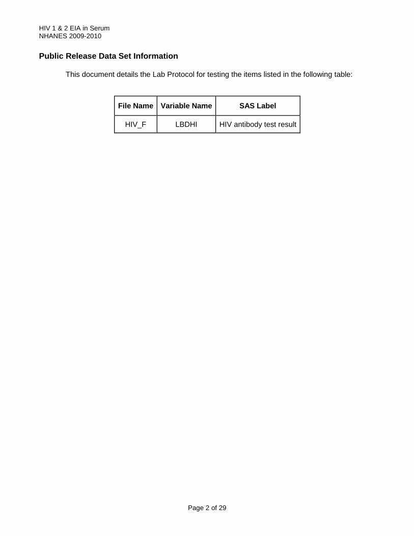
This document details the Lab Protocol for testing the items listed in the following table:
File Name Variable Name SAS Label
HIV_F LBDHI HIV antibody test result

HIV 1 & 2 EIA in Serum NHANES 2009-2010
Page 3 of 29
HIV-1/HIV-2 plus O EIA
1. Summary of Test Principle and Clinical Relevance- EIA
The Genetic Systems HIV-1/HIV-2 plus O EIA marketed by BioRad is an enzyme immunoassay utilizing recombinant proteins and synthetic peptides for the detection of antibodies to HIV-1(Groups M and O) and /or HIV-2 in human serum, plasma, or cadavered serum specimens. It is indicated as a screening test for serum, plasma, or cadavered serum specimens and as an aid in the diagnosis of infection with HIV-1 and/or HIV-2.
Summary and Explanation of the Test
The Genetic Systems HIV-1/HIV-2 plus O EIA incorporates highly conserved recombinant and synthetic peptide sequences representing HIV-1 (groups M and O) and HIV-2. Despite some degree of immunological cross-reactivity between types and subtypes of HIV, reliable detection of the more divergent strains may only be achieved by incorporating specific sequences into the assay design. Serologic studies have also shown that the core proteins of HIV-1 and HIV-2 display frequent cross-reactivity whereas the envelope proteins are more type specific. Any specimen that reacts in an initial test with the Genetic Systems HIV-1/HIV-2 plus O EIA must be retested in duplicate with the Genetic Systems HIV-1/HIV-2 plus O EIA. Initially reactive specimens that are reactive in wither one or both duplicates from the repeat testing are referred to as repeatedly reactive. Repeatedly reactive specimens may contain antibodies to either HIV-1 and /or HIV-2. Therefore, additional, more specific or supplemental tests for antibodies to both HIV-1 and HIV-2 such as Western blot or immunoflourescence must be performed to verify the presence or absence of antibodies to HIV-1 and HIV-2. Recommendations for appropriate use of such additional tests may be issued periodically by the United States Public Health Service.
Biological Principles of the Procedure The Genetic Systems HIV-1/HIV-2 plus O EIA is an immunoassay based on the principle of direct the antibody sandwich technique. Microwell strip plates (the solid phase) are coated with purified antigens: gp160 and p24 recombinant proteins derived from HIV-1; a peptide representing the immunodominant region of the HIV-2 transmembrane glycoprotein, gp36; and a synthetic polypeptide mimicking an artificial (i.e., encoded by no existing virus) HIV-1 group O specific epitope.
During the assay, specimens are evaluated for the presence of HIV-1 and HIV-2 antibodies by interaction with the absorbed peptides in the wells. Specimens are diluted in specimen diluent and added to each well. The wells are incubated and then washed. The next step is the addition of a Colored Conjugate Solution (green), which contains peroxidase-conjugated antigens (peptides mimicking various immunodominant epitopes of the HIV-1 and HIV-2 trasmembrane glycoprotein, and a p24 recombinant protein). If antibodies to either HIV-1 or HIV-2 are present, they bind to the adsorbed antigen and are not removed by washing. The working conjugate solution, peroxidase-labeled goat anti-human immunoglobulin, is then added to the wells and binds with the antibody-antigen complex, if present. Unbound conjugate is removed by a wash step. Working chromogen solution, TMB, is then added to the plate and allowed to incubate. A blue or blue-green color develops in proportion to the amount of antibody that has been bound to the antigen-coated plate. The enzyme reaction is stopped by the addition of acid, which results in a color change to yellow. The optical absorbance of controls and specimens is determined with a spectrophotometer with wavelength set at 450nm.
All repeatedly positive specimens are confirmed by Western blot (method at the end of this section).

HIV 1 & 2 EIA in Serum NHANES 2009-2010
Page 4 of 29
2. Safety Precautions
A. The positive and negative controls are heat treated to inactivate viruses. However, handle assay specimens and controls as if capable of transmitting infectious agents. Use of good laboratory practices and CDC-NIH guidelines is recommended.
B. Test operators should adhere to the Occupational Safety and Health Administration (OSHA) regulations (29 CFR 19.10).
C. Keep testing area separate from areas in which blood or blood products for transfusion are stored.
D. Do not use reagents beyond the expiration date printed on the reagent label.
E. With the exception of Substrate Solution, Substrate Diluent, Wash Solution, and Stop Solution, do not interchange reagents from different lots or kits.
F. Do not substitute Chromagen and/or Substrate Buffer with color development solutions from other Genetic Systems tests.
G. Mix all liquid reagents by gently inverting 3 to 5 times, just prior to use.
H. Prior to performing the test, bring to room temperature only as many strips of microwells as needed to perform the test run. Any strip of microwell that is not to be used in the current test run should be sealed in the foil bag with desciccant and stored at 2-8°C.
I. Remove reagents from the refrigerator storage approximately 60 minutes before beginning assay. Bring kit reagents to room temperature (15-30°C) prior to use. Return all kit components to their recommended storage conditions immediately after use.
J. For the manual pipetting of controls and specimens, use individual pipette tips to eliminate carryover of samples.
K. Handle negative and positive controls in the same manner as patient specimens.
L. If a specimen is inadvertently not added to well, the assay result will read nonreactive.
M. Inadequate adherence to package insert instructions may result in erroneous or invalid results.
N. The Genetic Systems HIV-1/HIV-2 plus O EIA performance is highly dependent upon incubation times and temperatures. Temperatures outside of the validated ranges may result in invalid assays. Incubation temperatures should be carefully monitored using calibrated thermometers, or equivalent.
O. Use only adequately calibrated equipment with this assay.
P. Use of dedicated equipment is recommended if equipment performance validations have not precluded the possibility of cross-contamination.
R. Use clean, disposable polypropylene (not polystyrene) tubes and reagent reservoirs for reagent
preparation and dispensing. S. Avoid exposing Chromagen or Working TMB solution to strong light during storage and
incubation. Do not allow chromagen solutions to come into contact with an oxidizing agent. T. Avoid contact with Stopping Solution. Do not allow Stopping Solution to come into contact with
strong bases, oxidizing agents or metals. U. Care must be exercised when dispensing samples to avoid cross contamination through
aerosols or carryover. Use a clean tip for dispensing each individual sample. V. Inadequate adherence to package insert instructions may result in erroneous results. W. Use only adequately calibrated equipment with this assay.

HIV 1 & 2 EIA in Serum NHANES 2009-2010
Page 5 of 29
WARNINGS FOR USERS
1. The HIV-1 and HIV-2 positive controls are heat-treated to inactivate viruses. However,
handle all the reagents as though capable of transmitting infection. All tests should be conducted using the precautions recommended for blood borne pathogens, as defined by OSHA regulations.
2. Do not pipette by mouth. 3. Do not smoke, drink, or eat in areas where specimens or kit reagents are being handled. 4. Wear protective clothing and disposable gloves while handling, the kit reagents. Wash
hands thoroughly after performing the test. 5. Handle Chromogen Reagent with care since DMSO is readily absorbed through the skin. 6. The Stopping Reagent is an acid. Wipe up spills immediately and flush the area with water.
If the Stopping Reagent contacts the skin or eyes, flush with copious amounts of water and seek medical attention.
7. BIOLOGICAL SPILLS: Spills not containing acid should be wiped thoroughly with an
effective disinfectant. Disinfectants that can be used include (but are not limited to) a solution of 10% bleach (O.5% solution of sodium hypochlorite), 70% ethanol, or 0.5% Wescodyne. Spills containing acid should be wiped dry. The area of the spill should be wiped with one of the chemical disinfectants. Materials used to wipe up spills should be disposed of as biohazardous waste.
8. Note: DO NOT PLACE SOLUTIONS CONTAINING BLEACH IN THE AUTOCLAVE.
9. Dispose of all specimens and materials used to perform the test as though they contain an
infectious agent. Disposal should comply with all applicable waste disposal requirements.
10. Sodium azide is included as a preservative in the positive and negative controls. Sodium azide has been reported to form lead or copper azide in laboratory plumbing. These azides are explosive. To prevent azide build-up if solutions containing azide are disposed of in the sink after biological inactivation, flush plumbing with a large volume of water.
3. Computerization; Data System Management
HIV antibody results are manually entered into a Microsoft Excel result file spreadsheet. After a run is complete and any additional corrections by the analyst are made, the Excel result file is prepared. Data is transmitted electronically to Westat’s ISIS computer system weekly and transferred from there to NCHS.
4. Specimen Collection, Storage, and Handling Procedures; Criteria for Specimen
Rejection
WARNING for in vitro diagnostic users: FDA has licensed this test for use with serum and plasma specimens only. Use of this licensed test kit with specimens other than those specifically approved for use with this test kit may result in inaccurate test results.

HIV 1 & 2 EIA in Serum NHANES 2009-2010
Page 6 of 29
A. Serum, plasma or cadavered serum specimens may be used in the test. B. The following anticoagulants have all been evaluated and found to be acceptable: EDTA,
sodium and lithium heparin, sodium citrate, CPD, CPDA 1, and ACD. Specimens which are collected into anticoagulant tubes should completely fill the tube as label indicates to avoid improper dilution. Specimens with observable particulate matter should be cleared by centrifugation prior to testing.
C. No clinically significant effect on assay results has been detected with increased levels of
protein, lipids, bilirubin, hemolysis, or after heat inactivation of patient samples. D. Specimens may be stored at 2–8°C for 7 days. For long, term storage, the specimens
should be frozen (at 20°C or colder). Samples should not be used if they have incurred more than 5 freeze-thaw cycles. Mix samples thoroughly after thawing.
E. If specimens are to be shipped, they should be packed in compliance with Federal
Regulations covering the transportation of etiologic agents. Studies have demonstrated that specimens may be shipped refrigerated (2–-8°C) or at ambient temperature for up to 7 days. For shipments that are in transit for more than 7 days, specimens should be kept frozen (20°C or lower).
F. The kit is not licensed for use with specimens other that serum, plasma, or cadavered
serum specimens. This kit is not intended for use on saliva/oral fluid or urine samples. 5. Procedures for Microscopic Examinations; Criteria for Rejection of Inadequately
Prepared Slides Not applicable for this procedure 6. Equipment and Instrumentation, Materials, Reagent Preparation, Calibrators
(Standards), and Controls
A. Reagent Preparation
1. Working Conjugate Solution
Note: Conjugate Concentrate should be kept in the refrigerator and immediately returned to the refrigerator after use. To avoid contamination of the Conjugate, wear clean gloves and do not touch tips of pipettes. Prepare a 1:11 dilution: Bring Conjugate Diluent to room temperature. Invert both the Conjugate Concentrate and Conjugate diluent to mix before using. Prepare a 1:11 dilution for each strip to be tested by adding 100µL of Conjugate Concentrate to 1ml Conjugate Diluent in a clean polypropylene tube. Mix well. Working Conjugate should be green. Note Concentrate lot number, Date and time of preparation, and date of expiration of the working Conjugate solution. Working Conjugate Solution is stable for 8 hours at room temperature. Working Conjugate solution should be mixed prior to use.
2. Working Chromogen Solution
Prepare a 1:11 dilution for each strip being tested. Bring chromogen reagent and chromogen diluent to room temperature. Invert the

HIV 1 & 2 EIA in Serum NHANES 2009-2010
Page 7 of 29
chromogen reagent and chromogen diluent to mix before using. Prepare a 1:11 dilution for each strip to be tested by adding 100µL of chromogen reagent to 1 ml of chromogen diluent in a clean polypropylene tube. (DO NOT USE A POLYSTYRENE CONTAINER.) Note chromogen reagent lot number, date and time of preparation and time of expiration of the working chromogen solution (8 hours from preparation). Mix working solution gently when combined. Working chromogen solution should be kept in the dark at room temperature prior to use. Chromogen reagent should be colorless to very pale yellow. Any other color indicates that the reagent is contaminated and should not be used. The working chromogen solution should be colorless. A distinct blue color indicates that the reagent is contaminated. Discard the working chromogen solution and prepare fresh reagent in a clean container. Prepare only the amount of the reagent to be used within 8 hours, ensuring that the volume of diluted reagent will be adequate for the entire plate(s). Extra chromogen reagent is provided.
3. Working Wash Solution
Prepare working wash solution as needed by adding one part wash solution concentrate (30×) to 29 parts of water (e.g. 120 mL of wash solution to 3480 mL of water). Use deionized or distilled water. Clinical laboratory reagent water Type I or Type II is acceptable. The working wash solution can be stored at room temperature for four weeks in a plastic container. Note lot number, date prepared and expiration date. Discard if no foaming is evident in the working-wash solution. Prepare a sufficient quantity of working wash solution to complete a full plate run.
7. Calibration and Calibration Verification Procedures
A. Validation of Results
Mean Negative Control absorbance value (NCx)
Determine the mean absorbance for the negative and positive controls by dividing the sum of their absorbance values by the number of acceptable controls. The individual negative control absorbance values must be greater than equal to 0.000 AU and less than or equal to 0.150 AU. One negative control absorbance value may be discarded if it is outside this range. The NCx may be calculated from the two remaining values.
Determine the mean of the negative controls as shown in the example below.
HIV-1 Negative Control (HIV-1 NCx)
Sample Number Absorbance Total absorbance/3 = 0.239/3 = 0...080(HIV-1 NCx) 1 0.075
2 0.083 3 0.081 0.239
B. Calculation of Results:
Cut-off Value
Determine the cutoff by adding 0.250 to NCx, as shown in the example below:
NCx = 0.080 Cutoff Value = 0.080 + 0.250 = 0.330
C. Assay Validation

HIV 1 & 2 EIA in Serum NHANES 2009-2010
Page 8 of 29
A run is valid if the following criteria are met:
The absorbance value of each negative control is greater than or equal to 0.000 AU and less than or equal to 0.150 AU. One negative control value may be discarded, and the mean of negative controls (NCx) may be calculated from the two remaining values. If two or more negative controls are out of limit, the plate is invalid and must be repeated.
The absorbance value of the HIV-1 Positive Control must be greater than or equal to 0.700 AU. The absorbance value of the HIV-2 positive Control must be greater than or equal to 0.700 AU. The absorbance value of the HIV-1 Group O positive Control must be greater than or equal to 0.700 AU.
If any of these criteria have not been met, the assay is invalid and must be repeated. D. Other Materials
Materials required but not provided:
1. Precision pipettes to deliver 20–200 µL, 1 mL, 5 mL and 10 mL, 25 ml (accurate within
+10%)and corresponding pipette tips, multichannel pipettors capable of delivering 25 µL and 100 µL (optional).
2. Appropriately sized graduated cylinders.
3. Dry heat incubator capable of maintaining 37 + 2°C. Calibrated thermometer.
4. Room temperature incubator 25 + 10 C. Calibrated thermometer
5. Microwell plate or strip washer qualified for use with this assay. The washer must be capable of dispensing at least 400 µL per well, cycling 5 times, and soaking for 30-60 seconds between each wash.
6. Microwell strip reader qualified for us with this assay. The spectrophotometer should have the following specifications at wavelength 450 nm:
a. Bandwidth: 10 nm HBW (half band width) or equivalent
b. Absorbance range: 0–2.0 AU (absorbance units)
c. Repeatability: + (0.5%+ 0.005) AU
d. Linearity or accuracy: 1% from 0 to 2.0 AU
7. The instrument should contain a reference filter for reading at 615–630 nm. An instrument without a reference filter can be used; however areas in the bottom of the wells that are opaque, scratched or irregular may cause absorbance readings that are falsely elevated.
8. Household bleach (5% to 8% sodium hypochlorite). Alternative disinfectants include: 70% ethanol or 0.5% Wescodyne (West Chemical Products).
9. Paper towels or absorbent pads for blotting.
10. Labeled null strips for testing partial plates
11. Clean polypropylene container for preparation of working TMB (chromogen) solution. (Do not use polystyrene) Clean container for preparation of working conjugate solution.
12. Deionized or distilled water. Clinical laboratory reagent water Type I or Type II is acceptable.
13. Gloves
14. Laboratory timer

HIV 1 & 2 EIA in Serum NHANES 2009-2010
Page 9 of 29
15. EIA polypropylene reagent reservoirs (optional) 8. Procedure Operating Instructions; Calculations; Interpretation of Results
A. Perform equipment maintenance and calibration where necessary, as required by the
manufacturer.
1. Remove kit (except for conjugate concentrate) from refrigerator and warm to room temperature on counter.
Be sure to confirm that kit is till in date and that all reagents in the kit are still in date. Record lot # & exp.
Prior to first time use of a kit with a new lot number to test patient samples, kit controls and external controls should be tested using the new kit.
2. Record all temperatures and gauge readings required for lab and reagents. Turn on
components required for biological safety hood. 3. Arrange samples to be tested in box in rows of 8. Plate map will be made from this
sequence of samples. 4. Turn on computer, reader and printer. ID is Imlab and type in password. Click on KC4 icon.
Click on protocol and choose Bio-Rad 1-2. Click on data and then new plate, and then samples. The screen for entering samples will appear.
5. Scan each sample ID found on each tube in the order in which they are in the box.
Progress to successive samples by the down arrow. The first row on the microwell plate will be the controls (neg., neg., neg, HIV-1+, HIV-2+, HIV-1 group O+, & accurun) This row is programmed in so the screen for entering the samples starts with row 2 and the first sample. Continue until all samples are entered in the correct order.
6. Print the plate as a map. Then choose OK. Then save plate as the date being run. 7. Insert required number of plate strips needed according to the plate map into the plate
frame. Number the strips. Only strips from kits with the same lot number may be mixed in any test run; however, if strips from a yet untested kit but of the same lot number, be sure to run one strip of controls from that set of strips as well. Return antigen coated strips not needed to the pouch and seal-these may be used at a later time. Place null strips in the rows not needed for the assay.
Each strip is coated with purified HIV 1 and HIV 2 antigens. Purified gp 160 and p24
recombinant antigens derived from HIV-1; a peptide representing the immunodominant region of the HIV-2 transmembrane glycoprotein, gp 36; and a synthetic polypeptide mimicking an artificial (i.e. encoded by no existing virus) HIV-1 group O specific epitope.
8. Dilute specimens and controls 3:4 in specimen diluent. Arrange tubes for diluting in rows of
8 corresponding to the rows on the microwell plate. Using a multi-channel pipette, add 35µL of specimen diluent (purple in color but will change to blue after the sample is added) to each tube. Deliver the specimen diluent to the bottom of the tube to avoid leaving any on the sides of tube. Using a single channel pipette, deliver 105µL of sample or control to appropriate tube using the plate map as a guide. Mix 3 times avoiding foaming. Be sure to use a new tip for each sample or control. After addition of each sample or control, move the tube to another rack in correct order to avoid confusion.

HIV 1 & 2 EIA in Serum NHANES 2009-2010
Page 10 of 29
9. After dilutions have been made of all controls and samples, use the multi-channel pipette to mix (carefully avoiding foaming) and deliver 100µL of each diluted sample or control to the appropriate microwell. Use the plate map as a guide. Be sure to use new tips for each row.
10. Cover the plate with a plate sealer being careful not to jar the plate causing spilling and
ensuring that all wells are covered. Place the plate in the 37oC incubator for 60 minutes. Set a clock and fill out the run sheet.
11. Return the specimens to the refrigerator. 12. Prepare wash buffer. Dilute 30x wash concentrate 1 part to 29 parts dIH2O (i.e. 60 mls 30x
wash concentrate plus 1740 parts dIH2O). Diluted wash buffer can be stored at RT for up to 4 weeks in a plastic container. Label the container with the date prepared, lot number and expiration date, and preparer’s initials. Mix using stir bar on stirrer until ready to set up Dynex Ultrawash Plus.
13. Fifteen minutes before the end of the incubation, set up the Dynex Ultrawash Plus. Attach
the plastic container with the diluted wash buffer to tubing 6 & 7. Be sure the top is screwed tightly so that the pressure gauge will be at 9. Be sure that the waste bottle is not full. Turn on the top switch in the back of the instrument. Then turn on the pump switch (the bottom switch). Be sure that a ‘wash plate’ is on the tray and then press purge. Verify that the program is set to deliver 500µL wash buffer/ well/ wash; to soak for 30-60 sec., & for 3 wash cycles. After the purge, turn of the pump switch (bottom switch and leave the top switch on).
14. When the 60 minute incubation is complete, carefully peel off the plastic seal avoiding and
spilling or cross contamination. 15. Place the plate on the washer tray. Be sure it is sitting securely inside the front pins and on
all sides. Press start. 3 wash cycles will be performed and then a beeper will sound. At that point turn
the plate around on the tray and press start again. 3 more wash cycles will be performed so that the plate will be washed a total of 6 wash cycles.
16. While the plate is washing, make up the working conjugate which is the next reagent to be
added to the plate. Working conjugate is made by diluting the Conjugate Concentrate 1:11. The Conjugate Diluent should be at room temperature before using. Invert both the Conjugate Diluent and the Conjugate Concentrate to mix before making the dilution. Prepare the 1:11 dilution in a clean polypropylene tube by adding 100µL of Conjugate Concentrate to 1ml Conjugate Diluent per strip.(i.e. for 3 microwell strips add 300µL Conjugate Concentrate to 3 mls. Conjugate Diluent). The Conjugate Concentrate is green and should be kept refrigerated. Working conjugate is good for 8 hours at room temperature.
17. Turn off the bottom switch. Remove the plate from the washer. Blot out any remaining liquid
by pinching the plate frame at the center sides and knocking the plate onto absorbent paper. If any of the strips fall out, be sure to place in the correct position in the plate frame according to the numbers and correct orientation.
18. Mix the working conjugate. Add 100µL of working conjugate to each well using a multi-
channel pipette. It is not necessary to change tips between strips if the tips do not touch the wells.
19. Cover the plate with the plate sealer so that all wells are covered. Place in the 37oC.
incubator for 30 min. (+/- 5 min.) Set a timer and enter the times on the run sheet.

HIV 1 & 2 EIA in Serum NHANES 2009-2010
Page 11 of 29
20. When the 30 min. incubation is complete, remove the plate from the incubator, carefully remove the plate seal, and wash and blot the plate as in steps # 16 & 17.
21. While the plate is washing, prepare the working TMB solution. Working TMB is made by
preparing a 1:11 dilution Chromagen: Subtrate Buffer. Bring both the Chromagen and Substrate Buffer to room temperature before using. Invert both to mix before making the dilution. Prepare the 1:11 dilution in a clean polypropylene tube by adding 100µL of Chromagen to 1ml Substrate Buffer per strip.(i.e. for 3 microwell strips add 300µL Chromagen to 3mls. Substrate Buffer). Working TMB should be colorless. A blue color indicates contamination; if present discard and prepare fresh working TMB. Working TMB should be kept in the dark at room temperature and used within 8 hours following preparation.
22. Turn off the bottom switch of the plate washer. Remove the plate from the washer. Blot out
any remaining liquid by pinching the plate frame at the center sides and knocking the plate onto absorbent paper. If any of the strips fall out, be sure to place in the correct position in the plate frame according to the numbers and correct orientation.
23. Add 100µL Working TMB to each well, seal with the plate sealer and incubate in the Room
Temperature incubator (18 to 30 o C) in the dark for 30 minutes ( +/- 5 min.). Set a timer and enter the times on the run sheet.
24. At the end of 30 minutes, carefully remove the plate sealer and add 100µL Stopping
Solution (1N Sulfuric Acid) to each well. The reaction mixture will turn yellow. Tap the plate gently to assure mixing. Complete mixing is required for acceptable results.
25. Read the absorbance within 30 minutes. Ensure that all strips are pressed flush into the
plate. Insert the plate into the plate reader. Make sure the selected protocol is for the Bio-Rad EIA test method. Open the appropriate file for the run. Go to file and click on Read Plate. Fill in the information on the screen which appears. Then click on Start reading.
26. The results will appear on the screen when the reader has completed reading the plate. If
the controls are not in acceptable range, a notice will appear. Negative control absorbance should be greater than 0.00 and less than or equal to 0.150 AU. For the assay to be valid 2 out of 3 Negative controls must be in the acceptable range. The cut -off will be calculated by averaging the O.D. of the 3 negative controls and adding that average to 0.250. Positive Control absorbance must be equal to or greater than 0.700 AU. If any of the criteria for the kit controls is not met, the assay is invalid and must be repeated.
27. Interpretation of Results: The presence or absence of HIV Ab is determined by relating the
absorbance value of the specimen to the cut-off value. Specimens with absorbance values less than 0.00 must be repeated.
Specimens with absorbance values less than the cut-off are considered non-reactive and no further testing is required.
Specimens with absorbance values greater than or equal to the cut-off are considered initially reactive and should be retested in duplicate to validate the initial test results. If, after repeat testing, the absorbance values of both duplicate specimens are less than the cut-off, the specimen may be considered non-repeatedly reactive and negative for HIV-1(Groups M and O) and HIV-2 antibodies.
Reasons for non-repeatedly reactive specimens include: a. Improper washing of microwell plates
b. Cross-contamination of non-reactive specimens with HIV-1 and/or HIV-2 Ab from a high titered specimen.
c. Contamination of the Working TMB solution

HIV 1 & 2 EIA in Serum NHANES 2009-2010
Page 12 of 29
d. Contamination of then Stopping Solution
If, after repeated testing, the absorbance value of either of the duplicates is greater than or equal to the cut-off value, the specimen must be considered repeatedly reactive.
8. Procedure Operating Instructions; Calculations; Interpretation of Results
The presence or absorbance of antibodies to HIV-1 and or HIV-2 is determined by relating the absorbance value of the specimens to the cutoff value. The cutoff value is determined by adding 0.250 to the mean absorbance value of the negative controls.
A. Specimens with absorbance values that are less than 0.000 must be repeated.
Those with values greater than the upper linearity limits of the reader should be reported as reactive.
B. Specimens with absorbance values less than the cutoff value are considered non-reactive by
the Genetic Systems HIV1/HIV-2 plus O EIA and may be considered negative for HIV-1 (M and O groups) and HIV-2 antibodies. Further testing is not required.
C. Specimens with absorbance values greater than or equal to the cutoff value are considered
initially reactive by the Genetic Systems HIV1/HIV-2 plus O EIA. Initially reactive specimens should be retested in duplicate to validate the initial test results. If, after repeat testing, the absorbance values of both duplicate specimens are less than the cutoff value, the original specimen may be considered non-repeatedly reactive and negative for HIV-1 (Groups M and O) and HIV-2 antibodies.
D. Reasons for non-repeatedly reactive specimens include: 1. Improper washing of microwell plates
2. Cross contamination of non-reactive specimens with HIV-1 and/or HIV-2 serum of a high titered specimen.
3. Contamination of the Working TMB solution by oxidizing agents (sodium hypochlorite, hydrogen peroxide, etc)
4. Contamination of the Stopping solution E. If after repeat testing, the absorbance value of either of the duplicates is greater than or
equal to the cutoff value, the specimen must be considered repeatedly reactive. F. Repeatedly reactive specimens according to the algorithm will undergo further testing:
Western blot and Multispot. G. Result entry
1. Open the NHANES master file on the shared drive. Enter the data being sure to match the correct specimen with the correct specimen number.
2. Have an objective review of the data entry.
3. Report results: Open correct shipment file. Enter the date received, the date analyzed,
the run number, the tech initials, the result code, and the result comment into the spreadsheet. Save as reports in the CSV file format. Save, ok, and yes. Do not save changes. Close shipment file.
4. Send report files by posting via FTP site (open and verify each file for accuracy before
sending). Go to the FTP site:
5. ftp.westat.com

HIV 1 & 2 EIA in Serum NHANES 2009-2010
Page 13 of 29
6. user: lab
7. Password: rta$6171
8. Find our folder: Lab03
9. Drag the report files to be reported to the Lab03 folder.
10. Close out of the site.
11. An e-mail will be sent to acknowledge receipt of the report files.
9. Reportable Range of Results
Reportable results are expressed as positive or negative. 10. Quality Control (Qc) Procedures
One negative and one positive control are included in each microplate in every analytical run (a set of consecutive assays performed without interruption).
Each sample is tested in duplicate and each assay is read by two individuals independently. Discrepancy in the results leads to repeat testing of the sample involved.
11. Remedial Action If Calibration Or Qc Systems Fail To Meet Acceptable Criteria
a. Repeat the test if both readers do not agree about the results in the duplicate wells.
b. Do not report results from runs in which the controls did not meet expected reactivities. 12. Limitations of Method; Interfering Substances and Conditions
A. The Genetic Systems HIV-1/HIV-2 plus O EIA procedure and the interpretation of Results must be followed closely when testing for the presence of antibodies to HIV-1 and/or HIV-2 in plasma, serum or cadavered serum specimens. The user of the kit is advised to read the package insert carefully prior to conducting the test. In particular, the test procedure must be carefully followed for sample and reagent pipetting, plate washing, and time and temperature of the incubation steps. Testing of other body specimens, pooled blood or processed plasma, and products made from such pools is not recommended. Data regarding the interpretation were derived from testing serum or plasma samples. Insufficient data are available to interpret test performed on other body specimens, pooled blood or processed plasma, and products made from such pools.
B. The Genetic Systems HIV-1/HIV-2 plus O EIA detects circulating antibodies to HIV-1 ( Groups
M and O) and HIV-2 and thus is useful in screening blood and plasma donated for transfusion and further manufacture, in screening cadaveric serum for tissue donation, in evaluating patients with signs or symptoms of AIDS, and in establishing prior infection with HIV-1 or HIV-2. Clinical studies continue to clarify and refine the interpretation and medical significance of the presence of antibodies to HIV-1 and /or HIV-2. Repeatedly reactive specimens must be investigated by additional tests, more specific or supplemental tests. Recommendations for appropriate use of such additional tests may be issued periodically by the United States Public Health Service. For individuals who are confirmed positive for antibodies, appropriate

HIV 1 & 2 EIA in Serum NHANES 2009-2010
Page 14 of 29
counseling and medical evaluation should be offered. Both confirmation of the test result on a freshly drawn sample and counseling should be considered an important part of testing for antibody to HIV-1 and HIV-2.
C. A negative test result at any point in the investigation of individual subjects does not preclude
the possibility of exposure to or infection with HIV-1 and /or HIV-2. D. False negative results can occur if the quantity of the marker present in the sample is too low
for the detection limits of the assay, or if the maker which is detected is not present during the stage of disease in which a sample is collected.
E. Failure to add specimen or reagent as instructed in the procedure could result in a falsely
negative test. Repeat testing should be considered where there is clinical suspicion of infection or procedural error.
F. AIDS and AIDS-related conditions are clinical syndromes and their diagnosis can only be
established clinically. Testing alone cannot be used to diagnose AIDS, even if the recommended investigation of reactive specimens suggests a high probability that the antibody to HIV-1 and /or HIV-2 is present.
G. The risk of an asymptomatic person with a repeatedly reactive serum developing AIDS or an
AIDS-related condition is not known, as the course of HIV infection may vary among individual patients and may be altered by antiretroviral therapy. However, in a prospective study, AIDS developed in 51% of homosexual men after 10 years of infection.
H. Data obtained from testing persons both at increased and at low risk for HIV-1/2 infection
suggest that repeatedly reactive specimens with high reactivity on the Genetic Systems HIV-1/HIV-2 plus O EIA may be more likely to demonstrate the presence of antibodies to HIV-1( Group M and O) and/or HIV-2 by additional, more specific or supplemental testing. Borderline reactivity is more frequently nonspecific, especially in samples obtained from persons at low risk for infection with HIV-1 or HIV-2; however the presence of antibodies to HIV-1 and/or HIV-2 in some of these specimens can be demonstrated by additional, more specific or supplemental testing, or by testing a subsequent sample drawn at a later date (e.g. 3 or 6 months).
I. It is generally recognized that detection of HIV antibody in infants born to seropositive
mothers is not adequate to diagnose HIV infection in the infant, since maternal IgG frequently persists for as long as 18 months after birth. Supplemental assays designed specifically for neonatal specimens may be helpful in resolving such cases.
J. An absorbance value of less than 0.000 AU may indicate a procedural or instrument error which
should be evaluated. The result is invalid and the specimen must be re-run. K. Factors that can affect the validity of results include failure to add the specimen or reagents to
the well, inadequate washing of microplate wells, failure to follow stated incubation times and temperatures, addition of the wrong reagents to specific wells, the presence of metals, or the splashing of bleach into wells.
L. Non-repeatedly reactive specimens can be caused by: Improper washing of microplate wells,
during the initial test; cross-contamination of non-reactive specimens with HIV antibody from a high-titered specimen; contamination of the chromogen reagent solution by oxidizing agents (sodium hypochlorite, hydrogen peroxide, etc.); contamination of the stopping reagent.
M. A person who has antibodies to HIV-1 is presumed to be infected with the virus. Except a
person who has participated in a vaccine study may develop antibodies to the vaccine and may or may not be infected with HIV. Clinical correlation is indicated with appropriate counseling, medical evaluation, and possibly additional testing to decide whether a diagnosis of HIV infection is accurate.

HIV 1 & 2 EIA in Serum NHANES 2009-2010
Page 15 of 29
13. Reference Ranges (Normal Values)
A normal sample is negative for HIV antibodies. 14. Critical Call Results (Panic Values)
Not applicable to this assay method. 15. Specimen Storage and Handling During Testing
Specimens are stored at ≤ –70°C until testing. After an aliquot of the thawed sample has been removed for testing, the residual is refrozen and stored at ≤ –70°C.
16. Alternate Methods for Performing Test or Storing Specimens If Test System Fails
If the analytical system fails, it is preferable to store specimens at ≤ –70°C until the system is returned to functionality.
17. Test Result Reporting System; Protocol For Reporting Critical Calls (If Applicable)
Not applicable to this assay method. 18. Transfer or Referral of Specimens; Procedures for Specimen Accountability and Tracking
Standard record keeping involves using the computerized database and the hard copy results themselves to track specimens. Records are maintained indefinitely. Only numerical identifiers (e.g., case ID numbers) should be used. All personal identifiers should be available only to the medical supervisor or project coordinator to safeguard confidentiality.
For the NHANES study, residual serum is retained at ≤ –70°C for 1 year and then returned to NCHS serum bank.
19. Summary Statistics and QC Graphs
Qualitative assays are assays with a positive, negative or borderline/indeterminate result. Since the controls do not generate quantitative values, plots are not generated for quality control purposes.

HIV 1 & 2 EIA in Serum NHANES 2009-2010
Page 16 of 29
References Centers for Disease Control: Provisional Public Health Service inter-agency recommendations for screening donated blood and plasma for antibody to the virus causing acquired immunodeficiency syndrome. Morbidity and Mortality Weekly Rep 34:5-7, 1985.
Delmonico FL, Snydman DR: Organ donor screening for infectious diseases. Transplantation 65(5):603-610, 1998.
Barre-Sinoussi F, Chermann JC, Rey F, et al: Isolation of a T-lymphotropic retrovirus from a patient at risk for acquired immune deficiency syndrome (AIDS). Science 220:868-871, 1983.
Gallo RC, Salahuddin SZ, Popovic M, et al: Frequent detection and isolation of cytopathic retroviruses (HTLV-III) from patients with AIDS and at risk for AIDS. Science 224:500-503, 1984.
Coffin J, Haase A, Levy JA, et al: What to call the AIDS virus? Nature 321:10, 1986.
Clavel F, Guetard D, Brun-Vezinet F: Isolation of a new human retrovirus from West African patients with AIDS. Science 233:343-346, 1986.
Clavel F, Manshino K, Chameret S, et al: Human immunodeficiency virus type 2 infection associated with AIDS in West Africa. New Engl J Med 316:1180-1185, 1987.
Schim van der Loeff MF and Aaby P: Towards a better understanding of the epidemiology of HIV-2. AIDS 13(Suppl. A):S69-S84, 1999.
Centers for Disease Control: AIDS due to HIV-2 infection - New Jersey. Morbidity and Mortality Weekly Rep 37:33-35, 1988.
Hoff R, Weiblen BJ, Schwerzler M, et al: Specific antibodies to HIV-2 detected in an anonymous newborn blood specimen from Massachusetts. Fourth Consensus Conference on Testing for Human Retroviruses, March 1989.
Ayanian JZ, Maguire JH, Marlink RG, et al: HIV-2 infection in the United States. New Engl J Med 320:1422-1423, 1989.
O’Brien TR, George JR, Holmberg SD: Human immunodeficiency virus type 2 infection in the United States. JAMA 267:2775-2779, 1992.
Centers for Disease Control: Update: HIV-2 infection among blood and plasma donors--United States, June 1992-June 1995. Morbidity and Mortality Weekly Rep 44:603-606, 1995.
Sullivan PS, Fleming PL: Surveillance for HIV-2 in the United States: Update and recommendations for future surveillance. Presented at the Association of Public Health Laboratories Conference, Charlotte, NC, March 6-9, 2000.
Brun-Vezinet F, Katlama C, Roulot D, et al: Lymphadenopathy associated virus type 2 in AIDS and AIDS-related complex. Lancet 1:128-132, 1987.
Quinn TC, Zacarias FRK, St. John RK: AIDS in the Americas: an emerging public health crisis. New Engl J Med 320:1005-1007, 1989.
Guyader M, Emerman M, Sonigo P, et al: Genome organization and transactivation of the human immunodeficiency virus type 2. Nature 326:662-669, 1987.
Cabrian K, Shriver K, Goldstein L, et al: Human immunodeficiency virus type 2: a review. J Clinical Immunoassay 11:107-114, 1988.
Janssens W, Buvé A, Nkengasong JN: The puzzle of HIV-1 subtypes in Africa. AIDS 11:705-712, 1997.
Charneau P, Borman AM, Quillant C, et al: Isolation and envelope sequence of a highly divergent HIV-1 isolate: definition of a new HIV-1 group. Virology 205:247-253, 1994.
HIV 1 & 2 EIA in Serum NHANES 2009-2010
Page 17 of 29
Simon F, Mauclère P, Rogues P, et al: Identification of a new human immunodeficiency virus type 1 distinct from group M and group O. Nature Medicine 4:1032-1037, 1998.
Gao F, Yue L, Robertson DL, et al: Genetic diversity of human immunodeficiency virus type 2: evidence for distinct subtypes with differences in virus biology. J Virology 68:7433-7447, 1994.
George JR, Rayfield M, Philips S, et al: Efficacies of U.S. FDA-licensed HIV-1 screening enzyme immunoassays for detecting antibodies to HIV-2. AIDS 4:321-326, 1990.
Loussert-Ajaka I, Ly TD, Chaix ML, et al: HIV-1/HIV-2 seronegativity in HIV-1 subtype O infected patients. Lancet 343:1393-1394, 1994.
Schable C, Leopold Z, Pau C-P, et al: Sensitivity of United States HIV antibody tests for detection of HIV-1 group O infections. Lancet 344:1333-1334, 1994.
Starcich BR, Hahn BH, Shaw GM, et al: Identification and characterization of conserved and variable regions in the envelope gene of HTLV-III/LAV, the retrovirus of AIDS. Cell 45:637-648, 1986.
Wang JJG, Steel S, Wisniewolski R, Wang CY: Detection of antibodies to human T-lymphotrophic virus type III by using a synthetic peptide of 21 amino acid residues corresponding to a highly antigenic segment of gp41 envelope protein. Proc Nat Acad Sci USA 83:6159-6163, 1986.
Cosand WL: Synthetic antigen for the detection of AIDS-related disease. U.S. Patent #4,629,783, 1986.
Fenouillet E, Sorensen A-M, Lacroix M, Coutellier A, Herson S, Fretz-Foucault C, Gluckman J-C: Early and specific diagnosis of seropositivity to HIVs by an enzyme-linked immunosorbent assay using env-derived synthetic peptides. AIDS 4:1137-41, 1990.
Gnann JW, McCormick, Mitchell S, Nelson J, Oldstone MBA: Synthetic peptide immunoassay distinguishes HIV type 1 and HIV type 2 infections. Science 237:1346-1349, 1987.
Gnann JW, Nelson JA, Oldstone MBA: Fine mapping of an immunodominant domain in the transmembrane glycoprotein of human immunodeficiency virus. J Virol 61:2639-2641, 1987.
Alizon M, Wain-Hobson S, Montagnier L, Sonigo P: Genetic variability of the AIDS virus: nucleotide sequence analysis of two isolates from African patients. Cell 46:63-74, 1986.
Bos ES, van der Doelen AA, van Rooy N, Schuurs AHWM: 3, 3’, 5, 5’ - tetramethylbenzidine as an ames test negative chromogen for horseradish peroxidase in enzyme immunoassay. J Immunoassay 2:187-204, 1981.
Garner RC, Walpole AL, Rose FL: Testing of some benzidine analogues for microsomal activation to bacterial mutagens. Cancer Letters 1:39-42, 1975.
Resnick L, Veren K, Salahuddin SZ, et al: Stability and inactivation of HTLV-III/LAV under clinical and laboratory environments. JAMA 255:1887-1891, 1986.
Sarngadharan MG, Markham PD: The role of human T-lymphotropic retroviruses in leukemia and AIDS, in Wormser GP (ed): AIDS and Other Manifestations of HIV Infection. New Jersey, Noyes Publications, 1987, pp 218-220.
Bond WW, Favero MS, Petersen NJ, et al: Inactivation of hepatitis B virus by intermediate-to-high level disinfectant chemicals. J Clin Micro 18:535-538, 1983.
Sehulster LM, Hollinger FB, Dreesman GR, Melnick JL: Immunological and biophysical alteration of hepatitis B virus antigens by sodium hypochlorite disinfection. Appl Environ Microbiol 42:762-7, 1981.
NCCLS: Preparation and testing of reagent water in the clinical laboratory - second edition; approved guideline. National Committee for Clinical Laboratory Standards, document C3-A3, 17(18), (ISBN1-562328-336-1), 1997.
Centers for Disease Control: 1993 Revised classification system for HIV infection and expanded surveillance case definition for AIDS among adolescents and adults. MMWR 41(No. RR-17):1-19, 1992.
Pantaleo G, Graziosi C, Fauci AS: The immunopathogenesis of human immunodeficiency virus infection. New Engl J Med 328:327-335, 1993.

HIV 1 & 2 EIA in Serum NHANES 2009-2010
Page 18 of 29
Cohen OJ, Fauci AS: Current strategies in the treatment of HIV infection. Adv Intern Med 46:207-246, 2001.
Rutherford GW, Lifson AR, Hessol NA, et al: Course of HIV Infection in a cohort of homosexual and bisexual men: an 11 year follow up study. Br Med J 301:1183-1188, 1990.
Carlson JR, Bryant ML, Hinrichs SH, et al: AIDS serology testing in low and high-risk groups. JAMA 253:3405-3408, 1985.
Schumacher RT, Garrett PE, Tegtmeier GE, Thomas D: Comparative detection of anti-HIV in early HIV seroconversion. J Clin Immunoassay 11:130-134, 1988.
Wara, DW, Luzuriaga K, Martin NL, et al: Maternal transmission and diagnosis of human immunodeficiency virus during infancy. Annals NY Acad Sci 693:14-19, 1993.

HIV 1 Western Blot in Serum NHANES 2009-2010
Page 19 of 29
HIV-1 Western Blot
1. Summary of Test Principle and Clinical Relevance- Western Blot
The enzyme-linked immunosorbent blot technique (Western Blot) has been used to detect antibodies to Human Immunodeficiency Virus Type I (HIV-1), recognized as the etiologic agent of Acquired Immunodeficiency Syndrome (AIDS). The combination of electrophoretic separation of complex mixtures of antigens with the highly sensitive immunoblotting technique has been useful in characterizing the antigenic profile of HIV-1 and describing the immune response to this virus in exposed or infected persons. Separate kits are used for urine or serum and plasma but the principles apply to both kits.
The Calypte HIV-1 Western Blot Kit, when used as directed in this insert, will detect antibodies to HIV-1 when present in the serum or plasma. The position of bands on the nitrocellulose strips allows this antibody reactivity to be associated with specific viral antigens.
Persons demonstrating antibodies to HIV-1 should be referred for medical evaluation, which may include testing by other techniques. A clinical diagnosis of AIDS can be made only if a person meets the case definition of AIDS established by the Centers for Disease Control and Prevention.
The Cambridge Biotech HIV-1 Western Blot Kit is manufactured by Calypte Biomedical Corporation from HIV-1 propagated in an H9/HTLV-IIIB T-Lymphocyte cell line. The partially purified virus is inactivated by treatment with psoralen and ultraviolet light, and detergent disruption. Specific HIV-1 proteins are fractionated according to molecular weight by electrophoresis on a polyacrylamide slab gel in the presence of sodium dodecylsulfate (SDS). The separated HIV-1 proteins are electrotransferred from gel to nitrocellulose membrane which is then washed, blocked (to minimize nonspecific immunoglobulin binding), and packaged. Individual nitrocellulose strips are incubated with serum or plasma specimens, or controls. During incubation, if HIV-1 antibodies are present in the specimen, they will bind to the viral antigens bound to the nitrocellulose strips. The strips are washed again to remove unbound material. Visualization of the human immunoglobulin specifically bound to HIV-1 proteins is accomplished in situ using a series of reactions with goat anti-human IgG conjugated with biotin, avidin conjugated with horseradish peroxidase (HRP), and the HRP substrate 4-chloro-1-naphthol. If antibodies to any of the major HIV-1 antigens are present in the specimen in sufficient concentration, bands corresponding to the position of one or more of the following HIV-1 proteins (p) or glycoprotiens (gp) will be seen in the nitrocellulose strip: p17, p24, p31, gp41, p51, p55, p66, gp120, gp160 (number refers to apparent molecular weight in kilodaltons).
2. Safety Precautions
A. Handle assay specimens, strips and reactive and non-reactive controls as if capable of
transmitting an infectious agent. Inactivated HIV-1 antigen has been electrophoresed and transferred onto nitrocellulose. Weakly and strongly reactive controls have been inactivated by heat treatment. In addition, plasma used to produce the controls was shown to be non-reactive for hepatitis B surface antigen. However, no known test method can offer assurance that products derived from human blood will not transmit infectious agents. Therefore, these components must be handled as if they are capable of transmitting infectious agents.
B. Do not pipette by mouth.
C. Wear disposable gloves throughout the test procedure. Dispose of gloves as biohazard waste. Thoroughly wash hands after handling test reagents.
D. Wipe spills promptly with a 1% sodium hypochlorite solution (1:5 dilution of liquid household bleach). Contaminated materials should be disposed of as biohazard waste.
E. Dispose of all specimens and materials in the Calypte HIV-1 Western Blot Kit procedure as biohazard waste. The recommended of disposal is autoclaving for a minimum of 1 hour at 121 degrees C. Disposable materials may be incinerated. Mix liquid wastes with an equal volume of

HIV 1 Western Blot in Serum NHANES 2009-2010
Page 20 of 29
5% sodium hypochlorite solution allowing at least 60 minutes for disinfection.
F. Do not permit substrate, especially 4-chloro-1-naphthol to contact the skin. If contact occurs, flush with water.
G. The controls contain sodium azide as a preservative. If these materials, either concentrated or diluted, are to be disposed of through a sink or other common plumbing systems, flush with generous amounts of water to prevent accumulation of potentially explosive compounds.
H. Avoid use of metal instruments in contact with substrate B and working substrate solution since metals can cause reduction in H2O2.
I. WARNING: FDA has licensed this test for use with serum and plasma specimens only
(separate kit for urine). Use of this licensed test kit with specimens other than those specifically approved for use with this test kit may result in inaccurate test results. 1. Do not interchange reagents between kit lots. 2. Do not use kit beyond its expiration date. The date is printed on kit boxes 3. Avoid contamination of reagents, when opening and withdrawing aliquots from the primary
vials. Keep all reagents refrigerated (2-8C) when not in use. 4. Do not interchange vial or bottle caps or stoppers; this will lead to cross contamination of
reagents. Designate specific reservoirs for specific reagents. 5. Grossly contaminated specimens or strips may result in the development of dark spots on
the strip which could not be interpreted. Careful attention must be given to the storage of specimens and kits to prevent this problem.
6. Shield working substrate solution from sunlight during preparation and use within 30
minutes of mixing. 7. Use reagent grade water (deionized water which is free of bacteria) to dilute reagents in
order to avoid substances which may interfere with the assay. 8. Do not remove nitrocellulose strips from the storage tube until immediately before use. To
prevent moisture from condensing inside the strip tube, open only after the strips have reached room temperature (approximately 30 minutes). Close the tube immediately after removing strips for use.
9. Allow all kit reagents and materials to reach room temperature before use (approximately
30 minutes).
10. Use only the controls supplied in the kit.
11. Do not cut strips. Narrower strips can lead to misinterpretation because strips may flip-over in the incubation tray, or artifacts in the reaction zones may be mistaken for possible bands or may prevent recognition of positive bands.
12. Measure all reagents. Use extreme care and calibrated pipettors with good quality tips
when preparing working conjugate solutions. 3. Computerization; Data System Management
HIV western blot results are manually entered into a Microsoft Excel result file spreadsheet. After a run is complete and any additional corrections by the analyst are made, the Excel result file is prepared Data is transmitted electronically weekly to Westat’s ISIS computer system and transferred from there to NCHS.
HIV 1 Western Blot in Serum NHANES 2009-2010
Page 21 of 29
4. Specimen Collection, Storage, and Handling Procedures; Criteria for Specimen Rejection
A. The Calypte HIV-1 Western Blot kit may be used with human serum or plasma (separate kit for urine). Reliability of test results with grossly lipemic, hemolyzed or cloudy specimens is not known.
B. Specimens may be stored at 2–8°C for up to two weeks. For longer intervals, the specimens should be frozen (at –18°C or colder).
C. Avoid multiple freeze/thaw cycles. Mix samples thoroughly after thawing.
D. Centrifuge if necessary to remove particulate matter prior to testing.
E. If specimens are to be shipped, they should be packed in compliance with Federal Regulations covering the transportation of etiologic agents.
5. Procedures for Microscopic Examinations; Criteria for Rejection of Inadequately Prepared Slides Not applicable for this procedure 6. Preparation of Reagents, Calibration (Standards), Controls, and All Other Materials; Equipment
and Instrumentation
A. Reagents
1. Nitrocellulose strips
2. Each nitrocellulose strip contain separated, bound antigenic proteins from partially purified inactivated HIV-1, insufficient quantity to detect human antibodies. Bovine protein is present as a blocking agent. Strips are consecutively numbered (1 through 27).
3. Non-Reactive Control
4. Normal serum non-reactive for HIV-1 antibodies and hepatitis B surface antigen. Contains 0.1%
sodium azide and 0.005% thimerosal as preservatives.
5. Strongly Reactive Control
6. Inactivated human serum containing a high titer of antibodies to HIV-1 antigens. Non-reactive for HBsAg. Contains 0.1% sodium azide and 0.005% thimerosal as preservatives.
7. Weakly Reactive Control
8. Inactivated human serum containing a low titer of antibodies to HIV-1 antigens. Non-reactive for
HBsAg. Contains 0.1% sodium azide and 0.005% thimerosal as preservatives.
9. Wash Buffer
10. Supplied as a 20X concentrate. When diluted contains 0.02M tris, 0.1 M NaCl, 0.3% Tween 20, and 0.005% thimerosal as preservative at pH 7.4.
11. Blotting Buffer
12. Supplied as a 10X concentrate. When diluted contains 0.02 M tris, 0.1 M NaCl, heat inactivated

HIV 1 Western Blot in Serum NHANES 2009-2010
Page 22 of 29
normal goat serum, and 0.01% thimerosal as preservative at pH 7.4.
13. Conjugate 1
14. Biotinulated Goat anti-human IgG (heavy and light chain) antibodies. Contains 0.002% thimerosal as a preservative.
15. Conjugate 2
Avidin conjugated horseradish peroxidase. Contains 0.01% thimerosal as a preservative.
16. Substrate A 7.8 mM solution of 40 chloro-I-naphthol in an alcohol solution.
17. Substrate B
Aqueous hydrogen peroxide solution (0.02%) in citrate buffer.
18. Blotting power Nonfat dry milk
Note: Allow reagents to reach room temperature before use (approximately 30 minutes).
B. Reagent Preparation
1. Diluted Wash Buffer a. Dilute 1 volume of wash buffer (20X) with 19 volumes reagent grade water. Mix well. b. Dilute wash buffer may be stored at room temperature for 3 months.
2. Working Blotting Buffer a. Working blotting buffer should be prepared fresh prior to use. b. Dilute 1 volume blotting buffer (10X) with 9 volumes of reagent grade water. Mix well c. Use 1.0 g of blotting power per 20 ml of the diluted blotting buffer prepared in step 2B above.
Mix thoroughly to dissolve the power. If the entire kit is to be used within five days, add 9.0g to 180 ml of diluted Blotting buffer. Store at 2-8 degrees C.
3. Working conjugate 1 solution a. Refer to the supplemental instruction sheet for the dilution appropriate for the conjugate lot
supplied with the kit. b. Working conjugate 1 solution should be prepared fresh prior to use.
4. Working conjugate 2 solution a. Refer to the supplemental instructions sheet for the dilution appropriate for the conjugate lot
supplied with the kit. b. Working conjugate 1 solution should be prepared fresh prior to use.
5. Working substrate solution a. Working substrate solution should be prepared fresh prior to use.
b. Prepare working substrate solution by mixing equal volumes of substrate A and substrate B. Mix well.
Reagents required (in mls) for Various Number of strips.
1 2 6 9 15 20 27 Diluted Wash Buffer
20.0 60.0 120.0 180.0 300.0 400.0 540.0
Blotting power 0.3g 0.9 g 1.8g 2.7g 4.5g 6.0g 8.1g

HIV 1 Western Blot in Serum NHANES 2009-2010
Page 23 of 29
Working Blotting buffer
6.0 18.0 36.0 54.0 90.0 120.0 162.0
Working conjugate 1** 2.0 6.0 12.0 18.0 30.0 40.0 54.0 Working conjugate 2** 2.0 6.0 12.0 18.0 30.0 40.0 54.0 Substrate A 1.0 3.0 6.0 9.0 15.0 20.0 27.0 Substrate B 1.0 3.0 6.0 9.0 15.0 20.0 27.0
Note: These are minimum volumes. Prepare a slight excess of each solution to compensate for loss during pipetting.
** See supplemental Instructions sheet for dilution calculation
Storage Instructions Store Cambridge Biotech HIV – 1 Western Blot Kits and/or individual reagent at 2-8 degrees C.
Unused Nitrocellulose strips should be kept dry and in the dark, in their storage tube at 2-8 degrees C. Indications of Instability or deterioration of reagents Changes in the physical appearance of the reagents supplied may indicate instability of deterioration of these materials. Substrate A should be colorless. If substrate A shows a color it has become oxidized and should not be used.
Materials provided
Each Calypte HIV-1 Western Blot Kit contains: Nitrocellulose Strips 27 strips Non-Reactive Control 1 vial (green) 160uL/vial Weakly Reactive Control 1 vial (lavender 160uL/vial Strongly Reactive Control 1 vial (red) 160uL/vial Wash Buffer (20x) 1 Bottle (60mL/bottle) Blotting Buffer (10x) 1 Bottle (18 mL/bottle Conjugate 1 1 vial (Blue) 160uL/vial Conjugate 2 1 vial (Black) 160uL/vial Substrate A 1 Bottle (30 mL/bottle) Substrate B 1 Bottle (30 mL/bottle) Blotting Power 1 Package (9.0+ g, minimum) Incubation Trays 3 Trays (9 well trays)
Materials required but not provided Rocker or rotary platform Pipettors and tips Tweezers and forceps 20 to 30 well incubation tray (in lieu of the small trays provided)
7. Calibration and Calibration Verification Procedures
A. Calibration Curve
No calibration curve is generated by the user as part of these assay methods.
B. Verification
Verification for this assay is not possible in the conventional manner. The investigators who read assay results are trained to analyze the positive and negative controls for each test series. If, within a testing series, positive or negative controls do not conform to specifications as defined in the protocol,

HIV 1 Western Blot in Serum NHANES 2009-2010
Page 24 of 29
the results for the entire series are invalidated, and the series is retested in duplicate to confirm the initial test result.
8. Procedure Operating Instructions; Calculations; Interpretation of Results
Caution: When handling the incubation tray supplied with the kits, take care not to splash or mix specimens. Remove the lid carefully to prevent moisture which may condense on the lid from falling into the tray. Do not handle samples or sample loaded pipette tips over uncovered incubation trays. Splashing or aerosols may lead to cross-contamination of sample wells. Reactions to allow the visualization of the human immunoglobulins bound to HIV-1 proteins bound to the viral antigens on the nitrocellulose strips can be performed manually as described in the following procedure or by using automated equipment according to the manufacturer’s instructions.
A. Bring all reagents to room temperature prior to use (approximately 30 minutes).
B. Add 2.0 mL of diluted wash buffer to each well to be used.
C. Using forceps, carefully remove a nitrocellulose strip from the vial and place numbered side up into
a well containing diluted wash buffer.
D. Place the tray on a rocker or rotary platform for 5 to 10 minutes at room temperature, then removes the buffer by aspiration.
E. Add 2.0 mL of working blotting buffer to each well.
F. Add 20 µL of each undiluted specimen or control to a well containing its assigned strip in working
blotting buffer. Caution: use a different pipette tip for each sample.
G. Cover the tray and incubate on the rocker or rotary platform overnight (14-20 hours) at room temperature (20-28 degrees ºC).
H. Carefully uncover the tray to avoid splashing or mixing specimens. Remove condensation or
droplets on the incubation tray lid by rinsing with diluted wash buffer or wiping with absorbent towels.
I. Aspirate the mixture from the wells into a trap containing disinfectant. Rinse aspirator tip with diluted
wash buffer or deionized water between samples to avoid cross contamination.
J. To each strip, add 2.0 mL of diluted wash buffer and rock by hand several times. Remove buffer by aspiration.
K. Add 2.0 mL of diluted wash buffer to each strip for a minimum of 5 minutes. Aspirate the wash
buffer between washes. Repeat a second time. Perform all wash steps at room temperature on a rocking rotary platform.
L. Add 2.0 mL of working conjugate 1 solution (prepared as directed in supplemental instructions) to
each well. Incubate for 60 minutes at room temperature on the rocker or rotary platform.
M. Aspirate the conjugate from the wells. Wash each strip three times for 5 minutes as in Step 11.
N. Add 2.0 mL of working conjugate 2 solution prepared as directed in supplemental instructions) to each well. Incubate for 60 minutes at room temperature on the rocker or rotary platform.
O. Aspirate the conjugate from the wells. Wash each strip three times for 5 minutes as in Step 11.
P. Add 2.0 mL of the working substrate solution to each well and incubate at room temperature on the

HIV 1 Western Blot in Serum NHANES 2009-2010
Page 25 of 29
rocker or rotary platform for 10 to 15 minutes (or until weak positives exhibits p24 and gp 160 bands).
Q. Aspirate the substrate and stop the reaction by rinsing the strips several times with distilled or
deionized water.
Note: Some specimens may cause spots to form on the strip due to precipitation. A cotton swab dipped in reagent grade water can be used to carefully remove the spots and allow for better visualization of results.
Air dry the strips between absorbent paper towels and score as directed in the Interpretation of Results section. For best results and consistency, strips should be scored soon after drying. When mounting with tape, do not tape over developed bands. This will cause bands to fade.
R. If desired, the strips may be photographed using high resolution film. Developed strips will retain
their color if stored in the dark. Exposure to light and air will eventually cause bands to fade.
9. Reportable Range of Results
The presence or absence of antibodies to HIV-1 in specimens and the identity of any antibodies present are determined by comparison of each nitrocellulose strip to the strips used for the non-reactive and weakly reactive controls tested with that run, and the strip used for the strongly reactive control tested once with the kit. The interpretation process requires three steps. First, each band which appears on the test strip must be identified based on the strongly reactive control strip. Second, each band is assigned a reactivity score based on its intensity. Third, the strip is interpreted based on the combination of band pattern and reactivity. The major HIV-1 gene products that have been identified are as follows: gp 160 - precursor of ENV glycoprotein gp 120 - outer ENV glycoprotein p66 - reverse transcriptase component of POL translate p55 - precursor of GAG proteins gp 41 - transmembrane ENV glycoprotein p31 - endonuclease component of POL translate p24 - GAG protein p17 - GAG protein Note: The gp 160 band may, in many cases, represent a multimer of gp 41. However, the presence of gp 120 has been verified using specific mono and polyclonal antibodies. The primary response of most any reactive antibody to Western blot is to the transmembrane part whether it is a tetramer or derived from the precursor.

HIV 1 Western Blot in Serum NHANES 2009-2010
Page 26 of 29
Intensity of bands present on strips used to test specific specimens may be scored as follows:
Intensity of band Reactivity Score
Absent –
Less than the intensity of p24 on the weakly reactive control strip
+/–
At least as intense as p24 on the weakly reactive control strip but less intense than p24 on the strongly reactive control strip
+
Greater than or equal to the intensity of p24 on the strongly reactive control strip ++
Using the strongly reactive control as a reference for position and the p24 band on the weakly reactive control strip as a reference for intensity, each band on a strip should be assigned a reactivity score. When analyzing test specimens, it is helpful to place the control strips side by side with unknown strips to facilitate the assignment of molecular weights and intensities of each band. The results of blotting are then interpreted as negative, indeterminate or positive based on the pattern which is present, according to the following table:
Pattern Interpretation No bands present
Negative
Any bands present but pattern does not meet criteria for positive
Indeterminate
Any two or more of the following bands present: p24, gp41 and gp120/160. Each band had a reactivity score of + or greater. Commonly, the bands at gp 41 or gp 160 are diffuse. Other viral bands may or may not be present.
Positive
The positive criteria follow the recommendations of the Centers for Disease Control and the Association of State and Territorial Public Health Laboratory Directors (ASTPHLD). These publications along with others have suggested that the additional requirement for p31 reactivity is unnecessary. Clinical studies with the Calypte HIV-1 Western Blot Kit have indicated that it is inappropriate to assign a positive interpretation to strips which display bands but lack any two of p24, gp41, gp120/160 with a reactivity score of + or greater for each band present. It is known that persons who have recently seroconverted may display incomplete patterns but will develop increased reactivity (both numbers and intensity of bands) when followed for a period of four to six months. Most blots with positive results will have other virus-specific bands present including p17, p31, p55, p66, gp120.
Conversely, persons at low risk for infection may have nonspecific reactions on the blot particularly in regions corresponding to p17, p24, p55 and p66, which will persist but which do not evolve into more extensive patterns over time. Although nonspecific reactivity may sometimes be attributed to autoantibodies, it is possible that in some cases the pattern may represent cross reaction with another human retrovirus. Persons with HIV-1 infection may also present incomplete patterns due to the natural

HIV 1 Western Blot in Serum NHANES 2009-2010
Page 27 of 29
history of AIDS or other immunodeficiency states. In particular, it has been noted that AIDS patients lose antibody reactions to p24 and p31, and in particular, infants may fail to seroconvert. In addition, infants may test positive for HIV-1 due to passive transfer of maternal antibodies which may persist for several months. Also, infected patients with malignancies and patients receiving immunosuppressive drugs may fail to develop a positive pattern.
Since reactivity of any degree with any of the virus-specific proteins (i.e. p24, p31, p66/51 or gp41/130/160) identified on the strip is presumptive evidence of antibodies to HIV-1, any such result (interpreted as Indeterminate) must be taken as suspicious and should trigger repeat testing and follow-up testing. Indeterminate assay results must not be considered positive or negative. (See Limitations of Procedure section)The correct evaluation in such situations must be based on the subsequent blot testing and clinical evaluation. In such cases, indeterminate blots may offer useful information. In some instances, non-viral bands have been observed with certain specimens. These bands are usually not accompanied by any of the other major viral bands of diagnostic significance (p24, gp41/120/160). The non-viral bands appear to be cell related with the most common in the molecular weight range of 70k, 51-55k(possible HLA DR) and 43 k(possible HLA-ABC).
10. Quality Control (QC) Procedures
The non-reactive and weakly reactive controls must be included with each run, regardless of the number of specimens tested or nitrocellulose strips used. The strongly reactive control is used to establish criteria for reactivity of bands and is to be included with the first run of specimens for each kit. The strongly reactive control need not be included in subsequent runs unless the strip is misplaced or faded. In order for the results obtained from any run of specimens be considered to be valid, the following criteria must be met:
A. Non-reactive control: No bands should be visible on the nitrocellulose strip used to test the non-reactive
control.
B. Strongly reactive control: All relevant molecular weight bands must be visible on the nitrocellulose strip used to test the strongly reactive control. These bands are p17, p24, p31, gp41, p51, and gp160. A gp 120 band may also be seen but is not a requirement for acceptable performance.
C. Weakly reactive control: The nitrocellulose strip used to test the weakly reactive control provides a
measure of the sensitivity of the Cambridge Biotech HIV-1 Western Blot Kit and must exhibit bands at p24 and gp160. Additional weak bands may appear but are not required to demonstrate acceptable performance.
11. Remedial Action If Calibration or QC Systems Fail To Meet Acceptable Criteria
A. Repeat the test if both readers do not agree about the results in the duplicate wells.
B. Do not report results from runs in which the controls did not meet expected reactivities. 12. Limitation of the Procedure
Optimal assay performance requires strict adherence to the assay procedure described in kit insert. Deviations from this procedure may lead to aberrant results. Highly reactive specimens may cause overdevelopment of the strip making interpretation of bands difficult due to strong background color development. Such specimens may be diluted 1:50 in working blotting buffer and the diluted specimen tested by the standard procedure described above.
HIV 1 Western Blot in Serum NHANES 2009-2010
Page 28 of 29
Slight ambiguities exist in the designation of the molecular weights of the HIV-1 antigens. The designations have been established by both internal testing with known markers and consensus of published literature. Although a blot positive for antibodies to HIV-1 indicates infection with the virus, a diagnosis of Acquired Immunodeficiency Syndrome or AIDS can only be made clinically if a person meets the case definition of AIDS established by the Centers for Disease Control and Prevention. Persons with positive blots for antibodies to HIV-1 should be referred for medical evaluation which may include additional testing. The clinical implications of antibodies to HIV-1 in an asymptomatic person are not known. However, a larger proportion of such persons have virus detectable in their peripheral blood and some will develop immunodeficiency.
Indeterminate blots should not be used as a basis for diagnosis of HIV-1 infection. However, such findings may provide useful information in the context of medical evaluation in which clinical information is available. Due to variations in test performance and the uncertainty associated with indeterminate blots, it is recommended that all indeterminate blots be repeated using the original specimen. Blood donors with an indeterminate blot should be retested using a fresh specimen after 6 months. A negative blot does not exclude the possibility of infection with HIV-1.
13. Reference Ranges (Normal Values)
A normal sample is negative for HIV antibodies. 14. Critical Call Results (Panic Values)
Not applicable to this assay method. 15. Specimen Storage and Handling during Testing
Specimens are stored at ≤ –70°C until testing. After an aliquot of the thawed sample has been removed for testing, the residual is refrozen and stored at ≤ –70°C.
16. Alternate Methods for Performing Test or Storing Specimens If Test System Fails
If the analytical system fails, it is preferable to store specimens at ≤ –70°C until the system is returned to functionality.
17. Test Result Reporting System; Protocol For Reporting Critical Calls (If Applicable)
Not applicable to this assay method. 18. Transfer or Referral of Specimens; Procedures for Specimen Accountability and Tracking
Standard record keeping involves using the computerized database and the hard copy results themselves to track specimens. Records are maintained indefinitely. Only numerical identifiers (e.g., case ID numbers) should be used. All personal identifiers should be available only to the medical supervisor or project coordinator to safeguard confidentiality.

HIV 1 Western Blot in Serum NHANES 2009-2010
Page 29 of 29
For the NHANES study, residual serum is retained at ≤ –70°C for 1 year and then returned to NCHS serum bank.
19. Summary Statistics and QC graphs
Qualitative assays are qualitative assays with a positive, negative or indeterminate result. The absorbance or reactivity values of specimens are compared with a cutoff value that is a ratio of the negative control mean and the positive control mean. Since the controls are read as cutoff values, plots of these values are not generated for quality control purposes.