lactate induces aberration in the mir-30a–dbf4 axis to
TRANSCRIPT

Wang et al. Cancer Cell Int (2021) 21:602 https://doi.org/10.1186/s12935-021-02291-2
PRIMARY RESEARCH
Lactate induces aberration in the miR-30a–DBF4 axis to promote the development of gastric cancer and weakens the sensitivity to 5-FuTengkai Wang1, Rui Ji2, Guanqun Liu2, Beilei Ma3, Zehua Wang3* and Qian Wang4*
Abstract
Background: Gastric cancer (GC) is one of the most common malignancies, molecular mechanism of which is still not clear. Aberrant expression of tumor-associated genes is the major cause of tumorigenesis. DBF4 is an important factor in cancers, although there is yet no report on its function and molecular mechanism in GC.
Methods: The expression of DBF4 in tumor tissues or cells of GC was detected by qRT-PCR and western blotting. Gas-tric cancer cell line MGC-803 and AGS were transfected with DBF4 siRNA or overexpression vector to detect the func-tion of DBF4 in proliferation, migration and the sensitivity to 5-Fu with CCK-8 assay, colony formation assay, transwell assay, and wound healing assay. miR-30a was found to be the regulator of DBF4 by online bioinformatics software and confirmed with qRT-PCR, western blot and dual-luciferase reporter assays.
Results: In our study, increased expression of DBF4 in GC tissues was first identified through The Cancer Genome Atlas (TCGA) and later confirmed using specimens from GC patients. Furthermore, functional experiments were applied to demonstrate that DBF4 promotes cell proliferation and migration in GC cell lines, moreover weakens the sensitivity of MGC803 and AGS cells to 5-Fu. We further demonstrated that miR-30a showed significantly lower expression in GC cells and inhibited the expression of DBF4 through 3ʹ-UTR suppression. Furthermore, rescue experi-ments revealed that the miR-30a-DBF4 axis regulated the GC cell proliferation, migration and the sensitivity to 5-Fu. The important composition in tumor microenvironment, lactate, may be the primary factor that suppressed miR-30a to strengthen the expression of DBF4.
Conclusions: Taken together, our study was the first to identify DBF4 as a regulator of cell proliferation and migration in GC. Furthermore, our study identified the lactate-miR-30a-DBF4 axis as a crucial regulator of tumor progression and the tumor sensitivity to 5-Fu, which maybe serve useful for the development of novel therapeutic targets.
Keywords: Gastric cancer, DBF4, miR-30a, Lactate, Proliferation, migration,5-Fu
© The Author(s) 2021. Open Access This article is licensed under a Creative Commons Attribution 4.0 International License, which permits use, sharing, adaptation, distribution and reproduction in any medium or format, as long as you give appropriate credit to the original author(s) and the source, provide a link to the Creative Commons licence, and indicate if changes were made. The images or other third party material in this article are included in the article’s Creative Commons licence, unless indicated otherwise in a credit line to the material. If material is not included in the article’s Creative Commons licence and your intended use is not permitted by statutory regulation or exceeds the permitted use, you will need to obtain permission directly from the copyright holder. To view a copy of this licence, visit http:// creat iveco mmons. org/ licen ses/ by/4. 0/. The Creative Commons Public Domain Dedication waiver (http:// creat iveco mmons. org/ publi cdoma in/ zero/1. 0/) applies to the data made available in this article, unless otherwise stated in a credit line to the data.
IntroductionGastric cancer (GC) is the fifth most common malig-nancy and ranks as the third leading cause of cancer-related death worldwide, with over 1 million estimated new cases each year [1–3]. Although there have been recent advances in GC diagnosis and therapy, the 5-year survival rate remains below 30 % for GC patients [4].
Open Access
Cancer Cell International
*Correspondence: [email protected]; [email protected] Department of Clinical Laboratory, Qilu Hospital, Shandong University (Qingdao), 758 Hefei Road, Qingdao, Shandong, P. R. China4 Department of Clinical Laboratory, Qilu Hospital, Shandong University, 107 Wenhuaxi Road, Jinan 250012, Shandong, P.R. ChinaFull list of author information is available at the end of the article
Page 2 of 12Wang et al. Cancer Cell Int (2021) 21:602
Therefore, it necessary to determine the underlying path-ogenic molecular mechanisms of GC to develop effective and novel strategies for treatment [5, 6].
A hallmark of cancer cells is aberrations in DNA rep-lication, a major cause of genomic instability and tumo-rigenesis [7]. DNA replication is initiated by assembly of the checkpoint effector enzyme cell division cycle 7-related protein kinase (CDC7), which then phosphoryl-ates the minichromosome maintenance (MCM) helicase complex on the MCM2 and MCM4 subunits [8–10]. The main regulatory activation subunit of CDC7 is DBF4, which is a highly conserved protein that represents an essential checkpoint at the G1/S transition [11]. DBF4 is reported to be scarcely expressed in normal tissues but significantly overexpressed in many cancer cells [10, 12]. Additionally, upregulation of DBF4 is associated with lower relapse-free survival in many cancers, indicat-ing that enhanced DBF4 may be a signal of malignancy in humans [13]. However, the effect of DBF4 in GC and the underlying molecular mechanisms regulating DBF4 remains largely unclear, and must be further elucidated.
MicroRNAs (miRNAs) are a class of endogenous non-coding small RNAs containing 18–25 nucleotides. The miRNAs regulate the expression of target genes by directly binding to the 3ʹ untranslated region (3ʹ-UTR) of mRNA, leading to degradation and/or inhibition of translation [14, 15]. Previous studies have identified many tumor suppressor genes as targets of microRNAs, and aberrant expression of miRNA has been reported in the development and progression of many types of cancer. Thus, microRNA dysregulation has the potential to serve as a novel biomarker for disease [15–20]. miR-30a was reported to be responsible for the intestinal transcripts in stomach cells during the progression of intestinal meta-plasia [21]. A previous study reported that miR-30a-5p may also play a suppressive role in GC [22]. However, there is poor characterization of the existing data related to the regulatory role of miR-30a-5p in GC growth and the regulation of miR-30a-5p expression.
Therefore, we conducted the first study on DBF4 in GC with the aim of elucidating the functional significance and molecular mechanisms of miR-30a-5p expression.
Materials and methodsPatient samplesWe collected GC tissues and corresponding adjacent noncancerous tissues from Qilu Hospital, Shandong Uni-versity (Shandong, China). The samples were collected from GC patients, immediately frozen, and stored at −80 °C until use. All samples were collected for research use only. The experimental protocols of this study were approved by the ethics committees of Qilu Hospital, Shandong University.
Cell cultureHuman GC cell lines (MGC-803 and AGS) were pur-chased from the Cell Resource Center (Shanghai, China). MGC-803 cells were cultured in Dulbecco’s Modified Eagle Medium (DMEM; Gibco, USA) and AGS cells were cultured in Ham’s F-12 K Medium with 10 % fetal bovine serum (FBS; Biological Industries, USA) and 100 U/mL penicillin-streptomycin (Solarbio, China). Cells were maintained in an incubator with 5 % CO2 at 37 °C.
Cell transfectionSmall interfering RNA (siRNA) targeting DBF4 (si-DBF4), negative control siRNA (si-NC), miR-30a mimics with corresponding negative controls (NC), the PEX-DBF4 overexpression vector, and the PEX vector were purchased from Genepharma (Shanghai, China). The mutated vector was constructed by KeyGEN BioTECH (Jiangsu, China). All of the constructs were verified by sequencing. Lipofectamine 2000 reagent (Invitrogen, Carlsbad, CA, USA) was used to perform cell transfec-tion, in accordance with the manufacturer’s protocol.
Quantitative real‑time polymerase chain reaction (qRT‑PCR)TRIzol reagent (Invitrogen) was applied to harvest total RNA from GC cells and GC tissues. The All-in-One™ miRNA qRT-PCR Detection Kit (GeneCopoeia, Fulen-Gen, China) or Prime Script™ RT reagent kit (Takara, Shiga, Japan) was used to generate cDNA. The qRT-PCR was performed on the ABI Real-Time PCR System. The primers used were synthesized by Sangon (Shanghai, China) as follows: DBF4 (forward, 5′-TGC AGT CCA TTT GAT GTA GAC AAG -3′; reverse, 5′- GAG GTT CCA CCA TAC TTA TCGCC-3′), β-actin (forward, 5′- AGT TGC GTT ACA CCC TTT C-3′; reverse, 5′- CCT TCA CCG TTC CAG TTT -3′). The conditions used for qRT-PCR were as follows: 94 °C for 10 s followed by 40 cycles of 95 °C for 10 s and 60 °C for 20 s. The relative expression of DBF4 and miR-30a was calculated using the 2−ΔΔCt method and β-actin or U6 snRNA served as an internal control.
Western blottingThe proteins from tissues and GC cells were extracted using cell lysis buffer (Beyotime, Shanghai, China) and rationed by BCA Reagent kit (Beyotime, Shanghai, China). Protein (50 µg) was loaded into 10 % sodium dodecyl sulfate-polyacrylamide gel electrophoresis (SDS-PAGE) gel and transferred onto polyvinylidene fluoride (PVDF) membranes (Millipore, Burlington, MA, USA). The PVDF membranes were blocked using Tris-buffered saline with Tween 20 (TBST) with 5 % nonfat milk at

Page 3 of 12Wang et al. Cancer Cell Int (2021) 21:602
room temperature for 2 h. Membranes were then washed with phosphate-buffered saline with Tween 20 (PBST) and incubated overnight at 4 °C with the correspond-ing primary antibodies: anti-DBF4 (1:2,000; ABclonal, Wuhan, China), anti-cyclin-A (1:2,000; ABclonal, China), anti-PCNA (1:2,000; ABclonal, China), and anti-β-actin (1:5,000; ABclonal, China). Membranes were washed and then incubated with matched secondary antibodies con-jugated with horseradish peroxidase (1:5,000; ABclonal, China) for 1 h at room temperature. Membranes were visualized using an enhanced chemiluminescence (ECL) kit (Thermo Fisher Scientific, USA). β-actin was used as a loading control.
Dual‑luciferase reporter assayThe interaction between miR-30a and DBF4 was pre-dicted by the bioinformatics database. MGC-803 and AGS cells were seeded in 24-well plates. GC cell were co-transfected with reporter plasmid and miR-30a mim-ics or negative control using Lipofectamine 2000. After 48 h, cells were lysed with passive lysis buffer for 10 min at 4 °C. Luciferase activity was assessed using the Dual Luciferase Assay Kit (Promega, Madison, WI, USA), according to the manufacturer’s protocol. Firefly lucif-erase was normalized to Renilla luciferase activity.
CCK‑8 assayThe treated cells were seeded in 96-well plates and were incubated with 100 µL DMEM, containing CCK-8 solu-tion (10 µL/well) for 30 min at 37 °C. The absorbance was measured at 450 nm with a spectrophotometer (Molecu-lar Devices, San Jose, CA, USA). All experiments were repeated three times in triplicate.
Transwell assayTranswell chambers with 8-µm pore filters (BD Bio-sciences, San Jose, CA, USA) were used to determine the migration of transfected GC cells. Transfected GC cells at a density of 5 × 10⁴cells/chamber were added to the upper chamber of each transwell containing 1 % FBS medium. Medium with 10 % FBS was added to the bot-tom chamber as a chemoattractant. The cells were nur-tured at 37 °C in a 5 % CO2 humidified atmosphere. The migration of transfected GC cells onto the lower surface of the membrane were visualized by staining with 0.1 % Crystal Violet for 30 min. The number of migrating trans-fected GC cells was counted using an inverted micro-scope (Olympus, Tokyo, Japan).
Woud healing assayThe transfected cells were re-digested and seeded into a 24-well cell plate. After the cells adhere to the wall, a 10 µl sterile pipette tip was used to draw a straight line in
the cell well. Rinse the floating cells repeatedly with ster-ile PBS and take pictures under the microscope. Later, change the medium to DMEM medium containing 1 % FBS and incubate for 24 h under 5 % CO2 37 °C. After 24 h, rinse repeatedly with sterile PBS, and take pictures under the microscope after removing the non-adherent cells. Cell migration was expressed as the migration rate: (original scratch area-new scratch area)/original scratch area ×100 %.
Cell proliferation with 5‑Fu treatmentCell proliferation was evaluated using a Cell Counting Kit-8 assay. According to the manufacturer’s instructions, cells were plated in 96-well plates at an initial number of 5 × 10³ cells per well. Then, 10µL of CCK-8 solution was added to each well followed by incubation at 37℃ for 2 h. Sample absorbances were measured at 450nm using a microplate reader. Then CCK-8 solution was thrown away and cells were treated with different concentrations of 5-Fu for 48 h. After that absorbances of each well were measured using the same way. Taken cells without 5-Fu treatment as a control group, the relative cell viability was calculated according to the formula: relative cell viability (%) = OD experiment/OD control × 100 %.
Statistical analysisComparisons between different groups were analyzed with GraphPad Prism v8.0 software (GraphPad Software, La Jolla, CA, USA) using unpaired t-tests. Data are pre-sented as means ± standard deviation. Linear regres-sion analysis was used to analyze the correlation between DBF4 and miR-30a expression in GC samples. Statisti-cal significance was reported as highly significant using *P<0.05, **P< 0.01, ***P< 0.001, or ****P< 0.0001.
ResultsDBF4 is upregulated in human gastric cancerTo explore gene expression profiles in GC, we per-formed gene set enrichment analysis (GSEA) with data from The Cancer Genome Atlas (TCGA; Fig. 1A, B). The analysis showed that the CELL_CYCLE signatures were significantly enriched in gastric tumor cells compared with adjacent tissues cells. We chose one of the most dif-ferentially expressed genes, DBF4, as our candidate for further experiments. DBF4 is a principle regulator of DNA replication, but its significant has not been previ-ously explored in GC. Statistical analysis of DBF4 expres-sion in 32 adjacent tissues and 375 tumor tissues from TCGA showed that DBF4 was dramatically upregulated in human GC (Fig. 1C). To further confirm the results from the TCGA analysis, an additional cohort with four pairs of GC tissues and adjacent normal tissues were col-lected to analyze DBF4 expression by western blots. As

Page 4 of 12Wang et al. Cancer Cell Int (2021) 21:602
shown in Fig. 1D, DBF4 was increased significantly in GC tumor tissues, in accordance with the results from TCGA database. qRT-PCR was then employed to evalu-ate DBF4 expression in GC cells and normal cells, find-ing that DBF4 was potently up-regulated in GC cell lines (Fig. 1E). Collectively, these findings evinced that DBF4 was highly expressed in GC.
DBF4 promotes the proliferation of gastric cancer cellsTo investigate the biological role of DBF4 in GC, we designed siRNAs to specifically target DBF4 and trans-fected the siRNAs into MGC-803 and AGS cells. The qRT-PCR and western blot analyses showed that the expression of DBF4 was significantly decreased in DBF4-siRNA-transfected cells (Fig. 2A, B). Next, we used CCK-8 assays and colony formation assays to detect proliferation of GC cells with decreased expression of DBF4. These assays showed that GC cell proliferation was decreased in the si-DBF4 group compared with the control group (P < 0.01; Fig. 2C, D). Expression of PCNA and cyclin-A was also decreased in GC cells transfected with si-DBF4 (Fig. 2E). To determine whether DBF4 overexpression had the opposite effect, we constructed the DBF4 overexpression vector, PEX-DBF4, and trans-fected this vector or the control vector (PEX) into GC cell lines (MGC-803 and AGS). The transfection efficiency
was validated by qRT-PCR and western blotting (Fig. 2F, G). Functionally, the CCK-8 assay and colony formation assay suggested that overexpression of DBF4 enhanced GC cell proliferation (Fig. 2H, I). Expression of PCNA and cyclin-A was increased in GC cells with DBF4 over-expression, as detected by western blotting (Fig. 2J). Together, these data demonstrated that DBF4 enhanced GC cell proliferation.
Expression of DBF4 weakens the sensitivity of MGC803 and AGS cells to 5‑Fu5-fluorouracil (5‐Fu) is considered as one of the first‐line chemotherapy drugs in advanced gastric cancer(GC). First, the effect of 5‐Fu treatment on cellular prolifera-tion‐associated MGC803 and AGS GC cells was assessed by CCK‐8 kit. As shown in Fig. 3 A, MGC803 and AGS cells were exposed to a series of concentrations of 5‐Fu for 48 h, taking the cell viability of the control sample (without 5‐Fu) as 100 %, viability of MGC803 and AGS cells was inhibited significantly after 5‐Fu treatment with the half‐maximal inhibitory concentration (IC50) values of 0.4111 and 0.3952 mM respectively. Then, DBF4 over-expression was performed in vitro based on MGC803 and AGS cells by plasmid transfection and IC 50 values of DBF4‐overexpressing MGC803-DBF4 and AGS-DBF4 cells were detected by the same way with 0.821 and 1.269
Fig. 1 DBF4 is upregulated in GC. A The volcano map shows the expression variations of genes in GC tissues compared to matched adjacent normal tissues from TCGA. B Gene set enrichment analysis (GSEA) of differentially expressed genes [Enrichment Score (ES) = 0.634, Normalized Enrichment Score (NES) = 2.066, Nominal p-value = 0.0, FDR q-value = 0.008] (left) and the heat map shows differentially expressed genes in the KEGG_CELL_CYCLE (right). C Analysis of DBF4 mRNA expression in 375 GC tissues and 32 adjacent tissues. D DBF4 protein expression was detected by western blotting in four paired GC samples. E DBF4 expression in normal cell line GES-1 and GC cell lines MGC-803, AGS, MKN45 and MKN28 was measured by qRT-PCR. Data are displayed as the mean ± SD of three independent experiments. **P < 0.01, ***P< 0.001, ****P< 0.0001

Page 5 of 12Wang et al. Cancer Cell Int (2021) 21:602
mM separately (Fig. 3B). Meanwhile, short interference siRNA for DBF4(siDBF4) were applied to knockdown the expression of DBF4 inMGC803 and AGS cells. IC50 values of MGC803-siDBF4 and AGS-siDBF4 cells were detected by the same way with 0.1453 and 0.1677 mM separately (Fig. 3C). Taken together, analysis for the IC 50 of 5‐Fu in treated cells (Fig. 3D) demonstrated that as an oncogene, DBF4 weakened the sensitivity of MGC803 and AGS GC cells to 5-Fu.
DBF4 promotes the migration of gastric cancer cellsTo investigate the biological function of DBF4 in GC, transwell assays and woud healing assay were used to determine the migration of MGC-803 and AGS cells. As shown in Fig. 4A, B, silencing of DBF4 repressed the migration of both gastric cancer cells. To further confirm the results, DBF4 was overexpressed in MGC-803 and AGS cells and the migration of both GC were detected by transwell assays and woud healing assay. Consistent with the results above, the migratory ability of MGC-803 and AGS cells was strongly enhanced following DBF4
overexpression (Fig. 4C, D). Collectively, these experi-ments indicated that DBF4 also perform an important role in migration of GC cells.
The miR‑30a inhibits the expression of DBF4The miRNAs are involved in the regulation of various biological processes such as cell proliferation [19], they regulate expression of many oncogenes and tumor sup-pressor genes, and they have been reported to play key roles in various human cancers, including GC [16, 17]. We first analyzed the miRNA expression profile in 436 GC tissues and 41 adjacent tissues tissues from TCGA. A total of 1881 miRNAs were expressed in these tissues and 139 showed differential expression. Of these, 79 miRNAs were downregulated and 60 were upregulated in GC (fold change ≥ 2 or ≤ 0.5, P < 0.05; Fig. 5A). A heat map was created showing differentially expressed microRNAs in GC tissues, relative to matched normal tissues (Fig. 5A). To determine the target regulatory miRNA for DBF4, two databases were screened, including TargetScan (https:// www. targe tscan. org) and TarBase (http:// starb ase. sysu.
Fig. 2 DBF4 promotes the proliferation of GC cells. A, F The qRT-PCR was conducted to measure DBF4 expression in MGC-803 (left) and AGS (right) cells transfected with si-DBF4 (A) or DBF4 overexpression vector (F). B, G DBF4 protein levels were detected by western blots in MGC-803 (above) and AGS (below) cells transfected with si-DBF4 (B) or DBF4 overexpression vector (G). C, H Cell growth analysis by CCK-8 assay in MGC-803 and AGS cells after DBF4 silencing (C) or DBF4 overexpression (H). D, I Colony formation assays were used to measure the proliferation of MGC-803 (left) and AGS (right) cells transfected with si-DBF4 (D) or DBF4 overexpression vector (I). Western blotting assays were used to analyze the expression of cyclin A and PCNA in MGC-803 and AGS cells transfected with si-DBF4 (E) or DBF4 overexpression vector (J). Data are displayed as the mean ± SD of three independent experiments. ***P< 0.001, ****P< 0.0001

Page 6 of 12Wang et al. Cancer Cell Int (2021) 21:602
edu. cn/ starb ase2/). The only overlapping result from the two databases was miR-30a-5p (Fig. 5B). As shown in Figs. 4B and 5A, miR-30a-5p also shows significant differ-ential expression in GC. Statistical analysis showed that miR-30a expression was significantly decreased in GC tumor tissue (Fig. 5C). In addition, the expression level of DBF4 was negatively correlated with the expression level of miR-30a in GC tissues, based on the data from TCGA (Fig. 5D). To further confirm the effect of miR-30a on DBF4 expression, both MGC-803 and AGS cells were transfected with miR-30a mimics and negative controls. DBF4 expression was analyzed by qRT-PCR and west-ern blotting assays, which showed that DBF4 expression was significantly downregulated in GC cells transfected with miR-30a mimics. This suggested that DBF4 may be the target of miR-30a in GC (Fig. 5E). Moreover, bioin-formatics analysis predicted a putative 8-mer-binding site with miR-30a in the 3ʹ-UTR of the DBF4 transcript (Fig. 5F). Dual-luciferase reporter assays were performed to determine whether miR-30a directly targets DBF4. Overexpression of miR-30a significantly decreased the luciferase activity of wild-type DBF4 in GC cells, but had no effect on luciferase activity of mutant DBF4 in GC
cells (Fig. 5G), demonstrating that miR-30a specifically binds to the 3ʹ UTR of miR-30a. Taken together, these data suggested that miR-30a, one of the most differen-tially downregulated miRNAs in GC, directly regulated DBF4 expression.
DBF4 overexpression restored the effects of miR‑30a on gastric cancer cellsHaving confirmed the interaction of miR-30a and DBF4, next we performed rescue experiments to determine whether miR-30a functions as a tumor suppressor gene via regulation of DBF4. Previous reports suggested that miR-30a may inhibit the proliferation and invasion of GC [22]. In agreement with these previous findings, MGC-803 and AGS gastric cell lines exhibited reduced prolifer-ation after transfection with miR-30a mimics (Fig. 6B, C). qRT-PCR analysis showed that DBF4 expression was sup-pressed in the miR-30a mimic group and upregulated in the DBF4 overexpression group (Fig. 6A). CCK-8 and col-ony formation assays showed that the suppressive effect of miR-30a overexpression on cell proliferation was abro-gated by overexpression of DBF4 (Fig. 6B, C). In order to further determine the effect of miR-30a-DBF4 axis on cell
Fig. 3 DBF4 weakens the sensitivity of MGC803 and AGS cells to 5-Fu. MGC803 and AGS cells were transfected with plasmid or siRNA for DBF4 (DBF4 or siDBF4) for 24 h. MGC803, AGS cells (A), DBF4-overexpressing MGC803-DBF4, AGS-DBF4 cells (B) and MGC803-siDBF4, AGS-siDBF4 cells (C) were incubated with control medium or various concentrations of 5-Fu as indicated at 37℃ for 48 h. After incubation, cell viability was measured by a CCK-8 assay. D Analysis for the IC50 of 5-Fu in treated cells. Results are expressed as percent of control and represent the mean±SD

Page 7 of 12Wang et al. Cancer Cell Int (2021) 21:602
Fig. 4 DBF4 enhanced the migration of GC cells. A, B MGC-803 and AGS cells were transfected with si-NC and si-DBF4. Transwell assay (A) and would healing assay (B) were applied to detect the migration of gastric cancer cells. C, D MGC-803 and AGS cells were treated with DBF4 overexpression. The migratory ability of gastric cancer cells were determined by transwell assay (C) and wound healing assay (D). Data are displayed as the mean ± SD of three independent experiments. **P< 0.01, ***P< 0.001

Page 8 of 12Wang et al. Cancer Cell Int (2021) 21:602
migration, would healing assay and transwell assays were used, and the results indicated that suppressive effect of miR-30a overexpression on cell migration was abro-gated following DBF4 overexpression. (Figure 6D and E). Next, miR-30a alone or along with PEX-DBF4 was introduced into the two GC cells with 5-Fu treatment.
CCK-8 analysis displayed that miR-30a mimics led to the decrease of the IC50 of 5-Fu, and DBF4 overexpression could reverse the miR-30 -overexpression effect on the IC50 of 5-Fu in MGC-803 and AGS cells (Fig. 6F). Taken together, these results suggested that DBF4 restored the
Fig. 5 The miR-30a inhibits the expression of DBF4. A The heat map (left) and volcano map (right) show significantly downregulated and upregulated miRNAs in GC tissues from TCGA. B A Venn diagram shows the overlap of target miRNAs of DBF4, predicted by TargetScan, Tarbase, and the miRNA prolife from TCGA. C The expression levels of miR-30a in GC tissues from TCGA were analyzed. D The correlation between DBF4 and miR-30a expression in GC tissues was assessed by Pearson’s correlation method. E The expression of DBF4 in MGC-803 and AGS cells transfected with miR-30a or NC was detected by qRT-PCR and western blotting. F Schematic illustration of the binding site between miR-30a and wild-type (WT) or mutant (Mut) DBF4 3ʹ-UTR. G Relative luciferase activity of WT or Mut DBF4 3ʹ-UTR in MGC-803 and AGS cell lines after overexpression of miR-30a. Data are presented as the mean ± SD (n = 3). **P < 0.01, ****P< 0.0001
(See figure on next page.)Fig. 6 Overexpression of DBF4 restores the effects of miR-30a upregulation on GC cells. MGC-803 and AGS cells were transfected with miR-NC, miR-30a, miR-30a+PEX, or miR-30a+PEX-DBF4. A qRT-PCR analysis was used to determine DBF4 mRNA expression levels in MGC-803 (above) and AGS (below) cells. B, C The proliferation of MGC-803 and AGS cells was assessed using CCK-8 (B) and colony formation (C) assays. Transwell assay (D) and wound healing assay (E) were performed to detect the migration of gastric cancer cells. F The IC 50 of 5-Fu in the two GC cells introduced with miR-NC, miR-30a, miR-30a+PEX, or miR-30a+PEX-DBF4. Data are presented as the mean ± SD (n = 3). **P < 0.01

Page 9 of 12Wang et al. Cancer Cell Int (2021) 21:602
Fig. 6 (See legend on previous page.)
Page 10 of 12Wang et al. Cancer Cell Int (2021) 21:602
effects of miR-30a on proliferation, migration and the 5-Fu-sensitivity of AGS and MGC-803 cells.
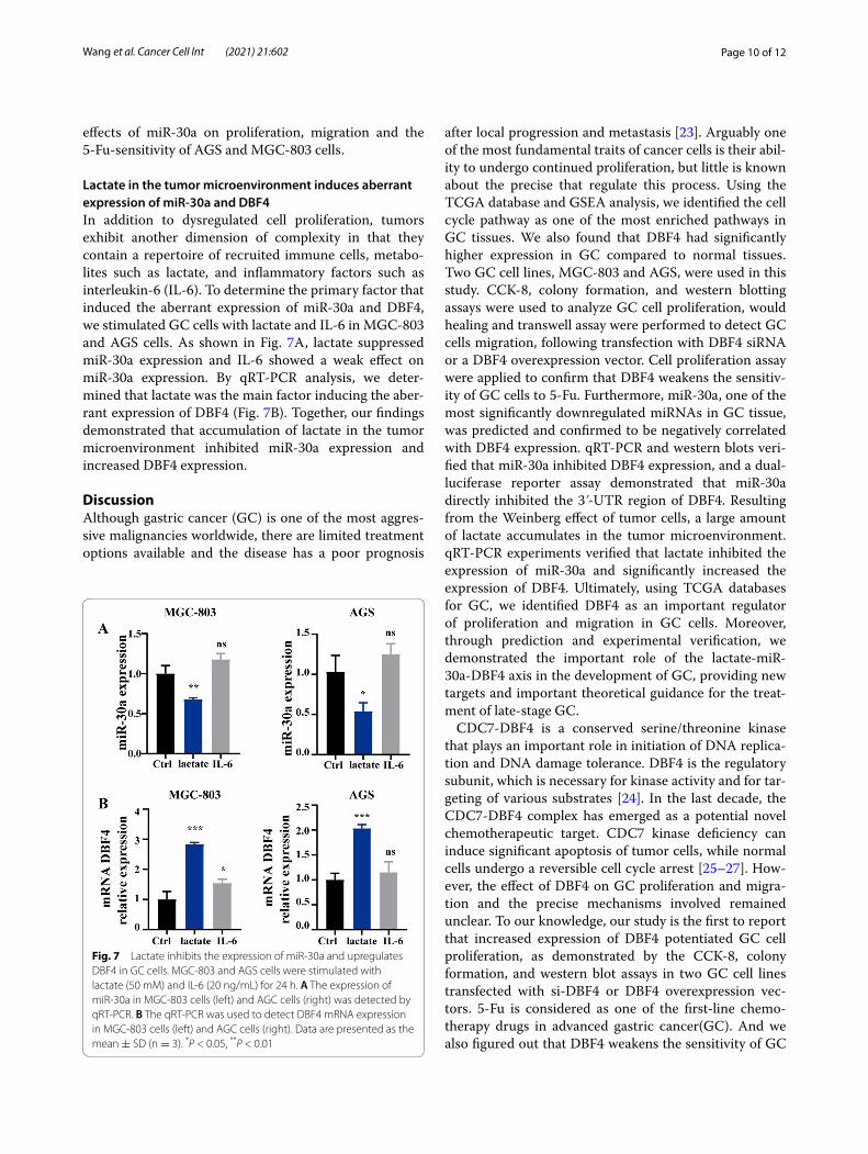
Lactate in the tumor microenvironment induces aberrant expression of miR‑30a and DBF4In addition to dysregulated cell proliferation, tumors exhibit another dimension of complexity in that they contain a repertoire of recruited immune cells, metabo-lites such as lactate, and inflammatory factors such as interleukin-6 (IL-6). To determine the primary factor that induced the aberrant expression of miR-30a and DBF4, we stimulated GC cells with lactate and IL-6 in MGC-803 and AGS cells. As shown in Fig. 7A, lactate suppressed miR-30a expression and IL-6 showed a weak effect on miR-30a expression. By qRT-PCR analysis, we deter-mined that lactate was the main factor inducing the aber-rant expression of DBF4 (Fig. 7B). Together, our findings demonstrated that accumulation of lactate in the tumor microenvironment inhibited miR-30a expression and increased DBF4 expression.
DiscussionAlthough gastric cancer (GC) is one of the most aggres-sive malignancies worldwide, there are limited treatment options available and the disease has a poor prognosis
after local progression and metastasis [23]. Arguably one of the most fundamental traits of cancer cells is their abil-ity to undergo continued proliferation, but little is known about the precise that regulate this process. Using the TCGA database and GSEA analysis, we identified the cell cycle pathway as one of the most enriched pathways in GC tissues. We also found that DBF4 had significantly higher expression in GC compared to normal tissues. Two GC cell lines, MGC-803 and AGS, were used in this study. CCK-8, colony formation, and western blotting assays were used to analyze GC cell proliferation, would healing and transwell assay were performed to detect GC cells migration, following transfection with DBF4 siRNA or a DBF4 overexpression vector. Cell proliferation assay were applied to confirm that DBF4 weakens the sensitiv-ity of GC cells to 5-Fu. Furthermore, miR-30a, one of the most significantly downregulated miRNAs in GC tissue, was predicted and confirmed to be negatively correlated with DBF4 expression. qRT-PCR and western blots veri-fied that miR-30a inhibited DBF4 expression, and a dual-luciferase reporter assay demonstrated that miR-30a directly inhibited the 3ʹ-UTR region of DBF4. Resulting from the Weinberg effect of tumor cells, a large amount of lactate accumulates in the tumor microenvironment. qRT-PCR experiments verified that lactate inhibited the expression of miR-30a and significantly increased the expression of DBF4. Ultimately, using TCGA databases for GC, we identified DBF4 as an important regulator of proliferation and migration in GC cells. Moreover, through prediction and experimental verification, we demonstrated the important role of the lactate-miR-30a-DBF4 axis in the development of GC, providing new targets and important theoretical guidance for the treat-ment of late-stage GC.
CDC7-DBF4 is a conserved serine/threonine kinase that plays an important role in initiation of DNA replica-tion and DNA damage tolerance. DBF4 is the regulatory subunit, which is necessary for kinase activity and for tar-geting of various substrates [24]. In the last decade, the CDC7-DBF4 complex has emerged as a potential novel chemotherapeutic target. CDC7 kinase deficiency can induce significant apoptosis of tumor cells, while normal cells undergo a reversible cell cycle arrest [25–27]. How-ever, the effect of DBF4 on GC proliferation and migra-tion and the precise mechanisms involved remained unclear. To our knowledge, our study is the first to report that increased expression of DBF4 potentiated GC cell proliferation, as demonstrated by the CCK-8, colony formation, and western blot assays in two GC cell lines transfected with si-DBF4 or DBF4 overexpression vec-tors. 5-Fu is considered as one of the first‐line chemo-therapy drugs in advanced gastric cancer(GC). And we also figured out that DBF4 weakens the sensitivity of GC
Fig. 7 Lactate inhibits the expression of miR-30a and upregulates DBF4 in GC cells. MGC-803 and AGS cells were stimulated with lactate (50 mM) and IL-6 (20 ng/mL) for 24 h. A The expression of miR-30a in MGC-803 cells (left) and AGC cells (right) was detected by qRT-PCR. B The qRT-PCR was used to detect DBF4 mRNA expression in MGC-803 cells (left) and AGC cells (right). Data are presented as the mean ± SD (n = 3). *P < 0.05, **P < 0.01

Page 11 of 12Wang et al. Cancer Cell Int (2021) 21:602
cells to 5-Fu, which suggested that DBF4 may become a novel therapeutic targets in GC treatment. Furthermore, our results suggested that DBF4 also play an important role in the regulation of cancer cells migration.
The miRNAs are small non-coding RNAs that post-transcriptionally regulate the expression of many genes to control physiological and pathological processes, including the occurrence, growth, and progression of cancer [28]. Until now, approximately 200 different miRNAs have been implicated in the occurrence and treatment of GC, and numerous studies have suggested that miRNAs may play an important role in various types of cancer, including GC [28–30]. Deregulation of miR-30a has been shown to play a role in many human cancers, and significant downregulation of miR-30a has been detected in anaplastic thyroid carcinomas and non-small cell lung cancer [31, 32]. In contrast, upregulation of miR-30a has been reported in glioma [33], which suggests a context-specific role for miR-30a across different cancer types. In the present study, miR-30a significantly inhibited the proliferation of GC cells, which is in agreement with the previous reports [22]. Furthermore, we demonstrated miR-30a regulated GC cells by inhibiting the expression of DBF4. Dual-lucif-erase reporter assays showed that miR-30a significantly suppressed the 3ʹ-UTR of DBF4. However, additional studies are required to determine the precise mecha-nism of this regulation. miR-30a was recently observed to be downregulated in GC, resulting from high pro-moter methylation induced by DNA methyltransferase [34]. In our study, we found that accumulation of lactate in the tumor microenvironment reduced the expression of miR-30a, suggesting that miR-30a may also be regu-lated by lactylation. Additional future experiments are needed to confirm this finding.
Our study is the first time to identify DBF4 as a criti-cal tumor suppressor in GC. Upregulation of DBF4 increased proliferation of GC cells. Mechanistically, miR-30 suppressed the 3ʹ-UTR of DBF4 and inhibited its expression. Lactate in the tumor microenvironment was the primary factor that induced aberrant expres-sion of miR-30a and DBF4. In conclusion, the lactate-miR-30a-DBF4 axis was a critical regulator of GC cell proliferation, migration, sensitivity to 5-Fu and may have potential to serve as a target for the treatment of gastric cancer.
AcknowledgementsNone.
Authors’ contributionsWTK: Investigation, data curation, writing-original draft. RJ, LGQ, and MBL: Investigation, data curation. WZH: Investigation, writing—review, editing and writing—original draft. WQ contributed reagents, material and equipment. All authors have read and approved the final manuscript.
FundingThis work was Supported by Qingdao Key Health Discipline Development Fund,and grants from the National Program on Key Basic Research Project of China (973 Program) (No. 2015CB755402), and Wu Jie Ping Medical Founda-tion (No. 320.6750.17181), Science and Technology Development Program of Qingdao City (No.19-6-1-20-nsh).
Availability of data and materialsWe hereby undertake that all data and materials are available.
Declarations
Ethics approval and consent to participateThe study was approved by the Ethics Committee of Qilu Hospital of Shan-dong University and All of the specimens were obtained with the patients’ informed consent.
Consent for publicationAll the authors agreed to be published.
Competing interestsThe authors declare no conflicts of interest.
Author details1 Department of Internal Medicine, Qilu Hospital, Shandong University, 107 West Wenhua Road, Jinan 250012, Shandong, P.R. China. 2 Department of Gastroenterology, Qilu Hospital, Shandong University, Jinan, Shandong, P.R. China. 3 Department of Clinical Laboratory, Qilu Hospital, Shandong University (Qingdao), 758 Hefei Road, Qingdao, Shandong, P. R. China. 4 Department of Clinical Laboratory, Qilu Hospital, Shandong University, 107 Wenhuaxi Road, Jinan 250012, Shandong, P.R. China.
Received: 5 June 2021 Accepted: 21 October 2021
References 1. Siegel RL, Miller KD, Jemal A. Cancer statistics 2017. CA Cancer J Clin.
2017;67:7–30. 2. Huang KK, Ramnarayanan K, Zhu F, Srivastava S, Xu C, Tan ALK, Lee M, Tay
S, Das K, Xing M, Fatehullah A, Alkaff SMF, Lim TKH, Lee J, Ho KY, Rozen SG, Teh BT, Barker N, Chia CK, Khor C, Ooi CJ, Fock KM, So J, Lim WC, Ling KL, Ang TL, Wong A, Rao J, Rajnakova A, Lim LG, Yap WM, Teh M, Yeoh KG, Tan P. Genomic and epigenomic profiling of high-risk intestinal metaplasia reveals molecular determinants of progression to gastric cancer. Cancer Cell. 2018;33:137-50 e135.
3. Smyth EC, Nilsson M, Grabsch HI, van Grieken NC, Lordick F. Gastric can-cer. Lancet. 2020;396:635–48.
4. Rugge M, Genta RM, Di Mario F, El-Omar EM, El-Serag HB, Fassan M, Hunt RH, Kuipers EJ, Malfertheiner P, Sugano K, Graham DY. Gastric cancer as preventable disease. Clin Gastroenterol Hepatol. 2017;15:1833–43.
5. Matuszcak C, Haier J, Hummel R, Lindner K. MicroRNAs: promising chem-oresistance biomarkers in gastric cancer with diagnostic and therapeutic potential. World J Gastroenterol. 2014;20:13658–66.
6. Shi J, Qu YP, Hou P. Pathogenetic mechanisms in gastric cancer. World J Gastroenterol. 2014;20:13804–19.
7. Hanahan D, Weinberg RA. Hallmarks of cancer: the next generation. Cell. 2011;144:646–74.
8. Araki H. Elucidating the DDK-dependent step in replication initiation. EMBO J. 2016;35:907–8.
9. Labib K. How do Cdc7 and cyclin-dependent kinases trigger the initiation of chromosome replication in eukaryotic cells? Genes Dev. 2010;24:1208–19.
10. Bonte D, Lindvall C, Liu H, Dykema K, Furge K, Weinreich M. Cdc7-Dbf4 kinase overexpression in multiple cancers and tumor cell lines is cor-related with p53 inactivation. Neoplasia. 2008;10:920–31.
11. Sheu YJ, Stillman B. Cdc7-Dbf4 phosphorylates MCM proteins via a dock-ing site-mediated mechanism to promote S phase progression. Mol Cell. 2006;24:101–13.

Page 12 of 12Wang et al. Cancer Cell Int (2021) 21:602
• fast, convenient online submission
•
thorough peer review by experienced researchers in your field
• rapid publication on acceptance
• support for research data, including large and complex data types
•
gold Open Access which fosters wider collaboration and increased citations
maximum visibility for your research: over 100M website views per year •
At BMC, research is always in progress.
Learn more biomedcentral.com/submissions
Ready to submit your researchReady to submit your research ? Choose BMC and benefit from: ? Choose BMC and benefit from:
12. Montagnoli A, Moll J, Colotta F. Targeting cell division cycle 7 kinase: a new approach for cancer therapy. Clin Cancer Res. 2010;16:4503–8.
13. Nambiar S, Mirmohammadsadegh A, Hassan M, Mota R, Marini A, Alaoui A, Tannapfel A, Hegemann JH, Hengge UR. Identification and functional characterization of ASK/Dbf4, a novel cell survival gene in cutaneous melanoma with prognostic relevance. Carcinogenesis. 2007;28:2501–10.
14. John B, Enright AJ, Aravin A, Tuschl T, Sander C, Marks DS. Human micro-RNA targets. PLoS Biol. 2004;2:363.
15. Ambros V. The functions of animal microRNAs. Nature. 2004;431:350–5. 16. Li PF, Chen SC, Xia T, Jiang XM, Shao YF, Xiao BX, Guo JM. Non-coding
RNAs and gastric cancer. World J Gastroenterol. 2014;20:5411–9. 17. Ueda T, Volinia S, Okumura H, Shimizu M, Taccioli C, Rossi S, Alder H, Liu
CG, Oue N, Yasui W, Yoshida K, Sasaki H, Nomura S, Seto Y, Kaminishi M, Calin GA, Croce CM. Relation between microRNA expression and progres-sion and prognosis of gastric cancer: a microRNA expression analysis. Lancet Oncol. 2010;11:136–46.
18. Pang Y, Young CY, Yuan H. MicroRNAs and prostate cancer. Acta Biochim Biophys Sin. 2010;42:363–9.
19. Bartel DP. MicroRNAs: genomics, biogenesis, mechanism, and function. Cell. 2004;116:281–97.
20. Miti A, Thamm S, Muller P, Csaki A, Fritzsche W, Zuccheri G. A miRNA biosensor based on localized surface plasmon resonance enhanced by surface-bound hybridization chain reaction. Biosens Bioelectron. 2020;167:112465.
21. Sousa JF, Nam KT, Petersen CP, Lee HJ, Yang HK, Kim WH, Goldenring JR. miR-30-HNF4gamma and miR-194-NR2F2 regulatory networks con-tribute to the upregulation of metaplasia markers in the stomach. Gut. 2016;65:914–24.
22. Liu Y, Zhou Y, Gong X, Zhang C. MicroRNA-30a-5p inhibits the prolifera-tion and invasion of gastric cancer cells by targeting insulin-like growth factor 1 receptor. Exp Ther Med. 2017;14:173–80.
23. Ratti M, Lampis A, Hahne JC, Passalacqua R, Valeri N. Microsatellite insta-bility in gastric cancer: molecular bases, clinical perspectives, and new treatment approaches. Cell Mol Life Sci. 2018;75:4151–62.
24. Sasi NK, Coquel F, Lin YL, MacKeigan JP, Pasero P, Weinreich M. DDK has a primary role in processing stalled replication forks to initiate downstream checkpoint signaling. Neoplasia. 2018;20:985–95.
25. Montagnoli A, Tenca P, Sola F, Carpani D, Brotherton D, Albanese C, Santo-canale C. Cdc7 inhibition reveals a p53-dependent replication checkpoint that is defective in cancer cells. Cancer Res. 2004;64:7110–6.
26. Montagnoli A, Valsasina B, Croci V, Menichincheri M, Rainoldi S, Marchesi V, Tibolla M, Tenca P, Brotherton D, Albanese C, Patton V, Alzani R, Ciavolella A, Sola F, Molinari A, Volpi D, Avanzi N, Fiorentini F, Cattoni M, Healy S, Ballinari D, Pesenti E, Isacchi A, Moll J, Bensimon A, Vanotti E, San-tocanale C. A Cdc7 kinase inhibitor restricts initiation of DNA replication and has antitumor activity. Nat Chem Biol. 2008;4:357–65.
27. Tudzarova S, Trotter MW, Wollenschlaeger A, Mulvey C, Godovac-Zim-mermann J, Williams GH, Stoeber K. Molecular architecture of the DNA replication origin activation checkpoint. EMBO J. 2010;29:3381–94.
28. Zhang M, Du X. Noncoding RNAs in gastric cancer: research progress and prospects. World J Gastroenterol. 2016;22:6610–8.
29. Tan P, Yeoh KG. Genetics and molecular pathogenesis of gastric adeno-carcinoma. Gastroenterology. 2015;149(2015):1153-1162 e1153.
30. Song JH, Meltzer SJ. MicroRNAs in pathogenesis, diagnosis, and treat-ment of gastroesophageal cancers. Gastroenterology. 2012;143:35–47 e32.
31. Visone R, Pallante P, Vecchione A, Cirombella R, Ferracin M, Ferraro A, Volinia S, Coluzzi S, Leone V, Borbone E, Liu CG, Petrocca F, Troncone G, Calin GA, Scarpa A, Colato C, Tallini G, Santoro M, Croce CM, Fusco A. Specific microRNAs are downregulated in human thyroid anaplastic carcinomas. Oncogene. 2007;26:7590–5.
32. Zhu J, Zeng Y, Xu C, Qin H, Lei Z, Shen D, Liu Z, Huang JA. Expression profile analysis of microRNAs and downregulated miR-486-5p and miR-30a-5p in non-small cell lung cancer. Oncol Rep. 2015;34:1779–86.
33. Wang K, Jia Z, Zou J, Zhang A, Wang G, Hao J, Wang Y, Yang S, Pu P. Analy-sis of hsa-miR-30a-5p expression in human gliomas. Pathol Oncol Res. 2013;19:405–11.
34. Qiao F, Zhang K, Gong P, Wang L, Hu J, Lu S, Fan H. Decreased miR-30b-5p expression by DNMT1 methylation regulation involved in gastric cancer metastasis. Mol Biol Rep. 2014;41:5693–700.
Publisher’s NoteSpringer Nature remains neutral with regard to jurisdictional claims in pub-lished maps and institutional affiliations.