laparoscopic hysterectomy - pennsylvania state university
TRANSCRIPT

Laparoscopic Hysterectomy Dr R K Mishra
Professor and Head of Minimal Access Surgery, TGO University, India
First University Qualified Master Minimal Access Surgeon of India (M.MAS)
Editor in Chief World Journal of Laparoscopic Surgery (WJOLS)
Member World Association of Laparoscopic Surgeon (WALS)
Member European Association for Endoscopic Surgery (EAES)
Member European Association for Transluminal Surgery (EATS)
Member Society of American Gastrointestinal and Endoscopic Surgeons (SAGES)
Member Society of Laparoendoscopic Surgeons (SLS)
Member Society of Robotic Surgery (SRS)
Member Clinical Robotic Surgery Association (CRSA)
Member Association of Surgeons of India (ASI)
Member Indian Medical Association (IMA)
President All India Specially Abled Association (AISAA)
Member Indian Association of Gastrointestinal Endosurgeons (IAGES)
Chairman, Delhi Laparoscopy Hospital, Pvt. Ltd. & Director, World Laparoscopy Hospital, Gurgaon Benign uterine diseases of uterus are very common and need hysterectomy and laparotomy. Most of these diseases can be performed laparoscopically. Laparoscopic assisted vaginal hysterectomy is increasingly becoming popular. Many women come to the doctor and say they want a “laser” hysterectomy. What they usually mean is a laparoscopically assisted vaginal hysterectomy or LAVH. Laparoscopically assisted vaginal hysterectomy (LAVH) is a procedure using laparoscopic surgical techniques and instruments to remove the uterus and/or tubes and ovaries through the vagina. The technique used to use lasers but now lasers have been mostly replaced by surgical clips, cautery or suturing. First laparoscopic hysterectomy was done by Reich et al in 1989. It is a technique made to replace abdominal hysterectomy.
LAPAROSCOPIC ANATOMY
The normal nulliparous uterus is approximately 8 cm in length and angled forward so the fundus lies over the posterior surface of the bladder. Uterus is all around covered with peritoneum except where the bladder touches the lower uterine segment at the anterior cul-de-sac and laterally at the broad ligament (Fig. 34.1).
Fig. 34.1: Anatomy of uterus, 1-Umbilical artery, 2-Ureter, 3-Uterine artery, 4-Internal iliac artery, 5-Ovarian artery, 6-Common iliac artery, 7-Utero-sacral ligament

Two important arteries, uterine and ovarian are of great significance in uterine surgery. The uterine arise from the internal iliac. They pass medially on the levator ani muscle, cross the ureter and ultimately divide into ascending and descending branch. The uterine artery runs in a tortuous course within the broad ligaments. The uterine arteries ascending branch terminates by anatomizing with the ovarian artery. From anterior to posterior, following important tubular structures are found crossing the brim of true pelvis: The round ligament of the uterus, the infundibulopelvic ligament, which contains the gonadal vessels and the ureter. The ovaries and fallopian tube are found between the round ligament and the infundibulopelvic ligament (Fig. 34.2).
Fig. 34.2: Position of uterus, 1-Uterus, 2-Round ligament, 3-Utero-ovarian ligament (proper ovarian ligament), 4-Uterosacral ligament, 5-Ovary, 6-Suspensory ligament of the ovary, 7-Ureter The ovarian ligaments run from the ovaries to the lateral border of the uterus. Ovary is attached to the pelvic side wall with infundibulopelvic ligament, which carries ovarian artery. One of the common mistakes is injury of the ureter during dissection of the infundibulopelvic ligament. If the uterus is deviated to the contralateral side with the help of uterine manipulator infundibulopelvic ligament is spread out and a pelvic side wall triangle is created. The base of this triangle is the round ligament, the medial side is the infundibulopelvic ligament, and the lateral side is the external iliac artery. The apex of this triangle is the point at which the infundibulopelvic ligament crosses the external iliac artery. The ureter always enters medial to this triangle into the pelvis. It is visible under the peritoneum overlying the external iliac artery. The ureters enter the pelvis in close proximity to the female pelvic organ and are at risk for injury during laparoscopic surgery of these organs. As the ureter course medially over the bifurcation of the iliac vessels, they pass obliquely under the ovarian vessels and then run in close proximity to the uterine artery. Laparoscopy hysterectomy needs careful identification of ureter with some dissection of retroperitoneum. An incision is made in the peritoneum overlying the pelvic side wall triangle between the fallopian tube and iliac vessel. Pelvic lymph node dissection is also necessary if gynecologist plan to perform radical laparoscopic hysterectomy. Node dissection as far distal as Cloquet’s node in the femoral triangle may be included and proximally dissection may be necessary up to para-aortic lymph node.
Indications of LAVH
Indications of LAVH are traditionally contraindications of vaginal hysterectomy. Indications include: • Previous pelvic surgery

• Endometriosis • Previous CS • Pelvic pain • Suspected adnexal pathology • Uterine myoma • Ectopic pregnancy • Acute or chronic pelvic inflammatory disease • Minimum uterine mobility and limited vaginal access. If a vaginal hysterectomy can be performed in the first place, there would be no point in adding the costs and complications of laparoscopy. Its greatest benefit is the potential to convert what would have been an abdominal hysterectomy into a vaginal hysterectomy. An abdominal hysterectomy requires both a vaginal incision and a four to six inch long incision in the abdomen, which is associated with greater post-operative discomfort and a longer recovery period than for a vaginal procedure. Another advantage of the LAVH may be the removal of the tubes and ovaries which on occasion may not be easily removed with a vaginal hysterectomy. The most common medical reasons for performing hysterectomies include uterine fibroids (30%), abnormal uterine bleeding (20%), endometriosis (20 %), genital prolapse (15%) and chronic pelvic pain (10%). For most of these conditions, other treatments should first be considered, and hysterectomy should be reserved as a last resort. LAVH result in a significantly shorter hospital stay, with a much more rapid return to normal activities, than total laparoscopic hysterectomy. The drug requirement to control pain and the level of pain patients experienced were also significantly less. Blood loss was not different for the two procedures (Tables 34.1 and 34.2).
Table 34.1: Postoperative pain levels
Day LAVH (n -= 19) TAH (n= 19) p
1 6.6 6.4 NS 3 4.4 4.3 NS
7 2.8 3.6 S
14 1.6 2.4 S 21 1.46 1.8 S
Week 6 1.35 1.4 NS
Wilcoxon’s signed rank test.
Ten-point activity scale: 1 = no pain, 10 = unbearable pain.
S = significant at p < 0.005; NS = not significant at p <0.01
Table 34.2: Postoperative activity levels
Day LAVH (n -= 19) TAH (n= 19) p
1 3.4 3.3 NS
3 5.4 4.4 NS
7 7.8 5.8 S 14 9.2 6.4 S
21 9.6 7.9 S
Week 6 9.95 8.5 S
Wilcoxon’s signed rank test.
Ten-point activity scale: 1 = extremely limited activity, 10 = no limits on activity
S = significant at p < 0.005; NS = not significant at p <0.01
Postoperative recovery times and pain levels were assessed in 37 patients with a primary complaint of pelvic pain and diagnoses of fibroid uterus, adenomyosis, and severe endometriosis who underwent LAVH. Women reported an activity level of 8.7 on a scale of 1 to 10 (10 no limits on activity) by postoperative day 14. In another study, those undergoing abdominal hysterectomy had a mean uterine weight of 418 g compared with 150 g for those undergoing LAVH. The hospital stay after abdominal hysterectomy was 4.5 days and after LAVH 2.5 days. An important public policy issue now confronts us. As it is currently performed, LAVH is more expensive than TAH. The issue is whether the benefits of shorter convalescence and faster return to the work force, shorter hospitalization, and less need for narcotics for postoperative pain outweigh the disadvantage of the higher cost. If total health care system costs are evaluated, the short-term disability costs of 2 weeks of recovery after laparoscopic hysterectomy should be compared with disability costs of 6 to 8 weeks of recovery after abdominal hysterectomy. For LAVH to be economically viable compared with TAH, savings in disability costs and the
increased contribution to the gross domestic product must offset the increased health care costs. In the current system, insurance companies and hospitals do not share in these benefits, only
the costs. The economic impact of laparoscopic surgery must take into account both the cost to
the hospital and insurance payers and these productivity and social issues. Insurance is based on a risk pool whereby the cost of a premium is based on the cost of treatment, not the ability of
the subscriber to return to work. An economic and social cost-benefit analysis must be
performed before decisions are made to modify or judge a procedure that provides substantial benefits to the patient. Since its introduction in 1989, continued improvement of techniques will likely progress rapidly so that LAVH will be performed on an outpatient basis for many women, and will result in shorter recovery time. Thus, the increased operating room time of approximately 46 minutes is significantly outweighed by the benefits available with widespread application of this procedure.
CLASSIFICATION
Garry and Reich Classification
• Type 1 Diagnostic lap + VH • Type 2 lap vault suspension after VH • Type 3 LAVH • Type 4 LH (lap ligation of uterine artery) • Type 5 TLH • Type 6 LSH (lap supracervical hysterectomy) • Type 7 LHL (lap hysterectomy with lymphadenectomy) • Type 8 LHL + O (as above + omentectomy) • Type 9 RLH (radical lap hysterectomy)
Preoperative Measures
Patients are evaluated same as that of any major surgery. Routine preoperative test include a complete blood count with differential, serum electrolyte, bleeding time and urinalysis. More comprehensive blood studies include thrombin time, partial thrombin time, ECG, chest X-ray

and endometrial biopsy. Mechanical and antibiotic bowel preparation is advised. Peglac powder 1 sachet with water a night prior to surgery is advised.
Patient Position
Patient should be in steep Trendelenburg’s and lithotomy position. One assistant should remain between the legs of patient to do uterine manipulation whenever required (Fig. 34.3).

Fig. 34.3: Per-vaginal examination should be routine
Position of Surgical Team (Fig. 34.4)
Surgeon stand left to the patient, camera assistant should be left to the surgeon. Second assistant should be the opposite side of the body of patient. One more assistant is required between the legs to handle uterine manipulator.
Fig. 34.4: Port position in LAVH
Port Position
A 10 mm umbilical port for camera should be along the inferior crease. Two 5 mm ports should be placed at 5 cm away from umbilicus on either side. Sometime, accessory port at right or left iliac region may be needed according to need. Port position should be in accordance with baseball diamond concept. If the left side of tube has to be operated, one port should be in right iliac fossa and another below left hypochondrium (Fig. 34.5).

Fig. 34.5: Port position for LAVH
Operative Technique
It is important throughout the procedure to be able to manipulate the uterus for optimal observation. Different types of uterine manipulators are available. Depending on the laparoscopic procedure, digital examination, probes, and sponge stick applicators are used in the cul-de-sac for identification of structures during laparoscopy. The direction and location of both ureters should be identified as much as possible (Fig. 34.6).
Fig. 34.6: LAVH using bipolar If adenexectomy is planned, following electrodesiccation and cutting of the round ligaments, 2 to 3 cm of the uterus, the infundibulopelvic ligament is desiccated and cut, taking progressive bites of tissue starting at pelvic brim and moving towards the round ligament. If endoscopic linear stapler is used the adnexae is grasped with forceps, it is retracted medially and caudally to stretch and outline the infundibulopelvic ligament, which is grasped and secured with the stapler. Stapler is not fired until the contained tissue is identified and the ureter safety is confirmed. Once transacted the staple line should be examined closely for any possible injury and hemostasis. Following infundibulopelvic ligament transaction the adnexae and uterine fundus are retracted in the opposite direction and the tissue of the upper broad ligament, including the round ligament, is grasped, secured and cut. The multifire GIA stapler can clamp and cut tissue efficiently. The device places six rows of small titanium staples and cuts the tissue in between, leaving three rows of staples on either side of the transected pedicle. This device leaves essentially bloodless pedicles. However, the instrument is disposable and expensive (Figs 34.7A and B).

Figs 34.7A and B: Successive desiccation and dissection If the adnexae is planned to preserve, the round ligament is desiccated and cut approximately 2 cm from the uterus. The anterior leaf of the broad ligament is opened towards the vesicouterine fold and bladder flap is developed. The anterior leaf of the broad ligament is grasped with forceps, elevated and dissected from the anterior lower uterine segment. The utero-ovarian ligament proximal tube and mesosalpinx are progressively dissected and cut and posterior leaf of the broad ligament is opened. Similarly, the round ligament fallopian tube and utero-ovarian ligament are grasped closed to their insertion into the uterus with endoscopic linear stapler, then secured stapled and cut. The distal end of the stapler or bipolar forceps must be kept free of the bladder and ureter (Fig. 34.8).

Fig. 34.8: Dissection of bladder peritoneum
Figs 34.9A to C: Steps of colpotomy The uterovesical junction is identified, grasped, and elevated with forceps while being cut with scissors. The bladder pillars are identified desiccated and cut. The bladder can be completely freed from the uterus by pushing downward with the tip of a blunt probe along the vesicocervical plane until the anterior cul-de-sac is exposed completely. In patients with severe anterior cul-de-sac endometriosis, previous CS or adhesions, sharp dissection can be performed. Injecting 5 ml of indigo caramine in the patient’s bladder helps to detect bladder trauma. After dissecting the bladder from the uterus, the uterine vessels are identified desiccated and cut to free the lateral border of uterus (Figs 34.10A to D). If sutures, clips or linear staplers are used, it is important to fully skeletonize the vessel. As the uterine vessel are grasped and cut, safety and position of the ureter should be checked. Ureter injury can be completely nullified if ureteric catheter is introduced before the procedure. Cardinal ligament dissection must be carefully done as ureter and uterine artery falls just lateral to that. The linear stapler can be used only if the parametrium has been dissected with the ample margins. Once the ureter is displaced laterally, the cardinal ligament tissue closest to the cervix is electrodesiccated and transacted. Alternatively, the linear stapler can be applied both on the uterine vessels and cardinal ligament (Figs 34.11 and 34.12).


Figs 34.10A to D: Successive clamping and desiccation of uterine pedical through the vaginal route, (A) Valsaleum holding cervix, (B) Application of ligasure clamp over left uterosacral, (C) Application of ligasure over the right uterine stump, (D) Application of ligasure over the left uterine stump
Fig. 34.11: Opening of anterior and posterior leaf broad ligament
Fig. 34.12: Separation of bladder
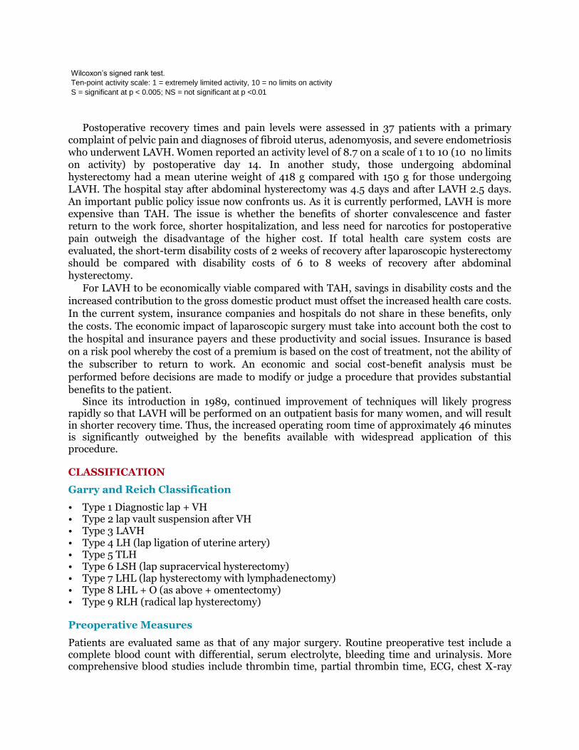
Colpotomy
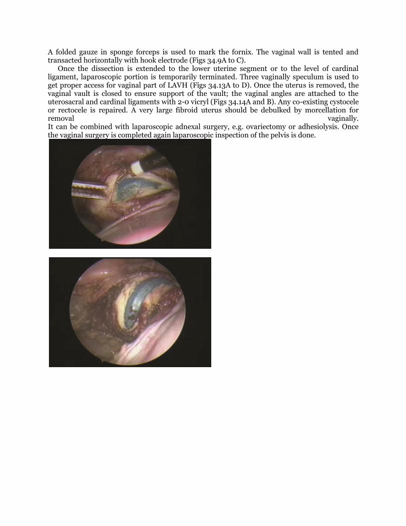
A folded gauze in sponge forceps is used to mark the fornix. The vaginal wall is tented and transacted horizontally with hook electrode (Figs 34.9A to C). Once the dissection is extended to the lower uterine segment or to the level of cardinal ligament, laparoscopic portion is temporarily terminated. Three vaginally speculum is used to get proper access for vaginal part of LAVH (Figs 34.13A to D). Once the uterus is removed, the vaginal vault is closed to ensure support of the vault; the vaginal angles are attached to the uterosacral and cardinal ligaments with 2-0 vicryl (Figs 34.14A and B). Any co-existing cystocele or rectocele is repaired. A very large fibroid uterus should be debulked by morcellation for removal vaginally. It can be combined with laparoscopic adnexal surgery, e.g. ovariectomy or adhesiolysis. Once the vaginal surgery is completed again laparoscopic inspection of the pelvis is done.

Figs 34.13A to D: Anterior and posterior colpotomy

Figs 34.14A and B: Closure of vault by extracorporeal knot
Total Laparoscopic Hysterectomy
Total laparoscopic hysterectomy requires vaginal seal to prevent gas leak. Two 4 × 4 inch wet sponges in the gloves can be used to insert into the vagina to prevent loss of pneumoperitoneum. By applying contralateral retraction to the uterus, vaginal wall surrounding the cervix is outlined, coagulated with the unipolar scissors or bipolar forceps and cut circumferentially until the cervix is separated. Specimen is pulled to mid vagina but not removed to preserve pneumoperitoneum. Vaginal vault is irrigated and inspected for any active bleeding. Once hemostasis is achieved, vaginal angles are sutured to the adjacent cardinal and uterosacral ligaments. Care is taken to avoid the ureter. Rest of the vaginal cuff is closed using intracorporeal knotting. Bipolar is used cautiously at the vaginal cuff to prevent tissue necrosis and subsequent wound breakdown if the sutures replaced in non-viable tissue. The hysterectomy can be performed laparoscopically up to the uterine size of 26 weeks. These patients must have adequate hemoglobin and hematocrit. The GnRH analogue should be given if the uterus is more than 18 weeks gestational size. According to baseball diamond concept the telescopic port should be placed between umbilicus and xiphoid in the patient whose uterus is more that 18 weeks size. The secondary ports should also be placed higher than usual. Big uterus with multiple myomas is difficult to manipulate. Sometime, 4 to 5 port may be necessary to handle this uterus. Anatomy is distorted and ureteral dissection may be necessary in these cases.
Subtotal Hysterectomy
Supracervical hysterectomy is performed to preserve libido of patient. The procedure is performed fully laparoscopically. After desiccating and cutting the uterine vessels at the level of cardinal ligaments above the uterosacral ligament, uterus is retracted and its lower segment is amputated with the scissors and unipolar cutting current. After transecting the uterus from the cervix, uterine manipulator is removed vaginally, the cervical stump is irrigated and hemostasis is achieved. The endocervical epithelium, lining the cervical canal is vaporized or coagulated with laser or electrosurgery. The rest of the endocervical canal is ablated vaginally to reduce the risk of intraepithelial cervical neoplasia. The cervical stump is closed with interrupted absorbable sutures and covered with peritoneum, which is stitched transversely with interrupted sutures. The dissected uterus is morcellated and removes through a 10 mm cannula. Mini-laparotomy or posterior colpotomy can also be performed to remove the uterus in case of subtotal hysterectomy. These patients are advised to annual examination for Pap smear.
Ending the Procedure
One of the benefits of LAVH or TLH over NDVH is inspection of pedicles at the end of surgery. The vaginal cuff can be closed from below or above but after that pneumoperitoneum is again
restored to see the pelvic and abdominal cavity. Irrigation and suction should be performed. In
case of any residual bleeding it can be controlled laparoscopically. At the end pelvis is filled with 300 ml Ringer’s lactate and it should be seen for change in color. Once inspection is satisfying
the fluid is sucked and instrument and cannula is removed after deflating the abdominal cavity.
It has been demonstrated that TLH and LAVH are associated with a shorter hospital stay and
patients require less pain medication compared to TAH. LAVH can replace most of the
abdominal hysterectomy for the benign disease of uterus and with the technology available
today it has definite benefit over non- descended vaginal hysterectomy.
DISCUSSION
Vaginal hysterectomy is part of repertoire of every trained gynecologist. It is considered as a
feasible option to abdominal hysterectomy and many studies have shown that vaginal
hysterectomy has fewer complications, short recovery, and hospital stay than laparotomy.
Laparoscopic hysterectomy requires greater surgical expertise and has a steep learning curve.
Randomized trials have shown advantages of laparoscopy versus laparotomy, including reduced
postoperative pain, shorter hospitalization, rapid recovery and substantial financial benefits to
society. The objective of performing hysterectomy laparoscopically can be achieved but the
question is does this offer any advantage over vaginal route. Every mode of hysterectomy has
advantages and disadvantages but the indications for each remain controversial. Good surgical
practice is when the indication for hysterectomy is considered as the primary criterion for
selecting the route of hysterectomy and not factors such as surgeon’s choice and experience. A
major determinant of the route of hysterectomy is not the clinical situation but the attitude of
the surgeon. There is no need for extra training and special skills or complicated equipment for
vaginal hysterectomy. Laparoscopic hysterectomy took a long time to perform in all studies. However with increasing weight of the uterus, there was a linear increase in operating time and blood loss in hysterectomy performed vaginally which was not observed in laparoscopic assisted vaginal hysterectomy. There is no statistically significant difference in postoperative analgesia requirement, hospital stay, recovery milestones or complication rates. The biggest drawback of laparoscopic route over vaginal one is its cost due to expensive disposable instruments, prolonged operating and anesthesia time and the need for a trained senior gynecologist. For laparoscopic assisted vaginal hysterectomy to be cost effective expensive disposable instruments have to be eliminated. Laparoscopic surgeons argue that subtotal hysterectomy can be performed laparoscopically but most randomized trials have failed to demonstrate any benefit of subtotal hysterectomy over total hysterectomy. In women who wish to retain their cervix vaginal subtotal hysterectomy described by Doderlein Kronig Technique can be performed. The disadvantage of vaginal approach is vault hematomas. The abdominal approach to hysterectomy does ensure good hemostasis under direct vision, while during the vaginal operation, the vault is closed and subsequent bleeding from the vagina between the mucosa and the peritoneum can give rise to problems, especially if a vasoconstrictor has been given that subsequently wears off. Laparoscopic approach can help check hemostasis and reduce the incidence of vault hema-tomas. However; this aspect needs to be evaluated in studies. Lack of uterine descent and nulliparity, fibroid uterus, need for oophorectomy, previous pelvic surgery are no more considered as contraindications to the vaginal route. With adequate

vaginal access and technical skill, and good uterine mobility, vaginal hysterectomy can easily be achieved Multiparity, lax tissues due to poor involution following multiple deliveries and lesser tissue tensile strength afford a lot of comfort to vaginal surgeon even in the presence of significant uterine enlargement. No evidence supports the use of laparoscopic hysterectomy rather than VH if latter can be performed safely. No outcomes are significantly worse for vaginal hysterectomy compared to LAVH. There are clinical situations where vaginal surgeries is not appropriate such as dense pelvic adhesions, severe endometriosis adnexal disease, when vaginal access is reduced when laparoscopic hysterectomy is indicated as it has advantages over the abdominal approach. Laparoscopic approach may be helpful postoperatively to rule out hemorrhage in some cases. Laparoscopic assistance should not be used to supplant inadequate skills of vaginal hysterectomy. Lack of training in vaginal surgery is not a reason for not removing uteri vaginally. The learning curve of VH is very short compared to laparoscopic surgery, however, the current scenario in residency programs is not providing a level of surgical competency in performing difficult vaginal hysterectomies. There is a need to improve this training. In order to compare the complication rates of different types of hysterectomies, considering an incidence of 4-5% of serious complications of hysterectomies 1460 women would be required in each arm of the study to detect 50% increase in the complication rate. Therefore, larger randomized controlled trials are required to compare different types of hysterectomies. When the size of the uterus is greater than 16 weeks gestation there is an increase in the operative time and blood loss in VH compared to LAVH which is statistically significant. Laparoscopically assisted vaginal hysterectomy is a useful adjunct to transvaginal hysterectomy for lysis of extensive adhesions and sometimes for certain concomitant adnexal surgery. Besides, LAVH can also secure almost all the main blood supplies to the uterus, i.e. the uterine vessels and the adnexal collaterals. Although a skilled surgeon can do transvaginal hysterectomy with a larger uterus by employing volume-reducing techniques, Kohler reported that laparoscopic coagulation hemostasis of the uterine vessels was associated with less blood loss. It may take time to achieve these goals, but they may make subsequent extirpation or volume reducing procedures easier and safer to perform. Therefore, the average operative time and estimated blood loss for the LAVH remained almost constant regardless of increasing uterine weight. Generally, the average operative time for LAVH is longer than that for transvaginal hysterectomy. It takes time to secure the uterine blood supply before extirpation and volume reducing procedures, but it also makes LAVH superior to transvaginal hysterectomy when dealing with a larger uterus. In our opinion, LAVH might be considered for a larger uterus in view of the relatively shorter operative time and less blood loss, whereas transvaginal hysterectomy is preferable for a small uterus, not only for shorter operative time and minimal wound, but also for much lower costs.
BIBLIOGRAPHY
1. Carley ME, McIntire D, Carley JM, Schaffer J. Incidence, risk factors and morbidity of unintended bladder or
ureter injury during hysterectomy. Int Urogynecol J Pelvic Floor Dysfunct 2002;13:18–21.
2. Chapron C, Dubuisson JB. Laparoscopic hysterectomy. Lancet 1995;345:593. Chapron C, Dubuisson JB, Aubert
V. Total laparoscopic hysterectomy: preliminary results. Hum Reprod 1994;9:2084–2089.
3. Chapron C, Fauconnier A, Goffinet F, Bre´art G, Dubuisson JB. Laparoscopic surgery is not inherently
dangerous for patients presenting with benign gynaecologic pathology. Results of a meta-analysis. Hum Reprod
2002;17:1334–1342.
4. Chapron C, Laforest L, Ansquer Y, Fauconnier A, Fernandez B, Breart G and Dubuisson JB. Hysterectomy
techniques used for benign disorders: results of a French multicentre study. Hum Reprod 1999;14:2464–70.
5. Chauveaud A, de Tayrac R, Gervaise A, Anquetil C and Fernandez H. Total hysterectomy for a nonprolapsed,
benign uterus in women without vaginal deliveries. J Reprod Med 2002;47:4–8.
6. Cosson M, Querleu D and Crepin G Hystérectomies pour pathologies bénignes. In Masson. Williams et Wilkins,
Paris, 1997;160.

7. Councell RB, Thorp JM Jr, Sandridge DA, Hill ST. Assessments of laparoscopic-assisted vaginal hysterectomy. J
Am Assoc Gynecol Laparosc 1994;2:49–56.
8. Dandolu V, Mathai E, Chatwani A, Harmanli O, Pontari M, Hernandez E. Accuracy of cystoscopy in the
diagnosis of ureteral injury in benign gynecologic surgery. Int Urogynecol J Pelvic Floor Dysfunct 2003;14:427–
31.
9. Daraï E, Soriano D, Kimata P, Laplace C and Lecuru F Vaginal hysterectomy for enlarged uteri, with or without
laparoscopy assistance: randomized study. Obstet Gynecol 2001;97:712–716.
10. Davies A, Vizza E, Bournas N, O’Connor H and Magos A. How to increase the proportion of hysterectomies
performed vaginally. Am J Obstet Gynecol 1998;179:1008–1012.
11. Dicker RC, Greenspan JR, Strauss LT, Cowart MR, Scally MJ, Peterson HB, DeStefano F, Rubin GL, Ory HW.
Complications of abdominal and vaginal hysterectomy among women of reproductive age in the United States.
The Collaborative Review of Sterilization. Am J Obstet Gynecol 1982;144:841–8.
12. Dorairajan G, Rani PR, Habeebullah S, Dorairajan LN. Urological injuries during hysterectomies: a 6-year
review. J Obstet Gynaecol Res 2004;30:430–5.
13. Dorsey JH, Steinberg EP and Holtz PM. Clinical indications for hysterectomy route: patient characteristics or
physician preference? Am J Obstet Gynecol 1995;173:1452–60.
14. Dwyer PL, Carey MP, Rosamilia A. Suture injury to the urinary tract in urethral suspension procedures for stress incontinence. Int Urogynecol J Pelvic Floor Dysfunct 1999;10:15–21.
15. Farquhar CM, Steiner CA. Hysterectomy rates in the United States 1990–1997. Obstet Gynecol 2002;99:229–
234. 16. Garry R, Fountain J, Brown J, Manca A, Mason S, Sculpher M, Napp V, Bridgman S, Gray J and Lilford R.
Evaluate hysterectomy trial. A multicentre randomised trial comparing abdominal, vaginal and laparoscopy
methods of hysterectomy. Health Technol Assess 2004a;8:1–154. 17. Garry R, Fountain J, Mason S, Hawe J, Napp V, Abbott J, Clayton R, Phillips G, Whittaker M, Lilford R et al.
(2004b) The evaluate study: two parallel randomised trials, one comparing laparoscopy with abdominal
hysterectomy, the other comparing laparoscopy with vaginal hysterectomy. BMJ 328, 129. Erratum in BMJ (2004) 328,494.
18. Gilmour DT, Das S, Flowerdew G. Rates of urinary tract injury from gynecologic surgery and the role of
intraoperative cystoscopy. Obstet Gynecol 2006;107:1366–1372. 19. Gilmour DT, Dwyer PL, Carey MP. Lower urinary tract injury during gynecologic surgery and its detection by
intraoperative cystoscopy. Obstet Gynecol 1999;94:883–9.
20. Gimbel H, Settnes A, Tabor A. Hysterectomy on benign indication in Denmark 1988–1998. Acta Obstet Gynecol Scand 2001;80:267–72.
21. Ha¨rkki-Siren P, Kurpi T, Sjo¨berg J, Tiitinen A. Safety aspects of laparoscopic hysterectomy. Acta Obstet
Gynecol Scand 2001;80:383–91. 22. Harkki-Siren P, Sjo¨berg J, Ma¨kinen J, Heinonen PK, Kaudo M, Tomas E, Laatikainen T. Finnish national
register of laparoscopic hysterectomies: A review and complications of 1165 operations. Am J Obstet Gynecol
1997;176:118–122.
23. Harkki-Siren P, Sjo¨berg J, Tiitinen A. Urinary tract injury after hysterectomy. Obstet Gynecol 1998;92:113–8.
24. Härkki-Siren P, Sjoberg J and Tiitinen A Urinary tract injuries after hysterectomy. Obstet Gynecol 1998;92;113–
8.
25. Harris MB and Olive DL. Changing hysterectomy patterns after introduction Gynecol 1994;171,340–3.
26. Hurd WW, Bude RO, De Lancey JO, Pearl ML. The relationship of the umbilicus to aortic bifurcation:
implications for laparoscopic technique. Obstet Gynecol 1992;80:48–51. 27. Hwang JL, Seow KM, Tsai YL, Huang LW, Hsieh BC and Lee C. Comparative study of vaginal, laparoscopically
assisted vaginal and abdominal hysterectomies for uterine myoma larger than 6 cm in diameter or uterus
weighing at least 450 g: a prospective randomized study. Acta Obstet Gynecol Scand 2002;81:1132–8. 28. Johns DA, Carrera B, Jones J, DeLeon F, Vincent R and Safely C. The medical and economic impact of
laparoscopically assisted vaginal hysterectomy in a large, metropolitan, not-for-profit hospital. Am J Obstet
Gynecol 1995;172:1709–15. 29. Johnson N, Barlow D, Lethaby A, Tavender E, Curr E and Garry R (2005a) Surgical approach to hysterectomy
for benign gynaecological disease. Cochran Database Syst Rev (2): CD003677. Johnson N, Barlow D, Lethaby A,
Taverder E, Curr L and Garry R (2005b) Methods of hysterectomy: systematic review and meta-analysis of randomised controlled trials. BMJ 330,1478.
30. Johnson N, Barlow D, Lethaby A, Tavender E, Curr L, Garry R. Methods of hysterectomy: systematic review and
meta-analysis of randomised controlled trials. Br Med J 2005;330:1478. 31. Kadar N. Dissecting the pelvic retroperitoneum and identifying the ureters. A laparoscopic technique. J Reprod
Med 1995;40:116–122.

32. Kreiker G, Bertoldi A, Sad Larcher J, Ruiz Orrico G, Chapron C. Prospective evaluation of the learning curve of
total laparoscopic hysterectomy in a universitary hospital. J Am Assoc Gynecol Laparosc 2004;11:229–235. 33. Le´onard et al. 2010 Ou CS, Beadle E, Presthus J, Smith M. A multicenter review of 839 laparoscopic-assisted
vaginal hysterectomies. J Am Assoc Gynecol Laparosc 1994;1:417–422.
34. Leonard F, Chopin N, Borghese B, Fotso A, Foulot H, Coste J, Mignon A, Chapron C. Total laparoscopic hysterectomy: preoperative risk factors for conversion to laparotomy. J Minim Invasive Gynecol 2005;12:312–
317.
35. Leonard F, Chopin N, Borghese B, Fotso A, Foulot H, Coste J, Mignon A and Chapron C. Total laparoscopy hysterectomy: preoperative risk factor for conversion to laparotomy. J Minim Invasive Gynecol 2005;12:312–7.
36. Liu CY, Reich H. Complications of total laparoscopic hysterectomy in 518 cases. Gynecol Endoscopy
1994;3:203–8. 37. Ma¨kinen J, Johansson J, Tomas C, Tomas E, Heinonen PK, Laatikainen T, Kauko M, Heikkinen AM, Sjo¨berg
J. Morbidity of 10 110 hysterectomy by type approach. Hum Reprod 2001;16:1473–8.
38. McMaster-Fay RA, Jones RA. Laparoscopic hysterectomy and ureteric injury: a comparison of the initial 275
cases and the last 1,000 cases using staples. Gynecol Surg 2006;3:118–21.
39. Meikle SF, Nugent EW, Orleans M. Complications and recovery from laparoscopy-assisted vaginal hysterectomy
compared with abdominal and vaginal hysterectomy. Obstet Gynecol 1997;89:304–11. 40. Mteta KA, Mbwambo J, Mvungi M. Iatrogenic ureteric and bladder injuries in obstetric and gynaecologic
surgeries. East Afr Med J 2006;83:79–85.
41. National Centre for Disease Control and Prevention 1997. Hysterectomy surveillance United States 1980–1993. CDC surveillance summaries, August. Mabille de Poncheville L (1998) Coeliochirurgie gynécologique en France,
instantanée 1996. Résultats d’une enquête nationale. Thèse de médecine, Tours, France. Moller C, Kehlet H and
Ottesen BS (1999) Hospitalization and convalescence after hysterectomy. Open or laparoscopy surgery? Ugeskr Laeger 161,4620–4624.
42. Nezhat F, Nezhat C, Admon D, Gordon S, Nezhat C. Complications and results of 361 hysterectomies performed
at laparoscopy. J Am Coll Surg 1995;180:307–16. 43. O’Shea RT, Petrucco O, Gordon S, Seman E. Adelaide laparoscopic hysterectomy audit (1991–1998): realistic
complications rates. Gynaecol Endoscopy 2000;9:369–372.
44. Oh BR, Kwon DD, Park KS, Ryu SB, Park YI, Presti JC Jr. Late presentation of ureteral injury after laparoscopic surgery. Obstet Gynecol 2000;95:337–339.
45. Paulson JD. Laparoscopically assisted vaginal hysterectomy. A protocol for reducing urinary tract complications.
J Reprod Med 1996;41:623–628. 46. Phipps JH, Tyrrell NJ. Transilluminating ureteric stents for preventing operative ureteric damage. Br J Obstet
Gynaecol 1992;99:81. Reich H, De Caprio J, McGlynn F. Laparoscopic hysterectomy. J Gynecol Surg
1989;5:213–216. 47. Ribeiro S, Reich H, Rosenberg J, Guglielminetti E, Vidali A. The value of intra-operative cystoscopy at the time
of laparoscopic hysterectomy. Hum Reprod 1999;14:1727–1729.
48. Ribeiro SC, Ribeiro RM, Santos NC and Pinotti JA (2003) A randomized study of total abdominal, vaginal and laparoscopy hysterectomy. Int J Gynaecol Obstet 83,37–43.
49. Rutkow IM. Obstetric and gynecologic operations in the United States, 1979 to 1984. Obstet Gynecol
1986;67:755–759. 50. Saidi MH, Sadler RK, Vancaillie TG, Akright BD, Farhart SA, White AJ. Diagnosis and management of serious
urinary complications after major operative laparoscopy. Obstet Gynecol 1996;87:272–276.
51. Shen CC, Wu MP, Kung FT, Huang FJ, Hsieh CH, Lan KC, Huang EY, Hsu TY, Chang SY. Major complications associated with laparoscopic-assisted vaginal hysterectomy: ten-year experience. J Am Assoc Gynecol Laparosc
2003;10:147–153.
52. Soriano D, Goldstein A, Lecuru F and Daraï E. Recovery from vaginal hysterectomy compared with laparoscopy-assisted vaginal hysterectomy: a prospective, randomized, multicenter study. Acta Obstet Gynecol Scand
2001;80,337–41.
53. University of York (UK) Centre for Health Economics. The management of menorrhagia. Effective healthcare 1991;1(9). Vessey MP, Villard-Mackintosh L, McPherson K, Coulter A and Yeates D. The epidemiology of
hysterectomy: findings in a large cohort study. Br J Obstet Gynaecol 1992;99:402–407.
54. Vakili B, Chesson RR, Kyle BL, Shobeiri SA, Echols KT, Gist R, Zheng YT, Nolan TE. The incidence of urinary tract injury during hysterectomy: a prospective analysis based on universal cystoscopy. Am J Obstet Gynecol
2005;192:1599–1604.
55. Visco AG, Taber KH, Weidner AC, Barber MD, Myers ER. Cost-effectiveness of universal cystoscopy to identify
ureteral injury at hysterectomy. Obstet Gynecol 2001;97:685–92.

56. Wattiez A, Soriano D, Cohen SB, Nervo P, Canis M, Botchorisvili R, Mage G, Pouly JL, Mille P, Bruhat MA. The
learning curve of total laparoscopic hysterectomy: comparative analysis of 1647 cases. J Am Assoc Gynecol
Laparosc 2002;9:339–45.
57. Wood EC, Maher P, Pelosi MA. Routine use of ureteric catheters at laparoscopic hysterectomy may cause unnecessary complications. J Am Assoc Gynecol Laparosc 1996;3:393–7.
58. Wu SM, Chao Yu YM, Yang CF and Che HL. Decision-making tree for women considering hysterectomy. J Adv
Nurs 2005;51:361–8.