laparoscopic surgery to treat ureterosciatic herniation ... · case report laparoscopic surgery to...
TRANSCRIPT
at SciVerse ScienceDirect
Urological Science 25 (2014) 25e27
Contents lists available
Urological Science
journal homepage: www.urol-sci .com
Case report
Laparoscopic surgery to treat ureterosciatic herniation after ureteralstent failure
Yi-Sheng Tai a,c, Kao-Lang Liu b, Mao-Yuan M. Su b, Hui-Ching Tai a, Kuo-How Huang a,*
aDepartment of Urology, National Taiwan University Hospital, College of Medicine, National Taiwan University, Taipei, TaiwanbDepartment of Medical Imaging, National Taiwan University Hospital, College of Medicine, National Taiwan University, Taipei, TaiwancDepartment of Urology, National Taiwan University Hospital Yun-Lin Branch, Taipei, Taiwan
Open access under CC BY-NC-ND license.
a r t i c l e i n f o
Article history:Received 14 December 2012Received in revised form14 January 2013Accepted 25 February 2013Available online 27 July 2013
Keywords:laparoscopic surgeryrepairureterosciatic herniation
* Corresponding author. Department of Urology,Hospital, Room 11-11, Number 7, Chung Shan South R
E-mail address: [email protected] (K.-H. H
1879-5226 Copyright � 2013, Taiwan Urological Assohttp://dx.doi.org/10.1016/j.urols.2013.05.006
a b s t r a c t
We report on a patient who presented with left flank pain for 6 months. Computed tomography andintravenous urography revealed left ureterosciatic herniation with severe hydronephrosis. Antegradeplacement of the ureteral double-J stent was performed and her symptoms subsequently subsided. Thesesymptoms recurred after the removal of the stent 1 year later with persistent hydronephrosis andherniation. We performed laparoscopic ureterolysis, ureteral fixation to psoas muscle, and sciatic herniarepair with hyaluronan-containing mesh. The result was encouraging and the follow-up image at 6months showed no hydronephrosis and no ureteral herniation.Copyright � 2013, Taiwan Urological Association. Published by Elsevier Taiwan LLC.
1. Introduction
A ureterosciatic hernia is extremely rare and often results inrecurrent infection or hydronephrosis. Computed tomography (CT)and magnetic resonance imaging are useful for diagnosis by theappearance of a curlicue ureter. Here we report a case of ureter-osciatic hernia successfully treated using laparoscopic surgery.
2. Case report
The 70-year-old woman visited our urology clinic in February2009 because she had left flank pain of 6 months’ duration. She hadundergone treatment for stress urinary incontinence with tension-free vaginal tape suspension in August 2008. Physical examinationrevealed knocking tenderness of the costovertebral angle on theleft side. Results of a urinalysis were normal; however, the patient’sserum creatinine level was elevated at 1.5 mg/dL (normal range0.6e1.3 mg/dL).
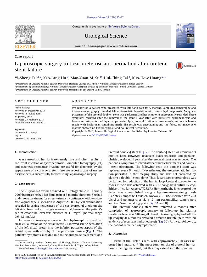
Intravenous urography revealed left hydronephrosis and noclear visualization of the distal ureter. CT showed sciatic herniationof the left distal ureter into the inferior posterior aspect of theischial spine with atrophy of the piriformis muscle (Fig. 1). Thepatient’s symptoms subsided due to the antegrade placement of a
National Taiwan Universityoad, Taipei 10016, Taiwan.uang).
ciation. Published by Elsevier Taiw
ureteral double-J stent (Fig. 2). The double-J stent was removed 3months later. However, recurrent hydronephrosis and pyelone-phritis developed 1 year after the ureteral stent was removed. Thepatient’s symptoms resolved after antibiotic treatment and double-J stent placement. The following year, the double-J stent wasreplaced every 6 months. Nevertheless, the ureterosciatic hernia-tion persisted in the imaging study and was not corrected byplacing a double-J stent alone. Thus, laparoscopic ureterolysis wasperformed for reduction of the hernial loop. Ureteral fixation to thepsoas muscle was achieved with a 2-O polyglactin suture (Vicryl,Ethicon, Inc., San Angelo, TX, USA). Herniorrhaphy for closure of thedefect was accomplished using a hyaluronan-containing mesh(Parietex Composite, Covidien, Norwalk, CT, USA) secured with 2-OVicryl and polymer clips via a 12-mm periumbilical camera portand two 5-mm working ports (Fig. 3A and B).
The ureteral double-J stent was removed 2 months aftercompletion of laparoscopic surgery. At follow-up, her plasmacreatinine level was 0.89 mg/dL. Renal ultrasonography and follow-up imaging at 6 months revealed a smooth ureteral path with noevidence of recurrent hydronephrosis (Fig. 3C). At 1-year follow-up,the patient remained asymptomatic.
3. Discussion
Hernia of the ureter is rare, with approximately 130 cases re-ported in literature.1e5 The most common site of ureteral hernia-tion is the inguinal canal, but this may extend into the scrotum.
an LLC. Open access under CC BY-NC-ND license.
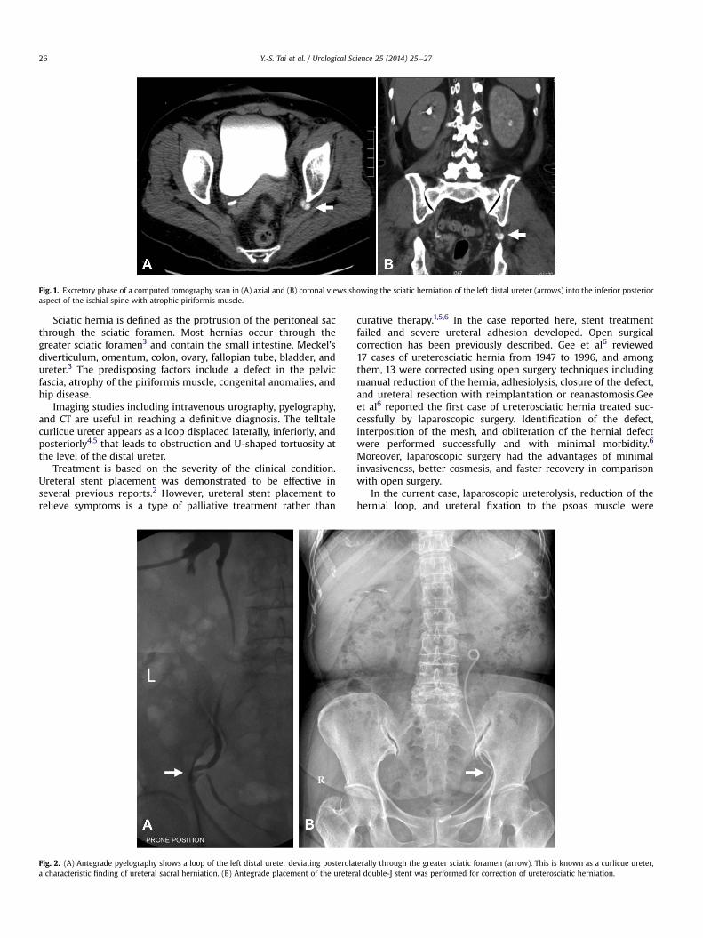
Fig. 1. Excretory phase of a computed tomography scan in (A) axial and (B) coronal views showing the sciatic herniation of the left distal ureter (arrows) into the inferior posterioraspect of the ischial spine with atrophic piriformis muscle.
Y.-S. Tai et al. / Urological Science 25 (2014) 25e2726
Sciatic hernia is defined as the protrusion of the peritoneal sacthrough the sciatic foramen. Most hernias occur through thegreater sciatic foramen3 and contain the small intestine, Meckel’sdiverticulum, omentum, colon, ovary, fallopian tube, bladder, andureter.3 The predisposing factors include a defect in the pelvicfascia, atrophy of the piriformis muscle, congenital anomalies, andhip disease.
Imaging studies including intravenous urography, pyelography,and CT are useful in reaching a definitive diagnosis. The telltalecurlicue ureter appears as a loop displaced laterally, inferiorly, andposteriorly4,5 that leads to obstruction and U-shaped tortuosity atthe level of the distal ureter.
Treatment is based on the severity of the clinical condition.Ureteral stent placement was demonstrated to be effective inseveral previous reports.2 However, ureteral stent placement torelieve symptoms is a type of palliative treatment rather than
Fig. 2. (A) Antegrade pyelography shows a loop of the left distal ureter deviating posterolaa characteristic finding of ureteral sacral herniation. (B) Antegrade placement of the ureter
curative therapy.1,5,6 In the case reported here, stent treatmentfailed and severe ureteral adhesion developed. Open surgicalcorrection has been previously described. Gee et al6 reviewed17 cases of ureterosciatic hernia from 1947 to 1996, and amongthem, 13 were corrected using open surgery techniques includingmanual reduction of the hernia, adhesiolysis, closure of the defect,and ureteral resection with reimplantation or reanastomosis.Geeet al6 reported the first case of ureterosciatic hernia treated suc-cessfully by laparoscopic surgery. Identification of the defect,interposition of the mesh, and obliteration of the hernial defectwere performed successfully and with minimal morbidity.6
Moreover, laparoscopic surgery had the advantages of minimalinvasiveness, better cosmesis, and faster recovery in comparisonwith open surgery.
In the current case, laparoscopic ureterolysis, reduction of thehernial loop, and ureteral fixation to the psoas muscle were
terally through the greater sciatic foramen (arrow). This is known as a curlicue ureter,al double-J stent was performed for correction of ureterosciatic herniation.
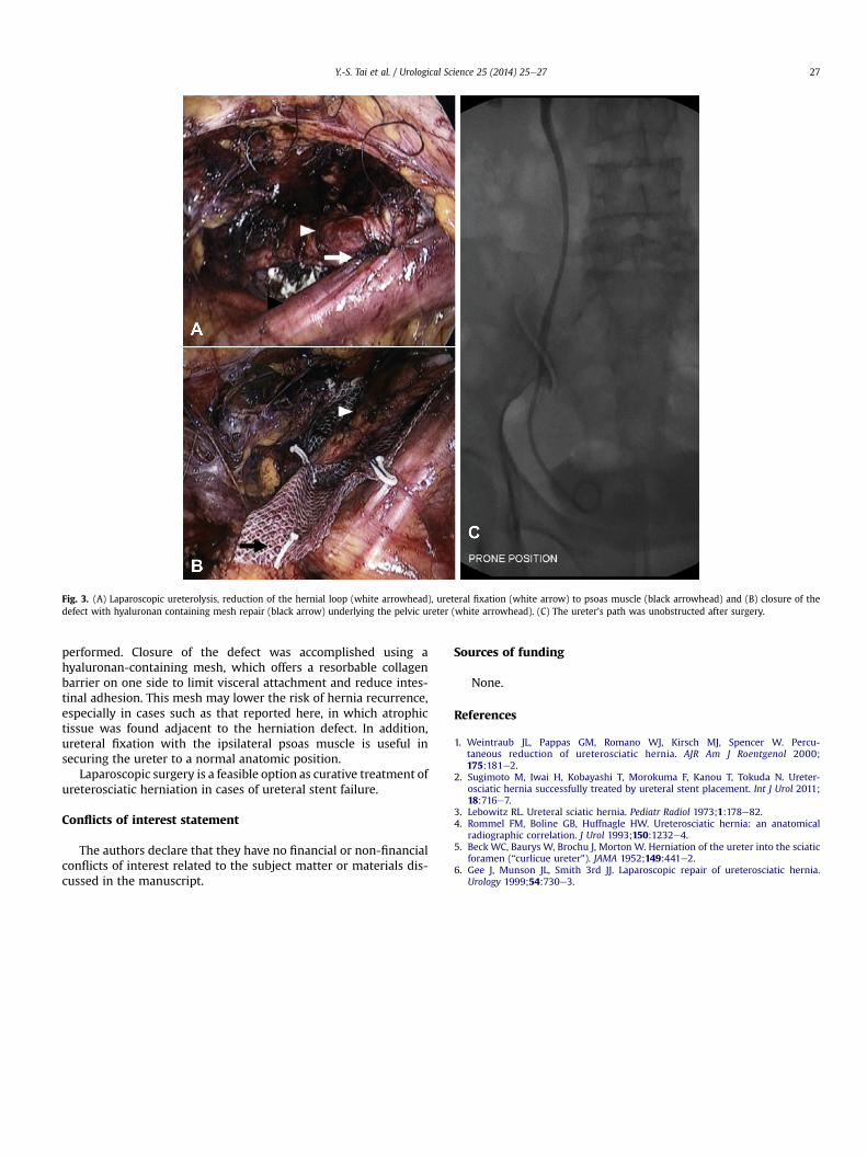
Fig. 3. (A) Laparoscopic ureterolysis, reduction of the hernial loop (white arrowhead), ureteral fixation (white arrow) to psoas muscle (black arrowhead) and (B) closure of thedefect with hyaluronan containing mesh repair (black arrow) underlying the pelvic ureter (white arrowhead). (C) The ureter’s path was unobstructed after surgery.
Y.-S. Tai et al. / Urological Science 25 (2014) 25e27 27
performed. Closure of the defect was accomplished using ahyaluronan-containing mesh, which offers a resorbable collagenbarrier on one side to limit visceral attachment and reduce intes-tinal adhesion. This mesh may lower the risk of hernia recurrence,especially in cases such as that reported here, in which atrophictissue was found adjacent to the herniation defect. In addition,ureteral fixation with the ipsilateral psoas muscle is useful insecuring the ureter to a normal anatomic position.
Laparoscopic surgery is a feasible option as curative treatment ofureterosciatic herniation in cases of ureteral stent failure.
Conflicts of interest statement
The authors declare that they have no financial or non-financialconflicts of interest related to the subject matter or materials dis-cussed in the manuscript.
Sources of funding
None.
References
1. Weintraub JL, Pappas GM, Romano WJ, Kirsch MJ, Spencer W. Percu-taneous reduction of ureterosciatic hernia. AJR Am J Roentgenol 2000;175:181e2.
2. Sugimoto M, Iwai H, Kobayashi T, Morokuma F, Kanou T, Tokuda N. Ureter-osciatic hernia successfully treated by ureteral stent placement. Int J Urol 2011;18:716e7.
3. Lebowitz RL. Ureteral sciatic hernia. Pediatr Radiol 1973;1:178e82.4. Rommel FM, Boline GB, Huffnagle HW. Ureterosciatic hernia: an anatomical
radiographic correlation. J Urol 1993;150:1232e4.5. Beck WC, Baurys W, Brochu J, Morton W. Herniation of the ureter into the sciatic
foramen (“curlicue ureter”). JAMA 1952;149:441e2.6. Gee J, Munson JL, Smith 3rd JJ. Laparoscopic repair of ureterosciatic hernia.
Urology 1999;54:730e3.