laryngeal and hypopharyngeal cancer · pdf fileradiotherapy is not good if there is vocal cord...
TRANSCRIPT
LARYNGEAL CARCINOMA
Surgical Anatomy and patterns of spread :
Common in old age 50 – 60yrs, M > F
Third most common cancer in males.
Anatomically 3 regions : Supraglottis, glottis and Subglottis
Hot Potato voice, Inspiratory dyspnoea � Supraglottic cancer
Dysphagia � Cricopharyngeal sphincter
Odynophagia � Epiglottic cancer
Each region is anatomically and embryologically distinct with separate Lymphatic Channels
SUPRAGLOTTIS :
Includes – arytenoids, epiglottis (laryngeal surface), aryepiglottic folds, false vocal cords and
ventricles
High lymphatics and so high propensity of Cervical Nodal metastasis. Lymph. Exit via the
Thyrohyoid membrane alongside superior laryngeal vessels into JDLN
GLOTTIS :
Both true vocal cords till 5mm below them
VC is membranous anterior 3/5th (Vocalis Muscle) and cartilagenous posterior 2/5th (vocal
process of arytenoid)
Anterior Commisure lies midway between thyroid notch and lower border of thyroid cartilage
in males and a little up in females
Sparse lymphatics of true vocal cords
Lymph from GLOTTIS AND SUBGLOTTIS pass via the cricothyroid membrane into
Delphian (Prelaryngeal), Pretracheal and deep cervical nodes alongside inferior thyroid
artery
SUBGLOTTIS :
Mobile part – 5 mm below VC to upper border of Cricoid
Fixed part – upper to lower border of cricoid
Pattern of Spread :
Due to its unique embryological development and lymphatics usually tumours arising remain
localised to particular sites of origin. Membranes and ligaments acting as natural barriers
Pressman identified pattern of spread
Spread preferentially occurs along lines of least resistance especially when connective tissue
barriers are traversed by blood vessels and nerves
Radiotherapy is not good if there is vocal cord fixity. If VC fixed then surgery
(VPL)treatment of choice always

POTENTIAL TISSUE SPACES :
Reinke’s space – Submucosal space between mucosa of glottis and underlying Vocalis
muscle. Acts as a bursa for sliding of mucosa over the muscle and provides fluency of speech
Early glottis cancers remain superficial and thus stripping of mucosa suffices
Preepiglottic Space of BOYER :
Bounded by :
� Anteriorly – Thyrohyoid membrane
� Superiorly – Hyoepiglottic ligament
� Inferiorly – Thyroepiglottic ligament
� Posteriorly – Epiglottis
Rich in lymphatics and Resistant to radiotherapy as sparse blood supply
Involvement directly puts staging to T3
Space continuous on either side with paraglottic space
Paraglottic Space : Paired
Bounded by :
� Laterally – Thyroid Ala
� Medially – Conus elasticus
� Superiorly – Ventricle and quadrangular membrane
� Posteriorly – pyriform sinus
Contains Thyroarytenoid muscle. Infiltration of this space causes fixity of vocal cords
Inferolaterally gap between cricoid and thyroid and so propensity for extralaryngeal spread
SUPRAGLOTTIC TUMOURS :
Preferential spread upwards to base of tongue and rarely involve below false cord
EPIGLOTTIC TUMOURS:
o SUPRAHYOID EPIGLOTTIS
o INFRAHYOID EPIGLOTTIS
Suprahyoid – Exophytic lesions and bulky proliferative masses with minimal underlying
invasion. Tend to be overstaged.
Infrahyoid – Infiltrative, may grow circumferentially to involve false cords, aryepiglottic
folds, medial wall of pyriform sinus and pharyngoepiglottic folds (lateral Glossoepiglottic
fold)
Preepiglottic space involvement is common (Radioresistant)
Epiglottic tumours rarely invade thyroid cartilage, implies transglottic spread and an absolute
contraindication for Supraglottic laryngectomy.
TUMOURS OF FALSE CORD –
Anterior commissure acts as barrier for tumour spread (Vocal ligament, Thyroepiglottic
ligament, conus elasticus and inner perichondrium of thyroid ala).
Can involve above i.e. epiglottis, posteriorly aryepiglottic folds n arytenoids and anteriorly
the anterior commisure.
TUMOURS OF VENTRICLE –
Paraglottic, Radioresistant, submucosal spread to medial wall of pyriform fossa. Most are
transglottic at presentation
TUMOURS OF ARYTENOIDS AND ARYEPIGLOTTIC FOLDS:
These tumours behave like pyriform fossa malignancies and are called MARGINAL ZONE
CANCERS
Post cricoid spread is common.
GLOTTIC CANCERS :
Most arise from anterior 2/3rd
(membranous part of VC)
Barriers – Reinke’s space and anterior commissure
Initial spread occurs horizontally and vertical spread occurs late.
Inferior spread is limited by conus elasticus which is barrier btw glottis and Subglottis.
Vertical spread directly upstages tumour to T2 and usually subglottic
Deep lateral invasion occurs in paraglottic space.
Cartilagenous portion rarely involved but involved in metastatic tumours from other sites.
Degree of vertical spread imp to plan conservative hemilaryngectomy and critical cut off is
limited to cricoid cartilage which lies 10mm anteriorly and 5mm posteriorly
Primary posterior lesions of glottis are rare but may be involved in secondaries from other
sites.
ANTERIOR COMMISURE LESIONS :
Uncommon
The anterior commissure is directly attached to the thyroid cartilage without intervening
tissues by the Broyle’s ligament. So early cartilage involvement occurs via this �
Tumour spreads superiorly involving base of epiglottis whereas inferiorly tumour exits the
cricothyroid membrane.
If tumour extends from the vocal cords onto the lower portion of the epiglottis at the anterior
commissure, the risk of thyroid cartilage invasion is extremely great
SUBGLOTTIC TUMOURS :
< 1 % of all laryngeal tumours
Spread along entire circumference of Subglottis
Cricoid cartilage involvement early as no intervening muscle layer.
Presenting feature in subglottis also is fixity of vocal cords ☺
Total Laryngectomy is Rx of choice
TRANSGLOTTIC MALIGNANCY :
Tumours that involve both supraglottis and glottis across the ventricle with cord fixity are
called transglottic malignancy. Can be supra to glottis or glottic spreading to supra.
Advanced at presentation
Cartilage involvement early, VC fixity and near total or total laryngectomy
Tumours of supraglottis can occasionally become tranglottic by ant commisure involvement,
invade cartilage early via broyle ligament.
Posterior extension tumours become transglottic by cricoarytenoid joint affection or
Interarytenoid area. Extralaryngeal spread via cricothyroid membrane or thyrohyoid
membrane.
PYRIFORM SINUS (Smugglers fossa)
Bounded by :
� Laterally – Mucosa covering lamina of thyroid cartilage
� Medially – arytenoids and aryepiglottic folds above and cricoid cartilage
below
� Superiorly – Lateral glossoepiglottic folds (pharyngeoepiglottic folds)
� Inferiorly – Oesophagus
No anatomical compartments and so tumour spreads aggressively
Sub mucous spread and so can’t be seen superficially much and hence resection margins
mapped out in cm and not mm
Apex situated at level of cricoid and close to paraglottic space
Spread :
� Anteriorly – Paraepiglottic space, Preepiglottic space
� Posteriorly – Postcricoid region, cricothyroid joint, extralaryngeal
involvement
� Medially – Arytenoid, cricoarytenoid and subglottis
Diagnosis and imaging
SUPRAGLOTTIC MALIGNANCY :
Present late, Usually a metastatic LN
Suprahyoid – overstaged and early diagnosed, non infiltrative
Pain (epiglottic), difficulty in swallowing (Cricopharyngeal sphincter involvement), FB
sensation, stridor, hoarseness, change of voice (hot potato voice), and aspiration (epiglottic Ca
causing failure of laryngeal closure or Ca pyriform fossa involving superior laryngeal
nerve causing sensory loss)
Lymphadenopathy common.
GLOTTIC :
Early presentation
Voice change mainly, Lymphadenopathy rare, stridor, hoarseness
SUBGLOTTIC :
Very uncommon and advanced presentation.
Can involve entire circumference of trachea and result in stridor
90 % of laryngeal cancers are SCC that involve mucosa and so visible on surface
Mobility of VC is the single most important factor to decide choice of treatment
Direct Laryngoscopy is gold standard for diagnosis.

IMAGING TECH. �
Barium Swallow of hypopharynx and upper oesophagus delineates lesions of pyriform sinus,
post pharyngeal wall and post cricoid area.
Imaging techniques are very helpful for evaluating the depth of tumour.
CT –
� Axial plane so accurate in judging vertical and lateral spread of tumour
� Particularly lesions of the anterior commissure and Subglottic region
� Paraglottic space is better visualised with CT
MRI –
� Preepiglottic space and base of tongue
� Superior than CT for Cartilage
STAGING
TNM
SUPRAGLOTTIS :
T1 � 1 subsite with normal VC mobility
T2 � Multiple with normal VC
T3 � Tumour limited to Larynx with VC fixation OR involvement of Preepiglottic space,
post cricoid region, medial wall of pyriform sinus
T4 � Extralaryngeal extension, destruction of thyroid cartilage, involvement of oropharynx
or soft tissues of neck.
GLOTTIS :
T1 �
� T1a – one VC
� T1b – both VC but no fixation yet
T2 �
� T2a – Supra or subglottic spread
� T2b – impaired cord mobility
T3 � Tumour limited to Larynx with VC fixation
T4 � Extralaryngeal extension, destruction of thyroid cartilage, involvement of oropharynx
or soft tissues of neck
SUBGLOTTIS :
T1 � limited to subglottis
T2 � extending to VC with normal or impaired mobility
T3 � Tumour limited to Larynx with VC fixation
T4 � Extralaryngeal extension, destruction of thyroid cartilage, involvement of oropharynx
or soft tissues of neck
N common of all
N1 � < 3 cm Single Ipsilateral
N2 �
� N2a – Ipsi single 3 – 6 cm
� N2b – Ipsi Multiple 3 – 6 cm
� N2c – Contralateral or bilateral 3 – 6 cm involvement
N3 � > 6cm
M1 � Distant metastasis
STAGING ::
Stage 0 – TisNoMo
Stage 1 – T1MoNo
Stage 2 – T2NoMo
Stage 3 –
� T3NoMo
� T1N1Mo
� T2N1Mo
� T3N1Mo
Stage 4 – T4NoN1Mo
� Any T N2N3 M0
� Any T any N M1
So if N1 then directly 3 or 4 and if N2 directly goes to stage 4
Treatment decisions
As a general rule if no VC fixity Radiotherapy gives best results (except pre and para
epiglottic space lesions)
RT alone not to be given in Pre para involvement, VC fixity, Cartilage invasion,
paralaryngeal soft tissue invasion, young indivisual, CIS of glottic cancer, Purely
anterior commisure lesions (surgery preferred)
Supragottic cancers N stage more important for treatment whereas glottis T stage more
important
Also influenced by general health and pulmonary reserve
Age � don’t use radio on young
Voice Quality � Best Radiotherapy > CO2 laser > Laryngofissure surgery
Glottic Cancers
Radiation avoided for CIS. CO2 laser OR Microlaryngoscopic stripping used.
Surgery RxOC in young, verrucous lesions, advanced lesions, patients refusing radiation or
unavailability
T1 – Mid cord lesions – Cure rates >90%
� Radiotherapy
� Endoscopic transoral CO2 laser coredectomy
� Laryngofissure surgery and Cordectomy with temporary tracheostomy

o Cord lesions Invol
� Radiotherap
� VPL (fronto
� Endoscopic
o Anterior Commisur
� VPL (Fronta
� Radiotherap
invasion is
recurrence)
T2 lesions –
o T2a – Radiotherapy
o T2b – VPL / Suprac
o RRSS (Radical radio
T3 and T4 –
o Total Laryngectomy
o RRSS
olving anterior commissure –
rapy – (wedging technique & uniform isodose curves)
ntolateral)
ic CO2 laser excision (only under expert hands)
ure lesions –
ntal)
rapy (cartilage invaded early so not a good option as
is difficult to diagnose also and thus only radiation
)
py with regular follow up
racricoid laryngectomy with CHEP (Cricohyodoepiglo
diotherapy and salvage surgery)
my / near total laryngectomy / extended partial larynge
)
as early cartilage
ion may lead to
lottopexy)
gectomy

SUPRAGLOTTIC LESIONS
MOBILE CORDS AND N
T1, T2 and those T3 where
son nodal mets, age, pulmon
:
NO CARTILAGE INVASION
re though Preepiglottic space involved but no cord fix
onary reserve, general health and subsite.
ixity, Rx depend

IF VOCAL CORD FIXED/CA
Total or near total laryngectomy
Partial
Transoral endoscopic resections
Once anterior commisure invo
Preepiglottic space invaded, Tra
Open partial laryngectomies
Faesable because of embryolo
function physiologically after su
Cricoid is the rostrum on which
complete collapse of larynx.
Arytenoid is essential as a basi
depend on it
Hence partial laryngectomy (
one mobile vibrating arytenoid
Thus only subglottic extens
conservative procedures.
Interarytenoid or retroarytenoid
For Sphincteric action � inner
atleast one SLN
Aim of reconstruction is to mai
normally provided by arytenoid
CARTILAGE INVADED :
y with permanent tracheostomy
Partial Laryngectomy
volved or deeper laryngeal spaces namely paraglo
ransoral endoscopic excision is oncologically unafe
logical compartmentalization of larynx and ability
such resections.
ch the entire larynx rests and resection of even a part o
asic functioning unit of larynx, phonation, aspiration
(voice and nasal respiration) � intact cricoid rin
oid utmost important.
nsion > 10mm anteriorly and 5 mm posterior
id involvement also necessitates resection of both aryt
ervation necessary so Subglottis � save RLN and f
aintain AP diameter to prevent stenosis & posterio
ids maintained to avoid aspiration and good quality of
glottic space or
ity of larynx to
rt of this leads to
n prevention all
ring and atleast
iorly is CI for
ytenoids.
for supraglottis
rior glottic bulk
of voice.

CONTRAINDICATIONS FOR VOICE CONSERVATION INCLUDE :
Subglottic extension (> 5mm posteriorly and > 10mm anteriorly)
Interarytenoid
Retroarytenoid involvement
Lesion too close to cricoid cartilage
Cricoarytenoid joint inv bilaterally
Arytenoid involved bilaterally
Loss of nerve supply bilaterally
GLOTTIC CANCER CONSERVATIVE SURGERIES
Report early so usually localised disease and thus mostly conservative approach used
LARYNGOFISSURE WITH CORDECTOMY:
Involved cord + ipsilateral soft tissue resected, thyroid cartilage divided for approach but not
resected.
VPL i.e. VERTICAL PARTIAL LARNGECTOMY :
Vertical segment of thyroid cartilage + Cord lesion + intervening paraglottic space.
Procedure can be extended in anterior commisure and/or arytenoids.
SUPRACRICOID LARYNGECTOMY WITH CHEP :
Entire thyroid cartilage + involved portion of glottis + paraglottic tissues
LARYNGOFISSURE WITH CORDECTOMY:
Now replaced by CO2 laser cordectomy
INDICATIONS :
Mid cord lesions
T1 lesions
No impairment of vocal cord mobility
No anterior commisure involvement
PROCEDURE :
GA through tracheostomy � Midline thyrotomy (incision from thyroid notch to inferior border of
thyroid cartilage) � Incision through cricothyroid membrane and larynx entered � Anterior cut
placed 2 to 3 mm posterior to anterior commisure and posterior cut anterior to arytenoid cartilage
� Superior resection line above false vocal cords and inferiorly lower border of thyroid cartilage.
The resultant mucosal defect is left to heal by granulations. In weeks dense fibrous pseudocord
forms making it difficult to differentiate from true cord.
Tracheostomy usually closed in a week.
Procedure well tolerated as doesnt cause aspiration.
Neither disturbs nerve supply nor pharyngeal musculature.
Cure rates 84-98%
VPL (Vertical Partial Laryngectomy aka Vertical Hemilaryngectomy)
Described by Billroth
INDICATIONS :
Impaired cord mobility glottic cancers
Anterior commisure involved

Extension to the anterior surface of arytenoid can be resected with VPL.
Resection depends on part of glottis involved :
HEMILARYNGECTOMY � cord lesion without anterior commisure or arytenoid involvement.
FRONTAL � Anterior commisure lesions
FRONTOLATERAL � Cord + anterior commisure
EXTENDED HEMILARYNGECTOMY � Cord lesion involving arytenoid
SUBTOTAL BIFRONTAL HEMILARYNGECTOMY � Horse shoe lesion involving both VC
but sparing arytenoids.
ELIGIBILITY CRITERIA FOR VPL
1. One VC + Anterior commisure + Not more than 1/3rd of opposite cord involved
2. Only extension till vocal process of arytenoid.
3. Subglottic Extension with not more than 10mm anteriorly and 5mm posteriorly
4. If paraglottic extension then should not cross ventricle above and has minimal subglottic
extension which will allow a clear margin of resection above the cricoid cartilage.
5. Cancer of true vocal cord with no involvement of the thyroid cartilage.
6. RT failure in early lesion i.e. RT lesion as well as residual lesion both satisfy eligibility criteria
for VPL.
PROCEDURE
Full thickness en block resection of involved portion of glottis along with overlying segment of
thyroid cartilage and intervening paraglottic tissues.
Upper margin of resection includes segment of false cord and lower margin above cricoid
cartilage
GA via tracheostomy � Horizontal incision over thyroid from one SCM to other � strap
muscles separated exposing larynx � reflect perichondrium on both sides of thyroid cartilage �
2 vertical thyroid cartilage cuts depending on lesion � entry via cricothyroid membrane �
first cut across glottis on less involved site � Epiglottis and both superior laryngeal nerves
preserved.
RECONSTRUCTION :
Reattachment of remnant vocal cords � the C/L true cord should be anchored to thyroid or
soft tissue to keep it taught.
Mucosal defect � need not be reconstructed in most cases except in FRONTAL or
FRONTOLATERAL laryngectomies as we have to prevent anterior fusion of two sides with
resultant stenosis.
Done using sialistic keel. Removed after 2 to 3 weeks
For Subtotal bifrontal laryngectomy epiglottis used for reconstruction as mucosa lining of
epiglottis is consistent with mucosa of remaining larynx. Epiglottis sutured to defect using
tensionless interrupted vicry sutures
Epiglottic recons. � distressing aspiration
Reconstruction of resected arytenoid � if posterior glottic bulk not restored � poor voice
quality and aspiration
Muscle,tendon, fat, cartilage, epiglottis can be used
Most prefer use of remnant thyroid cartilage

Raw area is resurfaced with mucosa of adjacent pyriform fossa.
COMPLICATIONS :
Aspiration, poor voice quality, cartilage necrosis, laryngeal stenosis.
SUPRACRICOID LARYNGECTOMY WITH CHEP ie CRICOHYOIDOEPIGLOTTOPEXY
Paraglottic spread with more likeliness of thyroid cartilage invasion
VPL removes only part of thyroid cartilage but CHEP removes full thyroid cartilage
It is essential that Preepiglottic space is free of tumour as laryngeal entry superiorly is via the
transepiglottic incision which cuts across the Preepiglottic space.
Cricoid, Hyoid and Suprahyoid portion of epiglottis (maintains sphincteric action, prevents
aspiration), SLN b/l is preserved.
INDICATIONS
Immobility or fixity of vocal cord (usually due to paraglottic inv of Thyroarytenoid) in presence
of mobile arytenoid
Ie absent cricoarytenoid joint involvement
Horseshoe lesions involving more than 1/3rd of C/L cord. Even VPL ie subtotal bifrontal
laryngectomy can be tried but resection will necessiate mucosal reconstruction.
CONTRAINDICATIONS :
Involvement of preepiglottic space
Involvement of cricoarytenoid joint
Subglottic extension involving cricoid
Poor pulmonary reserve
Poor general health of patient
ADVANTAGES OVER VPL
Entry in VPL via cricothyroid membrane and so limited exposure. In Supracricoid laryngectomy
entry via epiglottis above and cricothyroid membrane below both so EXCELLENT
EXPOSURE
Removal of thyroid cartilage has added oncological safety.
It also preserves nasal respiration and speech.
SUPRAGLOTTIC Ca PROCEDURES
MC i.e. 69% of all laryngeal cancers
Marginal zone and suprahyoid Ca more common than others
Rarely invade thyroid cartilage or cross ventricle to involve glottis
Spread upwards, involve preepiglottic space, valleculae, base of tongue usually
HORIZONTAL SUPRAGLOTTIC LARYNGECTOMY :
False cords, epiglottis, Preepiglottic space and upper 1/3rd of thyroid cartilage.
Hyoid bone included in resection when Preepiglottic space is grossly infiltrated.
Done for endolaryngeal lesions with mobile VC.
EXTENDED SGL
I/L arytenoid, base of tongue, valleculae, pyriform sinus
INDICATIONS
Cord mobility is an important prerequisite for SGL
Once paraglottic involvement occurs tumour becomes Transglottic and SGL cannot be done
Infrahyoid epiglottic lesions (as high propensity for preepiglottic space involvement) with mobile
cords
N2 N3 disease.
Suprahyoid lesions where radiotherapy CI as in young patients and CO2 laser unavailable
CONTRAINDICATIONS
VC fixity
Poor pulmonary reserve
Involvement of thyroid cartilage
Involvement of pyriform sinus upto apex
Involvement of interarytenoid or retroarytenoid region
Involvement of post cricoid region
Extensive involvement of base of tongue.
PROCEDURE :
GA via tracheostomy � 1 SCM to other horizontal incision � only perichondrium incised along
upper border and reflected down � save perichondrium as assists in closure � cuts made in
thyroid cartilage � avoid injury to anterior commisure as it may lead to permanent diff in speech
and swallowing � save hyoid if limited spread to preepiglottic space allows early
rehabilitation and more secure closure � in endolaryngeal central tumours only middle third
of hyoid resected � one sided disease divide hyoid at junction of body and lesser cornu which
helps preserve SLN � min one SLN has to be preserved �
Excision of tumour � Transvallecular approach except in extended where even valleculae
involved � epiglottis pulled downwards � aryepiglottic folds divided anterior to arytenoids �
dissection continued inferiorly through ventricles � preserving true vocal cords � false
removed
RECONSTRUCTION
Cricopharyngeal myotomy can be performed to facilitate postop swallowing
Closure performed by suturing cut edge of pyriform mucosa below to oropharyngeal mucosa
above starting laterally then going medially.
As region of resected supraglottis approached, primary mucosal closure is not possible
Now approximate upper end of remaining thyroid cartilage to base of tongue using 3 or 4 strong
nonabsorbable sutures.
Thyroid perichondrium which was preserved acts as second layer of closure
EXTENDED SUPRAGLOTTIC PROCEDURES
ARYTENOID RESECTION � Partial or complete � if complete carefully dislocate
cricoarytenoid joint to prevent injury to recurrent laryngeal nerve � I/L vocal cord medialized

and anchored to cricoid cartilage � lower plane of anaesthesia induce cough and check �
sometimes if extensive resection then posterior glottic bulk is maintained by using cartilage or
muscle � raw area resurfaced using pyriform mucosa
BASE OF TONGUE/VALLECULAE RESECTION � ENTRY VIA UNINVOLVED
VENTRICLE inferiorly � progress posteriorly � atleast one half of base of tongue has to be
preserved along with blood supply � if direct closure not possible due to excess loss of soft
tissue use PMMC flap.
RESECTION OF LATERAL WALL OF PYRIFORM SINUS :
Lateral wall + posterior and lateral wall of pharyngeal mucosa. Closure requires PMMC
COMPLICATIONS OF SGL :
Aspiration (can be prevented by preserving SLN), other surgical technique described are the
cricopharyngeal myotomy and suspension of larynx via mandible
Pharyngocutaneous fistula is a rare complication.
POSTOPERATIVE CARE
Nasogastric tube feeds started 24 to 48 hrs following surgery.
4 to 5 days later when oedema decreases tracheostomy tube corked and nasal respiration is
encouraged.
Once tracheostomy tube removed subglottic pressure rises and decreases cough and decrease
chances of aspiration.
After 1 week � oral feeds
After 2 to 12 weeks later � normal deglutition
Majority of SG cancers are Marginal zone
Seen mostly in elderly
Neck mets very common so Rx with radiotherapy of neck.
PROCEDURES FOR TRANSGLOTTIC CANCER
Transglottic aka transventricular cancers
Total or near total laryngectomy with permanent tracheostomy
Only in select few extended partial laryngectomy is feasible (vertical and horizontal laryngectomy
combinations).
Voice Conservation Procedure for transglottic cancers include �
Three Quarter laryngectomy
Supracricoid laryngectomy with CHEP
Minimum intact cricoid ring and atleast one functioning arytenoid.
THREE QUARTER LARYNGECTOMY :
Vertical Hemilaryngectomy + SGL
INDICATIONS
SG cancers spread to glottis and vice versa
CONTRAINDICATIONS
Subglottic involvement
Cord fixity due to cricoarytenoid joint involvement
Thyroid cartilage invasion

Interarytenoid or postcricoid involvement
RESECTION :
Body of Hyoid + thyroid cartilage + Preepiglottic space + I/L true vocal cord + Paraglottic space
and I/L arytenoid if involved.
GLOTTIC RECONSTRUCTION
Important to reconstruct ipsilateral VC for maintenance of functional laryngeal aditus.
RECONSTRUCTION USING MUSCLE FLAP
Sternohyoid muscle � Sutured as close to C/L cord and covered with pyriform mucosa flap.
Cartilage flaps better as muscle flaps can contract during healing thus distorting glottic
architecture and competence
USING CARTILAGE
Remaining thyroid cartilage can be restructured and refashioned to form i/l cord.
Cartilage can be used as free graft or pedicled flap
Free grafts like muscle flaps may contract during healing get dislodged
Pedicled cartilage flap � corner of ipsi or contra thyroid cartilage along with attached constrictor
muscle or simple fold over of the ipsi thyroid cartilage remnant.
SUPRACRICOID LARYNGECTOMY WITH CHP
Best suited for Transglottic cancers that have no extension into the pyriform or the base of tongue.
THYROID INVASION is NOT a CI for the procedure ☺ ☺
Entire thyroid + Pre + Paraglottic space + epiglottis resected
Cricoid + Hyoid and atleast one arytenoid preserved
INDICATION
Transglottic
Paraglottic spread with mobile arytenoids
Thyroid invasion
Anterior commisure involvement
CONTRAINDICATIONS
Cricoarytenoid jt involved
Subglottic extension > 10mm anteriorly and 5 mm posteriorly
Involvement of base tongue, valleculae, hyoid bone cant be saved as in massive preepiglottic
space involvement
Involvement of pyriform sinus
Post cricoid and interarytenoid regions
Prior tracheostomy is incompatible with procedure.
PROCEDURE
Preop tracheostomy AVOIDED as level of the tracheostome will change after the reconstructive
procedure is complete.
Here anaesthesia is administered via Orotracheal tube.
Freeing mucosa of pyriform sinus and dislocation of cricothyroid joint
Inferior constrictor muscle along with the perichondrium of the thyroid cartilage is incised along
the posterior border the thyroid cartilage � mucosa of pyriform fossa is then freed from the

cartilage using periosteum elevator � cricothyroid joint is gently dislocated taking care to avoid
injury to RLN and steps repeated on opposite side.
DISSECTION OF PREEPIGLOTTIC SPACE
The periosteum along the inferior border of hyoid bone is incised and stripped off its posterior
surface. Hyoid bone preserved
RESECTION OF TUMOUR :
Larynx entered through VALLECULAE above and cricothyroid membrane below.
Endotracheal tube passed via cricothyroid incision.
Incision is along the aryepiglottic fold to superior border of cricoid cartilage
Second cut between thyroid cartilage anteriorly and freed pyriform mucosa posteriorly.
RECONSTRUCTION :
The arytenoid cartilage if partially resected is fixed to the posterolateral border of cricoid cartilage
Cricoid to hyoid via 1-0 prolene sutures
To avoid tension on suture line the cervical and mediastinal trachea is mobilized using finger
dissection anteriorly and laterally.
Temp tracheostomy now made and closed after 4 days. Swallowing is promoted after one week
� pureed diet � remove nasogastric tube
CO2 LASER EXCISION
Precise, haemostasis, postop oedema less
Single sitting Rx (Radiation around 6 weeks treatment)
Resection performed as a day care procedure. No tracheostomy required. And if treatment
remains inadequate RT and surgery can still be done
INDICATIONS
BENIGN LESIONS
1. Vocal Nodule/polyp
2. Vapourization of papillomas
3. Laryngeal web release or post intubation stenosis
4. Arytenoidectomy in bilateral cord palsy.
5. Rienkes edema
PRE MALIGNANT / MALIGNANT LESIONS :
Early T1 T2 lesions of glottis
Keratosis / dysplasia of VC and Ca in situ
Supraglottic cancer confined to free border of epiglottis and aryepiglottic fold.
Localised recurrent or residulal disease following RT failure
Avoiding Tracheostomy in patients undergoing RT, residual disease.
With experience even T2b - T3 lesions can be treated with CO2 laser
PROCEDURE
Transorally using suspension laryngoscope and microscope
Laser beam focussed on a spot size and manipulated using micromanipulator to permit precise
excision in a relatively bloodless field.
Monobloc or multibloc resection based on area involved (avoids excessive sacrifice of normal
tissue).
Fibrosis due to previous neck surgery, prior radiotherapy, large tongue etc may prevent adequate
exposure and thus CO2 laser not preferred
CO2 laser is an excellent cutting tool but limited coagulation capability.
Minor ooze controlled thus with a defocussed laser beam or usually a adr. cottonoid.
For arterial bleeds, electrocautery is must usually required for supraglottic lesions.
ET tube wrapped with reflective aluminium foil to avoid cumbustion of inhaled gases.
Soaked cotton pledgets kept in larynx to prevent thermal damage.
Wear goggles to protect eyes.
Involvement of Anterior commisure, vocal process of arytenoid. Asso with higer recurrence so
CO2 avoided.
Early supraglottic lesions are more demanding for laser excisions as exposure not adequate,
haemostasis is a problem, defining margins difficult. Ideal lesion is one localised in free margin of
epiglottis without involving base or small lesion of aryepiglottic fold.
CO2 laser can also be used to create an airway in advanced laryngeal cancer to avoid
tracheostomy prior to definitive RT or surgery.
NEAR TOTAL LARYNGECTOMY :
Aka Pearson's procedure, extended vertical laryngectomy, subtotal laryngectomy or
Parsimonius laryngectomy. When pyriform fossa mucosa also removed its called
NTLpharyngectomy and if more extensive resection of pharynx with flap reconstruction then
called Extended NTLP.
Preserves voice but not nasal respiration.
Too extensive for partial laryngectomies but have sufficient laryngeal mucosa left with nerve
supply to form a shunt which diverts air into the pharynx for production of lung powered speech.
Cricoid ring involved but one arytenoid is supple and free of disease � NTL.
C/l RLN with arytenoid + Segment of cricoid forming cricoarytenoid joint + strip of posterior
tracheal wall leading upto permanent tracheostome are preserved
This innervated laryngeal remnant is fashioned in a sphincteric tube large act as a dynamic shunt
for voice production by diverting air from trachea to pharynx.
So like total laryngectomy there is an end tracheostome which needs occlusion for voice
production.
INDICATIONS
Subglottic extension
Involvement of pyriform apex.
Interarytenoid, retroarytenoid and postcricoid region must be free.
NTL may be done in patients on grounds of poor pulmonary reserve as an alternate to SGL.
CONTRAINDICATIONS :
Inter, retro arytenoid or post cricoid involvement
Mucosal involvement of > half of length of C/l cord anteriorly as this does not allow preservation
of sufficient laryngeal remnant for shunt formation.
Prior radiotherapy relative CI if tissues oedematous.

PROCEDURE :
RESECTION
Strap Muscles, I/L thyroid cartilage, thyroid lobe, I/L cricoid cartilage ring, upper tracheal rings
resected
Preepiglottic space, epiglottis, hyoid, if necessary valleculae resected
I/L VC with involved C/L cord resected
RLN needs preservation which innervated myomucosal shunt.
Preop tracheostomy
TRANSVALLEULAR entry / through uninvolved ventricle as told by pearson
Mucosa of Interarytenoid region and over posterior cricoid lamina in incised, posterior cricoid
lamina fractured in midline carefully avoiding damage to post cricoid mucosa.
Resection completed saving one arytenoid with posterior tracheal wall between arytenoid and
tracheostome.
CONSTRUCTION OF SHUNT
14F Foley’s or No 6 red rubber catheter used as a stent, adequate size and prevents blockage of
shunt. Airway diameter formed by this is 6mm which is important to generate adequate subglottic
pressure for phonation.
Catheter passed through nostril via shunt and taken out via tracheostome
PHARYNGEAL RECONSTRUCTION :
Neopharynx is closed as in total laryngectomy. Care taken near neoglottis to avoid post op
pharyngocutaneous fistula.
Patch pharyngoplasty using PMMC done in cases of extensive resection
POSTOP MANAGEMENT :
Rubber tube used as as a stent is removed 24 to 48 hours after surgery.
Once by 7th postop day wound completely healed and no salivary leak then oral feeds are started.
Tracheostomy tube used only if tendency of stoma towards stenosis
Speech after 3 to 4 weeks
Allow suture wounds to heal
In case of shunt stenosis dilate using bougies
VOICE PRODUCTION :
Lung powered speech
Unless shunt breakdown or stenosis
ADVANTAGES OF NTL OVER TEP :
Quality of voice is superior
TEP requires a Blom Singer prosthesis
Cost 1500 to 3000 and lasts only few months
Postoperative RT is difficult to tolerate in TEP.

COMPLICATIONS :
Stent stenosis, aspiration, phary
MANAGEMENT OF NECK IN L
Involvement of nodes decreas
percent
Patients of SCC respond well
(Cisplatin)
RATIONALE OF CHEMO :
Many patients receiving and
from chemo might benefit well
replace radical surgery as chose
Anterior chemotherapy may be
experience relief in stridor, impr
Thirdly, Chemo reduced tumou
Partial laryngectomy and laryng
ryngeal leak.
LARYNGEAL CANCERS
ases survival by 50
ell to Chemotherapy
nd BENEFITTING
ll with radio and can
sen Rx and thus laryngeal preservation
be used to reduce tumour burden before definitive the
provement i n voice, and symptomatically better prior
our bulk and so patients for total laryngectomy mig
ngeal preservation.
therapy (patients
ior to radiation)
ight land up for

SPEECH RESTORATION :
MC is Oesophageal speech
Patient swallows air into oesophagus by oropharyngeal trapping. Air released and exhalation
across pharyngoesophageal mucosa produces sound by the apposition of mucous membranes.
Electronic devices
TEP problems� aspiration, tissue breakdown, infection, stenosis
Shunt procedures better
Blom singer prosthesis is 3.3mm in diameter and is retained in the oesophagus by a circular
flange or collar in TEP. The Duck bill valve is easier to introduce but offers more resistance to air
flow.
About 40% if TEP do not develop prosthesis.
There can be primary or secondary TEP. Rarely done now
TEP PROCEDURE NOT MADE NOTES
HYPOPHARYNGEAL MALIGNANCY
AETIOLOGY
� Smoking
� Alcoholism
� Inadequate intake of fruits and vegetables
� Saturated fatty acids
� Lack of vitamins (C and E)
� Immune mediated
� Increased exposure to welding fumes
� Exposure to polycyclic aromatic hydrocarbons was associated with oesophageal cancer
� Radiation exposure
� Smoking and alcohol have multiplicative effect
� HPV 16 and 18 have been implicated (p53 underexpression by stabilizing its protein)
EPIDEMIOLOGY
� Pyriform fossa 43%
� Post cricoid 43%
� PPW 9%
� Cervical esophagus
� F:M = 2:1
� Postcricoid cancer is associated with Patterson browne Kelly syndrome
BENIGN TUMOURS AND OTHER NON NEOPLASTIC LESIONS
� Leiomyomas, fibrolipomas and papillomas, and carcinoma in situ
� Once diagnosed they are usually readily dealt with endoscopically or occasionally by lateral
pharyngotomy.
� Nasogastric tube syndrome
� Acquired stenosis of hypopharynx
MOLECULAR AND CELL BIOLOGY OF HYPOPHARYNGEAL CANCER
� Amplification of Chr 11q13
� Loss of tumour suppressor genes eg p53 (pathological stabilization of the p53 protein)
� Increased Ki67 is a proliferation marker expressed in the Gl and S phase of the cell cycle. Cells
with high proliferation had a lower five year survival than patients with low proliferation.
� Loss of retinoblastoma (Rb) tumour suppresser gene.
� Overexpression of growth factor receptors or over secretion of growth factors may be considered
as oncogenic events
� EGFR (epidermal growth factor receptor) overexpression in 30% patients
� Bax is proapoptotic and Bcl2 is antiapoptotic. (Bax expression is reduced)
� Cytokeratine 18 particularly appears to be expressed in nearly all cancers arising from the
hypopharynx and larynx.
� Cathepsin-D over expression (degrades extracellular matrix allowing tumour invasion and mets)
Is a potential independent predictor of cervical node metastases.
� Matrix-metalloproteases (MMP) also degrade the matrix. MMP 9 important. And under
expression of its inhibitor TIMP 1.
� Cancer cells largely fail to express relevant MHC antigens necessary for an effective host
response to the tumour.
� Increased Acute Phase Proteins regulated by IL6.
HISTOLOGY
� Most SCC Non Squamous are only 3.5%.
� Of the squamous cell carcinomas, the majority (70 percent) are moderately or well differentiated.
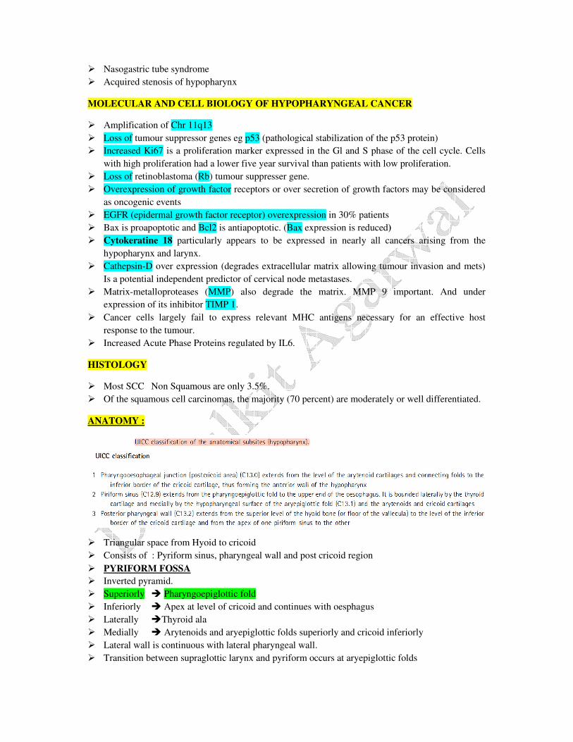
ANATOMY :
� Triangular space from Hyoid to cricoid
� Consists of : Pyriform sinus, pharyngeal wall and post cricoid region
� PYRIFORM FOSSA
� Inverted pyramid.
� Superiorly � Pharyngoepiglottic fold
� Inferiorly � Apex at level of cricoid and continues with oesphagus
� Laterally �Thyroid ala
� Medially � Arytenoids and aryepiglottic folds superiorly and cricoid inferiorly
� Lateral wall is continuous with lateral pharyngeal wall.
� Transition between supraglottic larynx and pyriform occurs at aryepiglottic folds

Posterior Hypopharyngeal wall :
� Posterior pharyngeal wall from Hyoid to cricopharynx. Lateral boundary is the lateral wall of
pyriform sinus
Postcricoid region
� Pyriform sinus and posterior hypoparyngeal wall lead below to post cricoid region which forms
inlet of oesophagus.
� This circular space behind cricoid encompasses cricopharyngeus muscle that provides sphincteric
action controlling passage of food from pharynx to oesophagus.
Patterns of spread
� Submucosal spread
� So more extensive than they appear externally
� Hence resection margins mapped out in cm and not mm
� Rich lymphatics and so nodal metastasis common
� Spread of PF malignancy � superiorly tongue, postcricoid inferomedially, posteriorly to
pharyngeal wall.
� Majority infiltrative so medially larynx and laterally thyroid cartilage with extralaryngeal
involvement
� Ossified cartilages are more prone to tumour invasion
� Paraglottic extension can occur anteromedially fixing vocal cords in apical lesions.
� About half the cases of pyriform fossa cancer demonstrate vocal cord fixation at the time of
presentation
� Even early lesions of pyriform apex are bad as very close to cricoid and voice conservative
surgeries usually not feasible
� Vocal cord fixation can be due to three causes: invasion of the cricoarytenoid joint, invasion of
the posterior cricoarytenoid muscle or involvement of the recurrent laryngeal nerve.
� Posterior pharyngeal wall tumours are relatively unusual.
� Mets to Jugular LN II,III,IV, Submandibular (I) and Posterior triangle (V) rarely involved
� Posterior hypopharynx will extend to Nodes of Rouvier and nodes around internal jugular. May
involve prevertebral space and leading to difficulty in extending neck and restricted side to side
mobility of larynx.
� Postcricoid carcinoma spreads circumferentially to cause obstruction to swallowing & extension
to cervical oesophagus is very common.
� Lateral or Parapharyngeal spread can cause widening of hypopharynx and may even involve
carotids.
� Sometimes anterolateral spread of Postcricoid Ca can present as swelling in region of thyroid
leading to misdiagnosis of goitre.
� Both goitre and post cricoid cancer are common in females.
� Often B/L cervical and mets along IJV with paratracheal LN involvement seen in postcricoid
cancer.
� Postcricoid cancers are less often associated with neck node metastases than piri form fossa
cancer.
� Distant mets to lungs

SYMPTOMS :
� Pharyngeal wall and pyriform u
unless gross in size
� 50% present with cervical LN
neck mass is first presenting fea
� PYRIFORM SINUS � Irrit
retention felt during swallowing
� Otalgia has been well describ
referred pain. The vagus n
supraglottis and piriform fossa m
� POST CRICOID � Progressive
� Voice change is late feature m
ie laryngeal musculature involve
SIGNS
� 90 Degree or 75 degree scope or
� Pooling of secretions � both py
� Loss of crepitus due to preverteb
� See for neck swellings as postcr
� Those patients without a node w
� See for Mobility of VC for Rx p
INVESTIGATIONS :
� Barium Swallow � filling def
cancers. Will also determine
postcricoid cancer.
� DLScopy and Biopsy
� FNAC neck node. Open biopsy
� CT scan
� CT-PET good for mapping out
and also postradiation residual d
� Disease active area will have h
FDG (fluorodeoxy glucose)
brighter.
� If lung mets present the th
carries out pneumonectomy or
head and neck surgery performe
STAGING
� T1 � < 2 cm with one subsite
� T2 � 2-4 cm more than one
fixation of hemilarynx
� T3 � >4 cm with fixation
usually symptomless
LN mets and usually
eature
ritation and mucous
ng, Otalgia.
ribed and is due to
nerve supplies the
a mucosa, as well as the mucosa of the postcricoid.
ive dysphagia
may be cause of RLN infiltration or paraglottic spac
lvement.
or flexible, fibreoptic nasopharyngolaryngoscope.
pyriform fossa and postcricoid lesions.
tebral space inv. in case of Postcricoid Ca (TROTTER
tcricoid can present like goitrous swelling.
will almost always harbour occult nodal disease.
planning
efect in pyriform
e lower limit of
sy avoided
ut occult LN mets
l disease
e high take up of
e) and appear
thoracic surgeon
or lobectomy and
med 6weeks later.
e subsite without
ace involvement
ER’S SIGN)
� T4a � Tumour involves thyroid cartilage or gland, cricoid cartilage, hyoid bone, esophagus or
central compartment soft tissue.
� T4b � prevertebral fascia, encases carotid artery or involves mediastinal structures
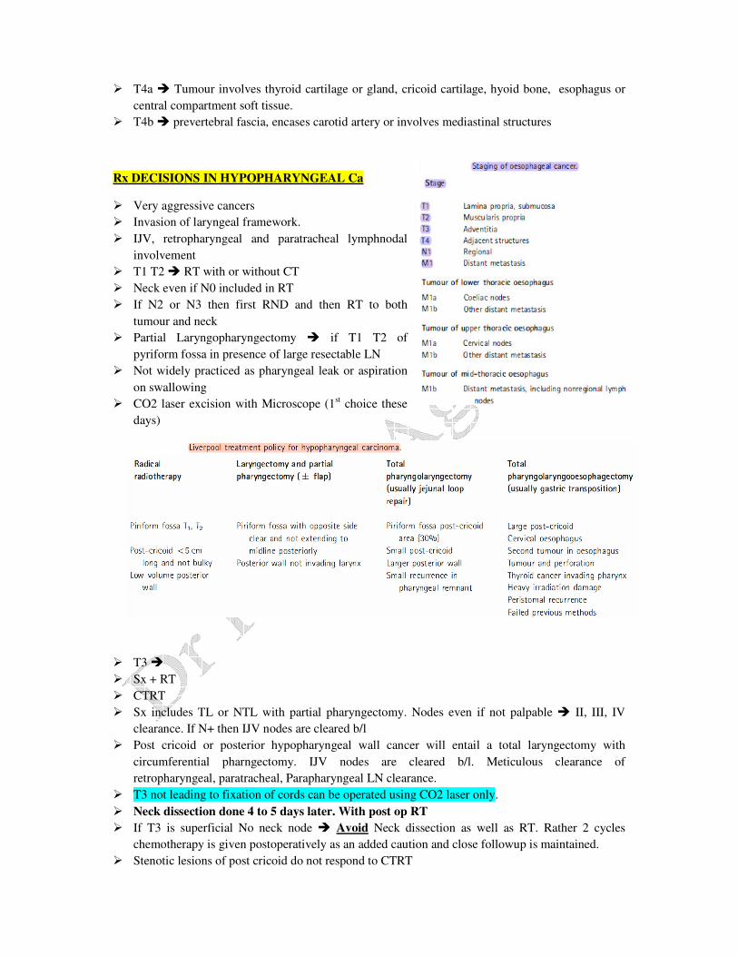
Rx DECISIONS IN HYPOPHARYNGEAL Ca
� Very aggressive cancers
� Invasion of laryngeal framework.
� IJV, retropharyngeal and paratracheal lymphnodal
involvement
� T1 T2 � RT with or without CT
� Neck even if N0 included in RT
� If N2 or N3 then first RND and then RT to both
tumour and neck
� Partial Laryngopharyngectomy � if T1 T2 of
pyriform fossa in presence of large resectable LN
� Not widely practiced as pharyngeal leak or aspiration
on swallowing
� CO2 laser excision with Microscope (1st choice these
days)
� T3 �
� Sx + RT
� CTRT
� Sx includes TL or NTL with partial pharyngectomy. Nodes even if not palpable � II, III, IV
clearance. If N+ then IJV nodes are cleared b/l
� Post cricoid or posterior hypopharyngeal wall cancer will entail a total laryngectomy with
circumferential pharngectomy. IJV nodes are cleared b/l. Meticulous clearance of
retropharyngeal, paratracheal, Parapharyngeal LN clearance.
� T3 not leading to fixation of cords can be operated using CO2 laser only.
� Neck dissection done 4 to 5 days later. With post op RT
� If T3 is superficial No neck node � Avoid Neck dissection as well as RT. Rather 2 cycles
chemotherapy is given postoperatively as an added caution and close followup is maintained.
� Stenotic lesions of post cricoid do not respond to CTRT
� Neck dissection always favoured in T3 lesions with large mets node
� Chemo includes � Cisplatinum + 5FU. Give 2 cycles , if response satisfactory the continue with
full dose RT ie around 70 Gy. If CT response poor RT wont work surely
� Continuous 5FU infusion might lead to cardiospasm and can be limitation in some individuals.
� Cisplatin can also be given concurrently with RT
� Mucosal toxicity is very High.
� Nutritional support mandatory via Ryles or feeding gastrostomy.
� Compl of CTRT is tight stenosis condemning the patient to a life time gastrostomy feeding
� For T4 lesions nonsurgical CTRT not recommended. SO only surgery.
VOICE CONSERVATIVE SURGERY IN HYPOPHARYNGEAL CANCER
� Transoral Microlaryngeal CO2 surgery
� Radiation
� T3 T4 � No scope
� Lateralised hypopharyngeal still can be taken up for NTLP with lung powered speech with
sacrifice of nasal respiration.
� Pyriform cancers are MC hypopharyngeal malignancies (Lateralised commonest)
� T1, T2, T3 superficial with mobile cords � Partial LP or CO2 excision
� Extended SGLP
Supracricoid Hemilaryngopharyngectomy (growth localised to pyriform)
� There should be no Cord fixity one sided, no thyroid erosion, no paraglottic inf, apex of pyriform
and retroarytenoid free of disease
� High rate of complications, don’t do if pulm insuff
Transoral Endoscopic CO2 Laser excision :
� Transoral CO2 provides early restoration of oral feeding and post op tracheostomy not needed.
� Oral feeds can be started 2 days postop
� Can be carried out on a day care basis
� Hypopharynx more vascular and thus procedure more demanding.
� Segment by segment resection carried out, submucosal resection can be carried out retaining
arytenoids and attached soft tissues and thus prevents post op aspiration
� For Superficially infiltrative lesions only
� Cervical spondylosis, fibrosis due to previous radiation or surgery, SMF etc are relative CI
� In cases where lateral pharyngeal wall is resected it is better to defer neck dissection by about 4-
5days.
� Post CO2 surgery for T1, T2 lesions, RT done only if incomplete resection but for T3 lesions its
mandatory to give adjuvant treatment even if resection margins free of tumour in form of
Cisplatin + 5FU two cycles at an interval of three weeks (RT is avoided).
� Never done merely to debulk tumour, always intent is to resect with tumour free margin.
� RT is reserved for recurrences only.
� RT for N1 with capsular invasion, N2, N3
� NTPL is only voice conservative procedure where large segment of cricoid ring is resected.
� In NTPL laryngeal remnant is tubed to form a myomucosal shunt.
MANAGEMENT OF THE NECK IN HYPOPHARYNGEAL CANCER :
� b/l spread common in postcricoid and posterior wall of hypopharynx.
� Level 1 and 5 are at least risk of metastasis, so only 2,3,4,6 and retropharyngeal nodes are
targeted.
� N0 Disease :
� CO2 laser with N0 �
� No RT if histologically normal, if present then CT
� Open surgery ie NTLP or TLP �
� Pyriform fossa malignancy � I,II,III,IV,VI cleared I/L and if frozen comes positive for mets, C/L
also cleared
� Posterior wall and postcricoid � b/l dissection.
� Post of radiotherapy is depends on advanced stage of primary.
� N+ disease :
� Managed surgically always except
1. in <3cm node RxOC = RT or CTRT with b/l neck included in RT. Salvage neck desection after 2 months for residual
disease.
2. In N2a/N3 if anterior chemo is dramatic and nodes shrink to very small size, RT preferred and then salvage surgery for
residual disease.
3. In cases where cure not possible then only CTRT
� CO2 laser excision with N+ � ND 4 to 5 days later I/L. If no residual disease or capsular
invasion of LN then only CT if invasions + then RT B/l.
� CT can downstage disease, can be curative for few, reduces incidence of distant mets.
� So it can be CTRT together or CT then RT or only RT.
� Concurrent chemo radiotherapy is better