laryngeal paralysis final
TRANSCRIPT
Laryngeal Paralysis
Vocal cord paralysis is a common problem found in the practice of Otolaryngology. It is a sign of disease and not a diagnosis.
The Vagus
The vagus nerve has three nuclei located within the medulla: 1. The nucleus ambiguus 2. The dorsal nucleus 3. The nucleus of the tract of solitarius
The nucleus ambiguus is the motor nucleus of the vagus nerve.
The efferent fibers of the dorsal (parasympathetic) nucleus innervate the involuntary muscles of the bronchi, esophagus, heart, stomach, small intestine, and part of the large intestine.
The afferent fibers of the nucleus of the tract of solitarius carry sensory fibers from the pharynx, larynx, and esophagus

The superior laryngeal nerve branches into internal and external branches.
The internal superior laryngeal nerve penetrates the thyrohyoid membrane to supply sensation to the larynx above the glottis.
The external superior laryngeal nerve innervates the one muscle of the larynx not innervated by the recurrent laryngeal nerve, the cricothyroid muscle.

Adductors of the Vocal Folds


The right vagus nerve passes anterior to the subclavian artery and gives off the right recurrent laryngeal nerve. This loops around the subclavian and ascends in the tracheo-esophageal groove, before it enters the larynx just behind the cricothyroid joint.
The left vagus does not give off its recurrent laryngeal nerve until it is in the thorax, where the left recurrent laryngeal nerve wraps around the aorta just posterior to the ligamentum arteriosum. It then ascends back toward the larynx in the TE groove.


The Laryngeal Musculature The intrinsic muscles of the larynx, all
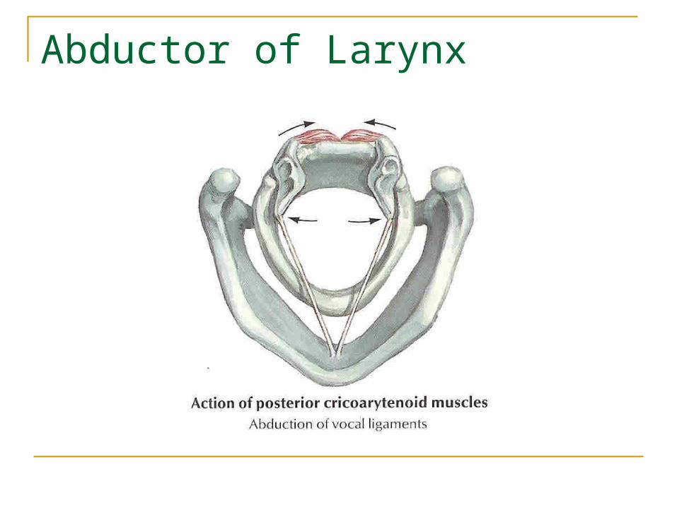
of which are innervated by the recurrent laryngeal nerve, include the: Posterior cricoarytenoid - the ONLY
abductor of the vocal folds. Functions to open the glottis by
rotary motion on the arytenoid cartilages.
Also tenses cords during phonation.
Abductor of Larynx

Lateral cricoarytenoid - - functions to close glottis by rotating arytenoids medially.
Transverse arytenoid - - only unpaired muscle of the larynx. Functions to approximate bodies of arytenoids closing posterior aspect of glottis.
Oblique arytenoid - - this muscle plus action of transverse arytenoid function to close laryngeal introitus during swallowing.
Thyroarytenoid - - very broad muscle, usually divided into three parts: Thyroarytenoideus internus (vocalis) - adductor
and major tensor of free edge of vocal fold. Thyroarytenoideus externus - major adductor of
vocal fold Thyroepiglotticus - shortens vocal ligaments

Anatomy of the Larynx - Motion Adductors of the Vocal Folds:
Wegner and Grossman Theory “In the absence of cricoarytenoid joint
fixation, an immobile vocal cord in paramedian position has total pure unilateral recurrent nerve paralysis, and an immobile vocal cord in lateral position has a combined paralysis of superior and recurrent nerves (the adductive action of cricothyroid muscle is lost)”

Causes of vocal cord paralysis Malignant : This accounts for 25% of cases,
one half being caused by carcinoma of lung

Causes of vocal cord paralysis Surgical/Traumatic: (20% cases)
Thyroidectomy Pneumonectomy CABG Penetrating neck or chest trauma. Post intubation Whiplash injuries Posterior fossa surgery
Causes of vocal cord paralysis Neurulogical (5-10%)
Wallenberg syndrome (lateral medullary stroke) Syringomyelia Encephalitis Parkinsons, Poliomyelitis Multiple Sclerosis Myasthenia Gravis, Guillian-Barre Diabetes
Causes of vocal cord paralysis Inflammatory:
Rheumatoid arthritis ,( really a "fixed" cord here) Infectious:
Syphilis Tuberculosis Thyroiditis Viral
Causes of vocal cord paralysis Idiopathic (20-25%):
Sarcoidosis, Lupus Polyarteritis nodosa Ortner's syndrome (left atrial hypertrophy).
Intracranial causes
Head injury CVA Bulbar
poliomyelitis
Distinctive features Other neurological
signs and symptoms due to combined paralysis of soft palate, pharynx and larynx

Cranial
Fracture base of skull Juglar foramen
lesions (Glomus tumours, Naspharyngeal Carcinoma)
Skull base osteomyelitis
Distinctive features Other cranial
nerve palsies (IX,X,XI)
Pharyngeal, superior and Recurrent Laryngeal nerve

Neck
Thyroidectomy Thyroid Tumours Post Cricoid
Carcinoma Malignant
Cervical Lymphnodes
Distinctive features
Superior and Recurrent Laryngeal nerves involved

Chest
Bronchogenic Carcinoma
Cardiothoracic Surgery Aortic Aneurysm Mediastinal
Lymphadenopathy Tracheal/Oesophageal
surgery
Distinctive feature Involvement of
Left Recurrent Laryngeal Nerve

Unilateral Superior Laryngeal Nerve Injury Normal vocal fold position
during quiet respiration. Noticeable deviation of
posterior commissure to paralyzed side during phonatory effort
At rest, the vocal fold on paralyzed side is slightly shortened and bowed, and may be depressed below level of normal side.
Unilateral Superior Laryngeal Nerve Injury Loss of sensation to the supraglottic larynx
can cause subtle symptoms such as frequent throat clearing, paroxysmal coughing, voice fatigue, vague foreign body sensations.
Loss of motor function to cricothyroid muscle can cause a slight voice change, which the patient usually interprets as hoarseness. Most common finding is diplophonia (with decreased range of pitch, most noticeable when trying to sing.
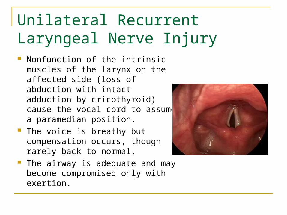
Unilateral Recurrent Laryngeal Nerve Injury Nonfunction of the intrinsic muscles
of the larynx on the affected side (loss of abduction with intact adduction by cricothyroid) cause the vocal cord to assume a paramedian position.
The voice is breathy but compensation occurs, though rarely back to normal.
The airway is adequate and may become compromised only with exertion.
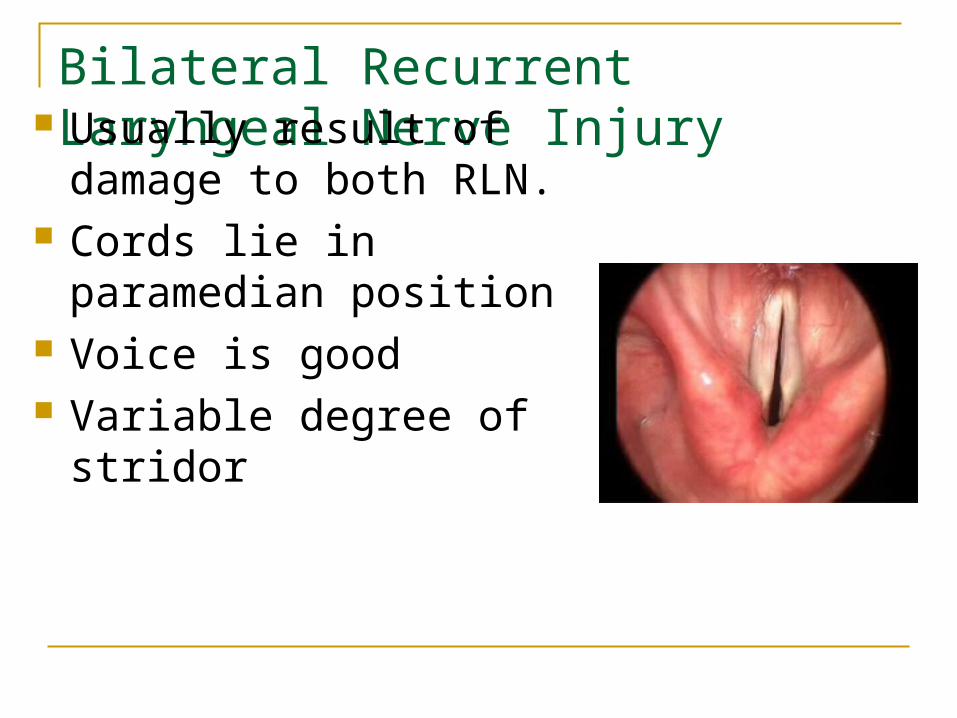
Bilateral Recurrent Laryngeal Nerve Injury Usually result of damage to both RLN.
Cords lie in paramedian position
Voice is good Variable degree of stridor
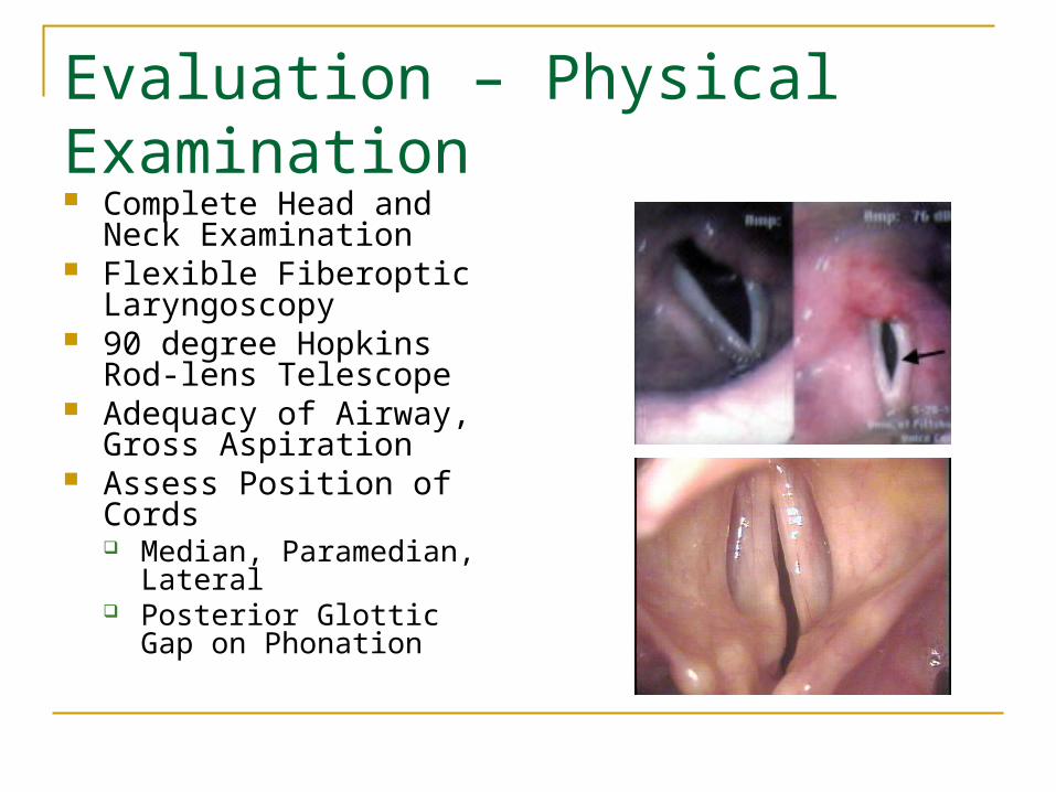
Evaluation – Physical Examination Complete Head and Neck
Examination Flexible Fiberoptic
Laryngoscopy 90 degree Hopkins Rod-
lens Telescope Adequacy of Airway, Gross
Aspiration Assess Position of Cords
Median, Paramedian, Lateral
Posterior Glottic Gap on Phonation

Evaluation – Unilateral Paralysis Manual Compression Test

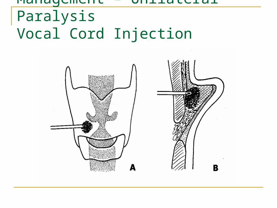
Management – Unilateral ParalysisVocal Cord Injection
Adds fullness to the vocal cord to help it better appose the other side
Injection technique is similar regardless of material used
Injection into thyroarytenoid/vocalis Injection can be done endoscopically or
percutaneiously Poor correction of posterior glottic gap
Management – Unilateral ParalysisVocal Cord Injection
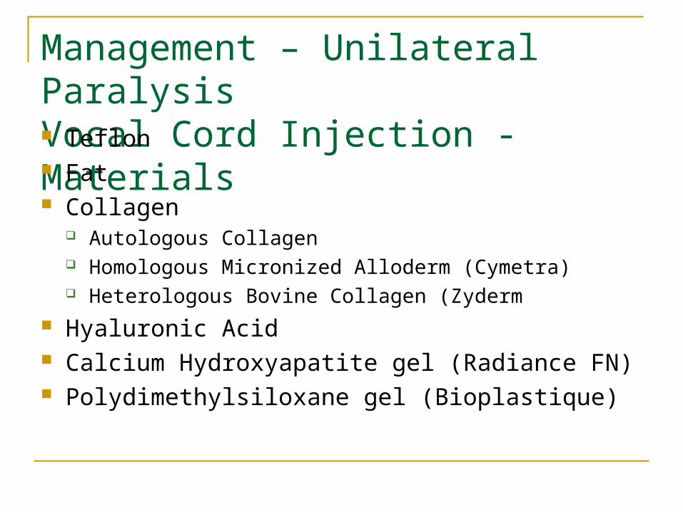
Management – Unilateral ParalysisVocal Cord Injection - Materials Teflon Fat Collagen
Autologous Collagen Homologous Micronized Alloderm (Cymetra) Heterologous Bovine Collagen (Zyderm
Hyaluronic Acid Calcium Hydroxyapatite gel (Radiance FN) Polydimethylsiloxane gel (Bioplastique)
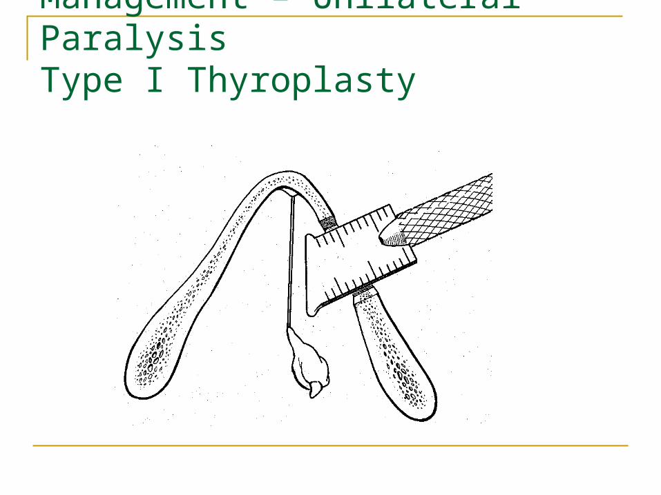
Management – Unilateral ParalysisType I Thyroplasty
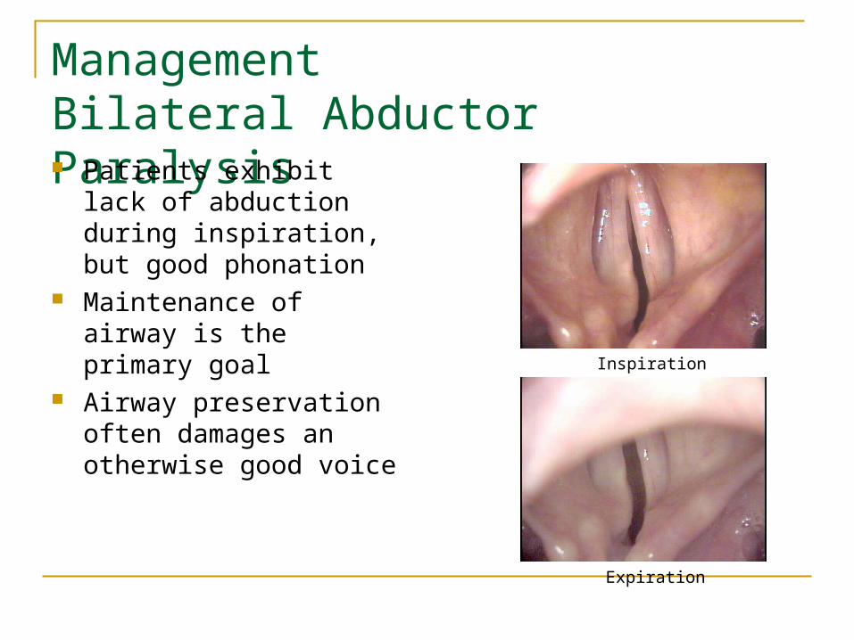
ManagementBilateral Abductor Paralysis Patients exhibit lack of
abduction during inspiration, but good phonation
Maintenance of airway is the primary goal
Airway preservation often damages an otherwise good voice
Expiration
Inspiration

ManagementBilateral Abductor Paralysis Tracheostomy
Gold standard Most adults will require this Speaking valves aid in phonation
Laser Cordectomy Laser Cordotomy Woodman Arytenoidectomy

Conclusions – Key Points
Management – Unilateral Paralysis Anterior and Posterior Glottic gap must be
addressed Arytenoid adduction is irreversible Continued improvement up to 1yr after Type I
thyroplasty Management – Bilateral Paralysis
Preservation of airway is most important goal

www.entlectures.com