larynx anatomy and laryngeal carcinoma
TRANSCRIPT

LEVELS OF THE LARYNX AND THEIR BOUNDARIES
The supraglottic division:From the superior-most tip of the epiglottis -to a
transverse plane through the laryngeal ventricle. The glottis:From this transverse plane to 1 cm inferiorly and
includes the true vocal cords. The subglottic regionFrom the inferior-most plane of the true cords -to the
inferior portion of the cricoid cartilage.


Supraglottis
• Extends from tip of epiglottis above to laryngeal ventricle below.
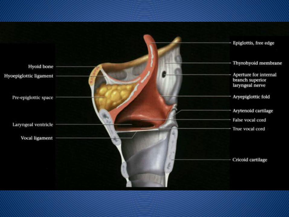
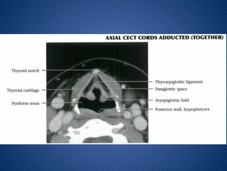
• Contains vestibule, epiglottis, pre-epiglottic fat, AE folds, FVC, paraglottic space, arytenoid cartilages

• Pre-epiglottic space: Fat-filled space between hyoid bone anteriorly & epiglottis posteriorly
• AE folds: Projects from cephalad tip of arytenoid cartilages to inferolateral margin of epiglottis• Represents superolateral margin of supraglottis, dividing it from pyriform sinus (hypopharynx)

• False vocal cords: Mucosal surfaces of laryngeal vestibule of supraglottis.• Beneath FVC are paired paraglottic spaces
• Paraglottic spaces: Paired fatty regions beneath false & true vocal cords• Superiorly they merge into pre-epiglottic space• Terminates inferiorly at under surface of TVC


Glottis
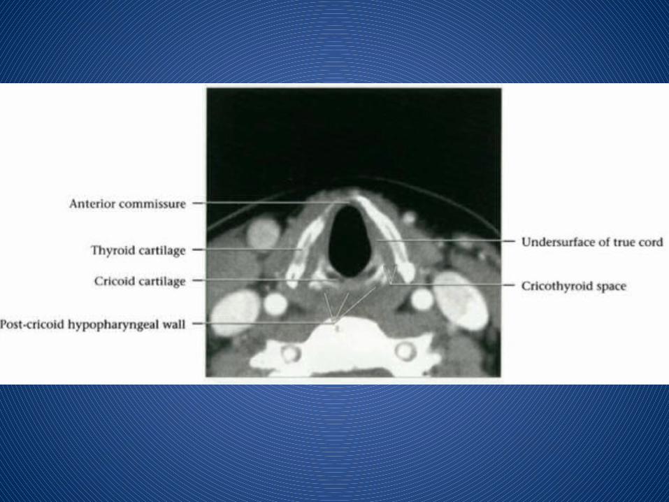
• TVC & anterior & posterior commissures
• Comprised of thyroarytenoid muscle (medial fibers are "vocalis muscle")• Anterior commissure: Midline, anterior meeting point of TVC


Subglottis
• Subglottis extends from under surface of TVC to inferior surface of cricoid cartilage
• Mucosal surface of subglottic area is closely applied to cricoid cartilage
• Conus elasticus: Fibroelastic membrane extends from medial margin of TVC above to cricoid below


CASE 1
Dr. Mohit GoelJR III

NAME : Mr. Harnapallu Baburao Abbanna Req No : 330Patient Code : 140102135 CT No:330AGE/SEX : 74 Yr(s) / Male Date: 09/05/2014
MSCT NECK WITH CONTRAST
HISTORY :
K/C/O CA hypopharynx.
Post radiotherapy status,
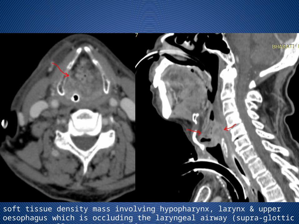
soft tissue density mass involving hypopharynx, larynx & upper oesophagus which is occluding the laryngeal airway (supra-glottic space) as well as upper end of oesophagus.
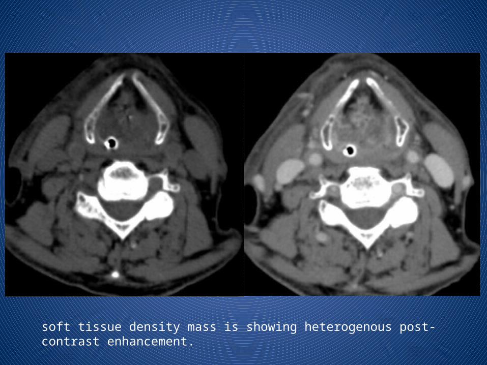
soft tissue density mass is showing heterogenous post-contrast enhancement.

Valecula appears nomral.Thyroid cartilage appears normal.

There is involvement of both thyro-arytenoid folds causing obliteration of both pyriform sinuses (L>R)

Pre-epiglottic space appears normal.There is involvement of para-glottic space on both sides.

Inferiroly the mass is extending upto the level of vocal cords.

NAME : Mr. Khan Ibrahim Amir Req No : 339Patient Code : 140403479 / IPD CT No:339AGE/SEX : 72 Yr(s) / Male Date: 10/05/2014 MSCT NECK /LARYNX
Clinical Profile: H/o change in voice.

Small mildly heterogeneously enhancing polypoidal mass lesion is noted involving right vocal cord and right para-glottic space, protruding into the laryngeal lumen
Plain Post-contrast

There is small non enhancing hypodense area noted within the lesion s/o necrosis.
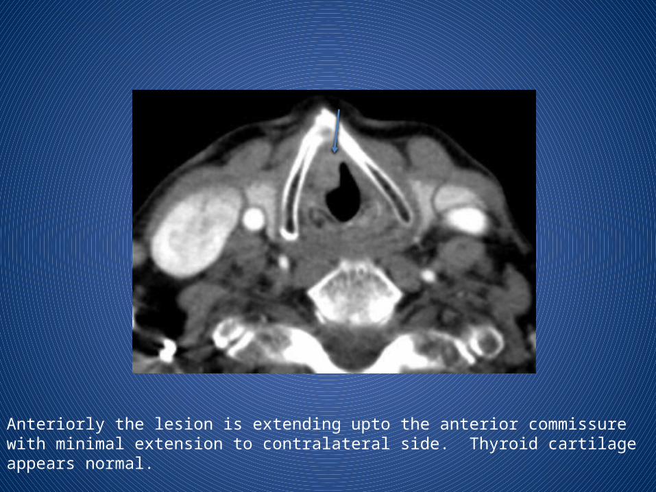
Anteriorly the lesion is extending upto the anterior commissure with minimal extension to contralateral side. Thyroid cartilage appears normal.

There is minimal extension of the enhancing soft tissue into the subglottic region noted at the anterior commissure. No obvious mass or thickening noted in posterior commissure.

Mucosal thickening in ethmoid sinus and right maxillary sinus.

Small lymph nodes noted in right level II

Calcified atherosclerotic changes are noted.

Supraglottic SCC
Approximately 30% of all laryngeal cancers arise in the supraglottis.
They often present in advanced stages, because symptoms (hoarseness, due to vocal cord involvement) do not occur until late.
Due to the rich lymphatic network of the supraglottis, nodal disease (level II and III) is a frequent finding in these patients.
Supraglottic SCC may arise in the • anterior compartment (epiglottis) or • the postero-lateral compartment (aryepiglottic fold and false
cords).
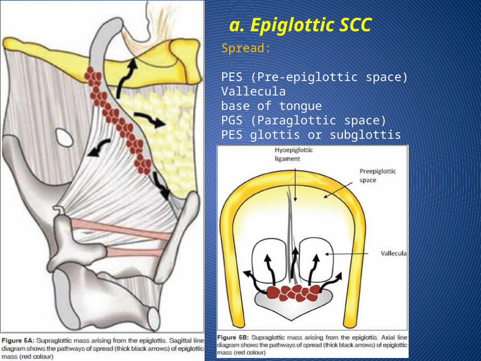
a. Epiglottic SCCSpread:
PES (Pre-epiglottic space)Valleculabase of tongue PGS (Paraglottic space)PES glottis or subglottis


b. Aryepiglottic fold (AE fold) SCC

c. False cord SCC

Glottic SCCGlottic SCCs represent about 65% of all laryngeal cancers.
Hoarseness of voice due to vocal cord involvement is the primary presenting symptom in these patients.
Metastatic nodal disease is rare in glottic carcinomas due to the sparse lymphatic drainage of the glottis.
Glottic SCCs commonly arise from the anterior half of the vocal cord and spread into the anterior commissure.
Anterior commissural disease is seen on CT or MRI as soft tissue thickening of more than 1-2 mm.
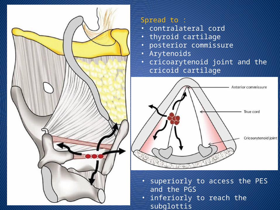
Spread to :• contralateral cord • thyroid cartilage • posterior commissure• Arytenoids• cricoarytenoid joint and the cricoid cartilage
• superiorly to access the PES and the PGS• inferiorly to reach the subglottis


Subglottic SCC
These cancers are rare, accounting for only 5% of all laryngeal cancers, clinically silent and present late in the course.
Subglottic cancer is diagnosed if any tissue thickening is noted between the airway and the cricoid ring.


Transglottic SCC
Laryngeal SCC encroaching on both, the glottis and supraglottis, with or without subglottic component and when the site of origin is unclear, is termed as transglottic tumor.
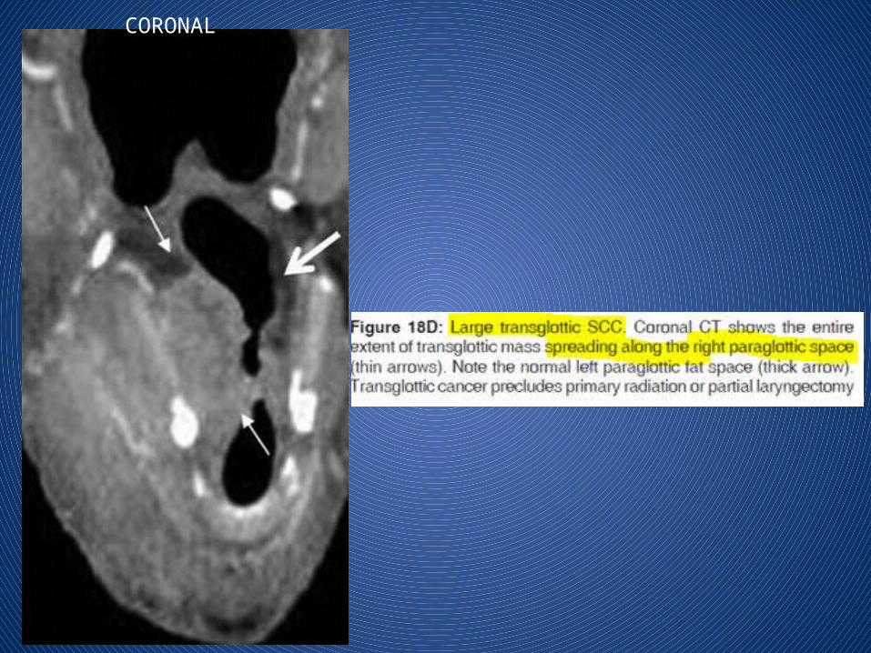
CORONAL

THANK YOU