lasereo 20140410-2
DESCRIPTION
Catalogo LASERETRANSCRIPT
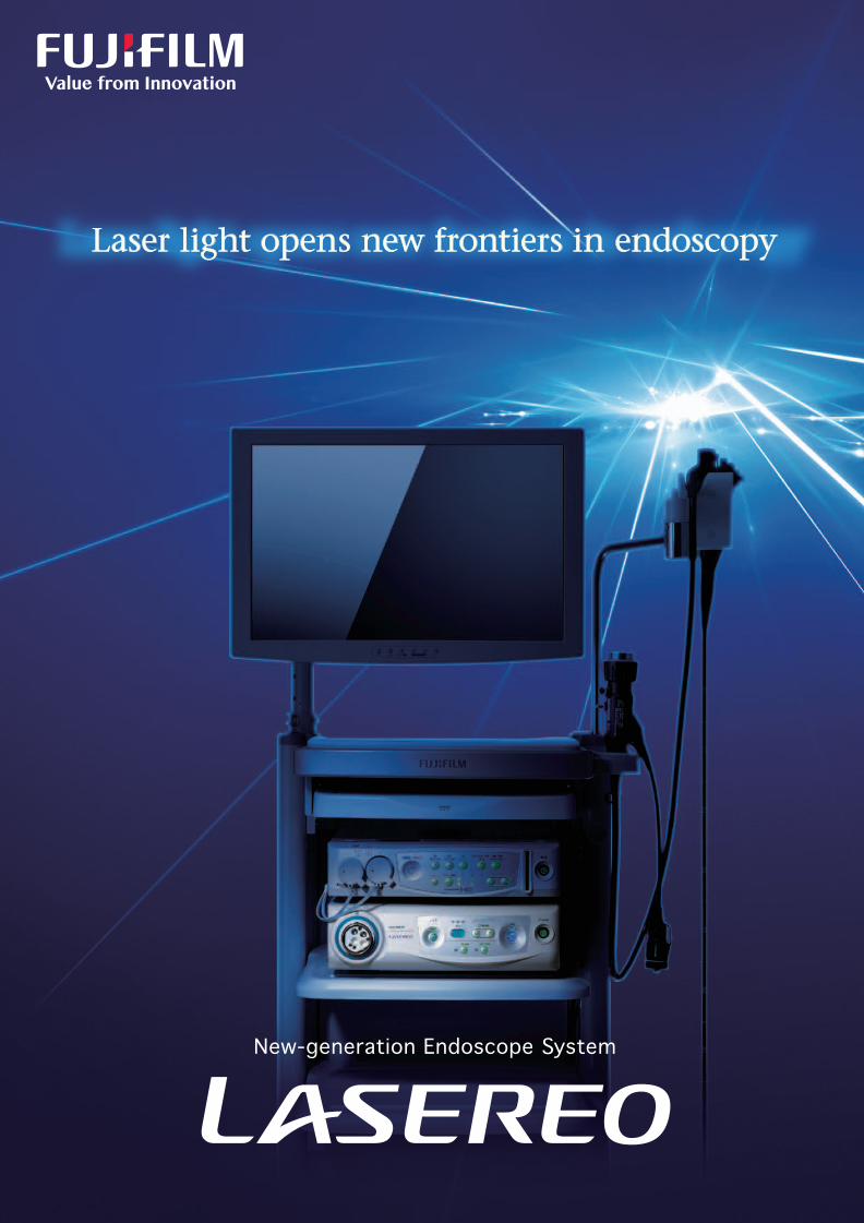
Laser light opens new frontiers in endoscopy
New-generation Endoscope System
Ref. No. XB-000E (SK·14·02·F1079·F9711) Printed in Japan ©2014 FUJIFILM Corporation

Lasereo is a trademark of FUJIFILM Corporation.
Fujifilm’s pursuit of a new light source that illuminates lesions
more clearly has resulted in an endoscope system equipped
with the laser light source.
2 3
A new generationof the endoscope system

Double laser imaging stepping into the new world
LASEREO is equipped with laser light sources that emit two different laser beams at different wavelengths: white light mode laser and BLI mode laser. The white light mode laser excites phosphors to create white light illumination (Oscillation wavelength: 450 nm ± 10 nm). The BLI mode laser enables the structures of vessels and mucosal surface to be captured through high contrast signals (Oscillation wavelength: 410 nm ± 10 nm). By controlling the emission intensity ratio of the two lasers and combining it with image processing, four types of observation modes are available.
Two laser sources
Conceptual diagram of laser illumination(The illustrations differ from laser light sources in actual use)
BLI (Blue LASER Imaging)
BLI produces high-contrast images of superficial mucosal structures and microvessels by combining two image components induced by narrowband light and white light.
The white light and BLI mode laser are emitted at a controlled intensity ratio to obtain the optimal light according to the observation purposes. In combination with image processing, four types of observation modes are available.
Four observation modes
[White light/FICE* mode] [BLI-bright mode] [BLI mode]
White light mode laser
BLI mode laser
BLI mode laser
White light mode laser
BLI mode laser
White light mode laser
Lower power consumption and longer life than with conventional lighting
This laser light source consumes only 10 W while a conventional xenon lamp needs 300 W to generate the same amount of light. In case a xenon lamp-installed endoscope has to be replaced regularly, there is no need with the laser source.
54
Image obtained from white light
Image obtained from narrowband light
Mucousmembranesurface layer
Mucosa Blood vessels
Narrowband lightWhite light
BLI image
Signalprocessing
White light mode laser White light
NarrowbandlightBLI mode laser
Phosphor
White light mode Provides images having the same color tone as with the conventional system (xenon light source).FICE* mode Constructs special images from rays having specific wavelenghs which are useful for better enhancemente of tissue aspects and vessels.
BLI-bright mode Emphasizes blood vessels and mucosal surface structures in middle to near distant views, which are brighter than the BLI mode.
BLI mode Emphasizes blood vessels and mucosal surface structures in near distant views or close up.
* Flexible spectral Imaging Color Enhancement(Image-Enhanced Endoscopy)

The white light mode provides bright, sharp stereoscopic images. The color tones are the same as with the conven-tional xenon light source.
By increasing the ratio of the narrowband light, BLI made aims at depicting sharp images of superficial microvessels and mucosal surface structures. BLI images support close-up and magnified observations.
FICE emphasizes minute differences in color tones between normal mucosa and lesions by the spectral image processing to clearly show mucosal structures and such lesions. The images are bright from near to distant, making it easy to identify a lesion and observe the boundary with the normal tissue.
The BLI-bright mode, in which the white light and narrowband light are used at an optimal balance, provides brighter images even from a middle to distant view compared to those obtained under typical narrowband light. This supports a wide-ranging observation including middle to near distant, close-up and magnified images.
White light mode
Four observation modesto suit various purposes
FICE* mode
BLI modeBLI-bright mode
* Flexible spectral Imaging Color Enhancement(Image-Enhanced Endoscopy)
• Up to four observation modes can be allocated to the scope switch.
Stomach
Stomach
Colon
Colon Esophagus
OFF (Stomach)
Esophagus
ON
White lightWhite light
BLI
BLI-bright
BLI
White light
BLI
FICE FICE
76

Gastroscopes for use with the laser light source
Colonoscopes for use with the laser light source
This endoscope is designed to support identification of lesions by using FICE or BLI images. The water and air supply systems are improved for better draining to ensure a clear view.
This endoscope offering close-up BLI images is suitable for observation of superficial microvessels and mucosal surface structures. The maximum optical magnification is approximately 135 times (on 19-inch LCD monitors).
For the upper G.I. Tract-Standard Type
EG-L590WRFor the upper Optical magnificationEG-L590ZW
This endoscope is designed to support identification of lesions by using FICE and BLI images. It has the large distal end of 12.0 mm in diameter, 140-degree field of view, 3.8-mm large forceps channel and water jetting function.
This endoscope offering close-up BLI images is suitable for observation of superficial microvessels and mucosal surface structures.The maximum optical magnification is approximately 135 times (on 19-inch LCD monitors).
For the Lower G.I. Tract-Standard Type
EC-L590WMFor the Lower G.I. Tract-Optical magnification
EC-L590ZW
Objective lens
Air/water nozzle
Forceps channel
Light guide 210°UP
100°LEFT
100°RIGHT
90°DOWN
Objective lens
Air/water nozzle
Forceps channel
Light guide 210°UP
100°LEFT
100°RIGHT
90°DOWN
Objective lens
Air/water nozzle
Water jet nozzle
Forceps channel
Light guide180°UP
160°LEFT
160°RIGHT
180°DOWN
Objective lens
Light guide
Air/water nozzle
Water jet nozzleForceps channel
180°UP
160°LEFT
160°RIGHT
180°DOWN
9
Entire endoscope Control portion Laser light source connecting section
Capturing clear images never seen before
An endoscope specially designed to make the best use of laser lights
8
Viewing direction
Field of view
Observation range
Distal end diameter
Flexible portion diameter
0° (Forward)
140°
4-100 mm
12.0 mm
12.0 mm
Bending capabilityUP: 180° / DOWN: 180°
RIGHT: 160° / LEFT: 160°
Minimum diameter offorceps channel 3.8 mm
Working length 1,330 mm
Total length 1,630 mm
Remark Water jet function
Image area &Forcept entry position
Viewing direction
Field of view
Observation range
Distal end diameter
Flexible portion diameter
0° (Forward)
WD: 140°/TL: 55°
WD: 6-100 mm/TL:2-3 mm
12.8 mm
12.8 mm
Bending capabilityUP: 180° / DOWN: 180°
RIGHT: 160° / LEFT: 160°
Minimum diameter offorceps channel 3.8 mm
Working length 1,330 mm
Total length 1,630 mm
Remark Water jet function
Image area &Forcept entry position
Viewing direction
Field of view
Observation range
Distal end diameter
Flexible portion diameter
0° (Forward)
140°
4-100 mm
9.6 mm
9.3 mm
UP: 210° / DOWN: 90°
RIGHT: 100°/ LEFT: 100°Bending capability
Working length 1,100 mm
Total length 1,400 mm
Minimum diameter offorceps channel 2.8 mm
Image area &Forcept entry position
Viewing direction 0° (Forward)
Field of view WD: 140°/TL: 55°
Observation range WD: 6-100 mm/TL: 2-3 mm
Distal end diameter 10.8 mm
Flexible portion diameter 9.8 mm
Bending capabilityUP: 210° / DOWN: 90°
RIGHT: 100° / LEFT: 100°
Working length 1,100 mm
Total length 1,400 mm
Minimum diameter ofForcept channel 2.8 mm
Image area &Forcept entry position

EXAMPLE OF SYSTEM CONFIGURATION
LASEREO System EPX-4450HD+LASEREO System
EXAMPLE OF SYSTEM CONFIGURATION
Processor VP-4450HD
System cart
Light Source LL-4450
19-inchLCD monitor
24-inchLCD monitor
Output devices
Printer HD recorder
Output devices
Printer HD recorder
System cart
19-inchLCD monitor
24-inchLCD monitor
Light Source LL-4450
Processor VP-4450HD
Light Source XL-4450
400-series scope 500,600-series scope
11
SpecificationsLight source LL-4450 Light source XL-4450Processor VP-4450HD
390 × 155 × 450 mmDimensions(W × H × D)
15 kgWeight
Power rating
Transmitted illumination
Air supply pump
Light control
Lamp rated value Main lamp:300 W xenon lamp LMP-002
Emergency lamp:75 W halogen lamp
Digital output
Analog output
HD-SDI: HDTV 1080 i(2 ch)DVI(Digital Visual Interface): 1280 × 1024 pEthernet: 100/10 Base
RGB:1280 × 1024 p
SDTV(120 V/NTSC, 230 V/PAL): RGB, Y/C, composite
Automatic light control
Lamp cooling method Forced air cooling
High, Middle, Low, Off
Light save On, Off
On, Off
AC 120 V ± 10 %, 60 Hz, 3.3 A
AC 230 V ± 10 %, 50 Hz, 1.7 A
AC 230 V ±10 %, 50 Hz, 1.2 APower rating
385 × 125 × 505 mm(Maximum)Dimensions(W × H × D)
12 kgWeight
Tranmitted illumination
Light control
Laser light 1 Light output wave length: 450 ±10nm
Maximum laser output: 3 W(CW)
Laser light 2 Light output wave length: 410 ± 10nm
Light control
Light cooling method Forced air cooling
Automatic light control
Air supply pump High, Middle, Low, Off
Automatic light control by the control signal from the prosessor
On, Off
Maximum laser output: 1.5 W(CW)
Brightness, Red, Green, Blue, R-Hue, Chroma; 9 stepsColor adjustmentHi, Lo; 9 stepsDetail3 stepsContrastHigh, Middle, Low, OffHyper-sharpnessColor Enhancement 10 presetsColor emphasisFlexible spectral Imaging
Color Enhancement 10 presetsFICE
Average/Peak/AutoIrisCF cardImage storageAC 120 V ± 10 %, 60 Hz, 0.8 A
AC 230 V ± 10 %, 50 Hz, 0.5 A
Power rating
9.5 kgWeight
390 × 105 × 460 mmDimensions(W × H × D)
MWL, StoreDICOM
10
L590-series scope
EC-L590WM EC-L590ZWEG-L590WR EG-L590ZW
L590-series scopes
Product name:Light sourceGMDN:35158
Generic name:Endoscopic light sources
Product name:ProcessorGMDN:18034
Generic name:Processing unit
Product name:Light sourceGMDN:35158
Generic name:Endoscopic light source

Laser light opens new frontiers in endoscopy
New-generation Endoscope System
Ref. No. XB-000E (SK·14·02·F1079·F9711) Printed in Japan ©2014 FUJIFILM Corporation