lec 04 blood groups - lecture notes - tiu
TRANSCRIPT

Lec 04Blood Groups
Assist. Prof. Dr. Mudhir S. Shekha

Introduction• Red cell membranes have antigens (protein / glycoprotein) on
their external surfaces. The antigens can be integral proteins where polymorphisms lie in the variation of amino acid sequence (e.g., rhesus [Rh], Kell), glycoproteins or glycolipids (e.g., ABO).
• These antigens areo unique to the individualo recognized as foreign if transfused into another individualo promote agglutination of red cells if combine with antibodyo more than 30 such antigen systems discovered→ complex
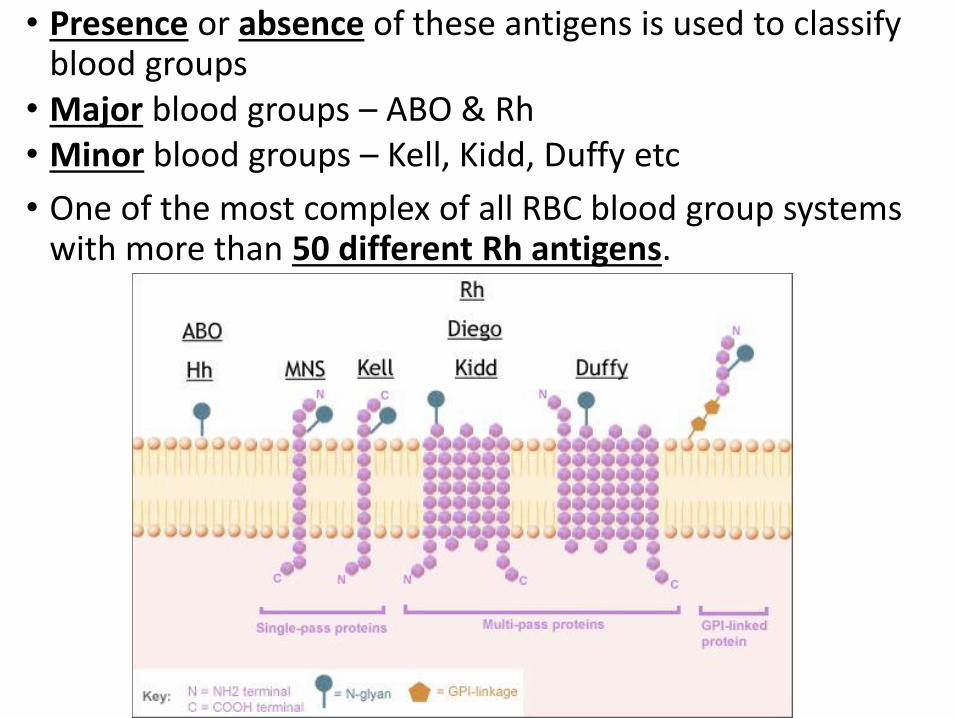
• Presence or absence of these antigens is used to classify blood groups
• Major blood groups – ABO & Rh• Minor blood groups – Kell, Kidd, Duffy etc
• One of the most complex of all RBC blood group systems with more than 50 different Rh antigens.

• The term “blood group” refers to the entire blood group system comprising red blood cell antigens whose specificity is controlled by a series of genes which can be allelic or linked very closely on the same chromosome.
• “Blood type” refers to a specific pattern of reaction to testing antisera within a given system.
• At present, 33 blood group systems representing over 300 antigens are listed by the International Society of Blood Transfusion.
• Most of them have been cloned and sequenced. The genes of these blood group systems are autosomal, except XG and XK which are X-borne, and MIC2 which is present on both X and Y chromosomes.


BLOOD GROUPING SYSTEM.• Major blood group system – based on Agglutinogens on
cell membrane, present widely & causes severe transfusion reaction• ABO• Rh system
• Minor blood group system – based on Agglutinogens but present in few populations & causes mild transfusion reaction.• MNS• P
• Familial blood group system – found in few families KELL. DUFFY, LUTHERAN, BOMBAY LEWIS, DEIGO, KIDD

Blood groups on the RBC
Gerbich
Knops
Kell
Lutheran
LW
Duffy
MNS
ABO
Ii
Rh
Indian
Cromer
Yt
Diego

ABO Blood Groups
• Most well known & clinically important blood group system.
• Discovered by Karl Landsteiner in 1901
• He discoved→major blood groups such as O, A, and B types, compatibility testing, and subsequent transfusion practices. He was awarded Noble Prize in 1930
• It is the ONLY system that the reciprocalantibodies are consistently and predictably present in the sera of people who have had no exposure to human red cells
• ABO blood group consist of o two antigens (A & B) on the surface of the RBCso two antibodies in the plasma (anti-A & anti-B)

ABO system• ABO remains the most important in transfusion and
transplantation since any person above the age of 6 monthspossess clinically significant anti-A and/or anti-B antibodies in their serum.
• Blood group A contains antibody against blood group B in serum and vice-versa, while blood group O contains no A/B antigen but both their antibodies in serum.
Antigens on RBCs
Antibody in plasma / serum
Blood group
A Anti-B A
B Anti-A B
AB None AB
None Anti-A, Anti-B O
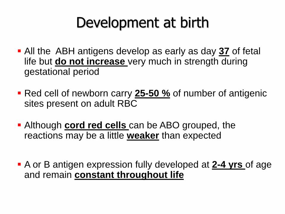
Development at birth
▪ All the ABH antigens develop as early as day 37 of fetal life but do not increase very much in strength during gestational period
▪ Red cell of newborn carry 25-50 % of number of antigenic sites present on adult RBC
▪ Although cord red cells can be ABO grouped, the reactions may be a little weaker than expected
▪ A or B antigen expression fully developed at 2-4 yrs of age and remain constant throughout life
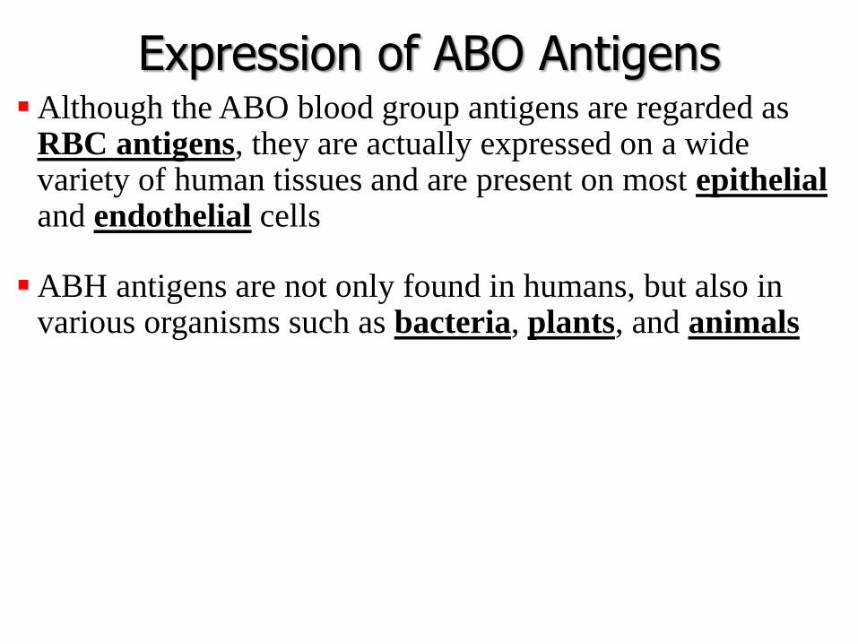
Expression of ABO Antigens▪Although the ABO blood group antigens are regarded as
RBC antigens, they are actually expressed on a wide variety of human tissues and are present on most epithelialand endothelial cells
▪ABH antigens are not only found in humans, but also in various organisms such as bacteria, plants, and animals

ABO and H Antigen Genetics• Genes at three separate loci control the occurrence and
location of ABO antigens
• The presence or absence of the A, B, and H antigens is controlled by the H and ABO genes
• The presence or absence of the ABH antigens on the red blood cell membrane is controlled by the H gene

ABO Antigen Genetics
• H gene – H and h alleles (h is an amorph)
• ABO genes – A, B and O alleles

H Antigen• The H gene codes for
an enzyme that adds the sugar fucose to the terminal sugar of a precursor substance(PS)
• The precursor substance (proteins and lipids) is formed on an oligosaccharide chain
Glucose
Galactose
N-acetylglucosamine
Galactose
H antigen
RBC
Fucose
Precursor
Substance
(stays the
same)
H antigen
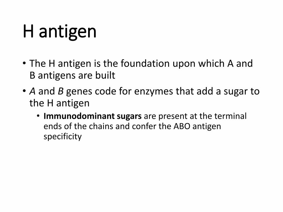
• The H antigen is the foundation upon which A and B antigens are built
• A and B genes code for enzymes that add a sugar to the H antigen• Immunodominant sugars are present at the terminal
ends of the chains and confer the ABO antigen specificity

A and B Antigen
• The “A” gene codes for an enzyme (transferase) that adds N-acetylgalactosamine to the terminal sugar of the H antigen• N-acetylgalactosaminyltransferase
• The “B” gene codes for an enzyme that adds D-galactose to the terminal sugar of the H antigen• D-galactosyltransferase

Formation of the A antigen
Glucose
Galactose
N-acetylglucosamine
Galactose
RBC
FucoseN-acetylgalactosamine
Glucose
Galactose
N-acetylglucosamine
Galactose
RBC
FucoseGalactose
Formation of the B antigen

Anti-A and anti-B antibodies▪ Not present in the newborn, appear in the first years of life (4-6
months), reach adult level at 5-10 years of age, decreases in elderly
▪ Naturally occurring as they do not need any antigenic stimulus
▪ However, some food & environmental antigens (bacterial, viral or plant antigens) are similar enough to A and B glycoprotein antigens and may stimulate antibody development
▪ Usually IgM, which are not able to pass through the placenta to the fetal blood circulation
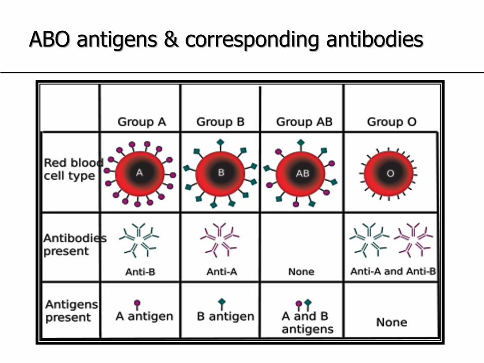
ABO antigens & corresponding antibodies

The Rh(D) Antigen• Rh is the most complex system, with over 50 antigens.
• The complexity of the Rh blood group Ags is due to the highly polymorphic genes that encode them.
• Discovered in 1940 after work on Rhesus monkeys.
• The 2nd most important after ABO in the crossmatch test.
• The genes that control the system are autosomal codominant located on the short arm of chromosome 1.

Rh blood group antigens are proteins
• The antigens of the Rh blood group are proteins (12 transmembrane).
• The RhD gene encodes the D antigen, which is a large protein on the red blood cell membrane, & the most important.
RHD gene RHCE gene
Chromosome 1
Proteins
C/c E/eD
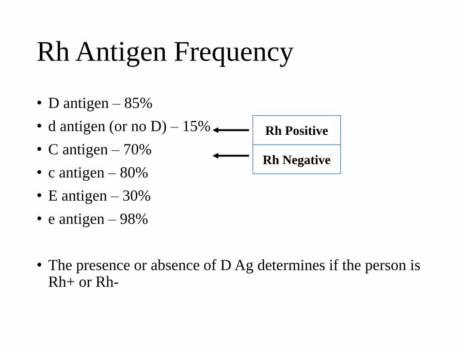
Rh Antigen Frequency
• D antigen – 85%
• d antigen (or no D) – 15%
• C antigen – 70%
• c antigen – 80%
• E antigen – 30%
• e antigen – 98%
• The presence or absence of D Ag determines if the person is Rh+ or Rh-
Rh Positive
Rh Negative
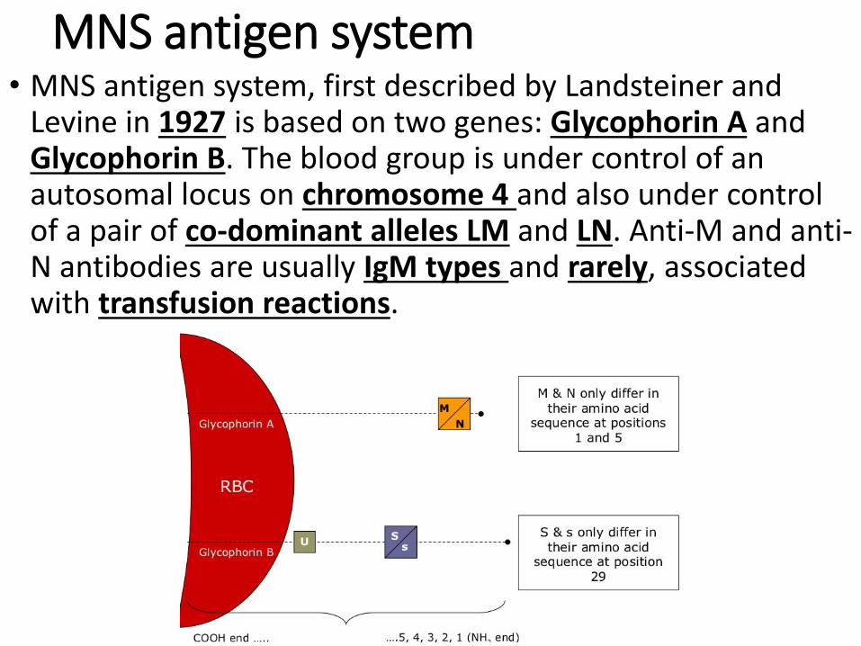
MNS antigen system• MNS antigen system, first described by Landsteiner and
Levine in 1927 is based on two genes: Glycophorin A and Glycophorin B. The blood group is under control of an autosomal locus on chromosome 4 and also under control of a pair of co-dominant alleles LM and LN. Anti-M and anti-N antibodies are usually IgM types and rarely, associated with transfusion reactions.

Lutheran system
• Lutheran system comprised of four pairs of allelic antigens representing single amino acid substitution in the Lutheran glycoprotein at chromosome 19. Antibodies against this blood group are rare and generally not considered clinically significant.

Kell system• These erythrocyte antigens are the third most potent
immunogenic antigen after ABO and Rh system, and are defined by an immune antibody, anti-K. It was first noticed in the serum of Mrs. Kellacher. She reacted to the erythrocytes of her newborn infant resulting in hemolytic reactions. Since then 25 Kell antigens have been discovered. Anti-K antibody causes severe hemolytic disease of the fetus and newborn (HDFN) and haemolytic transfusion reactions (HTR).

Duffy system• Duffy-antigen was first isolated in a patient called Duffy who had
haemophilia. It is also known as Fy glycoprotein and is present in the surface of RBCs. It is a nonspecific receptor for several chemokines and acts as a receptor for human malarial parasite, Plamodium vivax. Antigens Fya and Fyb on the Duffy glycoprotein can result in four possible phenotypes, namely Fy(a+b−), Fy(a+b+), Fy(a−b+), and Fy(a−b−). The antibodies are IgG subtypes and can cause HTR

Kidd system• Kidd antigen (known as Jk antigen) is a glycoprotein, present on
the membrane of RBCs and acts as a urea transporter in RBCs and renal endothelial cells. Kidd antibodies are rare but can cause severe transfusion reactions. These antigens are defined by reactions to an antibody designated as anti-Jka, discovered in the serum of Mrs. Kidd who delivered a baby with HDFN. Jka was the first antigen to be discovered by Kidd blood group system, subsequently, two other antigens Jkb and Jk3 were found.