lec 15 - brown€¦ · electron transitions such as these are common in organic compounds ... ......
TRANSCRIPT
1http://www.chem.ufl.edu/~itl/4411/lectures/lec_15.html
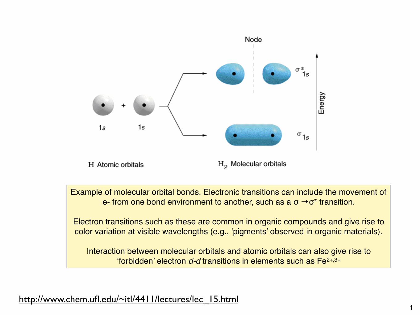
Example of molecular orbital bonds. Electronic transitions can include the movement of e- from one bond environment to another, such as a σ →σ* transition.
Electron transitions such as these are common in organic compounds and give rise to color variation at visible wavelengths (e.g., ‘pigments’ observed in organic materials).
Interaction between molecular orbitals and atomic orbitals can also give rise to ‘forbidden’ electron d-d transitions in elements such as Fe2+,3+
2http://www.chem.ufl.edu/~itl/4411/lectures/lec_15.html
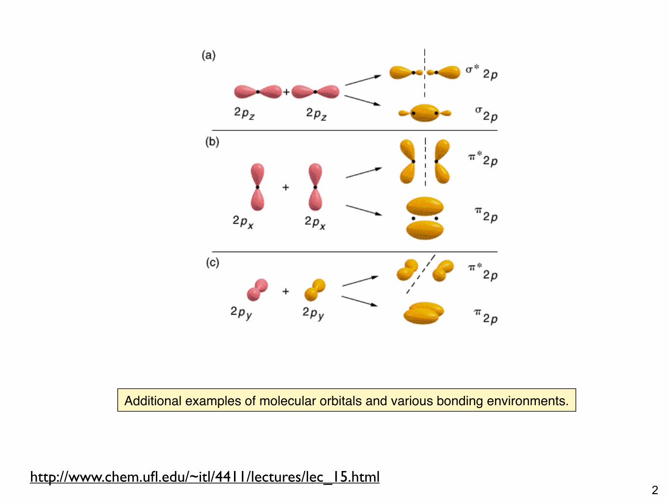
Additional examples of molecular orbitals and various bonding environments.
3http://www.organicchemistry.com/electronic-spectra-in-raman-spectroscopy/
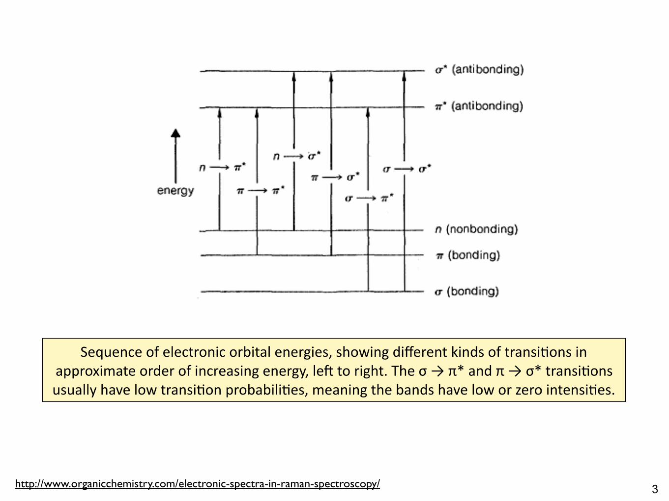
Sequenceofelectronicorbitalenergies,showingdifferentkindsoftransi8onsinapproximateorderofincreasingenergy,le=toright.Theσ→π*andπ→σ*transi8onsusuallyhavelowtransi8onprobabili8es,meaningthebandshaveloworzerointensi8es.
4

5Ar8s8crepresenta8onofthes,p,d,fatomicorbitals.
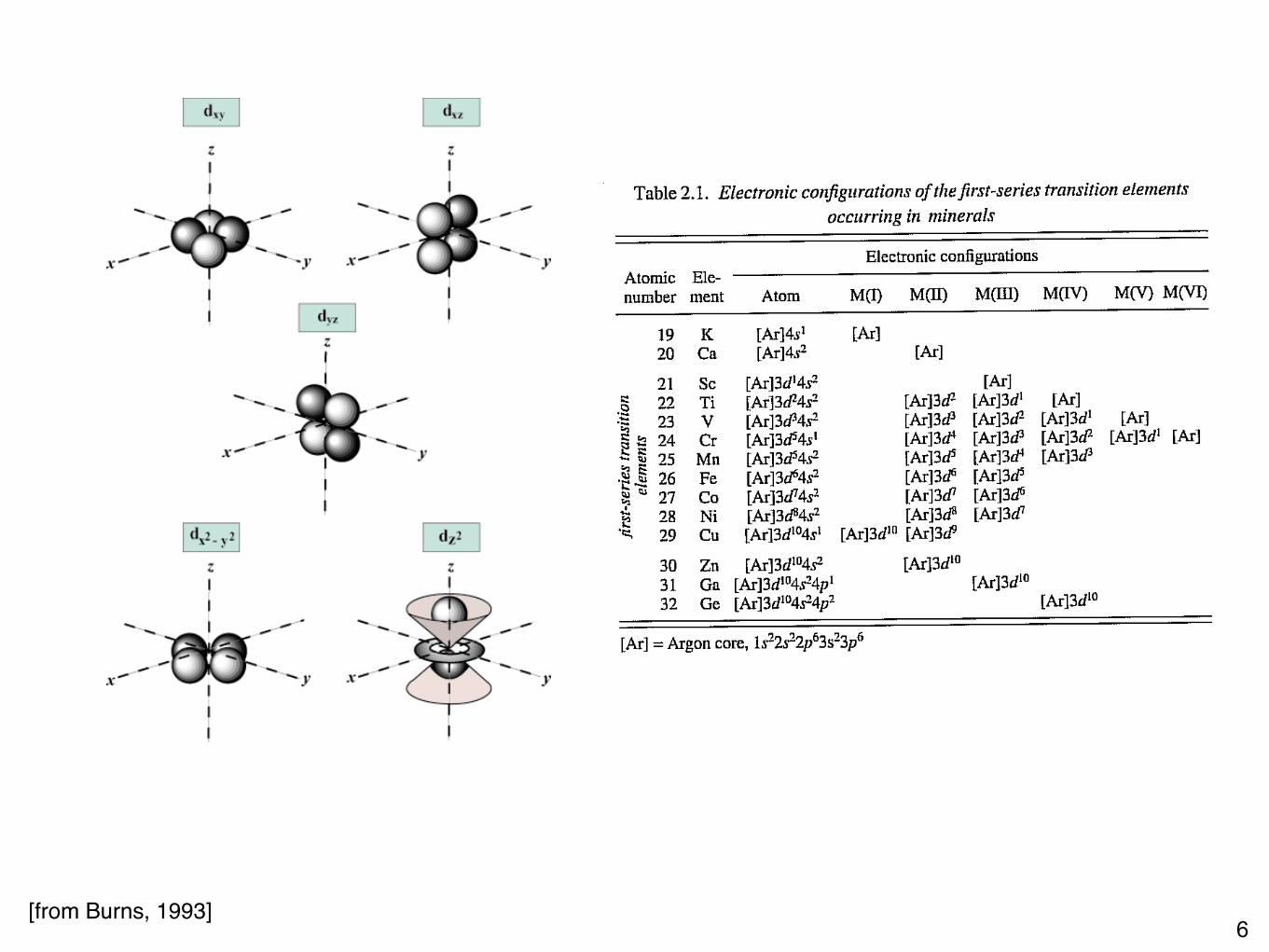
6[from Burns, 1993]
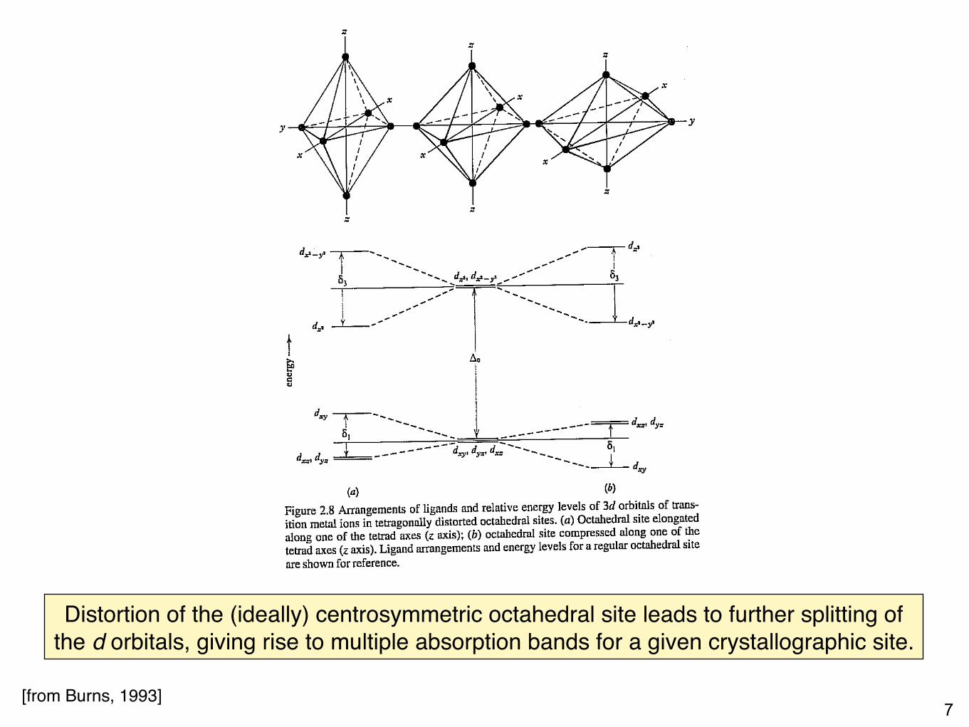
7[from Burns, 1993]
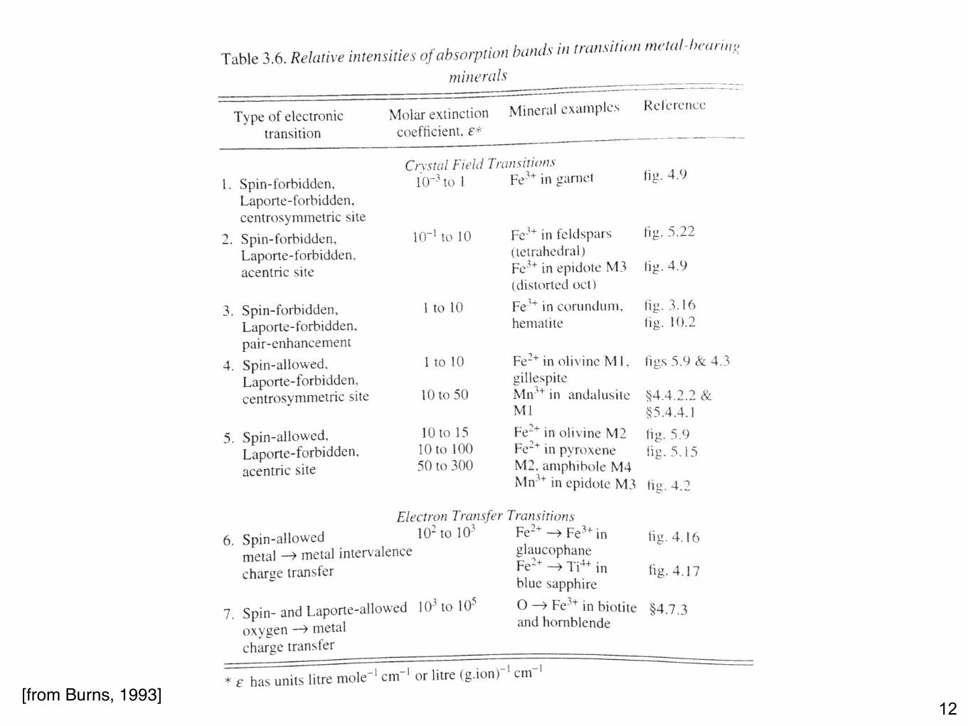
Distortion of the (ideally) centrosymmetric octahedral site leads to further splitting of the d orbitals, giving rise to multiple absorption bands for a given crystallographic site.
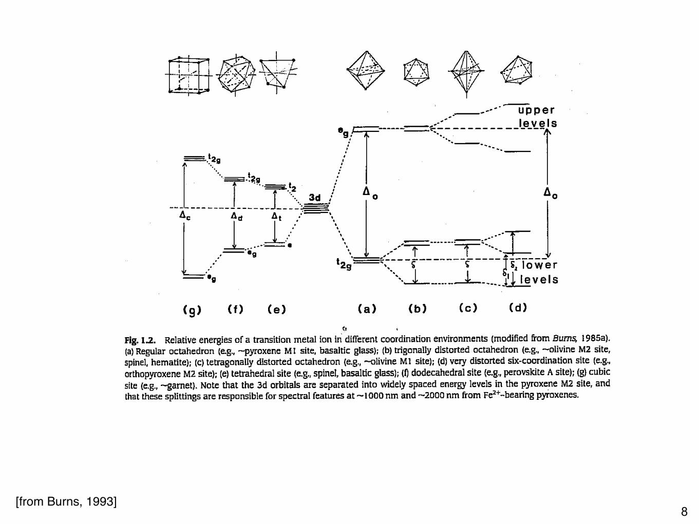
8[from Burns, 1993]
9[from Burns, 1993]
10
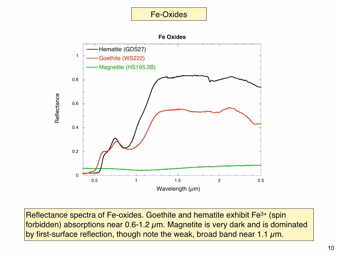
Fe-Oxides
Reflectance spectra of Fe-oxides. Goethite and hematite exhibit Fe3+ (spin forbidden) absorptions near 0.6-1.2 µm. Magnetite is very dark and is dominated by first-surface reflection, though note the weak, broad band near 1.1 µm.
11[from Burns, 1993]
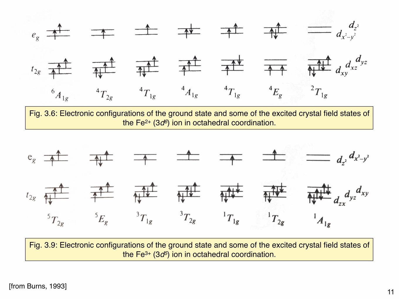
Fig. 3.6: Electronic configurations of the ground state and some of the excited crystal field states of the Fe2+ (3d6) ion in octahedral coordination.
Fig. 3.9: Electronic configurations of the ground state and some of the excited crystal field states of the Fe3+ (3d5) ion in octahedral coordination.
12[from Burns, 1993]
13
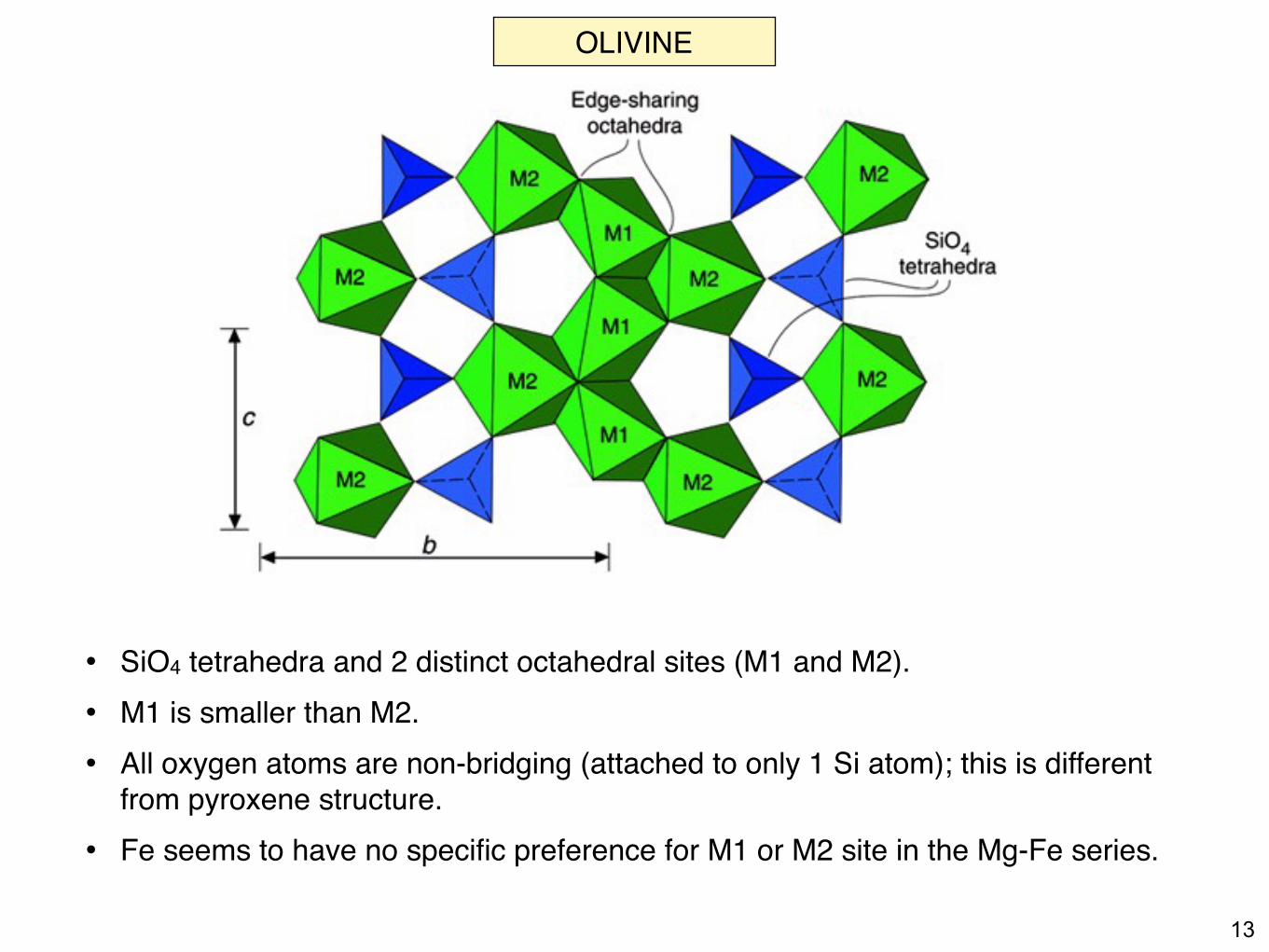
• SiO4 tetrahedra and 2 distinct octahedral sites (M1 and M2).• M1 is smaller than M2.• All oxygen atoms are non-bridging (attached to only 1 Si atom); this is different
from pyroxene structure.• Fe seems to have no specific preference for M1 or M2 site in the Mg-Fe series.
OLIVINE
14
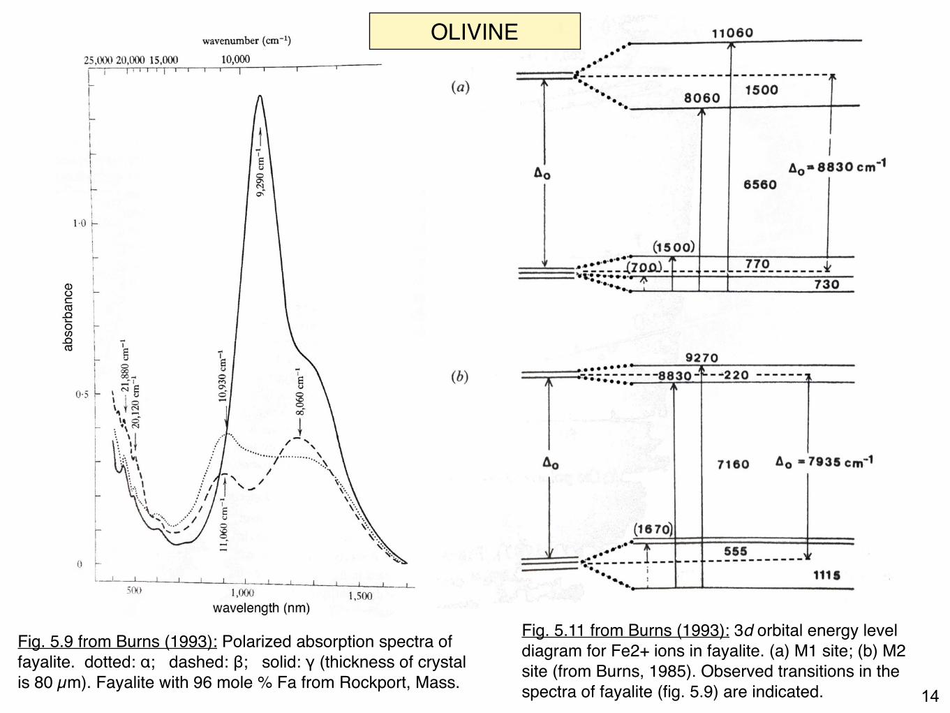
Fig. 5.11 from Burns (1993): 3d orbital energy level diagram for Fe2+ ions in fayalite. (a) M1 site; (b) M2 site (from Burns, 1985). Observed transitions in the spectra of fayalite (fig. 5.9) are indicated.
Fig. 5.9 from Burns (1993): Polarized absorption spectra of fayalite. dotted: α; dashed: β; solid: γ (thickness of crystal is 80 µm). Fayalite with 96 mole % Fa from Rockport, Mass.
OLIVINE
15
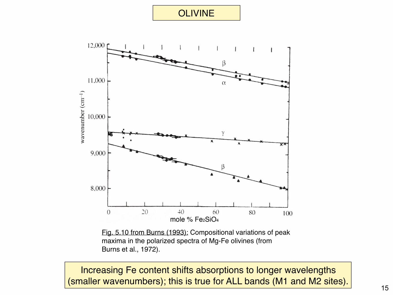
Fig. 5.10 from Burns (1993): Compositional variations of peak maxima in the polarized spectra of Mg-Fe olivines (from Burns et al., 1972).
Increasing Fe content shifts absorptions to longer wavelengths (smaller wavenumbers); this is true for ALL bands (M1 and M2 sites).
OLIVINE
16
OLIVINE
17
OLIVINE
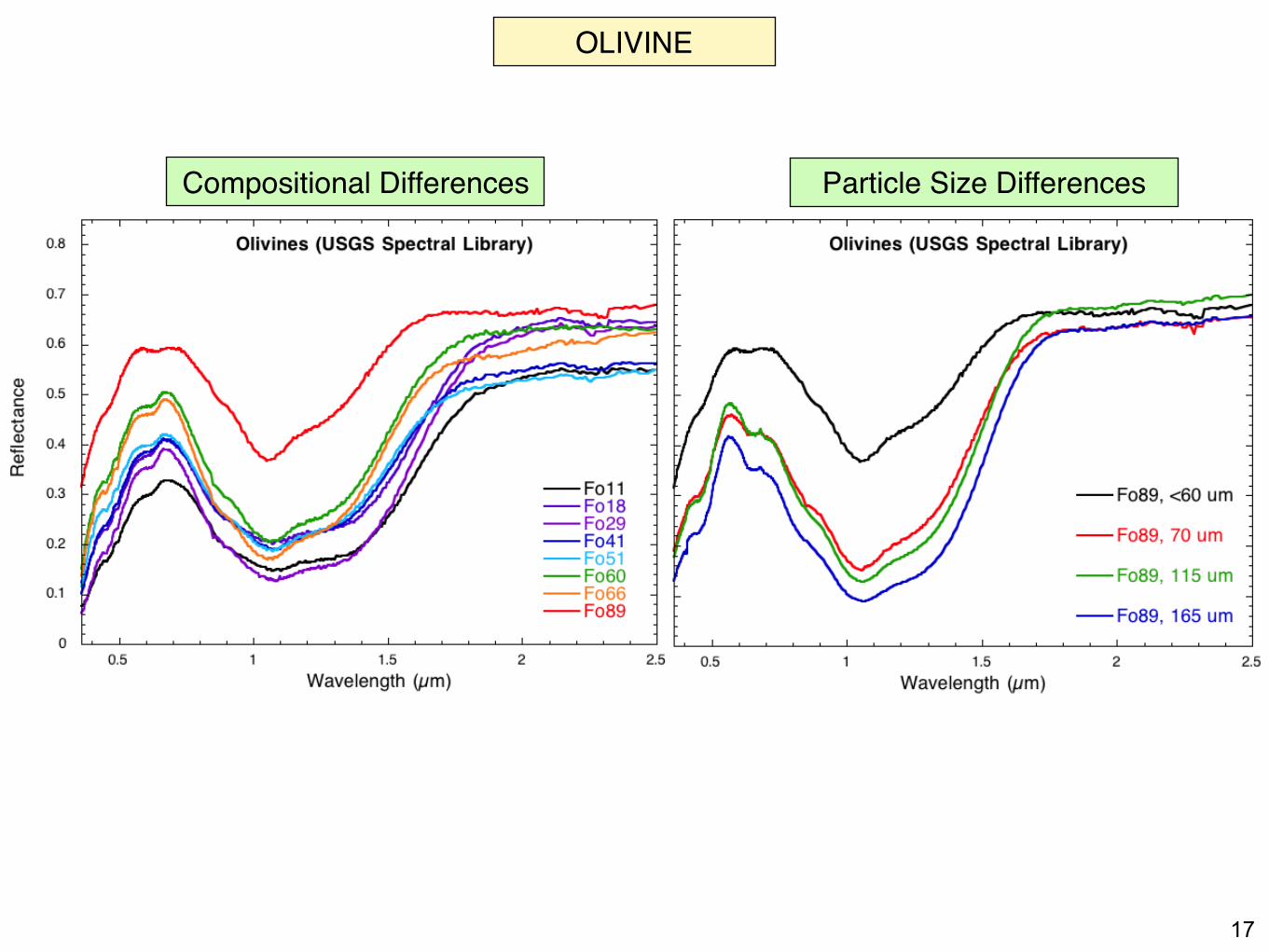
Compositional Differences Particle Size Differences