lectin microarray reveals binding profiles of ...aem.asm.org/content/77/13/4539.full.pdf ·...
TRANSCRIPT
APPLIED AND ENVIRONMENTAL MICROBIOLOGY, July 2011, p. 4539–4546 Vol. 77, No. 130099-2240/11/$12.00 doi:10.1128/AEM.00240-11Copyright © 2011, American Society for Microbiology. All Rights Reserved.
Lectin Microarray Reveals Binding Profiles of Lactobacillus caseiStrains in a Comprehensive Analysis of Bacterial
Cell Wall Polysaccharides�†Emi Yasuda,1* Hiroaki Tateno,2 Jun Hirabarashi,2 Tohru Iino,1 and Tomoyuki Sako3
Yakult Central Institute for Microbiological Research, 1796 Yaho, Kunitachi, Tokyo 186-8650, Japan1; Research Center forMedical Glycoscience, National Institute of Advanced Industrial Science and Technology, Central 2, 1-1-1 Umezono,
Ibaraki 305-8568, Japan2; and Yakult Europe B.V., Schutslussweg 1, 1332 EN Almere, The Netherlands3
Received 26 January 2011/Accepted 2 May 2011
We previously showed a pivotal role of the polysaccharide (PS) moiety in the cell wall of the Lactobacilluscasei strain Shirota (YIT 9029) as a possible immune modulator (E. Yasuda M. Serata, and T. Sako, Appl.Environ. Microbiol. 74:4746–4755, 2008). To distinguish PS structures on the bacterial cell surface of indi-vidual strains in relation to their activities, it would be useful to have a rapid and high-throughput method-ology. Recently, a new technique called lectin microarray was developed for rapid profiling of glycosylation ineukaryotic polymers and cell surfaces. Here, we report on the development of a simple and sensitive methodbased on this technology for direct analysis of intact bacterial cell surface glycomes. The method involveslabeling bacterial cells with SYTOX Orange before incubation with the lectin microarray. After washing, boundcells are directly detected using an evanescent-field fluorescence scanner in a liquid phase. Using this method,we compared the cell surface glycomes from 16 different strains of L. casei. The patterns of lectin-bindingaffinity of most strains were found to be unique. There appears to be two types of lectin-binding profiles: thefirst is characterized by a few lectins, and the other is characterized by multiple lectins with differentspecificities. We also showed a dramatic change in the lectin-binding profile of a YIT 9029 derivative with amutation in the cps1C gene, encoding a putative glycosyltransferase. In conclusion, the developed techniqueprovided a novel strategy for rapid profiling and, more importantly, differentiating numerous bacterial strainswith relevance to the biological functions of PS.
It is well documented that bacterial cell surface componentsand structures are critical factors for pathogenesis, host-mi-crobe interaction, immune modulation, and symbiosis. Toll-like receptors (TLRs) expressed on mammalian epithelial andimmune cells act as pattern recognition receptors, which areindividually responsible for a variety of different bacterial com-ponents, such as lipopolysaccharides (LPSs) from Gram-neg-ative bacteria, peptidoglycan (PG), lipoteichoic acid (LTA),and wall teichoic acid (WTA) from Gram-positive bacteria,flagella, lipoproteins, and nucleic acids. TLRs transfer sig-nals to the innate as well as the acquired immune system (1).Bacterial cell surface components are also recognized byother mammalian signaling molecules, such as nucleo-tide-binding oligomerization domain-containing protein 1(NOD1) and NOD2, which are intracellular proteins function-ing as cytosolic sensors in the regulation of inflammatory re-sponses (17). The human gut is inhabited by an enormousamount of microbes involved in maturation of the host im-mune system, establishment of commensalism, or induction ofthe inflammatory response through interaction with host cells.While the mechanism of a typical host-microbe interactionthrough TLRs and NODs is documented, there are a number
of ambiguous interactions between host and microorganism inwhich bacterial surface components are involved. It has re-cently been revealed that polysaccharide (PS) A from Bacte-roides fragilis stimulates T-cell activation (22), while type III PSfrom group B streptococci is known to be an immune stimu-lant, in addition to being a virulence factor (23).
Many strains of lactic acid bacteria (LAB) are used as pro-biotics, which are defined as microbes that exert a beneficialeffect on the host. Various factors produced by LAB have beenproposed to actively interact with mammalian host cells (12).For example, soluble proteins produced by probiotic bacteriaare known to regulate survival and growth of intestinal epithe-lial cells (11, 18, 30). Although the cell surface components ofLAB such as S-layer proteins, LTA, WTA, and PSs are pro-posed to be immune modulators (21), the active componentsdirectly involved in immune modulation and the molecularnature of these recognition processes are largely unknown.
The probiotic activity of the Lactobacillus casei strain Shi-rota (YIT 9029) has been extensively analyzed. While the bac-terium is known to be a strong Th1-type cytokine inducer (32),of which the active component was proposed to be LTA (12),it exerts anti-inflammatory activity against diabetes mellitus(26) and inflammatory bowel disease (24, 25) in animal mod-els. It was shown that the cell wall preparation containing PSand PG could be the active component of the anti-inflamma-tory activity (25). We have focused on the role of cell wall PSsin the immune modulation activities of YIT 9029 and havefound that high-molecular-mass cell wall PSs (PS-1) are a pre-requisite for anti-inflammatory activity (43). In this analysis, we
* Corresponding author. Mailing address: Yakult Central Institutefor Microbiological Research, 1796 Yaho, Kunitachi, Tokyo 186-8650,Japan. Phone: 81 42-577-8960. Fax: 81 42-577-3020. E-mail: [email protected].
† Supplemental material for this article may be found at http://aem.asm.org/.
� Published ahead of print on 20 May 2011.
4539
on August 22, 2018 by guest
http://aem.asm
.org/D
ownloaded from
on A
ugust 22, 2018 by guesthttp://aem
.asm.org/
Dow
nloaded from
on August 22, 2018 by guest
http://aem.asm
.org/D
ownloaded from
identified a cluster of genes essential for the biosynthesis of thehigh-molecular-mass PS-1 moiety, which are designated cps1A,cps1B, cps1C, cps1D, cps1E, cps1F, and cps1J.
In the light of recent research on the role of bacterial cellsurface structures/components in probiotic or symbiotic action(21), PSs from each bacterial cell wall could have individualand unique roles which are more important than has ever beenassumed (4, 19, 22, 46). As described in some reports concern-ing Bacteroides fragilis polysaccharides (7, 8, 27), bacterial cellshave different impacts on host cells, even when various mutantshaving defects in different predictive polysaccharide biosynthe-sis genes or deletions showed different immune-modulatingeffects on cultured splenocytes and T cells. However, the kindof PS structure important for their activity and how thesemolecules exert their activities on host cells are still openquestions. In addition, it has been shown that extracellular PSsof Streptococcus thermophilus (39) and group B Streptococcuscapsular PSs are highly diverse (6). It is well-known that patho-genic bacteria, such as hemolytic streptococci, often changetheir outer surface glycan profile to escape the host immunedefense mechanism (5). To investigate the dynamism and di-versity of bacterial outer surface structures, especially bacterialglycomes, in relation to their functional characterization, apowerful methodology with high throughput and versatility isneeded.
A novel technique called lectin microarray was developed toprofile the complex features of glycans expressed in variousforms (2, 14, 20, 37, 38). We adopted a unique evanescent-field-activated fluorescence detection principle to detect highlysensitive and reproducible lectin-glycoconjugate interactionson a glass side (20, 38). Using this detection principle, Tatenoet al. (2007) developed an application method enabling detec-tion of direct interaction between lectins and whole mamma-lian cells (37). Similarly, Hsu et al. (14–16) applied lectin mi-croarray technology to targeting of bacterial cells, though theyutilized a confocal detection principle.
In this study, we provide a practical methodology for profil-ing bacterial cell surface glycomes and show substantial differ-ences in glycan profiles between strains of the same Lactoba-cillus species.
MATERIALS AND METHODS
Bacterial strains and culture conditions. The bacterial strains used in thisstudy are listed in Table 1. Lactobacillus casei YIT 9029 is a commercial strainused in the production of fermented milk. A mutant of L. casei YIT 9029 ofwhich the cps1C gene was knocked out, YIT 9029 �cps1C (43), was analyzed.YIT 0180 is the neotype strain of L. casei (10). Cells were cultured in MRSmedium (Becton Dickinson and Company) for 22 h at 37°C under aerobicconditions.
Fluorescent staining of Lactobacillus casei cells. Cells cultured in 4 ml MRSmedium (1 � 109 to 2 � 1010) were harvested by centrifugation (4,000 � g for 5min at 4°C) and washed three times with 10 mM phosphate-buffered saline (PBS;pH 7.0). The cells were resuspended in 4 ml of 70% ethanol and agitated (100rpm for 30 min at room temperature [RT]) using a Personal-11 shaker (TaitecCo., Ltd.). The cells were harvested by centrifugation (4,000 � g for 5 min at 4°C)and washed three times with PBS. The cells were then resuspended in 4 ml ofPBS, before incubation with 1 to 50 �M SYTOX Orange nucleic acid stain(Molecular Probes Co., Ltd.) for 5 min at RT (41, 42). Cells labeled with SYTOXOrange were washed three times with PBS and finally resuspended in 360 �l ofPBS containing 1% bovine serum albumin (PBS-BSA). The fluorescence inten-sity of 2 � 108 labeled cells was measured using an ARVO X3 apparatus(PerkinElmer) within 1 h of labeling.
To visualize the cells labeled with SYTOX Orange, they were embedded in
Vectorshield with 4�,6-diamidino-2-phenylindole (DAPI; Vector Laboratories).Glass slides were examined using a Leica Q550FW system, and fluorescentimages were analyzed using Image-Pro Plus software (Media Cybernetics, SilverSpring, MD) (36).
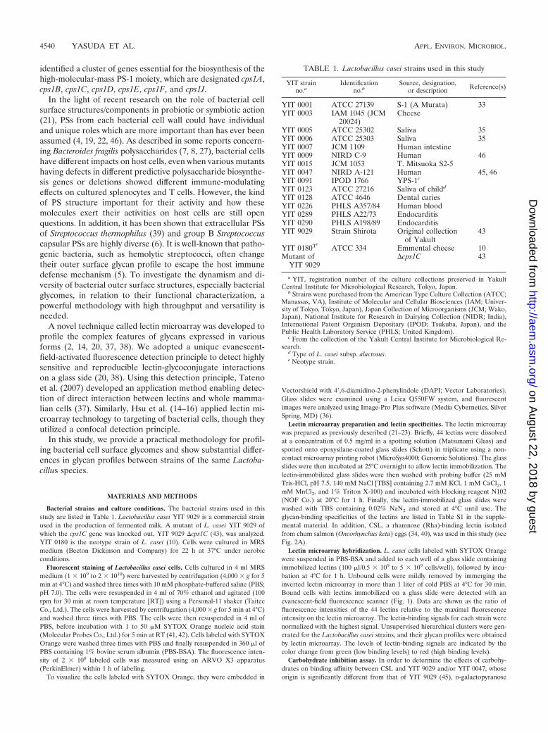
Lectin microarray preparation and lectin specificities. The lectin microarraywas prepared as previously described (21–23). Briefly, 44 lectins were dissolvedat a concentration of 0.5 mg/ml in a spotting solution (Matsunami Glass) andspotted onto epoxysilane-coated glass slides (Schott) in triplicate using a non-contact microarray printing robot (MicroSys4000; Genomic Solutions). The glassslides were then incubated at 25°C overnight to allow lectin immobilization. Thelectin-immobilized glass slides were then washed with probing buffer (25 mMTris-HCl, pH 7.5, 140 mM NaCl [TBS] containing 2.7 mM KCl, 1 mM CaCl2, 1mM MnCl2, and 1% Triton X-100) and incubated with blocking reagent N102(NOF Co.) at 20°C for 1 h. Finally, the lectin-immobilized glass slides werewashed with TBS containing 0.02% NaN2 and stored at 4°C until use. Theglycan-binding specificities of the lectins are listed in Table S1 in the supple-mental material. In addition, CSL, a rhamnose (Rha)-binding lectin isolatedfrom chum salmon (Oncorhynchus keta) eggs (34, 40), was used in this study (seeFig. 2A).
Lectin microarray hybridization. L. casei cells labeled with SYTOX Orangewere suspended in PBS-BSA and added to each well of a glass slide containingimmobilized lectins (100 �l/0.5 � 109 to 5 � 109 cells/well), followed by incu-bation at 4°C for 1 h. Unbound cells were mildly removed by immerging theinverted lectin microarray in more than 1 liter of cold PBS at 4°C for 30 min.Bound cells with lectins immobilized on a glass slide were detected with anevanescent-field fluorescence scanner (Fig. 1). Data are shown as the ratio offluorescence intensities of the 44 lectins relative to the maximal fluorescenceintensity on the lectin microarray. The lectin-binding signals for each strain werenormalized with the highest signal. Unsupervised hierarchical clusters were gen-erated for the Lactobacillus casei strains, and their glycan profiles were obtainedby lectin microarray. The levels of lectin-binding signals are indicated by thecolor change from green (low binding levels) to red (high binding levels).
Carbohydrate inhibition assay. In order to determine the effects of carbohy-drates on binding affinity between CSL and YIT 9029 and/or YIT 0047, whoseorigin is significantly different from that of YIT 9029 (45), D-galactopyranose
TABLE 1. Lactobacillus casei strains used in this study
YIT strainno.a
Identificationno.b
Source, designation,or description Reference(s)
YIT 0001 ATCC 27139 S-1 (A Murata) 33YIT 0003 IAM 1045 (JCM
20024)Cheese
YIT 0005 ATCC 25302 Saliva 35YIT 0006 ATCC 25303 Saliva 35YIT 0007 JCM 1109 Human intestineYIT 0009 NIRD C-9 Human 46YIT 0015 JCM 1053 T. Mitsuoka S2-5YIT 0047 NIRD A-121 Human 45, 46YIT 0091 IPOD 1766 YPS-1c
YIT 0123 ATCC 27216 Saliva of childd
YIT 0128 ATCC 4646 Dental cariesYIT 0226 PHLS A357/84 Human bloodYIT 0289 PHLS A22/73 EndocarditisYIT 0290 PHLS A198/89 EndocarditisYIT 9029 Strain Shirota Original collection
of Yakult43
YIT 0180TeATCC 334 Emmental cheese 10
Mutant ofYIT 9029
�cps1C 43
a YIT, registration number of the culture collections preserved in YakultCentral Institute for Microbiological Research, Tokyo, Japan.
b Strains were purchased from the American Type Culture Collection (ATCC;Manassas, VA), Institute of Molecular and Cellular Biosciences (IAM; Univer-sity of Tokyo, Tokyo, Japan), Japan Collection of Microorganisms (JCM; Wako,Japan), National Institute for Research in Dairying Collection (NIDR; India),International Patent Organism Depositary (IPOD; Tsukuba, Japan), and thePublic Health Laboratory Service (PHLS; United Kingdom).
c From the collection of the Yakult Central Institute for Microbiological Re-search.
d Type of L. casei subsp. alactosus.e Neotype strain.
4540 YASUDA ET AL. APPL. ENVIRON. MICROBIOL.
on August 22, 2018 by guest
http://aem.asm
.org/D
ownloaded from
(Gal), D-glucopyranose (Glc), D-galactosylpyranosyl-(134)-D-Glc (Lac), D-man-nopyranose (Man), L-rhamnopyranose (Rha), D-fructofuranosyl-(231)-D-gluco-pyranoside (Suc), or (3S,4R,5S,6S)-6-methyltetrahydro-2H-pyran-2,3,4,5-tetraol(Fuc) was added at a concentration of 0.1 �M to 2 � 109 cells labeled withSYTOX Orange in PBS-BSA. The effect of carbohydrate inhibition is shown asa percentage of the control signals with no added carbohydrate.
PS-PG. Cells grown overnight in 100 ml of MRS medium were harvested bycentrifugation (12,000 � g for 10 min at 4°C) and washed three times withdistilled water. Cells were resuspended in 4 ml of 5 mM Tris-malate–2 mMMgCl2 (pH 6.4). After boiling for 10 min, 1 mg of N-acetylmuramidase SG(Dainippon Sumitomo Pharma Co., Ltd.), to solubilize polysaccharides by di-gesting the peptidoglycan network of the cell wall, and 1 mg of benzonate (MerckJapan Ltd., Tokyo, Japan), as an endonuclease, were added to the cell suspen-sion, and the mixture was incubated at 37°C for 18 h. The reaction mix washeated at 100°C for 10 min and then centrifuged at 12,000 � g for 10 min at 4°C.One milligram of pronase (Roche Diagnostics K. K., Tokyo, Japan) in thesolution was added to the supernatant to digest all protein components into smallfragments, and the reaction mix was incubated at 37°C for an additional 20 h.The resulting solution was thoroughly dialyzed against deionized water using a3,500-molecular-mass-cutoff dialysis bag and more than 8 exchanges of water for2 or 3 days at RT. The samples obtained were designated the PS-PG fraction andstored in a refrigerator until further use.
The carbohydrate composition of the PS-PG was analyzed by labeling thesample using an ABEE labeling kit (J-Oil Mills Co. Ltd., Tokyo, Japan), followedby high-performance liquid chromatography analysis using a Honenpak C18
column (75 mm by 4.6 mm [inner diameter]) (44).
RESULTS
Optimization of labeling for Lactobacillus casei. Before usingSYTOX Orange, we examined the applicability of Cy3 label-ing, which is usually used in fluorescent labeling of glycopro-teins and cell surface proteins for microarray analysis. Thefluorescein isothiocyanate-labeling method (32) with 10 �g/mlCy3 successfully labeled 1 � 109 to 2 � 109 cells. However,nonspecific binding between Cy3-labeled cells and the glassmicroarray slides was detected. This was probably due to in-creased hydrophobicity of the cells. Labeling with Cell Tracker,another labeling reagent for whole-mammalian-cell staining(37), was also unsuccessful, since the fluorescence intensity ofL. casei cells (1 � 1010 to 2 � 1010) labeled with Cell Tracker(50 �g/ml) was too low to be analyzed by this technology.
We then examined the suitability of SYTOX Orange (41,42), whose maximum excitation wavelength is similar to that ofCy3 (32). We first measured the fluorescence intensity of 1 �
108 cells labeled with different concentrations of SYTOXOrange (Fig. 2B). The highest fluorescence intensity was ob-tained at 10 �M for all four strains. This concentration is100-fold higher than the maximum concentration recommendedin the standard protocol to label bacterial cells. This may becaused by the lower permeation of the dye across the cell walland cell membrane in lactobacilli treated with 70% ethanolthan in mammalian cells and other bacterial cells. The higherfluorescence intensity of labeled cells was more suitable forlectin microarray analysis. Another advantage of SYTOX Or-
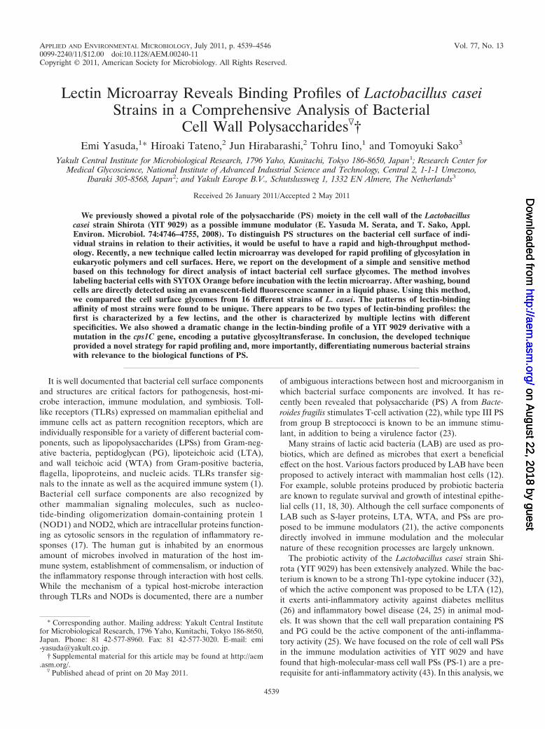
FIG. 1. Profiling system of bacterial cell wall polysaccharides usinglectin microarray.
FIG. 2. Optimization of lectin microarray analysis for bacterialcells. (A) Lectin microarray format. Glycan-binding specificities of thelectins used in this article are listed in Table S1 in the supplementalmaterial. The concentration of each lectin was 0.5 mg/ml. (B) Effect ofSYTOX Orange concentration on cell fluorescence intensity. Lacto-bacillus casei YIT 0005, YIT 0006, YIT 9029, and YIT 0180T cells weregrown to stationary phase, before labeling with SYTOX Orange (1 to50 �g/ml, as described in Materials and Methods). The fluorescenceintensity of 1 � 108 cells was measured by an ARVO X3 apparatus(PerkinElmer) using a Cy3 filter. The highest fluorescence intensitywas 10 �M for each strain. (C) Fluorescent images of L. casei cellslabeled with SYTOX Orange and DAPI. L. casei YIT 9029 and L. caseiYIT 0180T cells were labeled with 10 �M SYTOX Orange and rela-beled with DAPI on a glass slide. Red images were obtained withSYTOX Orange, and green images were obtained with SYTOX Or-ange and DAPI. The cells were observed to be thin and long usingSYTOX Orange and round using DAPI. (D) Dose-dependent fluores-cent signals of L. casei cells in the lectin microarray. Various numbersof cells labeled with SYTOX Orange were allowed to bind with thelectin array (0.5 to 4 � 109 cells/well). Bound cells were detected usingan evanescent-field fluorescent scanner.
VOL. 77, 2011 PROFILING OF L. CASEI CELL WALL POLYSACCHARIDES 4541
on August 22, 2018 by guest
http://aem.asm
.org/D
ownloaded from
ange is that the fluorescence intensity of cells labeled withSYTOX Orange was stable for 2 days at 4°C. Consequently,the concentration of SYTOX Orange used to label bacterialcells in this study was 10 �M.
Proper labeling of the cells with SYTOX Orange was con-firmed by monitoring the labeled cells with fluorescence mi-croscopy. As shown in Fig. 2C, it was possible to distinguish thearea of the cells stained by SYTOX Orange from that stainedby DAPI after double staining of L. casei cells. The latterstained only the center of the cells, as DAPI binds to AT-specific chromosomal DNA (31); SYTOX Orange stained thewhole area of the cell 2 or 3 times more strongly than DAPI.This is probably because the dye binds not only to double-stranded DNA, as described in the supplier’s manual, but alsoto RNAs that are distributed throughout the whole-cell cyto-plasm. It was essential to treat bacterial cells with 70% ethanolfor 30 min before staining with SYTOX Orange in order toobtain the full fluorescence intensity, and microscopic analysissuggested that this treatment did not damage cell morphology.Hence, we concluded that 10 �M SYTOX Orange was themost suitable labeling dye for lectin microarray analysis.
Differential profiling of 4 Lactobacillus casei strains. Wedetermined the required number of SYTOX Orange-labeledcells using 2 different strains of L. casei, YIT 0005 and YIT0006, at 0.5 � 109, 1.0 � 109, 2.0 � 109, or 4.0 � 109 cells/well.As shown in Fig. 2D, only 2.0 � 109 and 4.0 � 109 cells/wellgave the full fluorescence intensity and reproducible results inboth strains. It was concluded that 2 � 109 to 4 � 109 cells/wellwas suitable for this assay.
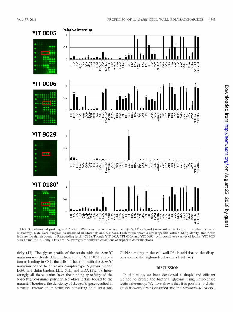
The lectin-binding activity of YIT 0005, YIT 0006, YIT 9029,and YIT 0180T was then analyzed in more detail. As shown inFig. 3, each strain had a unique binding profile. Similar topreviously reported glycoproteins (20) and mammalian cells(37), the bacterial cells had their own molecular patterns ofglycosylation on their cell surfaces. It can be also speculatedthat YIT 0005, YIT 0006, and YIT 0180T have similar outersurface PS structures. In particular, strong signals were com-monly detected in these three strains for ECA (asialo complex-type N-glycan binder), BPL and TJA-II (Gal binders), LEL(chitin binder), and O-glycan binders (WFA, MPA, HPA,VVA, SBA, GSL-I, and PTL-I).
However, YIT 9029 bound only to CSL, a rhamnose-specificlectin (32, 33, 37). It is known that PS from YIT 9018, theparental strain of YIT 9029 which was produced by removingbacteriophage �FSW from the YIT 9018 genome (33), con-tains Rha, Glc, Gal, N-acetylglucosamine (GlcNAc), and N-acetylgalactosamine (GalNAc) (28). We also confirmed thesugar composition of YIT 9029 PS using the ABEE labeling kit(44) and found the presence of Rha, Glc, Gal, and others (datanot shown), consistent with the results of Nagaoka et al. (28).Thus, the binding pattern of YIT 9029 does not directly reflectthe sugar composition of its cell wall PS. Similarly, to analyzethe relationship between lectin-binding profiles and sugar com-position, we confirmed the sugar composition of PSs from YIT0005, YIT 0006, and YIT 0180T. While the presence of Glc,GlcNAc, and GalNAc was confirmed in all strains, Rha was notdetected in YIT 0005 and Gal was confirmed to be present onlyin YIT 9029. The sugar compositions of YIT 0005 and YIT0006 in our study are consistent with those reported bySimelyte et al. (35) (YIT 0005 and YIT 0006 correspond to
L. casei ATCC 25302 and L. casei ATCC 25303, respectively).On the basis of our results, the binding profiles of the strainsanalyzed are not just determined by the sugar composition ofthe cell wall PSs, but they are also partly determined by thespecificity of lectin binding.
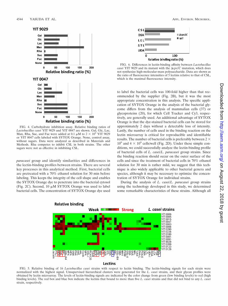
Next, a competitive inhibition assay was performed usingmonosaccharides and disaccharides as competitors (Gal, Glc,Man, Rha, Suc, Fuc, and Lac) to measure the binding speci-ficity of YIT 9029 and YIT 0047 (NIRD A-121) to CSL. Thebinding ratios of YIT 9029 and YIT 0047 are shown in Fig. 4.Rha completely inhibited the binding of YIT 9029 and YIT0047 to CSL, indicating that the binding of both strains to CSLwas rhamnose specific. Although the binding of YIT 0047 toCSL was partially inhibited by Gal and Glc, these sugars didnot affect the binding of YIT 9029 to CSL, suggesting that Galand Glc moieties in the cell surface structure of YIT 0047 butnot YIT 9029 are present near the binding site of CSL. Hence,the liquid-phase lectin microarray technique is sensitive enoughto distinguish similarities and differences in the glycome ofbacterial cell surfaces between strains, even in the same spe-cies, and is suitable in the identification of individual bacterialstrains. At the same time, this technique is very simple andreproducible for characterizing cell wall structures.
Profiling 16 L. casei strains. We examined 16 L. casei strainswhich are indistinguishable from each other by 16S rRNAsequences using the lectin microarray technique. As shown inFig. 5, each strain has a unique profile of binding to variouslectins, except for YIT 0001, YIT 0091, and YIT 9029 (Fig. 5).YIT 0091 is a clone of YIT 9029 personally stored by a re-searcher at the Yakult Central Institute for MicrobiologicalResearch a long time ago and is thus considered a clone of YIT9029 (M. Onoue [YIT 0091] and T. Sakurai [YIT 0001], per-sonal communication). YIT 0001 also had the same bindingprofile as YIT 9029 and will be discussed later.
Among 16 L. casei/L. paracasei strains, two types of lectin-binding characteristics could be recognized; one group has fewlectin responders which bind to only one or two different lec-tins, and the other group has multiple lectin responders whichbind to multiple lectins with different specificities. YIT 0001,YIT 0091, and YIT 9029 bound only to CSL, YIT 0009 boundonly to ABA, YIT 0123 bound only to jacalin, and YIT 0226bound only to BPL and SBA. Similar to YIT 9029, these strainsdo not necessarily have PSs with simple sugar composition butmay contain various sugar compositions. Alternately, there arestrains which bind to a number of lectins. For instance, YIT0003 binds to 23 lectins, YIT 0006 binds to 24 lectins, and YIT0290 binds to 27 lectins. From all the binding profiles, we couldnot draw a specific binding profile for L. casei/L. paracaseispecies but, rather, recognized profiles specific for individualstrains. None of the strains bound ACA and AOL, which arespecific for Gal-�1-3GalNAc-Thr/Ser and Fuc, respectively.
Next, we analyzed the lectin-binding profile of a mutant ofYIT 9029 as a typical example of a strain with few lectinbinders. As described above, the cell wall PS of YIT 9029contains several sugar molecules, including Glc, Gal, Rha,GlcNAc, and GalNAc, with different linkages. The mutant ofYIT 9029 with the �cps1C mutation was constructed by site-specific deletion mutagenesis within the cps1C gene encoding aputative glycosyltransferase, which is essential to synthesizePS-1 and most probably lacks a certain glycosyltransferase ac-
4542 YASUDA ET AL. APPL. ENVIRON. MICROBIOL.
on August 22, 2018 by guest
http://aem.asm
.org/D
ownloaded from
tivity (43). The glycan profile of the strain with the �cps1Cmutation was clearly different from that of YIT 9029: in addi-tion to binding to CSL, the cells of the strain with the �cps1Cmutation bound to an asialo complex-type N-glycan binder,DSA, and chitin binders LEL, STL, and UDA (Fig. 6). Inter-estingly all these lectins have the binding specificity of theN-acetylglucosamine polymer. No other lectins bound to themutant. Therefore, the deficiency of the cps1C gene resulted ina partial release of PS structures consisting of at least one
GlcNAc moiety in the cell wall PS, in addition to the disap-pearance of the high-molecular-mass PS-1 (43).
DISCUSSION
In this study, we have developed a simple and efficientmethod to profile the bacterial glycome using liquid-phaselectin microarray. We have shown that it is possible to distin-guish between strains classified into the Lactobacillus casei/L.
FIG. 3. Differential profiling of 4 Lactobacillus casei strains. Bacterial cells (4 � 109 cells/well) were subjected to glycan profiling by lectinmicroarray. Data were analyzed as described in Materials and Methods. Each strain shows a strain-specific lectin-binding affinity. Red boxesindicate the signals bound to Rha-binding lectin (CSL). Though YIT 0005, YIT 0006, and YIT 0180T cells bound to a variety of lectins, YIT 9029cells bound to CSL only. Data are the averages � standard deviations of triplicate determinations.
VOL. 77, 2011 PROFILING OF L. CASEI CELL WALL POLYSACCHARIDES 4543
on August 22, 2018 by guest
http://aem.asm
.org/D
ownloaded from
paracasei group and identify similarities and differences inthe lectin-binding profiles between strains. There are severalkey processes in this analytical method. First, bacterial cellsare pretreated with a 70% ethanol solution for 30 min beforelabeling. This keeps the integrity of the cell shape and enablesthe SYTOX Orange dye to penetrate into the bacterial cytosol(Fig. 2C). Second, 10 �M SYTOX Orange was used to labelbacterial cells. The concentration of SYTOX Orange dye used
to label the bacterial cells was 100-fold higher than that rec-ommended by the supplier (Fig. 2B), but it was the mostappropriate concentration in this analysis. The specific appli-cation of SYTOX Orange in the analysis of the bacterial gly-come differs from the analysis of mammalian cells (37) orglycoproteins (20), for which Cell Tracker and Cy3, respec-tively, are generally used. An additional advantage of SYTOXOrange is that the dye-stained bacterial cells can be stored forapproximately 2 days without a detectable loss of intensity.Lastly, the number of cells used in the binding reaction on thelectin microarray is critical for reproducible and identifiableresults. The number of bacterial cells is preferably between 2 �109 and 4 � 109 cells/well (Fig. 2D). Under these simple con-ditions, we could successfully analyze the lectin-binding profileof bacterial cells of L. casei/L. paracasei group strains. Sincethe binding reaction should occur on the outer surface of thecells and since the treatment of bacterial cells in 70% ethanolsolution for 30 min is rather mild, we suggest that this tech-nique is also widely applicable to other bacterial genera andspecies, although it may be necessary to optimize the concen-tration of SYTOX Orange for individual strains.
During the analysis of L. casei/L. paracasei group strainsusing the technology developed in this study, we determinedsome remarkable characteristics of these strains. Although all
FIG. 4. Carbohydrate inhibition assay. Relative binding ratios ofLactobacillus casei YIT 9029 and YIT 0047 are shown. Gal, Glc, Lac,Man, Rha, Suc, and Fuc were added at 0.1 �M to 2 � 109 YIT 9029or YIT 0047 cells labeled with SYTOX Orange. None, control assay,without sugars. Data were analyzed as described in Materials andMethods. Rha competes to inhibit CSL in both strains. The othersugars were not as effective in inhibiting CSL.
FIG. 5. Relative binding of 16 Lactobacillus casei strains with respect to lectin binding. The lectin-binding signals for each strain werenormalized with the highest signal. Unsupervised hierarchical clusters were generated for the L. casei strains, and their glycan profiles wereobtained by lectin microarray. The levels of lectin-binding signals are indicated by the color change from green (low binding levels) to red (highbinding levels). The red box and blue box indicate the lectins that bound to more than five L. casei strains and that did not bind to any L. caseistrain, respectively.
FIG. 6. Differences in lectin-binding affinity between Lactobacilluscasei YIT 9029 and its mutant with the �cps1C mutation, which doesnot synthesize high-molecular-mass polysaccharide. Data are shown asthe ratio of fluorescence intensities of 5 lectins relative to that of CSL,which is the maximal fluorescence intensity.
4544 YASUDA ET AL. APPL. ENVIRON. MICROBIOL.
on August 22, 2018 by guest
http://aem.asm
.org/D
ownloaded from
of the strains are classified into the same species group, L. ca-sei/L. paracasei, the individual strains have very unique lectin-binding profiles. YIT 0091 and YIT 9029 had the same bindingprofile (Fig. 5). They are known to originate from the sameancestor (M. Onoue, personal communication), and therefore,it could be anticipated that they have the same binding profile.Since YIT 0001 also showed the same binding profile as YIT0091 and YIT 9029, it is suggested that this strain also sharesthe same ancestor. Diversity in the outer surface PS structuresis often observed among streptococci, including the pathogenicspecies and other genera. It is believed that there is an activemechanism that enables these strains to frequently alter theirouter surface antigenic properties to escape from the hostimmune mechanism. Numerous reports have shown cell wallstructural and genetic diversities among the Lactobacillus spe-cies. We confirm this in the L. casei/L. paracasei group strains,using the lectin microarray. There may be a possibility thatglycosylated components other than cell wall polysaccharidescontribute to the binding of cells to lectins. However, we do nothave clear evidence that L. casei/L. paracasei group strainshave glycosylated proteins or (lipo)teichoic acids on the cellsurface. It is difficult to speculate about the mechanism andfrequency of such surface alteration; there may be hot spots ofgene conversion or gene exchange in the genomes of thesebacteria.
Though none of the lectins binds to every strain used in thisstudy, there are some lectins which bind to a number of L.casei/L. paracasei group strains. Those are CSL (Rha binder),BPL (Gal binder), LEL (chitin binder), ECA, RCA120, andDSA (asialo complex-type N-glycans binders), TJA-II, EEL,and ABA (Gal-type N-glycan binders), and jacalin, WFA,VVA, SBA, GSL-I, and PTL-I (O-glycan binders). In contrast,few strains bound to high mannose-type N-glycan binders(NPA, concanavalin A [ConA], GNA, and HHL) and sialicacid (Sia) binders (MAL, SNA, SSA, and TJA-I); furthermore,none of the strains bound ACA (Gal-�1-3GalNAc-Thr/Serbinder) or AOL (Fuc binder). This suggests that the surfacesugar moieties specific for these binders are generally absentamong strains of the L. casei/L. paracasei group.
The lectin-binding patterns of lactobacilli were previouslyreported using 6 different lectins (3). Interestingly, none of theLactobacillus acidophilus strains bound to any lectins becauseof autoagglutination. In fact, some strains in this study, forinstance, YIT 0005 and YIT 0180T, were found to autoagglu-tinate in MRS medium; however, agglutination itself did notaffect the binding to lectins (Fig. 3). These data may imply thatthe cell surface glycomes between L. casei/L. paracasei andL. acidophilus would be largely different from each other.
The lectin-binding profiles do not always reflect the sugarcomposition of the bacterial cell wall PSs. In some strains,lectin binding is limited, while in other strains many differenttypes of lectins bind to the cells. We speculate that limitedbinding is caused by physical hindrance of the binding sites forcertain lectins. For instance, YIT 9029 binds only to a Rha-specific lectin CSL in this assay, although the PS of YIT 9029consists of several sugar components (28; this study). The factthat the binding of CSL to YIT 9029 is inhibited by Rha but notby other sugars (Fig. 4) clearly indicates that this binding oc-curs in a Rha-specific manner. In one mutant of YIT 9029 withthe �cps1C mutation, which has a defect in a certain sugar
transferase gene, we suggest that a high-molecular-mass poly-saccharide (PS-1) structure is missing from the cell surface(43). This cps1C mutation has led the mutant to bind multiplelectins (Fig. 6). It is too early to speculate whether PS-1 con-tains the binding site for CSL and/or whether it would hindershorter PSs that potentially have the binding sites for CSL andother lectins. Differences in the patterns of inhibition againstthe binding of different L. casei strains to CSL by variouscarbohydrates have been indicated. These differences may re-flect the structural characteristics of the CSL binding site inthese strains. Further biochemical and molecular genetic anal-yses are required in order to clarify the exact structural fea-tures of the cell wall PSs.
The results obtained in this study show the actual interactionbetween lectins and microbial cells, which is probably via sur-face PSs. The lectins used in this assay are mostly derived fromvarious plants, except for AOL, which was isolated from Asper-gillus oryzae; CSL, which was isolated from salmon eggs (34,40); and HPA, which was isolated from Helix pomatia; somelectins were isolated from fungi. Recent advances in mam-malian cell biology reveal that various lectin-like proteinsare expressed on mammalian cell surfaces and may playpivotal roles in cell signaling. In addition, some plant lectinsare known to mimic mammalian lectins, but they could eas-ily be applied to the technology described here.
During the host-bacterium interaction in the gut, probioticbacteria may send their signals through various methods andfactors. The data presented in this study clearly show thatbacterial PSs can actively interact with lectins. Further analysison the interaction between bacterial PSs and host cells willenable us to identify novel factors involved in this response.
In conclusion, we have developed a liquid-phase lectin mi-croarray analysis technique to profile the bacterial glycome.The binding profile probably reflects, in part, the content andstructure of cell wall PSs. It is a simple and high-throughputsystem which enables us to distinguish and identify individualbacterial strains within the same species without performingcomplicated carbohydrate composition and structure analyses.In addition, the interactions between lectins and bacterial cellssuggest the presence and importance of this type of interactionin the probiotic actions.
ACKNOWLEDGMENTS
We thank Yoshiko Kubo and Jinko Murakami of the ResearchCenter for Medical Glycoscience at the National Institute of AdvancedIndustrial Science and Technology for help in preparation of the lectinmicroarray, Toshihiko Takada of the Yakult Central Institute for Mi-crobiological Research for help with bacterial labeling methods andthe electron microscopic photos, and Koich Watanabe for advice onchoosing Lactobacillus casei strains. We are deeply indebted to TeruoYokokura and the late Toshiaki Osawa, who both always encouragedus and gave helpful discussions.
REFERENCES
1. Akira, S., and K. Takeda. 2004. Toll-like receptor signalling. Nat. Rev.Immunol. 4:499–511.
2. Angeloni, S., et al. 2005. Glycoprofiling with micro-arrays of glycoconjugatesand lectins. Glycobiology 15:31–41.
3. Annuk, H., S. O. Hynes, S. Hirmo, M. Mikelsaar, and T. Wadstrom. 2001.Characterisation and differentiation of lactobacilli by lectin typing. J. Med.Microbiol. 50:1069–1074.
4. Baik, Y. S., and W. J. Cheong. 2007. Development of SPE for recovery ofpolysaccharides and its application to the determination of monosaccharidecomposition of the polysaccharide sample of a lactobacillus KLB 58. J. Sep.Sci. 30:1509–1515.
VOL. 77, 2011 PROFILING OF L. CASEI CELL WALL POLYSACCHARIDES 4545
on August 22, 2018 by guest
http://aem.asm
.org/D
ownloaded from
5. Bentley, S. D., et al. 2006. Genetic analysis of the capsular biosynthetic locusfrom all 90 pneumococcal serotypes. PLoS Genet. 2:e31.
6. Cieslewicz, M. J., et al. 2005. Structural and genetic diversity of group BStreptococcus capsular polysaccharides. Infect. Immun. 73:3096–3103.
7. Cobb, B. A., Q. Wang, A. O. Tzianabos, and D. L. Kasper. 2004. Polysac-charide processing and presentation by the MHCII pathway. Cell 117:677–687.
8. Coyne, M. J., et al. 2001. Polysaccharide biosynthesis locus required forvirulence of Bacteroides fragilis. Infect. Immun. 69:4342–4350.
9. Davis, G. H. 1955. The classification of lactobacilli from the human mouth.J. Gen. Microbiol. 13:481–493.
10. Dicks, L. M., E. M. Plessis, F. Dellaglio, and E. Lauer. 1996. Reclassificationof Lactobacillus casei subsp. casei ATCC 393 and Lactobacillus rhamnosusATCC 15820 as Lactobacillus zeae nom. rev., designation of ATCC 334 asthe neotype of L. casei subsp. casei, and rejection of the name Lactobacillusparacasei. Int. J. Syst. Bacteriol. 46:337–340.
11. Fang, Y., et al. 2007. Soluble proteins produced by probiotic bacteria regu-late intestinal epithelial cell survival and growth. Gastroenterology 132:562–575.
12. Feng, Y., and W. Xiao-Min. 2007. Difference in gene expression of macro-phage between normal spleen and portal hypertensive spleen indentified bycDNA microarray. World J. Gastroenterol. 13:3369–3373.
13. Heng, H. H., and L. C. Tsui. 1993. Modes of DAPI banding and simultane-ous in situ hybridization. Chromosoma 102:325–332.
14. Hsu, K.-L., and L. K. Mahal. 2006. A lectin microarray approach for therapid analysis of bacterial glycans. Nat. Protoc. 1:543–549.
15. Hsu, K.-L., K. T. Pilobello, and L. K. Mahal. 2006. Analyzing the dynamicbacterial glycome with a lectin microarray approach. Nat. Chem. Biol. 2:153–157.
16. Hsu, K.-L., J. C. Gildersleeve, and L. K. Mahal. 2008. A simple strategy forthe creation of a recombinant lectin microarray. Mol. Biosyst. 4:654–662.
17. Inohara, N., and G. Nunes. 2003. NODs: intracellular proteins involved ininflammation and apoptosis. Nat. Rev. Immunol. 3:371–382.
18. Kankainen, M., et al. 2009. Comparative genomic analysis of Lactobacillusrhamnosus GG reveals pili containing a human-mucus binding protein. Proc.Natl. Acad. Sci. U. S. A. 106:17193–17198.
19. Kullberg, M. C. 2008. Soothing intestinal sugars. Nature 453:602–604.20. Kuno, A., et al. 2005. Evanescent-field fluorescence-assisted lectin microar-
ray: a new strategy for glycan profiling. Nat. Methods 2:851–856.21. Lebeer, S., J. Vanderleyden, and S. C. J. De Keersmaecker. 2008. Genes and
molecules of Lactobacillus supporting probiotic action. Microbiol. Mol. Biol.Rev. 72:728–764.
22. Liu, C. H., S. M. Lee, J. M. VanLare, D. L. Kasper, and S. K. Mazmanian.2008. Regulation of surface architecture by symbiotic bacteria mediates hostcolonization. Proc. Natl. Acad. Sci. U. S. A. 105:3951–3956.
23. Mancuso, G., F. Tomasello, C. von Hunolstein, G. Orefici, and G. Teti. 1994.Induction of tumor necrosis factor alpha by the group- and type-specificpolysaccharides from III group B streptococci. Infect. Immun. 62:2748–2753.
24. Matsumoto, S., et al. 2005. Probiotic Lactobacillus-induced improvement inmurine chronic inflammatory bowel diseases is associated with the down-regulation of proinflammatory cytokines in lamina propria mononuclearcells. Clin. Exp. Immunol. 140:417–426.
25. Matsumoto, S., et al. 2008. A component of polysaccharide peptidoglycancomplex on Lactobacillus induced an improvement of murine model ofinflammatory bowel disease and colitis-associated cancer. Immunology 128:e170–e180.
26. Matsuzaki, T., et al. 1997. Prevention of onset in an insulin-dependentdiabetes mellitus model, NOD mice, by oral feeding of Lactobacillus casei.APMIS 105:643–649.
27. Mazmanian, S. K., C. H. Liu, A. O. Tzianabos, and D. L. Kasper. 2005. Animmunomodulatory molecule of symbiotic bacteria directs maturation of thehost immune system. Cell 122:107–118.
28. Nagaoka, M., et al. 1990. Structure of polysaccharide-peptidoglycan complexfrom the cell wall of Lactobacillus casei YIT 9018. J. Biochem. 108:568–571.
29. Nitta, K., T. Kawano, S. Sugawara, and M. Hosono. 2007. Regulation ofglobotriaosylceramide (Gb3)-mediated signal transduction by Rha-bindinglectin. Yakugaku Zasshi 127:553–561.
30. Pretzer, G., et al. 2005. Biodiversity-based identification and functional char-acterization of the mannose-specific adhesin of Lactobacillus plantarum. J.Bacteriol. 187:6128–6136.
31. Schweizer, D. 1981. Counterstain-enhanced chromosome banding. Hum.Genet. 57:1–14.
32. Shida, K., J. Kiyoshima-Shibata, M. Nagaoka, K. Watanabe, and M. Nanno.2006. Induction of interleukin-12 by Lactobacillus strains having a rigid cellwall resistant to intracellular digestion. J. Dairy Sci. 89:3306–3317.
33. Shimizu-Kadota, et al. 2000. Insertion of bacteriophage phiFSW into thechromosome of Lactobacillus casei Shirota (S-1): characterization of attach-ment sites and integrase gene. Gene 249:127–134.
34. Shirai, T., Y. Watanabe, M. Lee, T. Ogawa, and K. Muramoto. 2009. Struc-ture of Rha-binding Lectin CSL3: Unique pseudo-tetrameric architecture ofa pattern recognition protein. J. Mol. Biol. 391:390–403.
35. Simelyte, E., M. Rimpilainen, L. Lehtonen, X. Zhang, and P. Toivanen. 2000.Bacterial cell wall-induced arthritis: chemical composition and tissue distri-bution of four Lactobacillus strains. Infect. Immun. 68:3535–3540.
36. Takada, T., K. Matsumoto, and K. Nomoto. 2004. Development of multi-color FISH method for analysis of seven Bifidobacterium species in humanfeces. J. Microbiol. Methods 58:413–421.
37. Tateno, H., et al. 2007. A novel strategy for mammalian cell surface glycomeprofiling using lectin microarray. Glycobiology 17:1138–1146.
38. Uchiyama, N., et al. 2006. Development of a lectin microarray based on anevanescent-field fluorescence principle: a new strategy for glycan profiling.Methods Enzymol. 415:341–351.
39. Vaningelgem, F., et al. 2004. Biodiversity of exopolysaccharides produced byStreptococcus thermophilus strains is reflected in their production and theirmolecular and functional characteristics. Appl. Environ. Microbiol. 70:900–912.
40. Watanabe, Y., et al. 2009. The function of Rha-binding lectin in innateimmunity by restricted binding Gb3. Dev. Comp. Immunol. 33:187–197.
41. Yan, X., et al. 2000. Development of a mechanism-based, DNA stainingprotocol using SYTOX Orange nucleic acid stain and DNA fragment sizingflow cytometry. Anal. Biochem. 286:138–148.
42. Yan, X., et al. 2005. Probing the kinetics of SYTOX Orange stain binding todouble-stranded DNA with implications for DNA analysis. Anal. Chem.77:3554–3562.
43. Yasuda, E., M. Serata, and T. Sako. 2008. Suppressive effect on activation ofmacrophages by Lactobacillus casei strain Shirota genes determining thesynthesis of cell wall-associated polysaccharides. Appl. Environ. Microbiol.74:4746–4755.
44. Yasuno, S., K. Kokubo, and M. Kamei. 1999. New method for determiningthe sugar composition of glycoproteins, glycolipids, and oligosaccharides byhigh-performance liquid chromatography. Biosci. Biotechnol. Biochem. 63:1353–1359.
45. Yokokura, T., S. Kodaira, H. Ishiwa, and T. Sakurai. 1974. Lysogeny inlactobacilli. J. Gen. Microbiol. 84:277–284.
46. Yuki, N., et al. 1999. Survival of a probiotic, Lactobacillus casei strain Shi-rota, in the gastrointestinal tract: selective isolation from feces and identifi-cation using monoclonal antibodies. Int. J. Food Microbiol. 48:51–57.
4546 YASUDA ET AL. APPL. ENVIRON. MICROBIOL.
on August 22, 2018 by guest
http://aem.asm
.org/D
ownloaded from

APPLIED AND ENVIRONMENTAL MICROBIOLOGY, Aug. 2011, p. 5834 Vol. 77, No. 160099-2240/11/$12.00 doi:10.1128/AEM.06002-11Copyright © 2011, American Society for Microbiology. All Rights Reserved.
ERRATUM
Lectin Microarray Reveals Binding Profiles of Lactobacillus caseiStrains in a Comprehensive Analysis of Bacterial
Cell Wall PolysaccharidesEmi Yasuda,1* Hiroaki Tateno,2 Jun Hirabayashi,2 Tohru Iino,1 and Tomoyuki Sako3
Yakult Central Institute for Microbiological Research, 1796 Yaho, Kunitachi, Tokyo 186-8650, Japan1; Research Center forMedical Glycoscience, National Institute of Advanced Industrial Science and Technology, Central 2, 1-1-1 Umezono,Tsukuba, Ibaraki 305-8568, Japan2; and Yakult Europe B.V., Schutslussweg 1, 1332 EN Almere, The Netherlands3
Volume 77, no. 13, pages 4539–4546, 2011. Page 4539: The byline and affiliation line should read as shown above.
5834