lecture 1: the metastatic process general overview and
TRANSCRIPT

Lecture 1: The Metastatic Process
• General Overview and Mechanisms
• Pre-existent subpopulations
• Lymphangiogenesis
• Specificity of metastatic patterns of spread

Differences between Benign and Malignant Neoplasms
BENIGN MALIGNANT 1.Often encapsulated Non encapsulated 2.Well differentiated Poorly differentiated 3.Low mitotic rate High mitotic rate 4.Noninvasive Invasive 5.Non-metastasising Metastasising

Benign tumours are characterised by:
• Localised • Usually grow slowly • Compress, rather than infiltrate, adjacent
tissue, often forming a capsule • Do not spread to distant sites generally do
not recur after removal • May be self limiting or regress • Have cellular structure resembling that of
the tissue of origin

Malignant tumours are characterised by:
• Infiltration of malignant cells into surrounding tissues
• Invasion of neoplastic cells into blood and lymph vessels
• Spread of tumour cells to other parts of the body to establish secondary growths (metastasis)


METASTASIS
The transfer of disease from one
organ
or part of an organ to another not
directly connected with it

Spread of Cancer
• No invasion – in situ
• Local invasion • Dissemination
– Lymphatics – Blood stream – Intracavitary – Perineural

How Cancer Kills ?
• Late presentation is the norm – 109 cells per cubic cm of cancer
• Smallest lump detectable is about 1cm
• Local effects if anatomically problematic
- blocks tubes
- bleeds
- obstructs glands
• Metastatic burden = 1kg = death


Metastasis: the spread of tumours
Primary tumour
Secondary tumour
Intravasation
Extravasation

B16 Selection - 1973
F1
F10

Does it represent selection or adaptation?


Interpretation
• Metastatic sub-populations pre-exist within the parental population.
• These sub-populations breed true therefore there must be a genetic basis to their metastatic capacity.
• Added later- Gene expression signatures between primary and metastatic tumours should be different.

a
b c

Interpretation
Gene expression signatures of primary and secondary tumours
are similar.

Conclusions • Metastasis probably is regulated by genes we
already know about.
• How those genes are regulated though is not necessarily obvious.
• Technological developments ( gene expression arrays, miRNA arrays ) will allow us to interrogate this process.
• In future the focus may need to be on how disseminated cells grow at distant sites rather than how they accomplish this process.

Role of Angiogenesis in Tumour Spread

ANGIOGENESIS and METASTASIS
Weidner et al (1992) Prognostic Factor in Breast Cancer Horak et al (1992) Correlation with Survival in Breast Cancer MacChiarini et al (1992) Correlation with Metastasis in Lung Cancer Weidner et al (1993) Correlation with Metastasis in Prostate
Cancer


Angiogenic peptides
Angiogenic peptides

SS SS
NRP-1 NRP-1
VEGFR-1 VEGFR-2 VEGFR-3
VEGF-A
VEGF-B
VEGF-C
VEGF-D OV-VEGF
PIGF
VEGF-B
sVEGFR-1
P1114L
P P P P P P
Migration, permeability, DNA synthesis, survival
angiogenesis lymphangiogenesis

LYMPHANGIOGENESIS
• VEGF-C/VEGF-D binding to VEGF-R3.
• LYVE-1 as a marker of lymph vessels.
• Experimentally VEGF-C has promoted breast cancer metastasis.
• Clinical correlations with metastatic activity e.g. gastric,cervical and breast

LYMPHANGIOGENESIS
• Are the identified lymphatics functional?
• How specific is LYVE-1 as a marker?
• How solid are the clinical data?
• Is there variation between tumour types?

Metastasis: the spread of tumours
Primary tumour
Secondary tumour
Intravasation
Extravasation

“Cancer invasion can be viewed as a derangement in the proper sorting of cell populations, causing a violation of
normal tissue boundaries”
Liotta and Kohn
Nature 411: 375-379, 2001

Cellular Invasion: 3-step Process
• 1. Cell Attachment
• 2. Degradation of ECM
• 3 Cell Migration

Proteinases Serine proteinases uPA, thrombin,
Cathepsin G Cysteine proteinases Cathepsin B, L. Aspartyl proteinases Cathepsin D Matrix metalloproteinases Gelatinases,
Collagenases

SERINE PROTEASES
• Serine residue at active site
• Inactive pro-form
• uPA, trypsin, plasmin
Urokinase type Plasminogen activator (uPA)
• Plasminogen plasmin
• Concentrated at cell surface
• Tumour-stromal interface

MMPs • family of related enzymes with shared
domains • degrade collagens and other matrix proteins • Zinc dependent enzymes, work best at neutral
pH • generally secreted as inactive pro-enzymes • all inhibited by “TIMPs”

M2+ D S
P G R
G
Pax Tensin
FAK src G
M2+ D S
P G R
G
Pax Tensin
FAK G
a-actinin
actin
ECM glycoprotein
eg fibronectin
Cation-binding
site
SS SS
Small GTPases
-Rho,Rac,cdc42
a b

Selectins Integrins
Rolling Activation Flattening and
firm adhesion
Transendothelial
migration
CHEMOATTRACTANT (eg released after injury or infection)
Leukocyte Extravasation


The Nonrandom Nature of Metastatic
Spread
1.Hemodynamic Factors Increased bloodflow = increased frequency 2. Specific Cell Recognition Cell receptors = increased trapping 3.Seed and Soil Tumour/organ growth interaction = metastasis


Figure 1 The hypoxic response. a, Under conditions of normal oxygen, the von Hippel–Lindau tumour suppressor protein
(pVHL) modifies the protein HIF, which leads to its destruction. b, When oxygen is scarce (hypoxia), or when pVHL is
mutated, HIF accumulates inside cells and activates the expression of certain genes. This triggers two complementary
responses. First, tissue oxygenation is stimulated through the activation of genes such as VEGF (which stimulates the
outgrowth of new blood vessels) and erythropoietin (EPO, which stimulates the production of red blood cells). Second,
tumour cells are stimulated to move away from the site of hypoxia through the activation of genes such as c-Met, which
enhance cell motility and invasion. Now, Staller et al.3 show that the gene CXCR4 is also activated by HIF. CXCR4 not only
stimulates migration, it also enables tumour cells to

Cancer metastasis and chemokine signalling. Initiated epithelial cells are promoted by inflammation to undergo neoplastic progression, a process
that requires remodelling of the extracellular matrix, recruitment of inflammatory cells, angiogenesis and lymphangiogenesis. Out of this
microenvironment, carcinomas arise. These neoplastic cells then turn on expression of chemokine receptors, such as CXCR4. The production of
chemokine ligands for these receptors, in sites such as lymph nodes, bone marrow, liver and lung, then facilitates their invasion and migration to
secondary sites where malignant cells reside either in a dormant state, or proliferate to form a productive metastatic lesion. Blockade of
chemokine receptors, for example, anti-CXCR4 antibodies, attenuates metastatic spread in some experimental systems.

Metastasis: the spread of tumours
Primary tumour
Secondary tumour
Intravasation
Extravasation

Lecture 2: Intercellular adhesion and metastasis
• Cadherin superfamily
• Selectin family
• Integrin family

A functional classification of cell junctions
1. Occluding junctions (tight junctions)
2. Anchoring junctions
a. Actin filament attachment sites (adherens junctions)
i. cell-cell (e.g. adhesion belts)
ii cell-matrix (e.g. focal contacts)
b. Intermediate filament attachment sites
i cell-cell (desmosomes)
ii cell-matrix (hemidesmosomes)
3. Communicating junctions
a. gap junctions
b. chemical synapses
c. plasmodesmata (plants only)*

Tight Junction
(Zonula Occludens)
Lumen
Gap
Junction Hemidesmosome
Belt Desmosome
(Zonula Adherens)
Basement
Membrane
Extracellular Matrix

Cadherin Superfamily
Subfamily Members
Classical cadherins E-,N- or P-Cadherin
VE-Cadherin
Desmosomal cadherins Desmocollin
Desmoglein
Proto-cadherins -Protocadherin
CNR-Cadherin
Seven transmembrane (7TM) Flamingo
cadherins
T-cadherin T-cadherin
FAT family Dachsous
Fat

G protein
β-cat
DP
PG
PP
FYN
PDZ
β-cat α-cat
β-cat α-cat
vinc
PG
vinc
DP
DP
vinc
actin
IF
actin
IF
IF
mDAB1
Signalling cascades
eg. SRC protein tyrosine
kinases, G-proteins
?
E-, N- or
P-cadherin
VE-cadherin
classical
cadherins
desmocollin
desmoglein
μ-protocadherin
CNR-cadherin
protocadherins
7TM-cadherin
T-cadherin
FAT-family cadherin

Calcium dependent, cell-cell adhesion molecules Numerous family members, cell-lineage specificity of expression e.g. E-cadherin in epithelial cells Extracellular domain has 5 homologous repeats Binding specificity located to most distal repeats Establish and maintain intracellular connections Down-regulation results in cell dispersal
CADHERINS

Produced as 135kDa glycoprotein After cytoplasmic trimming expressed as 120kDa molecule Five extracellular domains of – 110 amino acids each, internal sequence homology, conserved Ca2+ binding motifs
E-Cadherin

N
Pre-region
a
b C
E1 E2 E3 E4 E5
HAV


Catenin complex
Catenin Size Chromosomal location a 102kDa 5q31 b 92kDa 3p21 (plakoglobin) 83kDa 17q21 p120ctn (p120 cas) 11q11

p120ctn (p120cas)
P120cas – major Src (tyrosine kinase) substrate
Arm domain – 11 copies of a 42-amino-acid motif originally described for the Drosophila segment polarity gene product, armadillo
At least 4 different isoforms

Reduced p120ctn expression correlates with poor survival in
patients with adenocarcinoma of the gastroesophageal junction
Wijnhoven, BP et al J Surg Oncol 92:116-123, 2005.


Conversion of Epithelial to Mesenchymal
Downregulation of E-cadherin activity (RNAi, Antibody)
Restoration of E-cadherin (cDNA)


Table 1 ECD expression and Dukes stage
ECD expression A&B C1&C2 ECD++/ECD+ 32 7 ECD- 4 29 36 36 Fisher exact test p<<0.001

Table 2 ECD expression and tumour grade
ECD expression Well Moderate Poor ECD++ 7 4 0 ECD+ 1 24 4 ECD- 0 8 24
8 36 28


E-Cadherin Downregulation
• Transcriptional regulation e.g. increased „Snail‟ activity, promoter methylation.
• Mutation in E-cad gene and LOH.
• Catenin mutation and downregulation leads to loss of functional cadherin/catenin complex.

Concept of EMT
• Phenotypic switch
• Co-ordinated set of molecular changes
• Change in function,fate and character

Transient loss of basement memranes in malignant progression: Brabletz model.
Tumour microenvironment
ZEBI?
EMT + BM Loss
MET MET
Invasive front
dissemination
Taken from:- Spaderna et al
Gastroenterology 131: 830, 2006

Concept of EMT
• Phenotypic switch
• Co-ordinated set of molecular changes
• Change in function,fate and character
• Change in morphological appearance

Gene expression changes said to be associated with EMT
• Loss of- • E-cadherin expression • Cytokeratin expression
• Gain of- • Vimentin expression • N-cadherin expression • MMP expression

Human lobular and ductal breast carcinomas
lobular ductal


Transient loss of basement memranes in malignant progression: Brabletz model.
Tumour microenvironment
ZEBI?
EMT + BM Loss
MET MET
Invasive front
dissemination
Taken from:- Spaderna et al
Gastroenterology 131: 830, 2006

Conclusion
• Loss of E-cadherin associated with tumour progression and metastasis

Wnt1 receptor
1111111
Dsh
P
P
P
b a
b-catenin
APC
GSK-3b
b
LEF/TFC
Cyclin D1
myc
Nucleus
P GSK-3p
LEF/TCF
GSK-3b
b
APC
Proteasome

WNT-wingless Pathway
Wnt-1 protein extracellular signalling factor
Frizzled seven-transmembrane-domain receptor
for Wnt-1
Dishevilled (Dsh) phosphorylated to active form by
casein kinase II
Glycogen synthase kinase-3b complexes with APC and cytoplasmic b-catenin
(GSK-3b)
Adenomatous polyposis coli phosphorylated, together with b-catenin, by GSK-3b
(APC)
Phosphorylated b-catenin Ubiquitinated. Proteasome degradation
Association of Wnt-1 and Frizzled
results in activation of Wnt-1 signalling pathway

b-catenin transcription
LEF/TCF leucocyte enhancer factor/T-cell factor
Free b-catenin Translocates to nucleus
Plus LEF/TCF Induces DNA binding and transcription
c-MYC LEF/TCF-responsive genes
cyclin D1
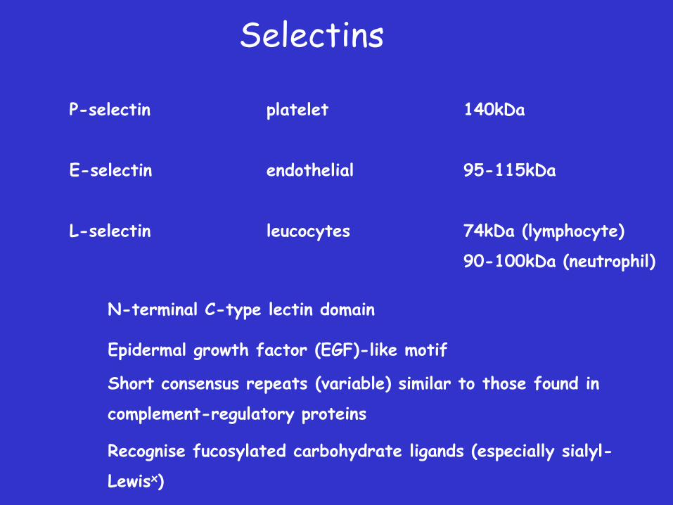
Selectins
P-selectin platelet 140kDa
E-selectin endothelial 95-115kDa
L-selectin leucocytes 74kDa (lymphocyte)
90-100kDa (neutrophil)
N-terminal C-type lectin domain
Epidermal growth factor (EGF)-like motif
Short consensus repeats (variable) similar to those found in
complement-regulatory proteins
Recognise fucosylated carbohydrate ligands (especially sialyl-
Lewisx)


L-selectin
Constitutively expressed by leukocytes -majority B cells, virgin T cells, neutrophils, monocytes and eosinophils Mediates binding to activated endothelium at inflammatory sites Ligands are highly glycosylated mucin-like molecules Direct binding lymphocytes to HEV of peripheral lymph nodes

E-selectin Expressed by cytokine-activated endothelial cells
Protein synthesis-dependent
Peaks 4-6 hours post cytokine stimulation
Mediates neutrophil, monocyte, memory
T-cell adhesion
Can be found in serum (proteolytic cleavage from cell
surface). Elevated in various inflammatory syndromes

P-selectin
Constitutively expressed in Weibel-Palade bodies of endothelial cells and alpha granules of platelets Rapidly mobilised to surface in response to inflammatory agents Cell surface expression short-lived Mediates adhesion of neutrophils and monocytes to activated platelets and endothelial cells

Integrin Ligands
• Extracellular matrix components
(many contain RGD motif)
• Ig superfamily molecules (e.g. VCAM)


Additional Reading
Ramachandran V et al
Dimerization of a selectin and its ligand stablizes cell rolling and enhances tether strength in shear flow
Proc. Natl. Acad. Sci. USA 98: 10166-10171 2001
Michaelson JS and Leder P
b-catenin is a downstream effector of Wnt-mediated tumorigenesis in the mammary gland
Oncogene 20: 5093-5099 2001
Runswick SK et al
Desmosomal adhesion regulates epithelial morphogenesis and cell positioning
Nature Cell Biology 3: 823-830, 2001