lecture 10 overview of the nervous system. outline organization of nervous system organization of...
TRANSCRIPT

Lecture 10Lecture 10
Overview of the Overview of the Nervous SystemNervous System
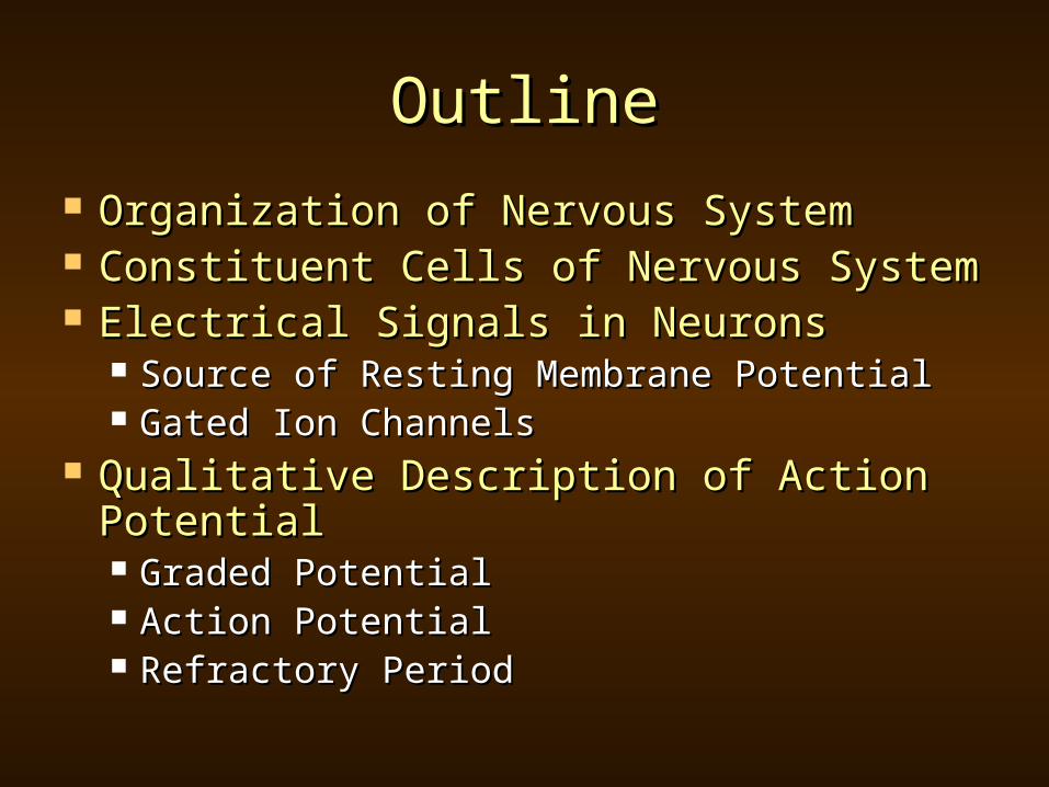
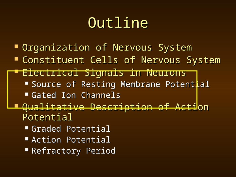
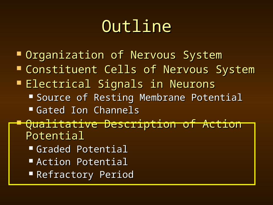
OutlineOutline
Organization of Nervous SystemOrganization of Nervous System Constituent Cells of Nervous SystemConstituent Cells of Nervous System Electrical Signals in NeuronsElectrical Signals in Neurons
Source of Resting Membrane PotentialSource of Resting Membrane Potential Gated Ion ChannelsGated Ion Channels
Qualitative Description of Action Qualitative Description of Action PotentialPotential Graded PotentialGraded Potential Action Potential Action Potential Refractory PeriodRefractory Period

Organization of the Nervous SystemOrganization of the Nervous System Central nervous systemCentral nervous system
Brain and spinal cordBrain and spinal cord Peripheral nervous systemPeripheral nervous system
Afferent neuronsAfferent neurons Sensory neuronsSensory neurons
Efferent neuronsEfferent neurons Send response to effector cellsSend response to effector cells Somatic motor divisionSomatic motor division
Control skeletal muscleControl skeletal muscle Autonomic divisionAutonomic division
Controls smooth and cardiac muscle and Controls smooth and cardiac muscle and exocrine/endocrineexocrine/endocrine
Two components:Two components: SympatheticSympathetic ParasympatheticParasympathetic Commonly exert antagonistic control over a single Commonly exert antagonistic control over a single
targettarget
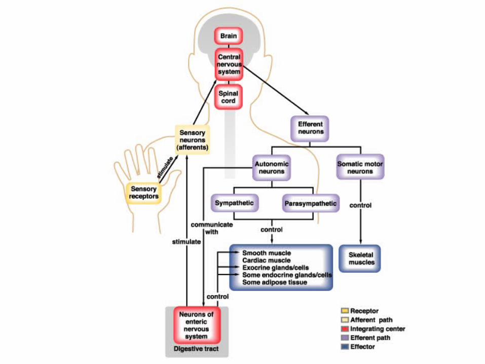
Fig 8.1 – Organization of the nervous system Silverthorn 2nd Ed

OutlineOutline
Organization of Nervous SystemOrganization of Nervous System Constituent Cells of Nervous SystemConstituent Cells of Nervous System Electrical Signals in NeuronsElectrical Signals in Neurons
Source of Resting Membrane PotentialSource of Resting Membrane Potential Gated Ion ChannelsGated Ion Channels
Qualitative Description of Action Qualitative Description of Action PotentialPotential Graded PotentialGraded Potential Action Potential Action Potential Refractory PeriodRefractory Period
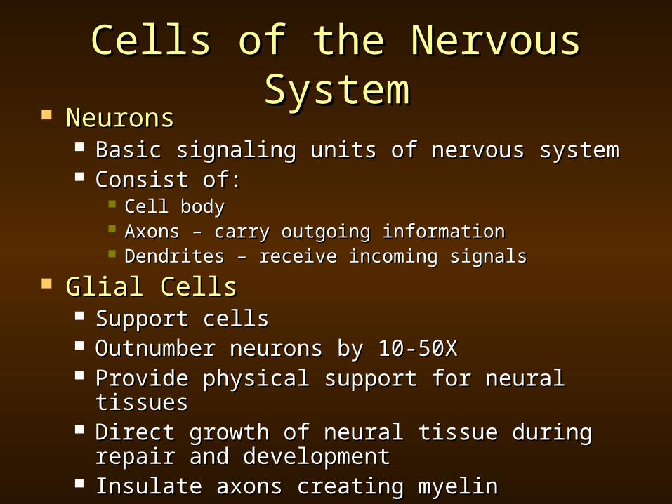
Cells of the Nervous SystemCells of the Nervous System NeuronsNeurons
Basic signaling units of nervous systemBasic signaling units of nervous system Consist of:Consist of:
Cell bodyCell body Axons – carry outgoing informationAxons – carry outgoing information Dendrites – receive incoming signalsDendrites – receive incoming signals
Glial CellsGlial Cells Support cellsSupport cells Outnumber neurons by 10-50XOutnumber neurons by 10-50X Provide physical support for neural tissuesProvide physical support for neural tissues Direct growth of neural tissue during repair and Direct growth of neural tissue during repair and
developmentdevelopment Insulate axons creating myelinInsulate axons creating myelin
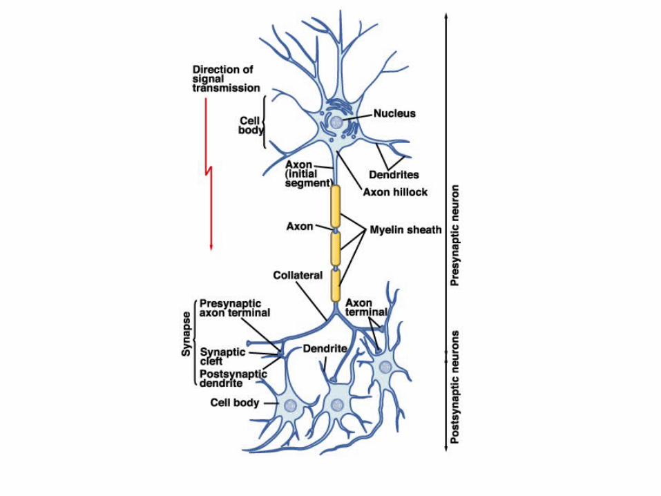
Fig 8.2 – Model neurons Silverthorn 2nd Ed
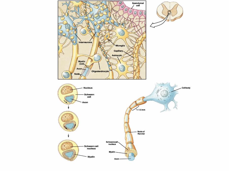
Fig 8.6 – Formation of myelin Silverthorn 2nd Ed
OutlineOutline
Organization of Nervous SystemOrganization of Nervous System Constituent Cells of Nervous SystemConstituent Cells of Nervous System Electrical Signals in NeuronsElectrical Signals in Neurons
Source of Resting Membrane PotentialSource of Resting Membrane Potential Gated Ion ChannelsGated Ion Channels
Qualitative Description of Action Qualitative Description of Action PotentialPotential Graded PotentialGraded Potential Action Potential Action Potential Refractory PeriodRefractory Period

Resting Membrane PotentialResting Membrane Potential Nernst EquationNernst Equation
GHK EquationGHK Equation
in
oution ion
ion
ZE log
61
outClinNainK
inCloutNaoutKm ClPNaPKP
ClPNaPKP
F
RTV
][][
][][ln
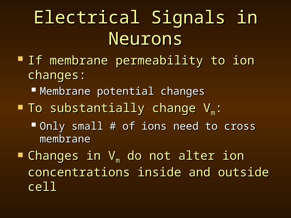
Electrical Signals in NeuronsElectrical Signals in Neurons
If membrane permeability to ion If membrane permeability to ion changes:changes: Membrane potential changesMembrane potential changes
To substantially change VTo substantially change Vmm:: Only small # of ions need to cross Only small # of ions need to cross
membranemembrane Changes in VChanges in Vmm do not alter ion do not alter ion
concentrations inside and outside cellconcentrations inside and outside cell

DepolarizationDepolarization
At rest:At rest: Membrane potential mostly due to KMembrane potential mostly due to K++
Membrane almost impermeable to NaMembrane almost impermeable to Na++
Depolarization:Depolarization: Cell becomes permeable to NaCell becomes permeable to Na++
NaNa++ rushes in, membrane potential rushes in, membrane potential dropsdrops
Moves towards +60mV of NaMoves towards +60mV of Na++
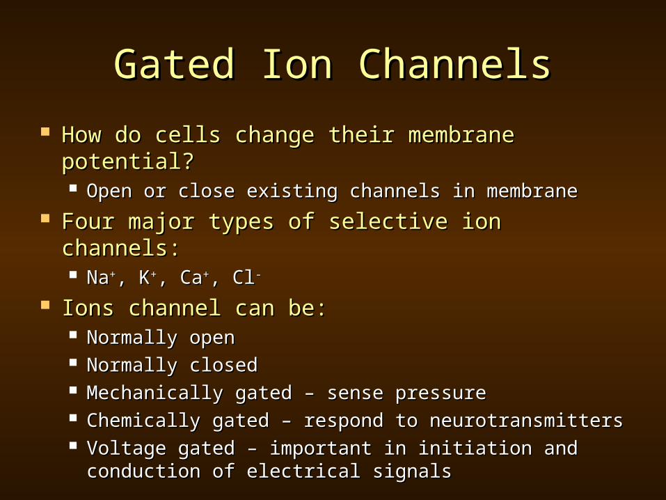
Gated Ion ChannelsGated Ion Channels
How do cells change their membrane How do cells change their membrane potential?potential? Open or close existing channels in membraneOpen or close existing channels in membrane
Four major types of selective ion channels:Four major types of selective ion channels: NaNa++, K, K++, Ca, Ca++, Cl, Cl--
Ions channel can be:Ions channel can be: Normally openNormally open Normally closedNormally closed Mechanically gated – sense pressureMechanically gated – sense pressure Chemically gated – respond to neurotransmittersChemically gated – respond to neurotransmitters Voltage gated – important in initiation and Voltage gated – important in initiation and
conduction of electrical signalsconduction of electrical signals
OutlineOutline
Organization of Nervous SystemOrganization of Nervous System Constituent Cells of Nervous SystemConstituent Cells of Nervous System Electrical Signals in NeuronsElectrical Signals in Neurons
Source of Resting Membrane PotentialSource of Resting Membrane Potential Gated Ion ChannelsGated Ion Channels
Qualitative Description of Action Qualitative Description of Action PotentialPotential Graded PotentialGraded Potential Action Potential Action Potential Refractory PeriodRefractory Period
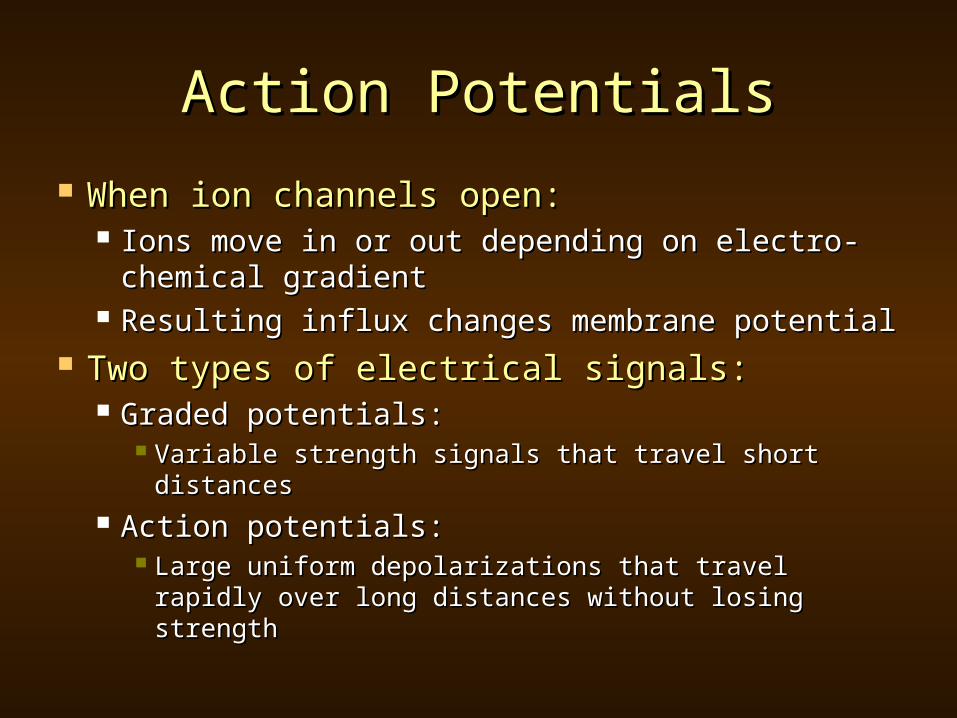
Action PotentialsAction Potentials
When ion channels open:When ion channels open: Ions move in or out depending on electro-Ions move in or out depending on electro-
chemical gradientchemical gradient Resulting influx changes membrane potentialResulting influx changes membrane potential
Two types of electrical signals:Two types of electrical signals: Graded potentials:Graded potentials:
Variable strength signals that travel short Variable strength signals that travel short distancesdistances
Action potentials:Action potentials: Large uniform depolarizations that travel rapidly Large uniform depolarizations that travel rapidly
over long distances without losing strengthover long distances without losing strength

Fig 8.7 – Graded potentials decrease in strength as they spread out from the point of origin Silverthorn 2nd Ed
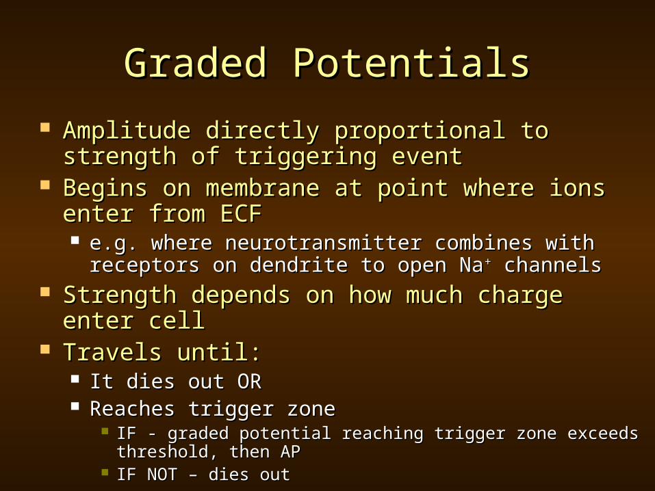
Graded PotentialsGraded Potentials
Amplitude directly proportional to strength of Amplitude directly proportional to strength of triggering eventtriggering event
Begins on membrane at point where ions Begins on membrane at point where ions enter from ECFenter from ECF e.g. where neurotransmitter combines with e.g. where neurotransmitter combines with
receptors on dendrite to open Nareceptors on dendrite to open Na++ channels channels Strength depends on how much charge enter Strength depends on how much charge enter
cellcell Travels until:Travels until:
It dies out ORIt dies out OR Reaches trigger zoneReaches trigger zone
IF - graded potential reaching trigger zone exceeds IF - graded potential reaching trigger zone exceeds threshold, then APthreshold, then AP
IF NOT – dies outIF NOT – dies out

Fig 8.8 – Subthreshold and suprathreshold graded potentials in a neuron Silverthorn 2nd Ed

Action PotentialsAction Potentials
All action potentials are identical in All action potentials are identical in strengthstrength
Do not diminish in strength as they Do not diminish in strength as they traveltravel
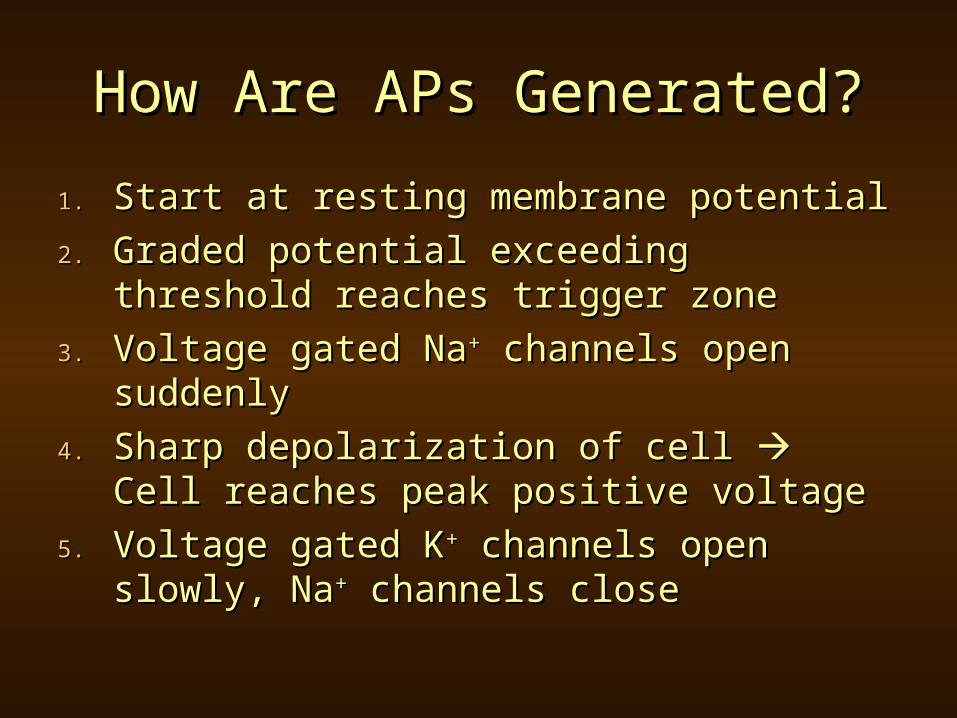
How Are APs Generated?How Are APs Generated?
1.1. Start at resting membrane potentialStart at resting membrane potential
2.2. Graded potential exceeding Graded potential exceeding threshold reaches trigger zonethreshold reaches trigger zone
3.3. Voltage gated NaVoltage gated Na++ channels open channels open suddenlysuddenly
4.4. Sharp depolarization of cell Sharp depolarization of cell Cell Cell reaches peak positive voltagereaches peak positive voltage
5.5. Voltage gated KVoltage gated K++ channels open channels open slowly, Naslowly, Na++ channels close channels close

How Are APs Generated?How Are APs Generated?
6.6. K+ moves out of cell K+ moves out of cell Reduces Reduces membrane potentialmembrane potential
7.7. K+ continues to leave, K+ continues to leave, hyperpolarizes cellhyperpolarizes cell
8.8. Voltage gated K+ channels close, Voltage gated K+ channels close, some K+ enters through leak some K+ enters through leak channelschannels
9.9. Cell returns to resting membrane Cell returns to resting membrane potentialpotential
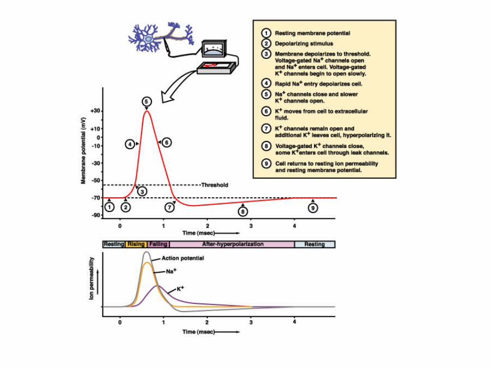
Fig 8.9 – the action potential Silverthorn 2nd Ed
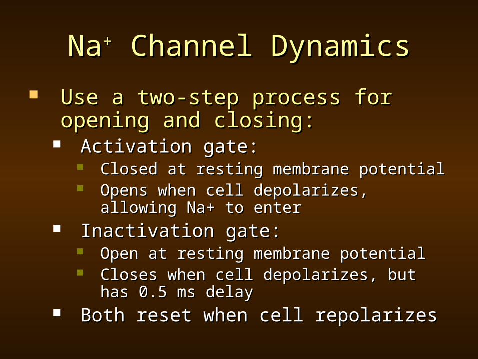
NaNa++ Channel Dynamics Channel Dynamics
Use a two-step process for opening Use a two-step process for opening and closing:and closing:
Activation gate: Activation gate: Closed at resting membrane potentialClosed at resting membrane potential Opens when cell depolarizes, allowing Na+ Opens when cell depolarizes, allowing Na+
to enterto enter Inactivation gate: Inactivation gate:
Open at resting membrane potentialOpen at resting membrane potential Closes when cell depolarizes, but has 0.5 Closes when cell depolarizes, but has 0.5
ms delayms delay Both reset when cell repolarizesBoth reset when cell repolarizes
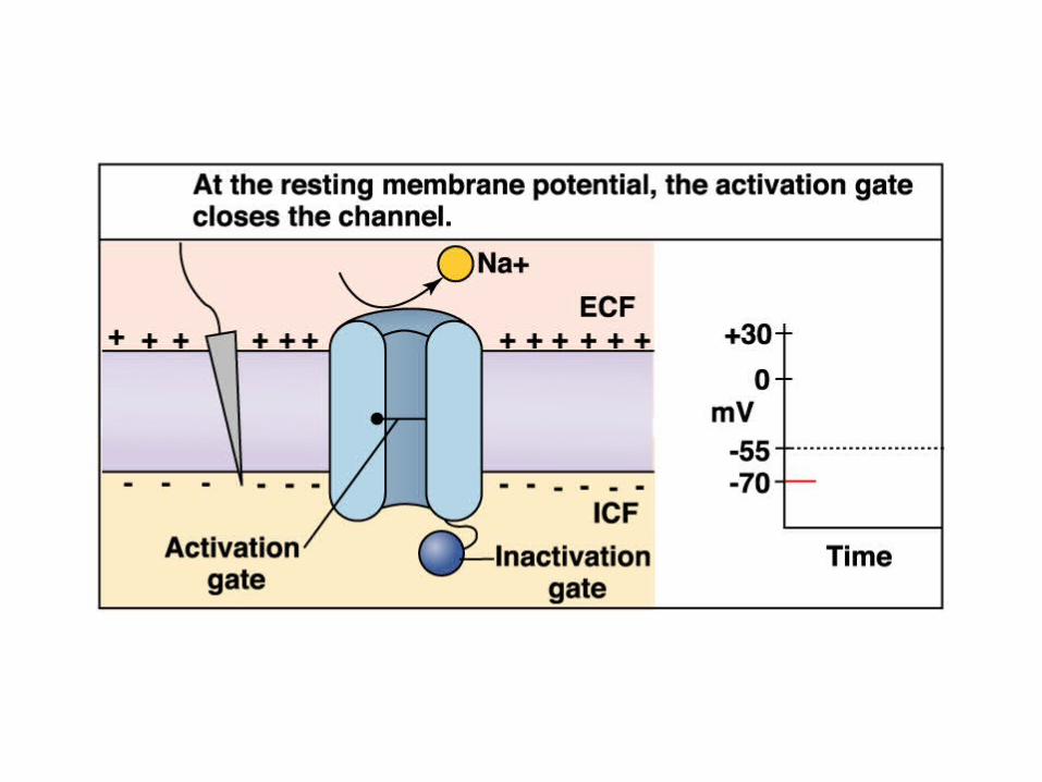
Fig 8.10 a – Model of the voltage-gated Na+ channel Silverthorn 2nd Ed

Fig 8.10 b – Model of the voltage-gated Na+ channel Silverthorn 2nd Ed
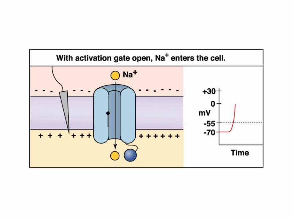
Fig 8.10 c – Model of the voltage-gated Na+ channel Silverthorn 2nd Ed

Fig 8.10 d – Model of the voltage-gated Na+ channel Silverthorn 2nd Ed
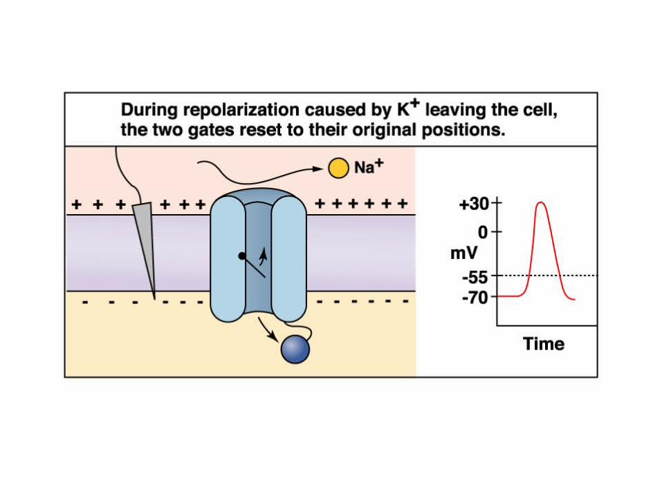
Fig 8.10 e – Model of the voltage-gated Na+ channel Silverthorn 2nd Ed
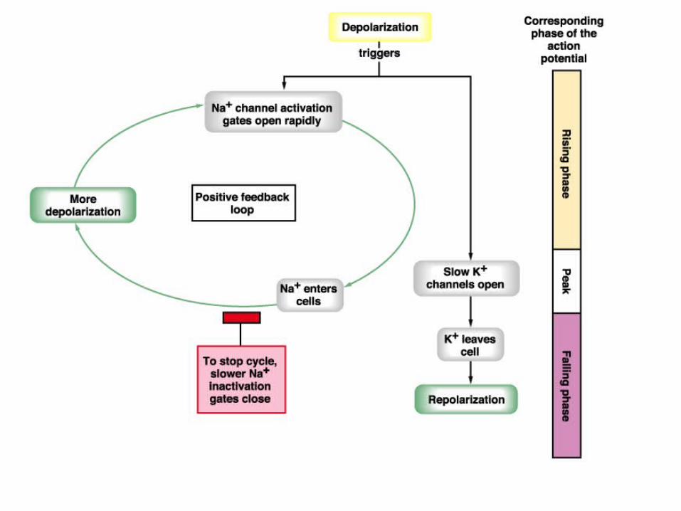
Fig 8.11 – Ion movements during the action potential Silverthorn 2nd Ed

Refractory PeriodRefractory Period Double gating of Na+ channel leads to Double gating of Na+ channel leads to
refractory periodrefractory period Absolute refractory periodAbsolute refractory period
Once an AP has begun, for about 1 ms, a 2Once an AP has begun, for about 1 ms, a 2ndnd AP can’t be generatedAP can’t be generated
Relative refractory periodRelative refractory period After NaAfter Na++ channel gates have been reset, but channel gates have been reset, but
before Vbefore Vmm has returned to normal, a STRONG has returned to normal, a STRONG graded potential can start a 2graded potential can start a 2ndnd AP AP
Graded potential opens NaGraded potential opens Na++ channels, but channels, but NaNa++ entry is offset by continuing K entry is offset by continuing K++ loss loss through Kthrough K++ channels that are still open channels that are still open

Fig 8.12 Refractory periods Silverthorn 2nd Ed
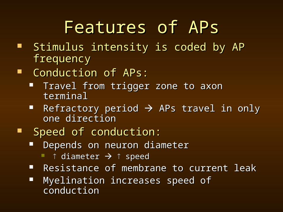
Features of APsFeatures of APs Stimulus intensity is coded by AP Stimulus intensity is coded by AP
frequencyfrequency Conduction of APs:Conduction of APs:
Travel from trigger zone to axon terminalTravel from trigger zone to axon terminal Refractory period Refractory period APs travel in only APs travel in only
one directionone direction Speed of conduction:Speed of conduction:
Depends on neuron diameter Depends on neuron diameter diameter diameter speed speed
Resistance of membrane to current leakResistance of membrane to current leak Myelination increases speed of Myelination increases speed of
conductionconduction
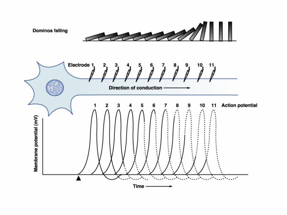
Fig 8.14 – Action potentials along an axon Silverthorn 2nd Ed
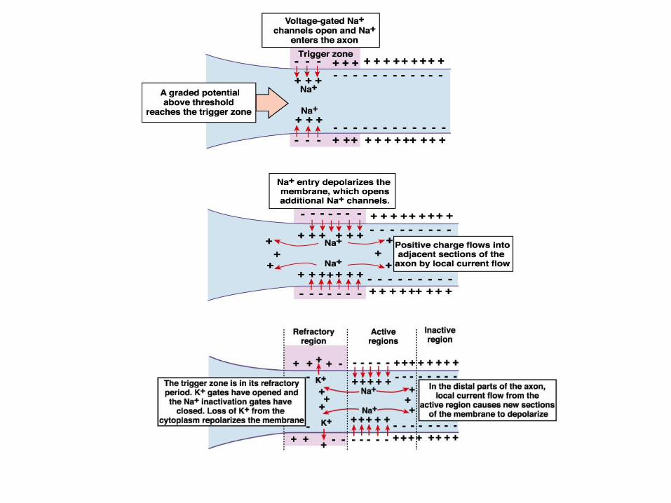
Fig 8.15 – Conduction of action potentials Silverthorn 2nd Ed
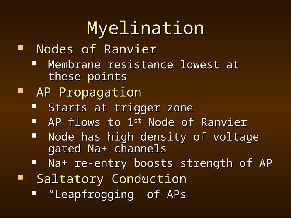
MyelinationMyelination Nodes of RanvierNodes of Ranvier
Membrane resistance lowest at these Membrane resistance lowest at these pointspoints
AP PropagationAP Propagation Starts at trigger zoneStarts at trigger zone AP flows to 1AP flows to 1stst Node of Ranvier Node of Ranvier Node has high density of voltage gated Node has high density of voltage gated
Na+ channelsNa+ channels Na+ re-entry boosts strength of APNa+ re-entry boosts strength of AP
Saltatory ConductionSaltatory Conduction ““Leapfrogging” of APsLeapfrogging” of APs
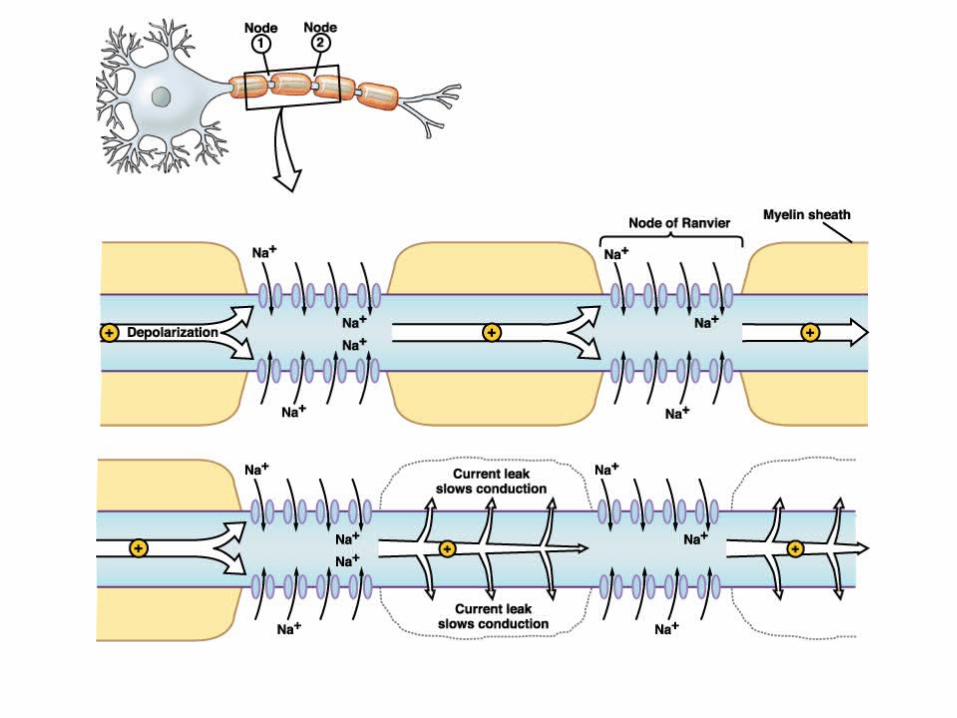
Fig 8.17 – Saltatory conduction Silverthorn 2nd Ed

SummarySummary
Organization of Nervous SystemOrganization of Nervous System Constituent Cells of Nervous SystemConstituent Cells of Nervous System Electrical Signals in NeuronsElectrical Signals in Neurons
Source of Resting Membrane PotentialSource of Resting Membrane Potential Gated Ion ChannelsGated Ion Channels
Qualitative Description of Action Qualitative Description of Action PotentialPotential Graded PotentialGraded Potential Action Potential Action Potential Refractory PeriodRefractory Period

Poem of the DayPoem of the Day
I Chop Some Parsley While Listening I Chop Some Parsley While Listening to Art Blakey’s Version of “Three to Art Blakey’s Version of “Three Blind Mice”Blind Mice” Billy CollinsBilly Collins
http://www.cduniverse.com/search/xx/mhttp://www.cduniverse.com/search/xx/music/pid/1230695/a/Three+Blind+Mice,+usic/pid/1230695/a/Three+Blind+Mice,+Vol+1.htmVol+1.htm

Due DatesDue Dates
Tuesday, October 5Tuesday, October 5thth HW5HW5