lecture 1,2,3
TRANSCRIPT

Purification of DNA Purification of DNA from Living Cellsfrom Living Cells

Purpose of DNA ExtractionPurpose of DNA ExtractionTo obtain DNA in a relatively purified form which can be used for further investigations, i.e. PCR, sequencing, etc.

Preparation of total cell DNA
Preparation of plasmid DNA Preparation of bacteriophage DNA

Basic ProtocolBasic Protocol Most DNA extraction protocols consist of two parts
1. A technique to lyse the cells gently and solubilize the DNA
2. Enzymatic or chemical methods to remove contaminating proteins, RNA, or macromolecules

Preparation of total cell Preparation of total cell DNADNA
The procedure for total DNA preparation from a culture of bacterial cells can be divided into four stages
1. A culture of bacteria is grown and then harvested.
2. The cells are broken open to release their contents.
3. This cell extract is treated to remove all components except the DNA.
4. The resulting DNA solution is concentrated.


Growing and harvesting a Growing and harvesting a bacterial culturebacterial cultureMost bacteria can be grown without too
much difficulty in a liquid medium (broth culture).
M9 is an example of a defined medium, in which all the components are known
Luria-Bertani (LB) is a complex or undefined medium, meaning that the precise identity and quantity of its components are not known.

Preparation of a cell Preparation of a cell extractextractTechniques for breaking open bacterial
cells can be divided into physical methods, in which the cells are disrupted by mechanical forces, and chemical methods, where cell lysis is brought about by exposure to chemical agents that affect the integrity of the cell barriers. Chemical methods are most commonly used with bacterial cells when the object is DNA preparation

LYSIS:
refers to the breaking of the cell wall and cellular membranes refers to the breaking of the cell wall and cellular membranes (esp, the plasma and nuclear membranes(esp, the plasma and nuclear membranes))
The cell wall is disrupted by mechanical force
Chemical lysis generally involves one agent attacking the cell wall and another disrupting the cell membrane
For cell wall lysis: lysozyme or ethylenediamine tetraacetate (EDTA), or a combination of both
For cell membrane lysis: sodium dodecyl sulphate (SDS)

Preparation of Cell extractPreparation of Cell extract
•Having lysed the cells, the final step in preparation of a cell extract is removal of insoluble cell debris. Components such as partially digested cell wall fractions can be pelleted by centrifugation ,leaving the cell extract as a reasonably clear supernatant
1. Lysozyme: digests the polymeric compounds that give the cell wall its rigidity
2. EDTA: remove magnesium, that are essential for preserving the overall structure of the cell envelope, and also inhibits cellular DNase
3. SDS: removes lipid molecules and thereby cause disruption of the cell membranes

Preparation of cell extractPreparation of cell extract

Purification of DNA from a cell extractIn addition to DNA, a bacterial cell extract
contains significant quantities of protein and RNA. Either:
1. treat the mixture with reagents which degrade the contaminants, leaving a pure solution of DNA
2. use ion-exchange chromatography to separate the mixture into its various components, so the DNA is removed from the proteins and RNA in the extract

Figure 3.5Two approaches to DNA purification. (a) Treating the mixture with reagents which degrade the contaminants, leaving a pure solution of DNA. (b) Separating the mixture into different fractions, one of which is pure DNA.

1. Removing contaminants by organic extraction and enzyme digestion
The standard way to deproteinize a cell extract is to add phenol or a 1 : 1 mixture of phenol and chloroform.
These organic solvents precipitate proteins but leave the nucleic acids (DNA and RNA) in aqueous solution.

Removal of protein by Phenol Removal of protein by Phenol ExtractionExtraction
(or phenol :chloroform = 1:1)
•Protease (pronase or proteinase K) before phenol extraction•Ribonuclease degrades RNA

Phenol Phenol extraction(continued…)extraction(continued…)cell extracts with high protein content needs
several phenol extractions because a single phenol extraction is not sufficient to completely purify the nucleic acids
But each mixing and centrifugation step results in a certain amount of breakage of the DNA molecules
treat the cell extract with a protease such as pronase or proteinase K before phenol extraction. These enzymes break polypeptides down into smaller units, which are more easily removed by phenol

Some RNA molecules, especially messenger RNA (mRNA), are removed by phenol treatment, but most remain with the DNA in the aqueous layer. The only effective way to remove the RNA is with the enzyme ribonuclease, which rapidly degrades these molecules into ribonucleotide subunits

2. Using ion-exchange chromatography to purify DNA from a cell extraction-exchange chromatography separates
molecules according to how tightly they bind to electrically charged particles present in a chromatographic matrix or resin.
The simplest way to carry out ion-exchange chromatography is to place the resin in a glass or plastic column and then add the cell extract to the top
If the extract contains very little salt ,all the negatively charged molecules bind to the resin and are retained in the column

Figure 3.7DNA purification by ion-exchange chromatography. (a) Attachment of DNA to ion-exchange particles. (b) DNA is purifiedby column chromatography. The solutions passing through the column can be collected by gravity flow or by the spincolumn method, in which the column is placed in a low-speed centrifuge.

If the salt solution of gradually increasing concentration is now passed through the column, the different types of molecule will elute (i.e., become unbound) in the sequence protein, RNA, and finally DNA.
If two salt solutions are used, one whose concentration is sufficient to elute the protein and RNA, leaving just the DNA bound, followed by a second of a higher concentration which elutes the DNA

Concentration of DNA samplesThe most frequently used method of
concentration is ethanol precipitation. In the presence of salt (strictly speaking,
monovalent cations such as sodium ions (Na+)), and at a temperature of −20°C or less, absolute ethanol efficiently precipitates polymeric nucleic acids.
With a thick solution of DNA the ethanol can be layered on top of the sample, causing molecules to precipitate at the interface.

Collecting DNA by ethanol precipitation. (a) Absolute ethanol is layered on top of a concentrated solution of DNA. Fibers of DNA can be withdrawn with a glass rod. (b) For less concentrated solutions ethanol is added (at a ratio of 2.5 volumes of absolute ethanol to 1 volume of DNA solution) and precipitated DNA collected by centrifugation.
Ethanol precipitationMonovalent cation (Na+)-20 ℃ or lessAbsolute ethanol
Concentration of DNA samples

Measurement of DNA concentrationDNA concentrations can be accurately
measured by ultraviolet (UV) absorbance spectrophotometry at 260 nm
The amount of UV radiation absorbed by a solution of DNA is directly proportional to the amount of DNA in the sample

Methods for the preparation of total cell DNA from plants

Plant tissues are particularly difficult in this respect as they often contain large amounts of carbohydrates that are not removed by phenol extraction. Instead a different approach must be used. One method makes use of a detergent called cetyltrimethylammonium bromide (CTAB), which forms an insoluble complex with nucleic acids.

When CTAB is added to a plant cell extract the nucleic acid–CTAB complex precipitates, leaving carbohydrate, protein, and other contaminants in the supernatant
The precipitate is then collected by centrifugation and resuspended in 1 M sodium chloride, which causes the complex to break down.
The nucleic acids can now be concentrated by ethanol precipitation and
the RNA removed by ribonuclease treatment

Purification of DNA for any Purification of DNA for any speciesspecies

Preparation of plasmid DNA there is an important distinction between plasmid
purification and preparation of total cell DNA. In a plasmid preparation it is always necessary to
separate the plasmid DNA from the large amount of bacterial chromosomal DNA that is also present in the cells.
The methods are based on the several physical differences between plasmid DNA and bacterial DNA
1. plasmids and bacterial DNA differ in size2. plasmids and bacterial DNA differ in conformation

Separation on the basis of size The largest plasmids are only 8% of the size of the E. coli
chromosome, and most are much smaller than this. Techniques that can separate small DNA molecules from large ones should therefore effectively purify plasmid DNA.
If the cells are lysed under very carefully controlled conditions, only a minimal amount of chromosomal DNA breakage occurs.
The resulting DNA fragments are still very large—much larger than the plasmids—and can be removed with the cell debris by centrifugation.
the bacterial chromosome is physically attached to the cell envelope, so fragments of the chromosome sediment with the cell debris if these attachments are not broken

Cell disruption carried out very gently to prevent wholesale breakage of the bacterial DNA.
Treatment with EDTA and lysozyme in the presence of sucrose, prevents the cells from bursting immediately.
Sphaeroplasts are formed, cells with partially degraded cell walls that retain an intact cytoplasmic membrane.
Cell lysis is now induced by adding a non-ionic detergent such as Triton X-100 (ionic detergents, such as SDS, cause chromosomal breakage).
This method causes very little breakage of the bacterial DNA, so centrifugation leaves a cleared lysate, consisting almost entirely of plasmid DNA.
If the plasmids themselves are large molecules, they may also sediment with the cell debris.


Separation on the basis of conformation
The term conformation refers to the overall spatial configuration of the molecule, with the two simplest conformations being linear and circular
Plasmids and the bacterial chromosome are circular, but during preparation of the cell extract the chromosome is always broken to give linear fragments. A method for separating circular from linear molecules will therefore result in pure plasmids.


Most plasmids exist in the cell as supercoiled Molecules Supercoiling occurs because the double helix
of the plasmid DNA is partially unwound during the plasmid replication process by enzymes called topoisomerases
The supercoiled conformation can be maintained only if both polynucleotide strands are intact, hence the more technical name of covalently closed circular (ccc) DNA.
If one of the polynucleotide strands is broken the double helix reverts to its normal relaxed state, and the plasmid takes on the alternative conformation, called open-circular (oc)

Supercoiling is important in plasmid preparation because supercoiled molecules can be fairly easily separated from non-supercoiled DNA
1. Alkaline denaturation2. Ethidium bromide–caesium chloride density
gradient centrifugation

Alkaline denaturation
The basic of the technique is the use of narrow pH range at which non-supercoiled DNA is denatured, whereas supercoiled plasmids are not.
If sodium hydroxide is added to a cell extract or cleared lysate, so that the pH is adjusted to 12.0–12.5, then the hydrogen bonding in non-supercoiled DNA molecules is broken, causing the double helix to unwind and the two polynucleotide chains to separate
If acid is now added, these denatured bacterial DNA strands reaggregate into a tangled mass.
The insoluble network can be pelleted by centrifugation, leaving plasmid DNA in the supernatant


Ethidium bromide–caesium chloride density gradient centrifugationA density gradient is produced by centrifuging a
solution of caesium chloride (CsCl) at a very high speedMacromolecules present in the CsCl solution when it is
centrifuged form bands at distinct points in the gradientExactly where a particular molecule bands depends on
its buoyant density DNA has a buoyant density of about 1.70 g/cm3, and
therefore migrates to the point in the gradient where the CsCl density is also 1.70 g/cm3. In contrast, protein molecules have much lower buoyant densities, and so float at the top of the tube, whereas RNA forms a pellet at the bottom

density gradient centrifugation in the presence of ethidium bromide (EtBr) can be used to separate supercoiled DNA from non-supercoiled molecules.
Ethidium bromide binds to DNA molecules by intercalating between adjacent base pairs, causing partial unwinding of the double helix
This unwinding results in a decrease in the buoyant density, by as much as 0.125 g/cm3 for linear DNA
Supercoiled DNA, with no free ends, has very little freedom to unwind, and can only bind a limited amount of EtBr.
The decrease in buoyant density of a supercoiled molecule is therefore much less, only about 0.085 g/cm3. As a consequence, supercoiled molecules form a band in an EtBr–CsCl gradient at a different position to linear and open-circular DNA


When a cleared lysate is subjected to this procedure, plasmids band at a distinct point, separated from the linear bacterial DNA, with the protein floating on the top of the gradient and RNA pelleted at the bottom.
The position of the DNA bands can be seen by shining ultraviolet radiation on the tube, which causes the bound EtBr to fluoresce.
The pure plasmid DNA is removed by puncturing the side of the tube and withdrawing a sample with a syringe
The EtBr bound to the plasmid DNA is extracted with n-butanol and the CsCl removed by dialysis .
The resulting plasmid preparation is virtually 100% pure and ready for use as a cloning vector.

Plasmid amplification Plasmid amplification offers a means of increasing the yield. Some multi copy plasmids (those with copy numbers of 20 or
more) have the useful property of being able to replicate in the absence of protein synthesis.
an inhibitor of protein synthesis(e.g., chloramphenicol) is added, and the culture incubated for a further 12 hours.
During this time the plasmid molecules continue to replicate, even though chromosome replication and cell division are blocked .
The result is that plasmid copy numbers of several thousand may be attained. Amplification is therefore a very efficient way of increasing the yield of multi copy plasmids

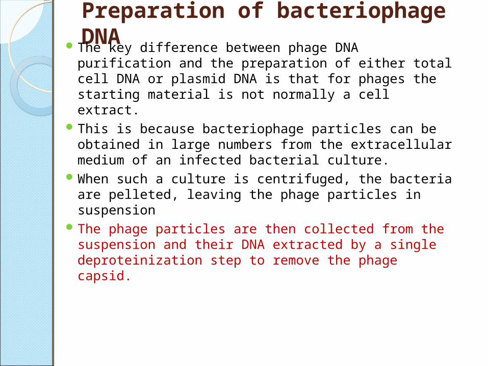
Preparation of bacteriophage DNAThe key difference between phage DNA purification and
the preparation of either total cell DNA or plasmid DNA is that for phages the starting material is not normally a cell extract.
This is because bacteriophage particles can be obtained in large numbers from the extracellular medium of an infected bacterial culture.
When such a culture is centrifuged, the bacteria are pelleted, leaving the phage particles in suspension
The phage particles are then collected from the suspension and their DNA extracted by a single deproteinization step to remove the phage capsid.

Growth of cultures to obtain a high λ titer
The naturally occurring λ phage is lysogenic ,and an infected culture consists mainly of cells carrying the prophage integrated into the bacterial DNA .The extracellular titer is extremely low under these circumstance.
To get a high yield of extracellular λ, the culture must be induced, so that all the bacteria enter the lytic phase of the infection cycle, resulting in cell death and release of λ particles into the medium.

Induction is normally very difficult to control, but most laboratory strains of carry a temperature-sensitive (ts) mutation in the cI gene.
This is one of the genes that are responsible for maintaining the phage in the integrated state.
If inactivated by a mutation, the cI gene no longer functions correctly and the switch to lysis occurs.
the cI gene is functional at 30°C, at which temperature normal lysogeny can occur. But at 42°C, the cI gene product does not work properly, and lysogeny cannot be maintained.


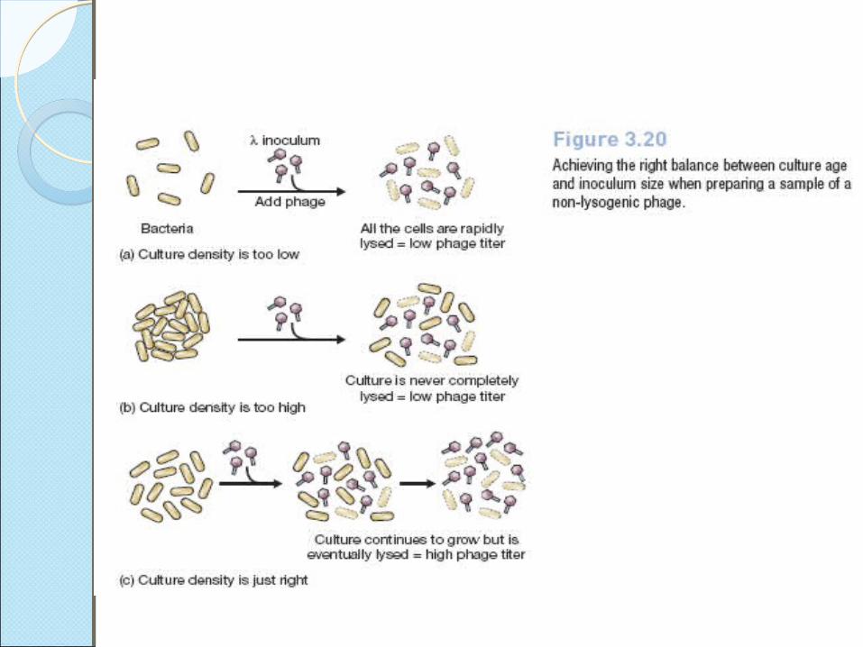
Preparation of non-lysogenic λ phages Although most strains are lysogenic, many cloning vectors
derived from λ are modified, by deletions of the cI and other genes, so that lysogeny never occurs. These phages cannot integrate into the bacterial genome and can infect cells only by a lytic cycle.
to obtain a high titer of phages, lies in the way in which the culture is grown, in particular the stage at which the cells are infected by adding phage particles.
If phages are added before the cells are dividing at their maximal rate, then all the cells are lysed very quickly, resulting in a low titer .
On the other hand, if the cell density is too high when the phages are added, then the culture will never be completely lysed, and again the phage titer will be low

The ideal situation is when the age of the culture, and the size of the phage inoculum, are balanced such that the culture continues to grow, but eventually all the cells are infected and lysed

Collection of phages from an infected cultureThe remains of lysed bacterial cells, along with
any intact cells that are can be removed from an infected culture by centrifugation, leaving the phage particles in suspension
Phage particles are so small that they are pelleted only by very high speed centrifugation.
Collection of phages is therefore usually achieved by precipitation with polyethylene glycol (PEG).

PEG is a long-chain polymeric compound which, in the presence of salt, absorbs water, thereby causing macromolecular assemblies such as phage particles to precipitate.
The precipitate can then be collected by centrifugation, and redissolved in a suitably small volume

Purification of DNA from λ phage particles Deproteinization of the redissolved PEG precipitate is sometimes
sufficient to extract pure phage DNA, but usually phages are subjected to an intermediate purification step.
This is necessary because the PEG precipitate also contains a certain amount of bacterial debris, possibly including unwanted cellular DNA.
These contaminants can be separated from the λ particles by CsCl density gradient centrifugation.
The λ particles band in a CsCl gradient at 1.45–1.50 g/cm3 and can be withdrawn from the gradient
Removal of CsCl by dialysis leaves a pure phage preparation from which the DNA can be extracted by either phenol or protease treatment to digest the phage protein coat.


Purification of M13 DNAPurification of M13 DNA