lecture-2 - bbau.ac.in › dept › dem › tm › lecture 2.pdf · lecture-2 m.sc 2nd semester...
TRANSCRIPT
Lecture-2M.Sc 2nd Semester (Environmental Microbiology)
Paper EM-202: Microbial physiology and adaptation
Unit IV: Basic Concepts of Radiation
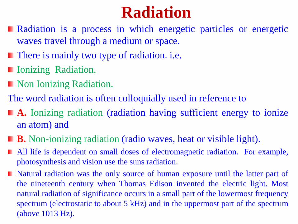
RadiationRadiation is a process in which energetic particles or energetic
waves travel through a medium or space.
There is mainly two type of radiation. i.e.
Ionizing Radiation.
Non Ionizing Radiation.
The word radiation is often colloquially used in reference to
A. Ionizing radiation (radiation having sufficient energy to ionize
an atom) and
B. Non-ionizing radiation (radio waves, heat or visible light).
All life is dependent on small doses of electromagnetic radiation. For example,
photosynthesis and vision use the suns radiation.
Natural radiation was the only source of human exposure until the latter part of
the nineteenth century when Thomas Edison invented the electric light. Most
natural radiation of significance occurs in a small part of the lowermost frequency
spectrum (electrostatic to about 5 kHz) and in the uppermost part of the spectrum
(above 1013 Hz).
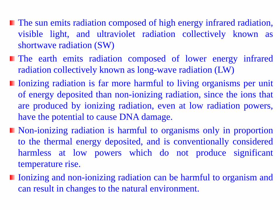
The sun emits radiation composed of high energy infrared radiation,
visible light, and ultraviolet radiation collectively known as
shortwave radiation (SW)
The earth emits radiation composed of lower energy infrared
radiation collectively known as long-wave radiation (LW)
Ionizing radiation is far more harmful to living organisms per unit
of energy deposited than non-ionizing radiation, since the ions that
are produced by ionizing radiation, even at low radiation powers,
have the potential to cause DNA damage.
Non-ionizing radiation is harmful to organisms only in proportion
to the thermal energy deposited, and is conventionally considered
harmless at low powers which do not produce significant
temperature rise.
Ionizing and non-ionizing radiation can be harmful to organism and
can result in changes to the natural environment.
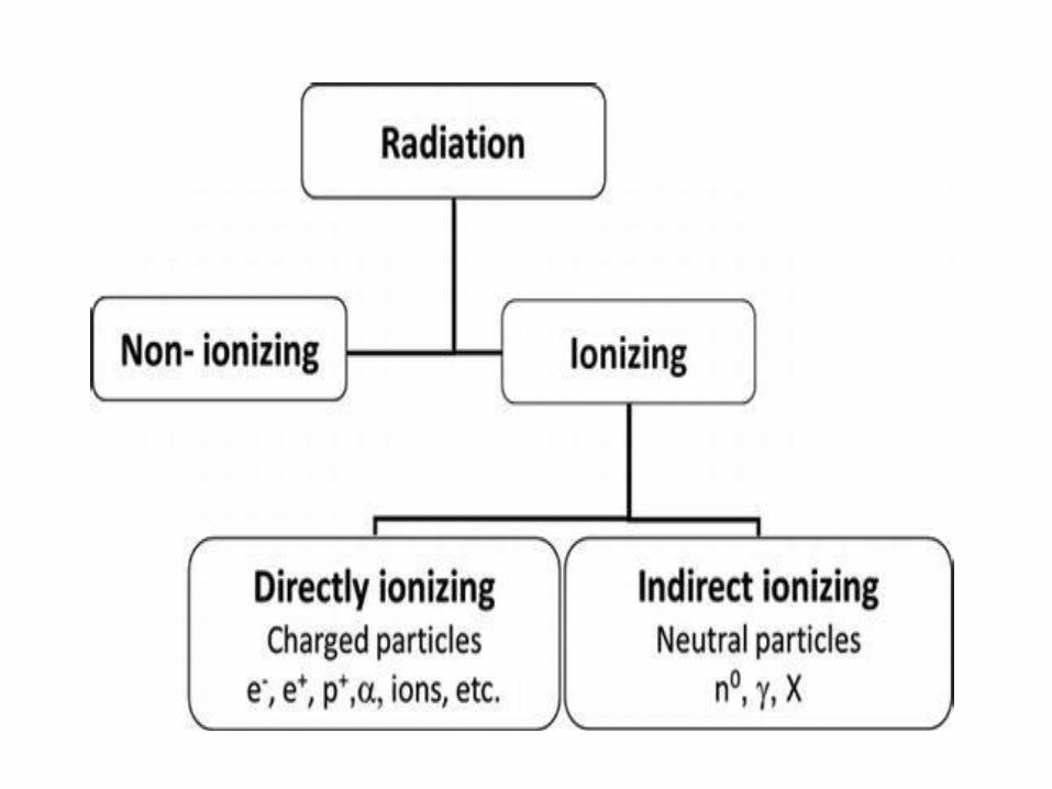
Ionizing RadiationIt is a type of radiation that is able to disrupt atoms and molecules
on which they pass through, giving rise to ions and free radicals.
It has enough energy to eject one or more electrons from the
atoms or molecules in the irradiated medium.
Ionizing radiation is produced by unstable atoms. Unstable atoms
differ from stable atoms because they have an excess of energy
and mass.
Each ionization releases approximately 33 electron volts (eV) of
energy. Material surrounding the atom absorbs the energy.
Compared to other types of radiation that may be absorbed,
ionizing radiation deposits a large amount of energy into a small
area. In fact, the 33 eV from one ionization is more than enough
energy to disrupt the chemical bond between two carbon atoms.
All ionizing radiation is capable, directly or indirectly, of
removing electrons from most molecules.
Ionizing Radiation cont.Higher frequency ultraviolet radiation begins to have enough
energy to break chemical bonds. X-ray and gamma ray radiation,
which are at the upper end of magnetic radiation have very high
frequency in the range of 100 billion billion Hertz and very short
wavelengths 1 million millionth of a meter. Radiation in this range
has extremely high energy. It has enough energy to strip off
electrons or, in the case of very high-energy radiation, break up the
nucleus of atoms.
Ionization is the process in which a charged portion of a molecule
(usually an electron) is given enough energy to break away from
the atom. This process results in the formation of two charged
particles or ions: the molecule with a net positive charge, and the
free electron with a negative charge.
Sources of Ionizing Radiation
Sources – x-rays, radioactive material produce alpha, beta, and
gamma radiation, cosmic rays from the sun and space.
Ionizing radiation comes from radioactive materials, X-ray
tubes, particle accelerators, and is present in the environment.
Present in uppermost part of the frequency spectrum (above 10
THz or 1013 Hz) and include IR infrared (heat), visible light,
and ionizing radiation such as ultraviolet (UV), X-rays, gamma
rays and cosmic rays.
Types of Ionizing Radiation
Alpha particles
Beta particles
Gamma rays (or photons)
X-Rays (or photons)
Neutrons
cosmic rays
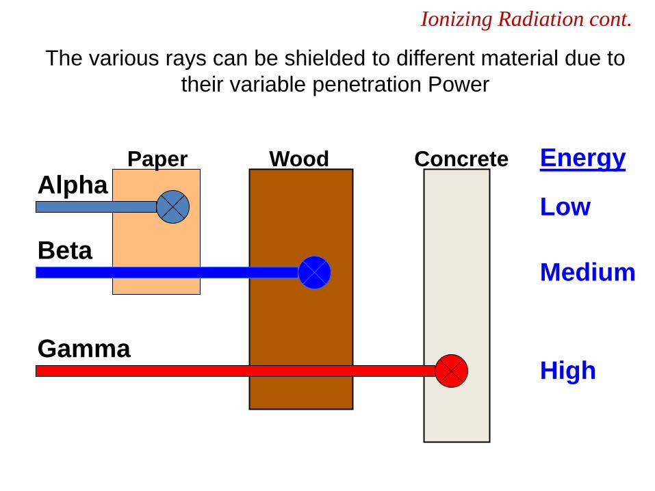
Ionizing Radiation cont.
Paper Wood Concrete
Alpha
Beta
Gamma
Energy
Low
Medium
High
The various rays can be shielded to different material due to
their variable penetration Power
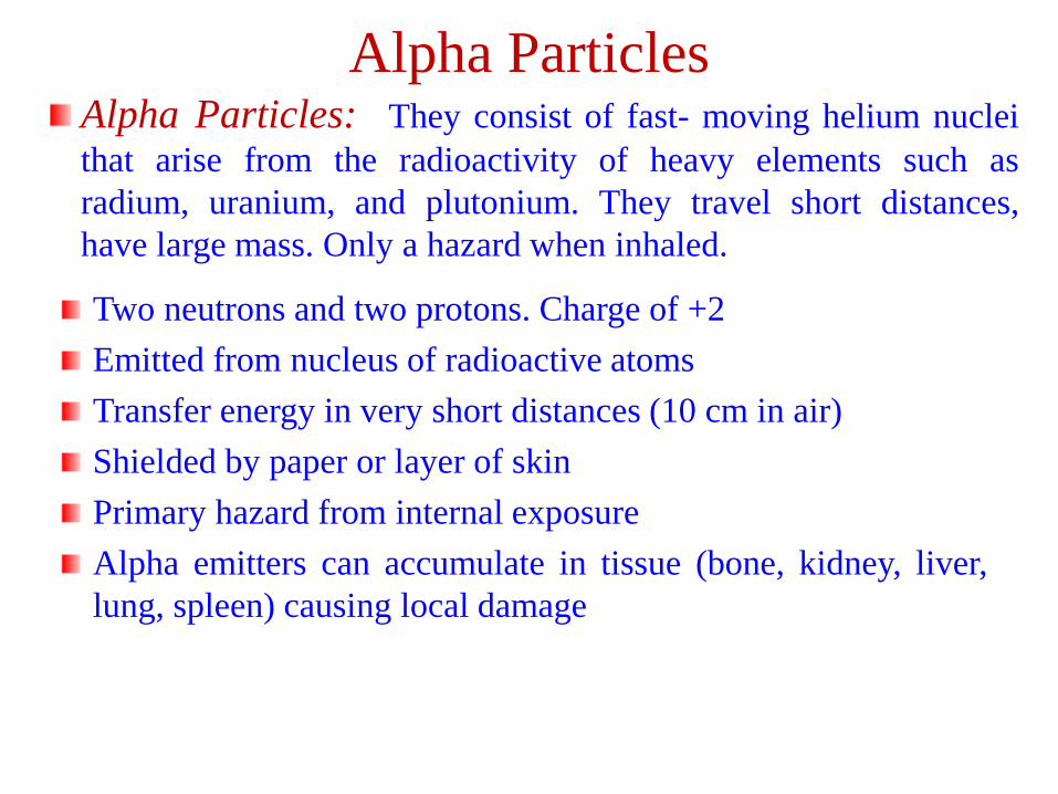
Alpha Particles: They consist of fast- moving helium nuclei
that arise from the radioactivity of heavy elements such as
radium, uranium, and plutonium. They travel short distances,
have large mass. Only a hazard when inhaled.
Alpha Particles
Two neutrons and two protons. Charge of +2
Emitted from nucleus of radioactive atoms
Transfer energy in very short distances (10 cm in air)
Shielded by paper or layer of skin
Primary hazard from internal exposure
Alpha emitters can accumulate in tissue (bone, kidney, liver,
lung, spleen) causing local damage
Beta Particles
Beta Particles: are streams of high speed electrons that can
arise from the disintegration of any radioactive nuclei. Primarily
an internal hazard, but high beta can be an external hazard to
skin. In addition, the high speed electrons may lose energy in the
form of X-rays when they quickly decelerate upon striking a heavy
material. This is called Breaking Radiation.
Small electrically charged particles similar to electrons
Charge of -1
Ejected from nuclei of radioactive atoms
Emitted with various kinetic energies
Shielded by wood, body penetration 0.2 to 1.3 cm depending on
energy
Can cause skin burns or be an internal hazard of ingested
Gamma Rays
Gamma Rays (or photons): are penetrating type of radiation
emitted by the nucleus of a radionuclide when it disintegrates. They
are similar to X-rays and ordinary light, but usually much more
energetic.
Electromagnetic photons or radiation (identical to x-rays except for
source)
Emitted from nucleus of radioactive atoms – spontaneous emission
Emitted with kinetic energy related to radioactive source
Highly penetrating – extensive shielding required
Serious external radiation hazard
Gamma rays are photons emitted from the nucleus, often as part of
radioactive decay. Gamma rays typically have higher energy than
X-rays.
X-raysX-Rays: Occur whenever an inner shell orbital electron isremoved and rearrangement of the atomic electrons results withthe release of the elements characteristic X-Ray energy.
Overlap with gamma-rays
Electromagnetic photons or radiation
Produced from orbiting electrons or free electrons – usuallymachine produced
Produced when electrons strike a target material inside and x-raytube
Emitted with various energies & wavelengths
Highly penetrating – extensive shielding required
External radiation hazard
Discovered in 1895 by Roentgen
X-rays are photons (Electromagnetic radiations) emitted fromelectron orbits.
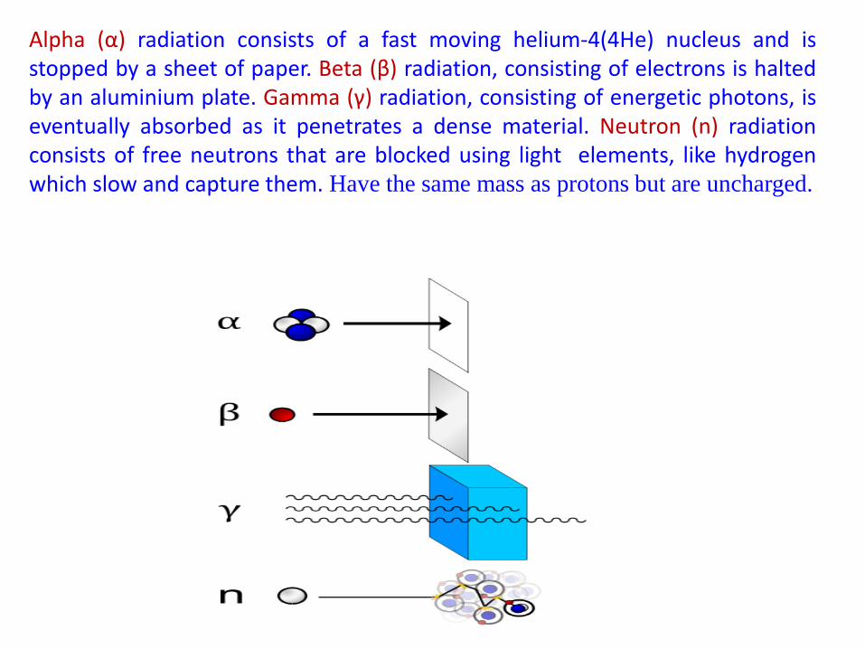
Alpha (α) radiation consists of a fast moving helium-4(4He) nucleus and isstopped by a sheet of paper. Beta (β) radiation, consisting of electrons is haltedby an aluminium plate. Gamma (γ) radiation, consisting of energetic photons, iseventually absorbed as it penetrates a dense material. Neutron (n) radiationconsists of free neutrons that are blocked using light elements, like hydrogenwhich slow and capture them. Have the same mass as protons but are uncharged.
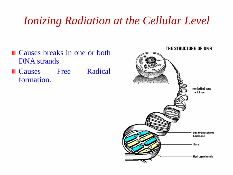
Ionizing Radiation at the Cellular Level
Causes breaks in one or bothDNA strands.
Causes Free Radicalformation.
All forms of radiation can have adverse health effects on cellularlevel when intense enough and/or time exposure long enough.
❖Generalizations: Biological effects are due to the ionizationprocess that destroys the capacity for cell reproduction or divisionor causes cell mutation. A given total dose will cause more damageif received in a shorter time period. A fatal dose is (600 R)
❖Acute Somatic Effects: Relatively immediate effects to a personacutely exposed. Severity depends on dose. Death usually resultsfrom damage to bone marrow or intestinal wall. Acute radio-dermatitis is common in radiotherapy; chronic cases occur mostlyin industry.
❖Delayed Somatic Effects: Delayed effects to exposed personinclude: Cancer, leukemia, cataracts, life shortening from organfailure, and abortion. Probability of an effect is proportional todose (no threshold). Severity is independent of dose. Doublingdose for cancer is approximately 10-100 rems.
Health effects of Ionizing Radiation
❖Genetic Effects: Genetic effects to off-spring of exposed
persons are irreversible and nearly always harmful. Doubling
dose for mutation rate is approximately 50-80 rems.
(Spontaneous mutation rate is approx. 10-100 mutations per
million population per generation.)
❖Critical Organs: Organs generally most susceptible to radiation
damage include: Lymphocytes, bone marrow, gastro-intestinal,
gonads, and other fast-growing cells. The central nervous system
is relatively resistant. Many nuclides concentrate in certain
organs rather than being uniformly distributed over the body, and
the organs may be particularly sensitive to radiation damage, e.g.,
isotopes of iodine concentrate in the thyroid gland. These organs
are considered "critical" for the specific nuclide.
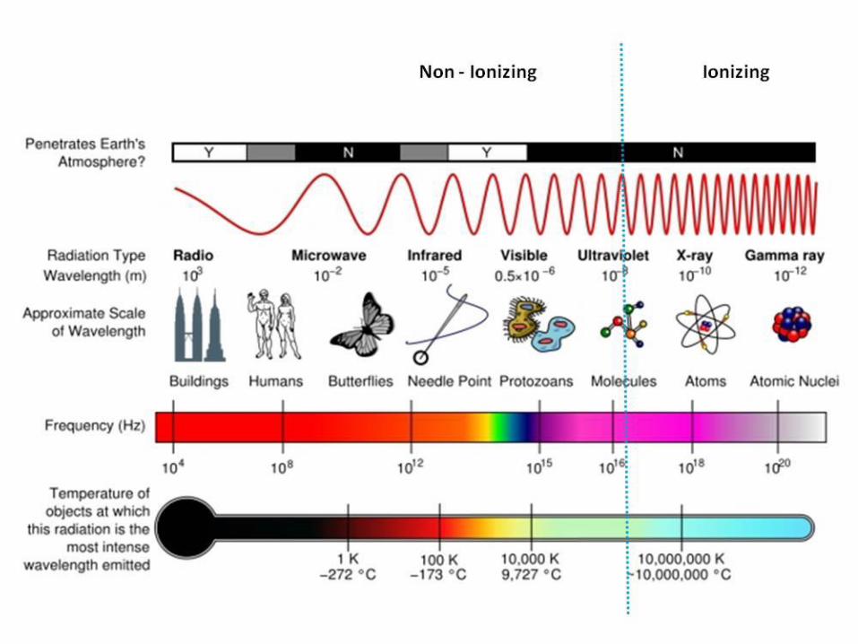
Non-ionizing RadiationNon-ionizing radiation ranges from extremely low frequencyradiation, shown on the far left through the audible, microwave, andvisible portions of the spectrum into the ultraviolet range.
advantage of the properties of non-ionizing radiation
microwave radiation tele-communications and heating food
infrared radiation infrared lamps to keep food warm inrestaurants
radio waves broadcasting
They are electromagnetic waves incapable of producing ions whilepassing through matter, due to their lower energy.
Extremely low-frequency radiation has very long wave lengths (onthe order of a million meters or more) and frequencies in the rangeof 100 Hertz or cycles per second or less. Radio frequencies havewave lengths of between 1 and 100 meters and frequencies in therange of 1 million to 100 million Hertz. Microwaves that we use toheat food have wavelengths that are about 1 hundredth of a meterlong and have frequencies of about 2.5 billion Hertz.
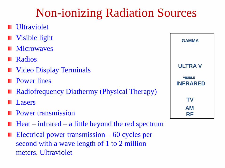
Non-ionizing Radiation SourcesUltraviolet
Visible light
Microwaves
Radios
Video Display Terminals
Power lines
Radiofrequency Diathermy (Physical Therapy)
Lasers
Power transmission
Heat – infrared – a little beyond the red spectrum
Electrical power transmission – 60 cycles per
second with a wave length of 1 to 2 million
meters. Ultraviolet
MICROWAVE
GAMMA
ULTRA V
VISIBLE
INFRARED
TV
AMRF
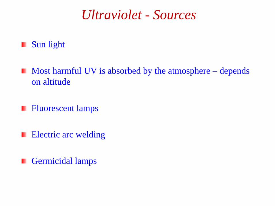
Ultraviolet - Sources
Sun light
Most harmful UV is absorbed by the atmosphere – depends
on altitude
Fluorescent lamps
Electric arc welding
Germicidal lamps
Ultraviolet - Effects
High ultraviolet – kills bacterial and other infectious agents
High dose causes - sun burn – increased risk of skin cancer
Pigmentation that results in suntan
Suntan lotions contain chemicals that absorb UV radiation
Reaction in the skin to produce Vitamin D that prevents
rickets
Strongly absorbed by air – thus the danger of hole in the
atmosphere
Skin cancer
Eye damage from sun (cornea)
Visible EnergyEnergy between 400 and 750 nm
High energy – bright light produces of number of adaptive
responses
Standards are set for the intensity of light in the work place
(measured in candles or lumens)
Infrared RadiationEnergy between 750 nm to 0.3 cm
The energy of heat – Heat is the transfer of energy
Can damage – cornea, iris, retina and lens of the eye (glass workers
– “glass blower’s cataract”)
Microwaves & Radio Waves
Energy between 0.1 cm to 1 kilometer
Varity of industrial and home uses for heating and information
transfer (radio, TV, mobile phones)
Produced by molecular vibration in solid bodies or crystals
Health effects – heating, cataracts
Long-term effects being studied
Effects Non-ionizing Radiation
Radiofrequency Ranges (10 kHz to 300 GHz)
Effects only possible at ten times the permissible exposure limit
Heating of the body (thermal effect)
Cataracts
Some studies show effects of teratoginicity and carcinogenicity.
UV radiation
Stimulates melanin (dark pigment) that absorbs UV protecting cells.
2 to 3 million non-malignant skin cancers
130,000 malignant melanomas
Sunburn – acute cell injury causing inflammatory response (erythema)
Accelerates aging process
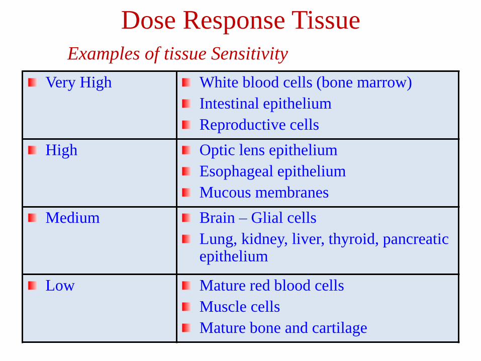
Dose Response Tissue
Examples of tissue Sensitivity
Very High White blood cells (bone marrow)
Intestinal epithelium
Reproductive cells
High Optic lens epithelium
Esophageal epithelium
Mucous membranes
Medium Brain – Glial cells
Lung, kidney, liver, thyroid, pancreatic epithelium
Low Mature red blood cells
Muscle cells
Mature bone and cartilage
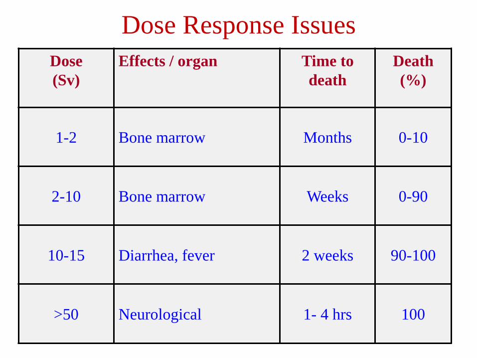
Dose Response Issues
Dose
(Sv)
Effects / organ Time to
death
Death
(%)
1-2 Bone marrow Months 0-10
2-10 Bone marrow Weeks 0-90
10-15 Diarrhea, fever 2 weeks 90-100
>50 Neurological 1- 4 hrs 100
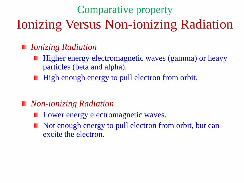
Comparative property
Ionizing Versus Non-ionizing Radiation
Ionizing Radiation
Higher energy electromagnetic waves (gamma) or heavy particles (beta and alpha).
High enough energy to pull electron from orbit.
Non-ionizing Radiation
Lower energy electromagnetic waves.
Not enough energy to pull electron from orbit, but can excite the electron.
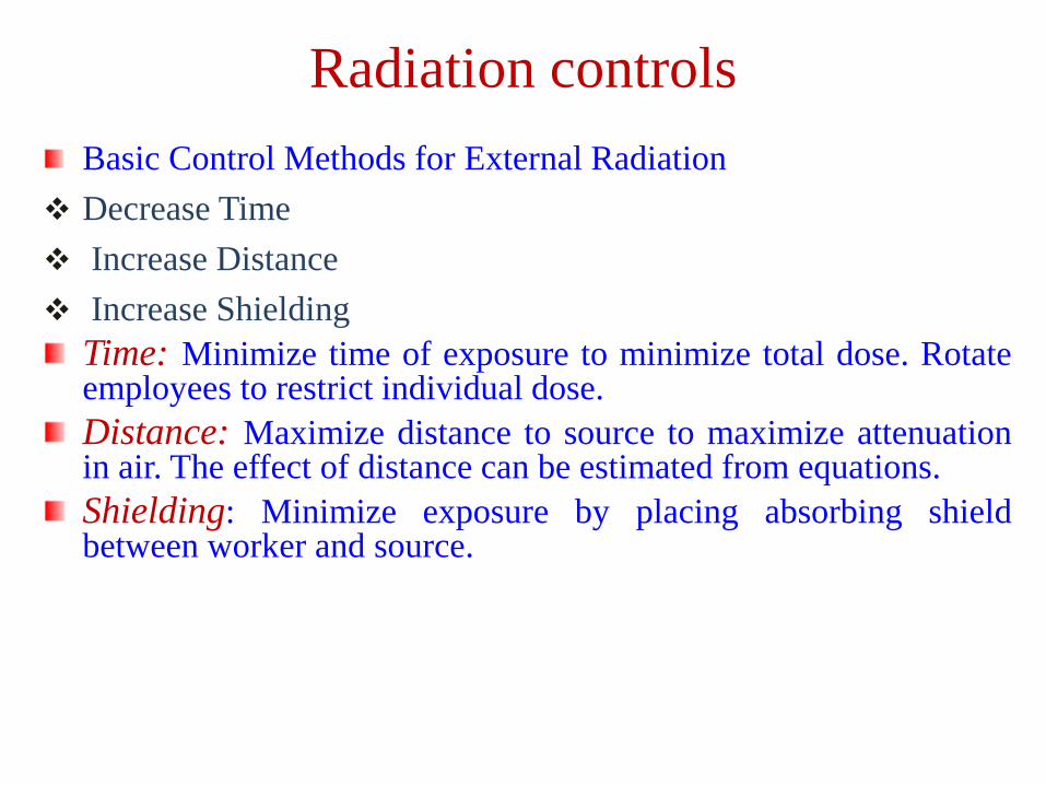
Radiation controls
Basic Control Methods for External Radiation
❖ Decrease Time
❖ Increase Distance
❖ Increase Shielding
Time: Minimize time of exposure to minimize total dose. Rotateemployees to restrict individual dose.
Distance: Maximize distance to source to maximize attenuationin air. The effect of distance can be estimated from equations.
Shielding: Minimize exposure by placing absorbing shieldbetween worker and source.
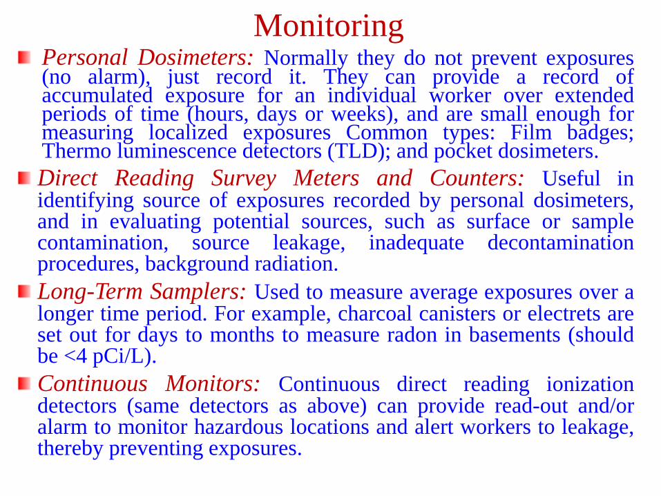
MonitoringPersonal Dosimeters: Normally they do not prevent exposures(no alarm), just record it. They can provide a record ofaccumulated exposure for an individual worker over extendedperiods of time (hours, days or weeks), and are small enough formeasuring localized exposures Common types: Film badges;Thermo luminescence detectors (TLD); and pocket dosimeters.
Direct Reading Survey Meters and Counters: Useful inidentifying source of exposures recorded by personal dosimeters,and in evaluating potential sources, such as surface or samplecontamination, source leakage, inadequate decontaminationprocedures, background radiation.
Long-Term Samplers: Used to measure average exposures over alonger time period. For example, charcoal canisters or electrets areset out for days to months to measure radon in basements (shouldbe <4 pCi/L).
Continuous Monitors: Continuous direct reading ionizationdetectors (same detectors as above) can provide read-out and/oralarm to monitor hazardous locations and alert workers to leakage,thereby preventing exposures.
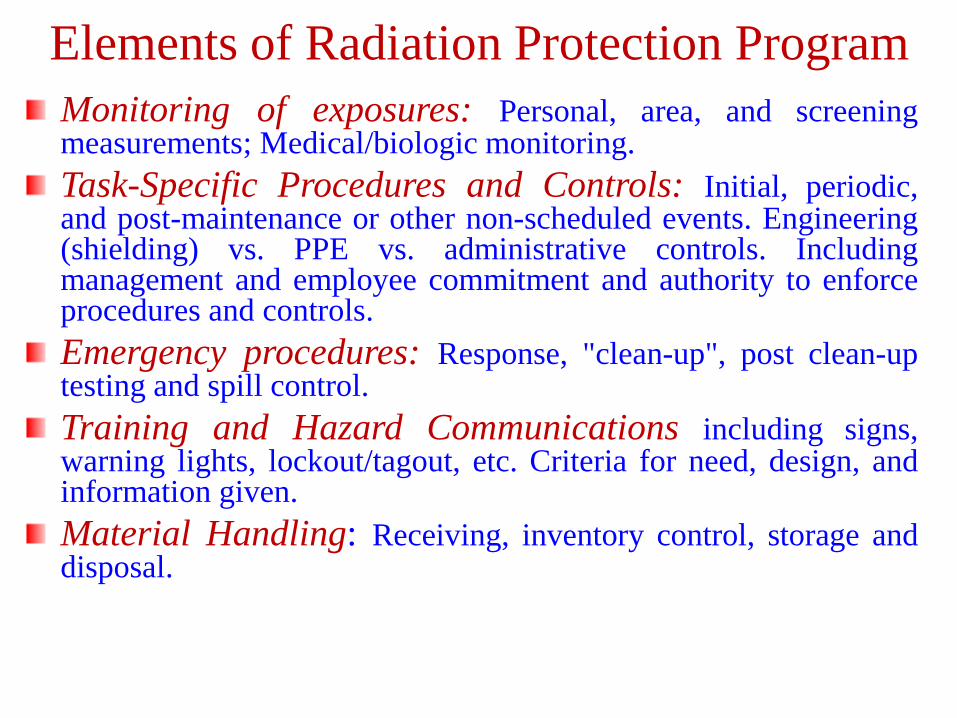
Elements of Radiation Protection Program
Monitoring of exposures: Personal, area, and screeningmeasurements; Medical/biologic monitoring.
Task-Specific Procedures and Controls: Initial, periodic,and post-maintenance or other non-scheduled events. Engineering(shielding) vs. PPE vs. administrative controls. Includingmanagement and employee commitment and authority to enforceprocedures and controls.
Emergency procedures: Response, "clean-up", post clean-uptesting and spill control.
Training and Hazard Communications including signs,warning lights, lockout/tagout, etc. Criteria for need, design, andinformation given.
Material Handling: Receiving, inventory control, storage anddisposal.