lecture 5 02-21-12 biomolecular...
TRANSCRIPT

Self-assembly and Nanotechnology 10.524
Lecture 5. Biomolecular Self-assembly (and Detection)
Instructor: Prof. Zhiyong Gu (Chemical Engineering& UML CHN/NCOE Nanomanufacturing Center)
Self-assembly and Nanotechnology
Lecture 5: Biomolecular Self-assembly
Table of Contents
Definitions
Fundamentals and principlesp p
Examples of biomolecular self-assembly
Nanostructure fabrication by bimolecular self assemblyNanostructure fabrication by bimolecular self-assembly
Case study: Biosensing and detection by nanowires
Self-assembly and Nanotechnology

Biomolecular Self-assembly
Definitions
A biomolecule is a chemical compound that naturally occurs in living organisms. Biomolecules consist primarily of carbon and hydrogen along with nitrogen oxygenBiomolecules consist primarily of carbon and hydrogen, along with nitrogen, oxygen, phosphorus and sulfur. Other elements sometimes are incorporated but are much less common.
P t iCell Membrane
Self-assembly and Nanotechnology
DNA Proteins

DNA Self-Assembly
Deoxyribonucleic acid (DNA) is a nucleic acid that contains thegenetic instructions for the development and function of living organisms.
The DNA double helix is held together by hydrogen bonds between the bases attached to the two strands. The four bases found in DNA are adenine (abbreviated A), cytosine (C), guanine (G) and thymine (T).
DNA self-assembly is the most advanced and versatile system known for programmable construction of patterned systems on the molecular scale
Self-assembly and Nanotechnology
patterned systems on the molecular scale
Reif, Duke Univ.

DNA Assisted Self-Assembly of Nanowires
Electroanalysis 19, 2007, No. 22, 2287 – 2293,

Nanostructure Fabrication by Bimolecular Self-assembly
DNA Based Nanoparticles AssemblyDNA Based Nanoparticles Assembly
Self-assembly and Nanotechnology Nature, 382, 607-609 (1996)
Protein Self-Assembly (Folding)
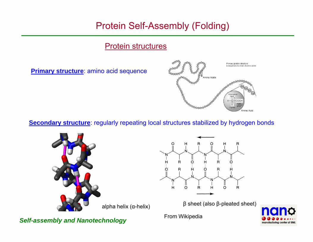
Protein structuresProtein structures
Primary structure: amino acid sequence
Secondary structure: regularly repeating local structures stabilized by hydrogen bonds
Self-assembly and Nanotechnology From Wikipedia
alpha helix (α-helix) β sheet (also β-pleated sheet)

Protein Self-Assembly (Folding)
Protein structures
Tertiary structure: the overall shape of a single protein l l th ti l l ti hi f th dmolecule; the spatial relationship of the secondary
structures to one another.
Tertiary structure is generally stabilized by nonlocal i t ti t l th f ti finteractions, most commonly the formation of a hydrophobic core, but also through salt bridges, hydrogen bonds, disulfide bonds, and even post-translational modifications.
Quaternary structure: the shape or structure that results from the interaction of more than one protein molecule, usually called protein subunits in this context, which function as part of the larger assembly or protein complex.
Self-assembly and NanotechnologyFrom Wikipedia

Antibody-Antigen Interactions
A tib d St tAntibody Structure
Antibodies are immune system-related proteins called immunoglobulins. Each antibody consists of four polypeptides–two heavy chains and two light chains joined to form a "Y"two heavy chains and two light chains joined to form a Y shaped molecule. The amino acid sequence in the tips of the "Y" varies greatly among different antibodies. This variable region, composed of 110-130 amino acids, give the antibody its specificity for binding antigen.
This image represents the structure of an antibody's variable region (Fab) complexed with an antigen, in this case hen egg white lysozyme.y y
Self-assembly and Nanotechnology
http://www.biology.arizona.edu/IMMUNOLOGY/tutorials/antibody/structure.html

Antibody-Antigen Interactions
The hypervariable (HV) regions of a Fab, representing both light and heavy chains, are highlighted in purple The antigen is greenhighlighted in purple. The antigen is green. The part of the antigen in direct contact with the antibody is called the antigenic determinant, or epitope
In this view, the HV regions of the Fab have been deleted. The framework (FR) regions of the antibody do not contact the antigenthe antibody do not contact the antigen.
Self-assembly and Nanotechnology
http://www.biology.arizona.edu/IMMUNOLOGY/tutorials/antibody/structure.html
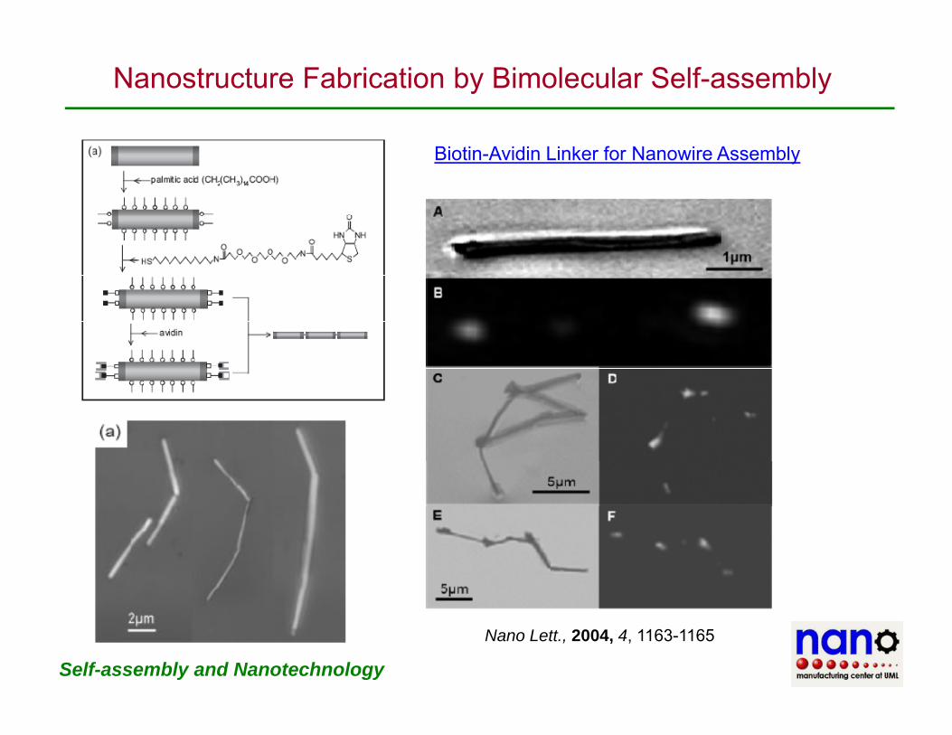
Nanostructure Fabrication by Bimolecular Self-assembly
Biotin-Avidin Linker for Nanowire Assembly
Self-assembly and Nanotechnology
Nano Lett., 2004, 4, 1163-1165
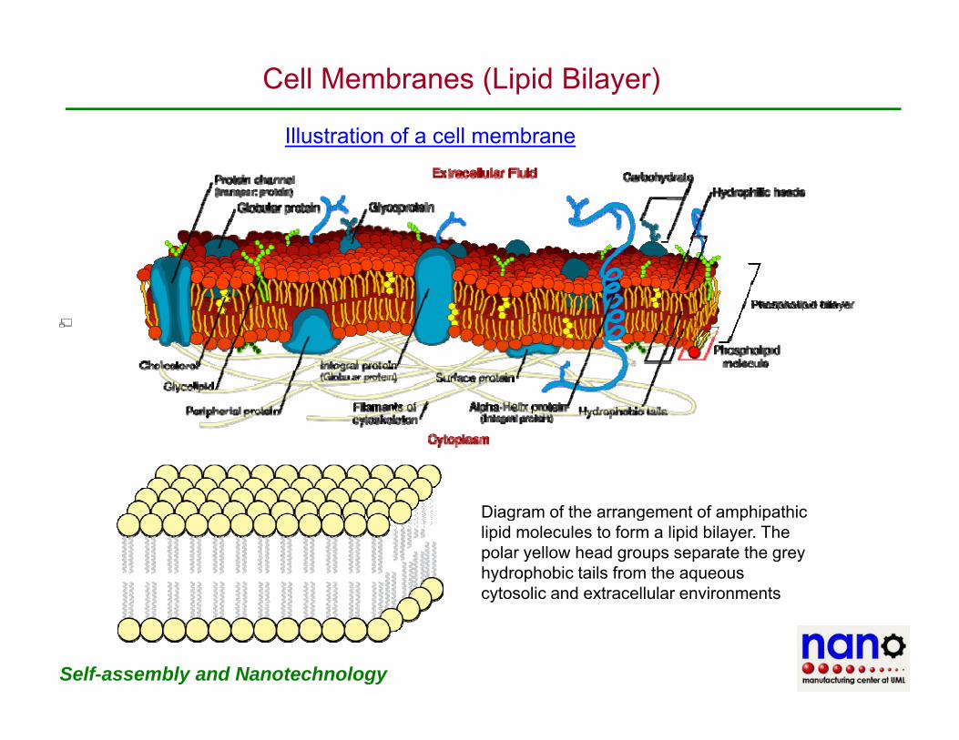
Cell Membranes (Lipid Bilayer)
Illustration of a cell membraneIllustration of a cell membrane
Diagram of the arrangement of amphipathicDiagram of the arrangement of amphipathic lipid molecules to form a lipid bilayer. The polar yellow head groups separate the grey hydrophobic tails from the aqueous cytosolic and extracellular environments
Self-assembly and Nanotechnology
Vesicles
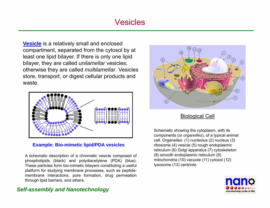
Vesicle is a relatively small and enclosed compartment, separated from the cytosol by at least one lipid bilayer. If there is only one lipid bilayer, they are called unilamellar vesicles;bilayer, they are called unilamellar vesicles; otherwise they are called multilamellar. Vesicles store, transport, or digest cellular products and waste.
Biological Cell
Schematic showing the cytoplasm, with its components (or organelles), of a typical animal cell. Organelles: (1) nucleolus (2) nucleus (3)
Biological Cell
E l Bi i ti li id/PDA i l ribosome (4) vesicle (5) rough endoplasmic reticulum (6) Golgi apparatus (7) cytoskeleton (8) smooth endoplasmic reticulum (9) mitochondria (10) vacuole (11) cytosol (12) lysosome (13) centriole.
A schematic description of a chromatic vesicle composed ofphospholipids (black) and polydiacetylene (PDA) (blue).These particles form bio-mimetic bilayers constituting a usefulplatform for st d ing membrane processes s ch as peptide
Example: Bio-mimetic lipid/PDA vesicles
Self-assembly and Nanotechnology
platform for studying membrane processes, such as peptide-membrane interactions, pore formation, drug permeationthrough lipid barriers, and others.
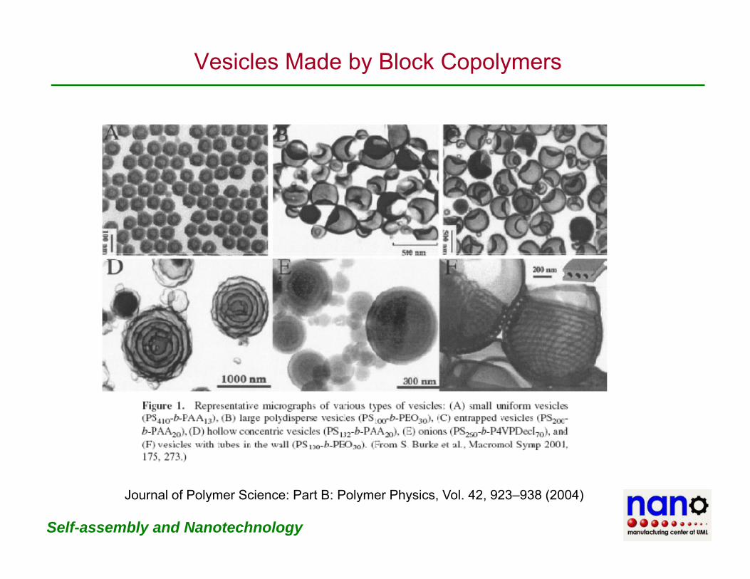
Vesicles Made by Block Copolymers
Self-assembly and Nanotechnology
Journal of Polymer Science: Part B: Polymer Physics, Vol. 42, 923–938 (2004)

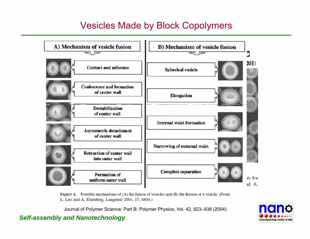
Vesicles Made by Block Copolymers
Self-assembly and Nanotechnology
Journal of Polymer Science: Part B: Polymer Physics, Vol. 42, 923–938 (2004)
Vesicles Made by Block Copolymers
Self-assembly and NanotechnologyJournal of Polymer Science: Part B: Polymer Physics, Vol. 42, 923–938 (2004)

Case Study: Nanowire-Based Biosensors
Materials Today, 2005, 20
Self-assembly and Nanotechnology Nanomedicine, 1(1), 51-65 (2006)

Case Study: Nanowire-Based Biosensors
(A) Schematic of a field-effect transistor (FET); (B) schematic of electrically-based sensing using FET devices The binding of aFET devices. The binding of a “charged or polar” biological or chemical species to the chemically modified gate dielectric is analogous to applying a voltageis analogous to applying a voltage using a gate electrode as shown in A. (C) A nanowire device configured as a sensor with antibody receptors (green) andantibody receptors (green) and binding of a protein with net negative charge yields an increase in the conductance. (D) A prototype nanowire sensor biochipprototype nanowire sensor biochip with integrated microfluidic sample delivery
Self-assembly and Nanotechnology Nanomedicine, 1(1), 51-65 (2006)

Case Study: Nanowire-Based Biosensors
(A) Schematic of a single virus binding and unbinding to the surface of a silicon nanowire device modified with antibody yreceptors; (B) simultaneous conductance and optical data for a silicon nanowire device with a low-density of antibody receptor units y y p(C) high-density of anti-body (D) schematic of multiplexsed single virus detection.
Self-assembly and NanotechnologyNanomedicine, 1(1), 51-65 (2006)