lecture 5 ess_3rd semester microscopic structure of female reproductive system ovary, oviduct,...
TRANSCRIPT
Lecture 5 ESS_3rd semester
MICROSCOPIC STRUCTURE OF FEMALE REPRODUCTIVE SYSTEM
Ovary, oviduct, uterus, vagina, and external genitalia Ovarian cycle, ovulation, and atresia. OogenesisMenstrual cycle - its relations to the ovarian cycle
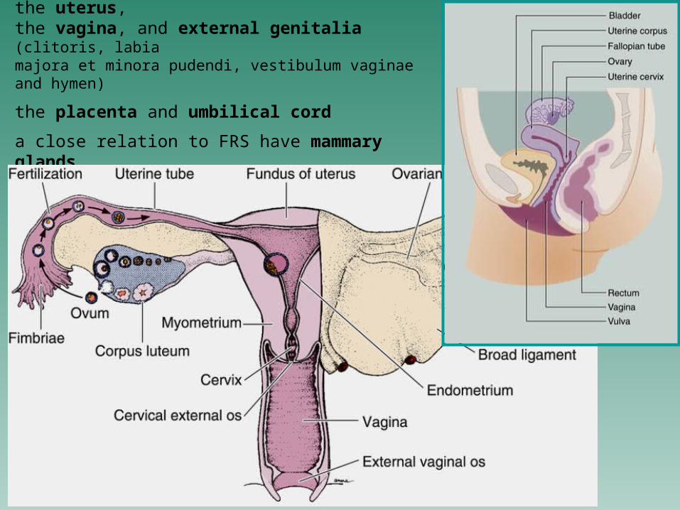
2 ovaries, 2 oviducts (uterine tubes), the uterus, the vagina, and external genitalia (clitoris, labiamajora et minora pudendi, vestibulum vaginae and hymen)
the placenta and umbilical cord
a close relation to FRS have mammary glands
function of female reproductive system: to produce and transport ova to support a developing embryo
in sexual mature females, some organs of this system undergoe cyclic changes in their structure and functional activity
the sexual maturity begins menarche = the time when the first menses
occur
and ends
menopause = a period, during which the cyclic changes become irregular or eventually disappear altogether
MICROSCOPIC ANATOMY OF THE OVARY(lat. ovarium, gr. oophoron)
an almond-shaped body approximately 3 cm long, 1.5 cm wide, and 1 cm thick
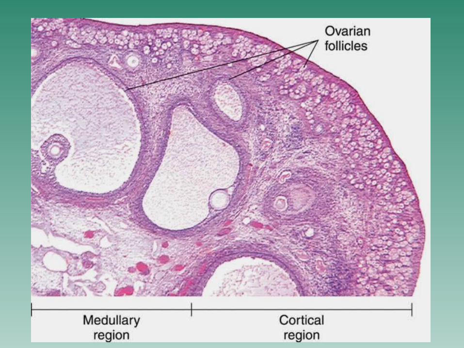
it is roughly divided into a central medulla that is occupied by a dense connective tissue stroma with a rich vascular bed a peripheral cortex composed of spindle-shaped form cells = fibroblasts
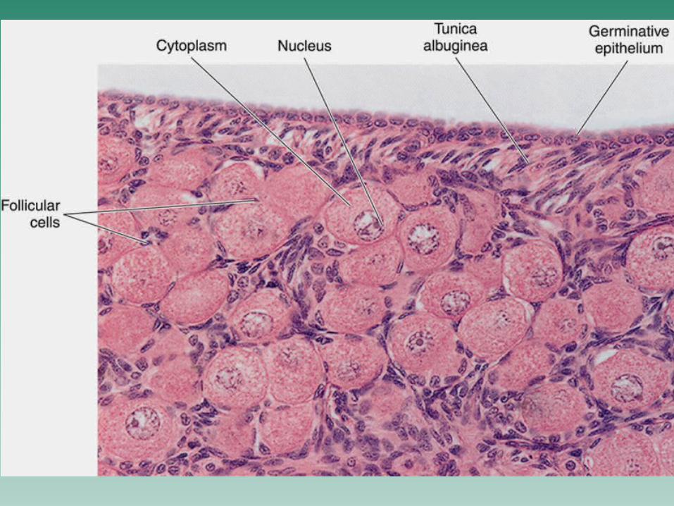
the surface of the organ is covered by simple squamous or cuboidal epithelium called the germinal epithelium
under the germinal epithelium fibroblasts are densely organized to form the capsule of the ovary known as the tunica albuginea
the cortex contains ovarian follicles and their derivatives that are corpus luteum and corpus albicans
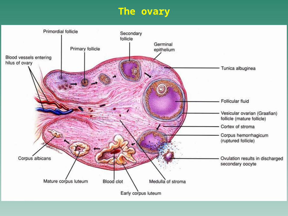
The ovary
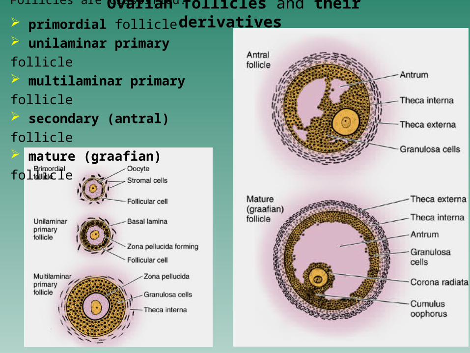
Ovarian follicles and their derivatives Follicles are classified:
primordial follicle unilaminar primary follicle multilaminar primary follicle secondary (antral) follicle mature (graafian) follicle
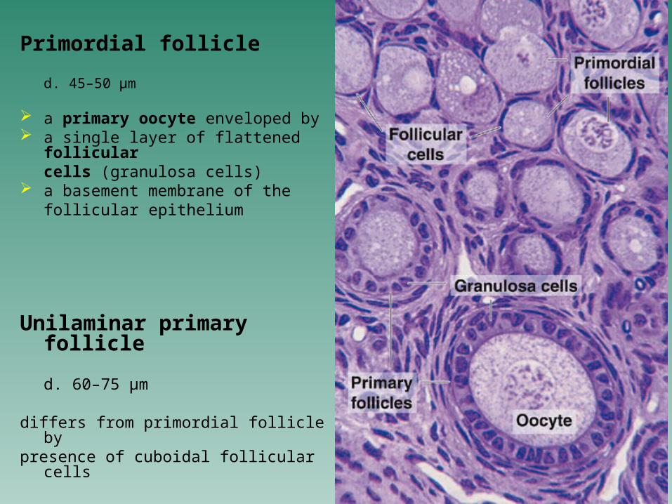
Primordial follicle
d. 45–50 µm
a primary oocyte enveloped by a single layer of flattened
follicularcells (granulosa cells)
a basement membrane of thefollicular epithelium
Unilaminar primary follicle
d. 60–75 µm
differs from primordial follicle bypresence of cuboidal follicular cells
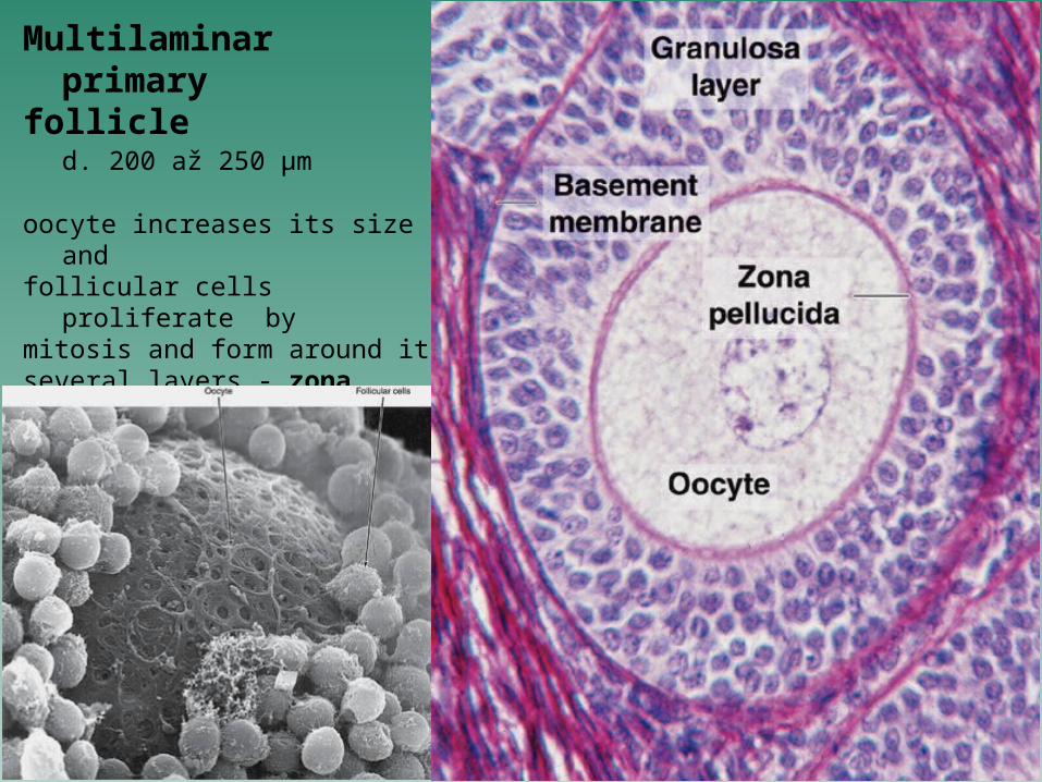
Multilaminar primary
follicle d. 200 až 250 µm
oocyte increases its size and follicular cells proliferate by mitosis and form around itseveral layers - zona granulosa
formation of the zona pellucida
the zona pellucida is composed of glycosaminoglycansit is thought that both oocyte and follicular cells take part in its synthesis
with this event, the cortical stroma around the follicle develops to form the
theca folliculiit differentiates subsequently into
the theca interna that is highly vascularized and and whose cells enlarge the theca externa formed by a connective tissue
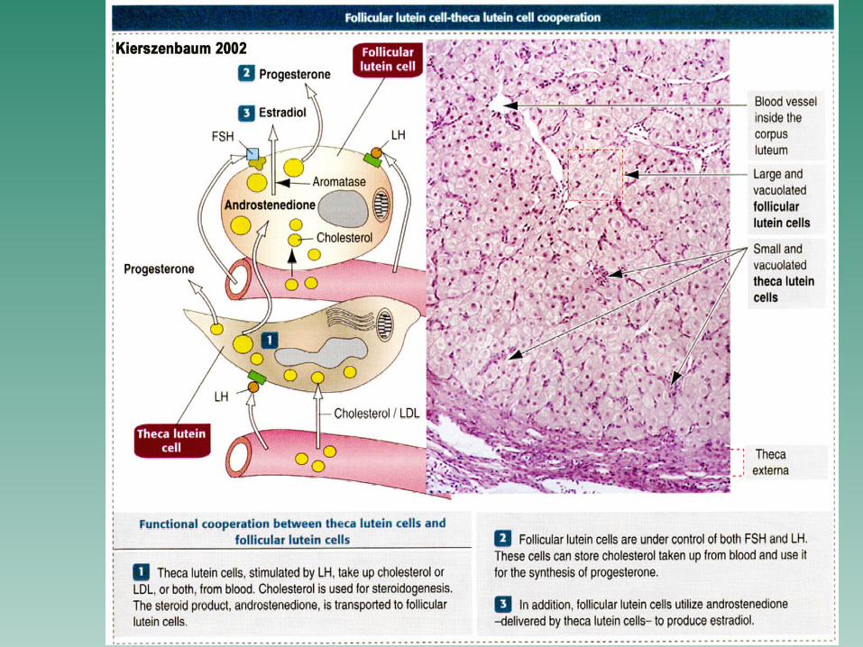
the cells of the theca interna synthesize androstenedione that is converted intoestradiol by cells of zona granulosa
Secondary (antral) follicle
is characterized by accumulation of fluid between follicular cells (cells of zonagranulosa) and by formation small cavities that gradually become to fuse in the
single, eccentrically placed cavity, the antrum folliculi filled with liquor folliculi
the lining of this single cavity is formed with several layers of follicular cells, the membrana granulosaat the follicle pole adjacent to the medulla, the membrana granulosa thickensinto the cumulus oophorus, protruding to the interior of the antrumthe oocyte is housed within the cumulus oophorus
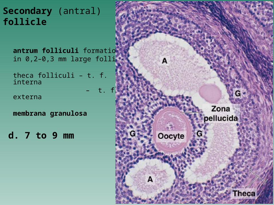
Secondary (antral) follicle
antrum folliculi formationin 0,2–0,3 mm large follicles
theca folliculi – t. f. interna – t. f. externa
membrana granulosa
d. 7 to 9 mm
Mature (graafian or preovulatory ) follicle
is about 1.5 - 2.5 cm in diameter and resembles transparent vesicle that bulges from the surface of the ovary
Its wall consists of: 4 - 5 layers of follicular cells - there is the membrana granulosa, the thickened basement membrane called as the membrane of
Slawjanski (firstly described by Slawjanski) the theca folliculi interna the theca folliculi externa
granulosa cells surrounding the oocyte are firmly attached to
the zona pellucida and accompany the oocyte during ovulation and its
expelling -
are usually called as the corona radiata
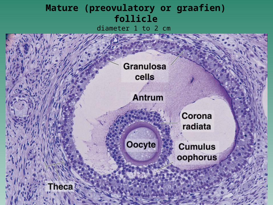
Mature (preovulatory or graafien) folliclediameter 1 to 2 cm
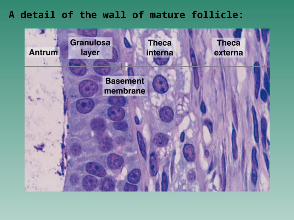
A detail of the wall of mature follicle:
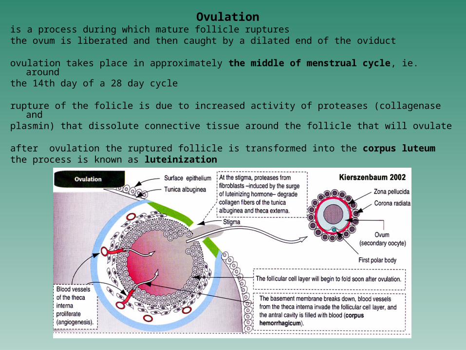
Ovulation is a process during which mature follicle rupturesthe ovum is liberated and then caught by a dilated end of the oviduct
ovulation takes place in approximately the middle of menstrual cycle, ie. aroundthe 14th day of a 28 day cycle
rupture of the folicle is due to increased activity of proteases (collagenase and plasmin) that dissolute connective tissue around the follicle that will ovulate
after ovulation the ruptured follicle is transformed into the corpus luteum the process is known as luteinization
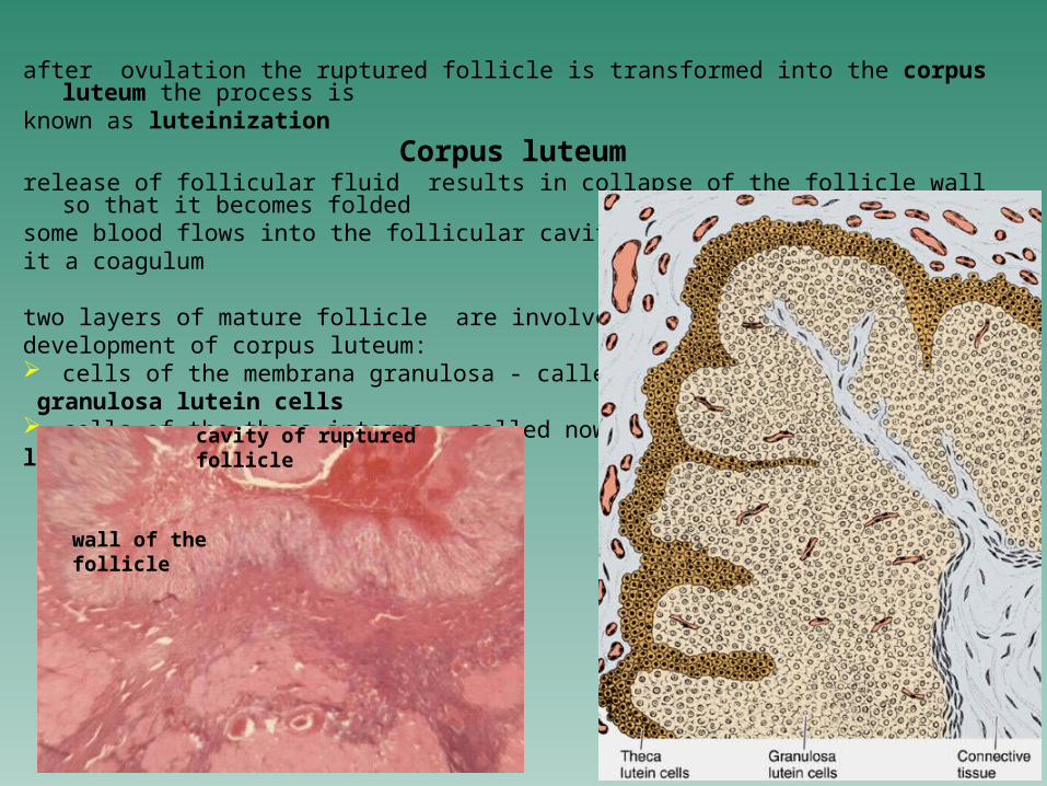
after ovulation the ruptured follicle is transformed into the corpus luteum the process is
known as luteinizationCorpus luteum
release of follicular fluid results in collapse of the follicle wall so that it becomes folded
some blood flows into the follicular cavity and forms in it a coagulum
two layers of mature follicle are involved in the development of corpus luteum: cells of the membrana granulosa - called now granulosa lutein cells cells of the theca interna - called now thecalutein cells
wall of the follicle
cavity of ruptured follicle
The granulosa lutein cellsare located at luminal border and increase greatly in size (35 m) the granulosa lutein cells show characteristics of steroid-secreting cells and
produce the progesterone
The theca lutein cells are located externally or in folds of the wall of then corpus luteum cells are smaller then granulosa lutein ones and stain more intesively in histological sections
they produce steroids other than progesterone
if the ovum is not fertilized, the corpus luteum functions only 10-12 days, and after this period starts to degenerate and disappear - the corpus luteum of menstruation
if pregnancy occurs, chorionic gonadotropin produced by the placenta stimulates the growth of corpus luteum, which becomes larger and is in function for about 3 months - the corpus luteum of pregnancy thereafter it gradually declines and definitively disappears after birth (during the childbed)
corpus albicans - is a final product occurring as a result of degeneration of corpus luteum
it appears as a region of dense connective tissue, later as a scar
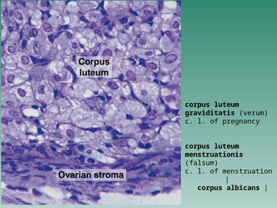
corpus luteum graviditatis (verum)c. l. of pregnancy
corpus luteum menstruationis (falsum) c. l. of menstruation | corpus albicans |
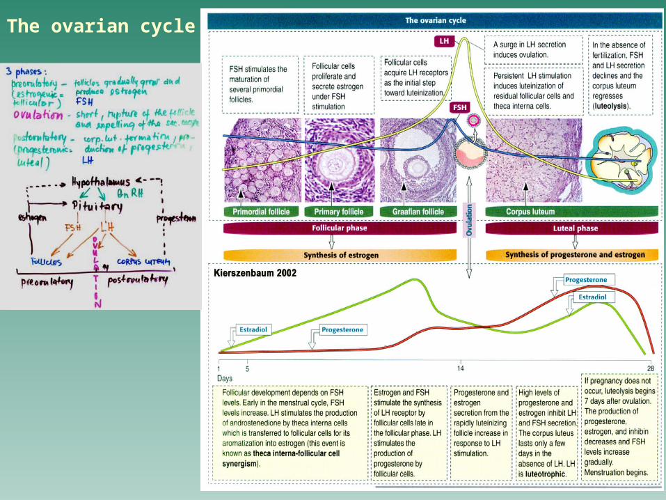
The ovarian cycle
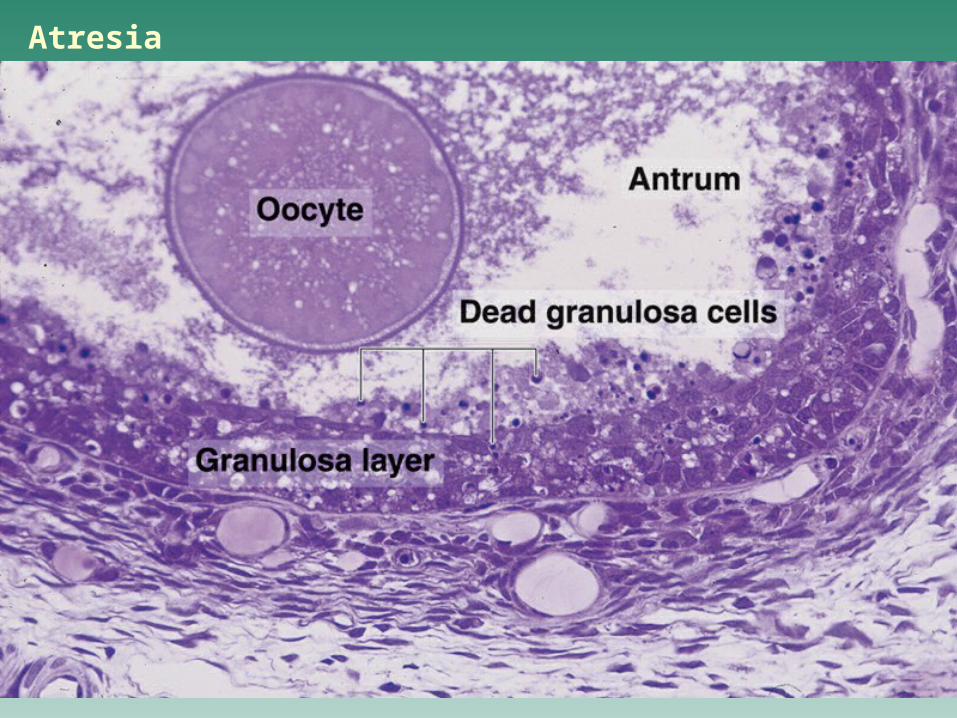
Atresia
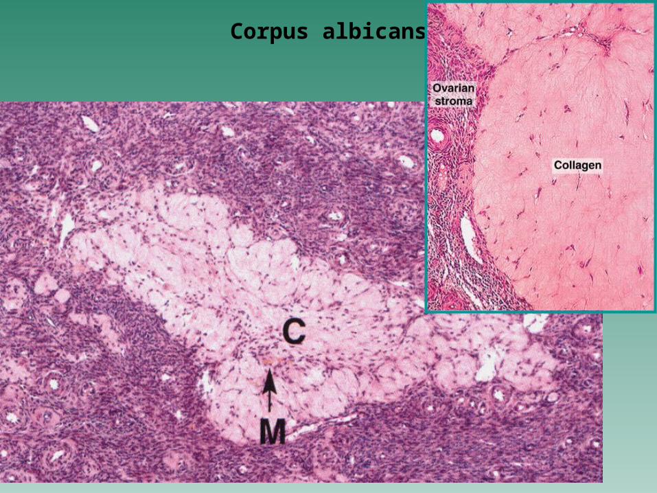
Corpus albicans
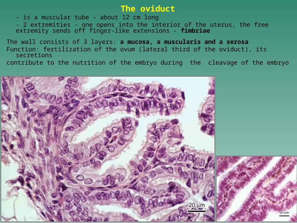
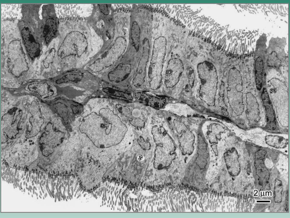
The oviduct - is a muscular tube - about 12 cm long- 2 extremities - one opens into the interior of the uterus, the free extremity sends off finger-like extensions - fimbriae
The wall consists of 3 layers: a mucosa, a muscularis and a serosaFunction: fertilization of the ovum (lateral third of the oviduct), its secretionscontribute to the nutrition of the embryo during the cleavage of the embryo
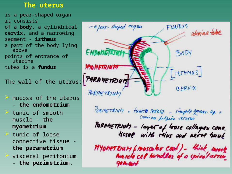
The uterusis a pear-shaped organit consistsof a body, a cylindricalcervix, and a narrowingsegment - isthmus a part of the body lying
abovepoints of entrance of uterinetubes is a fundus
The wall of the uterus:
mucosa of the uterus - the endometrium
tunic of smooth muscle - the myometrium
tunic of loose connective tissue - the parametrium
visceral peritonium - the perimetrium.
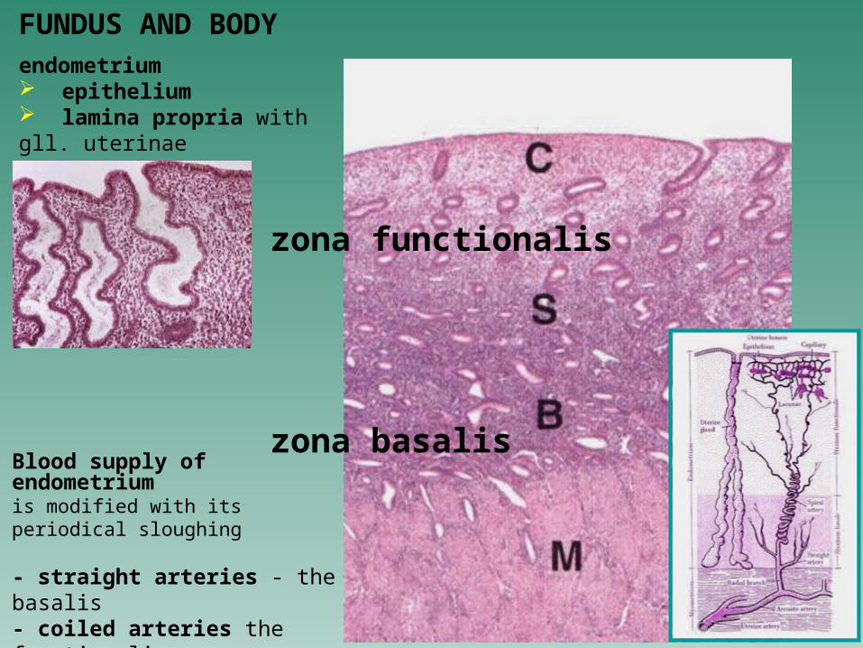
zona functionalis
zona basalis
Blood supply of endometrium is modified with its periodical sloughing
- straight arteries - the basalis- coiled arteries the functionalis
FUNDUS AND BODY
endometrium epithelium lamina propria with gll. uterinae
Histology of the endometrium closely depends on the ovarianhormones - estrogens and progesterone that are produced under stimulus of the anterior lobe of thepituitary
structural modifications have cyclic character and aresummarizingly called as the menstrual cycle
duration - in average 28 days
the menstrual cycle starts between 12 to 15 years of age and continues until about age 45-50 - only during these age limits the female is fertile
menopause is a period when the menstrual cycles are ceased
menstrual cycle includes 4 phases:
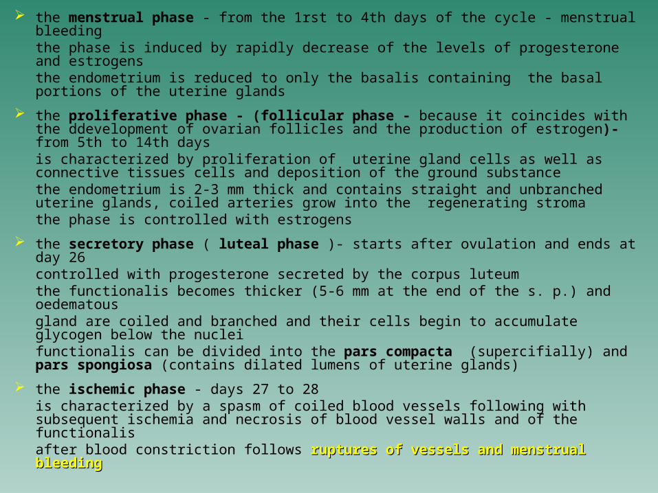
the menstrual phase - from the 1rst to 4th days of the cycle - menstrual bleeding the phase is induced by rapidly decrease of the levels of progesterone and estrogens the endometrium is reduced to only the basalis containing the basal portions of the uterine glands
the proliferative phase - (follicular phase - because it coincides with the ddevelopment of ovarian follicles and the production of estrogen)- from 5th to 14th daysis characterized by proliferation of uterine gland cells as well as connective tissues cells and deposition of the ground substancethe endometrium is 2-3 mm thick and contains straight and unbranched uterine glands, coiled arteries grow into the regenerating stromathe phase is controlled with estrogens
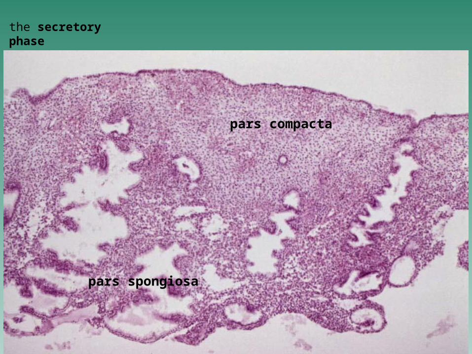
the secretory phase ( luteal phase )- starts after ovulation and ends at day 26controlled with progesterone secreted by the corpus luteumthe functionalis becomes thicker (5-6 mm at the end of the s. p.) and oedematousgland are coiled and branched and their cells begin to accumulate glycogen below the nucleifunctionalis can be divided into the pars compacta (supercifially) and pars spongiosa (contains dilated lumens of uterine glands)
the ischemic phase - days 27 to 28 is characterized by a spasm of coiled blood vessels following with subsequent ischemia and necrosis of blood vessel walls and of the functionalisafter blood constriction follows ruptures of vessels and menstrual bleedingruptures of vessels and menstrual bleeding
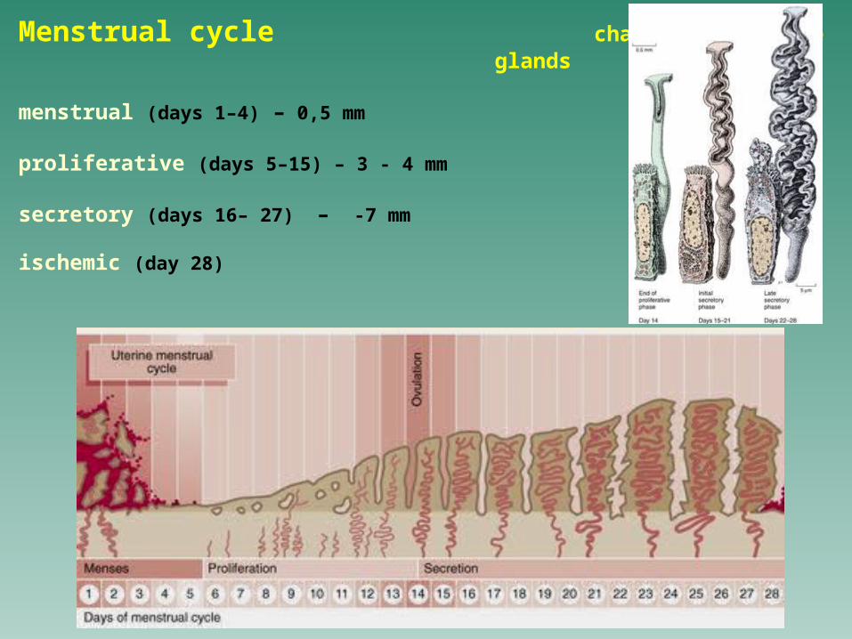
Menstrual cycle changes of uterine glands
menstrual (days 1–4) – 0,5 mm
proliferative (days 5–15) – 3 - 4 mm
secretory (days 16– 27) – -7 mm
ischemic (day 28)
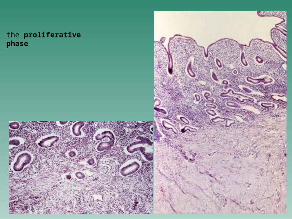
the proliferative phase
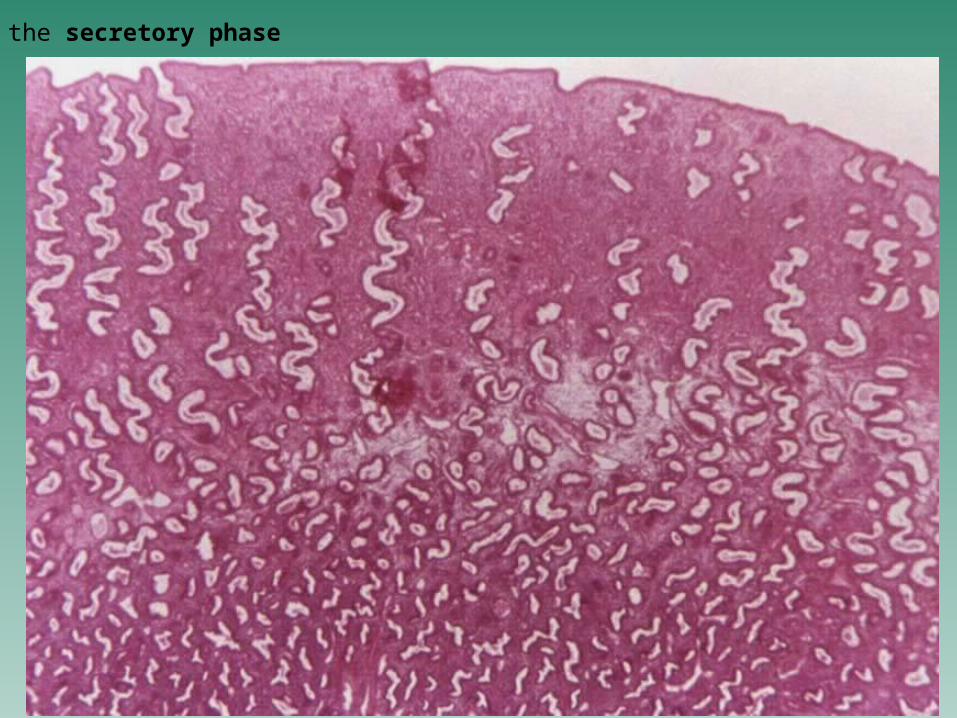
the secretory phase
the secretory phase
pars compacta
pars spongiosa

Relation between the menstrual and ovarian cycle
Remember: after fertilization of the ovum and implantation
of the embryo, the endometrium goes through profound changes and is called the decidua
cells of the stroma become enlarged and polygonal and are called decidual cells
the basal part of the decidua, decidua basalis =
the maternal part of the placenta
CERVIX
= inferior part of the uterusit is divided into an upper portion, the cervical canala lower vaginal portion that projects into the vagina
The cervical canal differs from the body of the uterus in (1) its wall consists largely of dense collagenous and elastic fibers
with only about 15 % of the wall being smooth muscle(2) the mucosa contains complex mucous glands and deep
branching folds - plicae palmatae glands may become occluded and form cysts (ovula Nabothi)
the mucosa does not participate in menstruation (however, the glands undergo cyclic changes during menstrual cycle: in the proliferative phase they produce thin and watery secretion, which becomes copious at ovulation; secretion shows the consistency of egg whites and forms semisolid mucous plug that prevents the passage of sperm, microorganisms etc. from entering the uterus from the vagina)
(3) on the vaginal portion, the simple columnar epithelium is replaced with stratified squamous, nonkeratinized epithelium
VAGINAis a fibromuscular, collapsed tube connecting the uterus to the exterior of the
bodywall: a mucosa, a muscularis, and an adventitia
Mucosa forms longitudinal folds - rugae. It is covered with 150 to 200 mm thick stratified squamous, nonkeratinized epithelium. Under the stimulus of estrogen, the vaginal epithelium synthesizes and accumulates a large quantity of glycogen, which is released into the vaginal lumen when the surface cells are exfoliated. Bacteria in the vagina (Lactobacillus acidophilus) metabolize glycogen and form lactic acid, which is responsible for the usually low pH of the vagina. Lamina propria is composed of loose connective tissue that is rich in elastic fibers. It also contains a few small lymph nodules and neutrophils. Lymphocytes and neutrophils invade the epithelium and pass into the lumen of the vagina during certain phases of the menstrual cycle - vaginal cytology. Lamina propria exhibits a rich vascularization that is the source of the fluid exusudate that seeps through the squamous epithelium into the lumen of the vagina during copulation.
Muscularis comprises two poorly develop smooth muscle layers: inner circular and outer longitudinal.
Adventitia surrounds the vagina and blends with adjacent organs. It is a coat of dense connective tissue, rich in thick elastic fibers. In this connective tissue are an extensive venous plexus, nerve bundles, and groups of nerve cells.
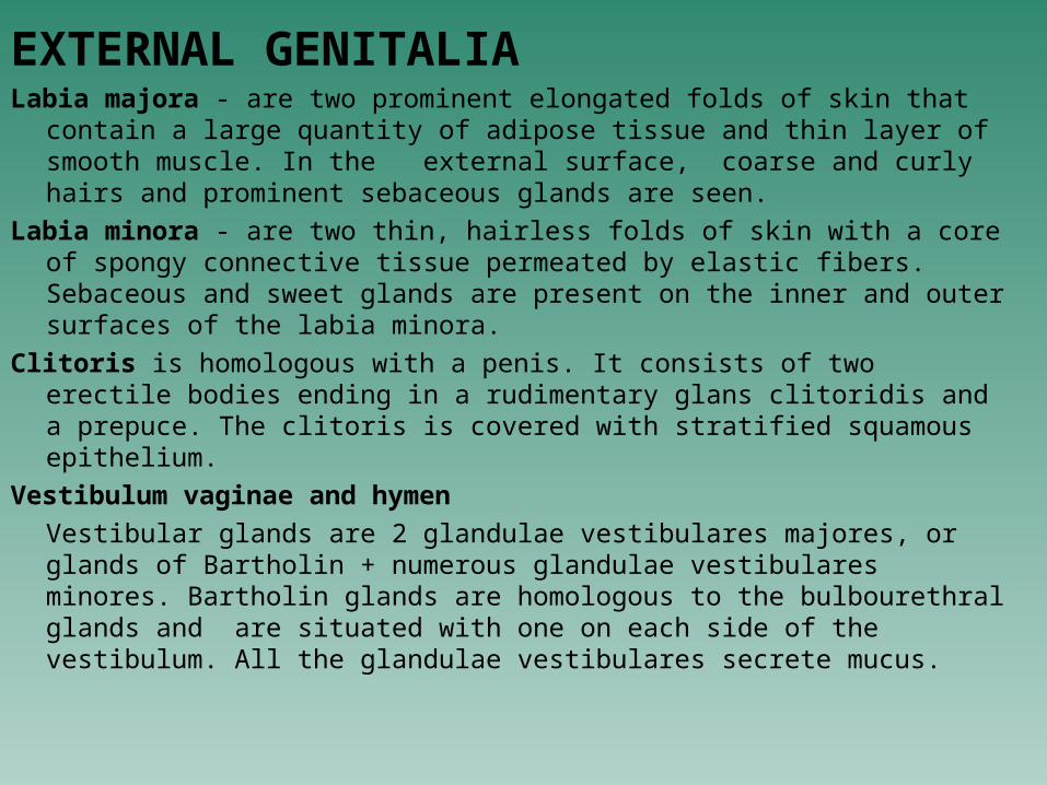
EXTERNAL GENITALIALabia majora - are two prominent elongated folds of skin that
contain a large quantity of adipose tissue and thin layer of smooth muscle. In the external surface, coarse and curly hairs and prominent sebaceous glands are seen.
Labia minora - are two thin, hairless folds of skin with a core of spongy connective tissue permeated by elastic fibers. Sebaceous and sweet glands are present on the inner and outer surfaces of the labia minora.
Clitoris is homologous with a penis. It consists of two erectile bodies ending in a rudimentary glans clitoridis and a prepuce. The clitoris is covered with stratified squamous epithelium.
Vestibulum vaginae and hymenVestibular glands are 2 glandulae vestibulares majores, or glands of Bartholin + numerous glandulae vestibulares minores. Bartholin glands are homologous to the bulbourethral glands and are situated with one on each side of the vestibulum. All the glandulae vestibulares secrete mucus.
PLACENTA
a temporary organ which develops during the second month of development
is the site of physiologic exchange between the mother and fetus
the human placenta is of discoidal shape measuring about 15 - 20 cm in
diameter and 2- 3 cm in thickness and weighing 500 - 600 g at full term.
it consists of 2 parts close associated each other:
the fetal part or villous chorion and
the maternal part or decidua basalis
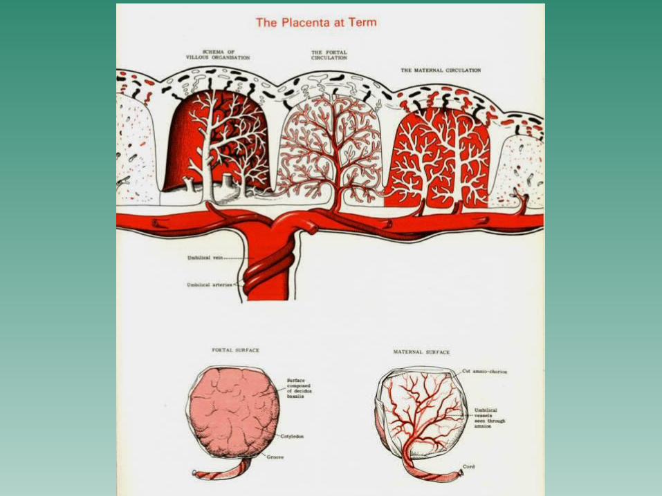
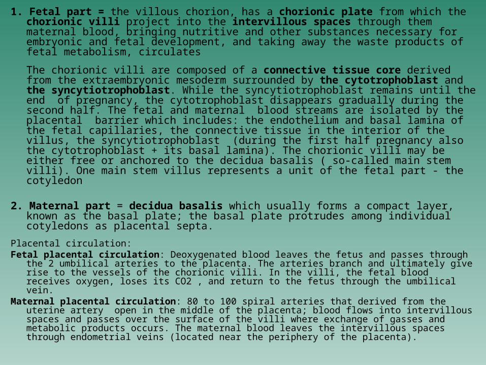
1. Fetal part = the villous chorion, has a chorionic plate from which the chorionic villi project into the intervillous spaces through them maternal blood, bringing nutritive and other substances necessary for embryonic and fetal development, and taking away the waste products of fetal metabolism, circulates
The chorionic villi are composed of a connective tissue core derived from the extraembryonic mesoderm surrounded by the cytotrophoblast and the syncytiotrophoblast. While the syncytiotrophoblast remains until the end of pregnancy, the cytotrophoblast disappears gradually during the second half. The fetal and maternal blood streams are isolated by the placental barrier which includes: the endothelium and basal lamina of the fetal capillaries, the connective tissue in the interior of the villus, the syncytiotrophoblast (during the first half pregnancy also the cytotrophoblast + its basal lamina). The chorionic villi may be either free or anchored to the decidua basalis ( so-called main stem villi). One main stem villus represents a unit of the fetal part - the cotyledon
2. Maternal part = decidua basalis which usually forms a compact layer, known as the basal plate; the basal plate protrudes among individual cotyledons as placental septa.
Placental circulation:Fetal placental circulation: Deoxygenated blood leaves the fetus and passes through
the 2 umbilical arteries to the placenta. The arteries branch and ultimately give rise to the vessels of the chorionic villi. In the villi, the fetal blood receives oxygen, loses its CO2 , and return to the fetus through the umbilical vein.
Maternal placental circulation: 80 to 100 spiral arteries that derived from the uterine artery open in the middle of the placenta; blood flows into intervillous spaces and passes over the surface of the villi where exchange of gasses and metabolic products occurs. The maternal blood leaves the intervillous spaces through endometrial veins (located near the periphery of the placenta).
Placental activitiesthree main activities: metabolic, transfer, and endocrine
• Placental metabolism - in placenta, particularly during early pregnancy, synthesizes glycogen, cholesterol, and fatty acids which all serve as a source of nutrients and energy for the embryo.
• Placental transfer - gases, nutrients, hormones, electrolytes, antibodies, wastes, and also several drugs are transported across the placental barrier. The transport is provided by 4 mechanisms: simple cell diffusion, facilitated diffusion, active transport, and pinocytosis.
• Placental endocrine secretion: the syncytiotrophoblast produces several hormones which are of 2 categories: protein hormones: human chorionic gonadotropin (hCG), human
chorionic somatomammotropin (hCS) or placental lactogen, human chorionic thyrotropin (hCT), and human chorionic corticotropin (hCACTH)
steroid hormones: progesterone + estrogens.
THE UMBILICAL CORD
is usually 1-2 cm in diameter and 30 -90 cm in length (average 55 cm)
the cord is attached near the center of the placenta
- amnionic ectoderm - simple squamous to cuboidal epithelium on the surface of the cord
- Wharton's jelly (gel-like connective tissue) - main umbilical tissue consisting of acid mucopolysaccharides, fibrocytes and thin collagen fibers arranged in a network
- umbilical blood vessels (vasa umbilicalia)– 1. umbilical arteries (arteriae umbilicales) - vessels with narrow
lumina in which smooth muscle cells are arranged circularly, spirally and longitudinally; outer and inner elastic membranes are missing
– 2. umbilical vein (vena umbilicalis) - a single vessel with a large lumen than in arteries; its wall is thin, with three distinct layers.
- rest of the allantoic duct (ductus allantoideus) - an all-defined patch of epithelium in the middle of a triangle demarcated by umbilical veins (sometimes appearing only as a thickening of connective fibers).