lecture: candidiasis - ksumsc.comksumsc.com/download_center/2nd/3) endocrine block/teams work... ·...
TRANSCRIPT

LECTURE: Candidiasis
• Important
• Do tor’s otes • Extra explanation
Editing File
" ي العظي هلل الع ة إذا داه "ال حول وال قوة إال ب ل هذه الجم وتقن يه يصع أو ، يستطيعه ال عظي أمر اإلنس . به القي ع

OBJECTIVES:
1. Acquire the basic knowledge about Candida as a pathogen
2. know the main infections caused by Candida species
3. Identify the clinical settings of such infections
4. Know the laboratory diagnosis, and treatment of these infections.

Candida (the organism):
Features • Candida is a unicellular yeast fungus
• It is imperfect* reproducing by budding**
Morphology • Microscopy: Budding yeast cells, and Pseudohyphae. • Culture: Creamy colony, fast growing on Sabouraud Dextrose agar (SDA), Blood agar (48 hr)***
species • There are many species of Candida (>150)
• The common species are:
Candida albicans, C.parapsilosis, C.tropicalis, C.glabrata, C.krusei.
Human commensal (normal flora)
Oral cavity - Skin - Gastrointestinal tract - Genitourinary tracts
Budding : if there is any weakness at the wall of the yeast cell --> there will
be pouching ( out growth at that side )--> it will continue until the daughter
cells are completely formed , the daughter cell could :
1-detach from the mother cell
2- or become elongated and make Pseudohyphae
**Asexual *Means :the sexual stage is unknown ***fast growing comparing with molds
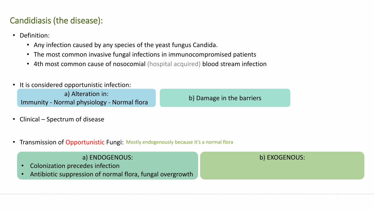
Candidiasis (the disease):
• Definition:
• Any infection caused by any species of the yeast fungus Candida.
• The most common invasive fungal infections in immunocompromised patients
• 4th most common cause of nosocomial (hospital acquired) blood stream infection
• It is considered opportunistic infection:
• Clinical – Spectrum of disease
• Transmission of Opportunistic Fungi:
a) Alteration in:
Immunity - Normal physiology - Normal flora b) Damage in the barriers
a) ENDOGENOUS:
• Colonization precedes infection
• Antibiotic suppression of normal flora, fungal overgrowth
b) EXOGENOUS:
Mostly e doge ously e ause it’s a or al flora
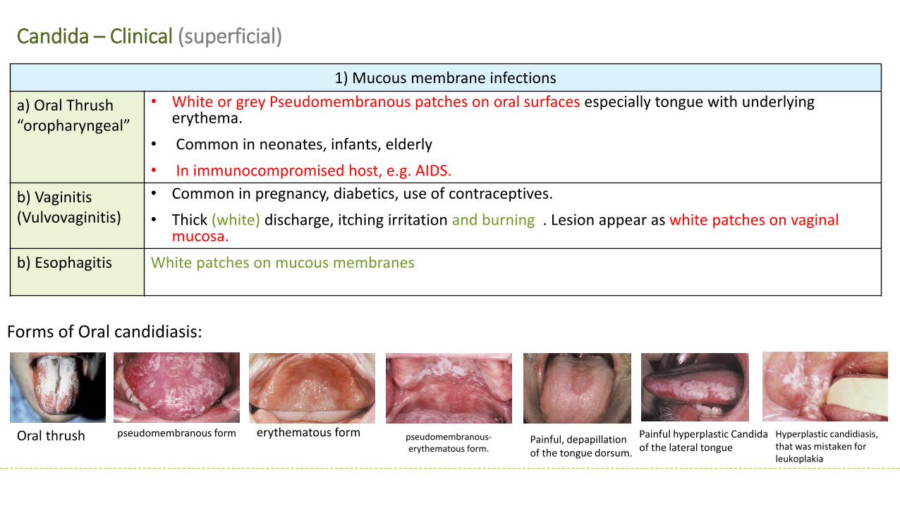
Candida – Clinical (superficial)
1) Mucous membrane infections
a) Oral Thrush
orophary geal
• White or grey Pseudomembranous patches on oral surfaces especially tongue with underlying erythema.
• Common in neonates, infants, elderly
• In immunocompromised host, e.g. AIDS.
b) Vaginitis
(Vulvovaginitis)
• Common in pregnancy, diabetics, use of contraceptives.
• Thick (white) discharge, itching irritation and burning . Lesion appear as white patches on vaginal mucosa.
b) Esophagitis
White patches on mucous membranes
Oral thrush
Forms of Oral candidiasis:
pseudomembranous-
erythematous form.
pseudomembranous form erythematous form Painful, depapillation
of the tongue dorsum.
Painful hyperplastic Candida
of the lateral tongue
Hyperplastic candidiasis,
that was mistaken for
leukoplakia

2) Cutaneous infections
Intertriginous1
candidiasis:
• Infections of skin folds eg. axilla, buttock, toe web, under breast.
• Erythematous lesion, dry or moist or whitish accompanied by itching and burning.
Nail infections: (pain -
nail discoloration)
• Onychomycosis (infection of nails) Discolored, hard, dislodge.
• Paronychia (infection of skin around nail bed)
Diaper rash
Chronic mucotaneous
candidiasis
children with T-cell abnormality. recurrent persistent superficial infection (non-invasive) mostly
caused by failure of T-cell immunity against candida. Most common cause: Candida albicans (in
children).
Candida – Clinical (superficial)
Chronic mucocutaneous candidiasis Intertriginous candidiasis Diaper rash Onychomycosis Paronychia
1 in between

Candida – Clinical (systemic)
• Urinary tract infection
• Candidemia (presence of candida in the blood; hematogenous spread to other organs)
• Disseminated (systemic, invasive) infection :Endophthalmitis (eye) - Liver and spleen – Kidneys – Skin – Brain – Lungs - Bone
Pulmonary Candidiasis
• Primary pneumonia is less common and could be a result of Aspiration
• Secondary pneumonia commonly seen with hematogenous candisiasis especially in:
Immunocompromised patients
Diagnosis Isolation of Candida from sputum, BAL bronchioalveolar lavage is not always significant because it is normal flora in
the mouth so the sample may be contaminated. So we have to correlate with:
Clinical features – Radiology - Other Lab investigations
But if we find candida in a normally sterile site (CSF, blood) then we consider it as significant.
Candidemia :Candida is the fourth most common in causing nosocomial* bloodstream infections (BSI)
Transmission: • Increased colonization (endogenous or exogenous factors) • Damage in host barriers by catheters, trauma, surgery • Immunosuppression (transplant patient, AIDs) • Central venous catheters (CVC)
Disseminated candidiasis (involvement of any organ): Septic shock – Meningitis - Ocular involvement (retinitis)
clinical manifestation • Fever could be the only clinical manifestation
% BSI% Crude
Mortality
Rank Pathogen
BSIper 10,000 admission
s
Total (n=20,978
)
ICU (n=10,5
15)
Non-ICU
(n=10,515) Total ICU
Non-ICU
1. CoNS 15.8 31.3 35.9 26.6 20.7 25.7 13.8
2. S aureus 10.3 20.2 16.8 23.7 25.4 34.4 18.9
3. Enterococcus spp
4.8 9.4 9.8 9.0 33.9 43.0 24.0
4. Candida spp 4.6 9.0 10.1 7.9 39.2 47.1 29.0
5. E coli 2.8 5.6 3.7 7.6 22.4 33.9 16.9
6. Klebsiella spp 2.4 4.8 4.0 5.5 27.6 37.4 20.3
7. P aeruginosa 2.1 4.3 4.7 3.8 38.7 47.9 27.6
8. Enterobacter spp 1.9 3.9 4.7 3.1 26.7 32.5 18.0
9. Serratia spp 0.9 1.7 2.1 1.3 27.4 33.9 17.1
10. A baumannii 0.6 1.3 1.6 0.9 34.0 43.4 16.3
*hospital acquired

Specimen: depend on site of infection we decide what sample to take: • Swabs, Urine, Blood, Respiratory specimens, CSF, Blood
1. Direct microscopy : What stains do we use? • Gram stain, KOH, Giemsa, GMS, or PAS stained smears. What do we see? • Budding yeast cells and pseudohyphae will be seen in stained
smear or KOH
2. Culture: • Media: SDA (Sabouraud Dextrose Agar ) & Blood agar at 37oC,
• Creamy moist colonies in 24 - 48 hours.
3. Blood culture:
In case of Candidemia , it is not blood agar .
We take blood sample from the patient and we put it in a bottle then in a machine
4. Serology: Patient serum Test for Antigen , e.g. Mannan antigen using ELISA Test for Antibodies
5. PCR:
Candidiasis – Laboratory diagnosis

Candidiasis – Laboratory diagnosis (Laboratory identification of Yeast) After identification we do susceptibility test in case of 1- sample from sterile body site or 2- recurrent infection
Because C. albicans is the most common species to cause infection
o The following tests are used to identify C. albicans:
If negative, then it could be any other yeast:
Use Carbohydrate assimilations and fermentation . ( not
used because it takes 72 hours for results )
Commercial kits available for this like: API 20C, API 32C
Culture on Chromogenic Media (CHROMagar™ Ca dida) gives different color for each species
1. Germ tube test : For atio of ger tu e whe ultured i seru at 37ᵒC in incubator for 2-4 hours
2. Chlamydospore production in corn meal Agar
3. Resistance to 500 μg/ml Cycloheximide
If these 3 are positive this yeast is C.albicans
عون بوزيتف الز كل الثالثة يط
Chlamydospores of C. albicans in CMA
Rounded and thickened wall could be
terminal or intercalary =in between
Germ tube test No constriction while pseudo hyphy has
constriction so there is continuation between the mother and the
daughter / it is the initial stage of formation of true hyphy
Carbohydrates assimilation test , API 20C

Chromagar
Morphology: producing green pigmented colonies on specially designed medium
to speciate certain yeasts based on color they produce
Candida species:
Sabouraud Dextrose Agar
Morphology: Creamy white yeast, may be dull, dry irregular and heaped up,
glabrous and tough
• Candida albicans

Candidiasis- Treatment
Oropharyngeal: Topical Nystatin suspension, Clotrimazole troches ,Miconazole, Fluconazole suspension. Systemic treatment in Immunocompromised patients
Vaginitis: Miconazole, Clotrimazole, Fluconazole
Systemic treatment: Fluconazole, Voriconazole, Caspofungin ( drug of choice ), Amphotericin B
Candidemia: • Treat for 14 days after last negative culture and resolution of signs and symptoms
• Remove catheters, if possible
oAntifungal susceptibility testing in not done routinely in the microbiology lab, It is done in the following cases:
For fungi isolated from sterile samples
If the patient is not responding to treatment
In case of recurrent infections
oPoints to consider:
C. glabrata can be less susceptible or resistant to fluconazole
C. krusei is resistant to fluconazole

SUMMARY:
Candidiasis:
Mucos membrane infections
Thrush (oropharyngeal)
Esophagitis
Vaginitis
Cutaneous infections
Paronychia (skin around nail bed)
Onychomycosis (nails)
Diaper rash
Chronic mucotaneous candidiasis (T-cell abnormality)
Urinary tract infections
Candidemia
Disseminated (systemic, invasive) infection
Endophthalmitis (eye)
Liver and spleen
Kidneys
Skin
Brain
Lungs (pulomnary candidiasis)
Bone
Laboratory diagnosis: Specimen depends on site of infection.
1. Direct microscopy: (gram stain, KOH, Giemsa, GMS, or
PAS stained)
2. Culture: (SDA + blood agar)
3. Blood culture
4. Serology: Antigens > ELISA, and Antibodies
5. PCR
Identification: (all positive > candida albicans)
1. Germ tube test
2. Chlamydospore production in corn meal Agar
3. Resistance to Cycloheximide
All negative > any other yeast: use CHO assimilations and
fermentation, culture on chromogenic media

QUIZ: 1. What is the most common type of candida?
a. Albicans
b. Parapsilosis
c. Tropicalis
d. Krusei
2.What is the most common route of transmission?
a. IV line
b. catheter
c. surgery
d. Use of broad spectrum antibiotics
3. Overweight patient comes in complaining of pain and burning
sensation in his axillary region. What is the most likely diagnosis?
a. chronic mucocutanous candidasis
b. Interiginous Candidasis
c. Nail bed infection
d. diaper rash
4. 27 year old male comes into the clinic. He has white and red
patches on his tongue and oral surfaces. You suspect its
oropharyneal candidasis. What does the patient most likely have as a
risk factor?
a. viral inefction
b. AIDs
. he’s healthy
d. on antibiotics
5. Which method is not used to confirm a diagnosis of pulmonary
candidasis?
a. isolation from sputum
b. clinical features
c. Radiology
d. other lab testing
6. How to confirm that the species is candida albicans?
a. SDA media stain culture
b. germ tube
c. chlamydospore
d. all the above
Answers: 1. a 2. d 3. b 4. b 5. a 6. d

THANK YOU FOR CHECKING OUR WORK, BEST OF LUCK!
Hamad Alkhudhairy Shrooq Alsomali
Reem Alshathri
Ghada Alskait
Jawaher Abanumy
Shoag Alahmari
Heba Alnasser
Najd Altheeb
Doctors slides