lecture nervous tissue. nerve terminations the nerve fiber traveling in peripheral nerves terminate...
TRANSCRIPT

LECTURE NERVOUS TISSUE

NERVE TERMINATIONS
The nerve fiber traveling in peripheral nerves terminate in peripheral structures to which or from which they convey nerve impulses.Accordingly the nerve terminations are classified into two major groups:1. Motor nerve endings2. Sensory nerve endings

1.MOTOR NERVE ENDINGS
• The motor nerve fiber terminate in tissue in which they excite activity by releasing neurotransmitter substances.
• At the termination of somatic efferent fiber(supplying skeletal muscles) the transmitter released is acetylcholine.
• On the other hand at the termination of the visceral efferent fibers (supplying smooth muscle and glandular epithelium) two different transmitter are released
1. Norepinephrine (noradrenaline) which is released at the most of the sympathetic nerve endings
2. Acetylcholine which is released at parasympathetic nerve endings

A-TERMINATIONS OF SOMATIC EFFERENT NERVE FIBERS
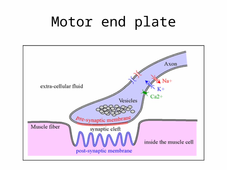
MOTOR END PLATES(MYONEURAL JUNCTIONS):
• Every skeletal muscle fiber has a motor end plate at the site where an axonal ending comes in close association with the fiber.
• As the myelinated nerves fiber reach a muscle they divide into several branches each passes to different muscle fibers, terminating there at the motor end plate.
• Close to the termination the myelin sheath disappears and the axon ends in a series of bulbous expansions that lie in troughs on the surface of the muscle fiber. These troughs are called synaptic troughs or synaptic gutters.
• Within the bulbous axonal termination are present numerous small, round, clear synaptic vesicles containing acetylcholine.
• In the base of the synaptic gutter the sarcolemma has folds which are called sub neural clefts or junctional folds.
Motor end plate

B-TERMINATION OF THE VISCERAL EFFERENT NERVE FIBERS
They are postganglionic unmyelinated nerve fibers .From autonomic ganglia they terminate into following effectors:I. Heart muscles(cardiomotor)II. Smooth muscle of blood vessels(vasomotor)III. Smooth muscle of viscera(viscermotor)IV. Smooth muscle of hair(pilomotor)V. Glandular epithelium(secretomotor)
• Near termination the visceral efferent fibers branch repeatedly forming complicated networks.
• Very fine fibers arise from these networks and terminate in bulbous enlargement.• These enlargements contains vesicles of acetylcholine in case of postganglionic
parasympathetic fibers and noradrenaline in most of the postganglionic sympathetic fibers.
• No specialized neuromuscular junctions are formed at the termination of visceral efferent nerve fibers.
• The neurotransmitter released at these terminations directly influence the target cells.

2.SENSORY NERVE ENDINGS(RECEPTORS)
The sensory nerve endings respond to stimuli in the periphery and send nerve impulses to the central nervous system (hence they may be called beginnings).Morphologically the sensory nerve endings can be divided into two main types:1. Encapsulated nerve endings2. Non-encapsulated (free) nerve endings
1-ENCAPSULATED NERVE ENDINGS
In this group of sensory nerve endings the nerve fiber terminals are enveloped by specialized connective tissue capsules.
Classified into following six types:1: TACTILE CORPUSCLES OF MEISSNER:• Meissner’s corpuscles are oval bodies covered by a connective
tissue capsule.• Within the capsule are present multilayered stacks of
transversely placed flattened cells.• Two or more myelinated nerve fibers supply each corpuscle
from which both myelinated and unmyelinated branches pass upward through the stacked cells, pursuing a zigzag or spiral course.

Location:• Located in dermal papillae• Found in nonhairy skin,espicially that of finger
tips, palms and soles.Function:It is clear from their name that the tactile corpuscles of meissner are concerned with perception of touch sensation.

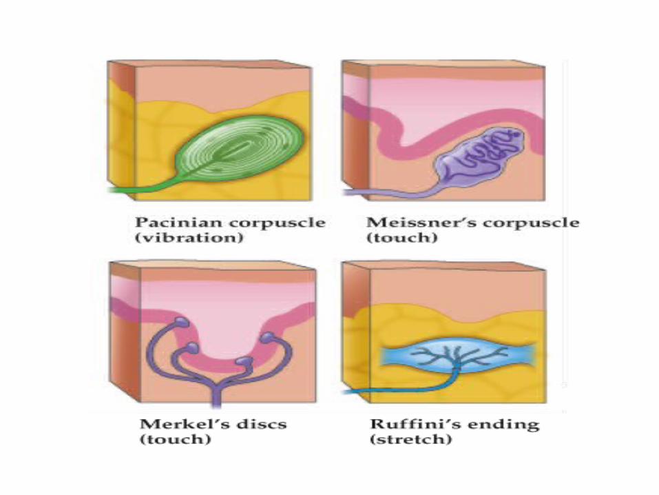
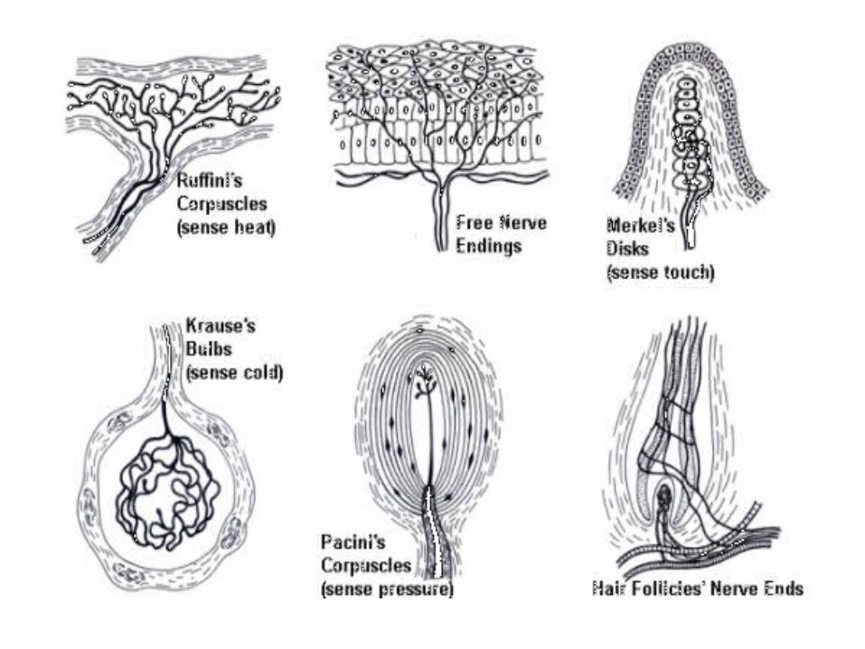
ENCAPSULATED NERVE ENDINGS….2.PACINIAN CORPUSCLES (CORPUSCLES OF VATER PACINI):• Pacinian corpuscles respond to pressure and vibration.
• They are large structures 1-2mm in length and 0.5-1mm in diameter.
• Characterized by highly developed connective tissue capsule which is composed of concentric lamellae of epithelioid fibroblast.
• In transverse section a pacinian corpuscles gives the appearance of a sliced onion.
• Each pacinian corpuscle is supplied by a single myelinated nerve fiber that looses its myelin sheath at the edge of the corpuscle and the naked axon passes through the core of the corpuscle to the end in bulbous swelling.
LOCATION:• Found in subcutaneous tissue especially that
of palms, soles and digits. • Also occur in external genitelia, periostium,
mesentery, tendons, ligament.
ENCAPSULATED NERVE ENDINGS..
3.RUFFINI’S ENDINGS:
• They are fusiform structures of 1-2mm in length.
• Each ruffini’s ending is consist of a thin connective tissue capsule enclosing a fluid filled space.
• A single myelinated nerve fiber enters the capsular space ,looses its myelin sheath and breaks up into a large number of unmyelinated branches .
LOCATION: Dermis of skin, subcutaneous tissue and in joint capsule.
FUNCTION:• They function as mechanoreceptors which are
activated by displacement of surrounding connective tissue.
• According to some investigators Ruffini’s endings are also responsible for the perception of warmth sensation.
ENCAPSULATED NERVE ENDINGS….
4.END BULBS OF KRAUSE:• They are spherical and consist of a thin connective tissue
capsule that surrounds a central cavity.• A myelinated fiber enter the cavity, looses its myelin sheath
and divides into a number of branches which terminate into club-like endings.
Location:Mainly found in dermis of the skin.Function:The end bulbs are primarily responsible for the cold sensation.

ENCAPSULATED NERVE ENDINGS….5.MUSCLE SPINDLES(NEUROMUSCULAR SPINDLES):• Fusiform structures ,ranging in length from 0.5-5mm.
• Each spindle consist of several striated muscle fibers enclosed in a connective tissue capsule these fibers are called intrafusal fibers .
• Intrafusal fibers are much smaller in diameter and length than extrfusal fibers.
Two varieties of intrafusal fibers may be distinguished in a neuromuscular spindle:

1. nuclear bag fibers• They have a central bag like dilation which has a cluster of
myonuclei.• The central dilated portion is devoid of myofibrils and
hence does not exhibit cross striations.
2. Nuclear chain fibers:The nuclear chain fibers are thinner than the nuclear bag fibers and exhibit only single row of myonuclei in their central portion which is devoid of myofibrils and cross striations.
ENCAPSULATED NERVE ENDINGS….
• A muscle spindle is supplied by both afferent (sensory) and efferent (motor) nerve fibers that enter the spindle by piercing the capsule.
• Each spindle receives a single large myelinated afferent fiber.
• This myelinated nerve fiber then looses its myelin sheath inside the capsule and divides into several branches that end in series of spirals which encircle the central portion of each nuclear bag and nuclear chain fiber.These endings are called annulospiral nerve endings.
ENCAPSULATED NERVE ENDINGS….
• ENCAPSULATED NERVE ENDINGS….
• Many spindles also receive one or more smaller afferent fibers.
• These fibers lose their myelin as they branch within the spindle capsule and terminate on nuclear chain fibers in small, multiple endings which are known as flower spray nerve endings.
• The motor nerve terminate at small motor end plates on striated portion of the intrafusal fibers .
ENCAPSULATED NERVE ENDINGS….
5.MUSCLE SPINDLES(NEUROMUSCULAR SPINDLES)………..
Location:Found in all striated muscles, often near a tendon.Function:They are concerned with regulation of reflex muscle tone.• ENCAPSULATED NERVE ENDINGS….

ENCAPSULATED NERVE ENDINGS….6.Tendon organs of Golgi (neurotendinous organs):• Consist of small bundles of collagenous fibers
enclosed in a connective tissue capsule.• A large myelinated nerve fiber enter the organ usually
at its middle and divides into numerous smaller unmyelinated branches which form an extensive network around the collagenous bundles of tendon organ.
Location:Located in tendons usually close to the muscle-tendon junctions.

Function:• They respond to an increase in the muscle tension.
• A heightened activity of these receptors exert an inhibitory effect, through interneurons of the central nervous system, upon the alpha motor neuron of the same muscle.
• Thus this reflex provides a negative feedback mechanism that prevents the development of too much tension on muscle.
2.NON-ENCAPSULATED (FREE) NERVE ENDINGS
• The most common type of sensory nerve endings in the body.
• Here the sensory nerve fibers are either unmyelinated or finely myelinated in type.
• The nerve fibers loose their covering before terminating and the naked axons divide into several branches which end blindly between the epithelial cells.
• Different fibers are functionally specialized to respond to the sensation of pain, temperature or light touch.

2.NON-ENCAPSULATED (FREE) NERVE ENDINGS
• The stratum basal of epidermis has some modified epidermal cells called Merkel cells.
• A Merkel cell and its associated nerve ending is collectively called as Merkel capsule.
• The Merkel capsule function as mechano-receptor, detecting the mechanical displacement of the skin.
• 2.NON-ENCAPSULATED (FREE) NERVE ENDINGS