lecture notes for neuroscience - studentvip · 2018-02-23 · o afferent fibres: sensory, to the...
TRANSCRIPT

Lecture notes for Neuroscience
Lecture 1: History * Ancient Egyptians: Idea of flow (too much or too little)centre of flow was the heart soul is from the heart
Hippocrates: everything comes from the brain (emotion and thoughts)
Galen: accepted brain as the seat intellect (Hippocrates) but also with flow/humers (instinct functions – liver, passion
– heart)
Aristotles: cardio-centric view of mind, opposed to Hippocrates centred beings in the heart(warmth), disregarded
the brain
Descarte’s – Thought nerves and the brain was a pump of liquid, mechanisms rather than spiritual explanations
Prof. paul broca: Bits of the nervous system that is associated with functions realised that left frontal lobe of brain
was responsible for speech “Tan” (patient)
What does nervous system do?
- Reveal universe(sensory) brain provides interpretation … exteroception
(detecting the outside world)
- Provide capacity for action
- Control homeostatic regulation
Lecture 2: How we can study it?* Neuroscience – multi-disciplinary science united by its subject – the nervous system
- CNS: brain and spine PNS: all nerves and clusters of neurons
- White matter: axon and fatty sheath Grey matter: cell bodies
- Most neurons are multipolar, 200 different types
Luigi Galvani – Italian doctor demonstrate that nerves convey electricity
- Experiments with frogs
- Simple unit of behaviour (reflexes)
Lecture 3: anatomy revision Para-sagittal – right next to midline
- Neuro-axis curves around thus transverse section can be vertical and horizontal
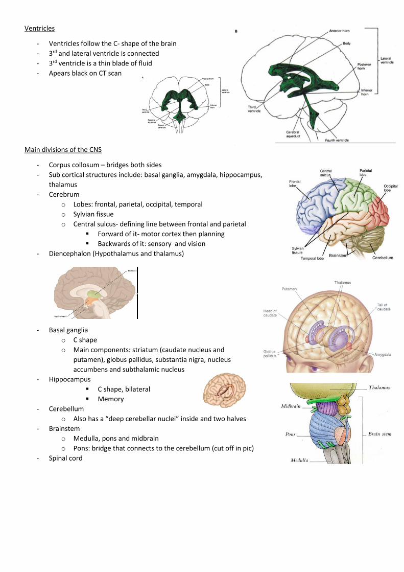
Ventricles
- Ventricles follow the C- shape of the brain
- 3rd and lateral ventricle is connected
- 3rd ventricle is a thin blade of fluid
- Apears black on CT scan
Main divisions of the CNS
- Corpus collosum – bridges both sides
- Sub cortical structures include: basal ganglia, amygdala, hippocampus,
thalamus
- Cerebrum
o Lobes: frontal, parietal, occipital, temporal
o Sylvian fissue
o Central sulcus- defining line between frontal and parietal
Forward of it- motor cortex then planning
Backwards of it: sensory and vision
- Diencephalon (Hypothalamus and thalamus)
- Basal ganglia
o C shape
o Main components: striatum (caudate nucleus and
putamen), globus pallidus, substantia nigra, nucleus
accumbens and subthalamic nucleus
- Hippocampus
C shape, bilateral
Memory
- Cerebellum
o Also has a “deep cerebellar nuclei” inside and two halves
- Brainstem
o Medulla, pons and midbrain
o Pons: bridge that connects to the cerebellum (cut off in pic)
- Spinal cord
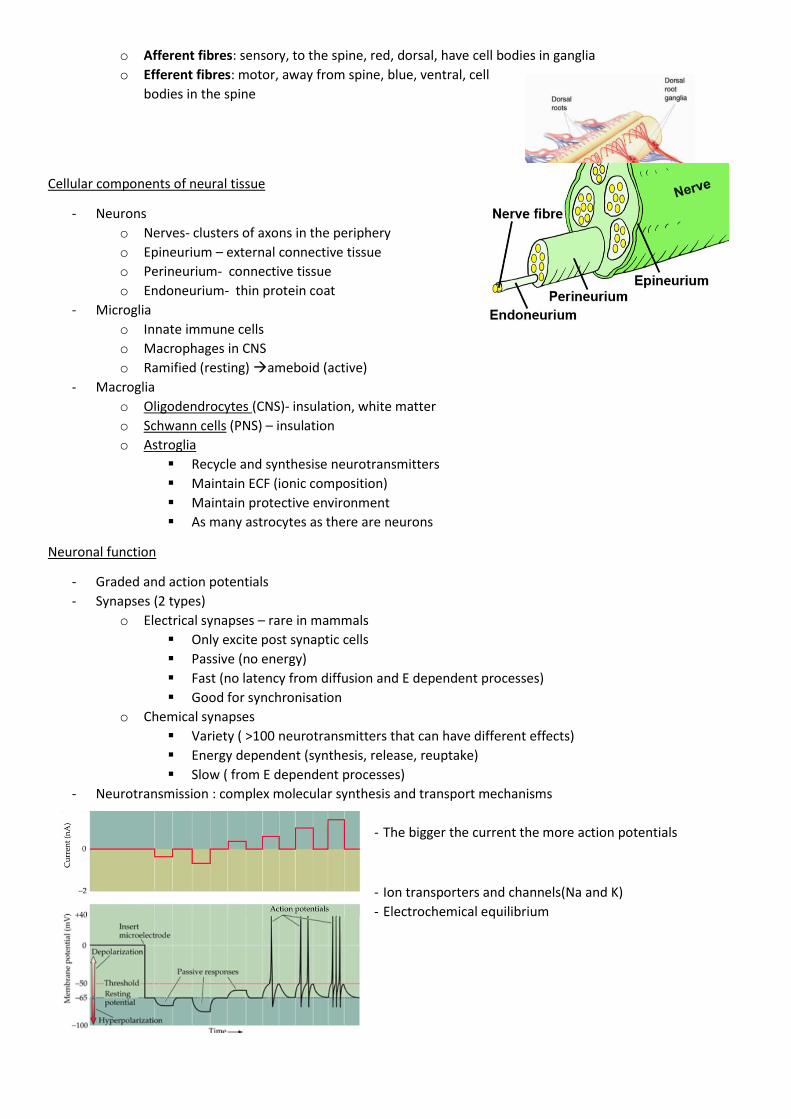
o Afferent fibres: sensory, to the spine, red, dorsal, have cell bodies in ganglia
o Efferent fibres: motor, away from spine, blue, ventral, cell
bodies in the spine
Cellular components of neural tissue
- Neurons
o Nerves- clusters of axons in the periphery
o Epineurium – external connective tissue
o Perineurium- connective tissue
o Endoneurium- thin protein coat
- Microglia
o Innate immune cells
o Macrophages in CNS
o Ramified (resting) ameboid (active)
- Macroglia
o Oligodendrocytes (CNS)- insulation, white matter
o Schwann cells (PNS) – insulation
o Astroglia
Recycle and synthesise neurotransmitters
Maintain ECF (ionic composition)
Maintain protective environment
As many astrocytes as there are neurons
Neuronal function
- Graded and action potentials
- Synapses (2 types)
o Electrical synapses – rare in mammals
Only excite post synaptic cells
Passive (no energy)
Fast (no latency from diffusion and E dependent processes)
Good for synchronisation
o Chemical synapses
Variety ( >100 neurotransmitters that can have different effects)
Energy dependent (synthesis, release, reuptake)
Slow ( from E dependent processes)
- Neurotransmission : complex molecular synthesis and transport mechanisms
- The bigger the current the more action potentials
- Ion transporters and channels(Na and K)
- Electrochemical equilibrium

- Gradient is propagated across the cell
- Myelin sheath makes the signal faster because there are only Aps happening at nodes
- Neurotransmitter synthesis made in cells body, travel through axon to dendrite
Lecture 4: Development of the Nervous System* - Trilaminar embryo: flat 3 layers disc of cells (endoderm, mesoderm, ectoderm)
o Nervous system appears in ectoderm via Neuralation
Neuralation
- Neural tube first appears as patch of specialised cells (1 cell thick) –
Neuroepithelium (stem cells of NS)
- Neural plate forms a crease which invaginates (fold)
- Forms neural tube
o 1st structure to identify as NS
o Forms brain and spinal cord
o Hollow
o Formation has a rostral to caudal gradient (rostral is older)
- Neural tube breaks free of ectoderm
Neural fold closure
- Order in which the grooves close
- Anacephaly – no head, point 2 doesn’t close brain doesn’t form
- Spina bifida- looks like a blister, fail to shut region 5, thin film of epithelium
Segmentation of Neural tube
- Rostral end of tube swells [vesiculation]3 distinct
vesicles
o Prosencephalon (forebrain)
Splits further to form telencephalon(cortex and basal ganglia) and diencephalon
Retinae form as optic vesicles
o Mesencephalon (Midbrain)
o Rhombencephalon (hindbrain)
Into 7 segments
Then splits into metencephalon and
myelencephalon (pons and medulla).
o The rest is spinal cord
- Note: optic vesicles (retina)
- The brain then forms a series of thin – walled bubbles around the fluid filled cavity of the brain

Neural crest
- Cells at the top of neural tube form neural crest which migrate away
- Establish PNS outside brain and spinal cord
o PNS
Dorsal root ganglia
Sympathetic and parasympathetic ganglia
Enteric ganglia (gut)
All glial cells in PNA (schwann cells)
o Melanocytes (pigment)
o Muscle, cartilage and bone of face, pharynx
o Dentine(part of teeth)