lecture - sensory development - embryology€¦ · lecture - sensory development ... and will form...
TRANSCRIPT

[Expand]
Human embryo sensory placodes (Week 5,stage 14)
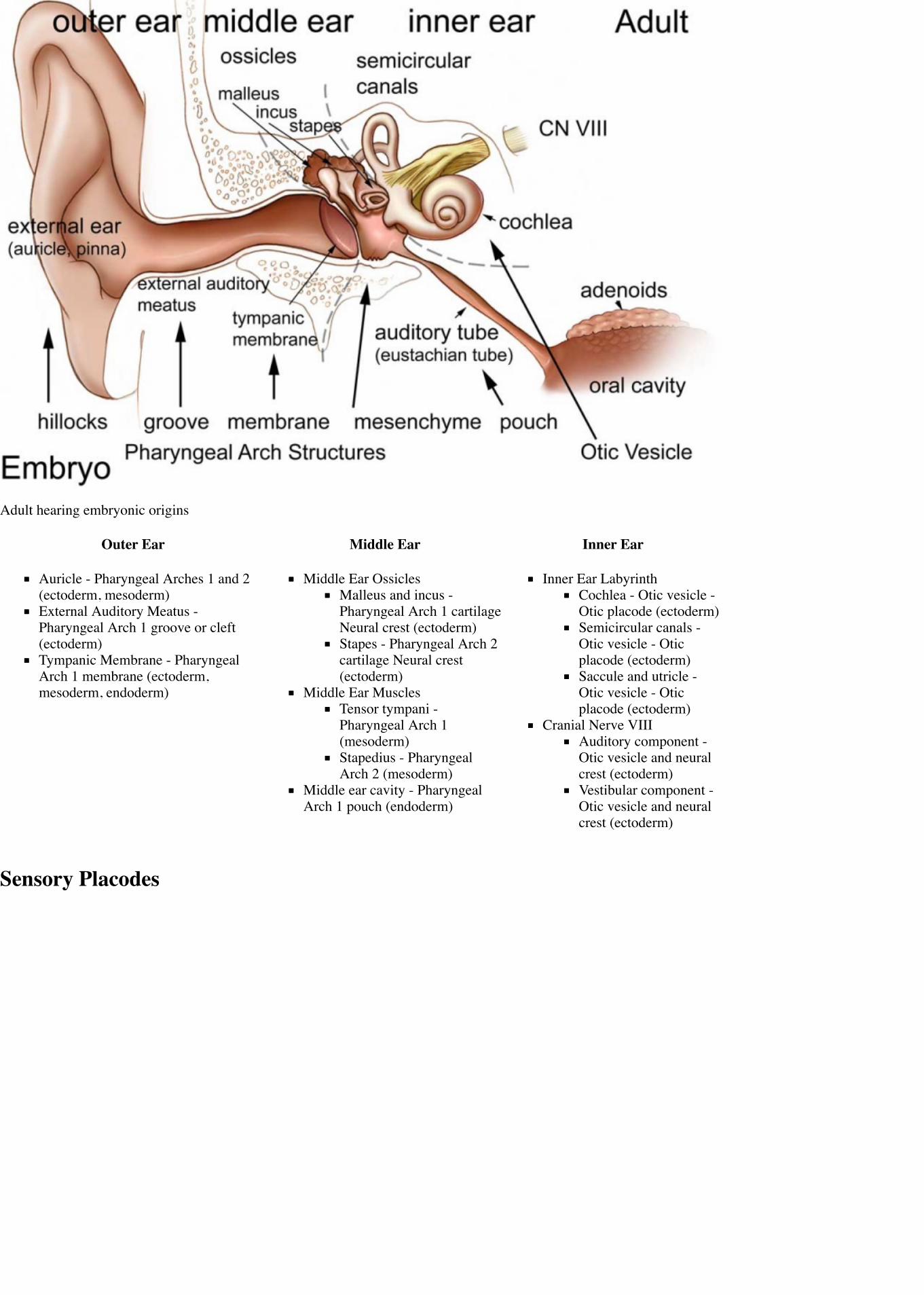
Adult hearing embryonic origins
Lecture - Sensory DevelopmentFrom Embryology
IntroductionThis lecture will introduce development of the special sensory structures associated with hearing, vision, smell andtaste. Due to time limitations the lecture will focus on hearing development and if time is available vision and othersenses will be introduced in general.
Hearing Development Vision Development Smell Development Taste Development
We use the sense of balance and hearing to position ourselves in space, sense our surrounding environment, and tocommunicate. Portions of the ear appear very early in development as specialized region (otic placode) on theembryo surface that sinks into the mesenchyme to form a vesicle (otic vesicle = otocyst) that form the inner ear.
This region connects centrally to the nervous system and peripherally through specialized bones to the external ear(auricle). This organisation develops different sources forming the 3 ear parts: inner ear (otic placode, otocyst),middle ear (1st pharyngeal pouch and 1st and 2nd arch mesenchyme), and outer ear (1st pharyngeal cleft and 6surface hillocks).
This complex origin, organisation, and timecourse means that abnormal development of any one system can impactupon the development of hearing.
ObjectivesUnderstanding of sensory placode developmentUnderstanding of inner, middle and external ear originsUnderstanding of timecourse of auditory developmentUnderstanding of abnormalities of auditory developmentBrief understanding of other sensory development
2015 Lecture 20 PDF
Also review your Head development lecture.
Lecture Resources
Movies and Virtual Slides

[Collapse]
[Expand]
[Expand]
Comparison of size at stage 14 to 23
References
Hill, M.A. (2015). UNSW Embryology (15thed.) Retrieved October 14, 2015, fromhttps://embryology.med.unsw.edu.au
Senses Links: Introduction | Placodes | Hearing and Balance | Vision | Smell | Taste | Touch | Stage22 | Category:Senses
Hearing Links: Introduction | Science Lecture | Medicine Lecture | Inner Ear | Middle Ear |Outer Ear | Balance | Hearing - Neural Pathway | Stage 22 | Abnormalities | NeonatalDiagnosis - Hearing | Hearing test | Sensory Introduction | Placodes | Student project |Category:Hearing
Historic Embryology
Vision Links: Introduction | Lens | Retina | Placodes | Extraocular Muscle | Cornea | Eyelid |Abnormalities | Student project 1 | Student project 2 | Category:Vision
Historic Embryology
Taste Links: Introduction | Student project | Tongue Development | Category:Taste
Archive: 2014 (https://embryology.med.unsw.edu.au/embryology/index.php?title=Lecture_-_Sensory_Development&oldid=149414) | 2014 PDF
Moore, K.L., Persaud, T.V.N. & Torchia,M.G. (2011). The developing human:clinically oriented embryology (9th ed.).Philadelphia: Saunders.
The following chapter links only work with a UNSW connection.
Chapter 18 – Development of Eyes and Ears (http://er.library.unsw.edu.au/er/cgi-bin/eraccess.cgi?url=http://www.mdconsult.com/books/page.do?eid=4-u1.0-B978-1-4377-2002-0..00018-7&isbn=978-1-4377-2002-0&uniqId=330028653-2#4-u1.0-B978-1-4377-2002-0..00018-7)
Schoenwolf, G.C., Bleyl, S.B., Brauer, P.R.& Francis-West, P.H. (2009). Larsen'shuman embryology (4th ed.). New York;Edinburgh: Churchill Livingstone.
The following chapter links only work with a UNSW connection.
Chapter 17 - Development of the Ears and Eyes (http://er.library.unsw.edu.au/er/cgi-bin/eraccess.cgi?url=http://er.library.unsw.edu.au/er/cgi-bin/eraccess.cgi?url=http://www.mdconsult.com/books/linkTo?type=bookPage&isbn=978-0-443-06811-9&eid=4-u1.0-B978-0-443-06811-9..10017-X)
Development TimingWeek 3 - otic placode, otic vesicleWeek 5 - cochlear part of otic vesicle elongates (humans 2.5 turns)Week 9 - Mesenchyme surrounding membranous labryinth (otic capsule) chondrifiesWeek 12-16 - Capsule adjacent to membranous labryinth undegoes vacuolization to form a cavity(perilymphatic space) around membranous labrynth and fills with perilymphWeek 16-24 - Centres of ossification appear in remaining cartilage of otic capsule form petrousportion of temporal bone. Continues to ossify to form mastoid process of temporal bone.3rd Trimester - Vibration acoustically of maternal abdominal wall induces startle response in fetus.
Embryonic Origin Overview
Adult hearing embryonic origins
Outer Ear Middle Ear Inner Ear
Auricle - Pharyngeal Arches 1 and 2(ectoderm, mesoderm)External Auditory Meatus -Pharyngeal Arch 1 groove or cleft(ectoderm)Tympanic Membrane - PharyngealArch 1 membrane (ectoderm,mesoderm, endoderm)
Middle Ear OssiclesMalleus and incus -Pharyngeal Arch 1 cartilageNeural crest (ectoderm)Stapes - Pharyngeal Arch 2cartilage Neural crest(ectoderm)
Middle Ear MusclesTensor tympani -Pharyngeal Arch 1(mesoderm)Stapedius - PharyngealArch 2 (mesoderm)
Middle ear cavity - PharyngealArch 1 pouch (endoderm)
Inner Ear LabyrinthCochlea - Otic vesicle -Otic placode (ectoderm)Semicircular canals -Otic vesicle - Oticplacode (ectoderm)Saccule and utricle -Otic vesicle - Oticplacode (ectoderm)
Cranial Nerve VIIIAuditory component -Otic vesicle and neuralcrest (ectoderm)Vestibular component -Otic vesicle and neuralcrest (ectoderm)
Sensory Placodes

Otic placodes (Stage 11 dorsal view) Sensory placodes ((Stage 14 ventral view)
week 4 a series of thickened surface ectodermal patches form in pairs in the head region.Recent research suggests that all sensory placodes may arise from common panplacodal primordium origin around the neural plate, and thendifferentiate to eventually have different developmental fates. PMID 20801420
sensory placodes will later contribute key components of each of our special senses (vision, hearing and smell).Other species have a number of additional placodes which form other sensory structures (fish, lateral line receptor).Note that their initial postion on the developing head is significantly different to their final position in the future sensory system.
Otic Placode
stage 13/14 embryo (shown below) the otic placode has sunk from the surface ectoderm to form a hollow epithelial ball, the otocyst, which now liesbeneath the surface surrounded by mesenchyme (mesoderm).The epithelia of this ball varies in thickness and has begun to distort, it will eventually form the inner ear membranous labyrinth.
Lens Placode
lies on the surface, adjacent to the outpocketing of the nervous system (which will for the retina) and will form the lens.
Nasal Placode
2 components (medial and lateral) and will form the nose olefactory epithelium.
Links: Placodes
Inner EarDescribed in detail later in lecture.

Stage 13 otocyst
Stage 22 ear
The inner ear is derived from a pair of surface sensory placodes (otic placodes) in the head region.These placodes fold inwards forming a depression, then pinch off entirely from the surface forming a fluid-filled sac or vesicle (otic vesicle, otocyst).The vesicle sinks into the head mesenchyme some of which closely surrounds the otocyst forming the otic capsule.The otocyst finally lies close to the early developing hindbrain (rhombencephalon) and the developing vestibulo-cochlear-facial ganglion complex.
Links: Inner Ear | Neuroscience - The Inner Ear (http://www.ncbi.nlm.nih.gov/books/bv.fcgi?rid=neurosci.section.894)
Middle Earderived from first pharyngeal pouch and 1st and 2nd arch mesenchymeextends as tubotympanic recess - during week 5 recess contacts outer ear canalmesoderm between 2 canals forms tympanic membraneexpands to form tympanic recessstalk of recess forms auditory tube(eustachian tube, pharyngotympanic tube)
Ossicles
The middle ear ossicles (bones) are derived from 1st and 2nd arch mesenchyme.The space in which these bones sit is derived from the 1st pharyngeal pouch.
remains connected to the oral cavity by the auditory tube.ossicles connected with tympanic cavity walls by ligaments (3 for the malleus, and 1

each for the incus and stapes).
Adult Ossicles
Malleus Incus Stapes
Pharyngeal arch cartilages
Tympanic Cavity
tympanic cavity enlargesto incorporatecoats with epitheliafirst arch mesoderm -tensor tympani musclesecond arch mesoderm -stapedius muscle
Cavities formed from the First Cleft
Middle Ear Genes - gooscoid, RARs, Prx1, Otx2, Hoxa1, Hoxb1, endothelian related molecules
Links: Middle Ear | Neuroscience - The Middle Ear (http://www.ncbi.nlm.nih.gov/books/bv.fcgi?rid=neurosci.section.893)
Outer EarThe external ear is derived from 6 surface hillocks, 3 on each of pharyngeal arch 1 and 2.The external auditory meatus is derived from the 1st pharyngeal cleft.The newborn external ear structure and position is an easily accessible diagnostic tool for potential abnormalities or further clinical screening.
Pinna- Auricle
Develops from six aural hillocks
3 on first arch3 on second archoriginally on neck, moves cranially during mandible development

arch 1 and 2 hillocks
External ear stages 14-23 and adult (not to scale)
Pharyngeal Arch Hillock Auricle ComponentArch 1 1 tragus
2 helix3 cymba concha
Arch 2 4 concha5 antihelix6 antitragus
Outer- external auditory meatus
derived from first pharyngeal cleftectodermal diverticulumweek 5 - extends inwards to pharynxuntil week 18 has ectodermal plug - plug forms stratified squamous epithelia of canal and outer eardrum
Timeline

Otic placodes (Stage 11 dorsal view)
Embryonic Period - Ectodermal cells proliferate and fill the entire lumen forming a meatal plug10 weeks - Meatal plug extends in a disc-like fashion. In the horizontal plane the meatus is boot-shapedwith a narrow neck and the sole of the meatal plug spreading widely to form the future tympanicmembrane medially. Proximal portion of the neck starts to be resorbed.13 weeks - Disc-like plug innermost surface in contact with the primordial malleus, contributes to theformation of the tympanic membrane.16.5 week - Meatus is fully patent throughout its length, lumen is still narrow and curved.18 week - Meatus is already fully expanded to its complete form.
(EAM data - Nishimura, 1992 PMID 1441991)
outer ear and external auditory meatus
Links: Outer Ear | Neuroscience - The External Ear (http://www.ncbi.nlm.nih.gov/books/bv.fcgi?rid=neurosci.section.891)
Inner
Otocyst
Week 8 cochlea
week 3 otic placode forms on surface ectodermotic placode sinks into mesodermforms otocyst (otic vesicle)branches form and generate endolymphatic duct and sacforms vestibular (dorsal) and cochlear (ventral) regionsdifferentiation of otic vesicle to membranous labyrinth
Vestibular Sac
generates 3 expansions - form semicircular ductsremainder forms utricleepithelia lining generates - hair cells, ampullary cristae, utricular maculaVestibular - Otoconia, otoconin- inner ear biominerals
Cochlear sac
generates coiled cochlear duct (humans 2 1/2 turns)remainder forms sacculeepithelia lining generateshair cellsstructures of organ of cortisaccular macula
Bony Labyrinth
formed from chrondified mesoderm
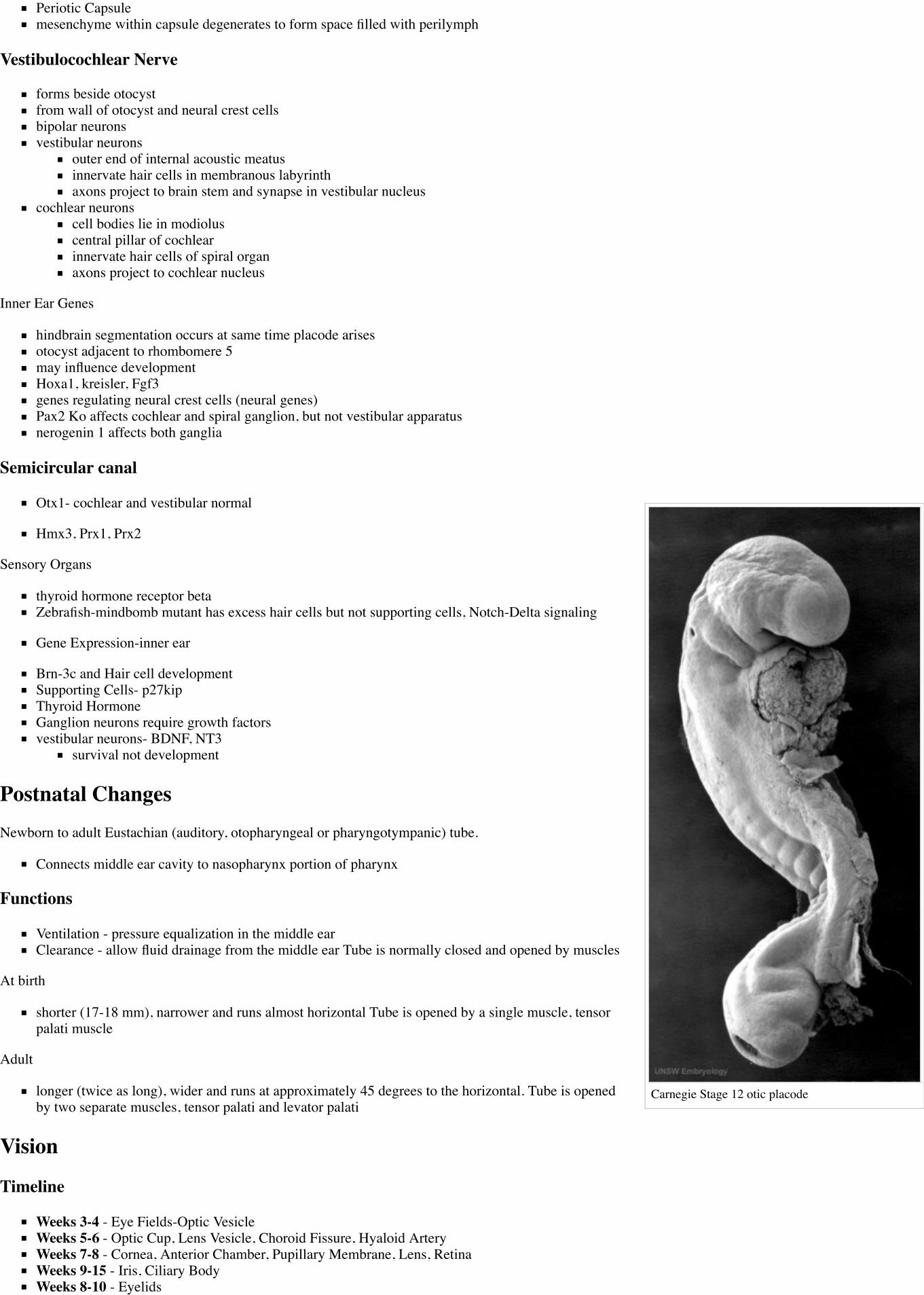
Carnegie Stage 12 otic placode
Periotic Capsulemesenchyme within capsule degenerates to form space filled with perilymph
Vestibulocochlear Nerve
forms beside otocystfrom wall of otocyst and neural crest cellsbipolar neuronsvestibular neurons
outer end of internal acoustic meatusinnervate hair cells in membranous labyrinthaxons project to brain stem and synapse in vestibular nucleus
cochlear neuronscell bodies lie in modioluscentral pillar of cochlearinnervate hair cells of spiral organaxons project to cochlear nucleus
Inner Ear Genes
hindbrain segmentation occurs at same time placode arisesotocyst adjacent to rhombomere 5may influence developmentHoxa1, kreisler, Fgf3genes regulating neural crest cells (neural genes)Pax2 Ko affects cochlear and spiral ganglion, but not vestibular apparatusnerogenin 1 affects both ganglia
Semicircular canal
Otx1- cochlear and vestibular normal
Hmx3, Prx1, Prx2
Sensory Organs
thyroid hormone receptor betaZebrafish-mindbomb mutant has excess hair cells but not supporting cells, Notch-Delta signaling
Gene Expression-inner ear
Brn-3c and Hair cell developmentSupporting Cells- p27kipThyroid HormoneGanglion neurons require growth factorsvestibular neurons- BDNF, NT3
survival not development
Postnatal ChangesNewborn to adult Eustachian (auditory, otopharyngeal or pharyngotympanic) tube.
Connects middle ear cavity to nasopharynx portion of pharynx
Functions
Ventilation - pressure equalization in the middle earClearance - allow fluid drainage from the middle ear Tube is normally closed and opened by muscles
At birth
shorter (17-18 mm), narrower and runs almost horizontal Tube is opened by a single muscle, tensorpalati muscle
Adult
longer (twice as long), wider and runs at approximately 45 degrees to the horizontal. Tube is openedby two separate muscles, tensor palati and levator palati
Vision
Timeline
Weeks 3-4 - Eye Fields-Optic VesicleWeeks 5-6 - Optic Cup, Lens Vesicle, Choroid Fissure, Hyaloid ArteryWeeks 7-8 - Cornea, Anterior Chamber, Pupillary Membrane, Lens, RetinaWeeks 9-15 - Iris, Ciliary BodyWeeks 8-10 - Eyelids

Carnegie Stage 13 otic vesicle
Inner ear hair cells
Eustacian tube angle changes
Stage 13 (week 5)
B1L B2L B3L B4L B5L B6L B7L
Lens
Surface ectoderm -> lens placode (optic placode) -> lens pit -> lens vesicle -> lens fibres -> lens capsuleand embryonic/fetal nucleus.
Retina
Neural plate ectoderm -> prosencephalon (forebrain) eye fields -> neural plate growth carries eye fieldregion forward -> eye field invaginates forming optic grooves (sulci) -> diencephalon optic groove interactswith surface ectoderm (induces optic placode) -> optic stalk -> optic vesicle -> folds inward (optic cup)forming double layer -> inner neural retina, outer pigmented retina
Links: Embryo Images - Eye Development (http://www.med.unc.edu/embryo_images/unit-eye/eye_htms/eyetoc.htm)
Neural Crest
Eye connective tissue
AbnormalitiesInner - common cavity, severe cochlear hypoplasia
Large vestibular aqueduct syndrome (LVAS) can be one of the common causes of hearing lossMiddle - rare and can be part of first arch syndrome, Malleus, Incus and Stapes Fixation
Cholesteatoma- Epithelium trapped within skull base in development, erosion of bones:temporal bone, middle ear, mastoid
Outer - Several genetic effects and syndromes, Environmental Effects
Outer Ear Abnormalities
Microtia - abnormally small external earPreauricular sinus - occurs in 0.25% births, bilateral (hereditary) 25-50%, unilateral (mainly the left),duct runs inward can extend into the parotid gland, Postnatally sites for infection
Fetal Alcohol Syndrome
Postion- Lower or uneven height, "railroad track” appearance, curve at top part of outer ear is under-developed, folded over parallel to curve beneath
Congenital Deafness
Sensorineural - cochlear or central auditory pathway
Hereditaryrecessive- severedominant- mild
can be associated with abnormal pigmentation (hair and irises)
Acquiredrubella (German measles), maternal infection during 2nd month of pregnancy, vaccination ofyoung girlsstreptomycinantibioticthalidomide
Conductive - disease of outer and middle ear
produced by otitis media with effusion, is widespread in young children.temporary blockage of outer or middle ear
Bionic EarCochlear Implant - Professor Graeme Clark (1960s, Australia) Array of electrodes implanted within cochlea, direct electrical stimulation to auditory nervefibres

vestibular sac abnormality
Microtia
Preauricular sinus
Conductive Hearing LossConductive Hearing Loss Produces a Reversible Binaural Hearing Impairment David R. Moore, Jemma E. Hine, Ze Dong Jiang, Hiroaki Matsuda, CarlH. Parsons, and Andrew J. King J. Neurosci. 1999;19 8704-8711 http://www.jneurosci.org/cgi/content/abstract/19/19/8704
tested ferrets by lon-term plugging of ear canalRepeated testing during the 22 months after unplugging revealed a gradual return to normal levels of unmasking.Results show that a unilateral conductive hearing loss, in either infancy or adulthood, impairs binaural hearing both during and after the hearingloss.Show scant evidence for adaptation to the plug and demonstrate a recovery from the impairment that occurs over a period of several months afterrestoration of normal peripheral function.
References
Textbooks
Before We Are Born (5th ed.) Moore and Persaud Chapter 20: p460-479Essentials of Human Embryology, Larson Chapter 12: p252-272
Online Textbooks
Developmental Biology (6th ed.) Gilbert, Scott F. Sunderland (MA): Sinauer Associates, Inc.;c2000. Evolution of the mammalian middle ear bones from the reptilian jaw(http://www.ncbi.nlm.nih.gov/books/bv.fcgi?rid=dbio.figgrp.5455%20) | Chick embryo rhombomereneural crest cells (http://www.ncbi.nlm.nih.gov/books/bv.fcgi?rid=dbio.figgrp.5460) | Somederivatives of the pharyngeal arches (http://www.ncbi.nlm.nih.gov/books/bv.fcgi?rid=dbio.table.3135) | Formation of the Neural Tube (http://www.ncbi.nlm.nih.gov/books/bv.fcgi?call=bv.View..ShowSection&rid=dbio.section.2871) | Differentiation of the Neural Tube(http://www.ncbi.nlm.nih.gov/books/bv.fcgi?call=bv.View..ShowSection&rid=dbio.section.2884) |Tissue Architecture of the Central Nervous System (http://www.ncbi.nlm.nih.gov/books/bv.fcgi?call=bv.View..ShowSection&rid=dbio.section.2894) | Neuronal Types(http://www.ncbi.nlm.nih.gov/books/bv.fcgi?call=bv.View..ShowSection&rid=dbio.section.2908) |Snapshot Summary: Central Nervous System and Epidermis(http://www.ncbi.nlm.nih.gov/books/bv.fcgi?call=bv.View..ShowSection&rid=dbio.section.2937)
Neuroscience Purves, Dale; Augustine, George J.; Fitzpatrick, David; Katz, Lawrence C.; LaMantia,Anthony-Samuel; McNamara, James O.; Williams, S. Mark. Sunderland (MA): Sinauer Associates,Inc. ; c2001 The Auditory System (http://www.ncbi.nlm.nih.gov/books/bv.fcgi?rid=neurosci.chapter.879) | The Inner Ear (http://www.ncbi.nlm.nih.gov/books/bv.fcgi?rid=neurosci.section.894) | The Middle Ear (http://www.ncbi.nlm.nih.gov/books/bv.fcgi?rid=neurosci.section.893) | The External Ear (http://www.ncbi.nlm.nih.gov/books/bv.fcgi?rid=neurosci.section.891) | Early Brain Development (http://www.ncbi.nlm.nih.gov/books/bv.fcgi?rid=neurosci.chapter.1447) | Construction of Neural Circuits(http://www.ncbi.nlm.nih.gov/books/bv.fcgi?rid=neurosci.chapter.1546) | Modification of BrainCircuits as a Result of Experience (http://www.ncbi.nlm.nih.gov/books/bv.fcgi?rid=neurosci.chapter.1640)
Molecular Biology of the Cell (4th Edn) Alberts, Bruce; Johnson, Alexander; Lewis, Julian; Raff,Martin; Roberts, Keith; Walter, Peter. New York: Garland Publishing; 2002. Neural Development(http://www.ncbi.nlm.nih.gov:80/books/bv.fcgi?db=Books&rid=mboc4.section.3963) | The threephases of neural development (http://www.ncbi.nlm.nih.gov:80/books/bv.fcgi?db=Books&rid=mboc4.figgrp.3966)
Clinical Methods 63. Cranial Nerves IX and X: The Glossopharyngeal and Vagus Nerves(http://www.ncbi.nlm.nih.gov/books/bv.fcgi?rid=cm.chapter.1949) | The Tongue(http://www.ncbi.nlm.nih.gov/books/bv.fcgi?rid=cm.chapter.3847) | 126. The Ear and AuditorySystem (http://www.ncbi.nlm.nih.gov/books/bv.fcgi?rid=cm.chapter.3777) | An Overview of theHead and Neck - Ears and Hearing (http://www.ncbi.nlm.nih.gov/books/bv.fcgi?rid=cm.chapter.3627#3654) | Audiometry (http://www.ncbi.nlm.nih.gov/books/bv.fcgi?rid=cm.chapter.3897)
Health Services/Technology Assessment Text (HSTAT) Bethesda (MD): National Library ofMedicine (US), 2003 Oct. Developmental Disorders Associated with Failure to Thrive(http://www.ncbi.nlm.nih.gov:80/books/bv.fcgi?db=Books&rid=hstat1a.section.25014#25029)
Eurekah Bioscience CollectionCranial Neural Crest and Development of the Head Skeleton(http://www.ncbi.nlm.nih.gov/books/bv.fcgi?rid=eurekah.chapter.53006)
Search
Bookshelf hearing development (http://www.ncbi.nlm.nih.gov/sites/entrez?db=Books&cmd=search&term=hearing+development)
Pubmed hearing development (http://www.ncbi.nlm.nih.gov/sites/gquery?itool=toolbar&cmd=search&term=hearing+development)
External LinksExternal Links Notice - The dynamic nature of the internet may mean that some of these listed links may no longer function. If the link no longer works searchthe web with the link text or name.
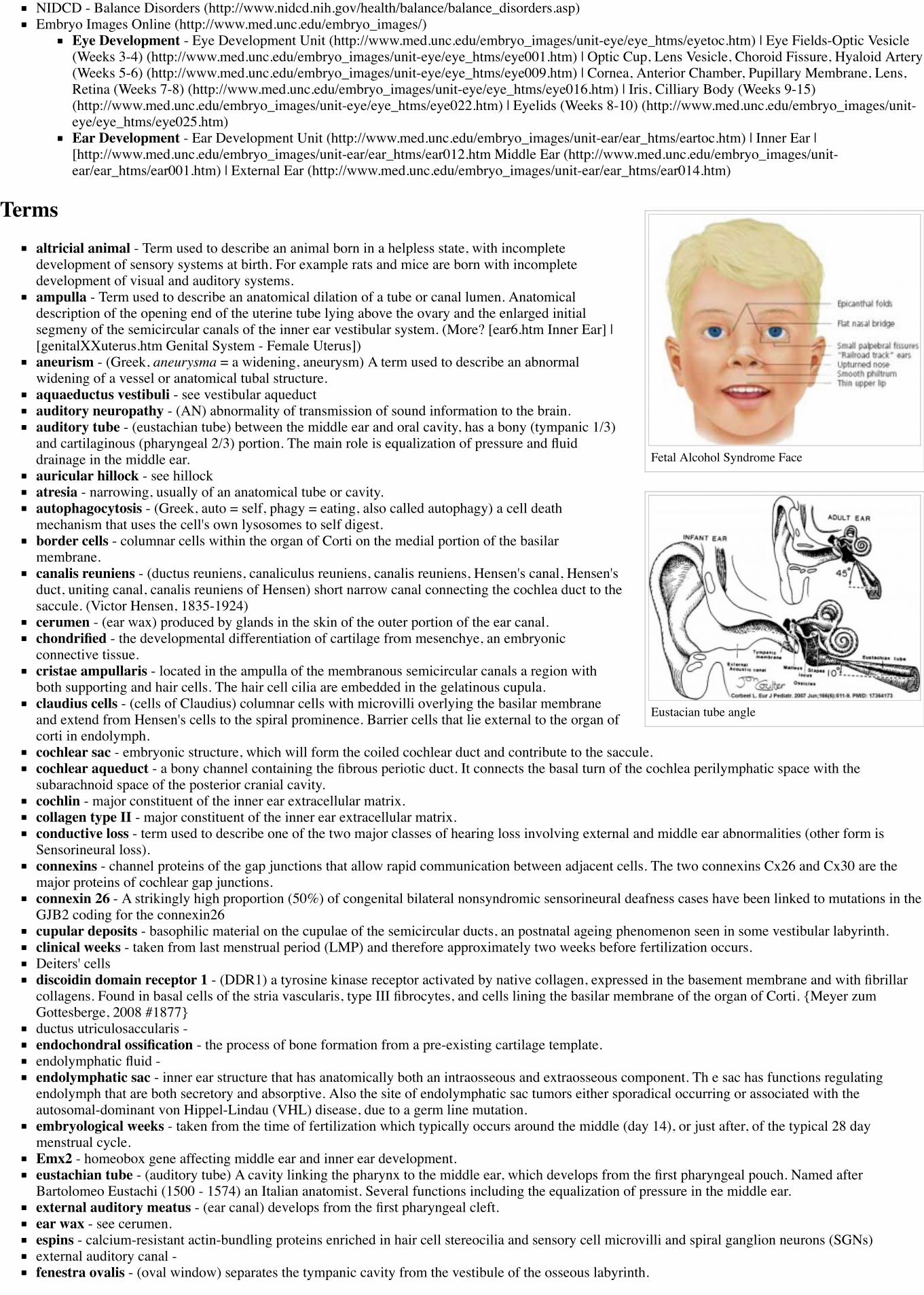
Fetal Alcohol Syndrome Face
Eustacian tube angle
NIDCD - Balance Disorders (http://www.nidcd.nih.gov/health/balance/balance_disorders.asp)Embryo Images Online (http://www.med.unc.edu/embryo_images/)
Eye Development - Eye Development Unit (http://www.med.unc.edu/embryo_images/unit-eye/eye_htms/eyetoc.htm) | Eye Fields-Optic Vesicle(Weeks 3-4) (http://www.med.unc.edu/embryo_images/unit-eye/eye_htms/eye001.htm) | Optic Cup, Lens Vesicle, Choroid Fissure, Hyaloid Artery(Weeks 5-6) (http://www.med.unc.edu/embryo_images/unit-eye/eye_htms/eye009.htm) | Cornea, Anterior Chamber, Pupillary Membrane, Lens,Retina (Weeks 7-8) (http://www.med.unc.edu/embryo_images/unit-eye/eye_htms/eye016.htm) | Iris, Cilliary Body (Weeks 9-15)(http://www.med.unc.edu/embryo_images/unit-eye/eye_htms/eye022.htm) | Eyelids (Weeks 8-10) (http://www.med.unc.edu/embryo_images/unit-eye/eye_htms/eye025.htm)Ear Development - Ear Development Unit (http://www.med.unc.edu/embryo_images/unit-ear/ear_htms/eartoc.htm) | Inner Ear |[http://www.med.unc.edu/embryo_images/unit-ear/ear_htms/ear012.htm Middle Ear (http://www.med.unc.edu/embryo_images/unit-ear/ear_htms/ear001.htm) | External Ear (http://www.med.unc.edu/embryo_images/unit-ear/ear_htms/ear014.htm)
Termsaltricial animal - Term used to describe an animal born in a helpless state, with incompletedevelopment of sensory systems at birth. For example rats and mice are born with incompletedevelopment of visual and auditory systems.ampulla - Term used to describe an anatomical dilation of a tube or canal lumen. Anatomicaldescription of the opening end of the uterine tube lying above the ovary and the enlarged initialsegmeny of the semicircular canals of the inner ear vestibular system. (More? [ear6.htm Inner Ear] |[genitalXXuterus.htm Genital System - Female Uterus])aneurism - (Greek, aneurysma = a widening, aneurysm) A term used to describe an abnormalwidening of a vessel or anatomical tubal structure.aquaeductus vestibuli - see vestibular aqueductauditory neuropathy - (AN) abnormality of transmission of sound information to the brain.auditory tube - (eustachian tube) between the middle ear and oral cavity, has a bony (tympanic 1/3)and cartilaginous (pharyngeal 2/3) portion. The main role is equalization of pressure and fluiddrainage in the middle ear.auricular hillock - see hillockatresia - narrowing, usually of an anatomical tube or cavity.autophagocytosis - (Greek, auto = self, phagy = eating, also called autophagy) a cell deathmechanism that uses the cell's own lysosomes to self digest.border cells - columnar cells within the organ of Corti on the medial portion of the basilarmembrane.canalis reuniens - (ductus reuniens, canaliculus reuniens, canalis reuniens, Hensen's canal, Hensen'sduct, uniting canal, canalis reuniens of Hensen) short narrow canal connecting the cochlea duct to thesaccule. (Victor Hensen, 1835-1924)cerumen - (ear wax) produced by glands in the skin of the outer portion of the ear canal.chondrified - the developmental differentiation of cartilage from mesenchye, an embryonicconnective tissue.cristae ampullaris - located in the ampulla of the membranous semicircular canals a region withboth supporting and hair cells. The hair cell cilia are embedded in the gelatinous cupula.claudius cells - (cells of Claudius) columnar cells with microvilli overlying the basilar membraneand extend from Hensen's cells to the spiral prominence. Barrier cells that lie external to the organ ofcorti in endolymph.cochlear sac - embryonic structure, which will form the coiled cochlear duct and contribute to the saccule.cochlear aqueduct - a bony channel containing the fibrous periotic duct. It connects the basal turn of the cochlea perilymphatic space with thesubarachnoid space of the posterior cranial cavity.cochlin - major constituent of the inner ear extracellular matrix.collagen type II - major constituent of the inner ear extracellular matrix.conductive loss - term used to describe one of the two major classes of hearing loss involving external and middle ear abnormalities (other form isSensorineural loss).connexins - channel proteins of the gap junctions that allow rapid communication between adjacent cells. The two connexins Cx26 and Cx30 are themajor proteins of cochlear gap junctions.connexin 26 - A strikingly high proportion (50%) of congenital bilateral nonsyndromic sensorineural deafness cases have been linked to mutations in theGJB2 coding for the connexin26cupular deposits - basophilic material on the cupulae of the semicircular ducts, an postnatal ageing phenomenon seen in some vestibular labyrinth.clinical weeks - taken from last menstrual period (LMP) and therefore approximately two weeks before fertilization occurs.Deiters' cellsdiscoidin domain receptor 1 - (DDR1) a tyrosine kinase receptor activated by native collagen, expressed in the basement membrane and with fibrillarcollagens. Found in basal cells of the stria vascularis, type III fibrocytes, and cells lining the basilar membrane of the organ of Corti. {Meyer zumGottesberge, 2008 #1877}ductus utriculosaccularis -endochondral ossification - the process of bone formation from a pre-existing cartilage template.endolymphatic fluid -endolymphatic sac - inner ear structure that has anatomically both an intraosseous and extraosseous component. Th e sac has functions regulatingendolymph that are both secretory and absorptive. Also the site of endolymphatic sac tumors either sporadical occurring or associated with theautosomal-dominant von Hippel-Lindau (VHL) disease, due to a germ line mutation.embryological weeks - taken from the time of fertilization which typically occurs around the middle (day 14), or just after, of the typical 28 daymenstrual cycle.Emx2 - homeobox gene affecting middle ear and inner ear development.eustachian tube - (auditory tube) A cavity linking the pharynx to the middle ear, which develops from the first pharyngeal pouch. Named afterBartolomeo Eustachi (1500 - 1574) an Italian anatomist. Several functions including the equalization of pressure in the middle ear.external auditory meatus - (ear canal) develops from the first pharyngeal cleft.ear wax - see cerumen.espins - calcium-resistant actin-bundling proteins enriched in hair cell stereocilia and sensory cell microvilli and spiral ganglion neurons (SGNs)external auditory canal -fenestra ovalis - (oval window) separates the tympanic cavity from the vestibule of the osseous labyrinth.

fenestra rotunda - (round window) separates the tympanic cavity from the scala tympani of the cochlea.fetus - (foetus) term used to describe human development after the 8th week (10th clinical week, LPM) and covers the developmental periods of secondand third trimester.fibroblast growth factor 1 - (Fgf-1) a growth factor released from cochlea sensory epithelium which stimulates spiral ganglion neurite branching.fibroblast growth factor 8 - (Fgf-8) a growth factor released by inner hair cells which regulates pillar cell number, position and rate of development.fibroblast growth factor receptor 3 - (Fgfr-3) a tyrosine kinase receptor with a role in the commitment, differentiation and position of pillar cells in theorgan of cortifundamental frequency - (natural frequency) the lowest frequency in a harmonic series, for the female voice this is about 225 Hz.helicotrema - term used to describe the cochlear apex.Hes - (hairy and enhancer of split) family of factors, which has been shown to be a general negative regulator of neurogenesis (Zheng, 2000).hillock - a small hill, used to describe the six surface elevations on pharyngeal arch one and two.Hindbrain - Invaginate -Incus - (anvil) auditory ossicleinner phalangeal cellsinner pillar cells - organ of Corti cells arranged in rows and form a boundary between the single row of inner hair cells and three rows of outer hair cells.These cells have surface-associated microtubule bundles.inner sulcus - area of the cochlear ductinterdental region -internal auditory meatus - (internal acoustic meatus, IAM) Anatomical canal in which CN VII and CN VIII ganglia reside and pass through to thebrainstem. This bony canal lies between the posterior surface of the petrous pyramid and the bony labyrinth within the dense petrous bone. Alsoassociated clinically with the site where acoustic neuromas may occur.Kolliker's organ - (Kollicker's organ, greater epithelial ridge) Developing cochlear structure consisting of columnar-shaped supporting cells filling theinner sulcus and lying directly under the tectorial membrane. This transient organ regresses and generates the space of the inner sulcus. Rudolph Albertvon Kolliker (1817-1905)??lateral semicircular duct -Limbus -LMP - acronym for last menstrual period, used to clinically measure gestation.malleus - (hammer) auditory ossiclemastoid process - of temporal boneMath1 - homolog of the Drosophila proneural gene atonal, necessary and sufficient for the production of hair cells in the mouse inner ear. {Chen, 2002#1932}Negatively regulated by Hes1 and Hes5meatal plug - temporary blockage of the external auditory meatus which forms at the end of the embryonic period and remains present until the seventhmonth.meatus - anatomical opening, cavity or space (external acoustic meatus,internal auditory meatus)Meckel's cartilage - first pharyngeal ach cartilage, located within the mandibular prominence. This cartilage first appears at stage 16, stage 20 thebeginning of membranous ossification. Named after Johann Friedrich Meckel, (1781 - 1833) a German anatomist.(http://www.whonamedit.com/doctor.cfm/1840.html)membranous labyrinth - Mesenchyme - Mesoderm - Microtia - Modiolus -mucopolysaccharidosis - (MPS IIIB, Sanfilippo Syndrome type B) abnormality caused by a deficiency in the lysosomal enzyme N-acetyl-glucosaminidase (Naglu). Children with MPS IIIB develop abnormal hearing, and mental functioning culminating in early death.netrin-1 - secreted growth factor, expressed in the organ of Corti and spiral ganglion cells, role in process outgrowth.neural tube -olivocochlear - brainstem cholinergic and GABAergic efferent system that innervates sensory cells and sensory neurons of the inner ear.organ of Corti - organ of Corti protein II - (OCP-II) cytosolic protein or transcription factor?otolithic membrane - extracellular matrix that cover the sensory epithelia of the inner ear.ossicle - (small bone) the individual bone of the three middle ear bones (auditory ossicles), which reduce vibrational amplitude but increase force to drivefluid-filled inner ear.ossify - the process of bone formation.otic capsule -otic cupotic placode -otic vesicle -otoconin - inner ear biominerals required for vestibular apparatus function.otogelin - (Otog) an inner ear specific glycoprotein expressed in cochlea cells at different developmental times.otolithic membrane - a membrane within the utricle and saccule containing embedded hair cell cilia and small crystalline bodies of calcium carbonate(otoliths). Functions to detect head motion.otoliths - small crystalline bodies of calcium carbonate found within the otolitic membrane of the utricle and saccule.ototoxic - compound or drug causing temporary or permanent hearing loss.outer hair cells - (OHCs) three rows of hair cells that function to increase basilar membrane motion through a local mechanical feedback process withinthe cochlea, the "cochlear amplifier".outer pillar cells - arranged in rows and form a boundary between the single row of inner hair cells and three rows of outer hair cells.paratubal musculature - muscles lying beside the auditory (Eustachian) tube. The tensor veli, palatini (TVP) and tensor tympani muscles.perilymph - perilymphatic space - Periotic Capsule - petrous portion - of temporal bonepejvakin gene - in humans, two missense mutations in this gene cause nonsyndromic recessive deafness (DFNB59) by affecting the function of auditoryneurons.pharyngeal archpharyngeal pouchpharyngeal membranePharynxpillar cells - (PC) form an inner and outer row of support cells that form a boundary between inner and outer hair cells.Placodepreyer reflex - ear flick in mouse in response to sound.presbyacusisprestin - a motor protein structurally similar to the anion transporter family expressed in cochlear outer hair cells.preauricular tag - skin tags located in front of the external ear opening, are common in neonates and in most cases are normal, though in some cases areindicative of other associated abnormalities.primordium-protocadherin 15 - (Pcdh15) required for initial formation of stereocilia bundles and changes in the actin meshwork within hair cells. The Ames waltzer(av) mouse mutant has both auditory and vestibular abnormalities from a mutation in this gene.Reichert's cartilage - pharyngeal ach 2 cartilage, named after Karl Bogislaus Reichert (1811 - 1883) a German anatomist.Reissner's membrane - (vestibular membrane, vestibular wall) is a membrane located inside the cochlea separating the scala media from scala vestibuli.Named after Ernst Reissner (1824-1878) a German anatomist. “It primarily functions as a diffusion barrier, allowing nutrients to travel from the

perilymph to the endolymph of the membranous labyrinth.rhombomere -Saccular macula -Saccule - (Latin, sacculus = a small pouch)sacculocollic reflex -scala tympani - one of the three Cochlea cavities, it is filled with perilymph.Scarpa's ganglion - (vestibular ganglion) primary afferent vestibular neuron ganglion of the vestibular nerve. Located within the internal auditorymeatus.semicircular canals - series of fluid-filled loops of the inner ear required for balance and sensing acceleration.sensorineural - term used to describe one of the two major classes of hearing loss involving the central pathway from the cochlear (other form isconductive loss).space of Nuel - within the cochlea, an organ of Corti space between the outer pillar cells and the phalangeal and hair cells. Named after Jean-Pierre Nuel(1847-1920) a Belgian ophthalmologist.spiral ganglion neurons - (SGN) innervate the inner (Type I) and outer (Type II) hair cells of the cochlea.stapedius muscle - (innervated by CN VII tympanic branch) one of the two muscles in the middle ear, contraction of this muscle pulls the stapes anddampens auditory ossicle movement.stapes - (stirrup) a middle ear auditory ossicle (bone).stapes footplate - startle response -stereocilia -finger-like projections from the apical surface of sensory hair cells forming the hair bundle in the cochlea. Formed by tightly cross-linkedparallel actin filaments in a paracrystalline array with cell surface specializations (tip links, horizontal top connectors, and tectorial membrane attachmentcrowns).stratified squamous epithelia - classification of epithelium which transiently forms a plug in external ear canal to the outer eardrum.stria vascularis - forms the outer wall of the cochlear duct of the mammalian cochlea is composed primarily of three types of cells. Marginal cells linethe lumen of the cochlear duct and are of epithelial origin. Basal cells also form a continuous layer and they may be mesodermal or derived from theneural crest. Intermediate cells are melanocyte-like cells, presumably derived from the neural crest, and are scattered between the marginal and basal celllayers. The stria forms endolymph and also contains a rich supply of blood vessels.sulcus -synostotically - anatomically normally separate skeletal bones fused together.tectorial membrane - extracellular matrix that cover the sensory epithelial hair cells of the organ of corti within the cochlea.alpha-tectorin and beta- (TECTA, TECTB) major non-collagenous protein component of the tectorial membrane forming a striated-sheet matrix.Synthesized as glycosylphosphatidylinositol-linked, membrane bound precursors.temporal bone -tensor tympani - (innervated by CN V mandibular nerve) one of the two muscles in the middle ear, contraction of this muscle pulls the malleus andtenses the tympanic membrane, dampening auditory ossicle movement. The muscle arises from auditory tube (cartilaginous portion) and is inserted intothe malleus (manubrium near the root).teratogens - trilaminar embryo -tonotopy - term describing the mapping along the tectorial membrane within the cochlea of the different sound frequencies.tympanic cavity - tympanic membrane -Utricle -Vacuolization - Vesicle - vestibular apparatus - vestibular evoked myogenic potential (VEMP) testvestibular ganglion - (Scarpa's ganglion) primary afferent vestibular neuron ganglion of the vestibular nerve. Located within the internal auditorymeatus.vestibular membrane - (Reissner's) extends from the spiral lamina to the outer wall and divides the cochlea into an upper scala vestibuli, a lower scalatympani.Vestibulocochlear Nerve - Cranial Nerve VIIIWhirlin - A PDZ scaffold protein expressed in hair cells at the stereocilia tips, essential for the stereocilia elongation process. The DFNB31 genemutations cause hearing loss in human and mouse. This protein can interact with membrane-associated guanylate kinase (MAGUK) protein, erythrocyteprotein p55 (p55).Wnt7a - signaling through the Wnt pathway regulates the development of hair cell unidirectional stereociliary bundle orientation.
2015 Course: Week 2 Lecture 1 Lecture 2 Lab 1 | Week 3 Lecture 3 Lecture 4 Lab 2 | Week 4 Lecture 5 Lecture 6 Lab 3 | Week 5 Lecture 7 Lecture 8Lab 4 | Week 6 Lecture 9 Lecture 10 Lab 5 | Week 7 Lecture 11 Lecture 12 Lab 6 | Week 8 Lecture 13 Lecture 14 Lab 7 | Week 9 Lecture 15 Lecture 16Lab 8 | Week 10 Lecture 17 Lecture 18 Lab 9 | Week 11 Lecture 19 Lecture 20 Lab 10 | Week 12 Lecture 21 Lecture 22 Lab 11 | Week 13 Lecture 23Lecture 24 Lab 12 | Projects: Group 1 | Group 2 | Group 3 | Group 4 | Group 5 | Group 6 | Students | Student Designed Quiz Questions | Moodle page(http://moodle.telt.unsw.edu.au/course/view.php?id=15814)
Retrieved from ‘https://embryology.med.unsw.edu.au/embryology/index.php?title=Lecture_-_Sensory_Development&oldid=205283’
Categories: Senses Hearing Sensory Vision Taste 2015 Science-Undergraduate
This page was last modified on 14 October 2015, at 14:19.This page has been accessed 7,432 times.