legionella pneumophila proteins that regulate rab1 membrane cycling
TRANSCRIPT

ARTICLES
Legionella pneumophila proteins thatregulate Rab1 membrane cyclingAlyssa Ingmundson1, Anna Delprato2, David G. Lambright2 & Craig R. Roy1
Rab1 is a GTPase that regulates the transport of endoplasmic-reticulum-derived vesicles in eukaryotic cells. The intracellularpathogen Legionella pneumophila subverts Rab1 function to create a vacuole that supports bacterial replication by amechanism that is not well understood. Here we describe L. pneumophila proteins that control Rab1 activity directly. Weshow that a region in the DrrA (defect in Rab1 recruitment A) protein required for recruitment of Rab1 to membranesfunctions as a guanine nucleotide dissociation inhibitor displacement factor. A second region of the DrrA protein stimulatedRab1 activation by functioning as a guanine nucleotide exchange factor. The LepB protein was found to inactivate Rab1 bystimulating GTP hydrolysis, indicating that LepB has GTPase-activating protein activity that regulates removal of Rabproteins from membranes. Thus, L. pneumophila encodes proteins that regulate three distinct biochemical reactions criticalfor Rab GTPase membrane cycling to redirect Rab1 to the pathogen-occupied vacuole and to control Rab1 function.
Rab proteins are molecular switches that have an important role inorganizing membranes to facilitate transport and fusion of vesicularcarriers1. Rab GTPases localize and control membrane dynamics onspecific subcellular organelles. Successful transport and maturationof vesicles coincides with a change in the composition of Rab pro-teins—a process called Rab conversion2. Several accessory proteinsregulate the Rab GTPase cycle. Prenylated Rab proteins in theirinactive GDP-bound state are solubilized from membranes by asso-ciating with Rab-guanine nucleotide dissociation inhibitor (RabGDI)3–5. There is evidence that soluble Rab proteins must be releasedfrom Rab GDI by a GDI-displacement factor (GDF) before mem-brane recruitment and activation by a cognate guanine nucleotideexchange factor (GEF)6. An active Rab protein recruits specific effec-tors that have a direct role in promoting membrane transport, vesicletethering and fusion7,8. To complete the process of membrane cyc-ling, Rab proteins are inactivated by cognate GTPase-activating pro-teins (GAPs)9,10 and are extracted from the membrane by Rab GDI5.
After uptake by phagocytic host cells, the bacterial pathogenL. pneumophila exploits the activities of the Rab1 protein to directthe maturation of the vacuole in which it resides and to create anendoplasmic-reticulum-derived organelle that supports intracellularreplication11–15. Because the vacuole, which is initially derived fromplasma membrane, is remodelled into an endoplasmic-reticulum-like compartment, host proteins involved in regulating endoplasmicreticulum to Golgi transport are recruited to the L. pneumophila-containing vacuole (LCV) through the activities of L. pneumophilaproteins delivered into the host cytosol by a bacterial type IV secre-tion apparatus called the Dot/Icm system14–18. Rab1 is one of the hostregulatory factors recruited to the LCV14,15. Although the mechanismby which Rab1 is recruited to the LCV is not well described, it wasdetermined that the association of Rab1 with the LCV requires theL. pneumophila protein DrrA (also known as SidM)19,20.
Previous biochemical studies have shown that DrrA functions as aRab1-specific GEF19,20. Nucleotide exchange reactions using purifiedRab1 protein revealed that a carboxy-terminal region of DrrA con-sisting of amino acid residues 451–647, DrrA(451–647), had the same
GEF activity and specificity for Rab1 as the full-length DrrA pro-tein19. Importantly, a DrrA derivative, consisting of residues 61–647, DrrA(61–647), could recruit Rab1 to the plasma membranewhen expressed ectopically in mammalian cells, whereas the plasma-membrane-localized DrrA(451–647) protein could not recruit Rab1(ref. 19). These data indicated that deletion of residues 61–450 inDrrA eliminated a second biochemical activity essential for the mem-brane recruitment and activation of Rab1 from the host cytosol. Herewe show that region 61–450 in DrrA is necessary to displace Rab GDIfrom a complex containing Rab1, indicating that DrrA has GDFactivity. In addition, we show that the L. pneumophila LepB proteinis a Rab1 GAP capable of inactivating Rab1.
DrrA has both GDF and GEF activity
The observation that the GEF domain alone is not sufficient forDrrA-mediated recruitment of Rab1 to the plasma membrane19 indi-cated that DrrA might also function as a GDF. We reasoned that, ifDrrA is able to displace Rab GDI in vivo, ectopic production of DrrAin eukaryotic cells should decrease the proportion of Rab1 that isbound to Rab GDI. Rab1 tagged with the Flag epitope (33Flag–Rab1) was affinity-purified using anti-Flag agarose from HEK293cells producing Rab GDI and green fluorescent protein (GFP)-taggedDrrA proteins. Rab GDI was not detected in association with33Flag–Rab1 isolated from cells producing GFP–DrrA(61–647)(Fig. 1a), a derivative shown previously to recruit Rab1 to theplasma membrane19. In contrast, Rab GDI was associated with33Flag–Rab1 isolated from cells producing either GFP alone orGFP–DrrA(451–647) with an intact GEF domain (Fig. 1a). Thesedata indicate that the DrrA protein can interfere with Rab1–Rab-GDI complex formation by a mechanism that requires residues out-side the GEF domain.
To determine whether DrrA could displace Rab GDI in vitro,purified DrrA was added to 33Flag–Rab1–Rab-GDI complexattached to anti-Flag agarose, and Rab GDI displacement into thesupernatant was measured over time. Rab GDI was efficiently dis-placed when DrrA protein was added to the reaction (Fig. 1b, c).
1Section of Microbial Pathogenesis, Yale University School of Medicine, Boyer Center for Molecular Medicine, 295 Congress Avenue, New Haven, Connecticut 06536, USA. 2Programin Molecular Medicine and Department of Biochemistry and Molecular Pharmacology, UMASS Medical School Two Biotech, 373 Plantation Street, Worcester, Massachusetts 01605,USA.
Vol 450 | 15 November 2007 | doi:10.1038/nature06336
365Nature ©2007 Publishing Group

DrrA also displaced Rab GDI from a complex containing 33Flag–Rab2 (Supplementary Information and Supplementary Fig. 1), indi-cating that DrrA may function in a similar manner to the eukaryoticprotein Rab acceptor 1 (prenylated) (Rabac1, also known as Yip3 inyeast and PRA1 in human) and promote the displacement of mul-tiple Rab proteins from the complex with Rab GDI21. DrrA(451–647)did not stimulate Rab GDI release, consistent with Rab GDI displace-ment activity requiring residues outside of the GEF domain.Importantly, DrrA(1–500), which lacks Rab1 GEF activity19, was suf-ficient to displace Rab GDI from Rab1, indicating that Rab1 nucleo-tide exchange is not required for Rab GDI displacement by DrrA.Thus, Rab GDI displacement and Rab1 guanine nucleotide exchangeare separate activities mediated by the DrrA protein.
Interactions between Rab GDI and a Rab protein can interfere withGEF activation because Rab GDI binding directly engages the Rabswitch regions and inhibits GDP release22,23. A GDF is predicted torecognize both Rab GDI and the Rab protein, and release Rab GDIfrom the complex, to enable a GEF to access the switch regions andcatalyse nucleotide exchange. Our data indicated that DrrA possessesboth GDF and GEF activity, predicting that DrrA should be ableto activate Rab1 even when Rab1–Rab-GDI is used as a substrate.The sensitized emission accompanying tryptophan to 29(39)-O-(N-methylanthraniloyl) (mant) fluorescence resonance energy transferwas used to measure the exchange of GDP for mant-GTP usingRab1–Rab-GDI as a substrate. Full-length DrrA activated Rab1,whereas the DrrA(451–647) protein containing the GEF domainbut deficient in GDF activity was unable to activate Rab1 when theRab1–Rab-GDI complex was used as a substrate (Fig. 1d). These dataare in contrast to previous data showing that full-length DrrA andDrrA(451–647) had similar catalytic activities in exchange reactionsfor Rab1 alone, and demonstrate that when Rab1–Rab-GDI is used asa substrate, Rab GDI displacement is essential for DrrA-mediatedactivation of Rab1.
LepB binds Rab1–GTP
Because Rab1 on the LCV would be available for binding to otherL. pneumophila proteins after DrrA activation, we investigated
whether any Dot/Icm-translocated proteins of unknown functioncould interact with Rab1 in a GTP-bound conformation. Thiswas done using the GTP-locked glutathione S-transferase (GST)–Rab1(Q70L) protein (in which Gln 70 was mutated to Leu) to co-precipitate GFP-tagged L. pneumophila proteins from transfectedmammalian cell lysates. This analysis revealed an interaction betweenGST–Rab1(Q70L) and the L. pneumophila protein LepB (Fig. 2a).The GDP-locked GST–Rab1(S25N) protein did not bind to LepB,indicating that LepB interacts preferentially with active Rab1–GTP(Fig. 2a). LepB did not bind to the GTP-locked GST–Rab7(Q67L),indicating specificity for the interaction between LepB and Rab1.Direct binding between purified His–LepB and GST–Rab1(Q70L)was detected, indicating that this interaction does not require othercellular factors (Fig. 2b).
LepB is translocated shortly after uptake
Previous studies have indicated that LepB is involved in programmedrelease of L. pneumophila from protozoan host cells, and not mam-malian cells, by an exocytic process that occurs after bacteria havecompleted their intracellular replication phase24. Thus, the possibilitythat Rab1 is a target of LepB function was surprising given that Rab1is conserved in protozoan and metazoan organisms, and becauseRab1 is found on the L. pneumophila vacuole shortly after bacterialuptake and is removed from the vacuole before bacterial replicationinitiates14,15. Spatial and temporal analysis of LepB localization inL. pneumophila-infected macrophages was conducted to determinewhether the kinetics of LepB and Rab1 association with the LCV weresimilar. Immunofluorescence microscopy revealed detectible levelsof LepB on LCVs containing a single bacterium at 3 h post infection(Fig. 2c). LepB was also present on mature endoplasmic-reticulum-derived vacuoles containing replicating L. pneumophila at 11 h postinfection (Fig. 2c). DrrA and Rab1 were present on early LCVs at the30-min time point after infection, whereas LepB was not detected thisearly (Fig. 2d). The percentage of LCVs positive for Rab1 peaked at2 h post infection, a time at which the presence of LepB on the LCVsbecame apparent. As the frequency of vacuoles staining positivefor LepB increased, the percentage of vacuoles staining positive for
His–DrrA
(451
–647
)
His–DrrA
(1–5
00)
His–DrrA
(1–6
47)
GFP
GFP
GFP
DrrA
(61–
647)
GFP
DrrA
(61–
647)
GFP
DrrA
(451
–647
)
Input Pulldown
4 8
0.95
1.00
1.05
1.10
1.15 DrrA(1–647)
DrrA(451–647) No DrrA
100
75
50
25
0
1.20
Rel
ativ
e flu
ores
cenc
e (I/
I 0)
12 16 20 24 28 0 Time (min)
Per
cent
age
of t
otal
R
ab G
DI r
elea
sed
c
His–DrrA(1–647)
His–DrrA(1–500)
His–DrrA(451–647)
15 30 45 60 75 Time (min)
d
Rab GDI
ba
GFP
DrrA
(451
–647
)
Beads
Rab GDI
Figure 1 | DrrA has GDF activity that is required for activation of Rab1bound to Rab GDI. a, The proteins indicated were produced with33Flag–Rab1 and Rab GDI in HEK293 cells. The amount of Rab GDIassociated with 33Flag–Rab1 in each lysate (Pulldown) was compared to thetotal amount in the cell lysate (Input) by immunoblot analysis. b, IsolatedRab1–Rab-GDI complex attached to anti-Flag agarose was incubated withthe indicated purified His-tagged DrrA proteins. The release of Rab GDI wasmeasured over 75 min after addition of the His–DrrA proteins by
immunoblot analysis of supernatants. Rab GDI that remained associatedwith the beads at the end of the reaction was eluted in 0.1 M glycine, pH 3.5,and measured by immunoblot analysis (beads). c, The amount of Rab GDIreleased after incubation with the indicated His-tagged DrrA proteins wasdetermined by measuring the intensity of the bands in b. d, The Rab1–Rab-GDI complex was incubated with the indicated DrrA protein or buffer alone(No DrrA), and increased fluorescence resulting from mant-GTP loadingwas used to measure Rab1 activation.
ARTICLES NATURE | Vol 450 | 15 November 2007
366Nature ©2007 Publishing Group
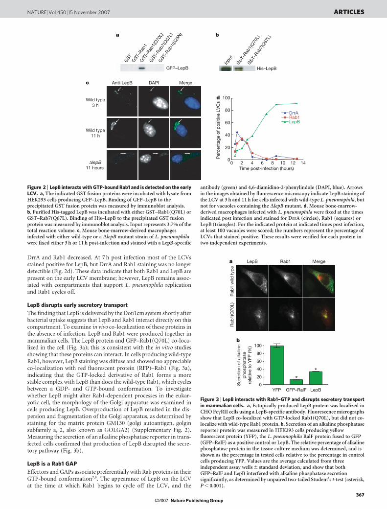
DrrA and Rab1 decreased. At 7 h post infection most of the LCVsstained positive for LepB, but DrrA and Rab1 staining was no longerdetectible (Fig. 2d). These data indicate that both Rab1 and LepB arepresent on the early LCV membrane; however, LepB remains assoc-iated with compartments that support L. pneumophila replicationand Rab1 cycles off.
LepB disrupts early secretory transport
The finding that LepB is delivered by the Dot/Icm system shortly afterbacterial uptake suggests that LepB and Rab1 interact directly on thiscompartment. To examine in vivo co-localization of these proteins inthe absence of infection, LepB and Rab1 were produced together inmammalian cells. The LepB protein and GFP–Rab1(Q70L) co-loca-lized in the cell (Fig. 3a); this is consistent with the in vitro studiesshowing that these proteins can interact. In cells producing wild-typeRab1, however, LepB staining was diffuse and showed no appreciableco-localization with red fluorescent protein (RFP)–Rab1 (Fig. 3a),indicating that the GTP-locked derivative of Rab1 forms a morestable complex with LepB than does the wild-type Rab1, which cyclesbetween a GDP- and GTP-bound conformation. To investigatewhether LepB might alter Rab1-dependent processes in the eukar-yotic cell, the morphology of the Golgi apparatus was examined incells producing LepB. Overproduction of LepB resulted in the dis-persion and fragmentation of the Golgi apparatus, as determined bystaining for the matrix protein GM130 (golgi autoantigen, golginsubfamily a, 2, also known as GOLGA2) (Supplementary Fig. 2).Measuring the secretion of an alkaline phosphatase reporter in trans-fected cells confirmed that production of LepB disrupted the secre-tory pathway (Fig. 3b).
LepB is a Rab1 GAP
Effectors and GAPs associate preferentially with Rab proteins in theirGTP-bound conformation7,8. The appearance of LepB on the LCVat the time at which Rab1 begins to cycle off the LCV, and the
His–LepB
GST
–Rab
1(Q
70L)
GST
–Rab
7(Q
67L)
GST
–Rab
1
GST
GFP–LepB
10 12 14Time post-infection (hours)
Wild type11 h
∆lepB11 hours
DAPI Merge
Inpu
t
GST
–Rab
1(Q
70L)
GST
–Rab
7(Q
67L)
20
40
60
80DrrARab1LepB
0
100d
Per
cent
age
of p
ositi
ve L
VC
s
0 2 4 6 8
Anti-LepB
Wild type3 h
c
ba
GST
–Rab
1(S2
5N)
Figure 2 | LepB interacts with GTP-bound Rab1 and is detected on the earlyLCV. a, The indicated GST fusion proteins were incubated with lysate fromHEK293 cells producing GFP–LepB. Binding of GFP–LepB to theprecipitated GST fusion protein was measured by immunoblot analysis.b, Purified His-tagged LepB was incubated with either GST–Rab1(Q70L) orGST–Rab7(Q67L). Binding of His–LepB to the precipitated GST fusionprotein was measured by immunoblot analysis. Input represents 3.7% of thetotal reaction volume. c, Mouse bone-marrow-derived macrophagesinfected with either wild-type or a DlepB mutant strain of L. pneumophilawere fixed either 3 h or 11 h post-infection and stained with a LepB-specific
antibody (green) and 4,6-diamidino-2-phenylindole (DAPI, blue). Arrowsin the images obtained by fluorescence microscopy indicate LepB staining ofthe LCV at 3 h and 11 h for cells infected with wild-type L. pneumophila, butnot for vacuoles containing the DlepB mutant. d, Mouse bone-marrow-derived macrophages infected with L. pneumophila were fixed at the timesindicated post infection and stained for DrrA (circles), Rab1 (squares) orLepB (triangles). For the indicated protein at indicated times post infection,at least 100 vacuoles were scored; the numbers represent the percentage ofLCVs that stained positive. These results were verified for each protein intwo independent experiments.
**
Rab
1(Q
70L)
80
60
40
20
YFP GFP–RalF LepB
b
LepB Rab1 Merge
Rab
1 w
ild t
ype
a
100
0 Sec
retio
n of
alk
alin
ep
hosp
hata
sere
lativ
e to
YFP
(%)
Figure 3 | LepB interacts with Rab1–GTP and disrupts secretory transportin mammalian cells. a, Ectopically produced LepB protein was localized inCHO FccRII cells using a LepB-specific antibody. Fluorescence micrographsshow that LepB co-localized with GTP-locked Rab1(Q70L), but did not co-localize with wild-type Rab1 protein. b, Secretion of an alkaline phosphatasereporter protein was measured in HEK293 cells producing yellowfluorescent protein (YFP), the L. pneumophila RalF protein fused to GFP(GFP–RalF) as a positive control or LepB. The relative percentage of alkalinephosphatase protein in the tissue culture medium was determined, and isshown as the percentage in tested cells relative to the percentage in controlcells producing YFP. Values are the average calculated from threeindependent assay wells 6 standard deviation, and show that bothGFP–RalF and LepB interfered with alkaline phosphatase secretionsignificantly, as determined by unpaired two-tailed Student’s t-test (asterisk,P , 0.001).
NATURE | Vol 450 | 15 November 2007 ARTICLES
367Nature ©2007 Publishing Group

preferential localization of LepB with the GTP-locked Rab1(Q70L)protein in vivo, are results that would be more consistent with LepBfunctioning as a GAP rather than as an effector of Rab1. Thus, puri-fied LepB was analysed for GAP activity in vitro using Rab1 as asubstrate. Rab1 GTP hydrolysis was stimulated on the addition ofpurified LepB (Fig. 4a). The catalytic efficiency (kcat/Km) of Rab1GTP hydrolysis stimulated by LepB was approximately 104 M21 s21,which was similar to the catalytic efficiency measured for yeast Gyp1,a previously characterized eukaryotic GAP that stimulates Rab1 GTPhydrolysis10,25. The specificity of the LepB GAP activity was testedusing several mammalian Rab proteins as substrates. Rab1 was theonly protein in which a significant increase in GTP hydrolysis wasobserved on the addition of LepB (Fig. 4b). These data show that LepBhas GAP activity specific for Rab1, making this the only bacterialprotein currently known that has the capacity to modulate Rab func-tion by stimulating GTP hydrolysis.
Discussion
These data indicate that proteins translocated into host cells byL. pneumophila have activities that control three critical eventsinvolved in the dynamic cycling of the Rab1 GTPase on membranes.DrrA is a multifunctional enzyme with both GDF and GEF activities.
Previously, the only cellular factor shown to have GDF activity wasthe eukaryotic Rabac1 protein21. Because Rabac1 is an integralmembrane protein that seems to have multiple functions in control-ling membrane traffic26, it has been difficult to demonstrate the spe-cific importance of the GDF activity in recruiting Rab GTPases tosubcellular organelles. Our data on the L. pneumophila DrrA protein,however, demonstrate clearly that both GDF and GEF activities areneeded for membrane recruitment of the Rab1 protein to a plasma-membrane-derived organelle. These findings raise the possibility thateukaryotic proteins that function as Rab GEFs might also have sepa-rate domains or subunits that mediate Rab GDI displacement. Thus,these studies not only show the importance of GDF activity in therecruitment of Rab proteins to membranes but also provide newinsight into a process that might be used by other Rab GEFs tomediate nucleotide exchange.
In addition to encoding an activator of Rab1, our data dem-onstrate that the L. pneumophila LepB protein is a GAP for Rab1.The ability of LepB to deactivate Rab1 through stimulating GTPhydrolysis could facilitate removal of the GTPase from the earlyLCV after this plasma-membrane-derived compartment has under-gone maturation events mediated by the association and fusion ofsecretory vesicles. Indeed, we determined that Rab1 association withthe LCV membrane was transient and the cycling of Rab1 off the LCVoccurred at a time at which LepB began to accumulate on this organ-elle. Being a large protein with a calculated molecular weight of148.6 kilodaltons, it is likely that LepB has multiple effector activities,as discovered for the smaller DrrA protein. It will be interesting tolocate the GAP domain in LepB and determine whether this region isrequired for the egress defect reported previously for a L. pneumo-phila lepB mutant24. Identifying the GAP domain might be challen-ging because LepB does not possess the canonical TBC (Tre-2, Bub2and Cdc16) domain found in most eukaryotic Rab GAPs9,10.Although LepB may still use arginine and glutamine residues for adual-finger mechanism to stabilize the transition state of the GTPhydrolysis reaction of the Rab, the lack of homology to other TBCproteins indicates that the structure of the domain in LepB contain-ing catalytic residues important for GAP activity is likely to be dif-ferent. Thus, future investigations into the structure and function ofLepB are likely to reveal how convergent evolutionary processes haveled to the emergence of a bacterial Rab GAP.
On the basis of these findings, we propose the following model forhow L. pneumophila proteins regulate Rab1 activity on the LCVmembrane. DrrA has the intrinsic ability to bind to the cytosolicsurface of the plasma membrane19. Because the LCV is initially aplasma-membrane-derived organelle, translocated DrrA proteinassociates with the immature LCV formed on bacterial internaliza-tion. The GDF activity of DrrA promotes the sampling of cytosolicRab proteins associated with Rab GDI. When Rab1–Rab-GDI com-plexes are sampled, the removal of Rab GDI permits the GEF domainin DrrA to activate Rab1, leading to the stable association of Rab1with the LCV membrane. Active Rab1 facilitates organization of theLCV membrane to promote the delivery and fusion of endoplasmic-reticulum-derived vesicles14,27. DrrA cycles off because the composi-tion of the vacuole membrane changes. The LepB protein accumu-lates on the LCV membrane and the GAP activity facilitates theremoval of Rab1 by stimulating GTP hydrolysis. The removal ofRab1 coincides with fusion of the LCV with the host endoplasmicreticulum and replication of L. pneumophila in a vacuole containingendoplasmic reticulum proteins.
METHODS SUMMARY
Plasmids encoding 33Flag–Rabs, Rab GDI and DrrA were transfected into
HEK293 cells, and 33Flag–Rab complexes were isolated using anti-Flag M2
affinity gel (Sigma). Recombinant His-tagged DrrA proteins were purified from
Escherichia coli as described19. Rab GDI release was detected by immunoblot
analysis using anti-Rab-GDI antibody (Synaptic Systems). For the GEF assays,
33Flag–Rab1–Rab-GDI complexes were eluted from the anti-Flag agarose
Rab33
Rab5Rab1 Rab2
150 nM LepB50 nM LepBNo LepB (intrinsic)
Time (min)
Time (min) Time (min) Time (min)
Time (min) Time (min)
0 20 60 100 120
0
0.02
0.04
0.06
0.08
40 80Time (min)
67 nM LepB
26 nM Gyp1
22 nM LepB
7 nM LepBRab1 alone
0.10
∆A36
0
a
0 33 66 100
0 33 66 100
0 33 66 100
0 33 66 100 0 33 66 100
0 33 66 100Time (min)
0 33 66 100
–0.01
0
0.01
0.02
0.03
0
0.01
0.02
–0.01
0
0.01
0.02
0.03
–0.01
0
0.01
0.02
0.03
–0.01
0
0.01
0.02
–0.01
0
0.01
0.02
0.03
–0.01
0
0.01
0.02
0.03
Rab7Rab6 Rab11
–0.01
0.03
∆A36
0
0.03b
Figure 4 | LepB is a Rab1-specific GAP. a, Inorganic phosphate release(DA360) resulting from Rab1–GTP hydrolysis was measured over time (min)after the addition of purified His-tagged LepB or yeast Gyp1 at theconcentrations indicated, and compared to the intrinsic rate of GTPhydrolysis by Rab1 (Rab1 alone). b, Inorganic phosphate release (DA360)resulting from GTP hydrolysis was measured over time (min) for theindicated Rab proteins after the addition of 150 nM LepB (red) or 50 nM LepB(green), and compared to the intrinsic rate of GTP hydrolysis (No LepB).Plots show results from two different assay wells for each condition tested.
ARTICLES NATURE | Vol 450 | 15 November 2007
368Nature ©2007 Publishing Group

using 33Flag peptide, and were concentrated using an Amicon centrifugal
filter unit with Ultracel-30 membrane (Millipore). The exchange reaction was
initiated on addition of the non-hydrolyzable GTP analogue mant-GppNHp,
and fluorescence was monitored at 460 nm following excitation at 290 nm using
a Sapphire multimode microplate spectrophotometer. Recombinant His–LepB
and GST–Rabs were purified from E. coli using standard procedures and were
used in binding assays as described28,29. Rabbits were immunized with purified
LepB to generate LepB-specific polyclonal antibodies. LepB co-localization with
RFP–Rab1 and GFP–Rab1(Q70L), and LepB-mediated inhibition of alkaline
phosphatase secretion, were analysed as described30. Bone-marrow macrophages
derived from A/J mice were infected with L. pneumophila, and LepB, Rab1 and
DrrA co-localization with vacuoles containing L. pneumophila was determined
by fluorescence microscopy as described19,31. Purified His–LepB and GTP-loaded
GST–Rab proteins were used in the GAP assays with purified yeast Gyp1 as a
control. The EnzChek phosphate assay (Molecular Probes) was used to measure
GTP hydrolysis as described25.
Full Methods and any associated references are available in the online version ofthe paper at www.nature.com/nature.
Received 8 September; accepted 4 October 2007.Published online 21 October 2007.
1. Zerial, M. & McBride, H. Rab proteins as membrane organizers. Nature Rev. Mol.Cell Biol. 2, 107–117 (2001).
2. Rink, J., Ghigo, E., Kalaidzidis, Y. & Zerial, M. Rab conversion as a mechanism ofprogression from early to late endosomes. Cell 122, 735–749 (2005).
3. Sasaki, T. et al. Purification and characterization from bovine brain cytosol of aprotein that inhibits the dissociation of GDP from and the subsequent binding ofGTP to smg p25A, a ras p21-like GTP-binding protein. J. Biol. Chem. 265,2333–2337 (1990).
4. Araki, S., Kikuchi, A., Hata, Y., Isomura, M. & Takai, Y. Regulation of reversiblebinding of smg p25A, a ras p21-like GTP-binding protein, to synaptic plasmamembranes and vesicles by its specific regulatory protein, GDP dissociationinhibitor. J. Biol. Chem. 265, 13007–13015 (1990).
5. Ullrich, O. et al. Rab GDP dissociation inhibitor as a general regulator for themembrane association of rab proteins. J. Biol. Chem. 268, 18143–18150 (1993).
6. Dirac-Svejstrup, A. B., Sumizawa, T. & Pfeffer, S. R. Identification of a GDIdisplacement factor that releases endosomal Rab GTPases from Rab-GDI. EMBOJ. 16, 465–472 (1997).
7. Markgraf, D. F., Peplowska, K. & Ungermann, C. Rab cascades and tetheringfactors in the endomembrane system. FEBS Lett. 581, 2125–2130 (2007).
8. Grosshans, B. L., Ortiz, D. & Novick, P. Rabs and their effectors: achievingspecificity in membrane traffic. Proc. Natl Acad. Sci. USA 103, 11821–11827 (2006).
9. Strom, M., Vollmer, P., Tan, T. J. & Gallwitz, D. A yeast GTPase-activating proteinthat interacts specifically with a member of the Ypt/Rab family. Nature 361,736–739 (1993).
10. Albert, S., Will, E. & Gallwitz, D. Identification of the catalytic domains and theirfunctionally critical arginine residues of two yeast GTPase-activating proteinsspecific for Ypt/Rab transport GTPases. EMBO J. 18, 5216–5225 (1999).
11. Horwitz, M. A. Formation of a novel phagosome by the Legionnaires’ diseasebacterium (Legionella pneumophila) in human monocytes. J. Exp. Med. 158,1319–1331 (1983).
12. Swanson, M. S. & Isberg, R. R. Association of Legionella pneumophila with themacrophage endoplasmic reticulum. Infect. Immun. 63, 3609–3620 (1995).
13. Kagan, J. C. & Roy, C. R. Legionella phagosomes intercept vesicular traffic fromendoplasmic reticulum exit sites. Nature Cell Biol. 4, 945–954 (2002).
14. Kagan, J. C., Stein, M. P., Pypaert, M. & Roy, C. R. Legionella subvert the functionsof Rab1 and Sec22b to create a replicative organelle. J. Exp. Med. 199, 1201–1211(2004).
15. Derre, I. & Isberg, R. R. Legionella pneumophila replication vacuole formationinvolves rapid recruitment of proteins of the early secretory system. Infect. Immun.72, 3048–3053 (2004).
16. Segal, G., Purcell, M. & Shuman, H. A. Host cell killing and bacterial conjugationrequire overlapping sets of genes within a 22-kb region of the Legionellapneumophila genome. Proc. Natl Acad. Sci. USA 95, 1669–1674 (1998).
17. Vogel, J. P., Andrews, H. L., Wong, S. K. & Isberg, R. R. Conjugative transfer by thevirulence system of Legionella pneumophila. Science 279, 873–876 (1998).
18. Nagai, H., Kagan, J. C., Zhu, X., Kahn, R. A. & Roy, C. R. A bacterial guaninenucleotide exchange factor activates ARF on Legionella phagosomes. Science 295,679–682 (2002).
19. Murata, T. et al. The Legionella pneumophila effector protein DrrA is a Rab1 guaninenucleotide-exchange factor. Nature Cell Biol. 8, 971–977 (2006).
20. Machner, M. P. & Isberg, R. R. Targeting of host Rab GTPase function by theintravacuolar pathogen Legionella pneumophila. Dev. Cell 11, 47–56 (2006).
21. Sivars, U., Aivazian, D. & Pfeffer, S. R. Yip3 catalyses the dissociation ofendosomal Rab-GDI complexes. Nature 425, 856–859 (2003).
22. Stroupe, C. & Brunger, A. T. Crystal structures of a Rab protein in its inactive andactive conformations. J. Mol. Biol. 304, 585–598 (2000).
23. Pylypenko, O. et al. Structure of doubly prenylated Ypt1:GDI complex and themechanism of GDI-mediated Rab recycling. EMBO J. 25, 13–23 (2006).
24. Chen, J. et al. Legionella effectors that promote nonlytic release from protozoa.Science 303, 1358–1361 (2004).
25. Pan, X., Eathiraj, S., Munson, M. & Lambright, D. G. TBC-domain GAPs for RabGTPases accelerate GTP hydrolysis by a dual-finger mechanism. Nature 442,303–306 (2006).
26. Geng, J., Shin, M. E., Gilbert, P. M., Collins, R. N. & Burd, C. G. Saccharomycescerevisiae Rab-GDI displacement factor ortholog Yip3p forms distinct complexeswith the Ypt1 Rab GTPase and the reticulon Rtn1p. Eukaryot. Cell 4, 1166–1174(2005).
27. Robinson, C. G. & Roy, C. R. Attachment and fusion of endoplasmic reticulum withvacuoles containing Legionella pneumophila. Cell. Microbiol. 8, 793–805 (2006).
28. Satoh, A., Wang, Y., Malsam, J., Beard, M. B. & Warren, G. Golgin-84 is a rab1binding partner involved in Golgi structure. Traffic 4, 153–161 (2003).
29. Christoforidis, S. & Zerial, M. Purification and identification of novel Rab effectorsusing affinity chromatography. Methods 20, 403–410 (2000).
30. Amor, J. C. et al. The structure of RalF, an ADP-ribosylation factor guaninenucleotide exchange factor from Legionella pneumophila, reveals the presence of acap over the active site. J. Biol. Chem. 280, 1392–1400 (2005).
31. Kagan, J. C., Murata, T. & Roy, C. R. Analysis of Rab1 recruitment to vacuolescontaining Legionella pneumophila. Methods Enzymol. 403, 71–81 (2005).
Supplementary Information is linked to the online version of the paper atwww.nature.com/nature.
Acknowledgements We thank G. Warren for his advice on the Rab1 bindingstudies, and M. P. Stein and L. Chesnel for technical suggestions and advice. Thiswork was supported by the NIH (C.R.R. and D.G.L.) and an NSF Graduate ResearchFellowship (A.I.).
Author Contributions C.R.R. supervised the project. A.I. conducted all of theexperiments in this study with the exception of the GEF assays, which wereconducted by A.D. D.G.L. assisted with the GAP assays. All authors contributed tothe writing of the manuscript
Author Information Reprints and permissions information is available atwww.nature.com/reprints. Correspondence and requests for materials should beaddressed to C.R.R. ([email protected]).
NATURE | Vol 450 | 15 November 2007 ARTICLES
369Nature ©2007 Publishing Group

METHODSIsolation of 33Flag–Rab–Rab-GDI complexes and Rab GDI release assays.HEK293 cells transfected with plasmids expressing 33Flag–Rab proteins and
Rab GDI were lysed on ice in 64 mM Tris-HCl, pH 8.0, 150 mM NaCl, 8 mM
MgCl2, 2 mM EDTA, 0.2 mM GDP, 0.1% CHAPS and protease inhibitors. After
clearing by spinning at 14,000g for 10 min, lysates were incubated with anti-Flag
M2 affinity gel (Sigma) (approximately 1.5 3 108 cells per ml of resin), and
subsequently the resin was washed with lysis buffer. To assess GDF activity in
vitro, purified His–DrrA protein was added (0.45 nmol per 20 ml anti-Flag resin
in 140ml total reaction volume) and incubated for 75 min at 25 uC. Solublefractions were recovered after centrifugation at 5,000g for 30 s. Bound protein
was eluted with protein sample buffer at 100 uC (Fig. 1a), 0.1 M glycine, pH 3.5,
at 25 uC (Fig. 1b, beads), or 100mg per ml 33Flag peptide (Sigma) (Fig. 1d) in
volumes equal to the reaction volume. Rab GDI was detected by immunoblot
analysis using anti-Rab-GDI antibody (Synaptic Systems).
Rab1–Rab-GDI GEF assays. Rab1–Rab-GDI complex (0.8mM) was incubated
in buffer (50 mM Tris, pH 8.0, 150 mM NaCl, 2 mM MgCl2, 0.1% CHAPS) with
either 0.25mM full-length DrrA (1–647) or the minimal DrrA GEF domain (451–
647). The exchange reaction was initiated on addition of 10 mM mant-GppNHp.
Samples were excited at 290 nm, and emission was monitored at 460 nm using
a Sapphire multimode microplate spectrophotometer to measure increased
sensitized emission accompanying tryptophan to mant fluorescence resonance
energy transfer that occurs on mant-GppNHp loading.
Binding of LepB to GST–Rabs. GST-tagged and His-tagged proteins were
purified from E. coli as described19. Binding assays were performed based on
described methods28,29. Human HEK293 cells transiently transfected with plas-
mid expressing LepB were lysed in 10 mM HEPES, pH 7.4, 150 mM KCl, 5 mM
MgCl2, 1 mM DTT, 1% Triton X-100 and protease inhibitors. Protein frompost-nuclear supernatants (100 mg) was added to purified GST–Rab proteins
(approximately 100mg) immobilized on glutathione sepharose 4B in the pre-
sence of 0.2 mM GDP (for GST and GST–Rab1(S25N)) or 0.2 mM GTP (for
GST–Rab1, GST–Rab1(Q70L) and GST–Rab7(Q67L)). Alternatively, purified
recombinant His–LepB (20mg) was added to the immobilized GST–Rabs
(100mg). Binding reactions were performed in 250ml of lysis buffer containing
0.2 mM guanine nucleotide for 14 h at 4 uC. After washing, bound protein was
eluted in 10 mM HEPES, pH 7.4, 1.5 M KCl, 20 mM EDTA and 1 mM DTT
containing either 5 mM GTP (for GST and GST–Rab1(S25N)) or 5 mM GDP
(for GST–Rab1, GST–Rab1(Q70L) and GST-Rab7(Q67L)).
Eukaryotic expression of LepB. For localization studies of LepB and GFP–Rab1
in eukaryotic cells, Chinese hamster ovary (CHO) FccRII cells expressing LepB
along with RFP–Rab1 or GFP–Rab1(Q70L) were fixed and stained with antibody
against LepB. Cells were subsequently visualized by fluorescence microscopy
using a Nikon Eclipse TE2000-S microscope. Digital images were acquired using
a Hamamatsu ORCA-ER camera and exported into Adobe Illustrator C2 for the
production of figures. Secretion assays were performed as described previously30.
Data are presented as the measured alkaline phosphatase activity in the cellculture media 7 h after replacement of the media of transfected cells, relative
to the cell-associated alkaline phosphatase activity. Displayed in Fig. 3b are the
average values from three independent assay wells 6 standard deviation.
Infection and staining of murine bone-marrow-derived macrophages.Macrophages were derived from bone marrow precursor cells and infected as
described31,32. After fixation, macrophages were stained for Rab1 and for DrrA as
described19,31, and for LepB with a rabbit polyclonal antibody raised against
purified His–LepB. Samples were visualized by fluorescence microscopy using
a Nikon Eclipse TE2000-S microscope. At least 100 LCVs were counted for each
protein examined, and the data are representative of two independent assays.
GAP assays. GAP assays were performed as described25. Purified GST–Rab pro-
teins were loaded with GTP, and unbound nucleotide was removed using a
desalting column. Phosphate release was measured in reactions containing
27 mM Rab1 using the EnzChek phosphate assay (Molecular Probes) after the
addition of purified LepB–His or yeast Gyp1. Catalytic efficiency of the LepB–
His GAP reaction using GST–Rab1 was calculated as described25. To compare the
activities of LepB–His for the different Rab proteins, initial velocities were cal-
culated: from 810–2,208 s of the reactions with Rab1, Rab5, Rab7 and Rab33;
from 973–1,980 s of the reactions with Rab2 and Rab11; and from 3,025–3,966 s
of the reaction with Rab6. These rates with and without LepB–His were analysed
with the following equation:
(kcat/Km)/kintr 5 ((Vg/Vi) – 1)/[GAP]
Where Vg is the rate with LepB–His, Vi is the rate without LepB–His, and [GAP]
is the concentration of LepB–His.
Construction of plasmids and DlepB mutant strain. His-tagged versions of
LepB were constructed by cloning the gene amplified from L. pneumophila
genomic DNA into the NdeI and BamHI sites of pET15b, and NdeI and XhoI
sites of pET26b. For cloning into pcDNA 4/TO, lepB was amplified and inserted
into the BamHI and XhoI sites of the digested vector. Full-length complementary
DNA encoding human GDP dissociation inhibitor 1 was obtained from Open
Biosystems (IHS1380-97430805). For mammalian cell expression, GDI1 was
amplified and inserted into the BamHI and XhoI sites of pcDNA 4/TO. A
33Flag expression vector was created for tagging Rabs by the Flag epitope into
the KpnI and BamHI sites of pcDNA 4/TO. This vector, 33Flag pcDNA 4/TO,
was digested at the BamHI and XhoI sites to insert the cDNA encoding Rab1 and
Rab2. To construct the clean deletion of lepB in L. pneumophila, pSR47S was
used33. The genomic regions proximal to lepB were amplified and inserted into
the XbaI and SacI sites of pSR47S. Construction of the isogenic DlepB mutant in
the L. pneumophila strain Lp01 by allelic replacement was as described34.
32. Celada, A., Gray, P. W., Rinderknecht, E. & Schreiber, R. D. Evidence for a gamma-interferon receptor that regulates macrophage tumoricidal activity. J. Exp. Med.160, 55–74 (1984).
33. Merriam, J. J., Mathur, R., Maxfield-Boumil, R. & Isberg, R. R. Analysis ofthe Legionella pneumophila fliI gene: intracellular growth of a definedmutant defective for flagellum biosynthesis. Infect. Immun. 65, 2497–2501(1997).
34. Campodonico, E. M., Chesnel, L. & Roy, C. R. A yeast genetic system for theidentification and characterization of substrate proteins transferred into host cellsby the Legionella pneumophila Dot/Icm system. Mol. Microbiol. 56, 918–933(2005).
doi:10.1038/nature06336
Nature ©2007 Publishing Group