leptin resistance is a secondary consequence of the obesity in
TRANSCRIPT

Leptin resistance is a secondary consequence of theobesity in ciliopathy mutant miceNicolas F. Berbaria,1, Raymond C. Paseka,1, Erik B. Malarkeya, S. M. Zaki Yazdia, Andrew D. McNaira, Wesley R. Lewisa,Tim R. Nagyb, Robert A. Kestersonc, and Bradley K. Yodera,2
Departments of aCell, Developmental, and Integrative Biology, bNutrition Sciences, and cGenetics, University of Alabama at Birmingham, Birmingham,AL 35294
Edited by Rudolph L. Leibel, Columbia University, New York, NY, and accepted by the Editorial Board March 20, 2013 (received for review June 15, 2012)
Althoughprimary cilia arewell establishedas important sensory andsignaling structures, their function in most tissues remains un-known. Obesity is a feature associated with some syndromes ofcilia dysfunction, such as Bardet-Biedl syndrome (BBS) and Alströmsyndrome, as well as in several cilia mutant mouse models. Recentdata indicate that obesity in BBS mutant mice is due to defects inleptin receptor trafficking and leptin resistance. Furthermore, induc-tion of cilia loss in leptin-responsive proopiomelanocortin neuronsresults in obesity, implicating cilia on hypothalamic neurons in reg-ulating feeding behavior. Here, we directly test the importance ofthe cilium as a mediator of the leptin response. In contrast to thecurrent dogma, a longitudinal study of conditional Ift88 cilia mutantmice under different states of adiposity indicates that leptin resis-tance is present only when mutants are obese. Our studies showthat caloric restriction leads to an altered anticipatory feeding be-havior that temporarily abrogates the anorectic actions of leptindespite normalized circulating leptin levels. Interestingly, preobeseBbs4 mutant mice responded to the anorectic effects of leptin anddid not display other phenotypes associated with defective leptinsignaling. Furthermore, thermoregulation and activity measure-ments in cilia mutant mice are inconsistent with phenotypes previ-ously observed in leptin deficient ob/ob mice. Collectively, thesedata indicate that cilia are not directly involved in leptin responsesand that a defect in the leptin signaling axis is not the initiatingevent leading to hyperphagia and obesity associated with ciliadysfunction.
Obesity is a major health issue associated with complicationsthat cause significant morbidity and mortality. Thus, iden-
tification of the protein hormone leptin in the spontaneous obesemouse mutant ob/ob was the source of much excitement (1), asleptin suppresses feeding activity and is secreted into serum inproportion to the amount of adipose tissue, the hormone’s pri-mary source (2). However, the therapeutic potential of leptin wasattenuated by a barrier to the action of leptin that must exist inan obese individual, as nearly all obese mice and human patientsexhibit elevated levels of circulating leptin (2, 3). This barrierphenomenon is known as leptin resistance, the precise mecha-nisms of which are unknown. Furthermore, age impacts leptinsensitivity, as older rodents are less responsive to the anorecticeffects of leptin than younger rodents (4–7). In the field ofobesity research, one challenge becomes determining how indi-viduals acquire leptin resistance and whether this is a primarycause of the obesity phenotype or simply a consequence.Recent findings revealed that obesity is associated with mu-
tations in proteins disrupting the function of primary cilia, whichare small, immotile, microtubule-based appendages protrudingfrom the surface of most mammalian cell types (8). Long thoughtto be vestigial, primary cilia are now known to serve as criticalsignaling hubs for diverse cellular pathways during development(9). The emergence of the primary cilium as a clinically impor-tant organelle was initiated by studies in model organisms suchas Chlamydomonas reinhardtii and Caenorhabditis elegans. Thesestudies led to the identification of proteins required for cilio-genesis through the bidirectional transport of cargo along the
cilium in a process known as intraflagellar transport (IFT) (10).Because IFT-null mutations are embryonically lethal, researchinto the roles of cilia in adults was initially limited. However, thegeneration of conditional IFT alleles in mice and the realizationthat several human genetic syndromes, known as ciliopathies,result from cilia dysfunction has expanded our understanding ofthe organelle (11). Mutations affecting cilia function in humanslead to a spectrum of diseases. Ciliopathy clinical features rangefrom prenatal lethality in Meckel-Grüber syndrome to obesity inAlström syndrome or Bardet-Biedl Syndrome (BBS).BBS is a pleiotropic, genetically heterogeneous syndrome asso-
ciated with retinopathy, cystic kidneys, cognitive deficits, poly-dactyly, anosmia, and obesity (8). Several of the implicated proteinsform a complex called the BBSome that functions in cilia mem-brane trafficking (12–15). Numerous mouse models of BBS re-capitulate many of the patients’ clinical features (16, 17). A majordistinction between mouse BBS and IFT mutants is that BBSmodels possess primary cilia, whereas IFTmutations generally leadto cilia ablation. Furthermore, data from multiple model systemssuggest that BBS-associated phenotypes result from defective ciliamediated signaling activity (12, 18, 19).Recent data show that Bbs1, a BBSome component, binds
the leptin receptor and may mediate leptin receptor trafficking(20). Furthermore, studies in Bbs2, Bbs4, and Bbs6 mutant micereveal that they are hyperleptinemic, and importantly, fail to re-duce food intake in response to leptin (21). Leptin resistance wasobserved in the mutant mice even after caloric restriction reducedfat mass and circulating leptin levels to those of control mice (20).Thus, defects in leptin signaling appear to contribute directly toBBS-associated obesity.We previously showed that conditional disruption of Ift88
throughout an adult mouse using the ubiquitously expressedCAGG-CreER (actin promoter) transgene leads to hyperphagiaand obesity (22). Intriguingly, the obesity phenotype can be re-capitulated using proopiomelanocortin (POMC)-Cre to disruptIft88 in POMC neurons of the hypothalamus, a key satiety centerthat regulates leptin responses (22). Thus, our goal was to in-vestigate whether cilia are directly involved in leptin signaling,whether cilia loss contributes to the development of leptin re-sistance, and whether the etiology of obesity in cilia mutant miceis similar to that observed in Bbs models where cilia are intactbut dysfunctional.
Author contributions: N.F.B., R.C.P., E.B.M., T.R.N., R.A.K., and B.K.Y. designed research;N.F.B., R.C.P., E.B.M., S.M.Z.Y., A.D.M., and W.R.L. performed research; N.F.B., R.C.P.,E.B.M., T.R.N., R.A.K., and B.K.Y. analyzed data; and N.F.B., R.C.P., E.B.M., and B.K.Y.wrote the paper.
The authors declare no conflict of interest.
This article is a PNAS Direct Submission. R.L.L. is a guest editor invited by the EditorialBoard.1N.F.B. and R.C.P. contributed equally to this work.2To whom correspondence should be addressed. E-mail: [email protected].
This article contains supporting information online at www.pnas.org/lookup/suppl/doi:10.1073/pnas.1210192110/-/DCSupplemental.
7796–7801 | PNAS | May 7, 2013 | vol. 110 | no. 19 www.pnas.org/cgi/doi/10.1073/pnas.1210192110

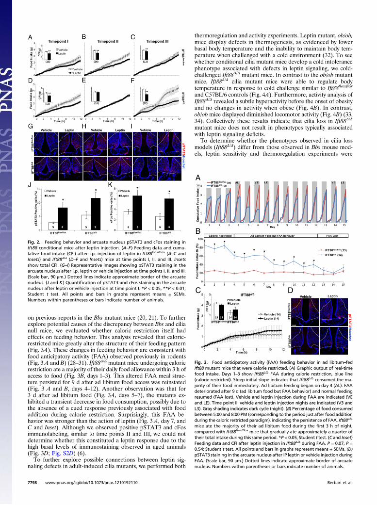
ResultsA longitudinal leptin sensitivity testing paradigm was designed todetermine whether leptin resistance in cilia mutant mice is a di-rect result of cilia loss or a secondary consequence of obesity(Fig. 1A). To initiate cilia ablation in adult mice, a previouslydescribed conditional allele of Ift88 and ubiquitously expressedinducible CAGG-CreER line were used (22, 23). Cohorts weregenerated such that transgene CAGG-CreER–positive animalswere always compared with control CAGG-CreER–negative lit-termates. Ift88 deletion was induced at 8 wk of age through ta-moxifen injection (Fig. 1A). Genotyping and Western blottingconfirmed CreER-mediated deletion of Ift88 within 10 d aftertamoxifen administration (Fig. S1 A and C). Subsequent immu-nostaining for the neuronal cilia marker adenylate cyclase III(ACIII) (24) confirmed the absence of hypothalamic neuronalcilia throughout the study (Fig. S1B). These induced cilia mutantmice are hereafter referred to as Ift88Δ/Δ and control mice asIft88flox/flox. Mice were challenged with i.p. injection of recombi-nant leptin, and subsequent feeding behavior was assessed usinga Biodaq feeding monitoring system (25) to record food intake ofindividual mice in real time. Mice were injected with leptin atthree time points at which they had different body and serumleptin compositions, hereafter referred to as time points I, II, andIII (Fig. 1 B and C), corresponding to preobese, obese, and food-restricted nonobese states, respectively.At time point I, Ift88Δ/Δ mice are hyperphagic but have not
diverged significantly in body weight or fat mass from Ift88flox/flox
controls. Serum leptin measurements demonstrated no signifi-cant difference between the two groups at this point (Ift88flox/flox,1.07 ng/mL; Ift88Δ/Δ, 1.05 ng/mL; P = 0.94, Student t test; Fig. 1B).Unexpectedly, leptin reduced food intake for both Ift88Δ/Δ
and Ift88flox/flox mice compared with vehicle injection (Fig. 2 A andD and Insets). This reduction would not be expected in Ift88Δ/Δ
mice if leptin was acting through the primary cilium. Furthermore,the difference in food intake between Ift88Δ/Δ mice treated withleptin and control Ift88flox/flox mice treated with leptin was notsignificant (Ift88flox/flox, 2.22 g; Ift88Δ/Δ, 2.26 g; P = 0.93, Studentt test; Fig. 2 A and D and Insets). Additionally, both Ift88flox/flox
and Ift88Δ/Δ mice at time point I displayed increased phosphor-ylated Stat3 (pSTAT3) and cFos within the arcuate nucleus 90min after injection, indicative of a response to leptin (Fig. 2 G, J,and K; Fig. S2A) (26). These results indicate that at time point I,after both the Ift88 protein and cilia have been lost, the responseto leptin remains intact as measured by both behavioral activityand neuronal markers of activation in cilia mutant Ift88Δ/Δ mice.
After 80 d of ad libitum feeding (time point II), Ift88Δ/Δ
mutants are significantly heavier with greater fat mass andgreater serum leptin levels (Fig. 1 A–C). Again, leptin was ad-ministered i.p., and as observed in many other models of in-creased adiposity and hyperleptinemia, the Ift88Δ/Δ mice did notdiminish feeding in response to leptin, whereas control Ift88flox/flox
responded with reduced food intake (Fig. 2 B and E and Insets).Both pSTAT3 and cFos labeling showed increased basal levelsof staining in the arcuate nucleus in all groups, showing no sig-nificant effects of leptin (Fig. 2H; Fig. S2B). However, theability to assess a response to leptin in rodents using markerssuch as pSTAT3 diminishes in aged animals (6, 7, 27). Theseresults indicate that obese Ift88Δ/Δ mice have elevated leptinlevels and display behavior consistent with hyperleptinemia-associated leptin resistance.To test whether the increased fat mass and serum leptin ob-
served at time point II drive the leptin resistant phenotype, micewere subjected to calorie restriction using a gradual stepwiseprocess over a period of several weeks (Fig. 1 A and B). Calorierestriction restored adiposity and serum leptin levels to thoseobserved in controls (hereafter referred to as time point III). Themice were then allowed ad libitum access to food for 10 d beforetesting the leptin response. Time point III leptin sensitivity wasassessed before ad libitum–associated changes in body weight andserum leptin, a condition similar to time point I. Although theserum leptin levels and fat mass were significantly higher in Ift88Δ/Δ
mice compared with Ift88flox/flox mice at time point III (Ift88flox/flox,2.54 ng/mL and 3.69 g; Ift88Δ/Δ, 4.14 ng/mL and 5.05 g; P <0.01, Student t test), they were not significantly different from thelevels measured at time point I (Ift88Δ/Δ time point III, 4.14 ng/mL and 3.69 g; Ift88Δ/Δ time point I, 1.06 ng/mL and 3.91 g; one-way ANOVA followed by post hoc Tukey’s HSD test; P = 0.72and P = 0.57, respectively). There was also a greater than 10-folddecrease in serum leptin levels compared with time point II.Strikingly, Ift88Δ/Δ mice again demonstrated a strong anorecticbehavior response to leptin administration (Fig. 2 C and F andInsets). Neither pSTAT3 nor cFos levels in the arcuate nucleusdisplayed differences in staining; however, as in time point II, thisresult is expected with older animals (Fig. 2I; Fig. S2C). Takentogether, these results, along with those from time points I and II,show that the leptin resistance observed in a mouse model of cilialoss is a consequence of the obesity and hyperleptinemia and nota primary defect associated with cilia loss.The finding that leptin administration causes a decrease in
feeding behavior in adult cilia mutants (both before becomingobese and again after caloric restriction) was unexpected based
Time (days)
Bo
dy
Wei
gh
t (g
)
15
20
25
30
35
40
45
50
0 20 40 60 80 100 120 140 160
IFT88flox/flox (18)IFT88 / (16)
CI
II
III
A
IFT88 /IFT88flox/flox
Wei
gh
t (g
)
BBody Weight Fat MassLean Mass Serum Leptin
0
10
20
30
40
50
0
5
10
15
20
25
0
5
10
15
20
25
0
10
20
30
40
50
Lep
tin
(ng
/mL
)
I II III I II III I II III I II III
** ****
**
*
*
* **
**
*
***11 16 13 14 13 14 20 23 13 13 1313
CFig. 1. Paradigm of leptin sensitivity testing, body com-position, and serum leptin in Ift88 conditional mutantmice. (A) Longitudinal paradigm showing body weightsof Ift88Δ/Δ and Ift88flox/flox mice after Cre induction (C),lean time point I (I), obese time point II (II), and lean timepoint III (III). (B) Body weight, fat mass, and lean mass ateach time point. (C) Serum leptin levels at each time point.All points and bars are means ± SEMs. *P < 0.05, **P < 0.01;Student t test. Numbers within parentheses or bars indicatenumber of animals.
Berbari et al. PNAS | May 7, 2013 | vol. 110 | no. 19 | 7797
GEN
ETICS
on previous reports in the Bbs mutant mice (20, 21). To furtherexplore potential causes of the discrepancy between Bbs and cilianull mice, we evaluated whether caloric restriction itself hadeffects on feeding behavior. This analysis revealed that calorie-restricted mice greatly alter the structure of their feeding pattern(Fig. 3A). These changes in feeding behavior are consistent withfood anticipatory activity (FAA) observed previously in rodents(Fig. 3 A and B) (28–31). Ift88Δ/Δ mutant mice undergoing calorierestriction ate a majority of their daily food allowance within 3 h ofaccess to food (Fig. 3B, days 1–3). This altered FAA meal struc-ture persisted for 9 d after ad libitum food access was reinstated(Fig. 3 A and B, days 4–12). Another observation was that for3 d after ad libitum food (Fig. 3A, days 5–7), the mutants ex-hibited a transient decrease in food consumption, possibly due tothe absence of a cued response previously associated with foodaddition during calorie restriction. Surprisingly, this FAA be-havior was stronger than the action of leptin (Fig. 3 A, day 7, andC and Inset). Although we observed positive pSTAT3 and cFosimmunolabeling, similar to time points II and III, we could notdetermine whether this constituted a leptin response due to thehigh basal levels of immunostaining observed in aged animals(Fig. 3D; Fig. S2D) (6).To further explore possible connections between leptin sig-
naling defects in adult-induced cilia mutants, we performed both
thermoregulation and activity experiments. Leptin mutant, ob/ob,mice display defects in thermogenesis, as evidenced by lowerbasal body temperature and the inability to maintain body tem-perature when challenged with a cold environment (32). To seewhether conditional cilia mutant mice develop a cold intolerancephenotype associated with defects in leptin signaling, we cold-challenged Ift88Δ/Δ mutant mice. In contrast to the ob/ob mutantmice, Ift88Δ/Δ cilia mutant mice were able to regulate bodytemperature in response to cold challenge similar to Ift88flox/flox
and C57BL/6 controls (Fig. 4A). Furthermore, activity analysis ofIft88Δ/Δ revealed a subtle hyperactivity before the onset of obesityand no changes in activity when obese (Fig. 4B). In contrast,ob/obmice displayed diminished locomotor activity (Fig. 4B) (33,34). Collectively these results indicate that cilia loss in Ift88Δ/Δ
mutant mice does not result in phenotypes typically associatedwith leptin signaling deficits.To determine whether the phenotypes observed in cilia loss
models (Ift88Δ/Δ) differ from those observed in Bbs mouse mod-els, leptin sensitivity and thermoregulation experiments were
0
5
10
15
CBA
FED
IF
T8
8flo
x/flo
xIF
T8
8/
VehicleLeptin
VehicleLeptin
0
1
2
3
4
5
Fo
od
In
ta
ke
(g
)
0 2 4 6 8 10 12Time (h)
1
3
5
**
CF
I (g
)
0
1
2
3
4
5
Fo
od
In
ta
ke
(g
)
CF
I (g
)
1
3
5
**
****
**
0 2 4 6 8 10 12Time (h)
0 2 4 6 8 10 12Time (h)
Timepoint I Timepoint II Timepoint III
G H I
IF
T8
8flo
x/flo
xIF
T8
8/
elciheVelciheVelciheV nitpeLnitpeLnitpeL
pS
TA
T3
/H
oech
st
pS
TA
T3
P
os
itive
c
ells
(%
)
cF
os
Po
sitive c
ells
(%
)
Vehicle
Leptin
J KVehicle
Leptin
*
IFT88flox/flox
IFT88/
)21()61()51(
(17) )21()61(
0
2
4
6
8
*
IFT88flox/flox
IFT88/
6 5 6 6
*
5 6 5 5
*
Fig. 2. Feeding behavior and arcuate nucleus pSTAT3 and cFos staining inIft88 conditional mice after leptin injection. (A–F) Feeding data and cumu-lative food intake (CFI) after i.p. injection of leptin in Ift88flox/flox (A–C andInsets) and Ift88Δ/Δ (D–F and Insets) mice at time points I, II, and III. Insetsshow total CFI. (G–I) Representative images showing pSTAT3 staining in thearcuate nucleus after i.p. leptin or vehicle injection at time points I, II, and III.(Scale bar, 90 μm.) Dotted lines indicate approximate border of the arcuatenucleus. (J and K) Quantification of pSTAT3 and cFos staining in the arcuatenucleus after leptin or vehicle injection at time point I. *P < 0.05, **P < 0.01;Student t test. All points and bars in graphs represent means ± SEMs.Numbers within parentheses or bars indicate number of animals.
0
1
2
3
4
5
0 2 4 6 8 10 12
Fo
od
In
ta
ke
(g
)
Time (h)
Vehicle (14)
Leptin (14)
CF
I (g
)
IFT88/
Cumula�
vefood
intake
(g)
0
1
2
3
4flox/flox
Δ/Δ
Cu
mu
lative F
oo
d In
take (g
)
Calorie Restricted Ad Libitum Food but FAA Behavior FAA Lost
Fo
od
In
ta
ke
in
itia
l 3
h (%
)
Day
IFT88flox/flox
(13)
IFT88/
(14)
*
**
*
*
*
*
*
*
**
0
20
40
60
80
100
1 2 3 4 5 6 7 8 9 10 11 12 13 14 15
*
1 2 3 4 5 6 7 8 9 10 11 12 13 14 15Day
Vehicle
Leptin
1
3
5
IFT88flox/flox
IFT88/
IF
T88
flo
x/flo
xIF
T88
/
Vehicle Leptin
pS
TA
T3
/H
oech
st
13 13 14 14
IFT88flox/flox (13)
IFT88/ (14)
AL V E L VE 3 L 3A
B
C D
Fig. 3. Food anticipatory activity (FAA) feeding behavior in ad libitum–fedIft88 mutant mice that were calorie restricted. (A) Graphic output of real-timefood intake. Days 1–3 show Ift88Δ/Δ FAA during calorie restriction, blue line(calorie restricted). Steep initial slope indicates that Ift88Δ/Δ consumed the ma-jority of their food immediately. Ad libitum feeding began on day 4 (AL). FAAdeteriorated after 9 d (ad libitum food but FAA behavior) and normal feedingresumed (FAA lost). Vehicle and leptin injection during FAA are indicated (VEand LE). Time point III vehicle and leptin injection nights are indicated (V3 andL3). Gray shading indicates dark cycle (night). (B) Percentage of food consumedbetween 5:00 and 8:00 PM (corresponding to the period just after food additionduring the caloric restricted paradigm), indicating the persistence of FAA. Ift88Δ/Δ
mice ate the majority of their ad libitum food during the first 3 h of night,compared with Ift88flox/flox mice that gradually ate approximately a quarter oftheir total intake during this same period. *P< 0.05, Student t test. (C and Inset)Feeding data and CFI after leptin injection in Ift88Δ/Δ during FAA. P = 0.07, P =0.54; Student t test. All points and bars in graphs represent means ± SEMs. (D)pSTAT3 staining in the arcuate nucleus after IP leptin or vehicle injection duringFAA. (Scale bar, 90 μm.) Dotted lines indicate approximate border of arcuatenucleus. Numbers within parentheses or bars indicate number of animals.
7798 | www.pnas.org/cgi/doi/10.1073/pnas.1210192110 Berbari et al.

performed in Bbs4 mutants (congenital knockout of Bbs4, here-after Bbs4−/−). To avoid confounding effects of calorie restrictionand altered body composition, Bbs4−/− mutants were analyzedbefore the onset of obesity. Bbs4−/− mice genotyped null andshowed loss of protein as previously described (Fig. S3 A and B)(16). Interestingly, much like Ift88Δ/Δ mice, preobese Bbs4−/− micedid not have significantly higher levels of serum leptin comparedwith controls (Bbs4+/+, 9.18 ng/mL; Bbs4−/−, 8.34 ng/mL; P = 0.75,Student t test; Fig. 5A), and responded to leptin injections com-pared with vehicle (Fig. 5D). Furthermore, increases in bothpSTAT3 and cFos were detected within the arcuate nucleus ofthe young, preobese Bbs4−/− mutants, indicating a leptin response(Fig. 5 B and C; Fig. S2E). Also consistent with normal leptinsignaling, nonobese Bbs4−/− mice did not display a defect inthermoregulation upon cold challenge (Fig. 5E). These resultsindicate that in both a model of cilia loss (Ift88Δ/Δ) and a model ofdefective cilia signaling (Bbs4−/−), leptin signaling is not directlyaffected. Importantly, these results indicate that a defect in a yetto be determined satiation pathway is dependent on the cilium.
DiscussionWe previously showed that loss of cilia on POMC cells in condi-tional mouse mutants resulted in hyperphagia and obesity (22).However, the molecular mechanisms behind this phenotype re-main elusive. Recent studies in mouse models of BBS suggest thatthe complex of BBS proteins known as the BBSome is critical toleptin receptor trafficking and pathway activity (20). Interestingly,Bbs mutant mice retain their cilia, albeit with disrupted cilia re-ceptor localization, whereas IFT mutations result in organelle loss(12, 22). Although both Bbs and IFT mutations lead to obesity, itis unclear whether similar mechanisms drive these pheno-types. Here we test whether the obesity phenotype observed inthese mouse models is initiated by defects in leptin signaling.To achieve this goal, the CAGG-CreER inducible transgene was
used. Although this model does not lead to total loss of Ift88, iteliminates confounding effects of cilia loss during development, anadvantage that the previously used POMC-Cre transgene does notpossess (23, 35). It was previously shown that other obese leptin-resistant animals regain leptin sensitivity on regulation of bodycomposition through controlled feeding (36, 37). Furthermore, thestudy presented here allowed for the repetitive assessment of leptinsensitivity in the same cohort of mice at different adiposity andleptin levels. Surprisingly, Ift88Δ/Δmice are resistant to the actions ofleptin when obese and have increased serum leptin but are sensitiveto exogenous leptin both before weight gain and after weight loss.
Although the exact mechanism behind leptin action and re-sistance remains unclear, the downstream effects of leptin havebeen characterized. Leptin signaling leads to pSTAT3 and in-duction of Socs3, and subsequent neuronal activity induces nu-clear cFos (38). In the young preobese states (time points I,Ift88Δ/Δ and Bbs4−/−), mice responded to leptin with an inductionof both pSTAT3 and cFos in the arcuate nucleus, in contrast towhat was shown for caloric-restricted lean Bbs mutants (20). Inaged animals (time points II, III, and entrained), pSTAT3 andcFos staining were readily apparent in all groups, yet significantdifferences between vehicle and leptin treatment groups werenot found. Similar results have been observed in several rodentmodels showing an age-dependent loss of leptin sensitivity (4, 39,40). Furthermore, decreases or complete loss of leptin inducedpSTAT3 response have been observed in aged rodents (6, 7, 27).This aspect of age-related emergence of leptin resistance is oftenoverlooked in the literature. It is important to note that in age-associated diminished pSTAT3 induction, the anorectic behavioralresponse to leptin remains intact (6). It is interesting to note that inBbs mutant mice, leptin-induced pSTAT3 is still observed, in-dicating that leptin signaling is not completely disrupted (20).Importantly, when Ift88Δ/Δ mice and Bbs4−/− mice are not hyper-leptinemic, leptin injection elicits an anorectic effect on feeding.The Bbs4−/−mice were only analyzed before the onset of obesity inorder to avoid confounding effects of altered body compositionand behavioral changes that we observed as a result of caloric re-striction. The cause of the altered behavior in the Bbs4 mutants isnot known but may be associated with the neurodevelopmentaland degenerative phenotypes that have been reported in thesemice (41, 42). Collectively, the results obtained in the Bbs4 andIft88 mutant mice indicate that the hyperphagia-associatedobesity in models of cilia dysfunction is not initiated by defectsin leptin signaling.To further investigate whether other phenotypes associated
with leptin signaling defects were observed in Ift88Δ/Δ mice, boththermoregulation and locomotor activity experiments were per-formed. Cold-challenged Ift88Δ/Δ mice demonstrated a normalthermoregulatory phenotype. This normal thermoregulationstands in stark contrast to leptin-deficient ob/ob mice, which areunable to thermoregulate (34). To further assess leptin signalingin cilia mutants, locomotor activity was evaluated. Ift88Δ/Δ micewere significantly more active in a 24-h period relative to ob/obmice. These data indicate that conditional Ift88 cilia loss in adultmice does not lead to a primary defect in leptin signaling.Previous reports demonstrated that restricted feeding in rodents
can alter both behavior and physiology, independent of light-darkcycles (43). For example, in response to food restriction, mice areknown to display FAA and alter their feeding behavior and mealstructure (28). Thus, FAA can confound results in experiments ifnot taken into account. Our analysis of Ift88Δ/Δmutant mice clearlyrevealed the FAA phenomenon. The mutants consume the ma-jority of their calories within the first 3 h of the dark cycle during thepaired feeding period (Fig. 3A andB). In contrast, control mice onan ad libitum diet normally consume their calories graduallythroughout the dark cycle. The FAA behavior in the mutantspersisted for 9 d after ad libitum food access was initiated (Fig. 3B).In addition, after ad libitum access, the mutants experienceda short period of depressed feeding activity (days 5–7). This di-minished feeding may result from the loss of cues established bydaily food administration during caloric restriction. This cue wouldnot occur during ad libitum access. If mutants are tested for leptinsensitivity during this FAAperiod, they appear leptin resistant withregard to their feeding. Thus, FAA is stronger than the appetitesuppression effects of leptin. After mutants emerged from theFAA period and returned to a normal feeding pattern (time pointIII), they again exhibited an anorectic response to leptin. Thepersistence of the FAA represents an important and under-appreciated aspect of feeding behavior analysis in the obesity field.
BA
32
33
34
35
36
37
38
0 120 240 360 480
IFT88flox/flox (5)
IFT88 / (4)ob/ob (4)
**
*
4°C 22°C ***
0
1
2
3
4
5 IFT88flox/flox
ob/obIFT88 /
Bea
m B
reak
s (1
04)/
24h
rs
Rec
tal T
emp
. (°C
)
Time (min)
12 8 4
Fig. 4. Thermoregulation and locomotor activity between Ift88 and ob/obmice. (A) Body temperature of Ift88flox/flox, Ift88Δ/Δ, and ob/ob mice mea-sured for baseline, after 30, 120, and 240 min of exposure to 4 °C, and at 270,390, and 480 min during room temperature recovery. The ob/ob mice werepulled from the experiment due to an inability to thermoregulate. *P < 0.05,one-way ANOVA followed by post hoc Tukey’s HSD test. (B) Locomotor ac-tivity at time point I comparing Ift88flox/flox, Ift88Δ/Δ, and ob/ob mice. *P <0.05, **P < 0.01; one-way ANOVA followed by post hoc Tukey’s HSD test.Numbers within parentheses or bars indicate number of animals.
Berbari et al. PNAS | May 7, 2013 | vol. 110 | no. 19 | 7799
GEN
ETICS

Interestingly, FAA is dependent on the suprachiasmatic nuclei(SCN) and not the arcuate nucleus where ciliary function is neededfor satiation responses (28). Future studies will address whetherthe FAA observed in Ift88 conditional mutants differs from thatobserved in WT mice.In contrast to our findings, current dogma indicates that cilia
are needed for normal leptin sensitivity based on previous studiesin the Bbs ciliopathy mouse models. Data from Seo et al. useda caloric restriction paradigm up until leptin responsiveness wasassessed to ensure that the Bbs mutant mice were kept lean (20).Although not directly addressed, this may have caused a FAAeffect similar to what we observed in the Ift88Δ/Δ mice overridingthe anorexigenic effects of leptin. To directly test whether Bbsmutant mice have a leptin signaling defect, we assessed bothleptin sensitivity and thermoregulation in Bbs4−/− mice before theonset of obesity. Interestingly, Bbs4−/− mice responded to i.p.leptin injection and maintain body temperature when cold chal-lenged. Collectively, these data suggest that the leptin signalingdefect reported previously in the Bbs mice is only secondary toeither weight gain or the FAA brought on by calorie restriction.These data ultimately leave the initiating molecular mechanismbehind cilia dysfunction-associated obesity unknown.Several potential molecular mechanisms for the obesity ob-
served in ciliopathy mouse models exist. One must consider thatthe mechanism leading to obesity in Ift conditional models andBbs models may be different. It is now well appreciated that thecilium functions as a key regulatory organelle for multiple path-ways. Some of the potential pathways that could be involved inthe obesity phenotype include altered G protein–coupled re-ceptor (GPCR) signaling or abnormal regulation of mTor orhedgehog (Hh) signaling pathways. Importantly, the orexigenicGPCR melanin concentrating hormone receptor 1 (Mchr1) ispresent on neurons of the hypothalamus, but is mistargeted in theBbs mutant mice (12). Thus, the possibility exists that alteredMchr1 signaling in the absence of cilia in the Ift88mutants, or dueto its exclusion from the cilia in the Bbs mutants, could result inhyperphagia induced obesity. Cilia loss alters mTOR activity,which has a well-documented role in energy homeostasis (44–46);however, altered mTor has not been evaluated in the Bbs mutantmice. Further, treatment of cilia mutant mice with rapamycin canpartially correct some mutant phenotypes (47). Arguably, defectsin the Hh pathway are currently the most directly associated withabnormal cilia function (48). Hh signaling is important for thedevelopment and patterning of numerous tissues, including thehypothalamus, and has critical roles during adult neurogenesis (49,50). Thus, obesity in congenital Bbs mutants could arise throughmispatterning of the hypothalamus. In fact, previous reportshave indicated loss of POMC neurons in Bbs mutant mice(20). The loss of POMC neurons seems less likely in the adultinducible Ift88 mutant, as the hyperphagia phenotype is evi-dent within 2 wk of inducing cilia loss. However, Hh pathwaycomponents are expressed in the hypothalamus of adult mice,and thus, it will be informative to evaluate whether feeding be-havior and energy homeostasis can be altered by modulating Hhsignaling activities specifically in the POMC neurons and whetherthis is influenced by the presence or absence of the cilium.
MethodsFor methodological details, see SI Methods.
All mice were maintained in an Association for Assessment and Accredi-tation of Laboratory Animal Care-approved facility in accordance with In-stitutional Animal Care and Use Committee guidelines at University ofAlabama at Birmingham. Ift88flox/flox; CAGG-CreER cohorts were generated,and tamoxifen induction of cilia loss was performed as previously described(22, 23). Bbs4 congenital mutant mice were obtained from Jackson Labs.Body composition was measured with an EchoMRI Quantitative MagneticResonance instrument as described previously (51). A BioDAQ episodic in-take monitor (Research Diets) was used to measure food intake. Locomotoractivity was measured using a tracker system (Phenotyper; Noldus).
0
2
4
6
8
10
12 VehicleLep�n
Bbs4+/+
Bbs4-/-
pS
TA
T3
p
os
itive
c
ells
(%
)
**
C
Bb
s4
+/+
Bb
s4
-/-
Vehicle Leptin
pS
TA
T3
/H
oech
st
Le
ptin
(n
g/m
L)
A
0
2
4
6
8
10
12
3 5 4 3 33
Time (h)
D
0
1
2
3
2 4 6 8 10 12
Vehicle (11)
Leptin (11)
Fo
od
in
ta
ke
(g
)
CF
I (g
) 45
21
3
Bbs4-/-
Bbs4+/+
Bbs4-/-
Vehicle
Leptin
**
(9) (11)
B
Bbs4+/+
Bbs4-/-
E
32
33
34
35
36
37
38
0 120 240 360 480
Bbs4+/+
(4)
Bbs4-/-
(5)
4°C 22°C
Time (min)
Re
cta
l T
em
p. (°C
)
Fig. 5. Serum leptin, thermoregulation and leptin sensitivity analysis inBbs4−/− mice. (A) Serum leptin levels of Bbs4+/+ and preobese Bbs4−/−
mice. (B) Quantification of pSTAT3 staining in the arcuate nucleus ofBbs4+/+ and Bbs4−/− mice after IP leptin and vehicle. *P < 0.05, Student ttest. (C) pSTAT3 staining in the arcuate nucleus after i.p. injection of leptinor vehicle in Bbs4+/+ and Bbs4−/− preobese mice. (Scale bar, 90 μm.) Dottedlines indicate approximate border of arcuate nucleus. (D) Feeding data afterleptin and vehicle injection in Bbs4−/− mutant mice. (Inset) Cumulative foodintake (CFI) for Bbs4+/+ and Bbs4−/− mice after leptin or vehicle i.p. injection.*P < 0.05, Student t test. (E) Body temperature of Bbs4+/+ and Bbs4−/− micemeasured for baseline, and then at 30, 120, and 240 min of exposure to 4 °C,and at 270, 390, and 480 min during room temperature recovery. All pointsand bars represent means ± SEMs. Numbers within parentheses or bars in-dicate number of animals.
7800 | www.pnas.org/cgi/doi/10.1073/pnas.1210192110 Berbari et al.
ACKNOWLEDGMENTS. We thank Dr. Mykytyn for the Bbs4 antibody, theUniversity of Alabama at Birmingham small animal phenotyping core forbody composition analyses (P30 DK056336 and P60 DK079626), and Mandy J.Croyle for technical assistance. This work was supported by the National
Institutes of Health Grants RO1 DK075996 (to B.K.Y.), F32 DK088404 (toN.F.B.), T32 HL7578 (to E.B.M.), and T32 GM008111, (to R.C.P.). AmericanRecovery and Reinvestment Act support (RO1DK075966-03S1) provided theBiodaq System.
1. Zhang Y, et al. (1994) Positional cloning of the mouse obese gene and its humanhomologue. Nature 372(6505):425–432.
2. Considine RV, et al. (1996) Serum immunoreactive-leptin concentrations in normal-weight and obese humans. N Engl J Med 334(5):292–295.
3. Maffei M, et al. (1995) Leptin levels in human and rodent: Measurement of plasmaleptin and ob RNA in obese and weight-reduced subjects. Nat Med 1(11):1155–1161.
4. Jacobson L (2002) Middle-aged C57BL/6 mice have impaired responses to leptin thatare not improved by calorie restriction. Am J Physiol Endocrinol Metab 282(4):E786–E793.
5. Scarpace PJ, Matheny M, Moore RL, Tümer N (2000) Impaired leptin responsiveness inaged rats. Diabetes 49(3):431–435.
6. Morrison CD, et al. (2007) Increased hypothalamic protein tyrosine phosphatase 1Bcontributes to leptin resistance with age. Endocrinology 148(1):433–440.
7. Scarpace PJ, Tümer N (2001) Peripheral and hypothalamic leptin resistance with age-related obesity. Physiol Behav 74(4-5):721–727.
8. Zaghloul NA, Katsanis N (2009) Mechanistic insights into Bardet-Biedl syndrome,a model ciliopathy. J Clin Invest 119(3):428–437.
9. Berbari NF, O’Connor AK, Haycraft CJ, Yoder BK (2009) The primary cilium as a com-plex signaling center. Curr Biol 19(13):R526–R535.
10. Pedersen LB, Rosenbaum JL (2008) Intraflagellar transport (IFT) role in ciliary assem-bly, resorption and signalling. Curr Top Dev Biol 85:23–61.
11. Sharma N, Berbari NF, Yoder BK (2008) Ciliary dysfunction in developmental abnor-malities and diseases. Curr Top Dev Biol 85:371–427.
12. Berbari NF, Lewis JS, Bishop GA, Askwith CC, Mykytyn K (2008) Bardet-Biedl syndromeproteins are required for the localization of G protein-coupled receptors to primarycilia. Proc Natl Acad Sci USA 105(11):4242–4246.
13. Nachury MV, et al. (2007) A core complex of BBS proteins cooperates with the GTPaseRab8 to promote ciliary membrane biogenesis. Cell 129(6):1201–1213.
14. Domire JS, et al. (2011) Dopamine receptor 1 localizes to neuronal cilia in a dynamic processthat requires the Bardet-Biedl syndrome proteins. Cell Mol Life Sci 68(17):2951–2960.
15. Jin H, et al. (2010) The conserved Bardet-Biedl syndrome proteins assemble a coat thattraffics membrane proteins to cilia. Cell 141(7):1208–1219.
16. Mykytyn K, et al. (2004) Bardet-Biedl syndrome type 4 (BBS4)-null mice implicate Bbs4in flagella formation but not global cilia assembly. Proc Natl Acad Sci USA 101(23):8664–8669.
17. Nishimura DY, et al. (2004) Bbs2-null mice have neurosensory deficits, a defect insocial dominance, and retinopathy associated with mislocalization of rhodopsin. ProcNatl Acad Sci USA 101(47):16588–16593.
18. Blacque OE, et al. (2004) Loss of C. elegans BBS-7 and BBS-8 protein function results incilia defects and compromised intraflagellar transport. Genes Dev 18(13):1630–1642.
19. Lechtreck KF, et al. (2009) The Chlamydomonas reinhardtii BBSome is an IFT cargo re-quired for export of specific signaling proteins from flagella. J Cell Biol 187(7):1117–1132.
20. Seo S, et al. (2009) Requirement of Bardet-Biedl syndrome proteins for leptin receptorsignaling. Hum Mol Genet 18(7):1323–1331.
21. Rahmouni K, et al. (2008) Leptin resistance contributes to obesity and hypertension inmouse models of Bardet-Biedl syndrome. J Clin Invest 118(4):1458–1467.
22. Davenport JR, et al. (2007) Disruption of intraflagellar transport in adult mice leads toobesity and slow-onset cystic kidney disease. Curr Biol 17(18):1586–1594.
23. Hayashi S, McMahon AP (2002) Efficient recombination in diverse tissues by atamoxifen-inducible form of Cre: A tool for temporally regulated gene activation/inactivation in the mouse. Dev Biol 244(2):305–318.
24. Bishop GA, Berbari NF, Lewis J, Mykytyn K (2007) Type III adenylyl cyclase lo-calizes to primary cilia throughout the adult mouse brain. J Comp Neurol 505(5):562–571.
25. Farley C, Cook JA, Spar BD, Austin TM, Kowalski TJ (2003) Meal pattern analysis ofdiet-induced obesity in susceptible and resistant rats. Obes Res 11(7):845–851.
26. Cui H, Cai F, Belsham DD (2006) Leptin signaling in neurotensin neurons involves STAT,MAP kinases ERK1/2, and p38 through c-Fos and ATF1. FASEB J 20(14):2654–2656.
27. Scarpace PJ, Matheny M, Shek EW (2000) Impaired leptin signal transduction withage-related obesity. Neuropharmacology 39(10):1872–1879.
28. Pitts S, Perone E, Silver R (2003) Food-entrained circadian rhythms are sustained inarrhythmic Clk/Clk mutant mice. Am J Physiol Regul Integr Comp Physiol 285(1):R57–R67.
29. Martínez-Merlos MT, et al. (2004) Dissociation between adipose tissue signals, be-havior and the food-entrained oscillator. J Endocrinol 181(1):53–63.
30. Escobar C, Cailotto C, Angeles-Castellanos M, Delgado RS, Buijs RM (2009) Peripheraloscillators: The driving force for food-anticipatory activity. Eur J Neurosci 30(9):1665–1675.
31. Li X, Cope MB, Johnson MS, Smith DL, Jr., Nagy TR (2010) Mild calorie restrictioninduces fat accumulation in female C57BL/6J mice. Obesity (Silver Spring) 18(3):456–462.
32. Mayer J, Barrnett RJ (1953) Sensitivity to cold in the hereditary obese-hyperglycemicsyndrome of mice. Yale J Biol Med 26(1):38–45.
33. Medina-Gomez G, et al. (2007) PPAR gamma 2 prevents lipotoxicity by controllingadipose tissue expandability and peripheral lipid metabolism. PLoS Genet 3(4):e64.
34. Trayhurn P, Thurlby PL, James WP (1977) Thermogenic defect in pre-obese ob/obmice. Nature 266(5597):60–62.
35. Xu AW, Ste-Marie L, Kaelin CB, Barsh GS (2007) Inactivation of signal transducer andactivator of transcription 3 in proopiomelanocortin (Pomc) neurons causes decreasedpomc expression, mild obesity, and defects in compensatory refeeding. Endocrinology148(1):72–80.
36. Enriori PJ, et al. (2007) Diet-induced obesity causes severe but reversible leptin re-sistance in arcuate melanocortin neurons. Cell Metab 5(3):181–194.
37. Shi H, et al. (2009) Diet-induced obese mice are leptin insufficient after weight re-duction. Obesity (Silver Spring) 17(9):1702–1709.
38. Woods AJ, Stock MJ (1996) Leptin activation in hypothalamus. Nature 381(6585):745.39. Gabriely I, Ma XH, Yang XM, Rossetti L, Barzilai N (2002) Leptin resistance during
aging is independent of fat mass. Diabetes 51(4):1016–1021.40. Ma XH, et al. (2002) Aging is associated with resistance to effects of leptin on fat
distribution and insulin action. J Gerontol A Biol Sci Med Sci 57(6):B225–B231.41. Davis RE, et al. (2007) A knockin mouse model of the Bardet-Biedl syndrome 1 M390R
mutation has cilia defects, ventriculomegaly, retinopathy, and obesity. Proc Natl AcadSci USA 104(49):19422–19427.
42. Eichers ER, et al. (2006) Phenotypic characterization of Bbs4 null mice reveals age-dependent penetrance and variable expressivity. Hum Genet 120(2):211–226.
43. Mistlberger RE (2009) Food-anticipatory circadian rhythms: Concepts and methods.Eur J Neurosci 30(9):1718–1729.
44. Berbari NF, et al. (2011) Mutations in Traf3ip1 reveal defects in ciliogenesis, embry-onic development, and altered cell size regulation. Dev Biol 360(1):66–76.
45. Boehlke C, et al. (2010) Primary cilia regulate mTORC1 activity and cell size throughLkb1. Nat Cell Biol 12(11):1115–1122.
46. Cota D, et al. (2006) Hypothalamic mTOR signaling regulates food intake. Science312(5775):927–930.
47. Shillingford JM, et al. (2006) The mTOR pathway is regulated by polycystin-1, and itsinhibition reverses renal cystogenesis in polycystic kidney disease. Proc Natl Acad SciUSA 103(14):5466–5471.
48. Goetz SC, Anderson KV (2010) The primary cilium: A signalling centre during verte-brate development. Nat Rev Genet 11(5):331–344.
49. Szabo NE, et al. (2009) Role of neuroepithelial Sonic hedgehog in hypothalamicpatterning. J Neurosci 29(21):6989–7002.
50. Breunig JJ, et al. (2008) Primary cilia regulate hippocampal neurogenesis by medi-ating sonic hedgehog signaling. Proc Natl Acad Sci USA 105(35):13127–13132.
51. Jones AS, Johnson MS, Nagy TR (2009) Validation of quantitative magnetic reso-nance for the determination of body composition of mice. Int J Body Compos Res7(2):67–72.
Berbari et al. PNAS | May 7, 2013 | vol. 110 | no. 19 | 7801
GEN
ETICS