lesions of the scalp eede-56 dilan samarawickrama, m.d. ashok srinivasan, m.d. university of...
TRANSCRIPT

Lesions of the ScalpeEdE-56
Dilan Samarawickrama, M.D.
Ashok Srinivasan, M.D.
University of Michigan

Disclosures
• The authors have no relevant financial disclosures.

Objectives
1. Review the anatomy of the scalp, including the blood and nerve supply
2. Review pathologies that involve the scalp

Scalp Anatomy
Layers of the Scalp
The scalp has 5 layers
1. Skin
2. Subcutaneous connective tissue
3. Gala aponeurotica
4. Subgaleal loose areolar connective tissue
5. Periosteum of the skull (also called the pericranium)
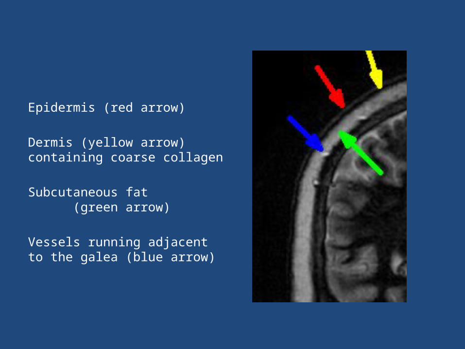
Epidermis (red arrow)
Dermis (yellow arrow) containing coarse collagen
Subcutaneous fat (green arrow)
Vessels running adjacent to the galea (blue arrow)

Epidermis (red arrow)
Dermis (yellow arrow) containing coarse collagen and vascular plexus
Subcutaneous fat (green arrow) with traversing fibrous strands
Occipitalis muscle (orange arrow)
BTFE 3D sequence
Skin of the Scalp
The skin contains hair follicles, sebaceous glands, and sweat glands.
It is the thickest skin in the body.
On imaging, the skin will enhance after contrast due to the rich subdermal plexus.

Subcutaneous Tissue
This is a thin layer of fat with fibrous strands splitting the fat into lobules. The fibrous strands connect the dermis to the galea.
This layer is rich in vessels and lymphatics.
The blood vessels in this layer are attached to the crossing the dermis. If they are cut, they are unable to vasoconstrict due to their attachments. This is why scalp lacerations can bleed profusely.
Galea Aponeurotica
This is a dense fibrous layer that extends over the vertex between the frontalis muscle anteriorly and the occipitalis muscle posteriorly. It blends in laterally with the temporalis fascia. The galea is thick at the vertex and thinner in the temporal region.
The major arteries course along the galea. From here, these arteries ramify superiorly into the subdermal plexus.

Subgaleal Connective Tissue
It is thought to be an avascular tissue, but there are fine vessels coming from the galea and skull. Emissary veins also course in this area and communicate with the sinuses.
This loose layer is what allows the scalp to glide over the skull. Because of this looseness, this layer is what results in scalp avulsions with trauma. This is also the layer surgeons use to create scalp flaps.
Blood that collects in this layer eventually pools around the eyes (“raccoon eyes”) due to obstruction in other directions from fascia or muscle.
Importance of the Emissary Veins
There are two important points regarding the emissary veins in the subgaleal tissue
1. Rupture of the veins can lead to subgaleal hemorrhage. These veins may be torn during delivery, particularly vacuum-assisted deliveries, leading to significant hematoma in the newborn scalp.
2. These vessels can spread infection intracranially as the emissary veins communicate with the dural venous sinuses.

Pericranium
The pericranium is firmly attached at the sutures. But in between, it is loosely attached to the bone.
Subperiosteal hematoma can lead to tension on the periosteum and ossification of the periosteum.
Sensory Innervation of the Scalp
The sensory innervation of the scalp is primarily from branches of cranial nerve V and the rami of C2 and C3.
The cranial nerve V1 branch innervates the forehead. The cranial V3 branches innervate the temple and anterior periauricular region.
The occipital nerves, which arise from C2 and C3, innervate the vertex, posterior scalp, and posterior periauricular region.

Importance of the Innervation of the Scalp
While headache can have many causes, compression of the nerves to a region of scalp may lead to the sensation of headache. For example, the zygomaticotemporal nerve passes through the temporalis muscle to innervate the temple and the occipital nerves pass through posterior cervical muscles to innervate the posterior scalp. Muscle tension may cause compression on these nerves and the feeling of headache.
Damage to the nerves during surgery may cause neuralgias of the head.
Arterial Supply of the Scalp
Each side of the scalp has 5 major arteries. Two arterial branches from the ophthalmic artery supply the forehead. However, the major arterial supply to the scalp is from the external carotid artery branches, including the superficial temporal artery, occipital artery, and posterior auricular artery.
The scalp has extensive arterial anastamoses. With just one temporal artery, the entire scalp can remain viable.
Tumors of the Scalp
Atypical Fibroxanthoma
AFX typically presents as an ulcerated nodular lesion on severely sun damaged skin in patients in elderly patients.
75% occur in the head and neck. The scalp and ear were identified as the most common sites of presentation in 65% and 23% of cases, respectively.
They are now regarded as cases of dermatofibrosarcoma protuberans (DFSP), which are more recently referred to as atypical benign fibrous histocytoma.
Atypical Fibroxanthoma
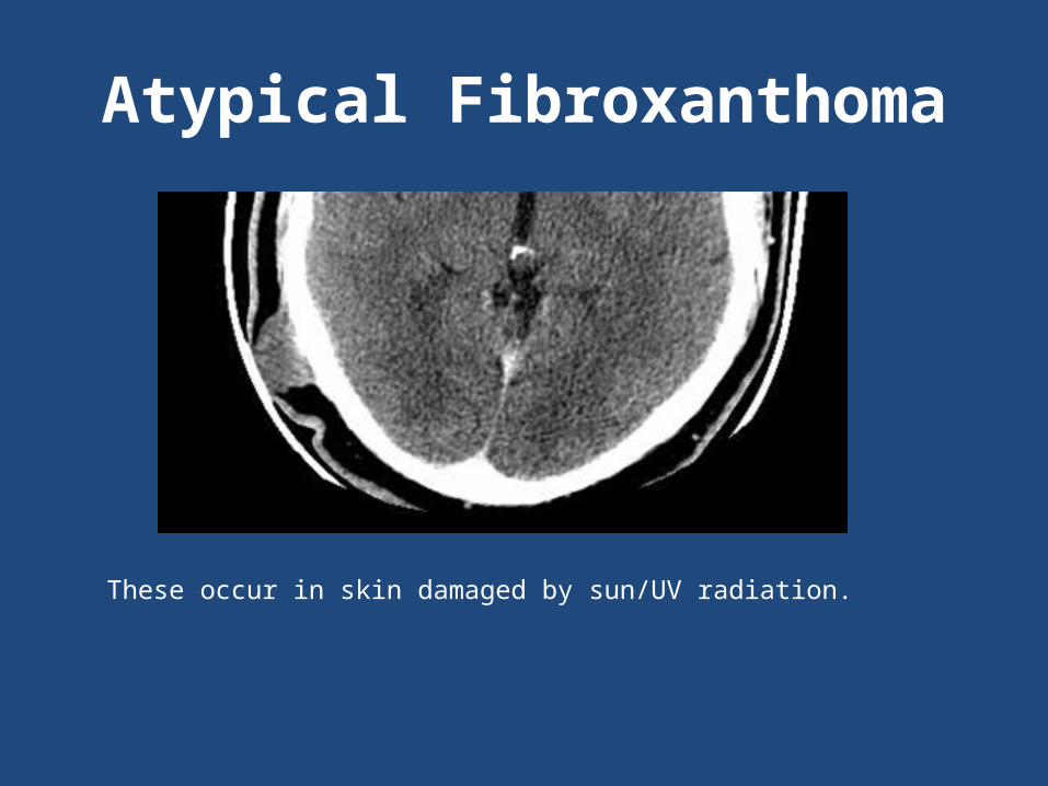
These occur in skin damaged by sun/UV radiation.

Differential for Malignant Scalp Masses in Adult
The most common malignant scalp tumors (in Taiwanese patients) in order of frequency found in one study by C.S. Chiu et al.
1. Basal cell carcinoma (44% of cases)
2. Squamous cell carcinoma (17% of cases)
3. Metastases to the scalp (13% of cases)
4. Angiosarcoma (7% of cases)
Melanoma only accounted for 2% of cases

Basal Cell Carcinoma

Squamous Cell Carcinoma
The vertex of the scalp is a common location for primary scalp tumors. Notice how even the scout shows subtle thickening of the skin.
Melanoma
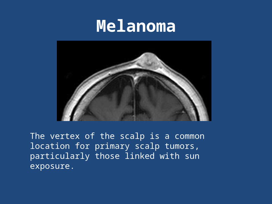
The vertex of the scalp is a common location for primary scalp tumors, particularly those linked with sun exposure.

Melanoma
This is just one section of a patient with a scalp melanoma that was much larger on adjacent sagittal slices. But notice the subtle thickening of the skin relative to the rest of the skin. Evaluation of the scalp on sagittal or coronal images is very useful as small lesions may be easily missed on axial images due to adjacent bone.
Metastasis to the Scalp
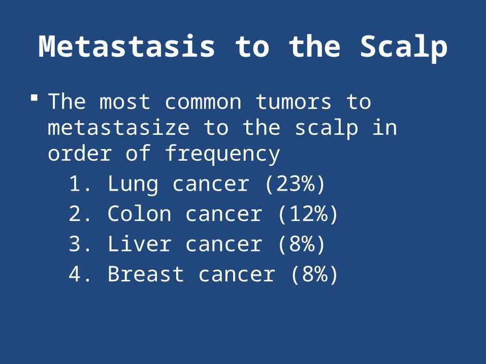
The most common tumors to metastasize to the scalp in order of frequency
1. Lung cancer (23%)
2. Colon cancer (12%)
3. Liver cancer (8%)
4. Breast cancer (8%)

Primary Undifferentiated Sarcoma
Congenital Scalp Mature Teratoma
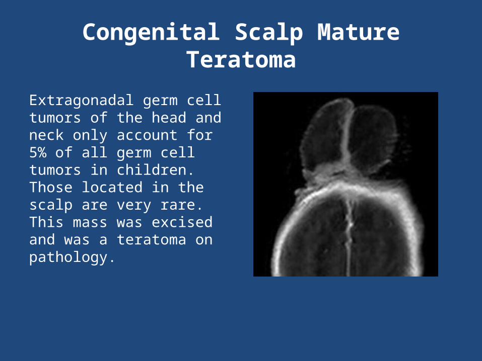
Extragonadal germ cell tumors of the head and neck only account for 5% of all germ cell tumors in children. Those located in the scalp are very rare. This mass was excised and was a teratoma on pathology.

Congenital Lesions

Congenital Inclusion Cysts of the Scalp
Dermoid cysts have a peak age distribution in the 1st decade. In general, dermoid cysts are more common that epidermoid cysts in children. They tend to occur in the midline, and can occur over the anterior fontanel.
Both epidermoid cysts and dermoid cysts are inclusion cysts due to epithelial rests. Dermoid cysts are distinguished from epidermoid cysts by the presence of hair, sebaceous glands, and sweat glands.

Congenital Inclusion Cysts of the Scalp
Inclusion cysts are usually present at birth and can develop gradually as secretion and internal desquamation accumulate. Exocrine glands in the cysts secrete a fluid similar to sweat.
Dermoid cyst have been described as hyperintense on T1 with variable signal on T2. But if the internal fat content is relatively low, the lesion will reveal CSF-like signal intensity on T2.

Dermoid Cyst

Dermoid Sinuses of the Scalp
In the head, the most common location is the external occipital protuberance (85%). The next most common locations are the nasion (11%) and the posterior parietal area (5%).
The depth to which they penetrate can vary. Occipital dermoid sinus may extend to the dura mater or further intracranially.

Fluid Collections in the Scalp
Fluid Collections in the Newborn Scalp
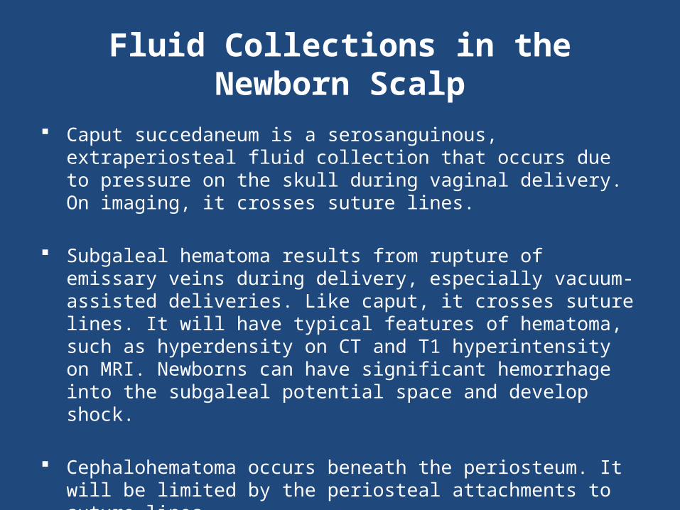
Caput succedaneum is a serosanguinous, extraperiosteal fluid collection that occurs due to pressure on the skull during vaginal delivery. On imaging, it crosses suture lines.
Subgaleal hematoma results from rupture of emissary veins during delivery, especially vacuum-assisted deliveries. Like caput, it crosses suture lines. It will have typical features of hematoma, such as hyperdensity on CT and T1 hyperintensity on MRI. Newborns can have significant hemorrhage into the subgaleal potential space and develop shock.
Cephalohematoma occurs beneath the periosteum. It will be limited by the periosteal attachments to suture lines.

Caput Succedaneum
Fluid collection crossing suture lines
Calcified Cephalahematoma
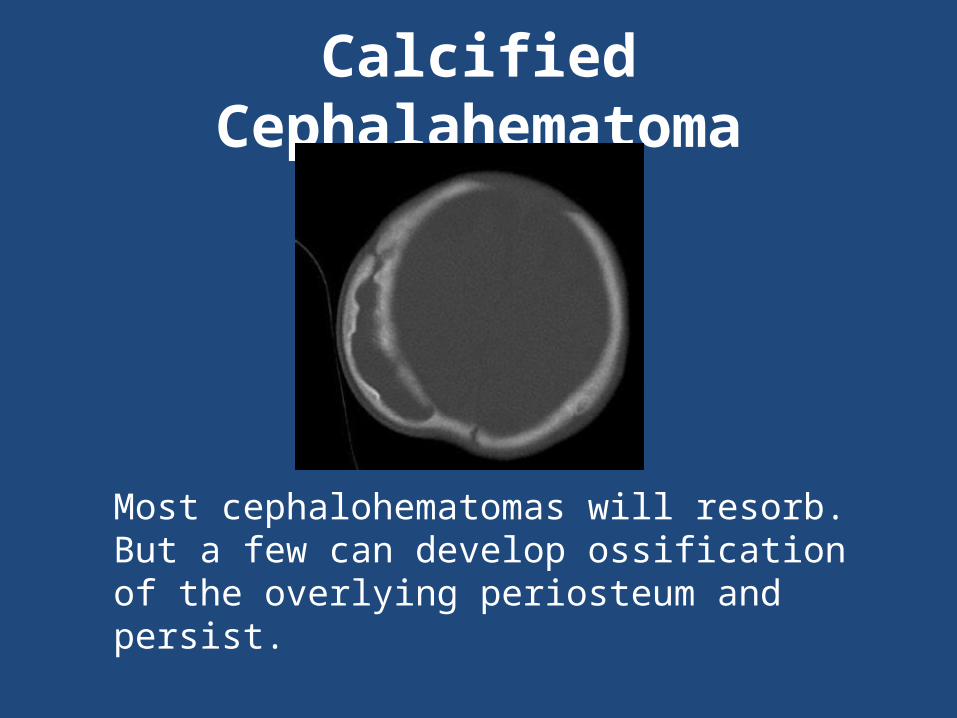
Most cephalohematomas will resorb. But a few can develop ossification of the overlying periosteum and persist.
Chronic Abscess
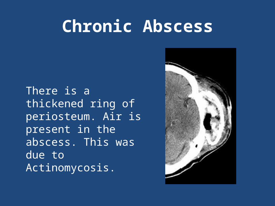
There is a thickened ring of periosteum. Air is present in the abscess. This was due to Actinomycosis.

Vascular Lesions
Atretic Encephalocele with Persistent Falcine Sinus
These rare lesions generally occur within a few centimeters of the lambda.
They contain meninges and neural rests.
Embryonic positioning of the straight sinus (persistent falcine sinus) has frequently been identified in these lesions.
Atretic Encephalocele with Persistent Falcine Sinus
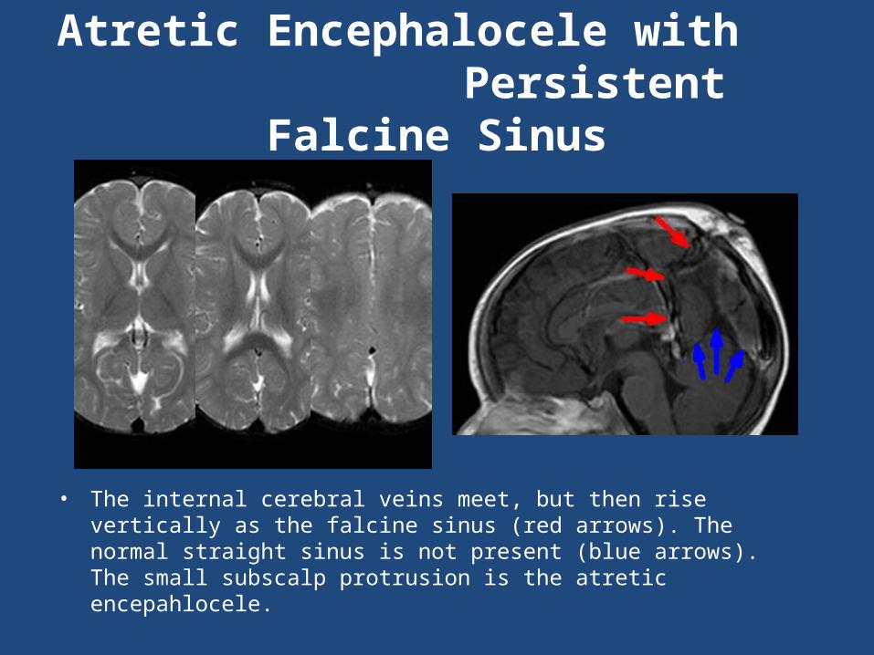
• The internal cerebral veins meet, but then rise vertically as the falcine sinus (red arrows). The normal straight sinus is not present (blue arrows). The small subscalp protrusion is the atretic encepahlocele.

Cirsoid Aneurysm
This is a misnomer. This is an arterio-venous fistula of scalp vessels. The term cirsoid aneurysm is used due to variceal dilatation of the draining veins (Greek kirsos = varix).
The majority occur as congenital anomalies, although 10-20% develops following penetrating trauma to the scalp.
In one series of 21 cases by Gurkalar et al., the superficial temporal artery was involved in 90% of cases. It was the primary arterial feeder in 70% of their cases.

Sinus Pericranii
This purely venous entity is characterized by an anomalous communication between the intracranial dural sinuses (usually the superior sagittal sinus) and dilated scalp veins
The varicosities are intimately associated with the periostium, are distensible, and vary in size with changes in intracranial pressure

Sinus Pericranii

AVM
• MRI shows increased flow voids in the scalp. Angiogram shows vessels from ophthalmic artery branches supplying the AVM. The ophthalmic artery branches normally supply the forehead.
Hemangiomas of Infancy
Hemangiomas of infancy (HOI) is an umbrella term for all the cutaneous hemangiomas that primary occur in infancy.
They are masses of plump, rapidly dividing endothelial cells.
They are often absent or small at birth and grow rapidly in early infancy.
In a large series, 60% of HOI occurred on the head and neck.
The growth characteristics of HOI are often divided
into phases: nascent, proliferating, involuting, and
involuted.

Hemangioma of Infancy
There are numerous flow voids within this proliferating scalp mass in a child.

References Bruckner AL, Frieden IJ. Hemangiomas of infancy. J Am Acad Dermatol. 2003
Apr;48(4):477-93. Gurkanlar D1, Gonul M, Solmaz I, Gonul E. Cirsoid aneurysms of the scalp.
Neurosurg Rev 2006; 29: 208–212. Meigel WN, Gay S, Weber L. Dermal architecture and collagen type distribution.
Arch. Derm. Res. 1977; 259:1-10. Withers AH, Brougham ND, Barber RM, Tan ST. Atypical fibroxanthoma and
malignant fibrous histiocytoma. J Plast Reconstr Aesthet Surg. 2011 Nov;64(11):e273-8.
Patterson RJ, Egelhoff JC, Crone KR, Ball WS Jr. Atretic parietal cephaloceles revisited: an enlarging clinical and imaging spectrum? Am J Neuroradiol 1998; 19:791–795.
de Carvalho GT, Fagundes-Pereyra WJ, Marques JA, Dantas FL, de Sousa AA. Congenital inclusion cysts of the anterior fontanelle. Surg Neurol. 2001 Dec;56(6):400-5
Ray M, Barnett D, Snipes G, Layton K, Opatowsky M. Ruptured intracranial dermoid cystProc (Bayl Univ Med Cent) 2012;25(1):23–25
References Hong SH, Chung HW, Choi JY, Koh YH, Choi JA, Kang HS. MRI findings of
subcutaneous epidermal cysts: emphasis on the presence of rupture. AJR 2006; 186:961–966.
Suster S, Ronnen M, Huszar M. Extraskeletal Ewing's sarcoma of the scalp. Pediatr Dermatol. 1988 May; 5(2):123-6.
Sharman AM, Kirmi O, Anslow P. Imaging of the skin, subcutis, and galea aponeurotica. Semin Ultrasound CT MRI 30:452-464.
Kemp WJ, Tubbs RS, Cohen-Gadol AA. The innervation of the scalp: A comprehensive review including anatomy, pathology, and neurosurgical correlates. Surg Neurol Int 2011;2:178.
Amar AP, Aryan HE, Meltzer HS, Levy ML Neonatal subgaleal hematoma causing brain compression: report of two cases and review of the literature. Neurosurgery. 2003 Jun;52(6):1470-4.
Bodkin PA, Bhangoo R, Walsh AR, Sgouros S. Beware of the midline scalp lump. J R Soc Med. 2004 May;97(5):239-41.
Carpenter JS1, Rosen CL, Bailes JE, Gailloud P. Sinus pericranii: clinical and imaging findings in two cases of spontaneous partial thrombosis. Am J Neuroradiol. 2004 Jan;25(1):121-5.

References Morón FE, Morriss MC, Jones JJ, Hunter JV. Lumps and bumps on the head in
children: use of CT and MR imaging in solving the clinical diagnostic dilemma. RadioGraphics 2004; 24:1655–1674.
Chao TK, Chang YL, Sheen TS. Extraskeletal Ewing’s sarcoma of the scalp. J Laryngol Otol. 2000 Jan;114(1):73-5.
Chiu CS, Lin CY, Kuo TT, Kuan YZ, Chen MJ, Ho HC, Yang LC, Chen CH, Shih IH, Hong HS, Chuang YH. Malignant cutaneous tumors of the scalp: a study of demographic characteristics and histologic distributions of 398 Taiwanese patients. J Am Acad Dermatol. 2007 Mar;56(3):448-52. Epub 2006 Dec 1.
Seyhan T, Sener L, Refik Ozerdem O, Bal N. Mature teratoma presenting as a scalp mass in a newborn. J Craniofac Surg. 2006 Sep;17(5):1009-11.
Bajoghli A, Agarwal S, Goldberg L, Mirzabeigi M. Melanoma arising from an epidermal inclusion cyst. J Am Acad Dermatol. 2013 Jan;68(1):e6-7.
Chang SJ, Sims J, Murtagh FR, McCaffrey JC, Messina JL. Proliferating trichilemmal cysts of the scalp on CT. Am J Neuroradiol. 2006 Mar;27(3):712-4.
Daniel Oppenheimer, M.D. [email protected] Brett Talbot, M.D. Shweta Bhatt, M.D. Ravinder Sidhu, M.D
Control # 209 Title: Life -Threatening Lytic lesion of the Mandible: A Lesson Learned eEdE# eEdE-157