lesson 1 the respiratory system 1-1. · pdf filelesson 1 the respiratory system 1-1. general...
TRANSCRIPT
LESSON 1
THE RESPIRATORY SYSTEM
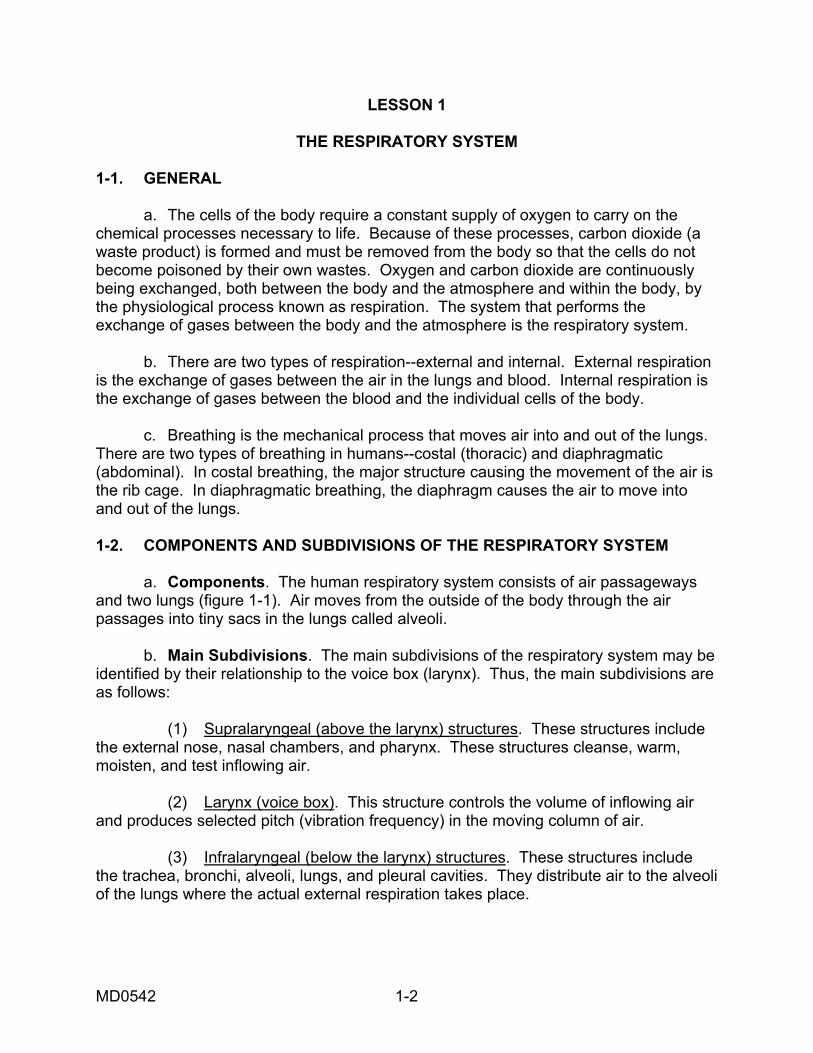
1-1. GENERAL a. The cells of the body require a constant supply of oxygen to carry on the chemical processes necessary to life. Because of these processes, carbon dioxide (a waste product) is formed and must be removed from the body so that the cells do not become poisoned by their own wastes. Oxygen and carbon dioxide are continuously being exchanged, both between the body and the atmosphere and within the body, by the physiological process known as respiration. The system that performs the exchange of gases between the body and the atmosphere is the respiratory system. b. There are two types of respiration--external and internal. External respiration is the exchange of gases between the air in the lungs and blood. Internal respiration is the exchange of gases between the blood and the individual cells of the body. c. Breathing is the mechanical process that moves air into and out of the lungs. There are two types of breathing in humans--costal (thoracic) and diaphragmatic (abdominal). In costal breathing, the major structure causing the movement of the air is the rib cage. In diaphragmatic breathing, the diaphragm causes the air to move into and out of the lungs. 1-2. COMPONENTS AND SUBDIVISIONS OF THE RESPIRATORY SYSTEM a. Components. The human respiratory system consists of air passageways and two lungs (figure 1-1). Air moves from the outside of the body through the air passages into tiny sacs in the lungs called alveoli. b. Main Subdivisions. The main subdivisions of the respiratory system may be identified by their relationship to the voice box (larynx). Thus, the main subdivisions are as follows: (1) Supralaryngeal (above the larynx) structures. These structures include the external nose, nasal chambers, and pharynx. These structures cleanse, warm, moisten, and test inflowing air. (2) Larynx (voice box). This structure controls the volume of inflowing air and produces selected pitch (vibration frequency) in the moving column of air. (3) Infralaryngeal (below the larynx) structures. These structures include the trachea, bronchi, alveoli, lungs, and pleural cavities. They distribute air to the alveoli of the lungs where the actual external respiration takes place.
MD0542 1-2
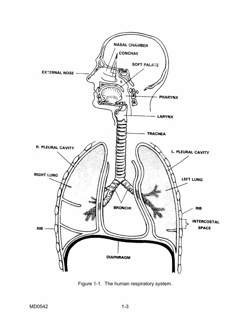
Figure 1-1. The human respiratory system.
MD0542 1-3
1-3. SUPRALARYNGEAL STRUCTURES Supralaryngeal structures are shown in figure 1-2. a. External Nose. The external nose is the portion projecting from the face. It is supported primarily by cartilages. It has a midline divider, called the nasal septum, which extends from the internal nose. There are paired openings (nostrils) that lead to paired spaces (vestibules). Guard hairs in the nostrils filter inflowing air. b. Nasal Chambers (Internal Nose). Behind each vestibule of the external nose is a nasal chamber. The two nasal chambers together form the internal nose. These chambers, too, are separated by the nasal septum. (1) The walls of the nasal chambers are lined with a thick mucous-type membrane known as the mucoperiosteum. It has a ciliated (hair-like projection that move fluids to the rear) epithelial surface. It also has a rich blood supply that provides warmth and moisture. At times, the membrane may become quite swollen. (2) The lateral wall of each chamber has three scroll-like extensions into the nasal chamber that help to increase the surface area exposed to the inflowing air. These scroll-like extensions are known as conchae.
Figure 1-2. Supralaryngeal structures.
MD0542 1-4
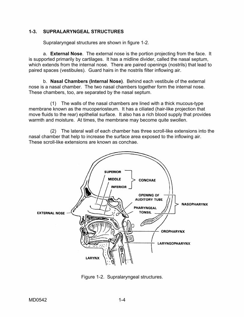
(3) The sense of smell is caused by special nerve endings located in the upper areas of the nasal chambers. The epithelium containing the sensory ending is known as the olfactory epithelium. (4) There are air "cells" or cavities in the skull called paranasal sinuses. The paranasal sinuses are connected with nasal chambers and are lined with the same ciliated mucoperiosteum. Thus, these sinuses are extensions of the nasal chambers into the skull bones. For this reason, they are known as paranasal sinuses. c. Pharynx. The pharynx is the common posterior space for the respiratory and digestive systems. (1) The portion of the pharynx specifically related to the respiratory system is the nasopharynx. It is the portion of the pharynx above the soft palate. The two posterior openings (nares) of the nasal chambers lead into the single space of the nasopharynx. The auditory (eustachian) tubes also open into the nasopharynx. The auditory tubes connect the nasopharynx with the middle ears. This allows the pressure between the outside and inside of the eardrum to be equalized. Lying in the upper posterior wall of the nasopharynx are the pharyngeal tonsils (adenoids). The soft palate floor of the nasopharynx is a trapdoor that closes off the upper respiratory passageways during swallowing. (2) The portion of the pharynx closely related to the digestive system is the oropharynx. It is the portion of the pharynx below the soft palate and above the upper edge of the epiglottis. The epiglottis is the flap that prevents food from entering the larynx when a person swallows. (3) The portion of the pharynx that is common to both the respiratory system and the digestive system is the laryngopharynx. It is the portion of the pharynx below the upper edge of the epiglottis. Thus, the digestive and respiratory systems lead into it from above and lead off from it below. 1-4. LARYNX The larynx, also called the Adam's apple or voice box, connects the pharynx with the trachea. The larynx is located in the anterior neck region and has a box-like shape (figure 1-3). The larynx of the male becomes larger and heavier during puberty and causes the voice to deepen. The adult male's larynx tends to be located lower in the neck. In the female, the larynx remains higher and smaller. a. The larynx has a vestibule ("entrance hallway") that can be covered over by the epiglottis. The glottis itself is the hole between the vocal cords. Through the glottis, air passes from the vestibule into the main chamber of the larynx (below the cords) and then into the trachea. The skeleton of the larynx is made up of a series of cartilages.
MD0542 1-5
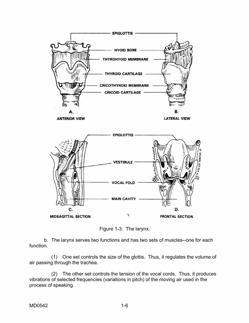
Figure 1-3. The larynx.
b. The larynx serves two functions and has two sets of muscles--one for each function. (1) One set controls the size of the glottis. Thus, it regulates the volume of air passing through the trachea. (2) The other set controls the tension of the vocal cords. Thus, it produces vibrations of selected frequencies (variations in pitch) of the moving air used in the process of speaking.
MD0542 1-6
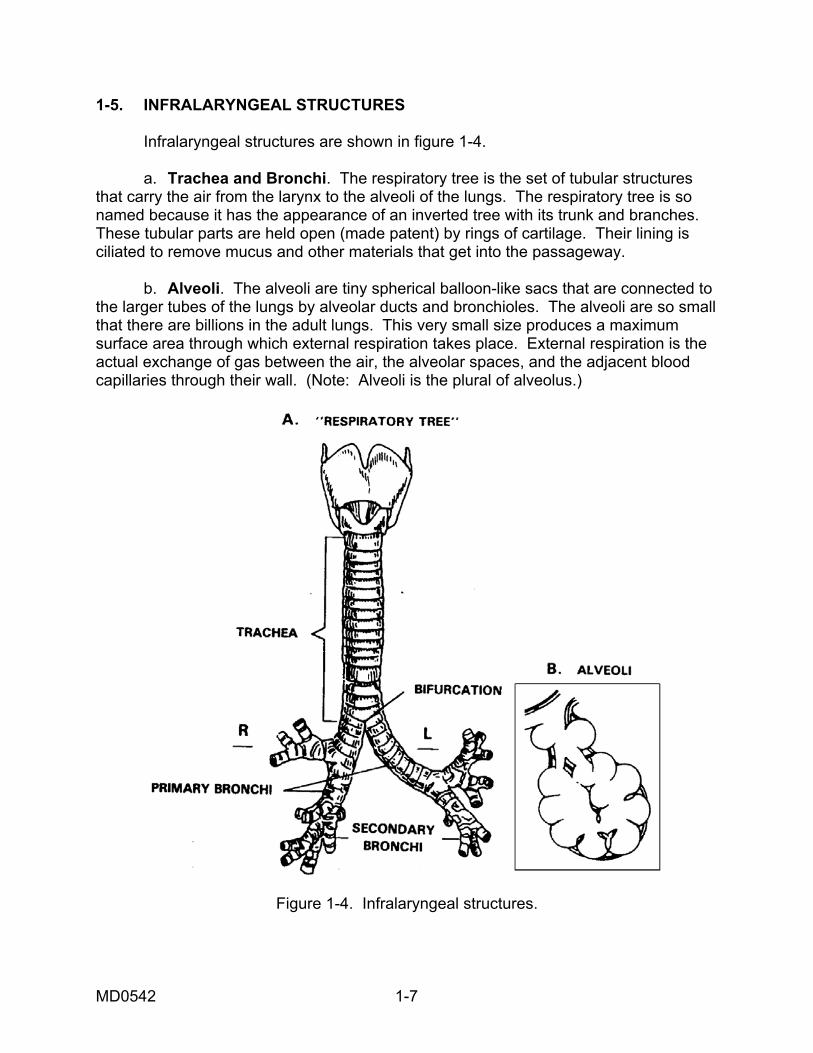
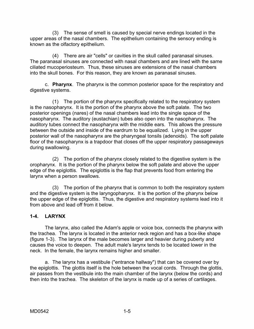
1-5. INFRALARYNGEAL STRUCTURES Infralaryngeal structures are shown in figure 1-4. a. Trachea and Bronchi. The respiratory tree is the set of tubular structures that carry the air from the larynx to the alveoli of the lungs. The respiratory tree is so named because it has the appearance of an inverted tree with its trunk and branches. These tubular parts are held open (made patent) by rings of cartilage. Their lining is ciliated to remove mucus and other materials that get into the passageway. b. Alveoli. The alveoli are tiny spherical balloon-like sacs that are connected to the larger tubes of the lungs by alveolar ducts and bronchioles. The alveoli are so small that there are billions in the adult lungs. This very small size produces a maximum surface area through which external respiration takes place. External respiration is the actual exchange of gas between the air, the alveolar spaces, and the adjacent blood capillaries through their wall. (Note: Alveoli is the plural of alveolus.)
Figure 1-4. Infralaryngeal structures.
MD0542 1-7
c. Lungs. A lung is an individual organ composed of tubular structures and alveoli, bound together by fibrous connective tissue. In the human, there are two lungs--right and left. Each lung is divided into lobes. A pulmonary lobe is a major subdivision of a lung marked by fissures (deep folds). Each lobe is further partitioned into bronchopulmonary segments. (1) The right lung is larger in volume than the left lung since the left lung must leave room for the heart. The right lung is divided into three pulmonary lobes (upper, middle, and lower) and ten bronchopulmonary segments (2+3+5). The left lung is divided into two pulmonary lobes (upper and lower) and eight bronchopulmonary segments (4+4). (2) Each lung is supplied by a primary or mainstream bronchus leading off the trachea. Each lobe is supplied by a secondary or lobar bronchus branching off of the primary bronchus. Each segment is supplied by a tertiary or segmented bronchus, a branch of the lobar bronchus. d. Pleural Cavities. Each lung is encased in a serous cavity called the pleural cavity. Each serous cavity has inner and outer membranes. The serous membranes secrete fluid that act as a lubricant between the membranes, allowing freer motion for the organs. The pleural cavities allow the lungs to move freely with a minimum of friction during the expansion and contraction phases of breathing. Located in the middle of the thorax, between the two pleural cavities, is the mediastinum ("I stand between"). The mediastinum is a tissue and organ-filled space. Within it is the heart, which is located at the same level as the lungs. 1-6. BREATHING AND BREATHING MECHANISMS a. Boyle's law (named after Robert Boyle, British physicist, 1627-1691) states that as the volume of a gas-filled container increases, the pressure inside decreases. Conversely, as the volume of a closed container decreases, the pressure inside increases. When two connected spaces of air have different pressures, the air moves from the space with greater pressure to the one with lesser pressure. In regard to breathing, we can consider the air pressure around the human body to be constant. The pressure inside the lungs may be greater or less than the pressure outside the body. Thus, a greater internal pressure causes air to flow out; a greater external pressure causes air to flow in. b. The human trunk can be compared to a hollow cylinder. This cylinder is divided into upper and lower cavities by the diaphragm. The upper is the thoracic cavity and is essentially gas-filled. The lower is the abdominopelvic cavity and is essentially water-filled. By changing the size (volume) of the thoracic cavity, air can be forced into and out of the cavity. The size of the thoracic cavity is changed by movement of the rib cage (coastal or thoracic breathing) and by movement of the diaphragm (diaphragmatic or abdominal breathing).
MD0542 1-8
1-7. COSTAL (THORACIC) BREATHING Muscles attached to the thoracic cage (rib cage) cause the chest to expand during inhalation and return to normal during exhalation. a. Inhalation. Muscles attached to the thoracic cage contract and raise the ribs. A typical rib might be compared to a bucket handle, attached at one end to the sternum (breastbone) and at the other end to the vertebral column (spine). The "bucket handle" is lifted by the overall upward and outward movement of the rib cage. These movements increase the thoracic diameters from right to left (transverse) and from front to back. Thus, the volume within the chest increases. Recalling Boyle's law, the increase in volume leads to a decrease in pressure. The higher air pressure outside the body then forces air into the lungs and inflates them. b. Exhalation. The rib cage movements and pressure relationships are reversed for exhalation. The muscles relax, the ribs return to their normal position, and the size of the chest decreases. The pressure within the chest increases and forces air outside the body. 1-8. DIAPHRAGMATIC (ABDOMINAL) BREATHING The diaphragm is a thin, but strong, dome-shaped muscular membrane that separates the abdominal and thoracic cavities. The abdominal wall is elastic in nature. The abdominal cavity is filled with soft, watery tissues. a. Inhalation. As the diaphragm contracts, the dome flattens and the diaphragm descends. This increases the depth (vertical diameter) of the thoracic cavity and thus increases its volume. This decreases air pressure within the thoracic cavity. The greater air pressure outside the body then forces air into the lungs. b. Exhalation. As the diaphragm relaxes, the elastic abdominal wall forces the diaphragm up again by pushing the watery tissues of the abdomen against the underside of the relaxed diaphragm. The dome moves upward, the volume of the thoracic cavity is decreased, and air is forced from the lungs and into the atmosphere. 1-9. NERVOUS CONTROL OF BREATHING Breathing can be controlled voluntarily to some extent, such as holding your breath for a short period of time or breathing deeply during a medical examination. However, breathing is usually controlled by the nervous system without the need to consciously order the body to inhale or exhale. Respiratory reflexes are controlled by the respiratory center found in the medullary portion of the brainstem. The respiratory center coordinates the actions of costal breathing and diaphragmatic breathing to ensure that they work together (chest and abdominal muscles contract at the same time and relax at the same time). The amount of carbon dioxide (CO2) in the circulating blood is one of the major influences on the actions of the respiratory center.
MD0542 1-9
1-10. MOVEMENT OF BLOOD a. The veins of the systemic blood circulatory system bring oxygen-poor blood from all parts of the body to the right atrium of the heart. From the right atrium, the blood flows into the right ventricle of the heart. Upon contraction of the right ventricle, blood is forced into the pulmonary arch. The pulmonary arch divides into the right and left pulmonary arteries that delivers the oxygen-poor blood to their respective lungs. Paralleling the branching of the respiratory tree, the arteries divide and subdivide within the lungs. These arteries lead to capillaries that surround the alveoli. The walls of these capillaries are thin enough to accommodate the passage of gases to and from the alveolus. The oxygen-poor blood gives up the carbon dioxide which it has been carrying and absorbs oxygen from the alveolus. Just as oxygen travels from the alveolus to the capillary, carbon dioxide travels from the capillary to the alveolus. b. The blood, now saturated with oxygen, is collected by the pulmonary venous system. The blood flows through the pulmonary veins into the left atrium of the heart. From the left atrium, it flows into the left ventricle. When the left ventricle contracts, the oxygen-rich blood is forced into the aorta of the systemic blood circulatory system. Other arteries branch off of this large artery and carry the oxygen-rich blood to all living cells within the body. As the arteries continue to subdivide and get smaller, they eventually reach the capillary stage. At this stage, oxygen moves from the blood into the surrounding body cells and carbon dioxide, a waste material, travels from the body cells to the blood. The blood then flows from the capillaries into veins and eventually returns to the right atrium of the heart. 1-11. TRANSPORTATION OF GASES Oxygen and carbon dioxide are the primary gases involved in respiration. Under special circumstances, nitrogen may also be of concern. Some of the gases are dissolved directly in the plasma of the blood. Most of the gasses, however, are carried within the erythrocytes (red blood cells, commonly called RBCs). The RBCs, found in great numbers in the blood, are specially constructed for transporting the gases. Hemoglobin, a substance found within RBCs, has a great affinity for oxygen. Yet, the hemoglobin can readily give up the oxygen wherever it is needed.
Continue with Exercises
MD0542 1-10
EXERCISES, LESSON 1 INSTRUCTIONS. Answer the following items by completing the statement or by writing the answer in the space provided at the end of the item. After you have completed all of these items, turn to "Solutions to Exercises" at the end of the lesson and check your answers. 1. Name and define the two types of respiration. a. ______________________________________________________________ ______________________________________________________________ b. ______________________________________________________________ ______________________________________________________________ 2. What does the larynx control? ______________________________________________________________ 3. What is found in the nose to filter inflowing air ______________________________________________________________ 4. The walls of the nasal chambers are lined with a thick mucous-type membrane called the ________________________________________________________. 5. The common posterior space for the respiratory and digestive systems is called the ______________________________________________________________. 6. The part of the pharynx that is related to the respiratory system is the: ______________________________________________________________.
MD0542 1-11
7. The tubes that are used to equalize the pressure between the outside and inside of the eardrum are called ____________________________________________. 8. How is the upper respiratory passageway closed when the person swallows food? ______________________________________________________________ ______________________________________________________________ 9. Which flap prevents food from entering the larynx during swallowing? ______________________________________________________________ 10. The two common terms that refer to the larynx are _______________________ and _______________________________________. 11. Describe the alveoli. ______________________________________________________________ ______________________________________________________________ 12. Which lung is the smaller lung and why? ______________________________________________________________ ______________________________________________________________ 13. Where is the mediastinum located? ______________________________________________________________ ______________________________________________________________
MD0542 1-12
14. How is Boyle's law related to a person's breathing? ______________________________________________________________ ______________________________________________________________ ______________________________________________________________ ______________________________________________________________ 15. During inhalation, how is the rib cage lifted? ______________________________________________________________ ______________________________________________________________ 16. Describe the action of the diaphragm during inhalation. ______________________________________________________________ ______________________________________________________________ ______________________________________________________________ ______________________________________________________________ 17. Which part of the brain controls the respiratory reflexes? ______________________________________________________________ ______________________________________________________________ 18. The primary gases involved in respiration are ______________________ and ________________________.
Check Your Answers on Next Page
MD0542 1-13
SOLUTIONS TO EXERCISES, LESSON 1 1. External respiration is the exchange of gases between the air in the lungs and blood. Internal respiration is the exchange of gases between the blood and the individual cells of the body. (para 1-1b) 2. The larynx controls the volume of inflowing air and produces selected pitch (vibration frequency) in the moving column of air. (para 1-2b(2)) 3. Hairs in the nose filter inflowing air. (para 1-3a) 4. Mucoperiosteum. (para 1-3b(1)) 5. Pharynx. (para 1-3c) 6. Nasopharynx. (para 1-3c(1)) 7. Auditory (eustachian) tubes. (para 1-3c(1)) 8. The soft palate floor of the nasopharynx is a trapdoor that closes off the upper respiratory passageways during swallowing. (para 1-3c(1)) 9. The epiglottis. (para 1-3c(2)) 10. Adam's apple and voice box. (para 1-4) 11. The alveoli are tiny spherical (balloon-like) sacs that are connected to the larger tubes of the lungs by alveolar ducts. (para 1-5b) 12. The left lung is smaller because it must leave room for the heart. (para 1-5c) 13. The mediastinum is found in the middle of the thorax, between the two pleural cavities. (para 1-5d) 14. When the volume of the chest cavity decreases, the air pressure inside the lungs increases and forces to flow out from the lungs. When the volume of the chest cavity increases, the air pressure inside the lungs decreases and causes air to flow in. (para 1-6a) 15. Muscles attached to the thoracic cage raise the rib cage during inhalation. (para 1-7a)
MD0542 1-14
MD0542 1-15
16. As the diaphragm contracts, the dome flattens and the diaphragm descends, thereby increasing the size of the thoracic cavity. This results in decreased air pressure within the thoracic cavity. The greater air pressure outside the body forces air into the lungs. (para 1-8a) 17. The respiratory center located in the brainstem. (para 1-9a) 18. Oxygen and carbon dioxide. (para 1-11)
End of Lesson 1