lessons from “mycobacterium-related” elephant necropsy cases scott p. terrell, dvm, dacvp
TRANSCRIPT

Lessons from “Mycobacterium-related”Elephant necropsy cases
Scott P. Terrell, DVM, DACVP


My “files”
• 41 “adult” animals spanning 11 years– Fetal and neonatal deaths excluded– Herpes virus cases excluded
• 21 Asians– Musculoskeletal, repro neoplasia, cardiac disease,
GI disease, Mycobacterial disease• 20 Africans– Musculoskeletal disease, GI disease,
Mycobacterial disease

Mycobacterial-related disease• 14 cases– 6 Africans
• 1 confirmed M. tuberculosis by post mortem culture– Gross and histologic granulomatous disease– Negative acid-fast stains
• 5 confirmed atypical Mycobacterial disease– Culture (4), PCR (1)
– 8 Asians (skewed sample?)
• 6 confirmed M. tuberculosis by post mortem culture• 2 gross and/or histologic granulomatous disease,
acid-fast negative, and culture negative*

African elephants

African elephants with Mycobacterium-related disease
1 confirmed M. tuberculosis by post mortem culture– Exposure and trunk wash history unknown– Gross and histologic granulomatous disease– Negative acid-fast stains***
5 confirmed atypical Mycobacterial disease–M. szulgai - 3 cases by culture–M. smegmatis – 1 case by culture–M. aurupense – 1 case by PCR

African elephant with Mycobacterium-related disease
1 confirmed M. tuberculosis by post mortem culture
–No mention of TB in history• Clinical evidence of musculoskeletal disease
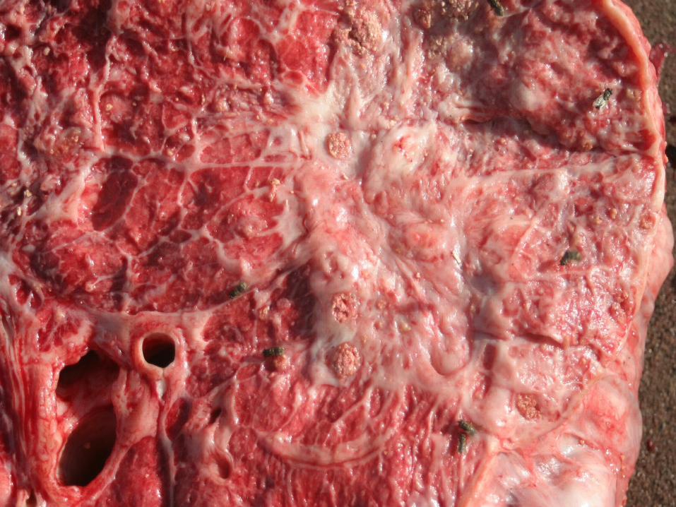
–Gross findings• Several pulmonary granulomas > 12cm diameter• Caseous material in bronchioles• Tracheobronchial lymph nodes enlarged• Sublumbar lymph node enlarged and caseous

African elephant with Mycobacterium-related disease
1 confirmed M. tuberculosis by post mortem culture
–Histopathology• Evaluated by highly qualified zoo pathologist• “Numerous” granulomas evaluated• Central caseous debris and mineralization• Multiple acid-fast stains negative• Diagnosed as highly suspicious for M tb.
– Culture (NVSL)• Positive from lung• Negative from lymph node and bronchial

Asian elephants

Mycobacterium-related diseasein Asian elephants
–6 confirmed M. tuberculosis by post mortem culture• 2 historically trunk wash positive, tx hist unknown• 4 had at least an exposure history to TB+ animal
–2 gross and/or histologic granulomatous disease, acid-fast negative, and culture negative• 1 historically trunk wash positive, treated• 1 pos STAT-pak, MAPIA, treated
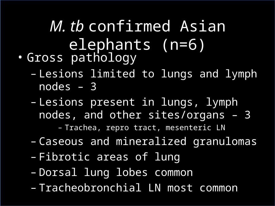
M. tb confirmed Asian elephants (n=6)• Gross pathology– Lesions limited to lungs and lymph nodes – 3– Lesions present in lungs, lymph nodes, and other
sites/organs – 3– Trachea, repro tract, mesenteric LN
– Caseous and mineralized granulomas– Fibrotic areas of lung– Dorsal lung lobes common– Tracheobronchial LN most common

Mycobacteriosis

Mycobacteriosis


Mycobacteriosis


Mycobacteriosis

M. tb confirmed Asian elephants (n=6)• Histopathology– Inflammation– Classic granulomatous pneumonia– Areas of histiocytic and necrosuppurative bronchopneumonia
– AF positive bacteria rare or very rare• Small % of granulomas examined are AF positive
– 10% • Bacteria can be as few as 3-5 organisms
• Cytology– My experience, not valuable in the field

H&E, 40xH&E, 4x

H&E, 40xH&E, 4x

H&E, 40x

H&E, 40xH&E, 40x

Mycobacteriosis
• Post mortem diagnosis–Numerous sections of lung and lymph node
must be cultured and examined• Acid-fast bacteria are exceedingly hard to find
on histopathology
Fites acid-fast400x

Mycobacteriosis
• Post mortem diagnosis–Numerous sections of lung and lymph node
must be cultured and examined• Acid-fast bacteria are exceedingly hard to find
on histopathology
Fites acid-fast400x

Mycobacteriosis
• Post mortem diagnosis–Numerous sections of lung and lymph node
must be cultured and examined• Acid-fast bacteria are exceedingly hard to find
on histopathology
Fites acid-fast1000x
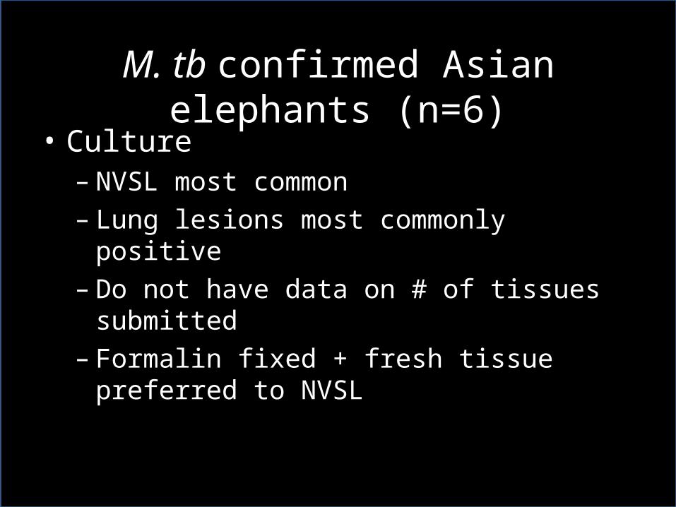
M. tb confirmed Asian elephants (n=6)• Culture– NVSL most common– Lung lesions most commonly positive– Do not have data on # of tissues submitted– Formalin fixed + fresh tissue preferred to NVSL

Tb related disease in Asians–2/8 animals• 1 - Gross and histopathologic granulomas in lung
– Acid-fast negative– Historically trunk wash positive
• 1 – Gross lesions in lymph node/trachea/lung – Granulomas in lymph node and trachea– Lung described as fibrosis– No granulomatous inflammation on histopath– No trachea listed on histopath report– Historically STAT-pak, MAPIA positive
• Both animals culture negative• Both animals had been treated

Summary
• Mtb more common in Asians than Africans• Lung and thoracic LN pathology most common• Histopathologic lesions vary• Acid-fast organisms are rare• Culture of post mortem lesions is often
successful• Sampling techniques are inconsistent• Historical information is lacking

Recommendations
• Good solid necropsy data can help…– Identify active cases– Define latency– Provide information with regard to accuracy of
diagnostic testing• Trunk wash• Serologic• Other

Recommendations
• For elephants with a “TB related history”… post mortem TB workup should be HIGH priority– Stat-PAK/MAPIA– Culture positive– Exposure history
– Human safety always takes priority

Recommendations• Post mortem TB work up should include– Peri-mortem serologic testing if possible (bank at least)
– Post-mortem “secretion” cultures• Trunk, trachea, airways
– Thorough sampling of lung and lymph node lesions• Individually labeled tissue• Multiple tissues for culture• Multiple tissues for histopathology• Tissue for culture rather than swab• Be sure to sample lesional tissue• NVSL +/- NJ• Histopathology by pathologist with TB experience

Recommendations
• Modify SSP necropsy/research protocol– Detailed procedures for TB sampling• Secretions• Tissues
– Detailed requests for exposure, trunk wash, serologic history, clinical signs