letter to the editor - princeton university to the editor ... with equal probability of clockwise or...
TRANSCRIPT

In Vitro Cell. Dev. Biol.--Animal 36:563-565, October 2000 9 2000 Society for In Vitro Biology 1071-2690/00 $10.00+0.00
Letter to the Editor SYMMETRY BREAKING IN CULTURED MAMMALIAN CELLS
Dear Editor: The generation of natural forms, such as characteristic tissue pat-
terns, involves a process of symmetry breaking in which structures with an initially uniform configuration spontaneously transform into structures with lower symmetry order (Ball, 1999). Symmetry break- ing has been examined in biological systems, including formation of fruiting bodies in slime mold (Palsson and Cox, 1996), pattern formation in bacterial colonies (Budrene and Berg, 1991), and gen- eration of animal coat patterns (Murray, 1993); however, the length scale of the characteristic patterns analyzed were orders of magni- tude larger than the individual cell components that drive pattern- ing. Here we describe a simplified in vitro model in which symmetry breaking and serf-organization can be visualized within small groups of mammalian cel!s migrating within a geometrically con- fined adhesive space on micropatterned culture substrates created with a microcontact printing technique (Chen et al., 1997).
Development of mammalian tissues, such as capillary blood ves- sels, is mediated by coordinated migration of cells, which, in turn, is controlled by cell interactions with soluble morphogens, other cells, and insoluble extracellular matrix proteins, such as fibronec- tin (FN). To create a minimal in vitro model of migration-dependent pattern generation, capillary endothelial cells were cultured in me- dium containing motility factors (basic fibroblast growth factor + 10% serum) on small (-<50 X 50 ~m 2) FN-eoated adhesive islands that were surrounded by nonadhesive barrier regions. When two cells were cultured on a single island, the high symmetry order associated with the ordinarily random movement of cells on uncon- fined adhesive surfaces (Dunn and Brown, 1987) changed sponta- neously. This transition resulted in directional cell migration and lower symmetry order, such that the entire muhicellular system syn- chronously rotated about its geometric center (Fig. 1A). Each cell in this dynamic pattern was led by a lamellipodium that wrapped around the trailing edge of the neighboring cell, thereby creating a sigmoid cell-cell interface reminiscent of the Yin-Yang (YY) sym- bol of Far Eastern religions (Capra, 1991) (Fig. 1B-D).
The self-organization of stably rotating cellular ensembles was a robust property of the system and not a random transient phe- nomenon: YYs were always found to be present when the two-cell systems were analyzed and, once the pattern established itself, the cells rotated continuously over the entire period of observation
(over 24 h). YYs were observed to rotate with similar angular velocity in groups of two to four cells cultured on islands of dif- ferent size (30-50 ~m) and shape (circle versus square) (Fig. 1A- D), with equal probability of clockwise or counter-clockwise mi- gration. Emergence of this stable pattern in a group containing only two cells represents the simplest example of symmetry break- ing in a mammalian cell population.
A computer simulation based on an idealized minimal model in which two cells migrated on a circular track driven by random walk (Dunn and Brown, 1987) suggests that correlated migration similar to that we observed will spontaneously arise and be stable against perturbations if the normally random motility of these cells exhibits a certain minimal value for two basic parameters: persistence and dynamic coupling (Fig. 2). Persistence is the tendency of a cell to keep moving in the same direction; dynamic coupling indicates that one cell has an increased tendency to move in the same direction as its neighbor when the ceils touch (e.g., due to mechanical in- duction of polarized motion, Verkhovsky et al., 1999). Endothelial cells exhibit persistence values that are many-fold higher than those of other migrating cells, such as leukocytes (Stokes et al., 1991) and fibroblasts (Ware et al., 1998), and thus, the robustness of cor- related migration we observed in endothelial cell populations is in agreement with the model. In fact, fibroblasts did not form YYs when cultured on the same FN-coated islands, and instead showed a straight cell-cell boundary (Fig 1E).
The transition from random cell motility to coordinated direc- tional migration of cellular ensembles in this simplified in vitro system represents a novel class of self-organization at the muhicel- lular level. Such a process generates patterns with characteristic features at the same length scale as the underlying components, namely, the individual cells, as is typical for tissue microarchitec- ture. This mechanism thus differs from the classical models based on reaction-diffusion (e.g., Turing model) which explain long-range (macroscopic) patterns (Murray, 1993) but averages out local inho- mogeneities. Instead, our experimental model incorporates local ad- hesive and structural cues that are also known to play a key role in mammalian tissue morphogenesis (Huang and Ingber, 1999). Thus, it may provide a useful tool to identify how structural cues may lead to symmetry breaking and the emergence of complex tis- sue patterns through collective behavior among different cellular and molecular components.
563
564 BRANGWYNNE ET AL.
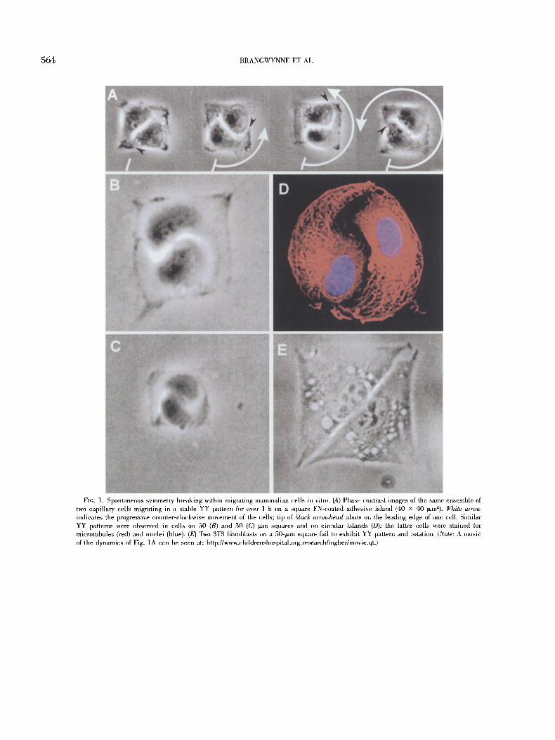
FIG. ]. Spontaneous symmetry breaking within migrating mammalian cells in vitro. (A) Phase contrast images of the same ensemble of two capillary cells migrating in a stable YY pattern for over 1 h on a square FN-coated adhesive island (40 x 40 tzm~). White arrow indicates the progressive counter-clockwise movement of the cells; tip of black arrowhead abuts on the leading edge of one cell. Similar YY patterns were observed in cells on 50 (B) and 30 (C) tzm squares and on circular islands (D); the latter cells were stained for microtubules (red) and nuclei (blue). (E) Two 3T3 fibroblasts on a 50qxm square fail to exhibit YY pattern and rotation. (Note: A movie of the dynamics of Fig. 1A can be seen at: http://www.childrenshospital.org.research/ingber/movie.qt.)

SYMMETRY BREAKING IN CULTURED MAMMALIAN CELLS 565
FIG. 2. Comparison between experimentally observed and computer-simulated transitions into the coordinated YY rotation. Experimen- tally determined changes in the position of each cell on an island plotted as a function of time (top) compared with results of a computer sinmlation (bottom). Vertical axis indicates the position of the centroid of the nucleus of each cell projected on an axis of the motion plane (arbitrary units). Note that after an initial pe- riod without coupled migration, cells in both the experiment and the simulation began ro- tating with a consistent phase shift of 180 ~ The discrete probabilistic model used was based on a biased one-dimensional random walk with persistence, P = 0.33, and cou- pling, C = 0.33 (P = 0; C = 0, represents a completely unbiased random walk. P = 1; C = 1, indicates complete persistence and cou- pling, respectively). Uncoordinated move- ment of cells was observed for values of P and C <0.3.
REFERENCES
Ball, P. The self-made tapestry: pattern fm~nation in nature. New York: Oxford University Press; 1999.
Bndrene, E. 0.; Berg, H. Complex patterns formed by motile cells of Esch- erichia coli. Nature 349:630-633; 1991.
Capra, F. The tao of physics. Boston, MA: Shambhala; 1991. Chen, C. S.; Mrksich, M.; Huang, S.; Whitesides, G. M.; Ingber, D. E. Geo-
metric control of cell life and death. Science 276:1425-1428; 1997. Dunn, G. A.; Brown, B. F. A unified approach to analysing cell motility. J.
Cell Sci. 8(Suppl.):81-102; 1987. Huang, S.; Ingber, D. E. The structural and mechanical complexity of cell-
growth control. Nat. Cell Biol. l:E131-E138; 1999. Murray, J. D. Mathematical biology. New York: Springer; 1993. Palsson, E.; Cox, E. C. Origin and evolution of circular waves and spirals in
Dictyostelium discoideum territories. Proc. Natl. Acad. Sci. USA 93: 1151-1155; 1996.
Stokes, C. L.; Lauffenburger, D. A.; Williams, S. K. Migration of individual microvessel endothelial cells: stochastic model and parameter mea- surement. J. Cell Sci. 99:419-430; 1991.
Verkhovsky, A. B.; Svitkina, T. M.; Borisy, G. G. Self-polarization and direc- tional motility of cytoplasm. Curt. Biol. 9:11-20; 1999.
Ware, M. F.; Wells, A.; Lauffenburger, D. A. Epidermal growth factor alters fibroblast migration speed and directional persistence reciprocally and in a matrix-dependent manner. J. Cell Sci. 111:2423-2432; 1998.
Clifford Brangwynne Sui Huang
Kevin Kit Parker Donald E. Ingber 1
Depar tments of Pathology and Surgery Children 's Hospital and Harvard Medical School
Enders 1007 300 Longwood Avenue
Boston, Massachuse t t s 02115
(Received 12 June 2000)
1 TO whom correspondence should be addressed at E-mail: ingber@ al.tch.harvard.edu