limbic system and psychiatric disorders
TRANSCRIPT
By : Dr. Karrar husain
Moderator: Dr. Piyush P Singh

The term "limbic" Latin word limbus, for "border" or
"edge“.
Paul Broca coined the term based on its physical location in
the brain, sandwiched between two functionally different
components.

Paul Broca first called this part of the brain "le grand lobe
limbique" in 1878,
but most of its putative role in emotion was developed only in
1937 when
the American physician James Papez described his anatomical
model of emotion, the Papez circuit.

James Papez published a journal article(1937) in which he
outlined a "new" circuit to account for emotion.
He hypothesized that the hippocampus, the cingulate gyrus,
the hypothalamus, the anterior thalamic nuclei,
and the interconnections among these structures constituted a
mechanism which elaborate the functions of emotions

Papez believed that the experience of emotion was determined
by activity in
the cingulate cortex and, less directly, other cortical areas.
Emotional expression was thought to be governed by the
hypothalamus.

NEURAL CIRCUIT FOR EMOTION AS ORIGINALLY PROPOSED BY JAMES PAPEZ

Paul D. MacLean (May 1, 1913 – December 26, 2007)
proposed triune brain theory.
MacLean's proposed that the human brain was in reality three
brains in one:
the reptilian complex,
the limbic system,
and the neocortex.

The Reptilian Brain : Core brainstem homeostasis and
survival.
The Paleomammalian Brain : the limbic system Social and
emotional attachment and motivated behaviours.
The Neomammalian Brain : neocortex and neocerebellum
skilled movements, logic thinking, languages and higher brain
functions.


There is no universal agreement on the total list of structures that should be included in limbic system.
All the authors include limbic cortex (cingulate and parahippocampal gyri), the hippocampal formation ,the amygdala, and the septal area.
Most include the hypothalamus, part of midbrain reticular formation and the olfactory areas.
Beyond that boundry get fuzzy; some authors include thalamic and neocortical regions

Hippocampus:Learning and Memory

Hippocampus:Learning and Memory
Amygdala:Emotions and Aggression

Hippocampus:Learning and Memory
Amygdala: Emotions and Aggression
Hypothalamus:Hunger, ThirstTemperature Control
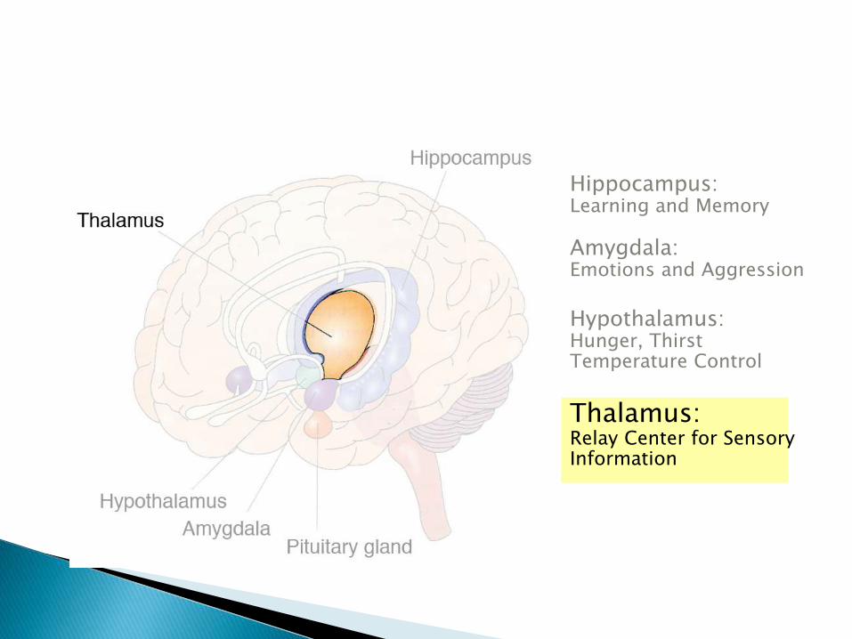
Hippocampus:Learning and Memory
Amygdala: Emotions and Aggression
Hypothalamus:Hunger, ThirstTemperature Control
Thalamus:Relay Center for SensoryInformation
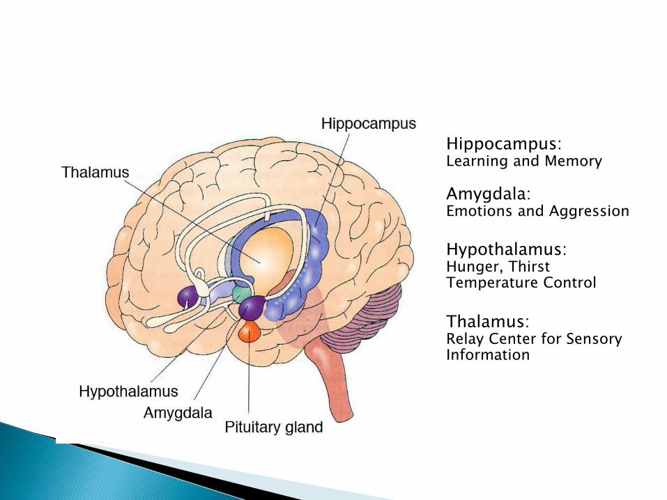
Hippocampus: Learning and Memory
Amygdala: Emotions and Aggression
Hypothalamus:Hunger, ThirstTemperature Control
Thalamus:Relay Center for SensoryInformation

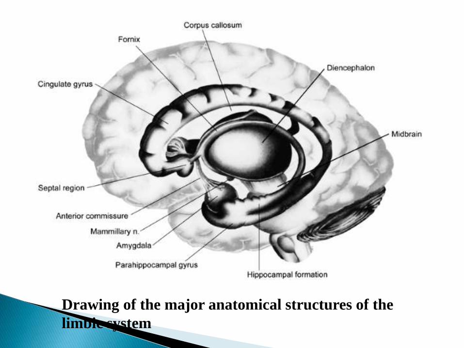
Drawing of the major anatomical structures of the
limbic system

limbic cortex the cingulate gyrus and the parahippocampal
gyrus.
The cingulate gyrus, located dorsal to the corpus callosum,
includes several cortical regions that are heavily
interconnected with the association areas of the cerebral
cortex.

posteriorly, it becomes continuous with the parahippocampal
gyrus, located in the medial temporal lobe.
Entorhinal cortex funnels highly processed cortical
information to the hippocampal formation,
and also is a major output pathway from the hippocampal
formation.

Located in the floor of the temporal horn of the lateral
ventricle
Three distinct zones—the dentate gyrus,
the hippocampus,
and the subicular complex.
These zones are composed of adjacent strips of cortical tissue,
and fold over each other mediolaterally in a spiral fashion,
giving rise to a C-shaped appearance

Dentate gyrus 3 layers:
outer acellular molecular layer
a middle layer granule cells, extend their dendritic trees
into the molecular and form the mossy fiber projection to the
hippocampus;
and an inner polymorphic layer.

Hippocampus is also a trilaminate structure
composed of molecular and polymorphic layers and a middle
layer of pyramidal neurons.
hippocampus is divided into three distinct fields CA3,
CA2, and CA, on the basis of cytoarchitecture.

subicular complex 3 components
the presubiculum,
the parasubiculum,
and the subiculum
It serve as transition regions between the hippocampus and the
parahippocampal gyrus.
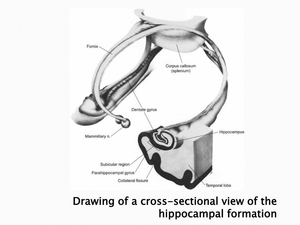
Drawing of a cross-sectional view of the hippocampal formation

major input to the hippocampal formation arises layers II and
III of the entorhinal cortex
that project to the dentate gyrus,
where they synapse on the dendrites of granule cells.

The mossy fiber axons of the granule cells then provide a
projection to CA3
Axon from CA3 project to the CA1 field of the hippocampus.
This region, in turn, projects to the subicular complex,
which provides output to the entorhinal cortex, completing the
circuit.

Drawing showing intrinsic connectivity of
hippocampal formation

Group of nuclei
Located in the medial temporal lobe just anterior to the hippocampal formation
These nuclei form several distinct clusters:
the basolateral complex,
the centromedial amygdaloid group,
and the olfactory group, includes the cortical amygdaloidnuclei.

Basolateral complex
largest group,
its connectivity and anatomical characteristics are more similar
to cortical regions than to the remaining amygdaloid nuclei.
the basolateral nuclei are directly and reciprocally connected
with the temporal, insular, and prefrontal cortices.

like cortical regions, the basolateral complex shares
bidirectional connections with the medial dorsal thalamic
nucleus
and receives projections from the midline and intralaminar
thalamic nuclei.
Neurons are pyramidal like and use excitatory
neurotransmitter.

the centromedial amygdala two major subdivisions.
The central subdivision central amygdaloid nucleus
+lateral portion of the bed nucleus of the stria terminalis.
reciprocally connected with brainstem viscerosensory and
visceromotor regions and with the lateral hypothalamus.
Also receives afferents from cortical limbic regions and the
basolateral amygdaloid complex.

the medial subdivision
composed of the medial amygdaloid nucleus
reciprocally connected with the medial or endocrine portions
of the hypothalamus

gray matter structure
above the anterior commissure.
The septal nuclei are reciprocally connected with the
hippocampus,
the amygdala,
and the hypothalamus and project to a number of structures in
the brainstem.

lies at the center of the limbic system
subdivided into three zones:
the supraoptic region,
the tuberal region
and the mammillary region.
The three zones are divided on each side into medial and
lateral areas by the fornix.


Drawing of the nuclei in the medial hypothalamus


The major structures of the limbic system are interconnected
with each other and with other components of the nervous
system
sensory information from the cingulate, the orbital and
temporal cortices, and the amygdala is transmitted to the
entorhinal cortex of the parahippocampal gyrus and then to the
hippocampal formation.

After traversing the intrinsic circuitry of the hippocampal
formation, information is projected through the fornix either to
the anterior thalamus, which, in turn, projects to the limbic
cortex, or to the septal area and the hypothalamus.
These latter two regions provide feedback to the hippocampal
formation through the fornix.
In addition, the mammillary bodies of the hypothalamus
project to the anterior thalamus.
Finally, the hypothalamus and the septal area project to the
brainstem and the spinal cord

Functional circuit b/w hippocampul formation,Thalamus,cerebral cortex and hypothalamus

Sensory information, primarily from the association regions of
the prefrontal and temporal cortices, projects to the amygdala.
Output from the amygdala is conducted through two main
pathways .
A dorsal route, the stria terminalis, project primarily to the
septal area and the hypothalamus..

The second major output route, the ventral amygdalofugal
pathway, terminate in the septal area, the hypothalamus, and
the medial dorsal thalamic nucleus.
The medial dorsal nucleus, in turn, projects heavily to
prefrontal and some temporal cortical regions

Functional circuit b/w amygdala,hypothalamusPrefrontal and temporal ass. cortices

Both of these pathways reveal how the limbic system is able to
integrate the highly processed sensory and cognitive
information content of the cerebral cortical circuitry with the
hypothalamic pathways that control autonomic and endocrine systems

the ventral amygdalofugal pathway also projects to the
nucleus accumbens (ventral striatum),
the area where the head of the caudate nucleus fuses with the
putamen.
This region sends efferents to the ventral palladium, an
extension of the globus pallidus.
This area, in turn, projects to the medial dorsal thalamic
nucleus.

Functional circuits b/w basal ganglia and limbic system


The limbic structures are closely related to the olfactory
cortex.
Amygdala is involved in the emotional response to smell
entorhinal cortex, is concerned with olfactory memories

Olfactory role of limbic system

Amygdala plays a role in food choice and emotional
modulation of food intake.
The lateral nucleus of the hypothalamus is the center for
control of feeding
whereas the ventromedial nucleus functions as the satiety
center.
PET and fMRI have shown that the limbic system is one of
the most active brain areas during dreaming.
The limbic system probably interweaves unconscious primal
emotions with our conscious cognitive thoughts and
perceptions
and thereby ties together emotions and memory during REM
sleep to form the content of dreams.

The suprachiasmatic nucleus of hypothalamus is the circadian
rhythm generator controlling the sleep-wake cycle.
The ventrolateral preoptic nucleus (VLPO) of the
hypothalamus sends projections to the histaminergic
tuberomamillary nucleus (TMN),
the serotonergic dorsal and median raphe nucleus
and the noradenergic locus coeruleus.

It also sends axons to the cholinergic basal forebrain,
the pedunculo pontine thalamic nucleus (PPT)
and lateral dorsal thalamic nucleus (LDT).
The VLPO projections to these areas are inhibitory.
The VLPO via its inhibition of the major arousal mechanisms,
functions as a 'sleep switch', promoting sleep

The VLPO by its disinhibition of the PPT-LDT also promotes
REM sleep.
The lateral hypothalamic area (LHA) contains orexinergic
neurons that promote wakefulness.
The orexinergic neurons inhibit the sleep-promoting VLPO
and the REM sleep-promoting neurons in the PPT-LDT.

FEAR
Fear responses are produced by the stimulation of the
hypothalamus and amygdala.
Amygdala destruction abolishes fear and its autonomic and
endocrine responses.
Amygdala is also involved in fear learning
Imaging studies have shown that viewing fearful faces
activates the left amygdala.

Rage and placidity
The destruction of the ventromedial hypothalamic nuclei and
septal nuclei in animals may induce rage.
Bilateral destruction of the amygdala results in placidity.
However, when the ventromedian nucleus is destroyed after
the destruction of the amygdala, the placidity generated is
converted to rage.
Autonomic and endocrine responses to emotion
Limbic stimulation causes changes in respiration and blood
pressure.
The stimulation of the cingulate gyrus and hypothalamus can
elicit autonomic responses.
Hypothalamic autonomic responses are mediated by the
cortical and limbic structures processing drives and emotions.

The fear and rage responses mediated by the limbic system
cause stimulation the hypothalamus, especially lateral areas
and produce diffuse sympathetic discharge.
The massive sympathetic discharge during stress is called the
"flight or fright response".

Stress via cortical and limbic connections causes release of
corticotropin-releasing hormone (CRH) from the
paraventricular nuclei of the hypothalamus.
CRH release mediates endocrine and immune responses

Limbic system and autonomic and endocrine responses

medial preoptic area of the hypothalamus is the central control
of male sexual behavior.
Chemosensory efferents olfactory systems project to the
medial amygdala (MeA).
MeA sends innervations (through the stria terminalis) to the
medial preoptic area (MPOA).
MPOA and MeA receive genitosensory input from the spinal
cord through the central tegmental field (CTF).

The MPOA sends efferents to the paraventricular nucleus of
the hypothalamus (PVN), the ventral tegmental area, the
nucleus paragigantocellularis and other autonomic and
somatomotor areas

Neural Circuit of Male Sexual BehaviorCTF: Central Tegmental Field,MeA: Medial Amygdala, BNST: Bed Nucleus of StriaTerminalis, VTA: Ventral Tegmental Area, MPOA: Medial Preoptic Area
The parvocellular part of the paraventricular nucleus (PVN) of
the hypothalamus send direct oxytocinergic and
vasopressinergic projections to the lumbosacral cord.
Activation of oxytocinergic neurons in the PVN by dopamine
and its agonists-excitatory amino acids (N-methyl-D-aspartic
acid)
or oxytocin itself or by electrical stimulation leads to penile
erection.

The inhibition of these neurons by GABA and its agonists or
by opioid peptides and opiate-like drugs, inhibits this sexual
response.
The activation of these neurons is secondary to the activation
of nitric oxide synthase (NOS), which produces nitric oxide.

glutamatergic inputs to the MPOA from the medial amygdala
(MeA) and bed nucleus of the stria terminalis (BNST),
mediate the female-stimulated increase in dopamine, which in
turn, enhances copulatory ability.
Extracellular glutamate in the MPOA increases during
copulation,
especially during ejaculation and increased glutamate
facilitates copulation and genital reflexes.

The reward circuitry underlying addictive behavior includes
amygdala and nucleus accumbens.
The amygdala plays a central role in cue-induced relapse.
Relapse associated with cues, stress and a single dose of a
drug of abuse results in release of excitatory neurotransmitters
in brain areas like hippocampus and amygdala.

The pathway of motivated behavior involves the prefrontal
cortex, the ventral tegmental area (VTA), the amygdala
especially the basolateral amygdala and extended amygdala,
the nucleus accumbens core and the ventral pallidum.
This pathway is involved in the motivation to take drugs of
abuse (drug-seeking) and the compulsive nature of drug-taking

Emotional memory
Emotion has powerful influence on learning and memory.
Amygdala, prefrontal cortex and medial temporal lobe, is involved in consolidation and retrieval of emotional memories.
Amygdala, prefrontal cortex and hippocampus are also involved in the acquisition, extinction and recovery of fears to cues and contexts.
Hippocampus is critical for long-term, declarative memory storage

Medial temporal lobe memory system
include the hippocampus and adjacent cortex,
the parahippocampal regions (PHG) and the entorhinal and
perirhinal regions.
involved in the storage of new memories.

Diencephalic memory system
consists of the hypothalamus, mammillary body and the
dorsomedial nucleus of thalamus.
important for the storage of recent memory;
a dysfunction of this circuit results in Korsakoff's syndrome.

Social cognition
thought processes involved in understanding and dealing with
other people.
Limbic structures involved are the cingulate gyrus and
amygdala.


Degenerative changes in the limbic system likely have a role
in neurodegenerative diseases,
particularly Pick's disease and Alzheimer's disease.
Marked atrophy is found in the limbic system, most notably
the dentate gyrus and hippocampus.

In Alzheimer's disease, senile plaques and neurofibrillary
tangles are dispersed throughout the cerebral cortex and basal
ganglia,
but the hippocampus and amygdala are often severely
involved

may be the result of a failure of the anterior cingulate and
hippocampus to modulate the activity of the amygdala.
A fear circuitry, involving the amygdala, prefrontal and
anterior cingulate has been described.

Studies have shown reduced limbic volumes.
The Papez circuit is probably involved in schizophrenia.
distortion of cortical neurons of layer II of the ERC,
decreased size of hippocampus

reduced number of GABAergic cells in the cingulate and
anterior thalamus
with glutamatergic excitotoxicity.
The other circuit involved is the basolateral circuit which
mediates the social cognition deficits in schizophrenia

Studies have shown variation in the volumes of the frontal lobes, basal ganglia, amygdala and hippocampus.
Functional studies have revealed decreased prefrontal and anterior cingulate activity.
Recently, researchers have posited that affective and cognitive symptomatology represents dysfunction within a network-the anterior limbic network,

which includes prefrontal regions and subcortical structures
such as the thalamus, striatum and the amygdala.
The dysfunction of this system (anterior limbic network) is
suggested in bipolar disorder, but its role in depression is
unclear.

The enlarged hippocampus represent a compensatory response
to the presence of disturbances in the perception of time,
temporal processing
and stimulus-seeking associated with ADHD.
Disrupted connections between the amygdala and
orbitofrontal cortex may contribute to behavioral disinhibition.
bilateral destruction of the amygdaloid body and inferior
temporal cortex.
characterized by visual agnosia,
placidity,
hypermetamorphosis,
hyperorality
and hypersexuality.

caused by cerebral trauma;
infections including herpes and other encephalitides;
Alzheimer's disease and other dementias;
Niemann-Pick disease and cerebrovascular disease

damage to mammillary bodies, dorsomedial nucleus of
thalamus and hypothalamus (diencephalic memory circuit).
chronic prominent impairment of recent and remote memory
(recent > remote)
Immediate recall is usually preserved.
Confabulation may be marked but is not invariably present

disproportionate impairment in specific aspects of social
cognition.
Limbic structures involved cingulate gyrus and amygdala,
which mediate cognitive and affective processing.
The basolateral circuit integral for social cognition is disrupted
in autism spectrum disorders.

Temporal lobe epilepsy is the most common epilepsy in adults
and is most often caused by hippocampal sclerosis.
Hippocampal sclerosis with involvement of amygdala and
parahippocampal gyrus mesial temporal sclerosis (MTS).
The frequency and widespread distribution of cerebral
abnormalities suggest that MTS is not limited to the medial
temporal lobe but instead, represents a limbic system disorder.

a paraneoplastic syndrome reported with carcinoma of the
lung, breast and some other primaries.
manifests as encephalitis that primarily involves the
hippocampus, amygdala, cingulate gyrus, insula and orbital-
frontal cortex.
Afflicted patients develop subacute onset of memory loss,
dementia, involuntary movements and ataxia.

• Kaplan and Sadock‘s Comprehensive textbook of psychiatry, 8th ed.
• Gray’s anatomy ,39th Edition
• Papez JW. A proposed mechanism of emotion. Arch Neurol Psychiatry
1937;38:725-43
• Maclean PD. The triune brain in evolution: Role in paleocerebral functions.Plenum
Press: New York; 1990
• Ganong WF. Smell and taste. In: review of medical physiology. 21st ed

• Ganong WF. Neural basis of instinctual behavior and emotions. In Review of
Medical Physiology. 21st ed.
• Benca RM, Cirelli C, Rattenborg MC, Tononi G. Basic science of sleep In: Kaplan
and Sadock‘s Comprehensive textbook of psychiatry, 8th ed.
• Saper CB, Chou TC, Scammell TE. The sleep switch: Hypothalamic control of
sleep and wakefulness. Trends Neurosci ;24:726-31.
• Adolphs R, Tranel D, Damasio H, Damasio AR. Fear and the human amygdala. J
Neurosci 1995;15:5879-91.
• Smith SM, Vale WW. The role of the hypothalamic-pituitary-adrenal axis in
neuroendocrine responses to stress.

• Hull EM, Dominguez JM. Getting his act together: Roles of glutamate, nitric oxide
and dopamine in the medial preoptic area. Brain Res 2006;1126:66-75
• Argiolas A, Melis MR. Central control of penile erection: Role of the
paraventricular nucleus of the hypothalamus.
• Kalivas PW, Volkow ND. The neural basis of addiction: A pathology of motivation
and choice. Am J Psychiatry 2005;162:1403-13.
• LaBar KS, Cabeza R. Cognitive neuroscience of emotional memory. Nat Rev
Neurosci 2006;7:54-64.
• Paller KA, Squire LR. Biology of memory. In: Kaplan and Sadock‘s
Comprehensive textbook of psychiatry, 8th ed

• Chan S, Erickson JK, Yoon SS. Limbic system abnormalities associated with
mesial temporal sclerosis: A model of chronic cerebral changes due to seizures.
Radiographics 1997;17:1095-110
• Bakheit AM, Kennedy PG, Behan PO. Paraneoplastic limbic encephalitis: Clinico-
pathological correlations. J Neurol Neurosurg Psychiatry 1990;53:1084-8.
• Kantarci K, Jack CR. Neuroimaging in Alzheimer disease: An evidence-based
review. Neuroimaging Clin N Am 2003;13:197-209.
• Deakin JF. Glutamate, GABA and cortical circuitry in schizophrenia. In The
psychopharmacology of schizophrenia. Reverly MA, Deakin JF, editor Arnold
Publishers: London; 2000. p. 56-70

• Rajkowska G. Anatomical pathology. In: Textbook of mood disorders. Stein DJ,
Kupfer DJ, Schatzberg AF
• Strakowski SM, DelBello MP, Adler CM. The functional neuroanatomy of bipolar
disorder: A review of neuroimaging › ndings. Mol Psychiatry 2005;10:105-16.
• Hechtman L. Attention-deficit/hyperactivity disorder. In: Kaplan and Sadock‘s
Comprehensive textbook of psychiatry, 8th ed.
