lipid metabolic reprogramming in cancer cells · 2018-12-21 · intracellular oncogenicsignalingand...
TRANSCRIPT
OPEN
REVIEW
Lipid metabolic reprogramming in cancer cellsS Beloribi-Djefaflia1,2,3,4, S Vasseur1,2,3,4 and F Guillaumond1,2,3,4
Many human diseases, including metabolic, immune and central nervous system disorders, as well as cancer, are the consequenceof an alteration in lipid metabolic enzymes and their pathways. This illustrates the fundamental role played by lipids in maintainingmembrane homeostasis and normal function in healthy cells. We reviewed the major lipid dysfunctions occurring during tumordevelopment, as determined using systems biology approaches. In it, we provide detailed insight into the essential roles exerted byspecific lipids in mediating intracellular oncogenic signaling, endoplasmic reticulum stress and bidirectional crosstalk between cellsof the tumor microenvironment and cancer cells. Finally, we summarize the advances in ongoing research aimed at exploiting thedependency of cancer cells on lipids to abolish tumor progression.
Oncogenesis (2016) 5, e189; doi:10.1038/oncsis.2015.49; published online 25 January 2016
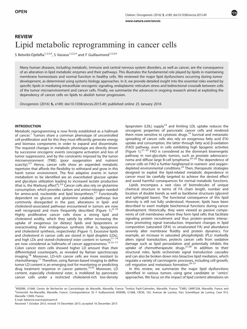
INTRODUCTIONMetabolic reprogramming is now firmly established as a hallmarkof cancer.1 Tumors share a common phenotype of uncontrolledcell proliferation and for this they must efficiently generate energyand biomass components in order to expand and disseminate.The required changes in metabolic phenotype are directly drivenby successive oncogenic events (oncogene activation and loss oftumor suppressors), and by the constraints imposed by the tumormicroenvironment (TME) (poor oxygenation and nutrientscarcity).2,3 Hence, cancer cells show an expanded metabolicrepertoire that affords the flexibility to withstand and grow in thisharsh tumor environment. The first adaptive events in tumormetabolism to be identified are an exacerbated glucose uptakeand glycolysis utilization leading to increased lactate production(that is, the Warburg effect4).5,6 Cancer cells also rely on glutamineconsumption, which provides carbon and amino-nitrogen neededfor amino-acid, nucleotide and lipid biosynthesis.6,7 Functionallydependent on glucose and glutamine catabolic pathways butcommonly disregarded in the past, alterations in lipid- andcholesterol-associated pathways encountered in tumors are nowwell recognized and more frequently described (Figure 1).8–10
Highly proliferative cancer cells show a strong lipid andcholesterol avidity, which they satisfy by either increasing theuptake of exogenous (or dietary) lipids and lipoproteins oroveractivating their endogenous synthesis (that is, lipogenesisand cholesterol synthesis, respectively) (Figure 1). Excessive lipidsand cholesterol in cancer cells are stored in lipid droplets (LDs),and high LDs and stored-cholesteryl ester content in tumors11–14
are now considered as hallmarks of cancer aggressiveness.13,15–17
Colon cancer stem cells showed higher LD amount than theirdifferentiated counterparts, as revealed by Raman spectroscopyimaging.18 Moreover, LD-rich cancer cells are more resistant tochemotherapy.11 Therefore, using Raman-based imaging to definetumor LD content is an emerging tool for monitoring or predictingdrug treatment response in cancer patients.19,20 Moreover, LDcontent, especially cholesteryl ester, is mobilized by pancreaticcancer cells under a restricted cholesterol-rich low-density
lipoprotein (LDL) supply14 and limiting LDL uptake reduces theoncogenic properties of pancreatic cancer cells and renderedthem more sensitive to cytotoxic drugs.14 Survival and metastaticspreading of cancer cells also rely on exogenous fatty acid (FA)uptake and consumption, the latter through fatty acid β-oxidation(FAO) pathway, even in cells exhibiting high lipogenic activities(Figure 1).21–23 FAO is considered as the dominant bioenergeticpathway in non-glycolytic tumors, such as prostate adenocarci-noma and diffuse large B-cell lymphoma.24–26 The dependence ofcancer cells on FAO is further heightened in nutrient- and oxygen-depleted environmental conditions.22 Then, therapeutic strategiesdesigned to exploit the lipid-related metabolic dependence incancer must be carefully targeted to achieve the desired effectand avoid harmful consequences for normal metabolic functions.Lipids encompass a vast class of biomolecules of unique
chemical structure in terms of FA chain length, number andlocation of double bonds as well as backbone structures (glyceroland sphingoid bases). The functional consequence of this lipiddiversity is still not fully understood. However, lipids have beendescribed to exert multiple biochemical functions during cancerdevelopment. Historically, they were viewed as passive compo-nents of cell membranes where they form lipid rafts that facilitatesignaling protein recruitment and thus protein–protein interac-tions promoting signal transduction. Important changes in lipidcomposition (saturated (SFA) vs unsaturated FA) and abundanceseverely alter membrane fluidity and protein dynamics. Forexample, an increase in saturated phospholipids (PLs) markedlyalters signal transduction, protects cancer cells from oxidativedamage such as lipid peroxidation and potentially inhibits theuptake of chemotherapeutic drugs.27,28 In addition to theirstructural roles, lipids orchestrate signal transduction cascadesand can also be broken down into bioactive lipid mediators, whichregulate a variety of carcinogenic processes, including cell growth,cell migration and metastasis formation.29–31
In this review, we summarize the major lipid dysfunctionsidentified in various tumors using gene candidate or ‘-omics’approaches. We focus on the impact of lipid content alterations on
1INSERM, U1068, Centre de Recherche en Cancérologie de Marseille, Marseille, France; 2Institut Paoli-Calmettes, Marseille, France; 3CNRS, UMR7258, Marseille, France and4Université Aix-Marseille, Marseille, France. Correspondence: Dr F Guillaumond, INSERM, U1068, CRCM, 163, Avenue de Luminy, Parc Scientifique de Luminy, Case 915,Marseille 13009, France.E-mail: [email protected] 1 October 2015; revised 10 December 2015; accepted 16 December 2015
Citation: Oncogenesis (2016) 5, e189; doi:10.1038/oncsis.2015.49
www.nature.com/oncsis
intracellular oncogenic signaling and on endoplasmic reticulum (ER)homeostasis. We also detail the lipid exchange between stromacellular components and cancer cells. Finally, we present advancesin the therapeutic targeting of metabolic actors associated withlipid pathways in preclinical and clinical development.
LIPID REPROGRAMMING IN TUMORSLipid alterations identified from tumor-specific gene expressionprofilingCandidate-gene expression studies identified upregulated tran-scripts involved in lipogenesis and cholesterol synthesis pathways(Figure 1), which are essential for development and progression ofa wide variety of tumors. Increased expression of lipogenicenzymes, such as acetyl-CoA carboxylase (ACC) and fatty acidsynthase (FASN), and ATP citrate lyase (ACLY) that promote alsocholesterol synthesis, represent a nearly-universal phenotypicalteration in most tumors.32,33 FASN overexpression predicts poorprognosis in cancer patients.34 Its expression levels appear at theprecancerous lesion stage and persist in metastatic breast andprostate tumors.34 As these initial observations, many othercandidate genes, involved in cholesterol-related pathways(uptake, synthesis and storage) and FAO, proved to be crucial insupporting malignancy.8,10,35 FAO-limiting enzymes, the carnitinepalmitoyltransferase 1 isoforms A and C (CPT1A and C) areoverexpressed in many human tumors.36–38 CPT1C upregulation,induced by AMPK and p53, has been shown to protect cancer cellsfrom death when they are under deprived glucose and oxygenconditions.36,38,39 Inversely, knockdown of CPT1 sensitizes cancercells to radiotherapy and apoptosis inducers.40–42
Our large-scale microarray profiling, centered on metabolicgenes, reveals lipid pathways as the most altered metabolic routesin pancreatic tumors, especially activated cholesterol and LDL
metabolisms.14 These tumors harbor also specific alterations inmetabolic pathways related to lipid messengers (phosphatidyli-nositols, PIs), lipid mediators (leukotrienes) and structural lipids(glycosphingolipids).14 This lipid signature unravels the highdependence of pancreatic tumors on cholesterol and identifiesexogenous cholesterol uptake, through LDLR, as the majorcholesterol pathway mediating tumor growth. Colorectal cancer(CRC) lipid signature, defined from a limited lipid-related genesexpression profiling, reveals four genes (ABCA1, ACSL1, AGPAT1and stearoyl-CoA desaturase (SCD)) overexpressed only in stage IICRC patients with a high risk of relapse. This signature displaysstronger power and accuracy than the currently used clinicalclassification.43
Lipid alterations identified from tumor-specific lipid profilingRecent advances in lipid analytical and imaging technologies,including electrospray ionization, matrix-assisted laser desorption/ionization, tandem mass spectrometry (MS/MS) and Ramanscattering microscopy, have greatly progressed such lipidomicanalysis.44 Raman-based imaging offers lipid compositionalmapping of cellular compartments, such as LDs.26,45 Thesecomplementary approaches provide crucial information on tumorlipid phenotype, in particular abundance, FA composition andspatial distribution of lipid classes within tumors. Over the pastfew years, much effort has surrounded establishing PL signature ofmalignant tumors. This signature segregates malignant tumorsfrom their benign counterparts as well as localized tumors fromadvanced ones. Indeed, breast tumors, when compared withadjacent normal tissue, have been characterized by a strikingincrease in membrane phosphatidylcholine and phosphatidy-lethanolamine and in PL-induced cell signaling, PI.46,47 In additionto these changes in PL amounts, the phosphatidylcholine content
Figure 1. A simplified map of the main altered lipid metabolic pathways in cancer cells. Lipid metabolic network (blue) includes import/exportand catabolic pathways (FAO) as well as de novo synthesis pathways, such as lipogenesis (that is, synthesis of TGs and PLs) and cholesterolsynthesis. Glucose- and/or glutamine-derived citrate, provided by the increased glycolysis and/or glutaminolysis (orange), are commonprecursors of lipogenesis and cholesterol synthesis. Cancer cells can also take up exogenous cholesterol, transported by LDL and very-low-density lipoproteins (VLDL), to meet their cholesterol requirement. When cholesterol, PLs and TGs are in excess in tumors, they are exportedinto circulation as high-density lipoproteins (HDLs) or locally stored into LDs. Exogenous FAs taken up by cancer cells are broken down toproduce energy through mitochondrial FAO process. TCA cycle, tricarboxylic acid cycle αKG, α-Ketoglutarate.
Lipid metabolic reprogramming in cancer cellsS Beloribi-Djefaflia et al
2
Oncogenesis (2016), 1 – 10
was found to be enriched in SFA, and this phosphatidylcholinecomposition was correlated with high tumor grade and pooreroverall survival.46 This membrane lipid saturation, a feature sharedby all lipogenic tumors,27 reduced membrane fluidity anddynamics48 and increased chemotherapy resistance.27 The specificPI signature revealed a shift toward polyunsaturated FA chaincomposition in PI from invasive breast cancers when comparedwith that in PI from in situ carcinoma.49 These findings highlightsignificant differences in FA composition depending on PL classand tumor grade. Unlike breast tumors, the lipid signature ofMyc-induced lymphoma is characterized by reduced phosphati-dylserine, phosphatidylethanolamine and PI amounts and byelevated monounsaturated FA-phosphatidylglycerol (PG) levelswhen compared with normal tissues.50 The increased PG is alsofound in renal cell and hepatocellular carcinomas.51,52 PG serves asa precursor of cardiolipin, which is found almost exclusively inmitochondrial membranes and intimately involved in maintainingmitochondrial functionality and membrane integrity. An abnormalcardiolipin molecular species distribution and a decrease in CLcontent in brain tumor mitochondria, revealed by shotgunlipidomic analysis, lead to irreversible respiratory injury and mayimpede the use of alternative energy sources to glucose.53
Lipidomic profiling has revealed unsuspected and recurrentlipid changes at the class and molecular species levels in cancercells. As previously discussed, PL-specific composition may help todiscriminate low- and high-grade tumors as well as malignant cellsfrom benign ones.46,47,49,50 Moreover, combined with transcrip-tome/proteome analyses, lipidomic data could also unravel newpotential lipid-related targets for drug development or newtreatments combining inhibitors of these targets with currentlyused chemotherapy.
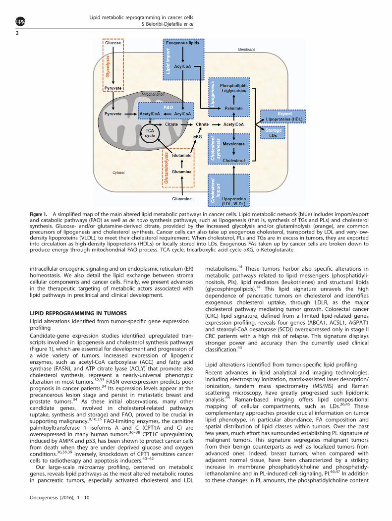
LIPID RAFTS IN CANCER CELL SIGNALINGIncreased lipid rafts in tumorsCell membranes contain different classes of lipids, some of which,in particular cholesterol and sphingolipids, form specific planar
microdomains known as lipid rafts (Figure 2).54 These differ fromthe cavin and caveolin protein-enriched invaginated-lipid raftsknown as caveolae.28 Both are essential not only for membraneprotein dynamics and trafficking but also for cell survival and celldeath program execution.55 In cancer cells, a wide range ofsignaling proteins and receptors regulating pro-oncogenic andapoptotic pathways during the early, advanced and metastaticstages of carcinogenesis reside in lipid rafts (Figure 2).55 Moreover,lipid rafts/caveolae and their main component, cholesterol, areenhanced in membrane of multiple cancer cells56–60 as well as inmembranes of tumor-released exosomes.61
Impact of disrupted lipid raft integrity on tumor cell fateDecreasing cholesterol content with membrane-depleting agents(methyl-β-cyclodextrin) or cholesterol synthesis inhibitors (statins)helped to decrypt the oncogenic signaling pathways whoseactivation is entirely dependent on lipid raft integrity. Anchored-lipid raft AKT protein has been extensively investigated in cancercells (Figure 2a).62,63 Its aberrant activation, contributing to tumordevelopment and invasiveness,64,65 correlates with increased lipidrafts in cancer cells.66,67 Lipid raft disruption inhibits AKTactivation63,67,68 and then reduces tumor cell proliferation(Figure 2a).66 Lipid rafts also exert crucial roles in cancerdissemination. By regulating cytoskeletal reorganization and focaladhesion dynamics, lipid rafts regulate cancer cell migration.69,70
They are also important for ligand-directed migration ofT-lymphoblastic lymphoma cells by maintaining C-X-C chemokinereceptor type 4 (CXCR4) dimer conformation.71
Novel raft-based entities, known as clusters of apoptoticsignaling molecule-enriched rafts (CASMERs), have been recentlydescribed (Figure 2b).55 These are constituted by co-aggregationof lipid rafts with death receptors (Fas/CD95, tumor necrosisfactor-related apoptosis-inducing ligand or TRAIL) and theirdownstream apoptotic molecules. This configuration activatedefficiently the apoptotic response independently of deathreceptor ligands (FasL and TNFα) (Figure 2b). CASMER formation
Figure 2. Lipid rafts as platforms for cell signaling. (a) Lipid rafts are formed by a phospholipid bilayer enriched in cholesterol, sphingolipidsand resident signaling proteins (AKT) and receptors (GPCR, G protein-coupled receptor; RTK, receptor tyrosine kinase including growth factorreceptor (GFR); CXCR4, C-X chemokine receptor 4). Once activated by their respective ligands, the receptors recruit different signalingeffectors that promote cell survival, cell migration and cell invasion, all of which contribute toward tumor growth. (b) Aggregation of deathreceptors (DR4/DR5, Fas) in lipid rafts forms CASMERs. Recruitment of CASMERs in a restricted space enhances fas-associated protein withdeath domain (FADD)/Caspase-8 death signaling pathway when compared with apoptotic signal induced by the activation of non-clustereddeath receptors.
Lipid metabolic reprogramming in cancer cellsS Beloribi-Djefaflia et al
3
Oncogenesis (2016), 1 – 10

and its subsequent Fas/CD95 or TRAIL-induced cell death can beinhibited by cholesterol-depleting agents, as described inleukemia cells and non-small cell lung carcinoma.55,72 Similarly,resveratrol induced-CASMER formation and sensitization of coloncarcinoma cells to death receptor-mediated apoptosis areprevented by cholesterol membrane depletion.73
These findings provide evidence for multiple oncogenic eventsdepending on lipid raft integrity. Disruption of these microdo-mains, which act as hubs linking receptors to their signalingeffectors, thus represents a valid therapeutic strategy in cancertreatment.
COMPLEX LIPID AND CHOLESTEROL ALTERATIONS INDUCINGER STRESSCancer cell fate following persistent ER stressThe ER ensures protein folding and maturation as well as calciumhomeostasis and regulates lipid metabolic processes. Accumula-tion of misfolded proteins, membrane lipid saturation orimbalanced calcium homeostasis leads to ER stress (ERS) andactivation of the unfolded protein response (UPR).74 UPR istransduced through three distinct ERS sensor proteins: ATF6(activating transcription factor 6), PERK (protein kinase RNA-likeendoplasmic reticulum kinase) and IRE1 (inositol-requiring trans-membrane kinase/endonuclease 1), which either reduce proteintranslation or increase ER-associated protein degradation tomaintain cell survival. UPR can evoke cell-cycle arrest in G1 phaseleading to the accumulation of quiescent cancer cells awaiting amore permissive environment to re-enter the cell cycle.75 Whencancer cells are submitted to persistent stresses (that is, hypoxia,membrane lipid saturation and nutrient deprivation), UPR leads tocell death.
Changes in complex lipid/cholesterol content and compositioncause ERS-induced apoptosisMembrane PL saturation disturbs ER structure66 and then impairsER homeostasis,76,77 a phenomenon commonly encountered invarious cancer cells.27 Lipid saturation, induced by the loss of theenzyme SCD1, was shown to promote ERS-activated apoptosis.78
A similar cancer cell fate is noticed following inactivation of sterolregulatory element-binding protein, the major transcriptionalregulator of lipogenic genes, in a lipid-poor environment.79 Thislipotoxic effect is abrogated by addition of exogenous unsatu-rated lipids80 or by re-expressing SCD1.79 Recently, imbalancedcholesterol homeostasis, leading to free cholesterol (FC) overload,was shown to induce ERS in cancer cells. Indeed, FC accumulationin HepG2 cells, induced by antitumor alkylphospholipids(perifosine, miltefosine and edelfosine),81 triggers an increase inthe ERS marker, CHOP (C/EBP homologous protein).82 Similarly,inhibitors of cholesterol esterification, targeting the enzymesterol-O-acyl transferase 1 (SOAT1), activated ERS markers inadrenocortical adenocarcinoma cells.83 High ceramide levels in ER,resulting from an increase either in membrane sphingomyelinhydrolysis or in ceramide de novo synthesis, can also induceERS.84,85 Cannabinoids, by increasing synthesized ceramidecontent, trigger ERS-induced cell death in human glioma andpancreatic adenocarcinoma.86–88 This process results from ap8-dependent upregulation of CHOP/ATF4 branch of UPR.87
Finally, increased exogenous ceramide uptake leads to apoptosisin various human cancer cells, including head and neck squamouscarcinoma cells89,90 and salivary adenoid cystic carcinoma cells.91
Data demonstrating ERS-induced apoptosis in cancer cellssubmitted to complex lipid and/or cholesterol homeostasisalterations open a promising therapeutic window. It allows us topredict that manipulating cholesterol and lipid supplies ormetabolic pathways leading to PL saturation, FC or ceramideaccumulation may impede tumor growth and dissemination.
TUMOR-STROMA COMMUNICATION MEDIATED BY LIPIDSIn human cancers, the TME, formed by extracellular matrixcomponents and numerous stromal cells including cancer-associated fibroblasts (CAFs), infiltrating immune cells, adipocytes,nerve cells, vascular/lymphatic endothelial cells, represents up to90% of the tumor mass.92 A molecular dialog between cancer cellsand adjacent CAFs or immune cells has been clearly demonstratedto support tumor growth and progression. Today, the central roleplayed by bioactive lipids and FAs as mediators of this crosstalkbetween cancer cells and stroma is increasingly recognized.
Cancer-stroma interplay through free FAsNumerous tumors grow in the vicinity of adipocytes ormetastasize to adipocyte-rich host environment. Metastaticovarian cancer cells home to omental adipose tissue, whichconstitutes an important reservoir of triglycerides (TGs).93 Hydro-lysis of these TG provides free FA (FFA), which are taken up andused as energy source by metastatic ovarian cancer cells(Figure 3).93 A similar FA exchanges also exist between adipocytesand metastatic bone marrow-derived prostate cancer cells.94 Thisadipocyte-cancer cell dialog is an adaptive metabolic process setup by cancer cells to take full advantage of the lipids stored inTME cells. FA translocation from stromal cells to cancer cells canbe mediated by lipoproteins, serum albumin and exosomes. It istempting to speculate that FA carried by serum albumin could betaken up by cancer cells through macropinocytosis: a non-receptor mediated endocytosis process constituting part of anancestral strategy used to salvage extracellular nutrients.95,96
Exosomes can also serve as carriers of FA and are taken up byrecipient cells (Figure 3). Their content, similar to that of parentalcells, is mostly enriched in SFA more than monounsaturated FAand polyunsaturated FA, the latter group of which is mostrepresented by arachidonic acid, the precursor of eico-sanoids (prostaglandins and leukotrienes).97 Once internalized,exosomes transfer their lipid material to the receiving cell.98 Theensuing lipid accumulation alters lipid homeostasis therebytriggering ERS-induced apoptosis and/or disturbing lipid raftsignaling, as discussed in the previous section. Beloribi et al.99
also demonstrated that synthetic exosome-like nanoparticles,mimicking the lipid composition of cancer exosomes, inhibit theNotch survival pathway leading to differentiated pancreatic cancercell death (Figure 3).
Tumor–stroma dialog orchestrated by prostaglandinsAn increase in prostaglandins (PGs) in cancer cells not onlypromotes tumor growth in a paracrine manner, but alsocoordinates the complex dialog between tumor cells and thesurrounding stromal cells. This crosstalk evades the immunesystem attack by promoting immunosuppression (Figure 3).30
Breast tumor-derived prostaglandin E2 (PGE2) has been shown toinduce, through an exosome-dependent transport, myeloid-derived suppressor cell activation, which in turn promotes tumorgrowth.100,101 Moreover, PGE2 was found to promote thedifferentiation of monocytes into tumor-associated suppressivemacrophages in cervical tumors.102 A pro-angiogenic activity oftumor-derived PGE2 has also been demonstrated in differentcancers.30,103–105 PGs released from cancer cells expressing therate-limiting enzyme of PG synthesis, cyclooxygenase-2 (COX-2),trigger endothelial cell migration in vitro and neovascularizationin vivo.103,104 Recently, a PGE2-dependent dialog between breasttumor cells and CAFs has been demonstrated. Tumor-derivedPGE2 activates the CAF-dependent secretion of a tryptophancatabolite, the kynurenine, which in turn increases cancer cellinvasiveness (Figure 3).106
Lipid metabolic reprogramming in cancer cellsS Beloribi-Djefaflia et al
4
Oncogenesis (2016), 1 – 10

Sphingolipid derivative as a mediator of tumor–stromal cellcommunicationSphingosine-1-phosphate (S1P), another bioactive lipid secretedby cancer cells, induces angiogenesis and lymphangiogenesisthrough its binding on S1P receptor 1, and facilitates tumorgrowth and metastasis formation.107,108 Moreover, high extra-cellular S1P levels, induced by overexpression of the upstreamregulatory sphingosine kinase, increases migration and tubeformation in co-cultured vascular or lymphatic endothelial cells(Figure 3).109
Together, these findings highlight the crucial role of lipids andtheir modes of transport in supporting the tumor-TME dialog,which is essential for tumor cell proliferation and dissemination.
LIPIDS AS CANCER THERAPY TARGETSTargeting the lipid and cholesterol dependence of cancer cellsInhibitor agents directed against lipogenic enzymes (FASN, ACLYand ACC) have been the subject of numerous studies; and theirefficacy as anticancer therapies have been proven in variouspreclinical models of carcinogenesis (Table 1).110–112 However,high adverse side effects of FASN-targeting drugs have precludedtheir clinical development. Numerous studies, using pharmacolo-gical agents targeting liver X receptor (LXR), a crucial transcrip-tional regulator of cholesterol homeostasis, have shown relevant
anticancer roles but also with undesired side effects.113 Recently,an LXR inverse agonist (SR9243), devoid of toxic side effects andwith similar impacts on colon cancer, holds significant promise forcancer therapy (Table 1).114 Alternative therapies directed againstSCD1 enzyme have shown a delay in tumor growth in variousmouse xenograft models.115 Interestingly, the high dependency ofcancer cells on SFA can be exploited to increase tumor-drugdelivery, as loading drugs in liposomes enriched in saturatedphosphatidylcholine has been shown to reduce the metastaticspread of pancreatic cancer in vivo.116 The use of CPT1 inhibitors(that is, etomoxir or ranolazine) provides beneficial effects inFAO-dependent tumors, notably in prostate cancer42 and inhuman leukemia when they are combined with pro-apoptoticagents.40 Recently, a novel CPT1a inhibitor, ST1326, hasbeen shown to drive leukemia cells toward apoptosis. Thisapoptotic effect results from an accumulation of palmitate.37
Several strategies have been developed to target cholesterol orcholesterol/isoprenoid synthesis. Oxidosqualene cyclase inhibitor(Ro 48-8071)117 or statins reduced tumor growth, angiogenesisand metastasis incidence in mouse carcinogenesis models(Table 1).118 However, despite promising preclinical results, theuse of statins as monotherapy failed to improve patient outcomein many cancers119 because in addition to inhibiting cholesterolsynthesis, statins increase circulating cholesterol supply throughLDLR. In contrast, cholesterol depletion in high LDLR-expressing
Figure 3. Tumor–stroma bidirectional dialog. Schematic representation of lipid exchanges between cancer cells and the different cell typesfound in the TME. In adipocytes adjacent to cancer cells, the hydrolysis of TG, stored in LDs, releases free fatty acids (FFAs) which are taken upby cancer cells, transported through fatty acid binding protein 4 (FABP4) and degraded to provide ATP needed for their growth. Bioactivelipids secreted by cancer cells, PGE2 and S1P, exert their effects on stromal cells through paracrine mechanisms. The PGE2, transported or notby exosomes, promotes angiogenesis and also immunosuppression. The latter effect results from an activation of myeloid-derived suppressorcells and differentiation of monocytes into suppressor macrophages. Moreover, tumor-derived PGE2 induces kynurenine secretion by CAFswhich in turn promote cancer cell invasiveness. S1P, by its binding on its specific receptor, promotes cancer cell proliferation andangiogenesis/lymphangiogenesis in an autocrine and paracrine manner, respectively. Taken together, FFA and free bioactive lipids contributetoward promoting tumor growth. Exosomes in TME contain high lipid levels within the membrane and lumen, and therefore constituteextracellular lipid sources which can be internalized by cancer cells and are responsible for the increased cell lipid concentration whichtriggers an ERS-induced cell death.
Lipid metabolic reprogramming in cancer cellsS Beloribi-Djefaflia et al
5
Oncogenesis (2016), 1 – 10
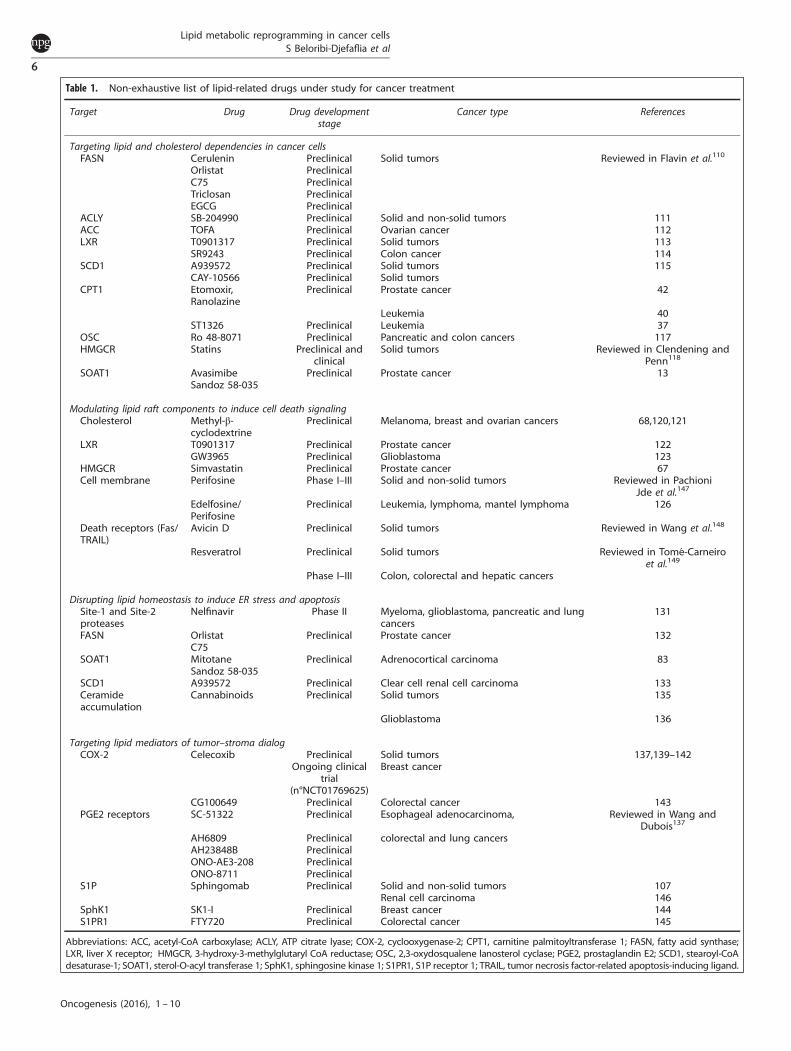
Table 1. Non-exhaustive list of lipid-related drugs under study for cancer treatment
Target Drug Drug developmentstage
Cancer type References
Targeting lipid and cholesterol dependencies in cancer cellsFASN Cerulenin Preclinical Solid tumors Reviewed in Flavin et al.110
Orlistat PreclinicalC75 PreclinicalTriclosan PreclinicalEGCG Preclinical
ACLY SB-204990 Preclinical Solid and non-solid tumors 111ACC TOFA Preclinical Ovarian cancer 112LXR T0901317 Preclinical Solid tumors 113
SR9243 Preclinical Colon cancer 114SCD1 A939572 Preclinical Solid tumors 115
CAY-10566 Preclinical Solid tumorsCPT1 Etomoxir,
RanolazinePreclinical Prostate cancer 42
Leukemia 40ST1326 Preclinical Leukemia 37
OSC Ro 48-8071 Preclinical Pancreatic and colon cancers 117HMGCR Statins Preclinical and
clinicalSolid tumors Reviewed in Clendening and
Penn118
SOAT1 AvasimibeSandoz 58-035
Preclinical Prostate cancer 13
Modulating lipid raft components to induce cell death signalingCholesterol Methyl-β-
cyclodextrinePreclinical Melanoma, breast and ovarian cancers 68,120,121
LXR T0901317 Preclinical Prostate cancer 122GW3965 Preclinical Glioblastoma 123
HMGCR Simvastatin Preclinical Prostate cancer 67Cell membrane Perifosine Phase I–III Solid and non-solid tumors Reviewed in Pachioni
Jde et al.147
Edelfosine/Perifosine
Preclinical Leukemia, lymphoma, mantel lymphoma 126
Death receptors (Fas/TRAIL)
Avicin D Preclinical Solid tumors Reviewed in Wang et al.148
Resveratrol Preclinical Solid tumors Reviewed in Tomé-Carneiroet al.149
Phase I–III Colon, colorectal and hepatic cancers
Disrupting lipid homeostasis to induce ER stress and apoptosisSite-1 and Site-2proteases
Nelfinavir Phase II Myeloma, glioblastoma, pancreatic and lungcancers
131
FASN OrlistatC75
Preclinical Prostate cancer 132
SOAT1 MitotaneSandoz 58-035
Preclinical Adrenocortical carcinoma 83
SCD1 A939572 Preclinical Clear cell renal cell carcinoma 133Ceramideaccumulation
Cannabinoids Preclinical Solid tumors 135
Glioblastoma 136
Targeting lipid mediators of tumor–stroma dialogCOX-2 Celecoxib Preclinical
Ongoing clinicaltrial
(n°NCT01769625)
Solid tumorsBreast cancer
137,139–142
CG100649 Preclinical Colorectal cancer 143PGE2 receptors SC-51322 Preclinical Esophageal adenocarcinoma, Reviewed in Wang and
Dubois137
AH6809 Preclinical colorectal and lung cancersAH23848B PreclinicalONO-AE3-208 PreclinicalONO-8711 Preclinical
S1P Sphingomab Preclinical Solid and non-solid tumors 107Renal cell carcinoma 146
SphK1 SK1-I Preclinical Breast cancer 144S1PR1 FTY720 Preclinical Colorectal cancer 145
Abbreviations: ACC, acetyl-CoA carboxylase; ACLY, ATP citrate lyase; COX-2, cyclooxygenase-2; CPT1, carnitine palmitoyltransferase 1; FASN, fatty acid synthase;LXR, liver X receptor; HMGCR, 3-hydroxy-3-methylglutaryl CoA reductase; OSC, 2,3-oxydosqualene lanosterol cyclase; PGE2, prostaglandin E2; SCD1, stearoyl-CoAdesaturase-1; SOAT1, sterol-O-acyl transferase 1; SphK1, sphingosine kinase 1; S1PR1, S1P receptor 1; TRAIL, tumor necrosis factor-related apoptosis-inducing ligand.
Lipid metabolic reprogramming in cancer cellsS Beloribi-Djefaflia et al
6
Oncogenesis (2016), 1 – 10

cancer cells by combining chemotherapy with the blockade ofLDLR represents a promising alternative therapeutic option tolimit pancreatic tumor growth. Indeed, LDLR silencing potentiatestumor regression induced by chemotherapy.14 Finally, pharmaco-logical inhibitors of SOAT1 enzyme (avasimibe, Sandoz 58-035)(Table 1), through limiting cholesteryl ester storage, have beenshown to suppress tumor growth in prostate cancer xenograftmodels.13
Lipid raft targetingAs discussed above, anticancer drugs that disturb membranecholesterol content can be used to impair lipid raft-dependent cellsurvival or cell death pathways. Methyl-β-cyclodextrin depletesmembrane cholesterol and inhibits human melanoma, breast andovarian cancer growth without elicited acute systemic cytotoxicity(Table 1).68,120 Moreover, when combined with tamoxifen, methyl-β-cyclodextrin slows down melanoma cancer progression byinhibiting AKT and favoring drug uptake,121 probably throughincreased membrane permeability. By increasing cholesterolABCG1-dependent efflux, LXR agonist abrogates the lipid raft-dependent AKT survival pathway and then induces prostatecancer cell apoptosis (Table 1).122 In glioblastoma, such LXRagonist induces tumor cell death in vivo; an effect resulting froman increase in both LDLR degradation and ABCA1-dependentcholesterol efflux.123 Other pharmacological treatments known toreduce lipid raft-associated cholesterol, such as inhibitors ofcholesterol synthesis, have been shown to promote prostatecancer growth arrest and cell death.67,124 Finally, differenttherapeutic drugs, promoting CASMER formation, are beinginvestigated. By their accumulation in cell membrane, syntheticalkylphospholipids (edelfosine, perifosine)125,126 and plant-derivedcompounds (Avicin D, resveratrol)127,128 promote the recruitmentof death receptors, including Fas and TRAIL, into lipid rafts(Table 1). This results in the activation of ligand-independent Fasand/or TRAIL apoptotic pathways in various cancer cells (Figure 2).
Therapies promoting ERS-induced apoptosisThe disruption of lipid homeostasis induces ERS and then cancercell death when the ERS exceeds the cell’s adaptive mechanisms.Nelfinavir and its analogs inhibit Site-1 and Site-2 proteases (S1Pand S2P), both of which are required for the release of the matureand transcriptionally-active form of sterol regulatory element-binding protein-1. The ensuing decrease in lipogenic geneexpression induces ERS and apoptosis in liposarcoma129 andcastration-resistant prostate cancer cells.130 This compoundassociated with chemotherapy is currently in phase II clinicaltrials for myeloma, glioblastoma, pancreatic and lung cancer(Table 1).131 Other chemical compounds, Orlistat and C75, byabrogating the activity of the rate-limiting enzyme of lipogenesisFASN, trigger activation of the UPR and cell death in prostatecancer cells (Table 1).132 Recently, mitotane has been demon-strated to have an anticancer role, impeding cholesterolesterification by inhibiting SOAT1 enzyme and consequentlyinducing an overload of cytotoxic FC within adrenocorticalcarcinoma cancer cells.83 This alteration in lipid homeostasiscauses ERS-induced apoptosis and seems to be specific tosteroidogenic cancer cells. A similar effect, with lower efficacy, isobserved with the Sandoz 58-035 SOAT inhibitor (Table 1).83 Asprostate tumors also exhibit high SOAT1 expression levels, it ispossible that mitotane may promote ERS-induced apoptosis.Preclinical investigations are ongoing on the use of an inhibitor ofSCD1 (A939572) which triggers SFA accumulation, in clear cellrenal cell carcinoma. Its combined administration with tyrosinekinase or mTOR inhibitors appears to improve its efficiencyand reduce its cytotoxicity.133 Impaired HIF2α/PLIN2-dependentlipid storage in clear cell renal cell carcinoma disturbs ERhomeostasis and enhances sensitivity to ERS-inducing agents.11
Hence, coupling proteasome inhibitors, such as bortezomibknown to induce the UPR,134 with HIF2α-specific inhibitorscurrently under development for treating clear cell renal cellcarcinoma patients, could be a rational therapeutic approach.Finally, treatments leading to ceramide accumulation and thenERS-induced apoptosis have shown encouraging results in manypreclinical cancer models (Table 1).135,136 Indeed, cannabinoidreceptor agonists, supporting ceramide-dependent pro-apoptoticcascade, in combination with conventional chemotherapycould be therapeutically exploited for the management ofglioblastoma.136
Disrupting the lipid-mediated dialog between cancer cells andTME cellsTargeting either the lipid messengers or their carriers betweenstromal and tumor cells constitutes an interesting anticancertherapeutic route to continue investigating. The use of COX-2enzyme inhibitor, Celecoxib, to disrupt PG synthesis has revealedits strong antitumoral and antimetastatic effect in variouspreclinical models.137,138 Moreover, it can attenuate patientchemoresistance as well as undesired side effects of anticancerdrugs in various cancers.137,139–142 One clinical study on breastcancer patients is ongoing to evaluate the effect of Celebrex (thatis, celecoxib) alone or in combination with vitamin D (Clinical TrialNo NCT01769625). New COX-2 inhibitors with lower adverse sideeffects, such as CG100649,143 and antagonists of PGE2 receptors30
have shown promising results in multiple cancer preclinicalmodels. Regarding the bioactive lipid S1P, its neutralizingmonoclonal antibody, sphingomab, has proven to be effective atinhibiting angiogenesis, tumor growth and metastasis in multiplecancer cell lines.107 Similar effects on cancer progressionhave been observed with inhibitors of its upstream regu-latory sphingosine kinase 1 (SphK1)144 or its receptor(S1P receptor 1).145 Moreover, sphingomab increases the sensitiv-ity of RCC-bearing mice to sunitinib treatment, which inhibitsVEGFR2 tyrosine kinase.146 Hence, sphingomab constitutes apromising therapy for RCC non-responder patients. In theextracellular space, all these bioactive lipids can be found withinexosomes, hence alternative strategies aiming at decreasingexosome generation and secretion or modifying the exosomecontent within tumors, should be considered in the future aspotential cancer treatments.
CONCLUSIONCompelling evidence gained from untargeted/targeted lipido-mics studies, cancer preclinical models and clinical trials, hasrevealed the crucial role of lipid classes and molecular species insupporting tumor growth and metastatic dissemination. Disrupt-ing lipid metabolic pathways to unbalance lipid homeostasis,through the targeting of enzymes, receptors or bioactive lipids,induces tumor regression and inhibits metastatic spread. Theseeffects result from: (1) fundamental changes in lipid raftcomposition; or (2) sustained ERS-induced UPR, both leading tocancer cell death; or (3) disruption of the lipid-mediated crosstalkbetween stromal and tumor cells, impeding the pro-tumoralfunction of stromal cells. Continued efforts to identify all the keyactors within these different processes may offer novel metabolictargets for cancer treatment. These clinical strategies, based onthe tumor dependence towards lipids, may hold promise for curethe most intractable cancers, including pancreatic and lungcancers, which will become the two deadliest cancers inhorizon 2030.
CONFLICT OF INTERESTThe authors declare no conflict of interest.
Lipid metabolic reprogramming in cancer cellsS Beloribi-Djefaflia et al
7
Oncogenesis (2016), 1 – 10

REFERENCES1 Hanahan D, Weinberg RA. Hallmarks of cancer: the next generation. Cell 2011;
144: 646–674.2 Boroughs LK, DeBerardinis RJ. Metabolic pathways promoting cancer cell
survival and growth. Nat Cell Biol 2015; 17: 351–359.3 Qiu B, Simon MC. Oncogenes strike a balance between cellular growth and
homeostasis. Semin Cell Dev Biol 2015; 43: 3–10.4 Warburg O. On the origin of cancer cells. Science 1956; 123: 309–314.5 Ying H, Kimmelman AC, Lyssiotis CA, Hua S, Chu GC, Fletcher-Sananikone E et al.
Oncogenic Kras maintains pancreatic tumors through regulation of anabolicglucose metabolism. Cell 2012; 149: 656–670.
6 Gaglio D, Metallo CM, Gameiro PA, Hiller K, Danna LS, Balestrieri C et al. Onco-genic K-Ras decouples glucose and glutamine metabolism to support cancercell growth. Mol Syst Biol 2011; 7: 523.
7 Son J, Lyssiotis CA, Ying H, Wang X, Hua S, Ligorio M et al. Glutamine supportspancreatic cancer growth through a KRAS-regulated metabolic pathway. Nature2013; 496: 101–105.
8 Baenke F, Peck B, Miess H, Schulze A. Hooked on fat: the role of lipid synthesis incancer metabolism and tumour development. Dis Model Mech 2013; 6:1353–1363.
9 Ackerman D, Simon MC. Hypoxia, lipids, and cancer: surviving the harsh tumormicroenvironment. Trends Cell Biol 2014; 24: 472–478.
10 Cruz PM, Mo H, McConathy WJ, Sabnis N, Lacko AG. The role of cholesterolmetabolism and cholesterol transport in carcinogenesis: a review of scientificfindings, relevant to future cancer therapeutics. Front Pharmacol 2013; 4: 119.
11 Qiu B, Ackerman D, Sanchez DJ, Li B, Ochocki JD, Grazioli et al. HIF2alpha-dependent lipid storage promotes endoplasmic reticulum homeostasis in clear-cell renal cell carcinoma. Cancer Discov 2015; 5: 652–667.
12 Accioly MT, Pacheco P, Maya-Monteiro CM, Carrossini N, Robbs BK, Oliveira SSet al. Lipid bodies are reservoirs of cyclooxygenase-2 and sites of prostaglandin-E2 synthesis in colon cancer cells. Cancer Res 2008; 68: 1732–1740.
13 Yue S, Li J, Lee SY, Lee HJ, Shao T, Song B et al. Cholesteryl ester accumulationinduced by PTEN loss and PI3K/AKT activation underlies human prostate canceraggressiveness. Cell Metab 2014; 19: 393–406.
14 Guillaumond F, Bidaut G, Ouaissi M, Servais S, Gouirand V, Olivares O et al.Cholesterol uptake disruption, in association with chemotherapy, is a promisingcombined metabolic therapy for pancreatic adenocarcinoma. Proc Natl Acad SciUSA 2015; 112: 2473–2478.
15 Bozza PT, Viola JP. Lipid droplets in inflammation and cancer. ProstaglandinsLeukot Essent Fatty Acids 2010; 82: 243–250.
16 de Gonzalo-Calvo D, Lopez-Vilaro L, Nasarre L, Perez-Olabarria M, Vazquez T,Escuin D et al. Intratumor cholesteryl ester accumulation is associated withhuman breast cancer proliferation and aggressive potential: a molecular andclinicopathological study. BMC Cancer 2015; 15: 460.
17 Abramczyk H, Surmacki J, Kopec M, Olejnik AK, Lubecka-Pietruszewska K,Fabianowska-Majewska K. The role of lipid droplets and adipocytes in cancer.Raman imaging of cell cultures: MCF10A, MCF7, and MDA-MB-231 compared toadipocytes in cancerous human breast tissue. Analyst 2015; 140: 2224–2235.
18 Tirinato L, Liberale C, Di Franco S, Candeloro P, Benfante A, La Rocca R et al. Lipiddroplets: a new player in colorectal cancer stem cells unveiled by spectroscopicimaging. Stem Cells 2015; 33: 35–44.
19 El-Mashtoly SF, Yosef HK, Petersen D, Mavarani L, Maghnouj A, Hahn S et al.Label-free Raman spectroscopic imaging monitors the integral physiologicallyrelevant drug responses in cancer cells. Anal Chem 2015; 87: 7297–7304.
20 Steuwe C, Patel II, Ul-Hasan M, Schreiner A, Boren J, Brindle KM et al. CARS basedlabel-free assay for assessment of drugs by monitoring lipid droplets intumour cells. J Biophotonics 2014; 7: 906–913.
21 Daniels VW, Smans K, Royaux I, Chypre M, Swinnen JV, Zaidi N. Cancer cellsdifferentially activate and thrive on de novo lipid synthesis pathways in a low-lipid environment. PLoS One 2014; 9: e106913.
22 Kamphorst JJ, Cross JR, Fan J, de Stanchina E, Mathew R, White EP et al. Hypoxicand Ras-transformed cells support growth by scavenging unsaturated fatty acidsfrom lysophospholipids. Proc Natl Acad Sci USA 2013; 110: 8882–8887.
23 Raynor A, Jantscheff P, Ross T, Schlesinger M, Wilde M, Haasis S et al. Saturatedand mono-unsaturated lysophosphatidylcholine metabolism in tumour cells: apotential therapeutic target for preventing metastases. Lipids Health Dis 2015;14: 69.
24 Liu Y, Zuckier LS, Ghesani NV. Dominant uptake of fatty acid over glucose byprostate cells: a potential new diagnostic and therapeutic approach. AnticancerRes 2010; 30: 369–374.
25 Caro P, Kishan AU, Norberg E, Stanley IA, Chapuy B, Ficarro SB et al. Metabolicsignatures uncover distinct targets in molecular subsets of diffuse large B celllymphoma. Cancer Cell 2012; 22: 547–560.
26 Li J, Cheng JX. Direct visualization of de novo lipogenesis in single living cells. SciRep 2014; 4: 6807.
27 Rysman E, Brusselmans K, Scheys K, Timmermans L, Derua R, Munck S et al. Denovo lipogenesis protects cancer cells from free radicals and chemotherapeuticsby promoting membrane lipid saturation. Cancer Res 2010; 70: 8117–8126.
28 Staubach S, Hanisch FG. Lipid rafts: signaling and sorting platforms of cells andtheir roles in cancer. Expert Rev Proteomics 2011; 8: 263–277.
29 Kunkel GT, Maceyka M, Milstien S, Spiegel S. Targeting the sphingosine-1-phosphate axis in cancer, inflammation and beyond. Nat Rev Drug Discov 2013;12: 688–702.
30 Wang D, Dubois RN. Eicosanoids and cancer. Nat Rev Cancer 2010; 10: 181–193.31 Nakanishi M, Rosenberg DW. Multifaceted roles of PGE2 in inflammation
and cancer. Semin Immunopathol 2013; 35: 123–137.32 Menendez JA, Lupu R. Fatty acid synthase and the lipogenic phenotype in
cancer pathogenesis. Nat Rev Cancer 2007; 7: 763–777.33 Zaidi N, Swinnen JV, Smans K. ATP-citrate lyase: a key player in cancer meta-
bolism. Cancer Res 2012; 72: 3709–3714.34 Kuhajda FP. Fatty acid synthase and cancer: new application of an old pathway.
Cancer Res 2006; 66: 5977–5980.35 Biswas S, Lunec J, Bartlett K. Non-glucose metabolism in cancer cells--is it all in
the fat? Cancer Metastasis Rev 2012; 31: 689–698.36 Zaugg K, Yao Y, Reilly PT, Kannan K, Kiarash R, Mason J et al. Carnitine palmi-
toyltransferase 1C promotes cell survival and tumor growth under conditions ofmetabolic stress. Genes Dev 2011; 25: 1041–1051.
37 Ricciardi MR, Mirabilii S, Allegretti M, Licchetta R, Calarco A, Torrisi MR et al.Targeting the leukemia cell metabolism by the CPT1a inhibition: functionalpreclinical effects in leukemias. Blood 2015; 126: 1925–1929.
38 Reilly PT, Mak TW. Molecular pathways: tumor cells Co-opt the brain-specificmetabolism gene CPT1C to promote survival. Clin Cancer Res 2012; 18:5850–5855.
39 Sanchez-Macedo N, Feng J, Faubert B, Chang N, Elia A, Rushing EJ et al.Depletion of the novel p53-target gene carnitine palmitoyltransferase 1C delaystumor growth in the neurofibromatosis type I tumor model. Cell Death Differ2013; 20: 659–668.
40 Samudio I, Harmancey R, Fiegl M, Kantarjian H, Konopleva M, Korchin B et al.Pharmacologic inhibition of fatty acid oxidation sensitizes human leukemia cellsto apoptosis induction. J Clin Invest 2010; 120: 142–156.
41 Hernlund E, Ihrlund LS, Khan O, Ates YO, Linder S, Panaretakis T et al. Poten-tiation of chemotherapeutic drugs by energy metabolism inhibitors2-deoxyglucose and etomoxir. Int J Cancer 2008; 123: 476–483.
42 Schlaepfer IR, Rider L, Rodrigues LU, Gijon MA, Pac CT, Romero L et al. Lipidcatabolism via CPT1 as a therapeutic target for prostate cancer. Mol Cancer Ther2014; 13: 2361–2371.
43 Vargas T, Moreno-Rubio J, Herranz J, Cejas P, Molina S, Gonzalez-Vallinas M et al.ColoLipidGene: signature of lipid metabolism-related genes to predict prognosisin stage-II colon cancer patients. Oncotarget 2015; 6: 7348–7363.
44 Loizides-Mangold U. On the future of mass-spectrometry-based lipidomics. FEBSJ 2013; 280: 2817–2829.
45 Le TT, Yue S, Cheng JX. Shedding new light on lipid biology with coherentanti-Stokes Raman scattering microscopy. J Lipid Res 2010; 51: 3091–3102.
46 Hilvo M, Denkert C, Lehtinen L, Muller B, Brockmoller S, Seppanen-Laakso T et al.Novel theranostic opportunities offered by characterization of alteredmembrane lipid metabolism in breast cancer progression. Cancer Res 2011; 71:3236–3245.
47 Guenther S, Muirhead LJ, Speller AV, Golf O, Strittmatter N, Ramakrishnan R et al.Spatially resolved metabolic phenotyping of breast cancer by desorption elec-trospray ionization mass spectrometry. Cancer Res 2015; 75: 1828–1837.
48 Ollila S, Hyvonen MT, Vattulainen I. Polyunsaturation in lipid membranes:dynamic properties and lateral pressure profiles. J Phys Chem B 2007; 111:3139–3150.
49 Kawashima M, Iwamoto N, Kawaguchi-Sakita N, Sugimoto M, Ueno T, Mikami Yet al. High-resolution imaging mass spectrometry reveals detailed spatialdistribution of phosphatidylinositols in human breast cancer. Cancer Sci 2013;104: 1372–1379.
50 Eberlin LS, Gabay M, Fan AC, Gouw AM, Tibshirani RJ, Felsher DW et al. Alterationof the lipid profile in lymphomas induced by MYC overexpression. Proc Natl AcadSci USA 2014; 111: 10450–10455.
51 Perry RH, Bellovin DI, Shroff EH, Ismail AI, Zabuawala T, Felsher DW et al. Char-acterization of MYC-induced tumorigenesis by in situ lipid profiling. Anal Chem2013; 85: 4259–4262.
52 Shroff EH, Eberlin LS, Dang VM, Gouw AM, Gabay M, Adam SJ et al. MYConcogene overexpression drives renal cell carcinoma in a mouse model throughglutamine metabolism. Proc Natl Acad Sci USA 2015; 112: 6539–6544.
53 Kiebish MA, Han X, Cheng H, Chuang JH, Seyfried TN. Cardiolipin and electrontransport chain abnormalities in mouse brain tumor mitochondria: lipidomicevidence supporting the Warburg theory of cancer. J Lipid Res 2008; 49:2545–2556.
Lipid metabolic reprogramming in cancer cellsS Beloribi-Djefaflia et al
8
Oncogenesis (2016), 1 – 10
54 Lingwood D, Simons K. Lipid rafts as a membrane-organizing principle. Science2010; 327: 46–50.
55 Mollinedo F, Gajate C. Lipid rafts as major platforms for signaling regulationin cancer. Adv Biol Regul 2015; 57: 130–146.
56 Lucken-Ardjomande S, Montessuit S, Martinou JC. Bax activation and stress-induced apoptosis delayed by the accumulation of cholesterol in mitochondrialmembranes. Cell Death Differ 2008; 15: 484–493.
57 Dessi S, Batetta B, Pulisci D, Spano O, Anchisi C, Tessitore L et al. Cholesterolcontent in tumor tissues is inversely associated with high-density lipoproteincholesterol in serum in patients with gastrointestinal cancer. Cancer 1994; 73:253–258.
58 Kolanjiappan K, Ramachandran CR, Manoharan S. Biochemical changes in tumortissues of oral cancer patients. Clin Biochem 2003; 36: 61–65.
59 Montero J, Morales A, Llacuna L, Lluis JM, Terrones O, Basanez G et al.Mitochondrial cholesterol contributes to chemotherapy resistance in hepato-cellular carcinoma. Cancer Res 2008; 68: 5246–5256.
60 Li YC, Park MJ, Ye SK, Kim CW, Kim YN. Elevated levels of cholesterol-rich lipidrafts in cancer cells are correlated with apoptosis sensitivity induced bycholesterol-depleting agents. Am J Pathol 2006; 168: 1107–1118; quiz 1404-5.
61 Llorente A, Skotland T, Sylvanne T, Kauhanen D, Rog T, Orlowski et al. Molecularlipidomics of exosomes released by PC-3 prostate cancer cells. Biochim BiophysActa 2013; 1831: 1302–1309.
62 Hill MM, Feng J, Hemmings BA. Identification of a plasma membrane Raft-associated PKB Ser473 kinase activity that is distinct from ILK and PDK1. Curr Biol2002; 12: 1251–1255.
63 Adam RM, Mukhopadhyay NK, Kim J, Di Vizio D, Cinar B, Boucher K et al.Cholesterol sensitivity of endogenous and myristoylated Akt. Cancer Res 2007;67: 6238–6246.
64 Courtney KD, Corcoran RB, Engelman JA. The PI3K pathway as drug target inhuman cancer. J Clin Oncol 2010; 28: 1075–1083.
65 Shukla S, Maclennan GT, Hartman DJ, Fu P, Resnick MI, Gupta S. Activation ofPI3K-Akt signaling pathway promotes prostate cancer cell invasion. Int J Cancer2007; 121: 1424–1432.
66 Borradaile NM, Han X, Harp JD, Gale SE, Ory DS, Schaffer JE. Disruption ofendoplasmic reticulum structure and integrity in lipotoxic cell death. J Lipid Res2006; 47: 2726–2737.
67 Zhuang L, Kim J, Adam RM, Solomon KR, Freeman MR. Cholesterol targetingalters lipid raft composition and cell survival in prostate cancer cells andxenografts. J Clin Invest 2005; 115: 959–968.
68 Fedida-Metula S, Elhyany S, Tsory S, Segal S, Hershfinkel M, Sekler I et al.Targeting lipid rafts inhibits protein kinase B by disrupting calcium homeostasisand attenuates malignant properties of melanoma cells. Carcinogenesis 2008; 29:1546–1554.
69 Wang R, Bi J, Ampah KK, Zhang C, Li Z, Jiao Y et al. Lipid raft regulates the initialspreading of melanoma A375 cells by modulating beta1 integrin clustering. Int JBiochem Cell Biol 2013; 45: 1679–1689.
70 Jeon JH, Kim SK, Kim HJ, Chang J, Ahn CM, Chang YS. Lipid raft modulationinhibits NSCLC cell migration through delocalization of the focal adhesioncomplex. Lung Cancer 2010; 69: 165–171.
71 Wang J, He L, Combs CA, Roderiquez G, Norcross MA. Dimerization of CXCR4 inliving malignant cells: control of cell migration by a synthetic peptide thatreduces homologous CXCR4 interactions. Mol Cancer Ther 2006; 5: 2474–2483.
72 Song JH, Tse MC, Bellail A, Phuphanich S, Khuri F, Kneteman NM et al. Lipid raftsand nonrafts mediate tumor necrosis factor related apoptosis-inducing ligandinduced apoptotic and nonapoptotic signals in non small cell lungcarcinoma cells. Cancer Res 2007; 67: 6946–6955.
73 Delmas D, Rebe C, Micheau O, Athias A, Gambert P, Grazide S et al. Redis-tribution of CD95, DR4 and DR5 in rafts accounts for the synergistic toxicity ofresveratrol and death receptor ligands in colon carcinoma cells. Oncogene 2004;23: 8979–8986.
74 Kato H, Nishitoh H. Stress responses from the endoplasmic reticulum in cancer.Front Oncol 2015; 5: 93.
75 Mylonis I, Sembongi H, Befani C, Liakos P, Siniossoglou S, Simos G. Hypoxiacauses triglyceride accumulation by HIF-1-mediated stimulation of lipin 1expression. J Cell Sci 2012; 125: 3485–3493.
76 Volmer R, van der Ploeg K, Ron D. Membrane lipid saturation activatesendoplasmic reticulum unfolded protein response transducers through theirtransmembrane domains. Proc Natl Acad Sci USA 2013; 110: 4628–4633.
77 Kitai Y, Ariyama H, Kono N, Oikawa D, Iwawaki T, Arai H. Membrane lipidsaturation activates IRE1alpha without inducing clustering. Genes Cells 2013; 18:798–809.
78 Ariyama H, Kono N, Matsuda S, Inoue T, Arai H. Decrease in membrane phos-pholipid unsaturation induces unfolded protein response. J Biol Chem 2010; 285:22027–22035.
79 Griffiths B, Lewis CA, Bensaad K, Ros S, Zhang Q, Ferber EC et al. Sterol regulatoryelement binding protein-dependent regulation of lipid synthesis supports cellsurvival and tumor growth. Cancer Metab 2013; 1: 3.
80 Williams KJ, Argus JP, Zhu Y, Wilks MQ, Marbois BN, York AG et al. An essentialrequirement for the SCAP/SREBP signaling axis to protect cancer cells fromlipotoxicity. Cancer Res 2013; 73: 2850–2862.
81 Rios-Marco P, Martin-Fernandez M, Soria-Bretones I, Rios A, Carrasco MP, MarcoC. Alkylphospholipids deregulate cholesterol metabolism and induce cell-cyclearrest and autophagy in U-87 MG glioblastoma cells. Biochim Biophys Acta 2013;1831: 1322–1334.
82 Rios-Marco P, Rios A, Jimenez-Lopez JM, Carrasco MP, Marco C. Cholesterolhomeostasis and autophagic flux in perifosine-treated human hepatoblastomaHepG2 and glioblastoma U-87 MG cell lines. Biochem Pharmacol 2015; 96: 10–19.
83 Sbiera S, Leich E, Liebisch G, Sbiera I, Schirbel A, Wiemer L et al. Mitotane inhibitsSterol-O-Acyl Transferase 1 triggering lipid-mediated endoplasmic reticulumstress and apoptosis in adrenocortical carcinoma cells. Endocrinology 2015; 156:3895–3908 en20151367.
84 Ponnusamy S, Meyers-Needham M, Senkal CE, Saddoughi SA, Sentelle D, SelvamSP et al. Sphingolipids and cancer: ceramide and sphingosine-1-phosphate inthe regulation of cell death and drug resistance. Future Oncol 2010; 6:1603–1624.
85 Henry B, Moller C, Dimanche-Boitrel MT, Gulbins E, Becker KA. Targeting theceramide system in cancer. Cancer Lett 2013; 332: 286–294.
86 Carracedo A, Gironella M, Lorente M, Garcia S, Guzman M, Velasco G et al.Cannabinoids induce apoptosis of pancreatic tumor cells via endoplasmicreticulum stress-related genes. Cancer Res 2006; 66: 6748–6755.
87 Carracedo A, Lorente M, Egia A, Blazquez C, Garcia S, Giroux V et al. The stress-regulated protein p8 mediates cannabinoid-induced apoptosis of tumor cells.Cancer Cell 2006; 9: 301–312.
88 Salazar M, Carracedo A, Salanueva IJ, Hernandez-Tiedra S, Lorente M, Egia et al.Cannabinoid action induces autophagy-mediated cell death through stimulationof ER stress in human glioma cells. J Clin Invest 2009; 119: 1359–1372.
89 Mehta S, Blackinton D, Omar I, Kouttab N, Myrick D, Klostergaard J et al.Combined cytotoxic action of paclitaxel and ceramide against the human Tu138head and neck squamous carcinoma cell line. Cancer Chemother Pharmacol2000; 46: 85–92.
90 Senkal CE, Ponnusamy S, Rossi MJ, Bialewski J, Sinha D, Jiang JC et al. Role ofhuman longevity assurance gene 1 and C18-ceramide in chemotherapy-inducedcell death in human head and neck squamous cell carcinomas. Mol Cancer Ther2007; 6: 712–722.
91 Liu Z, Xia Y, Li B, Xu H, Wang C, Liu Y et al. Induction of ER stress-mediatedapoptosis by ceramide via disruption of ER Ca(2+) homeostasis in humanadenoid cystic carcinoma cells. Cell Biosci 2014; 4: 71.
92 Hanahan D, Coussens LM. Accessories to the crime: functions of cells recruited tothe tumor microenvironment. Cancer Cell 2012; 21: 309–322.
93 Nieman KM, Kenny HA, Penicka CV, Ladanyi A, Buell-Gutbrod R, Zillhardt MRet al. Adipocytes promote ovarian cancer metastasis and provide energy forrapid tumor growth. Nat Med 2011; 17: 1498–1503.
94 Gazi E, Gardner P, Lockyer NP, Hart CA, Brown MD, Clarke NW. Direct evidence oflipid translocation between adipocytes and prostate cancer cells with imagingFTIR microspectroscopy. J Lipid Res 2007; 48: 1846–1856.
95 Palm W, Park Y, Wright K, Pavlova NN, Tuveson DA, Thompson CB. The Utilizationof Extracellular Proteins as Nutrients Is Suppressed by mTORC1. Cell 2015; 162:259–270.
96 Commisso C, Davidson SM, Soydaner-Azeloglu RG, Parker SJ, Kamphorst JJ,Hackett S et al. Macropinocytosis of protein is an amino acid supply route in Ras-transformed cells. Nature 2013; 497: 633–637.
97 Record M, Carayon K, Poirot M, Silvente-Poirot S. Exosomes as new vesicular lipidtransporters involved in cell-cell communication and various pathophysiologies.Biochim Biophys Acta 2014; 1841: 108–120.
98 Subra C, Grand D, Laulagnier K, Stella A, Lambeau G, Paillasse M et al. Exosomesaccount for vesicle-mediated transcellular transport of activatable phospholi-pases and prostaglandins. J Lipid Res 2010; 51: 2105–2120.
99 Beloribi S, Ristorcelli E, Breuzard G, Silvy F, Bertrand-Michel J, Beraud E et al.Exosomal lipids impact notch signaling and induce death of human pancreatictumoral SOJ-6 cells. PLoS ONE 2012; 7: e47480.
100 Xiang X, Poliakov A, Liu C, Liu Y, Deng ZB, Wang J et al. Induction of myeloid-derived suppressor cells by tumor exosomes. Int J Cancer 2009; 124: 2621–2633.
101 Sinha P, Clements VK, Fulton AM, Ostrand-Rosenberg S. Prostaglandin E2 pro-motes tumor progression by inducing myeloid-derived suppressor cells. CancerRes 2007; 67: 4507–4513.
102 Heusinkveld M, de Vos van Steenwijk PJ, Goedemans R, Ramwadhdoebe TH,Gorter A, Welters MJ et al. M2 macrophages induced by prostaglandin E2 andIL-6 from cervical carcinoma are switched to activated M1 macrophages by CD4+ Th1 cells. J Immunol 2011; 187: 1157–1165.
Lipid metabolic reprogramming in cancer cellsS Beloribi-Djefaflia et al
9
Oncogenesis (2016), 1 – 10
103 Chang SH, Liu CH, Conway R, Han DK, Nithipatikom K, Trifan OC et al. Role ofprostaglandin E2-dependent angiogenic switch in cyclooxygenase 2-inducedbreast cancer progression. Proc Natl Acad Sci USA 2004; 101: 591–596.
104 Chu J, Lloyd FL, Trifan OC, Knapp B, Rizzo MT. Potential involvement of thecyclooxygenase-2 pathway in the regulation of tumor-associated angiogenesisand growth in pancreatic cancer. Mol Cancer Ther 2003; 2: 1–7.
105 Tsujii M, Kawano S, Tsuji S, Sawaoka H, Hori M, DuBois RN. Cyclooxygenaseregulates angiogenesis induced by colon cancer cells. Cell 1998; 93: 705–716.
106 Chen JY, Li CF, Kuo CC, Tsai KK, Hou MF, Hung WC. Cancer/stroma interplay viacyclooxygenase-2 and indoleamine 2,3-dioxygenase promotes breast cancerprogression. Breast Cancer Res 2014; 16: 410.
107 Visentin B, Vekich JA, Sibbald BJ, Cavalli AL, Moreno KM, Matteo RG et al.Validation of an anti-sphingosine-1-phosphate antibody as a potential ther-apeutic in reducing growth, invasion, and angiogenesis in multiple tumorlineages. Cancer Cell 2006; 9: 225–238.
108 LaMontagne K, Littlewood-Evans A, Schnell C, O'Reilly T, Wyder L, Sanchez Tet al. Antagonism of sphingosine-1-phosphate receptors by FTY720 inhibitsangiogenesis and tumor vascularization. Cancer Res 2006; 66: 221–231.
109 Anelli V, Gault CR, Snider AJ, Obeid LM. Role of sphingosine kinase-1 inparacrine/transcellular angiogenesis and lymphangiogenesis in vitro. FASEB J2010; 24: 2727–2738.
110 Flavin R, Peluso S, Nguyen PL, Loda M. Fatty acid synthase as a potential ther-apeutic target in cancer. Future Oncol 2010; 6: 551–562.
111 Hatzivassiliou G, Zhao F, Bauer DE, Andreadis C, Shaw AN, Dhanak D et al. ATPcitrate lyase inhibition can suppress tumor cell growth. Cancer Cell 2005; 8:311–321.
112 Li S, Qiu L, Wu B, Shen H, Zhu J, Zhou L et al. TOFA suppresses ovarian cancer cellgrowth in vitro and in vivo. Mol Med Rep 2013; 8: 373–378.
113 Bovenga F, Sabba C, Moschetta A. Uncoupling nuclear receptor LXR andcholesterol metabolism in cancer. Cell Metab 2015; 21: 517–526.
114 Flaveny CA, Griffett K, El-Gendy Bel D, Kazantzis M, Sengupta M, Amelio AL et al.Broad anti-tumor activity of a small molecule that selectively targets the War-burg effect and lipogenesis. Cancer Cell 2015; 28: 42–56.
115 Uto Y. Recent progress in the discovery and development of stearoyl CoAdesaturase inhibitors. Chem Phys Lipids (e-pub ahead of print 3 September 2015;doi: 10.1016/j.chemphyslip.2015.08.018).
116 Graeser R, Bornmann C, Esser N, Ziroli V, Jantscheff P, Unger C et al. Antimeta-static effects of liposomal gemcitabine and empty liposomes in an orthotopicmouse model of pancreatic cancer. Pancreas 2009; 38: 330–337.
117 Maione F, Oliaro-Bosso S, Meda C, Di Nicolantonio F, Bussolino F, Balliano G et al.The cholesterol biosynthesis enzyme oxidosqualene cyclase is a new target toimpair tumour angiogenesis and metastasis dissemination. Sci Rep 2015; 5: 9054.
118 Clendening JW, Penn LZ. Targeting tumor cell metabolism with statins.Oncogene 2012; 31: 4967–4978.
119 Pisanti S, Picardi P, Ciaglia E, D'Alessandro A, Bifulco M. Novel prospects ofstatins as therapeutic agents in cancer. Pharmacol Res 2014; 88: 84–98.
120 Grosse PY, Bressolle F, Pinguet F. Antiproliferative effect of methyl-beta-cyclodextrin in vitro and in human tumour xenografted athymic nude mice. Br JCancer 1998; 78: 1165–1169.
121 Mohammad N, Malvi P, Meena AS, Singh SV, Chaube B, Vannuruswamy G et al.Cholesterol depletion by methyl-beta-cyclodextrin augments tamoxifen inducedcell death by enhancing its uptake in melanoma. Mol Cancer 2014; 13: 204.
122 Pommier AJ, Alves G, Viennois E, Bernard S, Communal Y, Sion B et al. Liver XReceptor activation downregulates AKT survival signaling in lipid rafts andinduces apoptosis of prostate cancer cells. Oncogene 2010; 29: 2712–2723.
123 Guo D, Reinitz F, Youssef M, Hong C, Nathanson D, Akhavan D et al. An LXRagonist promotes glioblastoma cell death through inhibition of an EGFR/AKT/SREBP-1/LDLR-dependent pathway. Cancer Discov 2011; 1: 442–456.
124 Brusselmans K, Timmermans L, Van de Sande T, Van Veldhoven PP, Guan G,Shechter I et al. Squalene synthase, a determinant of Raft-associated cholesteroland modulator of cancer cell proliferation. J Biol Chem 2007; 282: 18777–18785.
125 Gajate C, Gonzalez-Camacho F, Mollinedo F. Involvement of raft aggregatesenriched in Fas/CD95 death-inducing signaling complex in the antileukemicaction of edelfosine in Jurkat cells. PLoS ONE 2009; 4: e5044.
126 Gajate C, Mollinedo F. Edelfosine and perifosine induce selective apoptosis inmultiple myeloma by recruitment of death receptors and downstream signalingmolecules into lipid rafts. Blood 2007; 109: 711–719.
127 Reis-Sobreiro M, Gajate C, Mollinedo F. Involvement of mitochondria andrecruitment of Fas/CD95 signaling in lipid rafts in resveratrol-mediated anti-myeloma and antileukemia actions. Oncogene 2009; 28: 3221–3234.
128 Xu ZX, Ding T, Haridas V, Connolly F, Gutterman JU. Avicin D, a plant triterpe-noid, induces cell apoptosis by recruitment of Fas and downstream signalingmolecules into lipid rafts. PLoS ONE 2009; 4: e8532.
129 Guan M, Fousek K, Jiang C, Guo S, Synold T, Xi B et al.Nelfinavir induces liposarcoma apoptosis through inhibition of regulatedintramembrane proteolysis of SREBP-1 and ATF6. Clin Cancer Res 2011; 17:1796–1806.
130 Guan M, Su L, Yuan YC, Li H, Chow WA. Nelfinavir and nelfinavir analogs blocksite-2 protease cleavage to inhibit castration-resistant prostate cancer. Sci Rep2015; 5: 9698.
131 Koltai T. Nelfinavir and other protease inhibitors in cancer: mechanisms involvedin anticancer activity. F1000Res 2015; 4: 9.
132 Little JL, Wheeler FB, Fels DR, Koumenis C, Kridel SJ. Inhibition of fatty acidsynthase induces endoplasmic reticulum stress in tumor cells. Cancer Res 2007;67: 1262–1269.
133 von Roemeling CA, Marlow LA, Wei JJ, Cooper SJ, Caulfield TR, Wu K et al.Stearoyl-CoA desaturase 1 is a novel molecular therapeutic target for clear cellrenal cell carcinoma. Clin Cancer Res 2013; 19: 2368–2380.
134 Obeng EA, Carlson LM, Gutman DM, Harrington WJ Jr., Lee KP, Boise LH.Proteasome inhibitors induce a terminal unfolded protein response in multiplemyeloma cells. Blood 2006; 107: 4907–4916.
135 Sarfaraz S, Adhami VM, Syed DN, Afaq F, Mukhtar H. Cannabinoids for cancertreatment: progress and promise. Cancer Res 2008; 68: 339–342.
136 Torres S, Lorente M, Rodriguez-Fornes F, Hernandez-Tiedra S, Salazar M, Garcia-Taboada E et al. A combined preclinical therapy of cannabinoids and temozo-lomide against glioma. Mol Cancer Ther 2011; 10: 90–103.
137 Wang D, Dubois RN. The role of COX-2 in intestinal inflammation andcolorectal cancer. Oncogene 2010; 29: 781–788.
138 Xu L, Stevens J, Hilton MB, Seaman S, Conrads TP, Veenstra TD et al. COX-2inhibition potentiates antiangiogenic cancer therapy and prevents metastasis inpreclinical models. Sci Transl Med 2014; 6: 242ra84.
139 Kurtova AV, Xiao J, Mo Q, Pazhanisamy S, Krasnow R, Lerner SP et al. BlockingPGE2-induced tumour repopulation abrogates bladder cancer chemoresistance.Nature 2015; 517: 209–213.
140 Ng K, Meyerhardt JA, Chan AT, Sato K, Chan JA, Niedzwiecki D et al. Aspirin andCOX-2 inhibitor use in patients with stage III colon cancer. J Natl Cancer Inst2015; 107: 345.
141 Thill M, Reichert K, Woeste A, Polack S, Fischer D, Hoellen F et al. Combinedtreatment of breast cancer cell lines with vitamin D and COX-2 inhibitors.Anticancer Res 2015; 35: 1189–1195.
142 Knab LM, Grippo PJ, Bentrem DJ. Involvement of eicosanoids in the pathogen-esis of pancreatic cancer: the roles of cyclooxygenase-2 and 5-lipoxygenase.World J Gastroenterol 2014; 20: 10729–10739.
143 Kim SH, Margalit O, Katoh H, Wang D, Wu H, Xia D et al. CG100649, a novelCOX-2 inhibitor, inhibits colorectal adenoma and carcinoma growth inmouse models. Invest New Drugs 2014; 32: 1105–1112.
144 Nagahashi M, Ramachandran S, Kim EY, Allegood JC, Rashid OM, Yamada et al.Sphingosine-1-phosphate produced by sphingosine kinase 1 promotes breastcancer progression by stimulating angiogenesis and lymphangiogenesis. CancerRes 2012; 72: 726–735.
145 Liang J, Nagahashi M, Kim EY, Harikumar KB, Yamada A, Huang WC et al.Sphingosine-1-phosphate links persistent STAT3 activation, chronic intestinalinflammation, and development of colitis-associated cancer. Cancer Cell 2013;23: 107–120.
146 Zhang L, Wang X, Bullock AJ, Callea M, Shah H, Song J et al. Anti-S1P antibody asa novel therapeutic strategy for VEGFR TKI-resistant renal cancer. Clin Cancer Res2015; 21: 1925–1934.
147 Pachioni Jde A, Magalhães JG, Lima EJ, Bueno Lde M, Barbosa JF,de Sá MM et al. Alkylphospholipids - a promising class of chemotherapeuticagents with a broad pharmacological spectrum. Pharm Pharm Sci 2013; 16:742–759.
148 Wang H, Haridas V, Gutterman JU, Xu ZX. Natural triterpenoid avicins selectivelyinduce tumor cell death. Commun Integr Biol 2010; 3: 205–208.
149 Tomé-Carneiro J, Larrosa M, González-Sarrías A, Tomás-Barberán FA,García-Conesa MT, Espín JC. Resveratrol and clinical trials: the crossroadfrom in vitro studies to human evidence. Curr Pharm Des 2013; 19:6064–6093.
Oncogenesis is an open-access journal published by Nature PublishingGroup. This work is licensed under a Creative Commons Attribution 4.0
International License. The images or other third partymaterial in this article are includedin the article’s Creative Commons license, unless indicated otherwise in the credit line; ifthe material is not included under the Creative Commons license, users will need toobtain permission from the license holder to reproduce the material. To view a copy ofthis license, visit http://creativecommons.org/licenses/by/4.0/
Lipid metabolic reprogramming in cancer cellsS Beloribi-Djefaflia et al
10
Oncogenesis (2016), 1 – 10