lipoma arborescens arising in the extra-articular bursa of ... arborescens arising in the...
TRANSCRIPT
Lipoma arborescens arising in the extra-articular bursaof the knee joint
Shinji Minami1,*, Yusuke Miyake2, and Hirofumi Kinoshita1
1 Department of Orthopedic Surgery, Naga Municipal Hospital, 1282 Uchita, Kinokawa, Wakayama 649-6414, Japan2 Department of Orthopedic Surgery, Wakayama Medical University, 811-1 Kimiidera, Wakayama 641-8510, Japan
Received 17 December 2015, Accepted 15 May 2016, Published online 6 July 2016
Abstract – Lipoma arborescens arising in the extra-articular bursa of the knee joint is extremely rare. We describe an11-year-old boy who complained of a gradual swelling mass of the lateral knee joint. Magnetic resonance imaging(MRI) showed a high signal intensity tumor on T1- and T2-weighted images with a thickened septa and nodular lesionthat showed low signal intensity. The radiologist suggested the possible differential diagnosis of well-differentiatedliposarcoma. At operation, the tumor was found under the iliotibial tract and was not in contact with the knee joint.Histopathologically, this lesion was diagnosed as lipoma arborescens arising in the extra-articular bursa of the kneejoint. On MRI, the appearance of lipoma arborescens arising in the extra-articular bursa of the knee joint differed fromthat of conventional intra-articular lipoma arborescens. In this report, we describe a case of extra-articular lipomaarborescens of the knee joint bursa and discuss the diagnosis and etiology.
Key words: bursa, extra-articular, knee joint, lipoma arborescens.
Introduction
Lipoma arborescens is a rare synovial lesion, in which thetumor showed diffuse villous proliferation of the synoviumcharacterized by replacement of the subsynovial tissue bymature adipocytes. The most affected site is the knee joint,usually in the suprapatellar pouch of the knee joint [1]. Theselesions have been rarely reported in other locations, includingthe hand, wrist, elbow, shoulder, and hip [2–6], and a few caseshave been reported affecting the extra-articular bursa of thedeltoid and the tendon sheath [3, 7]; however, extra-articularbursa of the knee joint is thought to be extremely rare. Onlyone case of lipoma arborescens arising in the extra-articularbursa of the knee joint has been reported in the English litera-ture [8]. On magnetic resonance imaging (MRI), it has beenreported that the appearance of conventional lipoma arbores-cens arising in the knee joint usually shows a villous-likesynovial mass with signal intensity similar to that of fat onall sequences and reveals a frond-like villous structure associ-ated with joint effusion [9, 10]; however, our case showed highsignal intensity tumor on T1- and T2-weighted images with athickened septa and nodular lesion that showed low signalintensity. These findings on MRI were similar to those of otherlipogenic tumors, including well-differentiated liposarcoma.
In this report, we describe a case of extra-articular lipomaarborescens of the knee joint bursa and discuss the diagnosisand etiology.
Case report
An 11-year-old boy was admitted to our hospital becauseof swelling and a gradual growing tumor of the lateral kneejoint on the left side. He had no history of trauma or infection.The patient’s medical history and the family history were unre-markable, and he played in a football team. Physical examina-tion revealed swelling of the lateral knee joint. The lesion had anormal local temperature and normal skin coloration and wasnot tender to palpation. The range of motion of the knee jointwas normal. Plain radiographs did not show calcified andosseous lesions (Figure 1). MRI demonstrated a high signalintensity area on T1- and T2-weighted images in the axialand coronal views, and a thickened septa and nodular lesionthat showed low signal intensity (Figure 2). The tumor had a4 cm diameter. The radiologist suggested the possible differen-tial diagnosis of well-differentiated liposarcoma. Open biopsyof the tumor was performed. Histopathologically, lipogenicand chondral tissue was detected, but atypical lipoblasts werenot seen. Lipogenic tumor with chondral metaplasia and osteo-chondromatosis were considered for diagnosis. The tumor was*Corresponding author: [email protected]
SICOT J 2016, 2, 28� The Authors, published by EDP Sciences, 2016DOI: 10.1051/sicotj/2016019
Available online at:www.sicot-j.org
This is an Open Access article distributed under the terms of the Creative Commons Attribution License (http://creativecommons.org/licenses/by/4.0),which permits unrestricted use, distribution, and reproduction in any medium, provided the original work is properly cited.
OPEN ACCESSCASE REPORT

resected. At operation, the mass was found under the iliotibialtract and was not in contact with the knee joint (Figure 3).Histopathological examination of the excised tumor showedsynovial tissue in the margin of this tumor and, in thesubsynovia, diffuse proliferation of fat tissue was also detectedwith a few chondral nodules and thick fibrous septa. Thefindings were consistent with chondral metaplasia in lipomaarborescens (Figure 4). This tumor was diagnosed as lipomaarborescens arising in the extra-articular bursa of the knee jointunder the iliotibial tract. Informed consent was obtained fromthe patient for publication of this case report and anyaccompanying image.
Discussion
Lipoma arborescens is a rare synovial disorder and themean age of the reported patients is 43 (range 9–68 years)[11], and pediatric cases are uncommon [12]. Most affectedsites of this lesion occur in the knee, especially in thesuprapatellar pouch, but extra-articular lesions in the bursa ofthe deltoid and the tendon sheath are rarely reported.The extra-articular bursa of the knee joint is an extremely raresite. Only one case of lipoma arborescens arising in the extra-articular bursa of the knee joint has been reported [8]. In thisreport, Kurihashi et al. described that the lipoma arborescensmimicked synovial osteochondromatosis in the lateral kneebursa because the tumor was accompanied with osteochondralmetaplasiasa. In our case, histopathological examination alsorevealed osteochondral tissue in this tumor; therefore, synovialosteochondromatosis could be considered as a differentialdiagnosis. However, in our case, synovial tissue was alsorecognized and mature lipogenic tissues in the subsynoviamainly proliferated, but few chondral nodules were detected;
Figure 1. Plain radiographs of the knee joint did not show calcifiedand osseous lesions.
(a)
(b)
(c)
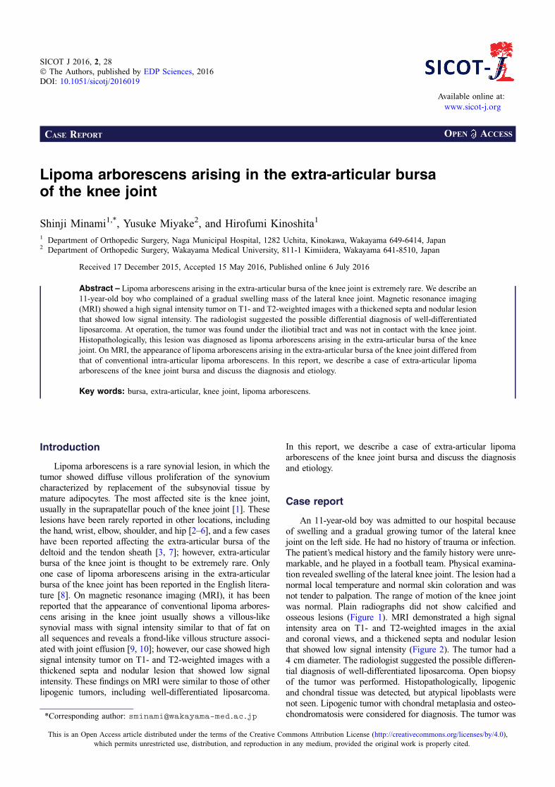
Figure 2. MRI of the tumor demonstrated a high signal intensityarea on the T1-weighted image in the axial (a) and T2-weightedimage in the axial and coronal views (b, c), and heterogeneously, alow signal intensity area was seen in the tumor. The tumor waslocated in the extra-capsular lesion of the lateral knee joint and hada diameter of 4 cm.
2 S. Minami et al.: SICOT J 2016, 2, 28

therefore, the diagnosis of this tumor was thought to be lipomaarborescens rather than synovial osteochondromatosis. On theother hand, it has been reported that lipoma arborescens onMRI showed a villous frond-like synovial mass with signalintensity similar to that of fat on all sequences [10, 13].However, these reports mainly explained the appearance ofintra-articular lipoma arborescens and it was thought that thesefindings did not correspond with extra-articular legions. In ourcase, the tumor mainly showed high signal intensity area onT1- and T2-weighted images in axial and coronal views, andshowed a low signal intensity area in the tumor that showeda thickened septa and nodular lesion. At first, the radiologistsuggested the possible differential diagnosis of other lipogenictumors, including well-differentiated liposarcoma. In cases ofwell-differentiated liposarcoma, the image of a predominantlyfatty mass with an irregularly thickened, nodular septa hasbeen reported [14], resembling our case; however, it has beenreported that well-differentiated liposarcoma occurs predomi-nantly in middle-aged patients, and atypical lipoblasts arecommonly detected. Our case showed diffuse proliferationsof fat tissue in the subsynovia and a thick fibrous septum with-out atypical lipoblasts. Coll et al. reported that the diagnosis oflipoma arborescens recognizes a diffuse synovial origin [15];therefore, our case could be distinguished from well-differentiated liposarcoma and could be diagnosed as lipomaarborescens. The etiology of lipoma arborescens remainsunknown and, as the cause of this lesion, trauma, inflammation,or neoplasm has been postulated [9, 16, 17]. A synovial reac-tion to a traumatic injury has been proposed [13] but mostpatients who have lipoma arborescens do not have a historyof trauma. In addition, it has been reported that there is anassociation between lipoma arborescens and degenerative jointdisease [18]; however, our patient was too young and degener-ative changes and meniscus tears were not observed on MRI.Jaffe suggested that lipoma arborescens represents a non-neoplastic villous synovial proliferation in response to chronicirritation of the synovium [19, 20]. Coll et al. also reported that
villous fatty proliferation likely reflects a rare synovialresponse to chronic irritation, because the lesions were recog-nized in relatively elderly patients with osteoarthritis [15]. Thisindicated that chronic irritation of the synovium could be a
(a)
(b)
(c)
Figure 4. Histopathological examination of excised tumor.(a) Synovial tissue was detected in the margin of this tumor (*).In the subsynovia, there was a thick fibrous septum and diffuseproliferation of fat tissue (Hematoxylin and eosin, · 20). (b) Diffuseproliferation of fat tissue was also detected in the tumor and atypicallipoblasts were not seen (Hematoxylin and eosin, · 50). (c) A fewchondral nodules and a thick fibrous septa were found in the fattissue (Hematoxylin and eosin, · 10).
Figure 3. Resection of the tumor. The tumor was a yellow soft masswith a thin capsule. The tumor was found under the iliotibial tractand was not in contact with the knee joint.
S. Minami et al.: SICOT J 2016, 2, 28 3

cause of lipoma arborescens. Although our patient did notshow degenerative changes of the knee joint, he played in afootball team and therefore vigorously moved his knee joint;therefore, it was thought that chronic mechanical irritation ofthe extra-articular bursa of the knee joint between the iliotibialtract and the knee joint might have occurred and it could bethought that this was the possible cause of this lesion.
Conclusion
Lipoma arborescens arising in the extra-articular bursa ofthe knee joint is extremely rare and, on MRI, the appearanceof extra-articular lipoma arborescens is different from that ofconventional intra-articular lipoma arborescens. It was consid-ered that extra-articular lipoma arborescens should bediagnosed differentially from synovial osteochondromatosisand other lipogenic tumors, including well-differentiatedliposarcoma.
Conflict of interest
SM, YM, and HK declare no conflict of interest in relationwith this paper.
References
1. Mulder JD, Schutte HE, Kroon HM, Taconis WK (1993)Radiologic Atlas of Bone Tumors. Amsterdam, ElsevierScience Publishers.
2. Laorr A, Peterfy CG, Tirman PF, Rabassa AE (1995) Lipomaarborescens of the shoulder: magnetic resonance imagingfindings. Can Assoc Radiol J 46(4), 311–313.
3. Nisolle JF, Blouard E, Baudrez V, Boutsen Y, De Cloedt P,Esselinckx W (1999) Subacromial-subdeltoid lipoma arbor-escens associated with a rotator cuff tear. Skeletal Radiol 28(5),283–285.
4. Levadoux M, Gadea J, Flandrin P, Carlos E, Aswad R, Panuel M(2000) Lipoma arborescens of the elbow: a case report. J HandSurg Am 25(3), 580–584.
5. Noel ER, Tebib JG, Dumontet C, Colson F, Carret JP, VauzelleJL, Bouvier M (1987) Synovial lipoma arborescens of the hip.Clin Rheumatol 6(1), 92–96.
6. Siva C, Brasington R, Totty W, Sotelo A, Atkinson J (2002)Synovial lipomatosis (lipoma arborescens) affecting multiple
joints in a patient with congenital short bowel syndrome.J Rheumatol 29(5), 1088–1092.
7. Dogramaci Y, Kalaci A, Sevinç TT, Atik E, Esen E, Yanat AN(2009) Lipoma arborescens of the peroneus longus andperoneus brevis tendon sheath: case report. J Am PodiatrMed Assoc 99(2), 153–156.
8. Kurihashi A, Yamaguchi T, Tamal K, Saotome K (1997)Lipoma arborescens with osteochondral metaplasia – a casemimicking synovial osteochondromatosis in a lateral kneebursa. Acta Orthop Scand 68(3), 304–306.
9. Hallel T, Lew S, Bansal M (1988) Villous lipomatousproliferation of the synovial membrane (lipoma arborescens).J Bone Joint Surg Am 70(2), 264–270.
10. Martín S, Hernández L, Romero J, Lafuente J, Poza AI, Ruiz P,Jimeno M (1998) Diagnostic imaging of lipoma arborescens.Skeletal Radiol 27(6), 325–329.
11. Kloen P, Keel SB, Chandler HP, Geiger RH, Zarins B,Rosenberg AE (1998) Lipoma arborescens of the knee. J BoneJoint Surg Br 80(2), 298–301.
12. Kataria H, Kapoor SK, Patra SR, Boruah T (2010) Lipomaarborescence of the knee in a child – a diagnostic dilemma:radiological and arthroscopic evaluation. J Orthop Sci 15(3),414–419.
13. Ryu KN, Jaovisidha S, Schweitzer M, Motta AO, Resnick D(1996) MR imaging of lipoma arborescens of the knee joint.Am J Roentgenol 167(5), 1229–1232.
14. Jelinek JS, Kransdorf MJ, Shmookler BM, Aboulafia AJ,Malawer MM (1993) Liposarcoma of the extremities: MR andCT findings in the histologic subtypes. Radiology 186(2),455–459.
15. Coll JP, Ragsdale BD, Chow B, Daughters TC (2011) Bestcases from the AFIP: lipoma arborescens of the knees in apatient with rheumatoid arthritis. Radiographics 31(2),333–337.
16. Arzimanoglu A (1957) Bilateral arborescent lipoma of theknee. J Bone Joint Surg Am 39(4), 976–979.
17. Bouraoui S, Haouet S, Mestiri H, Ennaïfar E, Chatti S, Kchir N,Zitouna MM (1996) Synovial lipoma arborescens. Ann Pathol16(2), 120–123.
18. Vilanova JC, Barceló J, Villalón M, Aldomà J, Delgado E,Zapater I (2003) MR imaging of lipoma arborescens and theassociated lesions. Skeletal Radiol 32(9), 504–509.
19. Matsumoto K, Okabe H, Ishizawa M, Hiraoka S (2001) Intra-articular lipoma of the knee joint. A case report. J Bone JointSurg Am 83(1), 101–105.
20. Jaffe HL (1958) Tumors and tumorous conditions of the bonesand joints. Philadelphia, Lea & Febiger. pp. 574–575.
Cite this article as: Minami S, Miyake Y & Kinoshita H (2016) Lipoma arborescens arising in the extra-articular bursa of the knee joint.SICOT J, 2, 28
4 S. Minami et al.: SICOT J 2016, 2, 28