lipopolysaccharide (lps)-binding protein stimulates …...tlr adaptor molecule 1 (trif)-dependent...
TRANSCRIPT
Lipopolysaccharide (LPS)-binding protein stimulates CD14-dependent Toll-like receptor 4 internalization and LPS-induced TBK1–IKKε–IRF3 axis activation Hiroki Tsukamoto1*, Shino Takeuchi1†, Kanae Kubota1†, Yohei Kobayashi1, Sao Kozakai1, Ippo Ukai1, Ayumi Shichiku1, Misaki Okubo1, Muneo Numasaki2, Yoshitomi Kanemitsu1, Yotaro Matsumoto1, Tomonori Nochi3,4, Kouichi Watanabe3,4, Hisashi Aso3,4, Yoshihisa Tomioka1* 1Laboratory of Oncology, Pharmacy Practice and Sciences, Graduate School of Pharmaceutical Sciences, Tohoku University, 6-3 Aoba, Aramaki, Sendai 980-8578, Japan 2Department of Geriatrics and Gerontology, Institute of Development, Aging and Cancer, Tohoku University, 4-1 Seiryo-machi, Sendai 980-8575, Japan 3Laboratory of Functional Morphology, 4International Education and Research Center for Food and Agricultural Immunology (CFAI), Graduate School of Agricultural Science, Tohoku University, 468-1, Aoba, Aramaki, Sendai 980-0845, Japan *Co-correspondence: Yoshihisa Tomioka, Ph.D., Professor Hiroki Tsukamoto, Ph.D., Assistant Professor Laboratory of Oncology, Pharmacy Practice and Sciences, Graduate School of Pharmaceutical Sciences, Tohoku University, 6-3 Aoba, Aramaki, Sendai 980-8578, Japan Phone: +81-22-795-6852 Fax: +81-22-795-6850 E-mail: [email protected] (YT), [email protected] (HT) †These authors contributed equally to this work. Running title: LBP mediates LPS-induced TLR4 internalization and signaling Keywords: CD14, cell surface receptor, endotoxin, innate immunity, LPS, LPS-binding protein (LBP), pathogen‐associated molecular pattern (PAMP), pattern recognition receptor (PRR), TIR‐domain‐containing adapter‐inducing interferon‐β (TRIF), Toll-like receptor 4 (TLR4), Abstract
Toll-like receptor 4 (TLR4) is an indispensable immune receptor for lipopolysaccharide (LPS), a major component of the Gram-negative bacterial cell wall. Following LPS stimulation, TLR4 transmits the signal from the cell surface and becomes internalized in an endosome. However, the spatial regulation of TLR4 signaling is not fully understood. Here, we investigated the mechanisms of LPS-induced TLR4 internalization and clarified the roles of the extracellular LPS-binding molecules, LPS-binding protein (LBP), and glycerophosphatidylinositol-anchored protein (CD14). LPS stimulation of CD14-expressing cells induced TLR4 internalization in the presence of serum, and an inhibitory anti-LBP mAb blocked its internalization. Addition of LBP to serum-free
cultures restored LPS-induced TLR4 internalization to comparable levels of serum. The secretory form of the CD14 (sCD14) induced internalization but required a much higher concentration than LBP. An inhibitory anti-sCD14 mAb was ineffective for serum-mediated internalization. LBP lacking the domain for LPS transfer to CD14 and a CD14 mutant with reduced LPS binding both attenuated TLR4 internalization. Accordingly, LBP is an essential serum molecule for TLR4 internalization, and its LPS transfer to membrane-anchored CD14 (mCD14) is a prerequisite. LBP induced the LPS-stimulated phosphorylation of TBK1, IKKε, and IRF3, leading to IFN-β expression. However, LPS-stimulated late activation of NFκB or necroptosis were not affected. Collectively, our results indicate that LBP controls LPS-induced TLR4 internalization, which induces
LBP mediates LPS-induced TLR4 internalization and signaling
1
http://www.jbc.org/cgi/doi/10.1074/jbc.M117.796631The latest version is at JBC Papers in Press. Published on May 14, 2018 as Manuscript M117.796631
by guest on February 13, 2020http://w
ww
.jbc.org/D
ownloaded from
TLR adaptor molecule 1 (TRIF)-dependent activation of the TBK1–IKKε–IRF3–IFN-β pathway. In summary, we showed that LBP-mediated LPS transfer to mCD14 is required for serum-dependent TLR4 internalization and activation of the TRIF pathway. Introduction
The innate immune system defends against invading pathogens by recognizing conserved pathogen-associated molecular patterns (PAMPs) (1,2). The induction of cytokines and chemokines by the innate immune system causes inflammation and initiates adaptive immunity (3). The recognition of PAMPs relies on pathogen recognition receptors (PRRs) on the cell surface or in intracellular compartments (1,2). Among the PRRs, Toll-like receptors (TLRs) have been intensively studied and broadly recognize pathogenic organisms (4,5). The subcellular locations where TLRs initiate signals determine the outcomes of such events (2,6,7). Therefore, TLR responses must be spatially and temporally regulated.
TLR4 is an indispensable receptor of LPS, which is a unique component of the Gram-negative bacterial cell wall (8). LPS forms a complex with MD-2, a secreted glycoprotein with an LPS binding pocket, via the extracellular domain of TLR4 (9,10). Following LPS binding via MD-2, TLR4 induces myeloid differentiation 88 (MyD88)-dependent signaling from the cell surface, which activates the NFκB/MAPK pathway (11). TLR4 is then internalized and initiates toll/IL-1 receptor (TIR)-domain-containing adapter-inducing IFN-β (TRIF)-dependent signaling and subsequent NFκB/IRF3 activation (12,13). However, the spatial regulation of TLR4 signaling is poorly understood.
Multiple LPS-binding proteins have been found in serum and cell membranes (5,14,15). Among those, CD14, a secreted and glycerophosphatidylinositol (GPI)-anchored protein, and the LPS-binding protein (LBP) are required for LPS recognition by TLR4 and MD-2 (5,16-18). The catalytic mechanism underlying LPS transfer by LBP to CD14 was recently revealed (17). LBP binds LPS micelles via the
N-terminal basic patch and forms transient ternary complexes with secreted (sCD14) or membrane-bound GPI CD14 (mCD14) on the C-terminus (17). Following the generation of CD14/LBP/LPS micelles, CD14 dissociates from LBP and receives monomeric LPS (17). The LPS transfer to mCD14 enhances the LPS sensitivity of TLR4/MD-2 in innate immune cells, including macrophages and monocytes. Thus, this is the first line of defense against bacterial invasion (19-21). Conversely, the transfer of LPS to sCD14 confers a response in CD14-deficient cells (22,23). In addition to its classical role of enhancing LPS sensitivity, CD14 expressed on the cell surface plays an essential role in the internalization of TLR4 and TRIF-mediated signaling from the endosome (13,24). In CD14-positive cells (e.g., macrophages), TLR4 is internalized in endosomes where it dissociates from MyD88 and interacts with TRIF to activate IRF3 (7,12).
LBP belongs to a LBP/bactericidal/permeability-increasing protein/palate, lung and nasal epithelial clone protein superfamily and is produced primarily in liver and epithelial cells of the lungs and gastrointestinal tract as an acute phase serum glycoprotein (25). The family members constitute physical barriers against bacterial infection in innate immunity (25,26). For example, the bactericidal/permeability-increasing proteins, which show the highest structural homology to LBP, neutralize LPS and also elicit bactericidal activity against Gram-negative bacteria (27). Among the LBP/bactericidal/permeability-increasing protein family, LBP is unique in that it binds LPS micelle and transfers a monomer to CD14, which enhances the TLR4/MD-2 response (17,25). Serum LBP increases ~10-fold during infection (28), peaking in the acute phase, which dampens the inflammatory response to LPS in some cases (29,30). LBP also interacts with other lipopeptides and enhances TLR2 responses (31,32). Thus, it could be designated a soluble PRR.
We previously showed that LBP is an essential mediator of TLR4/MD-2 dimerization in mCD14-expressing cells and responded to LPS (16). We also found that substantial LPS was bound to surface TLR4/MD-2 in mCD14-positive cells stimulated with LBP (16). Since TLR4 internalization relies on mCD14 expression (13,24),
LBP mediates LPS-induced TLR4 internalization and signaling
2
by guest on February 13, 2020http://w
ww
.jbc.org/D
ownloaded from
LBP may transfer LPS to TLR4 and transduce signals from the endosome via TLR4 internalization. Notably, LPS-induced type I IFN production was increased in monocytes stimulated with LBP (33), suggesting that LBP initiated TRIF-dependent IFN production. However, the mechanistic details for such events have not been resolved. In this study, we investigated the serum components that mediated TLR4 internalization and demonstrated an essential role for LPS transfer by LBP to mCD14, which was required for TLR4 internalization and activation of the TRIF-mediated pathway.
Results LPS-induced TLR4/MD-2 internalization is mediated by serum components
To identify the molecular mechanisms underlying LPS-induced TLR4 internalization, we examined the requirement for serum components using HEK293 cells expressing human TLR4/MD-2 and CD14. The surface expression of TLR4/MD-2 was assessed using flow cytometry in LPS-stimulated cells. In the presence of FCS and human serum, TLR4 was internalized upon LPS stimulation; however, internalization did not occur in the absence of serum, even in cells with mCD14 surface expression (Fig. 1A and B). Serum dependence was also observed in the mCD14-positive macrophage cell line, RAW264, and in bone marrow-derived macrophages (BMMs) (Fig. 1C and D). In contrast to the reduced surface expression, LPS stimulation did not change the total amount of TLR4 (Fig. 1E and F). These findings indicate that LPS-induced TLR4/MD-2 internalization was regulated by serum components. LBP is essential for LPS-induced TLR4/MD-2 internalization
Serum contains multiple LPS-binding molecules (5,14,15), including sCD14 and LBP, which are involved in LPS sensing by TLR4/MD-2 (5,16-18). We previously showed that LBP mediates mouse TLR4/MD-2 dimerization in mCD14-expressing cells (16). This was confirmed by co-immunoprecipitation (IP) assay using human TLR4/MD-2-expressing cells with or without
CD14 (Supplementary Figure 1). Additionally, we showed that LPS was bound to surface TLR4/MD-2 in the mCD14-expressing cells stimulated with LBP (16). Thus, we investigated whether LBP directly mediates the dimerization of TLR4 extracellular domain using co-IP assays. The soluble extracellular domains of human TLR4 (sTLR4F or sTLR4G), which were complexed with HA-tagged MD-2 but lacked the transmembrane (TM) and intracellular signaling domains, were FLAG- or GFP-tagged and incubated with LPS in the presence of FLAG-tagged sCD14 and LBP. sTLR4G was immunoprecipitated with an anti-GFP Ab, and sTLR4F was detected by Western blotting (WB) using an anti-FLAG mAb. sTLR4F co-precipitated with sTLR4G following LPS incubation with LBP and sCD14 (Fig. 2). Conversely, incubation of LPS with sCD14 or LBP alone did not result in co-precipitation. Dimerization also failed to occur in the absence of LPS. These findings indicate that LBP mediated LPS-induced TLR4 dimerization via the extracellular domain in a CD14-dependent manner. Thus, we hypothesized that LBP is an essential serum component that mediated TLR4 internalization following receptor dimerization.
To test this hypothesis, HEK293 cells expressing TLR4/MD-2/CD14 were stimulated in serum-free medium with recombinant human LBP prepared from CHO cells. LBP restored LPS-induced TLR4 internalization to levels comparable to those seen in the presence of complete serum (Fig. 3A and B). Internalization was also observed when BMM and RAW264 cells were stimulated with exogenous LBP (Fig. 3C and D) without apparent changes in the total amount of TLR4 (Fig. 3E and F). To confirm the internalization of surface TLR4, we also performed pulse-chase experiments with anti-TLR4 mAb in HEK293 cells expressing TLR4/MD-2/CD14. Following LPS stimulation with LBP, immunofluorescence of mAb-labeled surface TLR4 was detected as punctate foci in cytoplasmic regions (Supplementary Figure 2). Some internalized TLR4 was co-localized with the early endosome marker, EEA1, but not with the plasma membrane marker, wheat germ agglutinin (WGA), at 60 min of LPS stimulation (Fig. 3G and H). On the other hand, most cell surface TLR4 in unstimulated cells was co-localized with WGA but
LBP mediates LPS-induced TLR4 internalization and signaling
3
by guest on February 13, 2020http://w
ww
.jbc.org/D
ownloaded from
not EEA1. Cytoplasmic localization was rarely observed when stimulated in the absence of LBP (Supplementary Figure 2). Based on these results, TLR4 is internalized from the cell surface in an LBP-dependent manner.
The addition of sCD14 was also investigated since this molecule binds and responds to LPS in CD14-deficient cells (22,23). Exogenous sCD14 restored TLR4 internalization, albeit in a delayed manner compared to LBP (Fig. 3A). The concentrations of sCD14 and LBP required for TLR4 internalization were also evaluated to identify the most potent molecule. sCD14 required 100–1000 ng/ml to cause internalization, while 1–10 ng/ml of LBP was required (Fig. 3B).
The contribution of LBP from human serum to TLR4 internalization was also evaluated. Notably, TLR4 internalization was inhibited in HEK293 cells incubated with human serum that had been pre-treated with LBP-blocking mAb (Fig. 4A). A similar finding was also observed in BMMs, albeit to a less extent (Fig. 4B). The addition of human CD14-blocking mAb that inhibits LPS binding to human sCD14 in serum, but not that of mouse mCD14 on the surface of BMMs, was ineffective (Fig. 4B). These findings suggest that LBP is the serum component mediating LPS-induced TLR4 internalization, while the contribution of sCD14 is minimal. LPS transfer from LBP to mCD14 is required for TLR4/MD-2 internalization
Reconstitution assays were performed to determine the interaction between LBP and CD14. HEK293 cells were stimulated with a truncated version of LBP that lacked the C-terminal half (LBP-N). This version, which retains LPS binding, but not its transfer to CD14 (10,16,34), failed to internalize TLR4. Conversely, full-length LBP caused TLR4 internalization (Fig. 5A). TLR4 internalization was also investigated in CD14-negative HEK293 cells to determine whether LBP alone was sufficient for TLR4 internalization, or if sCD14 was required. The exogenous addition of neither LBP nor sCD14 was sufficient for internalization in CD14-negative cells, while their combined addition was marginally effective (Fig. 5B). This finding was consistent with the finding that serum containing bovine LBP and sCD14 was also ineffective (Fig. 5C).
A D10K mutation in the LPS-binding pocket of CD14 that introduced electrostatic repulsion decreased LPS binding (Fig. 5D and (22)). Cells transfected with the D10K mutant exhibited attenuated TLR4 internalization in the presence of LBP (Fig. 5E). These findings suggest that LBP requires CD14 for LPS binding to mediate TLR4 internalization. Moreover, internalization was conferred by mCD14, but not sCD14. Thus, LPS transfer from LBP to mCD14 is required for TLR4 internalization.
To determine whether the GPI moiety of CD14 was required for LBP-mediated TLR4 internalization, HEK293 cells were generated to express CD14 in which the GPI moiety was replaced with a TM domain. Cells stimulated with LPS in the presence of LBP exhibited the same rates of internalization as those expressing WT CD14, suggesting that GPI-anchoring is not required for TLR4 internalization (Fig. 5F and 5G).
LBP also binds lipoproteins derived from Gram-positive bacteria and induces TLR2 responses (31,32). TLR2 induces type I IFN and pro-inflammatory cytokine production (35); therefore, the role of serum and LBP on TLR2 internalization was also evaluated. Flow cytometry revealed that BMMs stimulated with the synthetic TLR2 agonist, Pam2CSK4, resulted in TLR2 internalization independently of serum and LBP (Fig. 6). These findings suggest that LBP selectively mediated LPS-stimulated TLR4 internalization. LBP mediates TBK1/IKKε/IRF3 phosphorylation, but not late-phase activation of NFκB or necroptosis
LPS-induced IFN-β secretion requires IRF3 activation, which is induced by TRIF from the endosomes in which TLR4 is internalized (12,13,36). MyD88 and TRIF activate NFκB sequentially in the LPS-induced TLR4 signaling pathway (1,37): MyD88 rapidly transmits the NFκB signal from plasma membrane and TRIF then induces the signal in a delayed fashion (1,7,11-13). Since LBP mediates TLR4 internalization, the impact of LBP on the activation of the TRIF pathway was evaluated. Phosphorylation of TBK1, IKKε, and IRF3 was evaluated in BMM and RAW264 cells treated with LBP. Notably, phosphorylation of TBK1, IKKε,
LBP mediates LPS-induced TLR4 internalization and signaling
4
by guest on February 13, 2020http://w
ww
.jbc.org/D
ownloaded from
and IRF3 occurred following 30–120 min of LPS stimulation with LBP (Fig. 7A and Supplementary Figure 3). Conversely, TBK1 and IKKε were only weakly phosphorylated in the absence of LBP, while phospho-IRF3 was undetectable. Consistent with these findings, LPS stimulation significantly induced IFN-β expression only in the presence of LBP (Fig. 7B). In contrast, MyD88-dependent TNF-α and IL-6 expression were not significantly affected but were slightly delayed in the absence of LBP (Fig. 7B). The role of LBP in late-phase activation of NFκB was also assessed. In contrast to the early phosphorylation (5-15 min) observed for IκB when stimulated with LBP, late-phase phosphorylation (60-120 min) was independent of LBP (Fig. 7C and Supplementary Figure 3). Additionally, LPS slightly induced NFκB activation in the absence of LBP, as assessed using a reporter assay with TLR4/MD-2/CD14-expressing Ba/F3 cells, although the response was attenuated compared to the response in the presence of LBP (Fig. 7D). To gain further insights into LBP-mediated TLR4 signaling, we also investigated the impact of LBP on the phosphorylation of MAPKs (e.g. ERK, p38, and JNK) in LPS-stimulated BMMs (Fig. 7E). ERK and p38 were weakly phosphorylated in the absence of LBP and significantly augmented by the addition of LBP. The phosphorylation of JNK was dependent on the presence of LBP.
TRIF was involved in the necroptosis of LPS-stimulated macrophages when caspase-8 was inhibited, while the same was not true for MyD88 (38). Therefore, the role of LBP in cell death was investigated in BMMs and resident peritoneal macrophages. Cytotoxicity and cell viability were assayed by lactate dehydrogenase (LDH) release and intracellular ATP content in cells stimulated with LPS when LBP was or was not added to culture. Cell death was induced by LPS stimulation with the pan-caspase inhibitor zVAD (Fig. 7F and G). However, LBP was not required for LDH release or ATP content. Consistent with these results, the phosphorylation of RIP1 was induced independent of LBP (Fig. 7C). Based on these results, TRIF-mediated necroptosis occurred independently of LBP.
LBP mediates Myddosome and Triffosome assembly
To explore the mechanism underlying LBP-mediated TLR4 signaling, we investigated the assembly of Myddosome and Triffosome in stable transfected cells expressing either MyD88-GFP or TRIF-GFP. Co-IP assays using an anti-GFP mAb in LPS-stimulated TLR4/MD-2/CD14-expressing HEK293 cells showed the LBP-dependent association of MyD88 with endogenous modified IRAK1 with a higher electrophoretic mobility (Fig. 8A). In addition, TRIF-TRAF3, TBK1 and TRAF6 association were also detectable when TLR4/MD-2/CD14- and TRIF-GFP-expressing Ba/F3 cells were stimulated with LBP (Fig. 8B). Only TRAF6 was associated with TRIF to a lesser extent in the absence of LBP. These results suggest that LBP efficiently mediates Myddosome and Triffosome assembly.
To gain further mechanistic insights underpinning how LBP regulates TLR4 signaling, we employed confocal microscopy to investigate the dissociation of surface TLR4 from the plasma membrane-anchored TIR domain containing adaptor protein (TIRAP) fused to GFP using pulse-chase experiments with TLR4/MD-2/CD14- and TIRAP-expressing HEK293 cells (Supplementary Figure 4A). Although, irrespective of LBP, labeled-surface TLR4 co-localized with TIRAP-GFP on several plasma membranes in resting and stimulated cells, TLR4 was also observed in intracellular compartments in LPS-stimulated cells with LBP. To confirm these results, we further investigated the association of TLR4 with TIRAP-GFP using co-IP (Supplementary Figure 4B). Consistent with the microscopic observation, TIRAP was constitutively associated with TLR4 independently of LBP. These results suggest that LBP did not promote the dissociation of TLR4 from the plasma membrane of TIRAP in LPS-stimulated cells. LPS-induced signal from TLR4 enhances LBP-mediated internalization
LPS triggers LBP-mediated TLR4 internalization; therefore, we investigated whether a specific TLR4 signal was required for internalization. C3H/HeJ-derived BMMs harboring a point mutation in the TIR signaling domain of TLR4 that rendered it unresponsive to LPS (39) failed to induce a signal, even if LPS was transferred from LBP and mCD14 to the TLR4
LBP mediates LPS-induced TLR4 internalization and signaling
5
by guest on February 13, 2020http://w
ww
.jbc.org/D
ownloaded from
extracellular domain. Internalization of TLR4 in the mutated BMMs was attenuated compared to that of the WT C3H/HeN-derived BMMs during the course of stimulation and at multiple concentrations of LPS (Fig. 9A and B). These findings indicate that the TIR-mediated signal from TLR4 enhances LBP-mediated internalization, but is not essential.
LPS-induced TLR4 internalization was reportedly mediated by GTPase dynamin, which is involved in most endocytic processes (12,40). Therefore, we investigated whether dynamin is also involved in the process of LBP-mediated TLR4 internalization. The pan-dynamin inhibitor dynasore significantly inhibited LBP-mediated TLR4 internalization in LPS-stimulated BMMs at 30 min but did so only modestly at 4 h compared to vehicle treatment (Fig. 9C and Supplementary Figure 5). Consistent with this result, dynasore modestly inhibited or delayed the phosphorylation of IRF3 (Fig. 9D). These findings indicated LBP-mediated TLR4 internalization and IRF3 signaling were, at least in part, dynamin-dependent processes. Discussion
This study revealed that LPS-induced TLR4 internalization required serum and mCD14. LBP mediated LPS-induced TLR4/MD-2 internalization, while recombinant LBP caused LPS-induced internalization in serum-free culture, provided that CD14 was expressed on the surface. Thus, interaction between LBP and mCD14, which transfers LPS to surface TLR4/MD-2, was required for internalization. The addition of LBP-N lacking the LPS transfer domain or the expression of mCD14 with decreased LPS binding attenuated internalization. Our present and previous studies showed that, when stimulated with LBP, TLR4 binds substantial amounts of LPS via MD-2 and induces the dimerization of TLR4 in mCD14-positive cells (16). Thus, TLR4/MD-2 dimers may form extensively in the presence of LBP and be a prerequisite for TLR4 internalization.
Similar to the case with TM receptors, TLR4 internalization is under the endocytic control of dynamin, clathrin, and their associated proteins (12,13,41). However, surface-exposed LPS-binding molecules, such as mCD14 and MD-2,
regulate LPS-induced TLR4 internalization (13,24). Extracellular interactions between CD14 and MD-2 with LPS initiate internalization (13,24). Although LBP also binds lipopeptides other than LPS (31,32), we showed that TLR2 internalization was independent of serum and LBP. Collectively, these findings suggest that the extracellular interaction between LBP with LPS is a selective mechanism for TLR4 internalization. Additionally, we showed that LBP-mediated TLR4 internalization depends, at least in part, on the dynamin-mediated process because treatment with dynasore significantly inhibited the internalization. However, the degree of inhibition was marginal at 4 h after stimulation, although it was more effective at 30 min. Consistent with our results, efficient but not complete inhibition was reported in a previous study in which internalization was assessed at 30 min by flow cytometry using BMMs (12). Significant but partial inhibition by dynasore at 4 h may be due to incomplete inhibition of dynamin. Therefore, we increased the concentration of dynasore up to 160 μM; however, this concentration was insoluble, and crystal deposits formed in the culture. In addition, the dynasore appeared toxic to the cells at this concentration (data not shown). Previous studies showed the involvement of dynamin in TLR4 internalization (12,40), and the dynamin-dependent process is a crucial part of the LBP-mediated TLR4 internalization mechanism; however, it is difficult to completely exclude the possibility of a dynamin-independent process.
As reported previously (13,24), membrane expression of CD14 was required for TLR4 internalization. A TLR4-independent signal is responsible for internalization in response to LPS (7,13), which may be transduced by CD14 via a lipid raft. Such a mechanism is unlikely, however, since the TM-type CD14 caused similar levels of internalization as the GPI-anchored CD14. Unexpectedly, C3H/HeJ BMMs had reduced TLR4 internalization compared to the C3H/HeN strain, which was inconsistent with previous findings indicating that the MyD88/TRIF-mediated TLR4 signal does not participate in internalization (13). Thus, it may be necessary to test the hypothesis that a TIR-mediated TLR4 signal contributes to LBP-mediated internalization in a MyD88- and TRIF-independent manner. It is also possible that
LBP mediates LPS-induced TLR4 internalization and signaling
6
by guest on February 13, 2020http://w
ww
.jbc.org/D
ownloaded from
the LPS-induced TLR4 signal enhances internalization through a CD14-mediated TLR4-independent signal (13).
In addition to LBP, exogenous sCD14 also facilitated TLR4 internalization. However, the concentration required was higher than that for LBP. An LBP blocking mAb decreased serum-mediated internalization in mCD14-positive HEK293 cells, while a CD14-blocking mAb failed to inhibit internalization in BMMs. These findings indicate that LBP is the primary serum component responsible for LPS-induced TLR4 internalization, while sCD14 plays little or no role.
We previously showed that sCD14 does not compensate for LBP in LPS binding or TLR4/MD-2 dimerization (16). This distinct function may be attributable to the binding and catalytic properties of LBP and CD14 for LPS. LBP binds to LPS micelles and repeats monomeric LPS transfer to CD14 multiple times (17). Conversely, sCD14 is not accessible to micelles, and directly binds LPS monomers via LBP. Thus, LBP may load LPS on TLR4 and induce a large cluster of TLR4 oligomers on the cell surface, triggering its internalization. In pulse-chase experiments, relatively large blots, presumably due to the accumulation of Ab-bound surface TLR4 oligomers, were observed in intracellular compartments, whereas distribution of TLR4 was dispersed on the cell surface in stimulated cells without LBP.
This study showed that LBP-mediated internalization leads to the phosphorylation of TBK1, IKKε, and IRF3. This pathway is also inducible by the TRIF-mediated signal from endosomes (42,43). Additionally, IRF3 dimerized upon LPS stimulation in LBP- and serum-dependent manners, which resulted in enhanced IFN-β production (33). Consistent with this finding, the induction of IFN-β mRNA occurs upon LPS stimulation with LBP in BMMs. In addition to the essential role of LBP on TBK1/IKKε/IRF3-mediated IFN-β production due to TLR4 internalization, we also revealed that signaling varied with each TRIF-mediated event. LBP did not impact late-phase NFκB activation or necroptosis, and phosphorylation of RIP1 was mostly independent of LBP. TRIF plays an essential role through an IRF3-independent mechanism for both processes (38). TRIF controls
signal transduction from the endosomes where TLR4 is internalized (1,7,12,13). The signals for delayed NFκB activation and the RIPK-mediated necroptosis may emanate from the cell surface or other intracellular organelles.
We showed the induction of the MyD88-dependent cytokines, TNF-α and IL-6, was not significantly impaired in the absence of LBP (i.e., TLR4 was not internalized). LPS-induced expression of TNF-α and IL-6 was reportedly mediated by a TRIF signal (37) as well as by a MyD88 signal (44). Therefore, based on our results, TRIF may transduce certain types of signals from the plasma membrane unless TLR4 is internalized into the endosome. In fact, we detected the TRIF-TRAF6 association in stimulated cells in the absence of LBP, although it was weaker than that in the presence of LBP. Phosphorylation of ERK and p38 MAPKs is augmented by LBP as TBK1 and IKKε. JNK was phosphorylated only when stimulated with LBP. LPS-induced phosphorylation of MAPKs is temporally regulated in the early phase by a MyD88 signal, and it is regulated in the late phase by a TRIF signal (1,37,44). Therefore, the potentiation of MAPK activation by LBP can be explained by several possible mechanisms. An intracellular TRIF signal could be initiated from TLR4 internalized by LBP, as suggested by the LBP-dependent association of TRIF with TRAF3 and TBK1, which constitutes a critical signaling complex for IRF3-dependent IFN-β production. Alternatively, the sensitivity and intensity of TLR4 signaling by the catalytic transfer of LPS to CD14 (and then TLR4/MD-2) by LBP (17) is possibly increased, which should enhance both MyD88- and TRIF-dependent pathways. As shown in our results, MyD88-dependent signaling was augmented; the association of IRAK1 with MyD88, an upstream event of the MyD88-dependent pathway, was detected when stimulated with LBP. Alternatively, a MyD88 signal could be induced from intracellular compartments in which TLR4 is internalized via the association with TIRAP, as described below. A comprehensive understanding of the spatiotemporal regulation of TLR4 signaling that includes LBP-regulated signaling will require further studies to identify the location of signal induction from TLR4 as well as to elucidate the dependence of adaptor proteins. To the best of our knowledge, the subcellular location for signal
LBP mediates LPS-induced TLR4 internalization and signaling
7
by guest on February 13, 2020http://w
ww
.jbc.org/D
ownloaded from
induction leading to late-phase activation of NFκB and MAPKs remains unidentified.
In pulse-chase experiments, we showed that, irrespective of LBP, TIRAP co-localized with surface TLR4 on the plasma membrane in resting and stimulated cells but did not dissociate TLR4 from TIRAP. These findings, biochemically confirmed by co-IP assays, are plausible because TIRAP acts as a sorting adaptor for MyD88 to induce the signal from the plasma membrane (11). Notably, TIRAP co-localized with internalized TLR4 in the intracellular compartment in response to LPS stimulation only in the presence of LBP. Recently, TIRAP was demonstrated to mediate MyD88 signaling from endosomal TLRs in addition to cell-surface TLRs (e.g., TLR4) (45). Therefore, internalized TLR4 may have induced the MyD88 signal that led to the activation of MAPKs.
We confirmed that internalized TLR4 was not co-localized on the plasma membrane in LBP-stimulated cells, and that some are located on EEA1-positive endosomes. However, we unexpectedly found that internalized TLR4 was not always positive for EEA1. From this finding, we speculate that the subcellular compartments of internalized TLR4 may be heterogeneous and contribute to different signaling events.
In conclusion, we demonstrated that LBP mediates TLR4 internalization by transferring LPS to mCD14, which activates the TBK1/IKKε/IRF3 pathway via a Triffosome assembly. In addition to the unspecific intracellular regulation, the internalization of TLR4 is uniquely regulated by extracellular machinery composed of LBP, CD14, and MD-2. These findings detail a mechanism by which TLR4 induces divergent signals from spatially different subcellular locations. Experimental procedures Materials
LPS from E. coli O111 and normal goat serum were obtained from Wako Pure Chemical Industries (Osaka, Japan). FITC-conjugated LPS from E. coli O111:B4 was obtained from Sigma (St. Louis, MO). Pam2CSK4 was from InvivoGen (San Diego, CA). Dynasore and zVAD-FMK were from AdooQ® Biosciences (Irvine, CA).
CF®488A-conjugated WGA was from Biotium Inc. (Fremont, CA). Mouse anti-human TLR4 (HT59) (46), mouse TLR4 (UT49) (47), rat anti-human CD14 inhibitory mAb (1B12) (48), and biotinylated mAbs were prepared as previously described. Other Abs were purchased from the following companies: mouse anti-FLAG M2 mAb (Sigma); rabbit anti-GFP Ab, anti-EEA1 Ab, mouse anti-HA (TANA2) mAb (MBL, Nagoya, Japan); mouse anti-human LBP mAb (biG412) (Biometec GmbH, Greifswald, Germany); mouse anti-IκBα (L35A4), anti-phospho-IκBα (Ser32/36) (5A5), rabbit anti-phospho-TBK1 (Ser172) (D52C2), anti-phospho-IKKε (Ser172) (D1B7), anti-IRF3 (D83B9), anti-phospho-IRF3 (Ser396) (4D4G), anti-IRAK1 (D51G7), anti-TLR4 (D8L5W) mAb, anti-phospho-p44/42 MAPK (Erk1/2) (Thr202/Tyr204), anti-phospho-p38 MAPK (Thr180/Tyr182), anti-phospho-SAPK/JNK (Thr183/Tyr185), anti-TRAF3 Ab, and HRP-conjugated goat anti-rabbit IgG Ab (Cell Signaling Technology, Danvers, MA); mouse anti-RIP mAb (38/RIP) (BD Biosciences, San Jose, CA); HRP-conjugated goat anti-mouse IgG Ab (Jackson ImmunoResearch Laboratories, West Grove, PA); mouse anti-human/mouse TLR2 (T2.5) mAb, allophycocyanin- and phycoerythrin (PE)-conjugated goat anti-mouse IgG, anti-rat IgG, PE- and HRP-conjugated streptavidin (stv) (BioLegend, San Diego, CA); and Alexa546-conjugated F(ab’)2 goat anti-mouse IgG (H+L) (Invitrogen, Carlsbad, CA); rabbit anti-NAK/TBK1 (EP611Y), anti-TRAF6 (EP591Y) mAb, Alexa488-conjugated preadsorbed goat anti-rabbit IgG H&L (Abcam, Cambridge, UK). Recombinant sCD14, LBP, and LBP-N tagged with FLAG sequences at the C-terminus were prepared as previously described (10,16). Rat anti-GFP mAb (GFP2) was established at Saga University Faculty of Medicine. In brief, two Wistar rats were immunized in the foot pads with EGFP (100 μg/head) (16) emulsified in CFA in a total volume of 1.5 ml. One week later, cells were harvested from the draining lymph nodes of the immunized rats and fused with SP2/O myeloma cells in polyethylene glycol 1500. After HAT selection, positive hybridoma clones were screened using immunofluorescent intracellular staining against Ba/F3 cells expressing mouse
LBP mediates LPS-induced TLR4 internalization and signaling
8
by guest on February 13, 2020http://w
ww
.jbc.org/D
ownloaded from
TLR4G/TLR4F/MD-2F/CD14 (16), and single clones were isolated by limiting dilution. Purified mAbs were obtained as previously described (46). Cells
Mouse macrophage RAW264 cells (RCB0535) and the human embryonic kidney cell line, HEK293 (CRL-1573), were purchased from Riken Cell Bank (Tsukuba, Japan) and American Type Culture Collection (Rockville, MD), respectively, and maintained in DMEM supplemented with 10% FCS. CHO-DG44 cells were provided by Dr. Fukudome (Saga University, Saga, Japan) and maintained in DMEM:Nutrient Mixture F-12 containing 5% FCS. HEK293-transfected cells stably expressing human TLR4 and FLAG-tagged MD-2 were previously generated (46). Cells were transfected with pEFBOS constructs expressing WT (10), D10K-mutated, or TM-type human CD14 with pBABEpuro. Selection was performed using puromycin. A series of human TLR4/MD-2/CD14-expressing stable HEK293 cells were generated. HEK293-transfected cells stably expressing mouse TLR4 and MD-2 were generated by co-transfection of pEFBOS constructs expressing C-terminally FLAG/6×His-tagged TLR4 (a kind gift from K. Miyake, the University of Tokyo) and pCAGGS1 constructs expressing C-terminally FLAG/6×His-tagged MD-2 followed by subsequent selection with G418. Additional co-transfection of a pEFBOS construct expressing mouse CD14 with pBABEpuro and selection with puromycin generated the HEK293 cells expressing TLR4/MD-2/CD14. A pcDNA3.1zeo(-) (Invitrogen) construct expressing human MyD88 fused with EGFP at the C-terminus (MyD88-GFP) were further transfected and then selected with zeocin to generate TLR4/MD-2/CD14- and MyD88-GFP-expressing HEK293 cells. Unless otherwise noted, lipofectamine® 2000 reagent (Invitrogen) was used for transfections in HEK293 cells. All HEK293 transfected cells were maintained as parental cells. BMM and peritoneal resident macrophages were prepared from C57BL/6N (Japan SLC, Hamamatsu, Japan), C3H/HeN, or C3H/HeJ mice (CLEA Japan, Tokyo, Japan), as previously described (48). Ba/F3-transfected cells expressing human TLR4/MD-2/CD14 and TRIF fused with EGFP at
the C-terminus (TRIF-GFP) were generated as follows: The Ba/F3-transfected cells stably carrying FLAG-tagged TLR4/MD-2 and NFκB reporter gene (46) were co-transfected with a pEFBOS construct expressing TRIF-GFP with a pBABEpuro by electroporation with GenePulser (Bio-Rad Laboratories, Hercules, CA). After puromycin selection, a pcDNA3.1/Hygro(+) construct expressing human CD14 was further transfected, and stably transfected clones were obtained by hygromycin selection. Construction of expression vectors
The CD14 D10K mutant vector was generated by site-directed mutagenesis of the pEFBOS construct carrying the WT CD14 ORF (48). Primers were as follows: 5′-CTTGTGAGCTGGACaagGAAGATTTCCGCTGCGTC-3′ and 5′-GACGCAGCGGAAATCTTCcttGTCCAGCTCACAAG-3′. A pEFBOS vector expressing human CD14 with the GPI anchor replaced with the TM domain was generated as follows: The TM domain (aa 218–244) containing a stop codon (49) and BamHI (5′) and NotI (3′) restriction sites was synthesized by FASMAC (Atsugi, Japan) and subcloned into a pEFBOS construct (16) expressing human sCD14 tagged with FLAG sequences at the C-terminus. The resulting construct expressed the signal peptide and amino acids 1–325, which are present in the GPI-anchored form of CD14, and is fused to the TM domain of the tissue factor.
The pCAGGS1 construct expressing C-terminally FLAG/6×His-tagged mouse MD-2 was generated by subcloning from the pEFBOS construct (a kind gift from K. Miyake) at XhoI and NotI sites. The pEFBOS construct expressing mouse CD14 was generated by subcloning the SalI- and BamHI-digested ORF fragment of the EST clone (Open Biosystems, Clone ID 4981093) into a XhoI- and BamHI-digested pEFBOS vector. A cDNA fragment coding for human MyD88 was amplified using PCR with a primer set (5′-agggtcgacaggaagcgctggcagacaATGCGACC-3′ and 5′- cagggatccGGGCAGGGACAAGGCCTTGGCAAG-3′). Amplified fragments were digested with SalI and BamHI and subcloned into an XhoI- and BamHI-digested pEFBOS vector with
LBP mediates LPS-induced TLR4 internalization and signaling
9
by guest on February 13, 2020http://w
ww
.jbc.org/D
ownloaded from
modifications to introduce a C-terminal EGFP tag. The MyD88-GFP fragment was further subcloned into pcDNA3.1zeo(-) vector at XbaI and NotI sites.
The pEFBOS construct expressing human TRIF-GFP was generated as follows: The TRIF ORF was amplified using PCR with a primer set (5′-ctcctcgagATGGCCTGCACAGGCCCATCACTTCC-3′ and 5′-acgggtaccTTCTGCCTCCTGCGTCTTGTCCTCGG-3′). Amplified fragments were digested with XhoI and KpnI and subcloned into a pEFBOS vector with modifications to introduce a C-terminal EGFP tag. Four crucial amino acids (VQLG) in the RHIM domain of TRIF were mutated to alanines by site-directed mutagenesis using a primer set (5′-CCACCACGCACAGATGGcAgcGgcGGcaCTGAACAACCACATGTGG-3′ and 5′-CCACATGTGGTTGTTCAGtgCCgcCgcTgCCATCTGTGCGTGGTGG-3′).
The pcDNA3.1/Hygro(+) (Invitrogen) construct expressing human CD14 was generated as follows: full-length human CD14 was subcloned from an EST clone (Open Biosystems, Clone ID 4149008) into pBluescript II-KS(+) at EcoRI and NotI sites (48). This insert was further subcloned into a pcDNA3.1/Hygro(+) vector at HindIII and NotI sites. Preparation of sTLR4/MD-2 complexed protein
Expression constructs for sTLR4 tagged at the C-terminus containing FLAG/6×His or EGFP/6×His tandem tags were generated as follows: A fragment of sTLR4 was subcloned into a pEFBOS vector, which was modified to introduce a FLAG/6×His tandem tag at the XhoI and BamHI sites at the C-terminus. An EGFP/6×His tandem tag was amplified from a pEGFP-N1 (Clontech, Palo Alto, CA) vector by PCR with a primer set (5′-gtcggatccATGGTGAGCAAGGGCGA-3′ and 5′-ggggcggccgctcaGTGGTGGTGGTGGTGGTGCTTGTACAGCTCGTCCATGCCGAGAGTG-3′), and subcloned into the pEFBOS vector described above. The FLAG/6×His- or EGFP/6×His-tagged sTLR4 sequences were subcloned from modified pEFBOS constructs into a pCAGGS1 vector (sTLR4FH/pCAGID and sTLR4GH/pCAGID), which was also modified by introducing IRES and DsRed-Express sequences, at XhoI and NotI sites. A cDNA fragment coding for human MD-2 was amplified by PCR with a sense primer
(5′-ttctcaagcctcagacagtgg-3′), and an anti-sense primer (5′-aatagatctATTTGAATTAGGTTGGTGTAGG-3′) (46). Amplified fragments were digested with XhoI and BglII and subcloned into an XhoI- and BamHI-digested pEFBOS vector with modifications to introduce a C-terminal HA tag (MD-2HA/pEFBOS). CHO-DG44 cells were transfected with sTLR4FH/pCAGID or sTLR4GH/pCAGID expression constructs using a Lipofectamine® 2000 reagent. Stable clones secreting sTLR4F and sTLR4G were established by G418 selection. Additional co-transfection of MD-2HA/pEFBOS and pBABEpuro was performed in stable clones and selected with puromycin. CHO clones producing sTLR4F/MD-2 and sTLR4G/MD-2 were cultured for two to three weeks in serum-free medium. The conditioned medium was collected and dialyzed in PBS, and proteins were purified by affinity chromatography using a HisTrap HP column (GE Healthcare, Buckinghamshire, UK). The eluted proteins were dialyzed in PBS and passed through a 0.22-μm sterile Millex-Gv filter (Millipore, Billerica, MA). Detection of sTLR4 dimerization
sTLR4 dimerization was detected by co-IP using FLAG and GFP Abs. Following 30 min of incubation of sTLR4F/MD-2 and sTLR4G/MD-2 (10 μg/ml each) with LPS (5 μg/ml) in 100 μl of PBS, a rabbit anti-GFP Ab-immobilized Affigel10 (25 μl) (16) was incubated for 1.5 h with gentle agitation, washed three times with 20 mM Tris buffer, pH 7.5, 150 mM NaCl, 1 mM EDTA, and 0.1% Triton X-100 and boiled in Laemmli buffer. Co-precipitated TLR4F and TLR4G were detected by WB using anti-FLAG and GFP Ab followed by HRP-conjugated secondary Abs. Cell stimulation for internalization and signal analysis
BMM (5–6 × 10⁵ cells), RAW264 (2.5–5 × 10⁵ cells), or HEK293-transfected cells (6 × 10⁵ cells) were inoculated in 24-well plates, cultured overnight in 1 ml of the serum-free medium, and stimulated with LPS in the presence or absence of FCS (5%), human serum (1%), LBP (100 ng/ml), and sCD14 (100 ng/ml), except where otherwise noted. To analyze internalization, cells were
LBP mediates LPS-induced TLR4 internalization and signaling
10
by guest on February 13, 2020http://w
ww
.jbc.org/D
ownloaded from
washed twice, collected in 1 mM EDTA containing PBS, and subjected to flow cytometry. For WB, cells were washed once with ice-cold PBS and lysed in 20 mM Tris buffer (pH 7.5) containing 150 mM NaCl, 1 mM EDTA, 1% Triton X-100, 1% protease inhibitor and phosphatase cocktails (Nacalai Tesque, Kyoto, Japan) for 10 min on ice. Following centrifugation at 15,000 × g for 15 min at 4°C, supernatants were subjected to WB.
RAW264 (5 × 10⁵ cells) or TLR4/MD-2/CD14/MyD88-GFP-expressing HEK293-transfected cells (1 × 10⁶ cells) were inoculated in 12-well plates, cultured overnight in 1 ml of the serum-free medium, and stimulated with LPS in the presence or absence of FCS (5%) or LBP (100 ng/ml). For IP, cell lysates were prepared as above and subjected to IP with anti-TLR4 mAb (UT49, 5 μg) conjugated with Protein G Sepharose® 4 Fast Flow (15 μl; GE Healthcare, Piscataway, NJ) or rat anti-GFP mAb (GFP2) covalently immobilized to AffiGel10 (15 μl, Bio-Rad Laboratories) as previously described (50). Following 1 h of incubation with gentle rotation, the protein-bound beads were washed three times with 20 mM Tris (pH 8.0), 150 mM NaCl, 1 mM EDTA, and 0.1% Triton X-100, and the bound proteins were eluted by boiling in Laemmli buffer.
TLR4/MD-2/CD14/TRIF-GFP-expressing Ba/F3-transfected cells (1 × 10⁷ cells) were suspended in serum-free medium and stimulated with LPS in the presence or absence of LBP (100 ng/ml). Cell lysates were subjected to IP with rat anti-GFP mAb (GFP2) immobilized to AffiGel10 as described above. Flow cytometry
Cells were stained on ice with primary (5 μg/ml) or biotinylated Ab (2 μg/ml) in staining buffer (PBS containing 2% FCS, 10 mM EDTA, and 100 U/ml penicillin), washed three times with staining buffer, and incubated with a PE-conjugated secondary Ab or stv. Flow cytometry was performed using a FACS caliber (Becton Dickinson, Franklin Lakes, NJ) or CytoFLEX (Beckman Coulter Life Sciences, Brea, CA). Data were analyzed using the WinMDI program (J. Trotter, Scripps Research Institute, La Jolla, CA) or FlowJo (Tree Star, Ashland, OR). For the detection of LPS, cells were incubated with FITC-conjugated LPS (20 μg/ml) for 20 min, washed, and analyzed as
described above. WB
Proteins were resolved on SDS-PAGE and transferred onto an Immobilon-P membrane (Millipore Co., Bedford, MA). After blocking with 1% skim milk or 1% BSA in 20 mM Tris buffer containing 150 mM NaCl and 0.05% Tween 20, blotted membranes were probed with primary mAbs and HRP-conjugated secondary Ab or stv. For phospho-TBK1, phospho-IKKε, IRF3, phospho-IRF3, phospho-p38, phospho-ERK1/2, phospho-SAPK/JNK, RIP, TBK1, TRAF3, TRAF6 and IRAK1, primary Abs were diluted in Can Get Signal® solution (Toyobo, Osaka, Japan). The immunoreactive protein was visualized by X-ray film (Fuji Film, Tokyo, Japan) using Chemi-Lumi One L or Super chemiluminescence kits (Nacalai Tesque). The Image J 1.51j8 software (NIH, Bethesda, MD) was used for quantification of immunoreactive bands. Cell cytotoxicity and viability assay
BMMs (1 × 10⁵ cells) were inoculated in a 96-well plate, cultured overnight in 100 μl of the serum-free medium, and then stimulated with LPS in the presence of 20–40 μM of z-VAD-FMK or DMSO (0.1%). Resident peritoneal macrophages and BMMs were stimulated without overnight incubation. Following 24 h of stimulation, LDH release to the culture medium was assayed using the LDH Cytotoxicity Detection Kit (Takara, Shiga, Japan). Intracellular ATP content was determined by CellTiter-Glo® Luminescent Cell Viability Assay (Promega, Madison, WI).
Immunofluorescence analysis
HEK293-transfected cells expressing mouse TLR4/MD-2/CD14 (1–2 × 10⁵ cells) were inoculated on poly-L-lysine-coated coverslips in 24-well plates and cultured overnight in 500 μl of the serum-free medium. Following a 15-min incubation with anti-TLR4 mAb (UT49, 5–10 μg/ml), cells were stimulated with LPS (1 μg/ml) in the presence or absence of LBP (100 ng/ml) in serum-free medium at 37°C, allowing internalization of Ab-bound TLR4. At different time points, cells were washed twice with ice-cold PBS, fixed with 4% paraformaldehyde in PBS for 30 min, permeabilized with 0.1% Triton X-100 in
LBP mediates LPS-induced TLR4 internalization and signaling
11
by guest on February 13, 2020http://w
ww
.jbc.org/D
ownloaded from
PBS for 5 min, washed twice, and blocked with normal goat serum for 30 min at room temperature. The cells were incubated with rabbit anti-EEA1 Ab (1:400) followed by Alexa488-conjugated goat anti-rabbit IgG and Alexa546-conjugated F(ab′)2 goat anti-mouse IgG (4–8 μg/ml each) for 1 h. For plasma membrane staining, the cells were fixed with 4% paraformaldehyde in PBS for 30 min, washed twice with HBSS, and then incubated with CF®488A-conjugated WGA (5 μg/ml) in HBSS for 30 min at room temperature. After washing three times with HBSS, the cells were permeabilized with 0.1% Triton X-100, blocked, and incubated with Alexa546-conjugated F(ab′)2 goat anti-mouse IgG, as above. Nuclei were stained with DAPI (1 μg/ml) for 10 min at room temperature. Secondary Ab and DAPI were diluted in 5% BSA/PBS. After the incubation, cells were washed four times with 0.05% Tween 20 in PBS and mounted with Fluoromount/Plus mounting medium (Diagnostic BioSystems, Pleasanton, CA). The specimens were visualized under a confocal laser scanning microscope LSM 700 (Carl Zeiss Microscopy, Jena, Germany) equipped with a Zeiss Axio Imager upright microscope with a 40×/0.95 Korr Plan-Apochromat objective. Fluorescent images were analyzed using the Zen lite software. The specificity of the staining was confirmed using isotype control mAb. NFκB reporter assay
Ba/F3-transfected cells (1–3 × 10⁴ cells) carrying mouse TLR4/MD-2/CD14/NFκB reporter gene were stimulated with LPS for 5–6 h on 96-well round-bottom plates, and luciferase
activity was measured as previously described (48). Quantitative real-time PCR
BMMs (1 × 10⁶ cells) were inoculated in 12-well plates, cultured overnight in 2 ml of the serum-free medium, and stimulated with LPS in the presence or absence of LBP (100 ng/ml). Following stimulation, total RNA was isolated using the RNAiso Plus (Takara) and reverse-transcribed to cDNA using ReverTra Ace® qPCR RT Master Mix with gDNA Remover (Toyobo). TNF-α, IL-6, IFN-β, and β-actin cDNA were detected using a KAPA® SYBR Fast qPCR kit (Kapa Biosystems, Wilmington, MA). The following primers were used: 5′-TGATCGGTCCCCAAAGG-3′ and 5′-GGTCTGGGCCATAGAACTGA-3′ for TNF-α; 5′-GAGGATACCACTCCCAACAGACC-3′ and 5′-AAGTGCATCATCGTTGTTCATACA-3′ for IL-6; 5′-CCACCACAGCCCTCTCCATCAACTAT-3′ and 5′-CAAGTGGAGAGCAGTTGAGGACATC-3′ for IFN-β; 5′-TGCGTGACATCAAAGAGAAG-3′ and 5′-CGGATGTCAACGTCACACTT-3′ for β-actin. Real-time PCR was performed in a CFX Connect™ Real-Time PCR Detection System (Bio-Rad Laboratories). The mRNA levels were determined using standard curves prepared by plotting defined amounts of plasmids containing the target gene (IL-6, IFN-β and β-actin) or serially diluted PCR products (TNF-α) against the respective threshold cycle. Specific amplification was confirmed using melting curves and agarose gel electrophoresis of the products.
Acknowledgments This work was supported in part by JSPS KAKENHI grant numbers 26460058 (HT), grants provided by the Takeda Science Foundation (HT), The Ichiro Kanehara Foundation (HT), the Research Foundation for Pharmaceutical Sciences (HT), and the Tokyo Biochemical Research Foundation (HT). Conflict of interest The authors declare no conflict of interest. Author contributions HT and YT conceived and supervised the study; HT, ST, KK and IU designed experiments; ST, KK, IU, SK, YK, MO, AS and YK performed experiments; HT, ST, KK, IU, SK, YK, TN, KW and HA analyzed data; HT wrote the manuscript; MN, YM, TN, KW, HA and YT reviewed the manuscript. All authors approved the final version of the manuscript.
LBP mediates LPS-induced TLR4 internalization and signaling
12
by guest on February 13, 2020http://w
ww
.jbc.org/D
ownloaded from
References 1. Kawai, T., and Akira, S. (2011) Toll-like receptors and their crosstalk with other innate receptors in
infection and immunity. Immunity 34, 637-650 2. Brubaker, S. W., Bonham, K. S., Zanoni, I., and Kagan, J. C. (2015) Innate immune pattern
recognition: a cell biological perspective. Annu. Rev. Immunol. 33, 257-290 3. Iwasaki, A., and Medzhitov, R. (2015) Control of adaptive immunity by the innate immune system.
Nat Immunol 16, 343-353 4. Kawai, T., and Akira, S. (2010) The role of pattern-recognition receptors in innate immunity: update
on Toll-like receptors. Nat Immunol 11, 373-384 5. Lee, C. C., Avalos, A. M., and Ploegh, H. L. (2012) Accessory molecules for Toll-like receptors and
their function. Nat Rev Immunol 12, 168-179 6. Kagan, J. C. (2012) Defining the subcellular sites of innate immune signal transduction. Trends
Immunol 33, 442-448 7. Rosadini, C. V., and Kagan, J. C. (2016) Early innate immune responses to bacterial LPS. Curr. Opin.
Immunol. 44, 14-19 8. Hoshino, K., Takeuchi, O., Kawai, T., Sanjo, H., Ogawa, T., Takeda, Y., Takeda, K., and Akira, S.
(1999) Cutting edge: Toll-like receptor 4 (TLR4)-deficient mice are hyporesponsive to lipopolysaccharide: evidence for TLR4 as the Lps gene product. J. Immunol. 162, 3749-3752
9. Park, B. S., Song, D. H., Kim, H. M., Choi, B. S., Lee, H., and Lee, J. O. (2009) The structural basis of lipopolysaccharide recognition by the TLR4-MD-2 complex. Nature 458, 1191-1195
10. Tsukamoto, H., Ihara, H., Ito, R., Ukai, I., Suzuki, N., Kimoto, M., Tomioka, Y., and Ikeda, Y. (2013) MD-2-dependent human Toll-like receptor 4 monoclonal antibodies detect extracellular association of Toll-like receptor 4 with extrinsic soluble MD-2 on the cell surface. Biochem. Biophys. Res. Commun. 440, 31-36
11. Kagan, J. C., and Medzhitov, R. (2006) Phosphoinositide-mediated adaptor recruitment controls Toll-like receptor signaling. Cell 125, 943-955
12. Kagan, J. C., Su, T., Horng, T., Chow, A., Akira, S., and Medzhitov, R. (2008) TRAM couples endocytosis of Toll-like receptor 4 to the induction of interferon-beta. Nat Immunol 9, 361-368
13. Zanoni, I., Ostuni, R., Marek, L. R., Barresi, S., Barbalat, R., Barton, G. M., Granucci, F., and Kagan, J. C. (2011) CD14 controls the LPS-induced endocytosis of Toll-like receptor 4. Cell 147, 868-880
14. Fenton, M. J., and Golenbock, D. T. (1998) LPS-binding proteins and receptors. J. Leukoc. Biol. 64, 25-32
15. Vreugdenhil, A. C., Snoek, A. M., van 't Veer, C., Greve, J. W., and Buurman, W. A. (2001) LPS-binding protein circulates in association with apoB-containing lipoproteins and enhances endotoxin-LDL/VLDL interaction. J. Clin. Invest. 107, 225-234
16. Tsukamoto, H., Fukudome, K., Takao, S., Tsuneyoshi, N., and Kimoto, M. (2010) Lipopolysaccharide-binding protein-mediated Toll-like receptor 4 dimerization enables rapid signal transduction against lipopolysaccharide stimulation on membrane-associated CD14-expressing cells. Int. Immunol. 22, 271-280
17. Ryu, J. K., Kim, S. J., Rah, S. H., Kang, J. I., Jung, H. E., Lee, D., Lee, H. K., Lee, J. O., Park, B. S., Yoon, T. Y., and Kim, H. M. (2017) Reconstruction of LPS Transfer Cascade Reveals Structural Determinants within LBP, CD14, and TLR4-MD2 for Efficient LPS Recognition and Transfer. Immunity 46, 38-50
18. Gioannini, T. L., Teghanemt, A., Zhang, D., Coussens, N. P., Dockstader, W., Ramaswamy, S., and Weiss, J. P. (2004) Isolation of an endotoxin-MD-2 complex that produces Toll-like receptor 4-dependent cell activation at picomolar concentrations. Proc. Natl. Acad. Sci. U. S. A. 101, 4186-4191
LBP mediates LPS-induced TLR4 internalization and signaling
13
by guest on February 13, 2020http://w
ww
.jbc.org/D
ownloaded from
19. Lee, J. D., Kato, K., Tobias, P. S., Kirkland, T. N., and Ulevitch, R. J. (1992) Transfection of CD14 into 70Z/3 cells dramatically enhances the sensitivity to complexes of lipopolysaccharide (LPS) and LPS binding protein. J. Exp. Med. 175, 1697-1705
20. Moore, K. J., Andersson, L. P., Ingalls, R. R., Monks, B. G., Li, R., Arnaout, M. A., Golenbock, D. T., and Freeman, M. W. (2000) Divergent response to LPS and bacteria in CD14-deficient murine macrophages. J. Immunol. 165, 4272-4280
21. Haziot, A., Ferrero, E., Kontgen, F., Hijiya, N., Yamamoto, S., Silver, J., Stewart, C. L., and Goyert, S. M. (1996) Resistance to endotoxin shock and reduced dissemination of gram-negative bacteria in CD14-deficient mice. Immunity 4, 407-414
22. Cunningham, M. D., Shapiro, R. A., Seachord, C., Ratcliffe, K., Cassiano, L., and Darveau, R. P. (2000) CD14 employs hydrophilic regions to "capture" lipopolysaccharides. J. Immunol. 164, 3255-3263
23. Pugin, J., Schurer-Maly, C. C., Leturcq, D., Moriarty, A., Ulevitch, R. J., and Tobias, P. S. (1993) Lipopolysaccharide activation of human endothelial and epithelial cells is mediated by lipopolysaccharide-binding protein and soluble CD14. Proc. Natl. Acad. Sci. U. S. A. 90, 2744-2748
24. Tan, Y., Zanoni, I., Cullen, T. W., Goodman, A. L., and Kagan, J. C. (2015) Mechanisms of Toll-like Receptor 4 Endocytosis Reveal a Common Immune-Evasion Strategy Used by Pathogenic and Commensal Bacteria. Immunity 43, 909-922
25. Schumann, R. R. (2011) Old and new findings on lipopolysaccharide-binding protein: a soluble pattern-recognition molecule. Biochem. Soc. Trans. 39, 989-993
26. Jack, R. S., Fan, X., Bernheiden, M., Rune, G., Ehlers, M., Weber, A., Kirsch, G., Mentel, R., Furll, B., Freudenberg, M., Schmitz, G., Stelter, F., and Schutt, C. (1997) Lipopolysaccharide-binding protein is required to combat a murine gram-negative bacterial infection. Nature 389, 742-745
27. Iovine, N. M., Elsbach, P., and Weiss, J. (1997) An opsonic function of the neutrophil bactericidal/permeability-increasing protein depends on both its N- and C-terminal domains. Proc. Natl. Acad. Sci. U. S. A. 94, 10973-10978
28. Prucha, M., Herold, I., Zazula, R., Dubska, L., Dostal, M., Hildebrand, T., and Hyanek, J. (2003) Significance of lipopolysaccharide-binding protein (an acute phase protein) in monitoring critically ill patients. Crit Care 7, R154-159
29. Zweigner, J., Gramm, H. J., Singer, O. C., Wegscheider, K., and Schumann, R. R. (2001) High concentrations of lipopolysaccharide-binding protein in serum of patients with severe sepsis or septic shock inhibit the lipopolysaccharide response in human monocytes. Blood 98, 3800-3808
30. Lamping, N., Dettmer, R., Schroder, N. W., Pfeil, D., Hallatschek, W., Burger, R., and Schumann, R. R. (1998) LPS-binding protein protects mice from septic shock caused by LPS or gram-negative bacteria. J. Clin. Invest. 101, 2065-2071
31. Schroder, N. W., Heine, H., Alexander, C., Manukyan, M., Eckert, J., Hamann, L., Gobel, U. B., and Schumann, R. R. (2004) Lipopolysaccharide binding protein binds to triacylated and diacylated lipopeptides and mediates innate immune responses. J. Immunol. 173, 2683-2691
32. Schroder, N. W., Morath, S., Alexander, C., Hamann, L., Hartung, T., Zahringer, U., Gobel, U. B., Weber, J. R., and Schumann, R. R. (2003) Lipoteichoic acid (LTA) of Streptococcus pneumoniae and Staphylococcus aureus activates immune cells via Toll-like receptor (TLR)-2, lipopolysaccharide-binding protein (LBP), and CD14, whereas TLR-4 and MD-2 are not involved. J. Biol. Chem. 278, 15587-15594
33. Kato, A., Ogasawara, T., Homma, T., Saito, H., and Matsumoto, K. (2004) Lipopolysaccharide-binding protein critically regulates lipopolysaccharide-induced IFN-beta signaling pathway in human monocytes. J. Immunol. 172, 6185-6194
34. Theofan, G., Horwitz, A. H., Williams, R. E., Liu, P. S., Chan, I., Birr, C., Carroll, S. F., Meszaros, K., Parent, J. B., Kasler, H., Aberle, S., Trown, P. W., and Gazzano-Santoro, H. (1994) An amino-terminal fragment of human lipopolysaccharide-binding protein retains lipid A binding but not CD14-stimulatory activity. J. Immunol. 152, 3623-3629
LBP mediates LPS-induced TLR4 internalization and signaling
14
by guest on February 13, 2020http://w
ww
.jbc.org/D
ownloaded from
35. Stack, J., Doyle, S. L., Connolly, D. J., Reinert, L. S., O'Keeffe, K. M., McLoughlin, R. M., Paludan, S. R., and Bowie, A. G. (2014) TRAM is required for TLR2 endosomal signaling to type I IFN induction. J. Immunol. 193, 6090-6102
36. Fitzgerald, K. A., Rowe, D. C., Barnes, B. J., Caffrey, D. R., Visintin, A., Latz, E., Monks, B., Pitha, P. M., and Golenbock, D. T. (2003) LPS-TLR4 signaling to IRF-3/7 and NF-kappaB involves the toll adapters TRAM and TRIF. J. Exp. Med. 198, 1043-1055
37. Yamamoto, M., Sato, S., Hemmi, H., Hoshino, K., Kaisho, T., Sanjo, H., Takeuchi, O., Sugiyama, M., Okabe, M., Takeda, K., and Akira, S. (2003) Role of adaptor TRIF in the MyD88-independent toll-like receptor signaling pathway. Science 301, 640-643
38. He, S., Liang, Y., Shao, F., and Wang, X. (2011) Toll-like receptors activate programmed necrosis in macrophages through a receptor-interacting kinase-3-mediated pathway. Proc. Natl. Acad. Sci. U. S. A. 108, 20054-20059
39. Poltorak, A., He, X., Smirnova, I., Liu, M. Y., Van Huffel, C., Du, X., Birdwell, D., Alejos, E., Silva, M., Galanos, C., Freudenberg, M., Ricciardi-Castagnoli, P., Layton, B., and Beutler, B. (1998) Defective LPS signaling in C3H/HeJ and C57BL/10ScCr mice: mutations in Tlr4 gene. Science 282, 2085-2088
40. Rajaiah, R., Perkins, D. J., Ireland, D. D., and Vogel, S. N. (2015) CD14 dependence of TLR4 endocytosis and TRIF signaling displays ligand specificity and is dissociable in endotoxin tolerance. Proc. Natl. Acad. Sci. U. S. A. 112, 8391-8396
41. Husebye, H., Halaas, O., Stenmark, H., Tunheim, G., Sandanger, O., Bogen, B., Brech, A., Latz, E., and Espevik, T. (2006) Endocytic pathways regulate Toll-like receptor 4 signaling and link innate and adaptive immunity. EMBO J. 25, 683-692
42. McWhirter, S. M., Fitzgerald, K. A., Rosains, J., Rowe, D. C., Golenbock, D. T., and Maniatis, T. (2004) IFN-regulatory factor 3-dependent gene expression is defective in Tbk1-deficient mouse embryonic fibroblasts. Proc. Natl. Acad. Sci. U. S. A. 101, 233-238
43. Fitzgerald, K. A., McWhirter, S. M., Faia, K. L., Rowe, D. C., Latz, E., Golenbock, D. T., Coyle, A. J., Liao, S. M., and Maniatis, T. (2003) IKKepsilon and TBK1 are essential components of the IRF3 signaling pathway. Nat Immunol 4, 491-496
44. Kawai, T., Adachi, O., Ogawa, T., Takeda, K., and Akira, S. (1999) Unresponsiveness of MyD88-deficient mice to endotoxin. Immunity 11, 115-122
45. Bonham, K. S., Orzalli, M. H., Hayashi, K., Wolf, A. I., Glanemann, C., Weninger, W., Iwasaki, A., Knipe, D. M., and Kagan, J. C. (2014) A promiscuous lipid-binding protein diversifies the subcellular sites of toll-like receptor signal transduction. Cell 156, 705-716
46 Tsukamoto, H., Fukudome, K., Takao, S., Tsuneyoshi, N., Ihara, H., Ikeda, Y., and Kimoto, M. (2012) Multiple potential regulatory sites of TLR4 activation induced by LPS as revealed by novel inhibitory human TLR4 mAbs. Int. Immunol. 24, 495-506
47. Bahrun, U., Kimoto, M., Tsukamoto, H., Tsuneyoshi, N., Kohara, J., and Fukudome, K. (2007) Preparation and characterization of agonistic monoclonal antibodies against Toll-like receptor 4-MD-2 complex. Hybridoma (Larchmt) 26, 393-399
48. Tsukamoto, H., Fukudome, K., Takao, S., Tsuneyoshi, N., Ohta, S., Nagai, Y., Ihara, H., Miyake, K., Ikeda, Y., and Kimoto, M. (2013) Reduced surface expression of TLR4 by a V254I point mutation accounts for the low lipopolysaccharide responder phenotype of BALB/c B cells. J. Immunol. 190, 195-204
49. Resta, R., Hooker, S. W., Laurent, A. B., Shuck, J. K., Misumi, Y., Ikehara, Y., Koretzky, G. A., and Thompson, L. F. (1994) Glycosyl phosphatidylinositol membrane anchor is not required for T cell activation through CD73. J. Immunol. 153, 1046-1053
50. Tsukamoto, H., Yamagata, Y., Ukai, I., Takeuchi, S., Okubo, M., Kobayashi, Y., Kozakai, S., Kubota, K., Mumasaki, M., Kanemitsu, Y., Matsumoto, Y., and Tomioka, Y. (2017) An inhibitory epitope of human Toll-like receptor 4 resides on leucine-rich repeat 13 and is recognized by a monoclonal antibody. FEBS Lett. 591, 2406-2416
LBP mediates LPS-induced TLR4 internalization and signaling
15
by guest on February 13, 2020http://w
ww
.jbc.org/D
ownloaded from
Footnotes The abbreviations used in this article: BMM, bone marrow-derived macrophages; GPI, glycerophosphatidylinositol; IP, immunoprecipitation; LBP, LPS-binding protein; LBP-N, truncated version of LBP that lacked the C-terminal half; LDH, lactate dehydrogenase; mCD14, membrane-bound GPI CD14; MFI, mean fluorescent intensity; MyD88, myeloid differentiation 88; PAMP, pathogen-associated molecular pattern; PE, phycoerythrin; PRR, pathogen recognition receptor; sCD14, secreted CD14; sTLR4, soluble extracellular domains of TLR4; stv, streptavidin; TIR, toll/IL-1 receptor; TIRAP, toll/IL-1 receptor domain containing adaptor protein; TLR, Toll-like receptor; TM, transmembrane; TRIF, toll/IL-1 receptor-domain-containing adapter-inducing IFN-β; WB, Western blotting; WCL, whole cell lysate; WGA, wheat germ agglutinin. Figure Legends Fig. 1 Serum mediates LPS-induced TLR4/MD-2 internalization. (A, B) HEK293 cells transfected with human TLR4/MD-2/CD14 were stimulated with LPS (100 ng/ml) with or without (A) 5% FCS, or (B) 1% human serum. Cells were stained with anti-human TLR4 mAb and PE-conjugated secondary Ab, and analyzed by FACS. The dotted and open histograms represent staining of unstimulated cells with or without primary mAb, respectively. The mean fluorescent intensity (MFI) is presented as the changes relative to unstimulated cells. (C) RAW264 cells and (D) BMMs were stimulated with LPS (100 ng/ml) in the presence or absence of 5% FCS. Cells were stained with biotinylated anti-mouse TLR4 mAb and PE-conjugated stv and analyzed as in (A, B). Data are representative of at least three independent experiments. (E) RAW264 cells were stimulated as in (C). Whole cell lysate (WCL) was subjected to IP with protein G-conjugated anti-TLR4 mAb and then analyzed using WB with anti-TLR4 mAb. As a control for sample preparation, WCL was analyzed using WB with anti-GAPDH mAb. The band intensities of TLR4 normalized to that of GAPDH are represented below the blot images as the changes relative to unstimulated cells in the absence of FBS. (F) BMMs were stimulated as in (D). WCL was prepared and analyzed using WB with anti-TLR4 and anti-GAPDH, respectively. Data are representative of three independent experiments. Fig. 2 LBP mediates LPS-induced dimerization of sTLR4/MD-2 in an CD14-dependent manner. Recombinant sTLR4F/MD-2HA and sTLR4G/MD-2HA were incubated with LPS (5 μg/ml) for 30 min in the presence of FLAG-tagged sCD14, LBP, or both, followed by sTLR4G immunoprecipitation using an anti-GFP Ab-immobilized gel. Precipitated sTLR4F and sTLR4G and the added recombinant proteins (input) were detected by WB using anti-FLAG M2 mAb (sTLR4F, sCD14, LBP), anti-GFP Ab (sTLR4G) or anti-HA mAb (MD-2), respectively. Data are representative of three independent experiments. Fig. 3 Exogenous LBP restored LPS-induced TLR4/MD-2 internalization under the serum-free conditions. (A) HEK293 cells transfected with human TLR4/MD-2/CD14 were stimulated with LPS (100 ng/ml) in the presence of sCD14 or LBP (1 μg/ml each), or (B) for 4 h in the presence of sCD14 or LBP at the indicated concentrations under the serum-free conditions. TLR4 internalization was analyzed as in Fig. 1A. The dotted and open histograms represent staining of unstimulated cells with or without primary mAb, respectively. (C) RAW264 and (D) BMMs were stimulated with LPS (100 ng/ml) in the presence of LBP (100 ng/ml). TLR4 internalization was analyzed as in Fig. 1C and D. Data are representative of at least three independent experiments. (E) RAW264 cells and (F) BMMs were stimulated as in (C, D), and total
LBP mediates LPS-induced TLR4 internalization and signaling
16
by guest on February 13, 2020http://w
ww
.jbc.org/D
ownloaded from
expression of TLR4 was analyzed as in Figure 1(E, F), respectively. Data are representative of three independent experiments. (G, H) HEK293 cells transfected with mouse TLR4/MD-2/CD14 were incubated with anti-TLR4 mAb and then stimulated with LPS (1 μg/ml) for 60 min in the presence of LBP (100 ng/ml). Following stimulation, (G) cells were fixed, stained with CF®488A-conjugated WGA (green), and then permeabilized with 0.1% Triton X-100. Internalized Ab-bound cell surface TLR4 was reacted with Alexa546-conjugated F(ab′)2 goat anti-mouse IgG (red). (H) Cells were fixed, permeabilized, and stained with rabbit anti-EEA1 Ab, followed by Alexa488-conjugated goat anti-rabbit IgG (green) and Alexa546-conjugated F(ab′)2 goat anti-mouse IgG (red). Nuclei were counterstained with DAPI (blue). Arrow indicates cell surface TLR4 on WGA-positive plasma membrane. Arrowhead indicates internalized TLR4 on EEA1-positive endosomes. Original magnification, 40× objective. Scale bar, 5 μm. Images are representative of two (G) and three (H) independent experiments. Fig. 4 Serum LBP is required for LPS-induced TLR4/MD-2 dimerization. (A) HEK293 cells transfected with human TLR4/MD-2/CD14 were incubated with anti-LBP or isotype control mAb (1 μg/ml each) for 5 min followed by stimulation with LPS (100 ng/ml) in the presence of 1% human serum. (B) BMMs were incubated with anti-LBP (1 μg/ml), anti-sCD14 (10 μg/ml), or isotype control mAb (10 μg/ml) for 5 min and stimulated with LPS (100 ng/ml) in the presence of 1% human serum. TLR4 internalization was analyzed by FACS. Data are representative of three independent experiments. Fig. 5 LPS transfer to mCD14 by LBP initiates LPS-induced TLR4/MD-2 internalization. (A) HEK293 cells transfected with human TLR4/MD-2/CD14 were stimulated with LPS (100 ng/ml) in the presence of LBP or LBP-N (1 μg/ml each). (B) HEK293 cells transfected with human TLR4/MD-2 were stimulated with LPS (100 ng/ml) in the presence of sCD14, LBP, or both (1 μg/ml each), or (C) in the presence of 5% FCS. TLR4 internalization was analyzed by FACS. (D) HEK293 cells expressing TLR4/MD-2 and WT or D10K CD14 were incubated with FITC-conjugated LPS (20 μg/ml) for 30 min in the presence of LBP (1 μg/ml). MFI was also determined by FACS. (E) Cells in (D) were stimulated with LPS (100 ng/ml) in the presence of LBP (1 ng/ml). TLR4 internalization was analyzed by FACS. (F) HEK293 cells expressing TLR4/MD-2 and WT or TM CD14 were stained with biotinylated anti-CD14 mAb and PE-conjugated stv, followed by FACS analysis. (G) The open histogram represents staining without primary mAb. Cells in (F) were stimulated with LPS (100 ng/ml) in the presence of LBP (100 ng/ml). TLR4 internalization was analyzed by FACS. Data are representative of three independent experiments. Fig. 6 Ligand-induced TLR2 internalization is independent of serum components. BMMs were stimulated with Pam2CSK4 (1 μg/ml) in the presence of LBP (100 ng/ml) or FCS (5%). TLR2 internalization was analyzed by FACS. Cells were stained with anti-mouse TLR2 mAb and allophycocyanin-conjugated secondary Ab, and surface TLR2 levels were analyzed by FACS. The dotted and open histograms represent the staining of unstimulated cells with or without primary mAb, respectively. Data are representative of three independent experiments. Fig. 7 LBP mediates LPS-induced TBK1/IKKε/IRF3 phosphorylation, but not delayed NFκB activation and necroptosis. (A, C, E) BMMs were stimulated with LPS (100 ng/ml) in the presence or absence of LBP (100 ng/ml). Following stimulation, cell lysates were prepared and analyzed by WB. Data are representative of at least three independent experiments. (B) BMMs were stimulated as in (A). The expression of TNF-α, IL-6, and IFN-β mRNA was determined using quantitative real-time PCR and normalized to β-actin mRNA. Data are shown as the mean ± SEM in triplicate measurements. Experiments were conducted three times with similar results. (D) Mouse TLR4/MD-2/CD14-expressing Ba/F3
LBP mediates LPS-induced TLR4 internalization and signaling
17
by guest on February 13, 2020http://w
ww
.jbc.org/D
ownloaded from
transfected cells carrying NFκB-responsive luciferase reporter genes were stimulated with LPS for 6 h in the presence or absence of LBP (400 ng/ml). Luciferase activity was shown as the mean ± SD of the fold increase against non-stimulated cells in triplicate cultures. (F) BMMs and (G) peritoneal resident macrophages were stimulated with LPS (100 ng/ml) and zVAD (30 μM) for 24 h in the presence or absence of LBP (100 ng/ml). Cytotoxicity was determined by LDH release into the culture medium. Cell viability was determined by intracellular ATP content. Data are representative of three independent experiments. Bars represent the mean ± SD. Significant differences were determined by the Student’s t-test; ns, not significant. Fig. 8 LBP mediates LPS-induced Myddosome and Triffosome assembly. (A) HEK293 cells stably expressing TLR4/MD-2/CD14 and MyD88-GFP and (B) Ba/F3 cells stably expressing TLR4/MD-2/CD14 and TRIF-GFP were stimulated with LPS (1 μg/ml) in the presence or absence of LBP (100 ng/ml). Following stimulation, cell lysates were prepared and then subjected to IP with AffiGel10-conjugated anti-GFP mAb. Precipitated samples and WCLs were analyzed using WB with the indicated Abs. Data are representative of three independent experiments. Fig. 9 LPS-induced signal from TLR4 enhances but is not required for LBP-mediated internalization. (A, B) BMMs from C3H/HeN and C3H/HeJ mice were stimulated with the indicated concentrations of LPS in the presence of LBP (100 ng/ml) for 1 h (A) or with LPS (100 ng/ml) in the presence of LBP (100 ng/ml) for the indicated periods (B). (C) BMMs from C57BL/6N mice were treated with dynasore (80 μM) for 15 min and then stimulated with LPS (100 ng/ml) in the presence of LBP (100 ng/ml). Surface TLR4 levels were analyzed by FACS. The dotted and open histograms represent staining of unstimulated cells with or without primary mAb, respectively. The shaded histograms represent the staining of LPS-stimulated cells with primary mAb. (D) BMMs were stimulated as in (C) for the indicated periods. Cell lysates were prepared and analyzed using WB with indicated Abs. Data are representative of three independent experiments.
LBP mediates LPS-induced TLR4 internalization and signaling
18
by guest on February 13, 2020http://w
ww
.jbc.org/D
ownloaded from
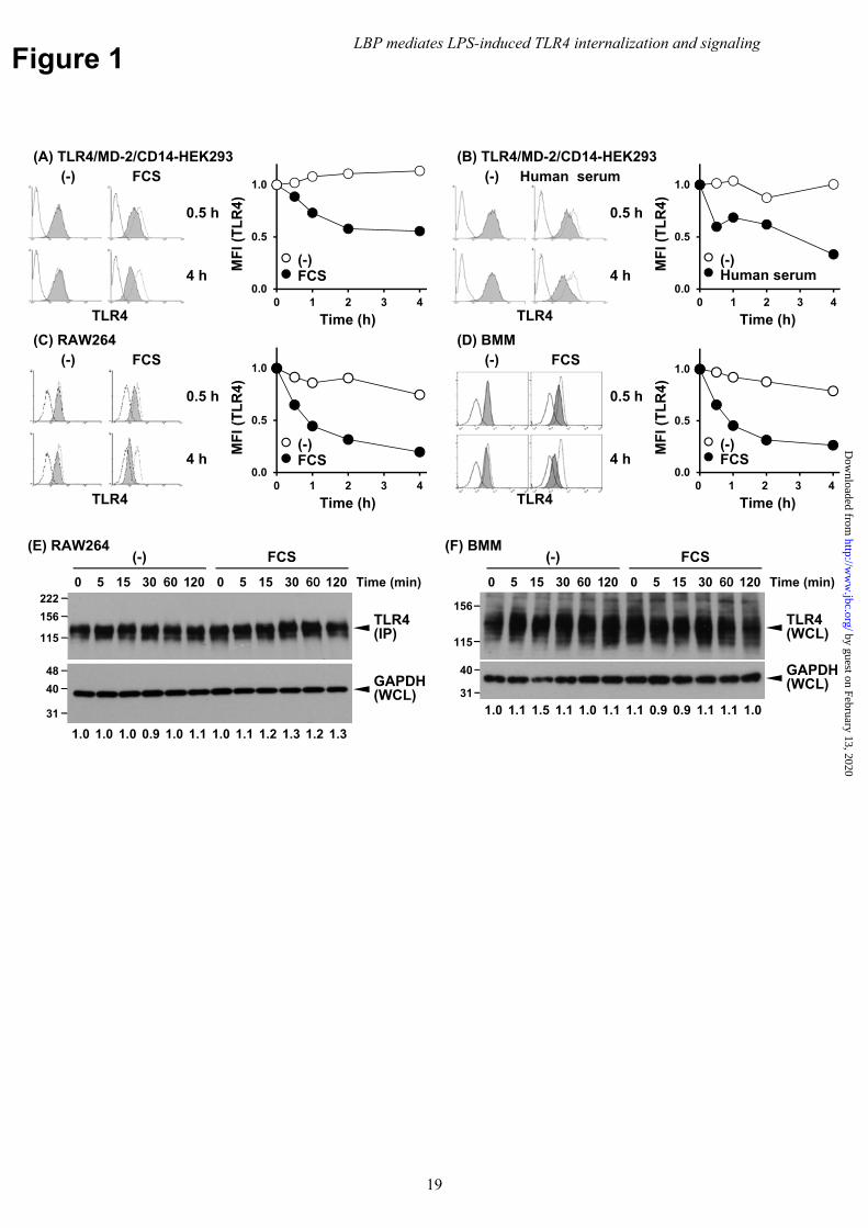
Figure 1
(C) RAW264
TLR4
0.5 h
4 h
(-) FCS
0.0
0.5
1.0
0 1 2 3 4
MF
I (T
LR
4)
Time (h)
FCS(-)
(D) BMM
TLR4
(-) FCS
0.5 h
4 h
(A) TLR4/MD-2/CD14-HEK293
MF
I (T
LR
4)
Time (h)
0.0
0.5
1.0
0 1 2 3 4
FCS(-)
TLR4
0.5 h
4 h
(-) FCS(B) TLR4/MD-2/CD14-HEK293
0.0
0.5
1.0
0 1 2 3 4
Time (h)
MF
I (T
LR
4)
Human serum(-)
0.5 h
TLR4
4 h
(-) Human serum
MF
I (T
LR
4)
Time (h)
0.0
0.5
1.0
0 1 2 3 4
FCS(-)
222
115
156
(E) RAW264
Time (min)
(-) FCS
1200 15 60305 0 15 60 120305
TLR4(IP)
GAPDH(WCL)
48
31
40
(F) BMM
Time (min)
(-) FCS
1200 15 60305 0 15 60 120305
TLR4(WCL)
GAPDH(WCL)
31
40
115
156
1.0 1.0 1.0 0.9 1.0 1.1 1.0 1.1 1.2 1.3 1.2 1.3
1.0 1.1 1.5 1.1 1.0 1.1 1.1 0.9 0.9 1.1 1.1 1.0
LBP mediates LPS-induced TLR4 internalization and signaling
19
by guest on February 13, 2020http://w
ww
.jbc.org/D
ownloaded from
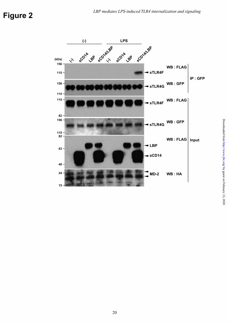
Figure 2
(kDa)
sTLR4FWB : FLAG
LPS(-)
WB : GFP
156
115
82
115
82
48
63LBP
sCD14
MD-2
115
156sTLR4G
sTLR4FWB : FLAG
WB : FLAG
WB : HA
15
24
115
156
sTLR4GWB : GFP
IP : GFP
Input
LBP mediates LPS-induced TLR4 internalization and signaling
20
by guest on February 13, 2020http://w
ww
.jbc.org/D
ownloaded from
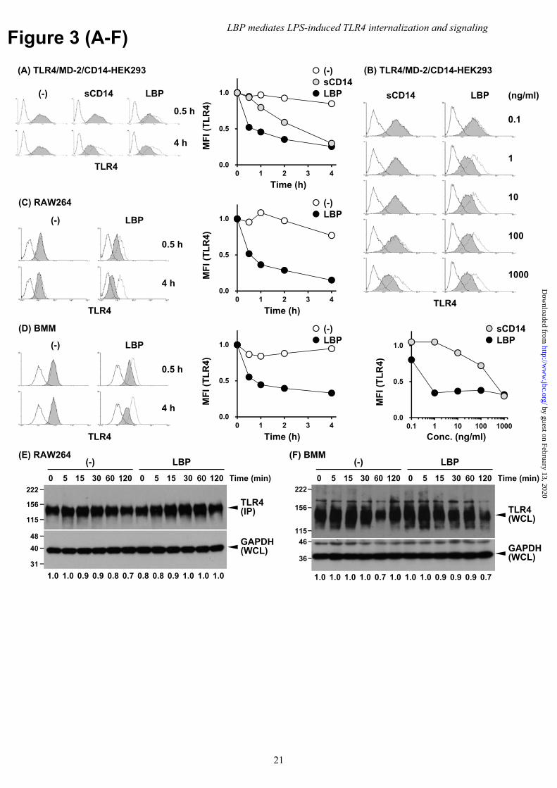
Figure 3 (A-F)
sCD14
TLR4
LBP (ng/ml)
1000
100
10
1
0.1
(B) TLR4/MD-2/CD14-HEK293
0.0
0.5
1.0
0 1 2 3 4
MF
I (T
LR
4)
Time (h)
4 h
0.5 h
TLR4
(-) LBPsCD14
(A) TLR4/MD-2/CD14-HEK293
LBPsCD14(-)
LBP
4 h
(-)
0.5 h
(C) RAW264
TLR4
0.0
0.5
1.0
0 1 2 3 4
MF
I (T
LR
4)
Time (h)
LBP(-)
LBP
4 h
(-)
0.5 h
(D) BMM
TLR4
0.0
0.5
1.0
0 1 2 3 4
MF
I (T
LR
4)
Time (h)
LBP(-)
0.0
0.5
1.0
0.1 1 10 100 1000
Conc. (ng/ml)
MF
I (T
LR
4)
LBPsCD14
222
115
156
(E) RAW264
Time (min)
(-) LBP
1200 15 60305 0 15 60 120305
TLR4(IP)
GAPDH(WCL)
48
31
40
(F) BMM
Time (min)
(-) LBP
1200 15 60305 0 15 60 120305
TLR4 (WCL)
GAPDH(WCL)
115
156
222
36
46
1.0 1.0 0.9 0.9 0.8 0.7 0.8 0.8 0.9 1.0 1.0 1.0 1.0 1.0 1.0 1.0 0.7 1.0 1.0 1.0 0.9 0.9 0.9 0.7
LBP mediates LPS-induced TLR4 internalization and signaling
21
by guest on February 13, 2020http://w
ww
.jbc.org/D
ownloaded from
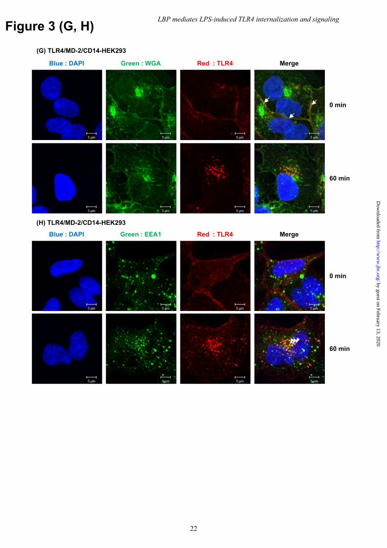
Figure 3 (G, H)
Blue : DAPI Red : TLR4Green : WGA Merge
(G) TLR4/MD-2/CD14-HEK293
0 min
60 min
Blue : DAPI Red : TLR4Green : EEA1 Merge
(H) TLR4/MD-2/CD14-HEK293
0 min
60 min
LBP mediates LPS-induced TLR4 internalization and signaling
22
by guest on February 13, 2020http://w
ww
.jbc.org/D
ownloaded from
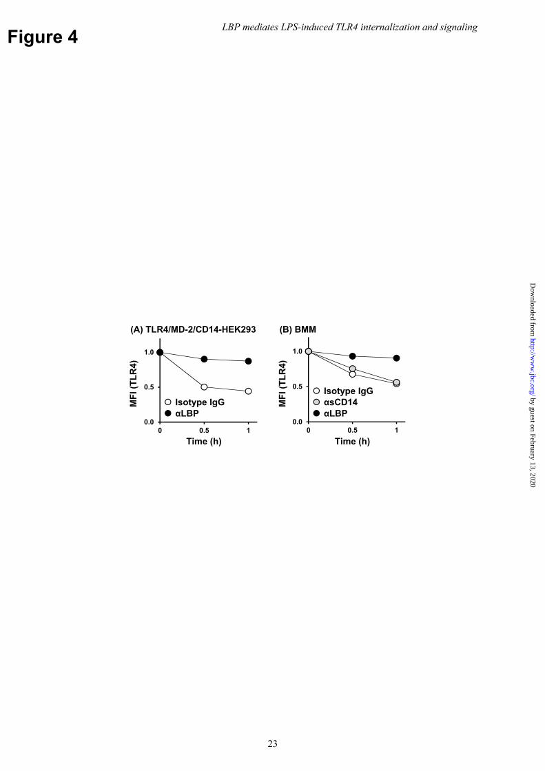
Figure 4
0.0
0.5
1.0
0 0.5 1
MF
I (T
LR
4)
Time (h)
(A) TLR4/MD-2/CD14-HEK293
0.0
0.5
1.0
0 0.5 1
Time (h)
MF
I (T
LR
4)
(B) BMM
αLBPαsCD14Isotype IgG
αLBPIsotype IgG
LBP mediates LPS-induced TLR4 internalization and signaling
23
by guest on February 13, 2020http://w
ww
.jbc.org/D
ownloaded from
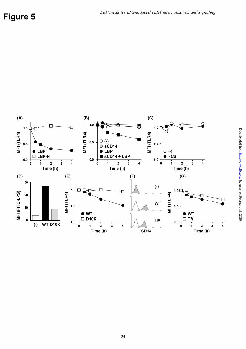
Figure 5M
FI (
TL
R4)
Time (h)
(A)
Time (h)
0.0
0.5
1.0
0 1 2 3 40.0
0.5
1.0
0 1 2 3 40.0
0.5
1.0
0 1 2 3 4
Time (h)
MF
I (T
LR
4)
(C)
0
10
20
30
MF
I (F
ITC
-LP
S)
WT D10K(-)0.0
0.5
1.0
0 1 2 3 4
LBPLBP-N sCD14 + LBP
LBPsCD14(-)
(-)FCS
WTD10K
MF
I (T
LR
4)
Time (h)
(D) (E)
WT
TM
(-)
(F) (G)
CD14
0.0
0.5
1.0
0 1 2 3 4
MF
I (T
LR
4)
Time (h)
WTTM
MF
I (T
LR
4)
(B)
LBP mediates LPS-induced TLR4 internalization and signaling
24
by guest on February 13, 2020http://w
ww
.jbc.org/D
ownloaded from
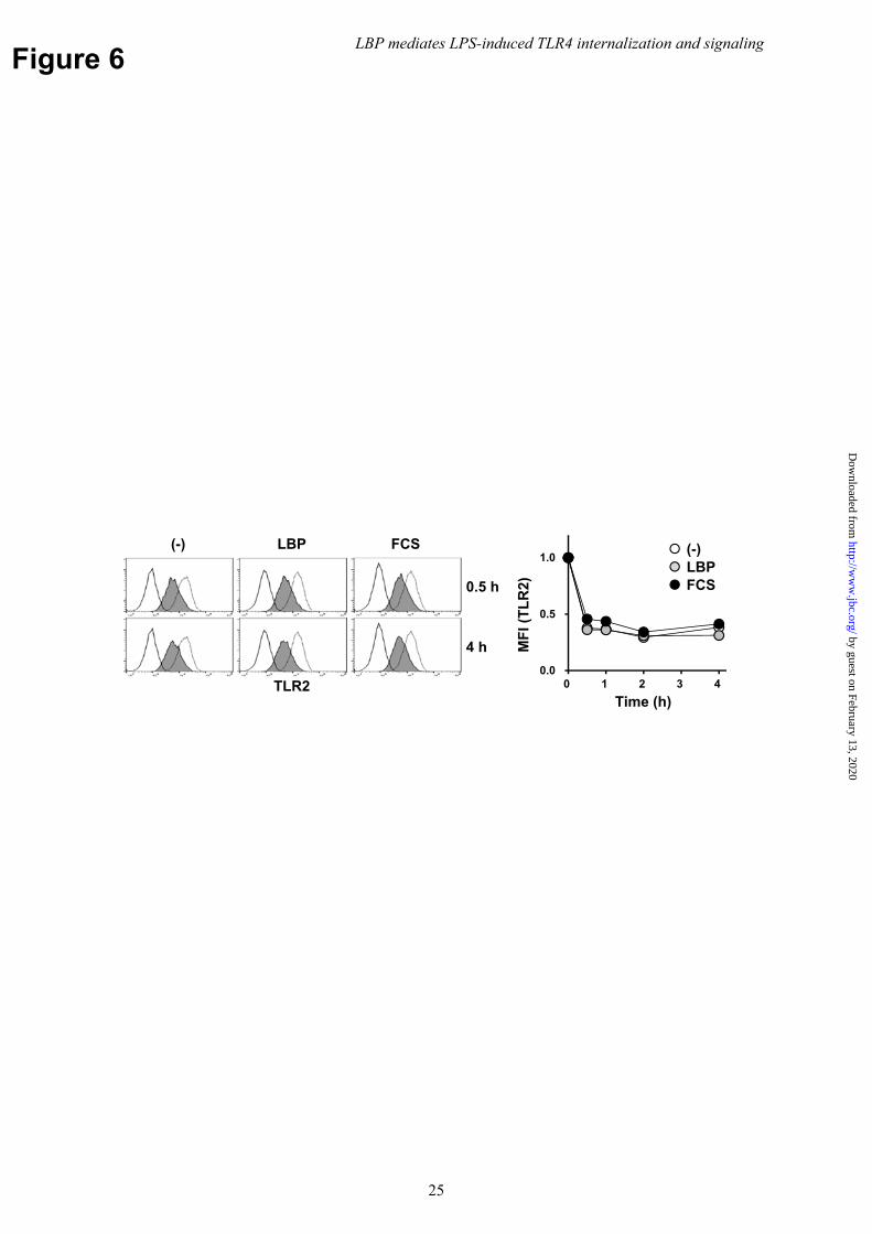
Figure 6
0.0
0.5
1.0
0 1 2 3 4
Time (h)
MF
I (T
LR
2) FCSLBP(-)
4 h
0.5 h
TLR2
(-) FCSLBP
LBP mediates LPS-induced TLR4 internalization and signaling
25
by guest on February 13, 2020http://w
ww
.jbc.org/D
ownloaded from
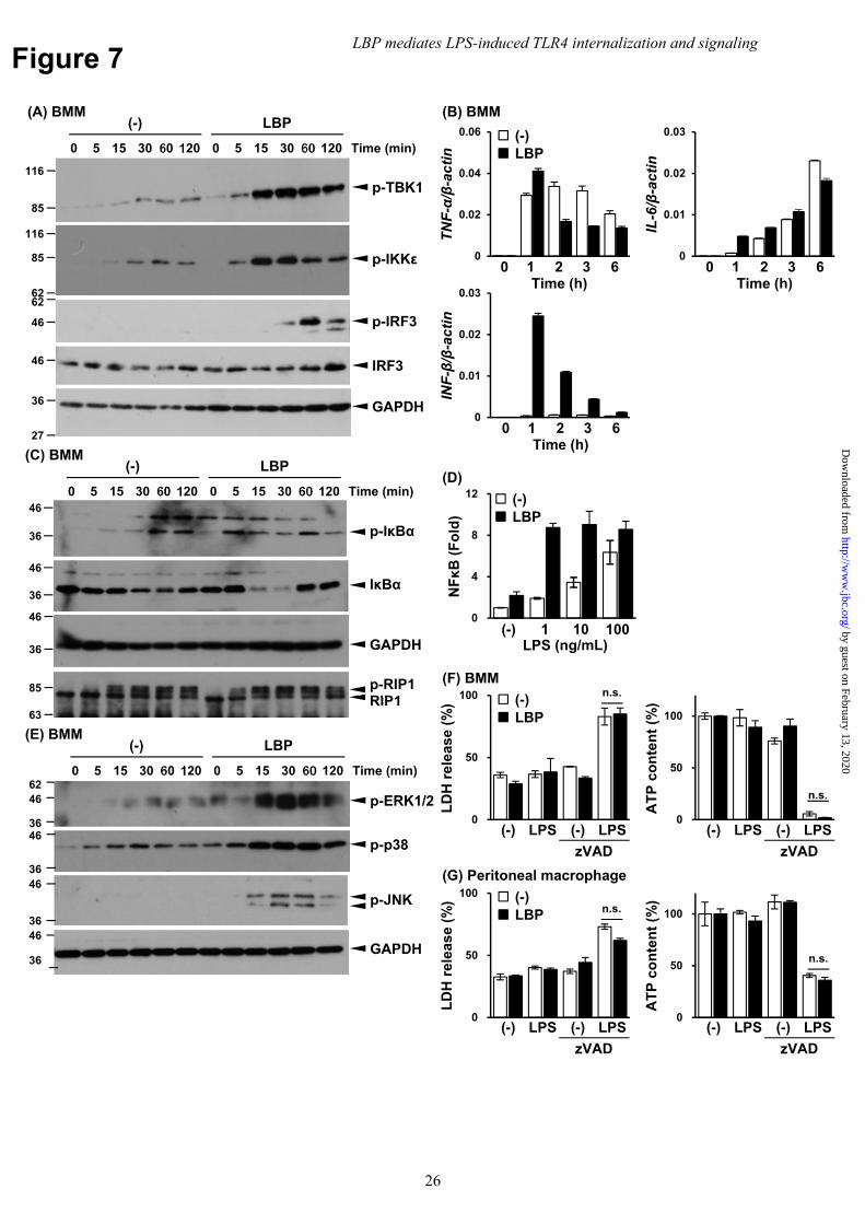
Figure 7
LD
H r
elea
se (
%)
0
50
100
(-) (-)
AT
P c
on
ten
t (%
)
0
50
100
(-) (-)
(F) BMM
(-)LBP
0
50
100
LD
H r
elea
se (
%)
(-) (-)0
50
100
AT
P c
on
ten
t (%
)
(-) (-)
(G) Peritoneal macrophage
(-)LBP
(A) BMM
Time (min)
(-) LBP
1200 15 60305 0 15 60 120305
p-TBK185
116
p-IKKε85
116
62
46
46
62
p-IRF3
IRF3
36
27
GAPDH
(C) BMM
Time (min)
(-) LBP
1200 15 60305 0 15 60 120305
p-IκBα
IκBα36
46
36
46
46
36 GAPDH
85
63RIP1p-RIP1
(E) BMM
Time (min)
(-) LBP
1200 15 60305 0 15 60 120305
p-ERK1/2
GAPDH36
46
62
36
46
36
46p-p38
36
46
p-JNK
(B) BMM
0
0.01
0.02
0.03
IL-6/β-actin
0 1 2 3 60
0.02
0.04
0.06
TNF-α/β-actin
0 1 2 3 6
(-)LBP
0
0.01
0.02
0.03
INF-β/β-actin
0 1 2 3 6
0
4
8
12
NFκB
(F
old
)
(-) 10
(-)LBP
(D)
LBP mediates LPS-induced TLR4 internalization and signaling
26
by guest on February 13, 2020http://w
ww
.jbc.org/D
ownloaded from
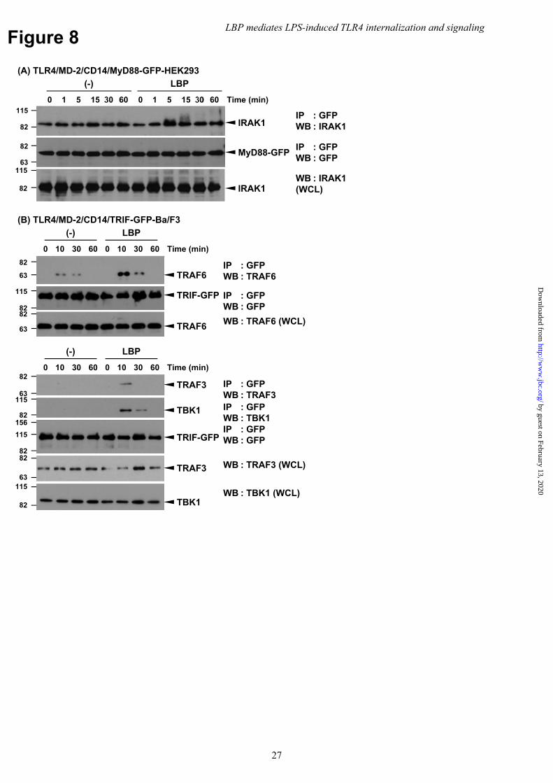
Figure 8
IP : GFPWB : IRAK1
(A) TLR4/MD-2/CD14/MyD88-GFP-HEK293
Time (min)
(-) LBP
600 5 30151 0 5 30 60151
IRAK1
115
82
63
82MyD88-GFP
IP : GFPWB : GFP
82
115
IRAK1WB : IRAK1(WCL)
82
115
63
82
63
82
(B) TLR4/MD-2/CD14/TRIF-GFP-Ba/F3
Time (min)
(-) LBP
600 3010 0 30 6010
TRAF6
TRIF-GFP
TRAF6
IP : GFPWB : TRAF6
IP : GFPWB : GFP
WB : TRAF6 (WCL)
82
115
82
115
15682
115
63
82
63
82Time (min)
(-) LBP
600 3010 0 30 6010
TRAF3
TRIF-GFP
TRAF3
IP : GFPWB : TRAF3
IP : GFPWB : GFP
WB : TRAF3 (WCL)
TBK1 IP : GFPWB : TBK1
WB : TBK1 (WCL)TBK1
LBP mediates LPS-induced TLR4 internalization and signaling
27
by guest on February 13, 2020http://w
ww
.jbc.org/D
ownloaded from
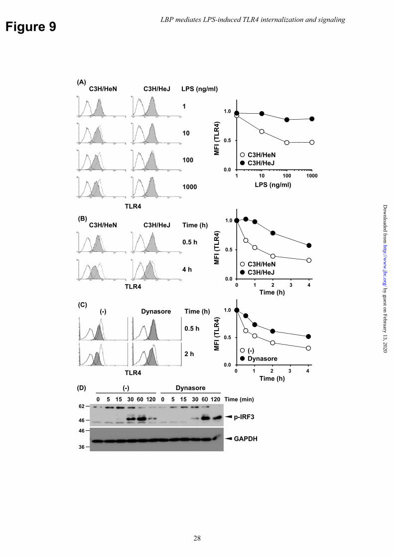
Figure 9
LPS (ng/ml)
MF
I (T
LR
4)
0.0
0.5
1.0
1 10 100 1000
TLR4
1000
100
10
1
C3H/HeJC3H/HeN LPS (ng/ml)(A)
C3H/HeNC3H/HeJ
Time (h)
MF
I (T
LR
4)
0.0
0.5
1.0
0 1 2 3 4
4 h
0.5 h
C3H/HeN C3H/HeJ Time (h)(B)
TLR4
C3H/HeNC3H/HeJ
(C)
Time (h)
MF
I (T
LR
4)
2 h
0.5 h
Time (h)
TLR4
(-)Dynasore
(-) Dynasore
(D)
Time (min)
(-) Dynasore
1200 15 60305 0 15 60 120305
p-IRF346
62
GAPDH36
46
0.0
0.5
1.0
0 1 2 3 4
LBP mediates LPS-induced TLR4 internalization and signaling
28
by guest on February 13, 2020http://w
ww
.jbc.org/D
ownloaded from
TomiokaYotaro Matsumoto, Tomonori Nochi, Kouichi Watanabe, Hisashi Aso and Yoshihisa
Ippo Ukai, Ayumi Shichiku, Misaki Okubo, Muneo Numasaki, Yoshitomi Kanemitsu, Hiroki Tsukamoto, Shino Takeuchi, Kanae Kubota, Yohei Kobayashi, Sao Kozakai,receptor 4 internalization and LPS-induced TBK1-IKK?-IRF3 axis activation
Lipopolysaccharide (LPS)-binding protein stimulates CD14-dependent Toll-like
published online May 14, 2018J. Biol. Chem.
10.1074/jbc.M117.796631Access the most updated version of this article at doi:
Alerts:
When a correction for this article is posted•
When this article is cited•
to choose from all of JBC's e-mail alertsClick here
by guest on February 13, 2020http://w
ww
.jbc.org/D
ownloaded from