liver stiffness evaluation in dm type 2, ng t hồng anh- ng thiện hùng
TRANSCRIPT

Liver Stiffness Evaluation by ARFI imaging in Type 2 Diabetes Mellitus Patients
Nguyễn Thị Hồng Anh, MDNguyễn Thiện Hùng, MD
Phan Thanh Hải, MDMEDIC CENTER

Introduction
NAFLD ( obesity, DM, metabolic syndrome): one of the most common chronic liver diseases worldwide.
NAFLD ↔ DM
NAFLD → (NASH) → liver cirrhosis → liver cancer .

Accurate liver fibrosis degree assessment → prognosis → deciding treatment course.
Limitations of liver biopsy → noninvasive
and reliable tests.
Ultrasound elastography:◦ Non-invasive
◦ Convenient
◦ Precise
◦ Measuring liver stiffness → liver fibrosis.

Background & Aims
Evaluate a population of OPD diabetic patients regarding the severity of liver steatosis and liver fibrosis by ARFI imaging.
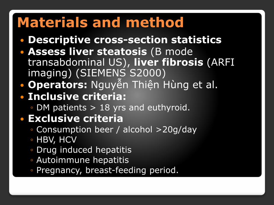
Materials and method Descriptive cross-section statistics Assess liver steatosis (B mode
transabdominal US), liver fibrosis (ARFI imaging) (SIEMENS S2000)
Operators: Nguyễn Thiện Hùng et al. Inclusive criteria:
◦ DM patients > 18 yrs and euthyroid.
Exclusive criteria◦ Consumption beer / alcohol >20g/day
◦ HBV, HCV
◦ Drug induced hepatitis
◦ Autoimmune hepatitis
◦ Pregnancy, breast-feeding period.

Staging liver steatosis and fibrosis
Steatosis severity:semi-quantitativescale/ B mode ◦ S0 = no steatosis
◦ S1 = mild steatosis
◦ S2 = moderate steatosis
◦ S3 = severe steatosis
Stages of liver fibrosis (ARFI technique)◦ F0 = 0.99 - 1.16 m/s
◦ F1 = 1.16 - 1.25 m/s
◦ F2 = 1.25 - 1.32 m/s
◦ F3 = 1.32 - 1.56 m/s
◦ F4 = 1.56 - 4.15 m/s

S1- mild steatosis (bright liver withdiscrete posterior attenuation).
S2 - moderate steatosis(bright liver withobvious posterior attenuation).
S3 - Severe steatosis(bright liver withintense posterior attenuation that makes it impossible to visualize the diaphragm).


Results
Nov 2016-Feb 2017.
80 type 2 diabetic patients (27 M, 53F).
Age=28-79.
Duration of acquired DM= first onset -20years.
4 obese patients (5%), 23 overweight patients (28.75%)

Results(80 cases)
Significant fibrosis (F2-F3): 23/80=28.75% ( steatosis)
Severe fibrosis (F4): 12/80= 15% ( steatosis)
100% severe steatosis(S3): significant /severe fibrosis.
5.0%(4/20) significant fibrosis (F2-F3) without steatosis: long time DM (7-15 y)
S0 S1 S2 S3 ∑
F0 5 6 6 0 17
F1 4 18 6 0 28
F2 2 3 6 1 12
F3 2 4 4 1 11
F4 0 5 4 3 12
∑ 13 36 26 5 80

Discussion 1
4/80 obese +23/80 over weight #33.75%
Lower percentage of male DM patient (27/80 #33.75%)
Liver steatosis : ◦ 67/80#83.75%
◦ Severe+moderate : 31/80 #38.75%

Discussion 2
Liver stiffness ≥ F2: ≥ 40% of DM patients,
not correspondence with steatosisseverity. Fibrosis seemed to depend on acquired DM duration.
Severe steatosis (S3) → significant
/severe fibrosis.
ARFI technique: fast, useful, valuable, comparable as transient elastography.

Discussion 3
ARFI TECHNIQUE:
The best accuracy: distinguish between patients with fibrosis ≤ F2 and those with
severe fibrosis or cirrhosis (F3-F4).
Less interference from obesity, ascites or narrow intercostal space .
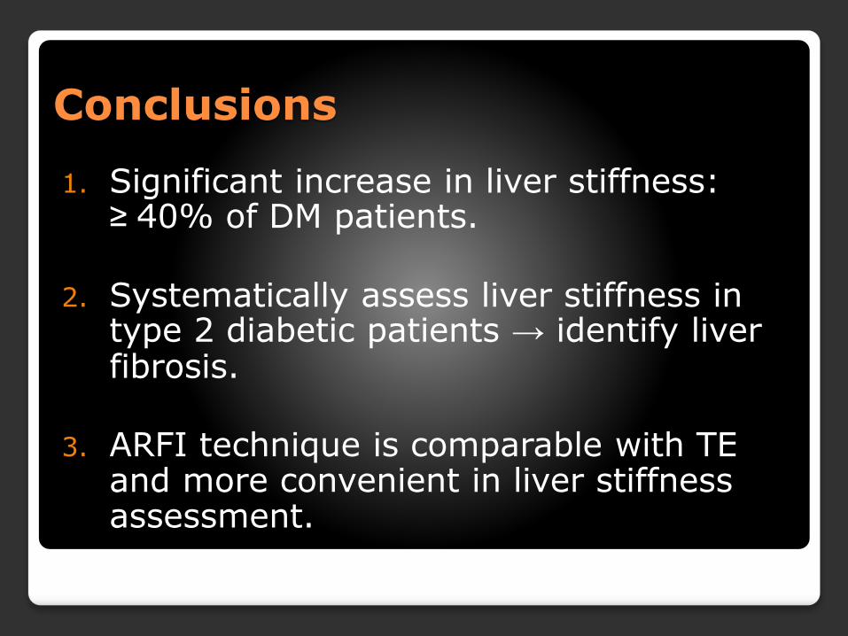
Conclusions
1. Significant increase in liver stiffness: ≥ 40% of DM patients.
2. Systematically assess liver stiffness in type 2 diabetic patients → identify liver fibrosis.
3. ARFI technique is comparable with TE and more convenient in liver stiffness assessment.

References
1.Liver Stiffness Evaluation by Transient Elastography in Type 2 Diabetes Mellitus Patients with Ultrasound-proven Steatosis -Ioan Sporea1, Ruxandra Mare1, Raluca Lupușoru1, Alexandra Sima2, Roxana Șirli1, Alina Popescu1, Romulus Timar2, J Gastrointestin Liver Dis, June 2016 Vol. 25 No 2: 167-174.
2.Liver Stiffness in Nonalcoholic Fatty Liver Disease:A Comparison of Supersonic Shear Imaging,FibroScan, and ARFI With Liver Biopsy. HEPATOLOGY, Month 2015
3.Principles and clinical application of ultrasound elastography for diffuse liver disease-Woo Kyoung Jeong1, Hyo K. Lim1, Hyoung-Ki Lee2, Jae Moon Jo2, Yongsoo Kim3. Ultrasonography 33(3), July 2014

Thanks for attention !