liver tumors in infancy -...
TRANSCRIPT
Chapter 18
Liver Tumors in Infancy
Julio C. Wiederkehr, Izabel M. Coelho,Sylvio G. Avilla, Barbara A. Wiederkehr andHenrique A. Wiederkehr
Additional information is available at the end of the chapter
http://dx.doi.org/10.5772/51764
1. Introduction
Hepatic tumors in children are relatively rare, accounting for 1 to 4% of all pediatric solid tu‐mors. [1] Primary liver masses constitute the third most common group of solid abdominal tu‐mors of childhood [2] with an incidence of 0.4 to 1.9 per million children each year. [3,4]
Liver masses in children can be malignant, benign, or indeterminate and they are a diversegroup of epithelial and mesenchymal tumors whose incidence can vary considerably withpatient age. [5] Two thirds of liver tumors in children are malignant. [6] Unlike liver tumorsin adults, in which the predominant histology is hepatocellular carcinoma, hepatoblastomaaccounts for two thirds of liver tumors in children. [7] Other liver malignancies in childreninclude sarcomas, germ cell tumors, and rhabdoid tumors, as well as the more familiar hep‐atocellular carcinoma. Benign tumors of the liver in children include vascular tumors, ha‐martomas, adenomas, and focal nodular hyperplasia. The histology and anatomy of apediatric liver tumor guides the treatment and prognosis. [8]
In this chapter we outline the epidemiology, etiology, pathology, clinical presentation, diag‐nosis and management of each of the most important types of liver tumor. Also aspects ofthe surgical anatomy and resection techniques and other ways to improve ressecability inliver tumors in childhood will be described such as portal vein thrombosis, chemotherapyand transarterial chemoembolization (TACE).
2. Epidemiology
The incidence of hepatic tumors in childhood is consistently quoted from many series as be‐ing in the region of 0.5-2.5 per million population [9] and approximately 100–150 new cases
© 2013 Wiederkehr et al.; licensee InTech. This is an open access article distributed under the terms of theCreative Commons Attribution License (http://creativecommons.org/licenses/by/3.0), which permitsunrestricted use, distribution, and reproduction in any medium, provided the original work is properly cited.
of liver tumors are diagnosed in the U.S. annually. [7] Two thirds of liver tumors in childrenare malignant. [6] acounting for slightly more than 1% of all pediatric malignancies andamong those there is a male preponderance of 1.8 : 1. [7,10]
Hepatoblastoma presents in a younger age group, being a uncommon diagnosis over theage of 4 years. Hepatocellular carcinoma has its peak onset in early adolescence, althoughthe range is wide. The older age at onset for hepatocarcinoma may well reflect its close asso‐ciation with other underlying disease processes. [10]
There are several suggestions that the incidence of malignant liver tumors is increasing inthe U.S. Surveillance, Epidemiology, and End Results data from 1972–1992 showed a 5% an‐nual increase. [7] Liver cancer represented 2% of all malignancies in infants in the early1980s with the incidence doubling to 4% 10 years later. [11]
At a population level, there has been a dramatic increase in survival in countries in which amodern health system has been implemented, although the increased survival is lower forhepatocarcinoma in comparison with hepatoblastomas. [10] According to Litten & Tomlin‐son [8], it has been suggested that the improvements in technology, care, and outcomes forpremature infants have been driving forces in the increase of the incidence in hepatic tu‐mors. Hepatoblastoma is more commonly diagnosed in children with a history of prematur‐ity than in full-term infants. Interestingly, those tumors that arise in ex-premature infants donot present at a younger age than those of term infants. [8]
3. Hepatoblastoma
Hepatoblastoma is the most common malignant tumor of the liver in children and is an em‐bryonal tumor in the classic sense of incomplete differentiation; [12] accounts for 1% of allpediatric malignancies and for 79% of all liver cancers in children under age. [13] Its overallincidence is 0.5–1.5 per million, however the incidence in children under the age of 18months is 11.2 cases per million. [14]
Hepatoblastoma is diagnosed in very young children with a peak in the newborn period re‐flecting those tumors that developed prenatally, and an overall median age at diagnosis of18 months; 90 percent of cases are manifest by the fourth birthday, several have beenpresent at birth, and there is an hypothesized association with prematurity. [15] Only 5% ofnew hepatoblastoma cases are diagnosed in children >4 years of age. [8]
The increased incidence of HB in children born before 28 weeks gestation (with birth weight<1500 g) compared with term gestations, may be explained by the exposure of rapidly divid‐ing hepatoblasts to endogenous metabolites and hormones as well as exogenous chemicalsthat would normally be eliminated via the placenta. Inefficiency and compromise of the im‐mature detoxification mechanisms could produce multiple somatic mutations and epigenet‐ic (ie, methylation) modifications of the genome. [16, 17]
For poorly understood reasons, hepatoblastoma occurs in males significantly more frequent‐ly than it does in females with a male:female ratio that ranges from 1.2 to 3.6:1. [14] Most
Hepatic Surgery424

commonly, these tumors present in the right lobe of the liver. [18] There is an increased inci‐dence of hepatoblastoma in Beckwith-Wiedemann Syndrome, which has a relative risk of2280 suggesting a role for genetic aberrations of chromosome 11 in the pathogenesis of hep‐atoblastoma,[19, 20] hemihypertrophy, and familial adenomatous polyposis (FAP) witch hasa relative risk of 1220 suggesting a role for aberrations of chromosome 5 in the pathogenesis.[21] Screening for cases in FAP kindred families is recommended by testing for germlinemutations in the APC tumor suppressor gene. [22, 23] Inactivation of the APC tumor-sup‐pressor gene (found on chromosome 5) is found in 67–89% of sporadic hepatoblastoma [24,25] This gene is known to regulate B-catenin and modulate the wnt signaling pathway, sug-gesting a role for this signaling pathway in the development of hepatoblastoma. [26] Addi‐tional biologic markers may include Trisomy 2, 8, and 20 and translocation of the NOTCH2gene on chromosome 1. [27]
Many etiological factors have been linked with the development of malignant hepatic tu‐mors in childhood (Table 1). Broadly speaking, genetic influences are particularly importantin the development of hepatoblastoma, whereas environmental factors and coexisting liverdisease are strongly associated with hepatocellular carcinoma. [10]
Hepatoblastoma Hepatocellular carcinoma
Beckwith-Wiedemann Syndrome Hepatitis B
Hemihypertrophy Hepatitis C
Familial adenomatous polyposis (FAP) Hereditary tyrosinemia
α1-Antitrypsin deficiency
Gardner syndrome Cirrhosis secondary to biliary atresia
Glycogen storage disease type I Glycogen storage disease type I
Trisomy 18 Neurofibromatosis
Fetal alcohol syndrome
Prematurity and low birth weight Familial adenomatous polyposis
Maternal exposure to: Drug/toxin exposure:
Oral contraceptives Androgens
Gonadotropins Oral contraceptives
Metals Methotrexate
Petroleum products Aflatoxins
Paints and pigments
Paternal exposure to: Fanconi anemia
Metals
Meckel diverticulum
Table 1. Conditions associated with hepatoblastoma and hepatocellular carcinoma.
Liver Tumors in Infancyhttp://dx.doi.org/10.5772/51764
425

Hepatoblastomas are composed of cells resembling the developing fetal and embryonic liv‐er, hence the classification as an embryonal tumor. Indeed, the cells comprising hepatoblas‐toma mark similarly to hepatic stem cells, defined as pluripotent hepatoblasts capable ofdifferentiating into hepatocytes or cholangiocytes. [28, 29]
According to the Childhood Epithelial Liver Tumors – International Criteria (CELTIC) group,the pathology of hepatoblastoma is classified into four groups based on the work of Weinbergand Finegold: fetal, embryonal, macrotrabecular and small-cell undifferentiated. [10]
Histologically, these tumors can be divided into epithelial (56%) or mixed epithelial/mesenchymal tissue. The epithelial group is further subdivided into fetal (31%), embry‐onal (19%), macrotrabecular (3%) and small-cell undifferentiated subtypes (3%). Thema‐jority of hepatoblastomas is epithelial and consist of a mixture of embryonal and fetalcell types (Fig. 1). [8, 30]
Figure 1. Distribution of histologic subtypes of hepatoblastoma. The majority are epithelial and consist of embryonaland fetal cell types. Pure fetal histology accounts for approximately 7% of hepatoblastomas and is associated with afavorable prognosis. Small cell undifferentiated hepatoblastoma accounts for 5% of hepatoblastoma cases and is as‐sociated with a poor prognosis. [8]
Of the five histologic subtypes—pure fetal, embryonal, mixed epithelial, mesenchymal/macrotrabecular, and small cell undifferentiated—fetal carries the most favorable prognosis.[31] Approximately 5% of hepatoblastomas are of the small cell undifferentiated subtype.This subtype is associated with a worse prognosis. [32] In the mixed epithelial/ mesenchy‐mal type, the presence of mesenchymal elements is associated with improved prognosis andthe most common mesenchymal elements are cartilage and osteoid. [33]
Hepatoblastomas usually presents as a palpable asymptomatic mass with abdominal disten‐sion. [10] Less common presentations include weight loss, anorexia, emesis and abdominalpain and usually indicate advanced disease. [34] One of the more unusual presenting featuresof hepatoblastoma is its association with sexual precocity due to the release of human chorion‐ic gonadotropic hormone (β-HCG) by the tumor. Osteoporosis is said to occur in up to 20% ofthe cases and when severe can lead to bone fractures and vertebral compression. [35] The tu‐mor may rupture spontaneously, producing an acute abdomen and hemoperitoneum. [10]
Hepatic Surgery426

Approximately 90% of patients demonstrate elevated serum AFP levels and there is a corre‐lation between AFP levels and extent of disease. [36]
The right lobe of the liver is most commonly involved with disease but in 35% of patientsthere is bilateral disease. [37] Distant metastasis are present in 20% of patients at the time ofdiagnosis with the lung being the most common site of metastasis; other common sites arethe brain and bone and metastasis occur more commonly with disease relapse. [38]
Hepatoblastoma (%) Hepatocellular carcinoma (%)
Abdominal mass 71 58
Weight loss 24 21
Anorexia 22 22
Pain 18 16
Vomiting 13 10
Jaundice 7 10
Table 2. Signs and symptoms of liver tumors in children. [10]
Overall, the diagnosis is based on laboratory tests (such as full blood count, liver functiontests, α-Fetoprotein – AFP and other markers), imaging (abdominal radiography, ultraso‐nography, computer tomography, magnetic resonance imaging, hepatic angiography, chestradiography and positron-emission tomography – PET) and biopsy.
The full blood count can reveal anemia (usually normocytic, normochromic) in at least 50% ofchildren with hepatoblastoma. [13, 39] The platelet count is also often abnormal with up to one-third of patients demonstrating thrombocytosis and fewer patients having thrombocytopenia.Thrombocytosis is thought to be related to increased levels of circulating thrombopoietin. [40]
Liver function tests are commonly normal in hepatoblastoma. [10] The serum alpha-fetopro‐tein (AFP) level is elevated in 90% of children with hepatoblastoma and tumors that fail toexpress AFP at diagnosis are felt to be biologically more aggressive. [41, 42] AFP levels mustbe interpreted with caution because AFP is commonly elevated in normal neonates up to 6months of age and may be slightly elevated in other tumors, as well as after hepatic damageor during regeneration of liver parenchyma.
The imaging study is important in evaluation liver neoplasms. CT, MRI and ultrasound arethe most commonly used modalities for pediatric doctors in their medical researches as wellas their clinical practice. Ultrasound is accepted as a first-line imaging method because of itsless irradiation, greater convenience and better real-time. [43] Ultrasound is extremely val‐uable in detecting much smaller lesions, especially in detecting fluid and blood-flow in a le‐sion, and it also can evaluate the hepatic vascular anatomy.[44] As a rule, the initialdiagnosis of live tumor is usually made by the abdominal ultrasound examination, whichwill identify the liver as the organ of origin. Hepatoblastoma are seen as a hyperechoic, sol‐id, intrahepatic mass on US. [45]
Liver Tumors in Infancyhttp://dx.doi.org/10.5772/51764
427

Both CT and MRI define the extent of tumor involvement showing its segmental extensionand its proximity to the portal vein, to help determine the resectability. Evaluation with CTdemonstrates a delineated hypoattentuated mass compared with the surrounding normaltissue and allows identification of calcifications. [46] The use of contrast allows assessmentof vascular involvement by the tumor. Combined MRI and contrast enhanced MR-angiogra‐phy gives the best evaluation of the vascular structures and the tumor blood supply, andthis best enables the planning of a resection. A diagnostic biopsy is recommended in all chil‐dren with a suspected hepatoblastoma. Given the potential side effects of chemotherapy, itis not a good clinical practice to start therapy in a patient in the absence of a tissue diagnosis.Additionally, it is necessary to rule out HCC. Although it is rare, HCC have been reported inchildren under the age of three and they carry a worse prognosis. [47]
Figure 2. CT scan of an infant with a large central hepatoblastoma.
Large multinodular expansile masses, hepatoblastomas radiographically appear well de‐marcated from the normal liver but are not encapsulated. They may invade hepatic veins,disseminate to the lungs, or penetrate the liver capsule to reach contiguous tissues. [12]
Historically, North Americans have staged liver tumors similar to other solid tumors, stag‐ing system continues to be used by the children’s oncology group (COG) and depends uponextent of surgery at the time of initial diagnosis.
Hepatic Surgery428
Relative number of patients presenting in each stage in the COG trial 9645 (1999–2003) is asfollows: Stage I (22%) indicates complete resection at diagnosis, Stage II (0.5%) microscopicresidual after attempted complete resection at diagnosis, Stage III (53%) biopsy at diagnosiswith gross residual tumor, and Stage IV (23%) metastatic disease at diagnosis.[48, 49] Thetraditional COG staging system has been criticized for being rather subjective, depending toa large extent on the surgeon rather than the tumor.[12, 50] To address this concern specificsurgical guidelines have been proposed by the COG liver tumor committee which define theanatomic and biologic characteristics of a tumor for which resection at diagnosis is recom‐mended. In addition the upcoming COG hepatoblastoma (AHEP 0731) protocol will add arisk-based stratification of treatment as follows: low risk (Stage I/II lacking any unfavorablebiologic feature); intermediate risk (Stage III or Stage I/II with small cell undifferentiated his‐tology); and high risk (Stage IV or Stage I/II/III with AFP <100 at diagnosis). [12]
3.1. Stage Information
There are two standard surgical staging systems for pediatric liver tumors. The Child‐hood Liver Tumour Strategy Group (SIOPEL) uses a presurgical-based staging system,while the Children's Oncology Group (COG) uses a postsurgical-based staging system.The staging systems support different treatment strategies. The presurgical staging systemis used with neoadjuvant chemotherapy followed by definitive surgery (with the excep‐tion of Pretreatment Extent of Disease [PRETEXT] stage 1), while the postsurgical stagingsystem has surgery as the initial strategy.
Both systems are used in the United States. In a retrospective comparison of the two stag‐ing systems at diagnosis using data from patients entered on a North American random‐ized trial, both staging systems predicted outcome. The presurgical PRETEXT stagingsystem may add prognostic information for patients staged postsurgically at stage 3. [51]The COG is investigating the use of PRETEXT stage before and after chemotherapy to de‐termine the optimal surgical approach. [52]
3.2. Presurgical Staging for Hepatoblastoma and Hepatocellular Carcinoma
The PRETEXT staging system for hepatoblastoma categorizes the primary tumor based onextent of liver involvement at diagnosis. The staging system was devised for use in an inter‐national hepatoblastoma treatment program in which only children with PRETEXT stage 1hepatoblastoma undergo initial resection of tumor. All others are treated with chemothera‐py prior to attempted resection of the primary tumor. The liver tumors are staged by inter‐pretation of computerized tomography or ultrasound with or without additional imagingby magnetic resonance. The presence or absence of metastases is noted in addition to thePRETEXT stage, but does not alter the PRETEXT stage. Tumor involvement of the venacava, hepatic veins, and portal vein, and extrahepatic extension are also noted.
The imaged liver is divided into four quadrants and involvement of each quadrant with tumoris determined. Stage increases and prognosis decreases as the number of quadrants radiologi‐cally involved with tumor increases from one to four. [53, 50] Experienced radiologist review is
Liver Tumors in Infancyhttp://dx.doi.org/10.5772/51764
429

important because it may be difficult to discriminate between real invasion beyond the ana‐tomic border of a given sector and displacement of the anatomic border. [50, 43]
Figure 3. Pretext stage 1 - Tumor involves only one quadrant; three adjoining liver quadrants are free of tumor.[http://www.cancer.gov/PublishedContent/MediaLinks/308970.html]
Figure 4. Pretext stage 2 - Tumor involves one or two quadrants; two adjoining quadrants are free of tumor. [http://www.cancer.gov/PublishedContent/MediaLinks/308970.html]
Hepatic Surgery430

Figure 5. Pretext stage 3 - Tumor involves three quadrants and one quadrant is free of tumor or tumor involves twoquadrants and two nonadjoining quadrants are free of tumor. [http://www.cancer.gov/PublishedContent/Medi‐aLinks/308970.html]
Figure 6. Pretext stage 4 - Tumor involves all four quadrants; there is no quadrant free of tumor. [http://www.cancer.gov/PublishedContent/MediaLinks/308970.html]
Liver Tumors in Infancyhttp://dx.doi.org/10.5772/51764
431
3.3. Treatment - Chemotherapy
During the past 30 years, there has been an improved survival for patients with HB basedon refinements in surgical techniques, a better understanding of the hepatic segmental anat‐omy, advances in chemotherapy, and the advent of liver transplantation as a therapeuticmodality for patients with unresectable disease. HB is a surgical neoplasm and only com‐plete tumor resection results in a realistic hope for cure. Long-term disappearance of tumorwith complete remission with chemotherapy alone has been anecdotally observed. Howev‐er, chemotherapy is a cornerstone in the management of HB. [55]
Although chemosensitivity varies between patients, it is an essential component of the man‐agement and complementary to radical surgical resection to affect a cure. In general, surgeonsagree that preoperative chemotherapy helps to reduce the size of most tumors and obtains bet‐ter demarcation between the tumor and surrounding liver tissue. [56, 57, 58] Consequently, tu‐mors are more likely to be completely resected without increasing perioperative morbidity ormortality. It is also speculated that residual microscopic disease may behave more aggressive‐ly under the influence of hepatotrophic factors stimulating liver regeneration if preoperativechemotherapy has not been used. [58] On the other hand, von Schweinitz et al. [59] have shownthat there is little to be gained from prolonging chemotherapy beyond the planned treatmentregimen, which incurs the risk of developing chemoresistance. [55]
Even if unresectable at diagnosis, most hepatoblastomas are unifocal and chemosensitive,especially to ‘‘platinum’’ derivative chemotherapeutic agents. With the routine addition ofcisplatin to the chemotherapy in the late 1980s, overall survival in hepatoblastoma increasedfrom 30% to 70%. [60, 61] Twenty years later, cisplatin remains the backbone of the chemo‐therapy regimen. In current trials by COG (America), SIOPEL (Europe, South America),GPOH (German), and JPLT (Japan) chemotherapeutic agents used in combination with cis‐platin have differed slightly. Although most use some form of doxorubicin, COG currentlyrecommends Cisplatin/5FU/Vincristin (C5V) for low-risk tumors, C5V+Doxorubicin for in‐termediate risk, and hopes to investigate new agents with up-front window therapy in high-risk tumors. [48, 49] Irinotectan, with or without doxorubicin, has been used in bothAmerica and Europe for patients with relapse. [62] Because tumor cells may become resist‐ant to chemotherapy over prolonged exposure [63] and because cumulative chemotherapytoxicity may be unwarranted, prolonged (44 cycles) courses of neoadjuvant chemotherapyare discouraged by all study groups. Early referral for complex surgical planning may be in‐dicated for large invasive tumors potentially requiring transplantation. [12]
Two principle strategies exist. In the United States, tumor resection at diagnosis, wheneverprudently possible, has been advocated with the argument that toxicity of chemotherapycan be reduced by avoidance of unnecessary neoadjuvant chemotherapy, that some tumorsmay become resistant to prolonged courses of chemotherapy [64] and the highest survivalrates have historically been observed in patients with initially resected tumors—althoughthese tumors also tend to be the smaller more favorable tumors. Proposed COG Surgicalguidelines advocate definitive surgical resection at diagnosis for localized, unifocal PRE‐TEXT I and II tumors followed by chemotherapy. When the tumor is large (PRETEXT III orIV), multicentric, shows radiographic evidence of portal or hepatic venous invasion, or pul‐
Hepatic Surgery432
monary metastatic lesions the chance of curative resection may be improved neoadjuvantchemotherapy and delayed primary resection. Alternatively, the SIOPEL study group dis‐courages resection of hepatoblastoma at diagnosis favoring neoadjuvant chemotherapy inall patients with the argument that the chemotherapy renders most tumors smaller, betterdemarcated, and more likely to be completely resected, and that the toxicity of neoadjuvantchemotherapy is offset by the increased rates of surgical resectability. Both COG and SIO‐PEL have invested considerable effort in attempts to decrease the significant ototoxiciy at‐tendant to the use of cisplatin based chemotherapy in young infants and toddlers. [12]
In the Intergroup Hepatoblastoma/ Hepatocellular Carcinoma Study, 28% of HB tumors werecompletely resected at diagnosis (Stage I) and 4% (Stage II) were incompletely excised. Thesepatients had a 91% and 100% 5-year survival, respectively. However, the surgical guidelines ofthe protocol lacked clear recommendations regarding which tumor should or should not be re‐sected at diagnosis. The study compared the use of cisplatin and doxorubicin in one treatmentarm to cisplatin, vincristine, and 5-fluorouracil (5-FU) in the other arm. The overall 3- year sur‐vival rates were 63% and 71%, respectively. [65] Although the difference between the groupswas not significant, the cisplatin/ doxorubicin group had a higher toxicity rate. A significant re‐sponse to preoperative chemotherapy was observed in Stage III patients allowing complete tu‐mor resection in 70–80% of these cases. Pre-operative chemotherapy had no effect on operativemortality; however, increased transfusion requirement and a higher operative morbidity wasobserved in patients that received chemotherapy preoperatively. [55]
The studies coordinated by the SIOPEL group have concentrated on using preoperative che‐motherapy. [56, 66] In SIOPEL-1, all patients were treated preoperatively with four coursesof cisplatin and doxorubicin (PLADO); surgical resection was followed by two more coursesof chemotherapy. If the tumor was judged unresectable by imaging after four courses of che‐motherapy, attempting surgical resection was delayed until after the sixth course. If the tu‐mor remained localized to the liver but was still unresectable, liver transplantation wasrecommended as the primary operative procedure if some response to chemotherapy hadbeen obtained in the absence of extrahepatic tumor extent or metastatic disease. The SIO‐PEL-2 pilot study [67]was designed to test the efficacy and toxicity of two chemotherapyregimens, one for patients with HB confined to the liver and involving no more then threehepatic sections ‘‘standard-risk (SR) HB”, and one for instances of HB extending into all foursections and/or with lung metastases or intra-abdominal extrahepatic spread or tumor rup‐ture at presentation or with serum AFP < 100 units at presentation ‘‘high-risk (HR) HB”.Those with SR-HB were treated with four courses of cisplatin monotherapy, delayed sur‐gery, and then two more courses of cisplatin. Patients with HR-HB were given cisplatin al‐ternating with carboplatin and doxorubicin, pre- and postoperatively. For SR-HB patients (n= 77), and HR-HB patients (n = 58), the 3-year progression-free survival rates were 89% and48%, respectively. For SR-HB patients, the efficacy of cisplatin monotherapy and the cispla‐tin/doxorubicin combination are now being compared in a prospective randomized trial(SIOPEL-3 study). For HR-HB patients, intensified chemotherapy with cisplatin, doxorubi‐cin, and carboplatin is being investigated in a SIOPEL-4 study. [55]
Liver Tumors in Infancyhttp://dx.doi.org/10.5772/51764
433
In unifocal HB, PRETEXT grouping based on imaging studies at diagnosis in some casesmay lead to overstaging the tumor from PRETEXT III to PRETEXT IV when the anatomicborder separating a lateral section from the sections of the liver harboring the bulging massis simply displaced (due to compression) but not invaded. [56, 68] Indeed, repeat imagingstudies after chemotherapy, when the tumor has shrunken, can demonstrate that the ana‐tomic border is free from invasion and allow for correct staging and performance of a partialhepatectomy (right or left trisegmentectomy). In multifocal HB with lesions scattered in thedifferent sections of the liver, clearance of one section, (e.g. the left lateral section) [69] canapparently be achieved by chemotherapy in some cases, tempting the surgeon to perform apartial rather than a total hepatectomy. However, this strategy is not recommended becauseof the high-risk of leaving viable malignant tumor cells in the remaining section. Therefore,in multifocal hepatoblastoma, liver transplantation is the best treatment option, whateverthe apparent result of chemotherapy. Further intensification of chemotherapy when the re‐sponse to completion of full courses of chemotherapy according to protocol is consideredunsatisfactory, and hazardous attempts at partial liver resection in order to avoid liver trans‐plantation ‘‘at any cost” are no longer justified since the efficacy of primary liver transplan‐tation for unresectable HB has been validated during the last decade. [55]
Even patients presenting with metastatic disease are potentially curable with a combination ofchemotherapy, complete tumor resection by partial hepatectomy or transplantation, and pul‐monary metastasectomy. The role of pulmonary metastasectomy has yet to be clearly defined,although it appears that surgical resection of lung deposits may be more likely to cure patientswith disease present at diagnosis but persistent after neoadjuvant therapy rather than patientswith pulmonary relapse. [12] Data from the most recent COG study, 9645, show 3-year event-free survival of 90% for Stage I–II, 50% for Stage III, and only 20% for Stage IV (Malogolowkinet al., 2007). In the European SIOPEL II 3-year survival for standard risk tumors was 90% andfor high-risk tumors was 50%. Cure from hepatoblastoma mandates a complete gross resectionof the primary tumor at some point during the treatment regimen. [12]
3.4. Surgical resection
The objective of the surgical procedure is to obtain a complete resection of the tumor, bothmacro- and microscopically, which is paramount for cure of HB (and other liver cancers).The surgical strategy should be based on a sound knowledge of segmental liver anatomy asdescribed by Couinaud, [70] vascular occlusion techniques and expertise in performing thedifferent types of liver resections, including the most extensive procedures (left or right tri‐segmentectomies). Intraoperative ultrasound is useful in confirming the location of majorvessels and other structures. Nonanatomical, atypical resections are best avoided, except inrare cases (i.e., pedunculated tumor), because of an increased risk of incomplete tumor re‐moval and a higher incidence of postoperative complications. [58] Very extensive liver re‐sections (up to 80% of the liver mass) can be tolerated by young children with HB andhepatic regeneration can be complete within 3 months, despite the administration of toxicagents since they usually have no underlying liver disease and excellent hepatic reserve. [71]Liver function rapidly returns to normal without long-term sequelae. Complete tumor resec‐
Hepatic Surgery434
tion can be easily achieved with a partial hepatectomy when the intrahepatic extent is limit‐ed to one or two sections (PRETEXT I and II). When the tumor involves three sections(PRETEXT III), preoperative neoadjuvant chemotherapy can make lesions initially consid‐ered ‘‘unresectable” become resectable with a trisegmentectomy. [55]
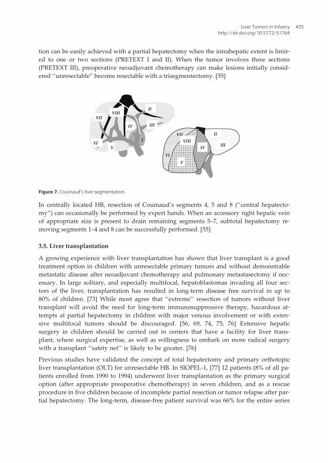
Figure 7. Couinaud’s liver segmentation.
In centrally located HB, resection of Couinaud’s segments 4, 5 and 8 (‘‘central hepatecto‐my”) can occasionally be performed by expert hands. When an accessory right hepatic veinof appropriate size is present to drain remaining segments 5–7, subtotal hepatectomy re‐moving segments 1–4 and 8 can be successfully performed. [55]
3.5. Liver transplantation
A growing experience with liver transplantation has shown that liver transplant is a goodtreatment option in children with unresectable primary tumors and without demonstrablemetastatic disease after neoadjuvant chemotherapy and pulmonary metastasectomy if nec‐essary. In large solitary, and especially multifocal, hepatoblastomas invading all four sec‐tors of the liver, transplantation has resulted in long-term disease free survival in up to80% of children. [73] While most agree that ‘‘extreme’’ resection of tumors without livertransplant will avoid the need for long-term immunosuppressive therapy, hazardous at‐tempts at partial hepatectomy in children with major venous involvement or with exten‐sive multifocal tumors should be discouraged. [56, 69, 74, 75, 76] Extensive hepaticsurgery in children should be carried out in centers that have a facility for liver trans‐plant, where surgical expertise, as well as willingness to embark on more radical surgerywith a transplant ‘‘safety net’’ is likely to be greater. [76]
Previous studies have validated the concept of total hepatectomy and primary orthotopicliver transplantation (OLT) for unresectable HB. In SIOPEL-1, [77] 12 patients (8% of all pa‐tients enrolled from 1990 to 1994) underwent liver transplantation as the primary surgicaloption (after appropriate preoperative chemotherapy) in seven children, and as a rescueprocedure in five children because of incomplete partial resection or tumor relapse after par‐tial hepatectomy. The long-term, disease-free patient survival was 66% for the entire series
Liver Tumors in Infancyhttp://dx.doi.org/10.5772/51764
435
and 85% and 40% for primary transplants and rescue transplants, respectively. Current fol‐low up is >10 years for all patients. All eight patients with PRETEXT IV tumors and all sixpatients with multifocal HB were cured of their disease. Of the seven patients with macro‐scopic extension into the portal vein and/or the hepatic veins/vena cava, 71% became long-term, disease-free survivors, as well as four of five (80%) children who had lung metastasesat presentation with complete clearance of lung lesions after chemotherapy. [55]
An extensive review of the world experience collected 147 cases of liver transplantation forHB. [77] Data were contributed by 24 centers (12 in North America, 10 in Europe, 1 in Japanand Australia each). Twenty-eight (19% of the total) patients presented with macroscopic ve‐nous extension and 12 (8%) with lung metastases. A total of 106 patients (72%) underwent aprimary transplant and 41 (28%) received a rescue transplant, either for incomplete resectionwith partial hepatectomy or for tumor relapse after previous partial hepatectomy. Twenty-eight (19%) received a live, donor-related liver transplant, and 119 (81%) received a de‐ceased donor liver graft. Median follow up since diagnosis for surviving patients was 38months (range 1– 121 months). Overall disease-free survival at 6 years post-transplant was82% and 30% for primary transplants and for rescue transplants, respectively. Multivariatestatistical analysis showed no difference in regard to gender, age, and lung metastases atpresentation or type of transplant. For primary transplants, the only parameter significantlyrelated to overall survival was macroscopic venous invasion (P = 0.045). Remarkably, the 6-year, disease-free survival (82%) for the 106 patients who received a primary transplant wassimilar to the 3-year, progression-free survival (89%) for the 77 HB patients with standard-risk hepatoblastoma confined to the liver and involving no more than 3 hepatic sections thatwere enrolled in the SIOPEL-2 study. [67] In a recent review of the UNOS database in theUSA concerning liver transplantation in 135 children transplanted for unresectable or recur‐rent HB (1987–2004), the one, five, and 10-year survival was 79%, 69%, and 66% respectively.[78] The median age at transplantation was 2.9 ± 2.5 years. Sixteen percent received a graftfrom a live donor. Fifty-five percent of the deaths were due to metastases or recurrent dis‐ease. The latest ELTR report, including 129 patients transplanted for HB has shown a 1- and5-year survival of 100% and 74%, respectively. [55, 79]
3.6. Timing of transplantation
Timing of liver transplantation should not be delayed in excess of a few weeks after the lastcourse of chemotherapy (as per protocol). An expeditious access to organ donors is requiredto meet this requirement. If this is not possible with deceased donors (including split livergrafts), a live-related donor is a valuable option. [55]
According to the results of published studies, the following guidelines have been developedfor early consultation with a transplant surgeon: [55]
1. Multifocal PRETEXT IV HB is a clear and undisputed indication for primary liver trans‐plantation, whatever the result of chemotherapy. Apparent clearance of one liver lobeshould not distract from this guideline because of the high probability of persistent mi‐croscopic viable neoplastic cells. Pediatric oncologists should resist the temptation to in‐
Hepatic Surgery436

tensify chemotherapy in a vain effort to avoid transplantation. These patients should betreated within the same protocol as patients with localized tumors amenable to partialhepatectomy, with as many cycles of chemotherapy before and after transplantation aspatients submitted to partial hepatectomy for a localized HB.
2. Primary liver transplantation may be the best option for large, solitary PRETEXT IVHB, involving all four sections of the liver, unless tumor downstaging is clearlydemonstrated after initial chemotherapy. If this is the case, a clear retraction of thetumor from the anatomic border of one lateral sector would allow performance of aradical trisegmentectomy.
3. Unifocal, centrally located PRETEXT II and III tumors involving main hilar structuresor all three main hepatic veins should be considered for primary liver transplantationbecause these venous structures would presumably not become free of tumor after che‐motherapy. Heroic attempts at partial hepatectomy would be best avoided because ofthe risk of incomplete resection of malignant tissue.
3.7. Contraindications
Persistence of viable extrahepatic tumor deposit after chemotherapy, not amenable to surgi‐cal resection, is the only absolute contraindication for liver transplantation. Macroscopic ve‐nous invasion (portal vein, hepatic veins, vena cava) is not a contraindication if completeresection of the invaded venous structures can be accomplished. When there is evidence orsuspicion of invasion of the retrohepatic vena cava, it should be resected ‘‘en-bloc” and re‐constructed. Review of the world experience showed that venous extent was associated witha significantly shorter survival (P = 0.045). [77] Of the nine TNM IV A/IVB patients (eightwith major intrahepatic venous invasion) reported by Reyes and associates, seven were aliveand disease-free 21–146 months after transplantation. [80]
Patients with lung metastases at presentation should not be excluded from liver transplanta‐tion if the metastases clear completely after chemotherapy and/or surgical resection. Long-term, disease-free survival was obtained in 80% of such patients in the SIOPEL-l study and 58%in the world experience. Complete eradication of metastatic lesions by chemotherapy and sur‐gical resection of any suspicious remnant after chemotherapy is a paramount pre-requisite fortransplantation. [81] When tumor resection by partial hepatectomy is incomplete or when in‐trahepatic relapse is observed after a previous partial hepatectomy, performing a rescue livertransplantation may be a relative contraindication because of the disappointing results ob‐served in the SIOPEL-l study and in the reported world experience. [55]
3.8. Outcomes
In experienced surgical units, major intraoperative complications of liver resection for HBsuch as severe bleeding, air embolism, and unrecognized bile duct injury are infrequent andoperative mortality is very low, even after extended hepatectomies, since children with HBhave no underlying liver disease. As an example, summarizes the 25 years (1978–2003) ofexperience gained at Cliniques Saint-Luc, Brussels [82] with 53 children treated for HB.
Liver Tumors in Infancyhttp://dx.doi.org/10.5772/51764
437

There were 39 partial hepatectomies, including 23 right or left trisegmentectomies, and 13primary liver transplants (two from deceased donors and 11 from living related donors).Only one child died from surgical complications (extensive portal vein thrombosis presentat diagnosis). Postoperative bleeding requiring reoperation was encountered in 2 patients(3.5%). The incidence of biliary complications was 7.6% after partial hepatectomy and 23%following liver transplantation. Actuarial disease-free survival was 89% and 79% in trans‐plant patients and in children treated with partial hepatectomy, respectively. [55]
Although individual centers treat relatively small numbers of patients with liver cancer, thebest overall survival rates are obtained in experienced units that include liver transplanta‐tion in their surgical armamentarium. [55, 83, 84, 85]
The most recent report from King’s college, London [86] confirms that the modern strategyof combining chemotherapy and radical tumor resection enables the majority of childrenwith HB to be cured. From October 1993 to February 2007, 25 liver transplantations wereperformed for HB: 18 from deceased donors and 7 from living donors. Fifteen and ten pa‐tients were PRETEXT IV and III, respectively. All patients received preoperative chemother‐apy following the successive SIOPEL protocols. Patient and graft survival after cadaverictransplantation was 91%, 77.6% and 77.6% after 1, 5 and 10 years, respectively, without re‐transplantation. Patient and graft survival after living related liver transplantation was100%, 83.3% and 83.3%, respectively. All surviving children but one remain disease-free,with a median follow up of 6.8 years (range: 0.9–14.9). There were five deaths at a median of13 months post-OLT, secondary to tumor recurrence in 4 and respiratoryfailure in one. [55]
A remote data entry system is accessible online, worldwide, and free of charge. Registrationis open for patients transplanted since January 1st, 2006 (http://www.pluto.cineca.org). PLU‐TO stands for Pediatric Liver Unresectable Tumor Observatory and was developed by theSIOPEL strategy group. This will allow online registration of children undergoing livertransplantation for a malignant liver tumor. The aim is to establish an international multi‐center database with prospective registration of children (<18 years) presenting with unre‐sectable tumor (HB, HCC, epithelioid hemangioendothelioma and other rare malignanttumors) undergoing primary orrescue liver transplantation.
4. Hepatocellular carcinoma
Hepatocellular carcinoma (HCC) in childhood is rare and accounts for less than 0.5% of allpediatric malignancies, [87, 88] is the second most common malignant hepatic neoplasm inchildren. HCC presents at an older age than does hepatoblastoma, with most HCC cases di‐agnosed in children older than 5 years. [89] Its relative frequency is 0.5 to 1.0 cases per mil‐lion children. It is more frequently encountered in older children and teenagers than ininfants. [88,90] HCC is more often encountered in males and older children between age 10and 14 yr and the median age of onset is 12 year. [88]
Previous reports from Southeast Asia cite an annual incidence of pediatric hepatic tumorsthat is roughly four times higher than western reports in children with less than 15 years of
Hepatic Surgery438

age. [91] This finding is largely based on the high hepatitis carrier rate, with a Taiwanesereport stating that 80% of primary liver tumors in children were hepatocellular carcinoma.With the introduction of hepatitis B vaccine in Southeast Asia, however, there has been amarked reduction in the incidence of hepatocellular carcinoma, although the impact of thehepatitis B vaccine has mainly reduced the incidence of liver tumors in males. [92] Occasion‐ally, malignant tumors in children are seen with features of both hepatocellular carcinomaand hepatoblastoma. These tumors are more common in children with a diagnosis at laterages than that typical of hepatoblastoma.
There is an association with pediatric HCC and pre-existing liver cirrhosis, most often becauseof biliary atresia, Fanconi’s syndrome, and hepatitis B. However, most pediatric HCC are denovo tumors and are not necessarily related to cirrhosis. [75] In certain metabolic diseases suchas hereditary tyrosinemia and glycogen storage disease type IA, there is an increased inci‐dence of HCC. Hereditary tyrosinemia, caused by a deficiency in fumarylacetoacetate hydro‐lase, results in a greatly increased susceptibility to HCC. This is because of the accumulation oftoxic metabolites in the liver, and the incidence of HCC is 50% by age two. Current medicaltherapies for tyrosinemia markedly reduce but do not eliminate the risk of development ofHCC. Glycogen storage disease type IA is caused by a deficiency in glucose- 6-phosphatase.This results in the development of hepatic adenomas in 50% of patients, and about 11% of pa‐tients with adenomas because of glycogen storage disease type IA will undergo malignanttransformation into HCC. [93] Other risk factors for HCC include previous treatment with an‐drogenic steroids, oral contraceptives and methotrexate. [94] Unlike adult HCC, pediatricHCC often demonstrate reduced levels of cyclin D1 expression. [95] Whether this is involvedin the pathogenesis of pediatric HCC is still unclear. [96]
HCC is a malignancy of hepatocyte origin. The tumor is noted to have a fibrous capsule andis also predisposed to vascular invasion. [97] There are two distinct groups of HCC patientsin childhood: those developing HCC in the context of advanced chronic liver disease (CLD),and children who develop sporadic HCC without preceding liver disease. The latter grouptypically affects older children. Their clinical behavior and biologic behavior are similar toHCC in adults. Approximately 26% of cases are histologically of a fibrolamellar type, [98]which does not appear to make a prognostic difference. Sporadic HCC in children has a rel‐atively poor outcome, [75] while the several small series that report on HCC developing inCLD do so in the context of liver transplantation (LT) [82, 99, 100, 101, 102] The fibrolamellarsubtype of HCC (FLHCC) accounts for 3% of HCCs and is not associated with underlyingliver disease. FLHCC lesions are solitary, encapsulated, and well defined. Up to 75% of pa‐tients will have elevated serum AFP levels. [89, 97].
As for the pathology, HCC macroscopically are usually multifocal and invasive, commonlyinvolving both lobes and frequently associated with vascular invasion, extrahepatic exten‐sion, or both at the time of diagnosis. Areas of hemorrhage and necrosis are common, andthe lesions themselves vary in consistency from soft to firm. This significantly reduces theresectability rate. Czauderna et al report only a 36% complete tumor resection rate in a seriesof 39 children recorded by the International Society of Pediatric Oncology over a 4-year timeperiod. [75] The microscopic features distinguishing hepatocellular carcinoma from hepato‐
Liver Tumors in Infancyhttp://dx.doi.org/10.5772/51764
439

blastoma are the presence of tumor cells larger than normal hepatocytes, broad cellular tra‐beculae, considerable nuclear pleomorphism, nucleolar predominance, frequent tumor giantcells, and absence of hemopoiesis. [33,94] The fibrolamellar variant of HCC is probably aseparate clinical entity. Histologically, the tumor cells are plump, with deeply eosinophiliccytoplasm and a marked fibrous stroma separating epithelial cells into trabeculae. [103]
HCC often present as abdominal swelling associated with dull aching pain and discomfort.Other frequent complaints are of rapid weight loss and weakness. [75] The most commonclinical sign is hepatomegaly. HCC frequently presents at the time of diagnosis with meta‐static spread, most commonly to the regional lymph nodes, lungs and bones. [96]
4.1. Laboratory findings
Although most children with HB have an elevated serum AFP level, this marker is elevatedin 50–70% of patients with HCC and less markedly than in HB. Approximately 60–80% ofHCC present with significantly elevated AFP levels. [96] All children with HCC should bescreened for exposure to viral hepatitis B and C. Similar to HB, some children with HCCmay be anemic and others may demonstrate thrombocytosis. Children with cirrhosis-associ‐ated HCC may present with elevated serum liver enzyme levels (AST) and those with sple‐nomegaly may show pancytopenia. Careful assessment of hepatic functional reserve inchildren with cirrhosis is important prior to embarking on major hepatic resection. Howev‐er, no specific data are available for children regarding tests used in adults (Iodocyanine-green (ICG) dye clearance, galactose elimination capacity). Therefore, the evaluation of thehepatic functional reserve in children is based on standard liver tests including total biliru‐bin, prothrombine time and INR. [55]
4.2. Imaging
The diagnostic imaging in children with HCC is not different from HB. HCC is often multi‐focal and may present with a variable number and distribution of tumor nodules. Whileidentifying larger nodules is not difficult, recognizing lesions less than 1.0 cm is still a chal‐lenge. Positron emission tomography (PET) using 18- fluorodeoxyglucose may be useful inidentifying unsuspected extrahepatic disease. [104]
Three-dimensional CT image analysis techniques are now available to estimate tumor vol‐ume and provide detailed intrahepatic anatomy that resembles the actual intraoperativefindings. CT volumetry may permit calculation of resected tumor volume and anticipatedsize of the remnant liver in planning resection. [105] Diagnostic laparoscopy is useful to de‐termine if extra- hepatic disease is present and may avoid unnecessary attempts at resection.Plain radiograph and CT of the chest should be obtained to rule out lung metastases. Hepat‐ic arteriography is currently limited to instances of HCC managed by hepatic artery infusionor transcatheter chemoembolization which can be performed in older children. [55]
On US imaging, HCC may appear as a solitary or multicentric mass most commonly involv‐ing the right lobe of the liver, or as a diffusely infiltrating lesion. At diagnosis, these massesappear solid, rarely contain calcification, and have variable echogenicity. Small lesions ap‐
Hepatic Surgery440

pear homogeneous and are most often hypoechoic. The capsule can be seen as a hypoechoichalo. Larger lesions become necrotic, and therefore demonstrate a more heterogeneous ap‐pearance. Doppler US may detect the high-velocity flow that is related to neovascularity, butDoppler US is most useful for identifying venous invasion. Portal venous invasion is identi‐fied in up to 60% of cases, [106] with hepatic venous invasion identified less commonly.Doppler US may differentiate neoplastic thrombus from bland (benign) thrombus by detect-ing internal neovascularity in the former. [97]
Potentially curative therapies can treat the very early and early stages of the disease. How‐ever, less than 30% of HCC patients are detected with the disease in those stages. [107] An‐other 20% of patients with terminal stage HCC receive recommendations for the bestsupportive treatment. Since HCC is unresectable in the majority of patients at the time of thefirst diagnosis, patients are often directed to nonsurgical treatments. Physicians have longoverlooked radiotherapy (RT) for HCC as radiation might induce fatal hepatic toxicity atdoses lower than the therapeutic doses. [108] However, such limitation has been overcomeby recent developments in RT technology involving precise delivery of focused high-doseon partial volume of the liver. [109, 110, 111, 112, 113, 114] According to the Korean LiverCancer Study Group (KLCSG) practice guidelines, RT is considered appropriate for unre‐sectable, locally advanced HCC without extrahepatic metastasis, Child-Pugh class A or B,and tumors occupying less than two-thirds of the liver. [115]
4.3. Results of resection
Based on recent experience, the optimal treatment should have been total hepatectomy andliver transplantation. Katzenstein et al. reported on 46 children enrolled in the POG andCCG studies - 8 with stage I, 25 with stage III, 13 with stage IV. [49] The overall event-freesurvival at 5 years was 17%. The outcome was not more favorable in 10 children with FL-HCC. No difference in survival was observed whatever the chemotherapy regimen was giv‐en. 369 The German Cooperative Liver Study Group [116] reported the results of twoprospective trials. The survival rate of HCC was 33% and 25% in HB-89 (12 patients – 1989–1993) and 25% in HB-94 (25 patients – 1994–1998), respectively. The SIOPEL-1 study (1990–1994) enrolled 39 patients with HCC who were treated with neoadjuvant chemotherapy(PLADO). Thirty-one percent had metastases, 39% had extrahepatic extension/vascular inva‐sion, 56% had multifocal HCC while 31% had pre-existing liver disease. A partial responseto PLADO was observed in 49%, a complete tumor resection was possible in 36% (2 withliver transplantation). The 5-year event-free survival was 17%. Adverse prognostic factorsincluded multifocality, metastases and vascular invasion. In SIOPEL-2 pilot study (1994–1998), 21 patients were treated with ‘‘super-PLADO” (carboplatin, cisplatin and doxorubi‐cine). Eighteen percent had metastases, 35% had extrahepatic extension/vascular invasionand 53% had multifocal HCC. Partial response to SUPER-PLADO was observed in 46%;complete tumor resection was performed in 47% (one with liver transplantation). The 3-yearoverall survival was 22%. In SIOPEL-3 (1999–2004), 65 patients were treated with SUPER-PLADO with a partial response in 40%. Thirteen underwent primary surgery. Forty-fourpercent were never resectable. The 3-year event-free survival was 10%. Currently, the new
Liver Tumors in Infancyhttp://dx.doi.org/10.5772/51764
441
SIOPEL-5 study is evaluating non-cirrhotic HCC patients staged according to the PRETEXTsystem and receiving neoadjuvant PLADO chemotherapy and thalidomide (an anti-angio‐genic agent) followed by surgery and postoperative metronomic chemotherapy.
4.4. Liver transplantation for hepatocellular carcinoma
Experience with liver transplantation in children with unresectable HCC is somewhat limit‐ed but results have significantly improved over the recent years. Beaunoyer et al. reportedon 10 children with underlying liver disease in 5 and cirrhosis in 5. Six had one nodule >5cm and 7 had >3 nodules. The 5-year actuarial survival was 83%; two died, one of recur‐rence, while 2 with macrovascular invasion survived. Number and size of lesions or grossvascular invasion did not significantly impact survival. [82] Reyes et al. reported on 19 chil‐dren with HCC who underwent total hepatectomy and liver transplantation in 1989–1998;two thirds had underlying liver disease. [80] The 5-year disease-free survival was 63% (3/6died of recurrent HCC). In their experience, risk factors for recurrence were tumor size, vas‐cular invasion and lymphnode involvement. [80] Austin et al. analyzed the aggregated out‐come for OLT in HCC in 41 children <18 years (UNOS data). Patient survival was 63% at 5year and 58% at 10 year. Recurrence was the primary cause of death in 86%. [78]
The most conventional criteria for transplantation are the so-called Milan criteria: [117] nomore than three tumors, each not more than 3 cm in size, or a single tumor, not more than 5cm in diameter, and no evidence of extrahepatic disease or vascular invasion. Recent studiessuggest that, in an otherwise normal liver, the present cut-off for tumor size might be ex‐panded to 6.5 cm or 7 cm. [118, 119] The evidence supports the moderate expansion of theMilan criteria although findings from different studies lack consistency and prospective val‐idation by pretransplant imaging. [79] There are no hard data implying that Milan criteriacan appropriately select children with a low risk of recurrence of HCC after transplantation.Indeed, Milan criteria are derived from experience in adults with cirrhosis, whereas the ma‐jority of children with HCC have no underlying cirrhosis. There is no prospective trial inchildren while the role of OLT in non-cirrhotic liver is unknown. Moreover, there are differ‐ences in biology [120] between adult and pediatric HCC with different molecular findings:mutation of c- met gene in children with HCC, not in adults, level of glycin D1 (regulatoryprotein of G1 phase cycle) expression is lower in children, loss of heterozygosity on chromo‐somal arm, 13q, higher in children. There is evidence that childhood HCC might be less che‐moresistant than adult HCC; a partial response was observed in 49% enrolled in SIOPEL-1study. [75] The SIOPEL group has launched in 2005 a new SIOPEL-5 trial directed to non-cirrhotic hepatocellular carcinoma in children and adolescents. It is based on the hypothesisthat the addition of an antiangiogenic drug (Thalidomide) to PLADO will result in an im‐provement of survival with acceptable toxicity. Most likely, Sorafenib will be substituted forThalidomide on the basis of data obtained in adults with advanced HCC. [121]
Patients with unresectable disease restricted to the liver will be submitted to liver transplan‐tation. Since the majority of children with HCC in western countries have no underlying liv‐er disease, recent data suggest that liver transplantation may be quite useful treatment incarefully selected unresectable cases. [78, 80, 82] Unlike the adult population, the frequency
Hepatic Surgery442

of HCC in the pediatric population is low; therefore, the experience in the application of liv‐er transplantation in the pediatric population for HCC is limited. [122, 123, 124, 125] In pa‐tients whose disease is confined to the liver, the use of liver transplantation is indicated.Because chemotherapy is not beneficial at present in this group, results in patients withmore extensive disease are poor. [126]
5. Benign tumors
In general, benign tumors of the liver may arise from hepatocytes, bile duct epithelium, thesupporting mesenchymal tissue, or a combination of two or more of these. In addition totrue neoplastic conditions of the liver, a variety of nodular diseases may occur that resem‐ble, and must therefore be differentiated from, tumours. Although most patients with be‐nign hepatic tumors are asymptomatic, a minority may present with symptoms that may belocal or systemic. In these patients, the relationship between the symptoms and the hepaticlesions may be difficult to correlate, and additional evaluation is necessary to rule out othercauses for the patients complaints. In most cases patients with benign hepatic lesions haveno preexisting liver disease, and the finding of a coexisting chronic liver disease such as cir‐rhosis, chronic hepatitis B or C, or hemochromatosis should raise a suspicion for a malig‐nant tumor. A conclusive diagnosis of a focal hepatic lesion is essential because it mayrepresent a primary or secondary malignancy, which may require immediate treatment. Inaddition, some benign lesions carry specific risks such as rupture, bleeding, malignant trans‐formation, consumptive coagulopathy, and disseminated intravascular coagulation. [127]
Primary liver masses constitute the third most common group of solid abdominal tumors ofchildhood, [2, 128, 129] with an incidence of 0.4 to 1.9 per million children each year. [129,130] Benign primary liver masses described in children include hemangioma/infantile hep‐atic hemangioendothelioma, focal nodular hyperplasia, simple hepatic cysts, mesenchymalhamartomas, adenomas, nodular regenerative hyperplasia, hematomas, arterial venous mal‐formations, granulomas, and lymphangiomas. [2, 12, 128, 129, 130, 131, 132, 133]
Infantile hepatic hemangioendothelioma is a tumor derived from vascular endothelial cells,which is the most diagnosed benign hepatic tumor in children. Hence it accounts for ap‐proximately 12% of all childhood hepatic tumors,the most common benign vascular tumorof the liver in infancy, and the most common symptomatic liver tumor during the first 6months of life. [134, 135, 136, 137]
While the majority of benign masses may be of little consequence, morbidity and mortalitycan occur from benign masses, mass effect from a tumor can cause pain, biliary obstructionand inferior vena cava obstruction, limit lung capacity, or cause feeding difficulty. [2, 12,129, 138] Most of the recent radiology literature concerning the liver has focused on lesionsdetection or identification of specific features (enhancement patterns) that may help distin‐guish benign from malignant hepatic tumours. Except for hemangioma and focal nodularhyperplasia (FNH), little is know about imaging characteristics that can help identify anddistinguish among the many less common bening liver masses. [139]
Liver Tumors in Infancyhttp://dx.doi.org/10.5772/51764
443

5.1. Infantile hepatic hemangioendothelioma
More than 90% are diagnosed before the age of 6 years. The typical presentation is of hepa‐tomegaly, hemangiomas of the skin, and heart failure resulting from massive arteriovenousshunting. [127, 140] In addition to heart failure, this tumor may cause consumption coagul‐opathy (Kasabach–Merritt syndrome) and obstructive jaundice. [127, 141] Although well cir‐cumscribed, this tumor is not encapsulated and often has scattered calcifications.Microscopically, this tumor consists of multiple small vessels lined by plump endothelialcells and surrounded by fibrous stroma.
Ultrasonography usually shows hepatomegaly and solitary or multiple hepatic lesions,which may vary from anechoic to hyperechoic. The unenhanced CT scan demonstrates thelesion as a well-defined hypo-attenuating mass, occasionally with calcifications. After con‐trast injection, the lesion may show enhancement resembling hemangioma and may becomeisodense on delayed images. Angiography shows dilated, irregular vascular lakes that com‐monly persist beyond the venous phase. 99mTc-sulfur colloid scintigraphy shows the lesionas a cold spot because of a lack of Kupffer cells within the tumor. [127]
The prognosis of this lesion is dependent on its size and its effect on the heart function.Spontaneous regression is frequent but death may occur within the first 6 months of life be‐cause of cardiac failure or replacement of the normal hepatic parenchyma. [127, 142] Theprognosis is usually good if heart failure is managed successfully.
Treatment is dictated by tumor-related symptoms produced by tumor size. Management ofcongestive heart failure may be sufficient in some cases. If symptoms are not relieved, treat‐ment should be aimed at decreasing the tumor size. [127]
Other treatments include hepatic artery ligation, transcatheter endovascular embolization,and radiation therapy. [127, 143, 144] Liver transplant is increasingly recognized as a viabletreatment modality for infantile hemangioendothelioma when other treatments fail. [127, 145]
5.2. Focal Nodular Hyperplasia (FNH)
FNH is very rare in pediatric population with an age prevalence in children 7-8 years old,although some cases are diagnosed in early childhood or even in the prenatal period.[146, 147] The female sex is predominant with a M/F ratio of less than 1/10 in one of thelargest series. [147, 148]
The majority (70-90%) of FNH at presentation is asymptomatic and the most common waythat the disease is discovered is when, during an occasional physical examination, hepato‐megaly or a palpatory abdominal mass are detected. The lesion is more often unique, butabout 8% of cases may show multiple nodules, up to 30. The diameter of lesions is extremelyvariable, from less than 1 cm to more than 15 cm but usually is less than 5 cm. [147]
The diagnosis in the majority of cases could be by Ultrasound, CT Scan and MRI. Needlebiopsy or open air biopsy are necessary when the radiological investigations are doubtful,above all in case of absence of the central scar, and not rarely the differential diagnosis from
Hepatic Surgery444

other nodular lesions of liver may be difficult. The differential diagnosis includes differentnodular lesions of the liver. [147]
The natural evolution of FNH is unpredictable. In about 2/3 of cases, remain stable and inabout 1/3-1/4 of cases show a gradual spontaneous improvement as far as a complete remis‐sion. In rare instances an increase in number as well as in size may occur [9]. The recent studiesin molecular biology have confirmed that FNH is not a pre-neoplastic lesion: the tissue paren‐chymal organization is pretty the same of usual liver tissue and, moreover, even though insome cases a clonal origin of FNH nodules have been demonstrated, until now no somatic mu‐tation in the β-catenin gene or in the other genes implicated in the hepatocellular adenoma(where a malignant transformation is possible) have been discovered. [147, 149, 150]
About the management the first step is, of course, the stop of oral contraceptive. Consideringthe body of evidence that FNH doesn't undergo malignant transformation and that there areonly sporadic cases followed by spontaneous rupture and consequent abdominal bleeding, weagree with the opinion that in asymptomatic cases it is opportune a careful follow-up with anultrasound scan every 6-12 months, and that elective surgery has probably to be limited to thepatients suffering of abdominal pain or with a voluminous or growing mass. [147, 149]
5.3. Nodular regenerative Hyperplasia (NRH)
Nodular regenerative hyperplasia (NRH) is a disease characterized by multiple nodulescomposed by hepatocytes, without a fibrous tissue or central scar. The rare pediatric casesare mostly in association with the congenital absence of portal vein (sometimes complicatedby heart disease or multi-cystic kidney dysplasia). Indeed, only about 200 cases have beenreported. Symptoms, when present, are mainly associated with the complication of portalhypertension. [151, 152, 153, 154]
CT presentation is really different from FNH, as there are multiple hypodense lesions withpoor or absent enhancement after contrast administration. [147, 155] The typical imagingshowing anechoic and regular profile of the mass at ultrasound, easily recognize cystic le‐sions: however CT and MRI may be necessary in selected cases. [147]
5.4. Hamartomas
Mesenchymal hamartoma is a rare, benign, developmental tumor of the liver, with occasion‐al risk of malignancy. Histologically, it appears as a disordered arrangement of the mesen‐chyme, bile ducts, and hepatic parenchyma. Cords of normal appearing hepatocytes areseparated by zones of loose, poorly cellular mesenchyme. The porous nature of the mesen‐chyme permits accumulation of fluid. [156, 157] Grossly, it has stromal and cystic compo‐nents with no capsules, and can grow to large sizes. [157, 158] The typical presentation isone of asymptomatic, rapid abdominal distention with a palpable mass on physical exami‐nation. The rapid expansion of the tumor is believed to be due to degeneration of the mesen‐chyme and fluid accumulation. Other uncommon associated symptoms are vomiting, fever,constipation, diarrhea, and weight loss. [156, 157] Laboratory investigations usually revealnormal liver function with elevated alpha-fetoprotein, which is believed to be secreted by
Liver Tumors in Infancyhttp://dx.doi.org/10.5772/51764
445

the proliferating hepatocytes within the tumor. [157, 159] The radiological appearance is oneof a large, uni or multi-cystic, avascular mass occupying part of the liver. [157, 158] Surgicalresection has been the standard treatment for this tumor.
6. Sarcoma
The third most common hepatic malignancy, after hepatoblastoma and hepatocellular carci‐noma, is undifferentiated embryonal sarcoma. [8, 160, 161] It is believed to be a primitivemesenchymal neoplasm, which usually behaves in a highly malignant fashion. [162] It wasfirst recognized as a clinicopathologic entity by Stocker and Ishak in 1978. [156] Before theirreport, this tumor had been described under different names such as embryonal sarcoma[163] mesenchymoma, [164] primary sarcoma [165] or fibromyxosarcoma. [166]
These tumors occur in children 5–10 years of age and are mesenchymal in appearance. [8, 167]Diagnosis of primary hepatic sarcoma is challenging due to the lack of specific presentingsymptoms, lack of serological markers, non-specific findings on radiological imaging and therarity of the disease. [86] However, leukocytosis and elevated aspartate aminotransferase andalkaline phosphatase are not uncommon laboratory findings. [156, 161, 162, 168, 169] The se‐rum α-fetoprotein level is always normal. [156, 161, 162, 169] There is no correlation with hepa‐titis B or C virus infection. Most tumors have prominent areas of cystic degeneration. [161, 162]Multinucleated giant tumor cells with eosinophilic cytoplasm and frequent mitosis are usuallypresent. (Stocker and Ishak,1978 and [162] et al.,2001) PAS-positive, diastase-resistant hyalineglobules, which are believed to be lysosomes or apoptotic bodies, are frequently seen withintumor cells as well as in extracellular stromata. [156, 162, 168, 170, 171]
Regarding the radiological imaging, undifferentiated embryonal sarcoma often show a mis‐leading cystic appearance on CT and magnetic resonance imaging (MRI) in contrast to a pre‐dominantly solid appearance on ultrasound. [86, 172]
Undifferentiated embryonal sarcoma of the liver behaves in a highly malignant fashion, [162,173] and the median survival has been less than a year. [156, 162] Complete surgical resection isthe key to a favorable outcome. However, despite apparent complete resectability in somecas‐es, local recurrence and distant metastases have been major impediments to achieving long-term disease-free survival. [162, 173] Multidisciplinary treatment (chemotherapy andradiotherapy) has been used to achieve superior and local control and disease-free survival inpatients with Undifferentiated embryonal sarcoma of the liver. [160, 167, 173]
Author details
Julio C. Wiederkehr1,2*, Izabel M. Coelho1,2, Sylvio G. Avilla1,2, Barbara A. Wiederkehr2 andHenrique A. Wiederkehr2
*Address all correspondence to: [email protected]
Hepatic Surgery446
1 Federal University of Paraná, Curitiba, Brazil
2 Hospital Pequeno Príncipe, Curitiba, Brazil
References
[1] Kim, E. H., Koh, K. N, Park, M, Kim, B. E, Im, H. J, & Seo, J. J. (2011). Clinical featuresof infantile hepatic hemangioendothelioma. Korean Journal of Pediatrics, 54(6), 260,doi:10.3345/kjp.2011.54.6.260.
[2] Luks, F. I., Yazbeck, S., Brandt, M. L., et al. (1991). Benign liver tumors in children: a25- year experience. J Pediatr Surg, 26, 1326-30.
[3] Reymond, D., Plaschkes, J., Luthy, A. R., et al. (1995). Focal nodular hyperplasia ofthe liver in children: review of follow-up and outcome. J Pediatr Surg, 30, 1590-3.
[4] Ehren, H., Mahour, G. H., & Isaacs, H., Jr. (1983). Benign liver tumors in infancy andchildhood. Report of 48 cases. Am J Surg, 145, 325-9.
[5] Emre, S., & Mc Kenna, G. J. (2004). Liver tumors in children. Pediatric transplantation,8(6), 632-8.
[6] Weinberg, AG, & Finegold, MJ. (1983). Primary hepatic tumors of childhood. HumPathology, 14, 512-537.
[7] Multerys, M., Goodman, M. T., Smith, MA, et al. (1999). Hepatic Tumors. In RiesLAG, SmithMA,GurneyJGet al. (eds). Cancer Incidence, SurvivalamongChildren,Adolescents: United States SEER Program 1975-1995. SEER Program, NIH Pub.[99-4649], Bethesda, MD, National Cancer Institute, 91-97.
[8] Litten, J. B., & Tomlinson, G. E. (2008). Liver tumors in children. The oncologist, 13(7),812-20.
[9] Dimmick, J. E., Rogers, P. C. J., & Blair, G. (1994). Hepatic Tumors. In: Pochedly C, ed.Neoplastic Siseases of Childhood, Chur, Switzerland, Harwood Academic, 973-1010.
[10] Kelly, D. (2008). Diseases of the Liver and Biliary System in Children ed., Wiley-Black‐well, Oxford.
[11] Kenney, LB, Miller, B. A., Ries, L. A., et al. (1998). Incidence of cancer in infants in theU.S.: 1980-1990. Cancer, 82, 1396-1400.
[12] Meyers, R. L. (2007). Tumors of the liver in children. Surgical oncology, 16(3), 195-203.
[13] Mann, J. R., Kasthuri, N., Raafat, F., et al. (1990). Malignant hepatic tumours in chil‐dren: incidence, clinical features and aetiology. Paediatr Perinat Epidemiol, 4, 276-289.
Liver Tumors in Infancyhttp://dx.doi.org/10.5772/51764
447
[14] Bulterys, M., Goodman, M. T., Smith, M. A., et al. (1999). Cancer Inci- dence and Sur‐vival Among Children and Adolescents: United States SEER Program1975-1995. Na‐tional Cancer Institute SEER Program. NIHPublication [99-4649], 91-97.
[15] Owe, T., Kubota, A., Okuyama, H., et al. (2003). Hepatoblastoma in children of ex‐tremely low birth weight: a report from a single prenatal center. Journal of PediatricSurgery, 38, 134-7.
[16] Honda, S., Haruta, M., Sugawara, W., et al. (2008). The methylation status ofRASSF1A promoter predicts responsiveness to chemotherapy and eventual cure inhepatoblastoma patients. Int J Cancer, 5, 1117-25.
[17] Sakamoto, L. H., De Camargo, B., Cajaiba, M., et al. (2010). MT1G hypermethylation:a potential prognostic marker for hepatoblastoma. Pediatr Res, 67, 387-93.
[18] Exelby, P. R., Filler, R. M., & Grosfeld, J. L. (1975). Liver tumors in children in theparticular reference to hepatoblastoma and hepatocellular carcinoma: AmericanAcademy of pediatrics surgical section survey- 1974. Journal of pediatric surgery, Saun‐ders, Retrieved from, http://linkinghub.elsevier.com/retrieve/pii/0022346875900950?showall=true.
[19] DeBaun, M. R., & Tucker, M. A. (1998). Risk of cancer during the first four years oflife in children from the Beckwith-Wiedemann Syndrome Registry. J Pediatr, 132,398-400.
[20] Steenman, M., Westerfeld, A., & Mannens, M. (2000). Genetics of Beckwith-Weide‐mann Syndrome associated tumours: common genetic pathways. Genes ChromosomesCancer, 28, 1-13.
[21] Giardello, F. M., Offerhaus, G. J., Krush, A. J., et al. (1991). Risk of hepatoblastoma infamilial adenomatous polyposis. J Pediatr, 119, 766-768.
[22] Aretz, S., Koch, A., Uhlhaas, S., et al. (2006). Pediatric Blood Cancer, 47, 811-8.
[23] Hirschman, B. A., Pollock, B. H., & Tomlinson, G. E. (2005). The spectrum of APCmutations in children with hepatoblastoma from familial adenomatous polyposiskindreds. Journal of Pediatrics, 147, 263-6.
[24] Wei, Y., Fabre, H., Branchereau, S., et al. (2000). Activation of B-catenin in epithelialand mesenchymal hepatoblastomas. Oncogene, 19, 498-506.
[25] Jeng, Y. M., Wu, M. Z., Chang, M. H., et al. (2000). Somatic mutations of B-cateninplay a crucial role in the tumorigenesis of sporadic hepatoblastoma. Cancer, 152, 45-5.
[26] Udatsu, Y., Kusafuka, T., Kuroda, S., et al. (2001). High frequency of beta catenin mu‐tations in hepatoblastoma. Pediatr Surg Int, 17, 508-512.
[27] Tomlinson, G. E., Douglass, E. C., Pollock, B. H., et al. (2006). Cytogenetic analysis ofa large series of hepatoblastoma: numerical aberrations with recurring translocationsinvolving 1q12-21. Genes Chromosomes Cancer, 44, 177-84.
Hepatic Surgery448
[28] Ruck, P., Xiao, J. C., Pietsch, T., et al. (1997). Hepatic stem-like cells in hepatoblasto‐ma: Expression of cytokeratin 7, albumin and oval cell associated antigens detectedby OV-1 and OV-. Histopathology, 31, 324-329.
[29] Ruck, P., & Xiao, J. C. (2002). Stem-like cells in hepatoblastoma. Med Pediatr Oncol, 39,504-507.
[30] Stocken, J. T. (1994). Hepatoblastoma. Semin Diagn Pathol, 11, 136-143.
[31] Malogolowkin, M. H., Katzenstein, H. M., Krailo, M., et al. (2006). Intensified plati‐num therapy is an ineffective strategy for improving outcome in pediatric patientswith advanced hepatoblastoma. Journal of Clinical Oncology, 24, 2879-84.
[32] Haas, J. E., Feusner, J. H., & Finegold, M. J. (2001). Small cell undifferentiated histolo‐gy in hepatoblastoma may be unfavorable. Cancer, 92, 3130-4.
[33] Hass, J. E., Mczynski, K. A., Krailo, M., et al. (1989). Histopathology and prognosis inchildhood hepatoblastoma and hepatocellular carcinoma. Cancer, 64, 1082-1095.
[34] Perilongo, G., & Shafford, E. A. (1999). Liver tumours. Eur J Cancer, 19, 953-958.
[35] Teng, C. T., Daeschner, C. W., Jr., Singleton, E. B., Rosenberg, H. S., Cole, V. W., Hill,L. L., & Brennan, J. C. (1961). Liver disease and osteoporosis in children. I. Clinicalobservations. Journal of Pediatrics, 59, 684-702.
[36] Van Tornout, J. M., Buckley, J. D., Quinn, J. J., et al. (1997). Timing and magnitude ofdecline in alpha-fetoprotein levels in tested children with unresectable or metastatichepatoblastoma are predictors of outcome: a report from the Children’s CancerGroup. J Clin Oncol, 15, 1190-1197.
[37] Hartley, A. L., Birch, J. M., Kelsey, A. M., et al. (1990). Epidemiological and familialaspects of hepatoblastoma. Med Pediatr Oncol, 18, 103-119.
[38] Feusner, J. R., Krailo, M. A., Hass, J. E., et al. (1993). Treatment of pulmonary meta‐stasis of initial stage I hepatoblastoma in child- hood: report from the children’s can‐cer group. Cancer, 71, 859-864.
[39] Lack, E. E., Neave, C., & Vawter, G. F. (1982). Hepatoblastoma- A clinical and patho‐logic study of 54 cases. Am J Suj Pathol, 6, 693-705.
[40] Nickerson, H. J., Silberman, T. L., & McDonald, T. P. (1980). Hepatoblastoma, throm‐bocytosis and increased thrombopoetin. Cancer, 315-7.
[41] Meyers, R. L., Katzenstein, H. M., Rowland, J. H., et al. (2008). PRETEXT and otherprognostic factors in hepatoblastoma. Pediatric Blood Cancer.
[42] Perilongo, G. (2006). State of the art: Treatment of childhood liver tumors. Geneva,Switzerland. In: 38th annual meeting of SIOP.
[43] Roebuck, D. J., Olsen, O., & Pariente, D. (2006). Radiological staging in children withhepatoblastoma. Pediatr Radiol, 36, 176-82.
Liver Tumors in Infancyhttp://dx.doi.org/10.5772/51764
449

[44] Roebuck, D. (2008). Focal liver lesion in children. Pediatr Radiol, 38(3), 518-22.
[45] De Campo, M., & De Campo, J. F. (1988). Ultrasound of primary hepatic tumors inchildhood. Pediatric Radiol, 19, 19-24.
[46] Helmberger, J. R., Ros, P. R., Medgo, P. J., et al. (1999). Pediatric liver neoplasms: aradiology-pathological correlation. Eur Radiol, 9, 1339-1347.
[47] Von Schweiniz, D., Burger, D., Weiner, P., et al. (1992). Therapy of malignant livertumors in childhood. An intermittent report of the HB-89 multicenter. Clin Pediatr,204, 214-220.
[48] Katzenstein, H. M., Krailo, M., Malogolowkin, M. H., et al. (2007, February). Biologyand treatment of children with all stages of hepatoblastoma: COG proposalAHEP-0731. submitted to CTEP and NCI.
[49] Katzenstein, H. M., Krailo, M., Malogolowkin, M. H., et al. (2002). Hepatocellular car‐cinoma in children and adolescents: results from the Pediatric Oncology Group andthe Children’s Cancer Group intergroup study. J Clin Oncol, 20(12), 2789-97.
[50] Aronson, D. C., Schnater, J. M., Staalman, C. R., et al. (2005). Predictive value of pre‐treat- ment extent of disease system in hepatoblastoma: Results from the Internation‐al Society of Pediatric Oncology Liver Tumor Study Group SIOPEL-1 study. J ClinOncol, 23, 1245-1262.
[51] Meyers, R. L., Rowland, J. R., Krailo, M., et al. (2009). Predictive power of pretreat‐ment prognostic factors in children with hepatoblastoma: a report from the Chil‐dren’s Oncology Group. Pediatr Blood Cancer, 53(6), 1016-22.
[52] Douglass, E. C., Reynolds, M., Finegold, M., et al. (1993). Cisplatin, vincristine, andfluorouracil therapy for hepatoblastoma: a Pediatric Oncology Group study. J ClinOncol, 11(1), 96-9.
[53] Brown, J., Perilongo, G., Shafford, E., et al. (2000). Pretreatment prognostic factors forchildren with hepatoblastoma-- results from the International Society of PaediatricOncology (SIOP) study SIOPEL 1. Eur J Cancer, 36(11), 1418-25.
[54] http://www.cancer.gov/PublishedContent/MediaLinks/308970.html.
[55] Otte, J. B. (2010). Progress in the surgical treatment of malignant liver tumors in chil‐dren. Cancer treatment reviews, 36(4), 360-71, Elsevier Ltd.
[56] Czauderna, P., Otte, J. B., Aronson, D. C., et al. (2005). Guidelines for surgical treat‐ment of hepatoblastoma in the modern era : recommendations from the childhoodliver tumour strategy group of the international society of paediatric oncology (SIO‐PEL). European Journal of Cancer, 41, 1031-6.
[57] Stringer, M. (2006). Liver tumors. Semin Pediatr Surg, 9, 196-208.
[58] Fuchs, J., Rydzynski, J., Hecker, H., et al. (2002). The influence of preoperative che‐motherapy and surgical technique in the treatment of hepatoblastoma-a report from
Hepatic Surgery450

the German cooperative liver tumours studies HB-89 and HB-94. Eur J Pediatr Surg,12, 255-61.
[59] Von Schweinitz, D., Faundez, A., Teichmann, B., et al. (2000). Hepatocyte growth-fac‐tor- scatter-factor can stimulate postoperative tumor-cell proliferation in childhoodhepatoblastoma. Int J Cancer, 85, 151-9.
[60] Ortega, J. A., Douglass, E. C., Feusner, J. H., et al. (2000). Randomized comparison ofcisplatin/vincristin/5-fluorouracil and cisplatin/doxorubicin for the treatment of pe‐diatric hepatoblastoma (HB): a report from the Children’s cancer group and the pe‐diatric oncology group. Journal of Clinical Oncology, 18, 2665-75.
[61] Schnater, J. M., Aronson, D. C., Plaschkes, J., et al. (2002). Surgical view of the treat‐ment of patients with hepatoblastoma. Cancer, 94, 1111-20.
[62] Malogolowkin, M. H., Katzenstein, H. M., Krailo, M., et al. Redefining the role ofdoxorubicin for the treatment of children with hepatoblastoma. Journal of Clinical On‐cology.
[63] Von Schweinitz, D., & Haberle, B. (2007, March). German liver tumor study: HB 99.Poland, Gdansk. In: First international symposium childhood hepatoblastoma.
[64] Von Schweinitz, D., Hecker, H., Harms, D., et al. (1995). Complete resection beforedevelopment of drug resistance is essential for survival from advanced hepatoblasto‐ma-a report fro the German cooperative pediatric liver tumor study HB-89. Journal ofPediatric Surgery, 30, 845-52.
[65] Ortega, J. A., Douglass, E., Feusner, J., et al. (1994). A randomized trial of cisplatin/vincristine/5-fluorouracil vs. CCP/doxorubicin continuous infusion for the treatmentof hepatoblastoma: results from the pediatric inter-group hepatoma study (abstr).Proc Am Soc Clin Oncol (ASCO), 13, 416.
[66] Pritchard, J., Brown, J., Shafford, E., et al. (2000). Cisplatin, doxorubicin and delayedsurgery for childhood hepatoblastoma: a successful approach-results of the first pro‐spective study of the International Society of Pediatric Oncology. J Clin Oncol, 18,3819-28.
[67] Perilongo, G., Shafford, E., Maibach, R., et al. (2004). Risk-adapted treatment forchildhood hepatoblastoma Final report of the second study of the International Soci‐ety of Pediatric Oncology- SIOPEL 2. Eur J Cancer, 40, 411-21.
[68] Meyers, R. L., Malogolowkin, M. H., Rowland, J. M., & Krailo, M. (2006, May 27).Predictive value of the PRETEXT staging system in children with hepatoblastoma. In:Presented at the 37th annual meeting American Pediatric Surgical Association, Hilton Head,SC.
[69] Dall’Igna, P., Cecchetto, G., Toffolutti, T., et al. (2003). Multifocal hepatoblastoma isthere a place for partial hepatectomy? Med Pediatr Oncol, 40, 113-6.
[70] Couinaud, C. (1992). The anatomy of the liver. Ann Ital Chir, 63, 693-7.
Liver Tumors in Infancyhttp://dx.doi.org/10.5772/51764
451
[71] Wheatley, J. M., Rosenfield, N. S., Berger, L., & La Quaglia, M. P. (1996). Liver regen‐eration in children after major hepatectomy for malignancy-evaluation using a com‐puter-aided technique of volume measurement. J Surg Res, 61, 183-9.
[72] Von Schweinitz, D. (2006). Management of liver tumors in childhood. Semin PediatrSurg, 15, 17-24.
[73] Otte, J. B., & De Ville de Goyet, J. (2005). The contribution of transplantation to thetreatment of liver tumors in children. Semin Pediatr Surg, 14, 233-8.
[74] Chardot, C., Sant Martin, C., Gilles, A., et al. (2002). Living related liver transplanta‐tion and vena cava reconstruction after total hepatectomy including the vena cavafor hepatoblastoma. Transplantation, 73, 90-2.
[75] Czauderna, P., Mac Kinley, G., Perilongo, G., et al. (2002). Hepatocellular carcinomain children: results of the first prospective study of the international society of pedia‐tric oncology group. Journal of Clinical Oncology, 20, 2798-804.
[76] Millar, A. J. W., Hartley, P., Khan, D., et al. (2001). Extended hepatic resection withtransplantation back-up for an unresectable tumor. Pediatric Surgery International, 17,378-81.
[77] Otte, J. B., Pritchard, J., Aronson, D. C., et al. (2004). Liver transplantation for hepato‐blastoma: Results from the International Society of Pediatric Oncology (SIOP) studySIOPEL-1 and review of the world experience. Pediatr Blood Cancer, 42, 74-83.
[78] Austin, M. T., Leys, C. M., Feurer, I. D., et al. (2006). Liver transplantation for child‐hood hepatic malignancy: a review of the United Network for Organ Sharing(UNOS) database. J Pediatr Surg, 41, 182-6.
[79] Hoti, E., & Adam, R. (2008). Liver transplantation for primary and metastatic livercancers. Transplant Int, 21, 1107-17.
[80] Reyes, J. D., Carr, B., Dvorchik, I., et al. (2000). Liver transplantation and chemothera‐py for hepatoblastoma and hepatocellular cancer in childhood and adolescence. J Pe‐diatr, 136(6), 795-804.
[81] Perilongo, G., Brown, J., Shafford, E., et al. (2000). Hepatoblastoma presenting withlung metastases: treatment results of the first cooperative, prospective study of theInternational Society of Pediatric Oncology on childhood liver tumors. Cancer, 89,1845-53.
[82] Beaunoyer, M., Vanatta, J. M., Ogihara, M., et al. (2007). Outcomes of transplantationin children with primary hepatic malignancy. Pediatr Transplant, 11(6), 655-60.
[83] Pimpalwar, A. P., Sharif, K., Ramani, P., et al. (2002). Strategy for hepatoblastomamanagement: transplant versus nontransplant surgery. J Pediatr Surg, 37, 240-5.
[84] Tiao, G. M., Bobey, N., Allen, S., et al. (2005). The current management of hepatoblas‐toma: a combination of chemotherapy, conventional resection, and liver transplanta‐tion. J Pediatr, 146, 204-11.
Hepatic Surgery452

[85] Molmenti, E. P., Wilkinson, K., Molmenti, H., et al. (2002). Treatment of unresectablehepatoblastoma with liver transplantation in the pediatric population. Am J Trans‐plant, 6, 535-8.
[86] Faraj, W., Mukherji, D., El Majzoub, N., Shamseddine, A., Shamseddine, A., & Kha‐life, M. (2010). Primary undifferentiated embryonal sarcoma of the liver mistaken forhydatid disease. World journal of surgical oncology, 8(58).
[87] Moore, S. W., Hesseling, P. B., Wessels, G., et al. (1997). Hepatocellular carcinoma inchildren. Pediatr Surg Int, 12, 266-70 .
[88] Bellani, F. F., & Massimino, M. (1993). Liver tumors in childhood: Epidemiology andclinics. J Surg Oncol, 3, 119-121.
[89] Dubois, J., Garel, L., Russo, P., et al. (1993). Pediatric case of the day. Radiographics,13, 691-2.
[90] Parkin, D. M., Stiller, C. A., Draper, G. J., et al. (1988). The international incidence ofchildhood cancer. Int J Cancer, 42, 511-520.
[91] Chen, J. C., Chang, M. L., Lin, J. N., et al. (2005). Comparison of childhood hepaticmalignancies in a hepatitisBhyper-endemic area. World J Gastroenterol, 11, 5289-5294.
[92] Chang, M. L., Chen, J. C., Lai, M. S., et al. (1997). Universal hepatitis B vaccination inTaiwan and the incidence of hepatocellular carcinoma in children. Taiwan Child‐hood Hepatoma Study Group. N Engl J Med, 336, 1855-1859.
[93] Howell, R. R., Stevenson, R. E., Ben-Menachem, Y., et al. (1976). Hepatic adenoma intype I glycogen storage disease. JAMA, 236, 1481-1489.
[94] Weinberg, A. G., & Finegold, M. J. (1983). Primary Hepatic Tumor of Childhood.Hum Pathol, 14, 512-537.
[95] Kim, H., Lee, M. J., Kim, M. R., et al. (2000). Expression of cyclin D1, cyclin E, cdk4and loss of heterozygosity of 8p13q 17p in hepatocellular carcinoma. Comparisonstudy of childhood and adult hepatocellular carcinoma. Liver, 20, 173-178.
[96] Emre, S., & Mc Kenna, G. J. (2004). Liver tumors in children. Pediatric transplantation,8(6), 632-8.
[97] Varich, L. (2010). Ultrasound of Pediatric Liver Masses. Ultrasound Clinics, 5(1),137-152, Elsevier Ltd.
[98] Katzenstein, H. M., Krailo, M. D., Malogolowkin, M. H., et al. (2003). Fibrolamellarhepatocellular carcinoma in children and adolescents. Cancer.
[99] Arikan, C., Kilic, M., Nart, D., et al. (2006). Hepatocellular carcinoma in children andeffect of living-donor liver transplantation on outcome. Pediatr Transplant, 10, 42-7.
[100] Sevmis, S., & Karakayali, H. (2008). Ozc carcinoma in children. Pediatr Transplant, 12,52-6.
Liver Tumors in Infancyhttp://dx.doi.org/10.5772/51764
453

[101] Hadzic, N., Quaglia, A., Portmann, B., et al. (2011). Hepatocellular carcinoma in chil‐dren with biliary atresia; King’s College Hospital Experience. J Pediatr.
[102] Hadzic, N., & Finegold, M. J. (2011). Liver neoplasia in children. Clinics in liver dis‐ease, 15(2), 443-62, vii-x., Elsevier Ltd.
[103] Craig, J. R., Peters, R., Edmondson, H. A., & Omata, M. (1980). Fibrolamellar carcino‐ma of the liver: a tumor of adolescentes and Young adults with distinctive clinicopa‐thologic features. Cancer, 46, 372-9.
[104] Hain, S. F., & Fogelman, I. (2004). Recent advances in imaging hepatocellular carcino‐ma: diagnosis, staging and response assessment functional imaging. Cancer J, 10,121-7.
[105] Shoup, M., Gonen, M., D’Angelica, M., et al. (2003). Volumetric analysis predicts hep‐atic dysfunction in patients undergoing major liver resection. J Gastrointest Surg, 7,325-30.
[106] Rumack, C. M., Wilson, S. R., & Charboneau, J. W. (2005). Diagnostic ultrasound (3rdedition), St Louis (MO), Mosby.
[107] Bruix, J., & Sherman, M. (2005). Practice Guidelines Committee, American Associa‐tion for the Study of Liver Diseases. Management of hepatocellular carcinoma. Hepa‐tology, 42, 1208-1236.
[108] Cochrane, A. M., Murray-Lyon, I. M., Brinkley, D. M., & Williams, R. (1977). Quadru‐ple chemotherapy versus radiotherapy in treatment of primary hepatocellular carci‐noma. Cancer, 40, 609-6.
[109] Lawrence, T. S., Tesser, R. J., & ten Haken, R. K. (1990). An application of dose vol‐ume histograms to the treatment of intrahepatic malignancies with radiation therapy.Int J Radiat Oncol Biol Phys, 19, 1041-1047.
[110] Lawrence, T. S., Ten Haken, R. K., Kessler, M. L., et al. (1992). The use of 3-D dosevolume analysis to predict radiation hepatitis. Int J Radiat Oncol Biol Phys, 23, 781-788.
[111] Robertson, J. M., Mc Ginn, C. J., Walker, S., et al. (1997). A phase I trial of hepatic ar‐terial bromodeoxyuridine and conformal radiation therapy for patients with primaryhepatobiliary cancers or colorectal liver metastases. Int J Radiat Oncol Biol Phys, 39,1087-1092.
[112] Seong, J., Keum, K. C., Han, K. H., et al. (1999). Combined transcatheter arterial che‐moembolization and local radiotherapy of unresectable hepatocellular carcinoma. IntJ Radiat Oncol Biol Phys, 43, 393-397.
[113] Shim, S. J., Seong, J., Han, K. H., et al. (2005). Local radiotherapy as a complement toincomplete transcatheter arterial chemoembolization in locally advanced hepatocel‐lular carcinoma. Liver Int, 25, 1189-1196.
[114] Park, W., Lim, D. H., Paik, S. W., et al. (2005). Local radiotherapy for patients withunresectable hepatocellular carcinoma. Int J Radiat Oncol Biol Phys , 61, 1143-1150.
Hepatic Surgery454
[115] Park, JW. (2004). Korean Liver Cancer Study Group and National Cancer Center.Practice guideline for diagnosis and treatment of hepato- cellular carcinoma. Korean JHepatol, 10, 88-98.
[116] Von Schweinitz, D. (2004). Treatment of liver tumors in children. In: Clavian PA, FongY, Lyerly H, et al. editors. Liver tumors: current and emerging therapies, Boston, Jones andBartlett.
[117] Mazzaferro, V., Regalia, E., Doci, R., et al. (1996). Liver transplantation for the treat‐ment of small hepatocellular carcinoma in patients with cirrhosis. New Engl J Med,334, 693-9.
[118] Yao, F. Y., Ferrell, L., Bass, N. M., et al. (2001). Liver transplantation for hepatocellu‐lar carcinoma: expansion of the tumor size limits does not adversely impact survival.Hepatology, 33, 1394-403.
[119] Roayaie, S., Frischer, J. S., Emre, S. H., et al. (2002). Long-term results with multimo‐dal adjuvant therapy and liver transplantation for the treatment of hepatocellularcarcinoma larger than 5 centimetres. Ann Surg, 235, 533-9.
[120] Terracciano, L., & Tornillo, L. (2003). Cytogenetic alteration in liver cell tumors as de‐tected by comparative genomic hybridization. Pathologica, 95, 71-82.
[121] Llovet, J. M., Ricci, S., Mazzaferro, V., et al. (2008). Sorafenib in advanced hepatocel‐lular carcinoma. New Engl J Med, 359, 420-2.
[122] Srinivasan, P., Mc Call, J., Pritchard, J., et al. (2002). Orthotopic liver transplantationfor unresectable hepatoblastoma. Transplantation, 74, 652-5.
[123] Tagge, E. P., Tagge, D. U., Reyes, J., et al. (1992). Resection, including transplan- ta‐tion, for hepatoblastoma and hepatocellular carcinoma: impact on survival. J PediatrSurg, 27, 292-6, discussion 297.
[124] Freeman, R. B., Jr., & Edwards, E. B. (2000). Liver transplant waiting time does notcorrelate with waiting list mortality: implications for liver allocation policy. LiverTransplant, 6, 543-52.
[125] Organ Procurement and Transplantation Network-HRSA. (1998). Final rule withcomment period. Fed Regist, 63, 16296-338.
[126] Tiao, G. M., Alonso, M. H., & Ryckman, F. C. (2006). Pediatric liver transplantation.Seminars in pediatric surgery, 15(3), 218-27.
[127] Schiff, E. R., Maddrey, W. C., & Sorrel, M. F. (2011). Schiff’s Disease of the Liver (11thed.), Wiley-Blackwell.
[128] Ehren, H., Mahour, G. H., & Isaacs, H., Jr. (1983). Benign liver tumors in infancy andchildhood. Report of 48 cases. Am J Surg, 145, 325-9.
[129] Kochin, M. D., Tamir, A., Miloh, M. D., Ronen Arnon, M. D., Kishore, R., Iyer, M. D.,Frederick, J., Suchy, M. D., Nanda Kerkar, M., Zenge, J. P., Fenton, L., Lovell, M. A.,
Liver Tumors in Infancyhttp://dx.doi.org/10.5772/51764
455
Grover, T. R., et al. (2002). Case report: infantile hemangioendothelioma. Curr OpinPediatr, 14, 99-102.
[130] Reymond, D., Plaschkes, J., Luthy, A. R., et al. (1995). Focal nodular hyperplasia ofthe liver in children: review of follow-up and outcome. J Pediatr Surg, 30, 1590-3.
[131] Bakshi, P., Srinivasan, R., Rao, K. L., et al. (2006). Fine needle aspiration biopsy in pe‐diatric space- occupying lesions of liver: a retrospective study evaluating its role anddiagnostic efficacy. J Pediatr Surg, 41, 1903-8.
[132] Schwartz, M. E., Konstadoulakis, M. M., Roayaie, S., et al. (2008). The Mount Sinaiexperience with orthotopic liver transplantation for benign tumors: brief report andliterature review: case reports. Transplant Proc, 40, 1759-62.
[133] Finegold, M. J., Egler, R. A., Goss, J. A., et al. (2008). Liver tumors: pediatric popula‐tion. Liver Transpl, 14, 1545-56.
[134] Zenge, J. P., Fenton, L., Lovell, M. A., & Grover, T. R. (2002). Case report: infantilehemangioendothelioma. Curr Opin Pediatr, 14, 99-102.
[135] Mortelé, K. J., Vanzieleghem, B., Mortelé, B., Benoit, Y., & Ros, P. R. (2002). Solitaryhepatic infantile hemangioendothelioma: dynamic gadolinium-enhanced MR imag‐ing findings. Eur Radiol, 12, 862-865.
[136] Ingram, J. D., Yerushalmi, B., Connell, J., Karrer, F. M., Tyson, R. W., & Sokol, R. J.(2000). Hepatoblastoma in a neonate: a hypervascular presentation mimicking he‐mangioendothelioma. Pediatr Radiol, 30, 794-797.
[137] Roos, J. E., Pfiffner, R., Stallmach, T., Stuckmann, G., Marincek, B., & Willi, U. (2003).Infantile hemangioendothelioma. Radiographics : a review publication of the Radiologi‐cal Society of North America, 23(6), 1649-55.
[138] Stringer, M. D., & Alizai, N. K. (2005). Mesenchymal hamartoma of the liver: a sys‐tematic review. J Pediatr Surg, 40, 1681-90.
[139] Horton, K. M., Bluemke, D. A., Ralph, H., Soyer, P., & Fishman, E. K. (1999). CT andMR Imaging of Benign Hepatic, 431-451.
[140] Zafrani, E. S. (1989). Update on vascular tumours of the liver. J Hepatology, 8(1),125-30.
[141] Linderkamp, O., Hopner, F., Klose, H., et al. (1976). Solitary hepatic hemangioma in anewborn infant complicated by cardiac failure, consumption coagulopathy, microan‐giopathic hemolytic anemia, and obstructive jaundice. Case report and review of theliterature. Eur J Pediatr, 125(1), 239.
[142] Hobbs, K. E. (1990). Hepatic hemangiomas. World J Surg, 14(4), 468-71.
[143] DeLorimier, A. A., Simpson, E. B., Baum, R. S., et al. (1967). Hepatic-artery ligationfor hepatic hemangiomatosis. N Engl J Med, 277(7), 333-7.
Hepatic Surgery456

[144] Warmann, S., Bertram, H., Kardorff, R., et al. (2003). Interventional treat- ment of in‐fantile hepatic hemangioendothelioma. J Pediatr Surg, 38(8), 1177-81.
[145] Walsh, R., Harrington, J., Beneck, D., et al. (2004). Congenital infantile hepatic he‐mangioendothelioma type II treated with orthotopic liver transplantation. J PediatrHematol Oncol, 26(2), 121-3.
[146] Lack, E. E., & Ornvold, K. (1986). Focal nodular hyperplasia and hepatic adenoma: areview of eight cases in the pediatric age group. J Surg Oncol, 33, 129-35.
[147] Farruggia, P., Alaggio, R., Cardella, F., Tropia, S., Trizzino, A., Ferrara, F., & D’Ange‐lo, P. (2010). Focal nodular hyperplasia of the liver: an unusual association with dia‐betes mellitus in a child and review of literature. Italian journal of pediatrics, 36, 41,doi: 10.1186/1824-7288-36-41.
[148] Luciani, A., Kobeiter, H., Maison, P., Cherqui, D., Zafrani, E. S., Dhumeaux, D., &Mathieu, D. (2002). Focal nodular hyperplasia of the liver in men: is presentation thesame in men and women? Gut, 50, 877-80.
[149] Rebouissou, S., Bioulac-Sage, P., & Zucman-Rossi, J. (2008). Molecular pathogenesisof focal nodular hyperplasia and hepatocellular adenoma. J Hepatol, 48, 163-170.
[150] Raidl, M., Pirker, C., Schulte-Hermann, R., Aubele, M., Kandioler-Eckersberger, D.,Wrba, F., Micksche, M., Berger, W., & Grasl-Kraupp, B. (2004). Multiple chromoso‐mal abnormalities in human liver (pre)neoplasia. J Hepatol, 40, 660-668.
[151] Vernier-Massouille, G., Cosnes, J., Lemann, M., Marteau, P., Reinisch, W., Laharie,D., & Cadiot, G. (2007). Nodular regenerative hyperplasia in patients with inflamma‐tory bowel disease treated with azathioprine. Gut, 56(10), 1404-9.
[152] Stromeyer, F. W., & Ishak, K. G. (1981). Nodular transformation (nodular ‘‘regenera‐tive’’ hyperplasia) of the liver.A clinicopathologic study of 30 cases. Hum Pathol, 12,60-71.
[153] Wanless, I. R., Godwin, T. A., Allen, F., et al. (1980). Nodular regenerative hyperpla‐sia of the liver in hematologic disorders: a possible response to obliterative portal ve‐nopathy. A morphometric study of nine cases with an hypothesis on thepathogenesis. Medicine, 59, 367-79.
[154] Naber, A. H., Van Haelst, U., & Yap, S. H. (1991). Nodular regenerative hyperplasiaof the liver: an important cause of portal hypertension in non-cirrhotic patients. JHepatol, 12, 94-9.
[155] Reshamwala, P. A., Kleiner, D. E., & Heller, T. (2006). Nodular regenerative hyper‐plasia: not all nodules are created equal. Hepatology, 44, 7-14.
[156] Stocker, J. T., & Ishak, K. G. (1983). Mesenchymal hamartoma of the liver: Report of30 cases and review of the literature. Pediatr Pathol, 1, 245-67.
[157] Gupta, R., Parelkar, S. V., & Sanghvi, B. (2009). Mesenchymal hamartoma of the liver.Indian J Med Paediatr Oncol, 30, 141-143, doi:.
Liver Tumors in Infancyhttp://dx.doi.org/10.5772/51764
457

[158] Kirks, D. R., & Griscom, N. T. (1990). Practical pediatric imaging. Lippincott Williamsand Wilkins, 3rd ed, Boston, Little, Brown, Diagnostic radiology of infants and children,808-815.
[159] Ito, H., Kishikawa, T., Toda, T., Arai, M., & Muro, H. (1984). Hepatic mesenchymalhamartoma of an infant. J Pediatr Surg, 19, 315-7.
[160] Bisogno, G., Pilz, T., Perilongo, G., et al. (2002). Undifferentiated sarcoma of the liverin childhood: A curable disease. Cancer, 94, 252-257.
[161] Lack, E. E., Schloo, B. L., Azumi, Net, et al. (1991). Undifferentiated (embryonal) sar‐coma of the liver.Clinical and pathological study of 16 cases with emphasis on immu‐nohistochemical features. Am J Surg Pathol, 15, 1-16.
[162] Chuang, W.-yu., Lin, J.-nan., Hung, I.-jih., & Hsueh, C. (2001). Undifferentiated Sarco‐ma of the Liver, 399-404.
[163] Foster, J. H., & Berman, M. M. (1977). Solid Liver Tumors, Philadelphia, W. B. Saun‐ders, 198-202.
[164] Donovan, E. J., & Santulli, T. V. (1946). Resection of the left lobe of the liver for mes‐enchymoma- Report of case. Ann Surg, 124, 90-3.
[165] Willeford, G., & Stembridge, V. A. (1950). Primary sarcoma of liver- Report of a case.Am J Dis Child, 80, 404-7.
[166] Dintzman, M., Reiss, R., & Haimoff, H. (1966). Right hepatectomy. Isr J Med Sci, 2,743-9.
[167] Noguchi, K., Yokoo, H., Nakanishi, K., Kakisaka, T., Tsuruga, Y., Kamachi, H., Mat‐sushita, M., et al. (2012). A long-term survival case of adult undifferentiated embry‐onal sarcoma of liver. World journal of surgical oncology, 10(1), 65.
[168] Walker, N. I., Horn, M. J., Strong, R. W., Lynch, S. V., Cohen, J., Ong, T. H., & Harris,O. D. (1992). Undifferentiated (embryonal) sarcoma of the liver: Pathologic findingsand long-term survival after complete surgical resection. Cancer, 69(1), 52-59.
[169] Aoyama, C., Hachitanda, Y., Sato, J. K., Said, J. W., & Shimada, H. (1991). Undifferen‐tiated (embryonal) sarcoma of the liver. A tumor of uncertain histogenesis showingdivergent differentiation. Am J Surg Pathol, 15, 615-24.
[170] Chou, P., Mangkornkanok, M., & Gonzalez-Crussi, F. (1990). Undifferentiated (em‐bryonal) sarcoma of the liver: ultrastructure, immunohistochemistry, and DNA ploi‐dy analysis of two cases. Pediatr Pathol, 10, 549-62.
[171] Keating, S., & Taylor, G. P. (1985). Undifferentiated (embryonal) sarcoma of the liver:ultrastructural and immunohistochemical similarities with malignant fibrous histio‐cytoma. Hum Pathol, 16, 693-9.
Hepatic Surgery458
[172] Buetow, P. C., Buck, J. L., Pantongrag-Brown, L., Marshall, W. H., Ros, P. R., Levine,M. S., & Goodman, Z. D. (1997). Undifferentiated embryonal sarcoma of the liver:pathological basis of imaging findings in 28 cases. Radiology, 203, 779-783.
[173] Urban, C. E., Mache, C. J., Schwinger, W., Pakisch, B., Ranner, G., Riccabona, M.,Schimpl, G., Brandesky, G., Messner, H., Pobegen, W., Becker, H., & Grienberger, H.(1993). Undifferentiated (embryonal) sarcoma of the liver in childhood. Successfulcombined-modality therapy in four patients. Cancer, 72, 2511-6.
[174] Newman, K. D., Schisgall, R., Reaman, G., & Guzzetta, P. C. (1989). Malignant mes‐enchymoma of the liver in cildren. J Pediatr Surg, 24, 781-3.
[175] Kirks, D. R., & Griscom, N. T. (1990). Practical pediatric imaging. editors. LippincottWilliams and Wilkins, 3rd ed., Boston, Little, Brown, Diagnostic radiology of infantsand children, 808-815.
Liver Tumors in Infancyhttp://dx.doi.org/10.5772/51764
459
