liver x receptor-retinoid x receptor (lxr … · liver x receptor-retinoid x receptor (lxr-rxr)...
TRANSCRIPT
1
LIVER X RECEPTOR-RETINOID X RECEPTOR (LXR-RXR)
HETERODIMER CISTROME REVEALS CO-ORDINATION OF LXR AND AP1 SIGNALING IN KERATINOCYTES
Qi Shen#, Yuchen Bai#, Ken C.N. Chang, Yongjun Wang*, Thomas P. Burris*, Leonard P. Freedman, Catherine C. Thompson, and Sunil Nagpal
Women's Health & Musculoskeletal Biology, Nuclear Receptors & Dermatology, Wyeth Research, 500 Arcola Road, Collegeville, PA 19426 and *The Scripps Research Institute,
Jupiter, FL 33458 Running head: LXR-RXR cistrome in keratinocytes
#QS and YB contributed equally to the manuscript Address correspondence to: Sunil Nagpal, PhD, Respiratory & Inflammation, External
Discovery & Preclinical Sciences, Merck Research Laboratories, 770 Sumneytown Pike, West Point, PA 19846. Phone: 215-652-3281; Fax: 215-993-0561; E-mail:
[email protected] Present address: Leonard P. Freedman, Department of Biochemistry & Molecular
Biology, Thomas Jefferson University, Philadelphia, PA Liver X receptors (LXRs) play a critical role in regulating lipid synthesis and transport in numerous tissues. In the skin, activation of LXR induces keratinocyte differentiation and improves epidermal permeability barrier homeostasis. To elucidate the mechanism of LXR action in skin, we mapped its cistrome by identifying LXRβ-RXRα binding sites using ChIP-on-chip in normal human epidermal keratinocytes (NHEKs). The cistrome was integrated with transcription data to obtain a global view of LXR action in keratinocyte biology. Here, we identify 2035 LXRβ-RXRα binding sites containing 4794 LXR response elements in NHEKs, and show the presence of consensus heterodimer active regions in genes involved in keratinocyte lipid transport/synthesis and terminal differentiation. Bioinformatics analysis of the cistrome revealed an enrichment of AP1 cis-regulatory motifs in the vicinity of the LXRβ-RXRα binding sites. Importantly, we
demonstrate a direct interaction between LXR and Jun/Fos, indicating that the co-operation between LXR and AP1 may orchestrate keratinocyte differentiation. Finally, we corroborate these results by genome-wide mapping of the c-Fos and c-Jun cistromes in NHEKs, and demonstrate that 77% of all the LXRβ-RXRα binding regions show the presence of AP1 motifs at adjacent locations. Our findings provide new insight into the mechanism of LXR action in keratinocyte differentiation, lipid production and barrier formation, and further strengthen the validation of LXR as a potential therapeutic target for skin disorders including skin aging, psoriasis and atopic dermatitis. Liver X receptors (LXRα/NR1H3 and LXRβ/NR1H2) are nuclear receptors that play critical roles in lipid metabolism and transport. Both LXR isoforms are also expressed in the skin, including in epidermal
http://www.jbc.org/cgi/doi/10.1074/jbc.M110.165704The latest version is at JBC Papers in Press. Published on February 24, 2011 as Manuscript M110.165704
Copyright 2011 by The American Society for Biochemistry and Molecular Biology, Inc.
by guest on June 18, 2018http://w
ww
.jbc.org/D
ownloaded from

2
keratinocytes and fibroblasts (1-3). An ordered process of keratinocyte differentiation and lipid production in the epidermis results in the formation of a water impermeable barrier, which is crucial for all of the physiological functions of the skin. LXRs are important regulators of epidermal biology, since their activation by specific ligands leads to stimulation of keratinocyte differentiation, epidermal lipid synthesis and anti-inflammatory responses in skin cells (1-3). LXR ligands stimulate keratinocyte differentiation by inducing the expression of genes involved in cornified envelope formation, namely transglutaminase 1 (TGM1), involucrin (IVL), loricrin (LOR) and filaggrin (FLG) (3,4). Epidermally produced neutral lipids such as cholesterol, ceramides and fatty acids are essential for skin barrier formation and maintenance. Accordingly, LXR agonists upon topical application augment epidermal lipid synthesis in murine skin (5), presumably by inducing expression of the ABC family of lipid transporters and lipid synthesis genes. LXR ligands induce the expression of lipid transporters (ABCA1, ABCG1, ABCA2, ABCA12 and ABCA13) and lipid synthesis genes (SCD and FASN) in keratinocytes (3,6). ABCA1 and ABCG1 are known cholesterol transporters (1), and ABCA12 gene mutations in mice and humans severely affect epidermal barrier formation (7,8). In addition, a synthetic LXR ligand also increased the expression of genes involved in ceramide synthesis (3). These studies indicate that an LXR ligand might be an important regulator of cutaneous physiology in normal and disease states. Accordingly, LXR ligands have shown efficacy in murine models
of atopic dermatitis, irritant dermatitis, epidermal proliferation and photoaging (2,3). Therefore, LXR ligands have tremendous potential as therapeutic agents for the treatment of skin inflammatory indications including skin aging. However, the mechanism of LXR action in skin, particularly in epidermal keratinocytes is poorly understood. A limited number of LXR-responsive genes have been identified in skin cells, and of these only 6 (SREBF1, FASN, SCD1, ABCA1, ABCG1 and ApoE) have been shown to harbor an LXR-responsive element (LXRE) in their regulatory sequences (9-13). In addition, oxysterol LXR ligands have been shown to enhance the expression of IVL gene by inducing AP1 (Jun/Fos) family proteins (14). Therefore, LXR agonists also have the potential to increase the expression of AP1-responsive keratinocyte differentiation genes by enhancing the expression of AP1 family members.
In recent years, genome-wide cistrome identification studies have elucidated the mechanism of ERα, AR and PPARγ action in breast cancer cells, prostate cancer cells and adipocytes, respectively (15-18). These cistrome studies not only resulted in the identification of previously unknown cis-regulatory motifs, but also revealed the identity of interacting transcription factors that co-operate with these nuclear receptors (15-18). Taking a cue from these studies, we decided to explore LXR action in the epidermis by genome-wide identification of LXR-RXR heterodimer binding sites in normal human epidermal keratinocytes (NHEKs) using chromatin immunoprecipitation (ChIP) coupled with on-chip (ChIP-on-chip) detection of genomic fragments. Combination of
by guest on June 18, 2018http://w
ww
.jbc.org/D
ownloaded from
3
LXR cistrome and transcriptome studies described herein delineate an elaborate role of this nuclear receptor in the processes of epidermal differentiation, lipid synthesis and transport by identifying new LXR-responsive genes involved in these pathways. Notably, we have uncovered co-enrichment of LXR and AP1 motifs indicating intersection of these two major pathways in keratinocytes. Our studies validate LXR as a target for skin aging and atopic dermatitis, and for the first time provide evidence that it may also be a novel target for the treatment of psoriasis.
Experimental Procedures
Plasmid construction. Three copies of PADI LXRE (5'-GGATCAGTTAAGGTCA-3') were cloned into pTAL-Luc (Clontech, Mountain View CA) through MluI and BglII. LXRα was cloned into pcDNA3.1 vector (Invitrogen/Gibco, Carlsbad, CA) to construct PADI-LXRE-Luc reporter. pcDNA3.1-LacZ was from Invitrogen and pGL4.73 was from Promega, Madison, WI. For the construction of a reporter containing the PADI4 upstream region (921 bp) reporter containing the LXRE (PADI4-921bp-Luc), a fragment (chromosome 1 : 17616464-17617385) including the LXRE was cloned into pTAL-Luc vector (Clontech, Mountain View CA) through MluI and XhoI. The primers used in PCR were: AGGGCTGGGCAGGGCATCAT (forward) and GGCCCTAAGCTTGCTGGGGA (reverse). The mutant (MT) construct was made using QuikChange II site-directed mutagenesis kit (Agilent Technologies, Santa Clara, CA) according to the manufacturer’s instructions. The mutant primers targeting LXRE were:
GAGGCTAAGGCAGGTGGATCAGTTAAGGcCgGGAGTTTGAGACGG (forward) and CCGTCTCAAACTCCcGgCCTTAACTGATCCACCTGCCTTAGCCTC (reverse) Cell culture and cotransfections. HEK293 cells were maintained in Dulbecco’s modified Eagle’s medium supplemented with 10% fetal bovine serum at 37 °C under 5% CO2. Cells were plated in 96-well plates at a density of 15 X 103 cells/well 24 h before transfection. Each transfection contained 100 ng of the PADI-LXRE-Luc reporter, 50 ng of pGL4.73 reporter (renilla luciferase internal control reporter), and 50 ng of receptor or control vector. Eight hour post-transfection, the cells were treated with LXR ligand GW3965 or DMSO. Twenty four hour post-treatment, the luciferase activity was measured using the Dual-GloTM luciferase assay system (Promega, Madison, WI). The values indicated represent the means ± S.E. from three independently transfected wells. The experiments were repeated three times, and representative experiments are shown. Skin cells. NHEKs (Cambrex/Lanza, Walkersville, MD) were cultured as per vendor's recommendations. In general, cells were trypsinized and seeded on day 0, and treated with T1317 (1 μM) on day 1. The cells were harvested on day 2 with lysis buffer (Applied Biosystems/Ambion, Foster City, CA) directly added to the cultured cells after a PBS wash. NHEKs were either used for RNA purification using Qiagen RNeasy RNA purification column (Qiagen, Hilden, Germany) as per vendor’s protocol or directly processed to cDNA using “Cell-to-cDNA” lysis
by guest on June 18, 2018http://w
ww
.jbc.org/D
ownloaded from
4
buffer (Ambion, Foster City, CA). LXRβ WT and KO skin and keratinocytes. LXRβ KO mice were obtained from Deltagen (San Carlos, CA) in the 129 strain and backcrossed for seven generations into black C57BL/6J mice. LXR-β KO was accomplished using LXR-β gene sequence deletion from base 226 to 395 by using a homologous recombination vector (Deltagen, San Carlos, CA). Skins from newborn mice (2-3 days old) were isolated and floated on 2.5 mg/ml dispase (Invitrogen/Gibco, Carlsbad, CA) overnight at 4 °C and separated into epidermal and dermal layers using small forceps. The epidermal layer was minced and keratinocytes isolated as previously described (19). Keratinocytes were cultured in Eagle’s minimal essential medium containing fetal bovine serum (8 %) in 24-well culture plates (day 0). Cells were treated with vehicle or T1317 on day 2, followed by isolation and purification of RNA on day 3 using RNeasy column (Qiagen, Hilden, Germany). Gene expression profiles were analyzed by Taqman QRT-PCR gene assays (Applied Biosystems, Foster City, CA). The skin from LXRβ KO and C57BL/6J mice obtained as described above was rinsed with PBS, harvested and stored at -80°C. Total RNA was isolated from the tissue and purified using Qiagen RNeasy RNA purification column. Hairless mice (SKH1, Charles River Lab) were treated by T1317 (10mM) topically twice a day for 3 days and skin was harvested as described above. RNA was isolated and analyzed by real-time PCR. Chromatin immunoprecipitation. NHEKs were fixed with 1% formaldehyde for 15 min and quenched with 0.125 M glycine. Chromatin was isolated by adding lysis buffer followed
by disruption with a Dounce homogenizer. Lysates were sonicated and the DNA sheared to an average length of 300-500 bp. Genomic DNA (Input) was prepared by treating aliquots of chromatin with RNase, proteinase K and heated for de-crosslinking, followed by ethanol precipitation. Pellets were resuspended and the resulting DNA was quantified using a Nanodrop spectrophotometer. Extrapolation to the original chromatin volume allowed quantitation of the total chromatin yield. An aliquot of chromatin (30 μg) was precleared with protein A/G agarose beads (Invitrogen, Carlsbad, CA). Factor-bound DNA sequences were isolated using antibodies against LXRβ (sc-1000, sc-13068, sc-1001, sc-34343), RXRα (sc-553), c-Fos (sc-7202) or c-Jun (sc-1694) (Santa Cruz, CA). After incubation at 4 °C overnight, protein A/G agarose beads isolated immune complexes were washed, eluted with SDS buffer and subjected to RNase and proteinase K treatments. Crosslinks were reversed by incubation overnight at 65 °C, and ChIP DNA was purified by phenol-chloroform extraction and ethanol precipitation. Quantitative PCR (QPCR) reactions were carried out in triplicate on specific genomic regions using SYBR Green Supermix (Bio-Rad). The resulting signals were normalized for primer efficiency by carrying out QPCR for each primer pair using Input DNA. ChIP-chip analysis. ChIP and Input DNAs were amplified by Whole Genome Amplification using the GenomePlex WGA kit (Sigma). The resulting amplified DNA was purified, quantified, and tested by QPCR at the same specific genomic regions as the original ChIP DNA to assess quality of the amplification reactions. Amplified
by guest on June 18, 2018http://w
ww
.jbc.org/D
ownloaded from
5
DNA was fragmented and labeled using the DNA Terminal Labeling Kit (Affymetrix), and then hybridized to Affymetrix GeneChip Human Tiling 2.0R array sets at 45°C overnight. Arrays were washed, scanned, and the resulting CEL files were analyzed with TAS (Affymetrix) or with MAT software (20). The Affymetrix Tiling Analysis Software (TAS) analysis was conducted by GenPathway Inc. Briefly a two-sample analysis was performed comparing each CEL file of the ChIP IP samples against the CEL file from the input DNA array. The resulted BAR files contain the signal values for all probes on the arrays. Signal values are "estimates of fold enrichment" of ChIP/IP-DNA, which in essence are ratios (in linear scale) between the intensity of the probes on the ChIP/IP DNA array divided by the intensity of the corresponding probe on the input DNA array. To make the values more significant, however, these ratios are computed by applying averaging and ranking steps to a set of probes within a 400 bp sliding window. An Interval is a discrete genomic region, defined by the chromosome number and a start and end coordinate that represents the locations of signal peaks. For each BAR file, Intervals are calculated and compiled into BED files. Three TAS parameters that determine the Intervals: (1) Threshold=2.5, (2) MaxGap = 300 and (3) MinRun =180. Overlapping Intervals among different samples are grouped into Active Regions. Microarray Analysis Tool 2.09 (MAT-2.09) was used with probes remapped to the hg18 Human Genome Assembly. The threshold cutoffs for binding regions were FDR <= 1%. LXR, RXR or AP-1 binding sites were considered overlapped when there was at
least 1 bp in common between the binding regions. Screenshots of LXR and RXR binding regions relative to individual RefSeq genes in there native chromosomal locations were obtained from the UCSC Genome Browser (http://genome.ucsc.edu). An active region within 10kb upstream or downstream of a gene is counted as being associated with that gene. Sequence conservation and enriched motif analysis. The LXR, RXR or AP-1 ChIP active regions were aligned at their centers and uniformly expanded to 3 kb in each direction, and phastCons scores were retrieved (http://genome.ucsc.edu) and averaged at each position. CEAS (21) was used to analyze the transcription factor motifs that are located in ChIP binding regions. The well-defined position weight matrices (PWMs) were obtained from TRANSFAC and JASPAR (22). Enrichment of motifs within the LXR, RXR and AP-1 ChIP-chip data set was calculated relative to the frequency of motif occurrence in the entire human genome. Immunoprecipitation and western blot. Cell lysate with overexpression of Myc-LXRβ, Myc-c-Fos or Myc-c-Jun were obtained (Origene, Rockville, MD). 10μg of each lysate was used for the Western blot. 100μg of LXRβ was incubated with c-Fos or c-Jun lysates for 4 hours at 4°C. Anti-LXRβ antibody (sc-1001) was used to immunoprecipitate the target protein complexes by rotation overnight at 4°C. Immune complexes were isolated by protein A/G beads, proteins were eluted and analyzed by 10% SDS-PAGE. Western blot was performed using an anti-Myc antibody (Invitrogen, Carlsbad, CA).
RESULTS
by guest on June 18, 2018http://w
ww
.jbc.org/D
ownloaded from
6
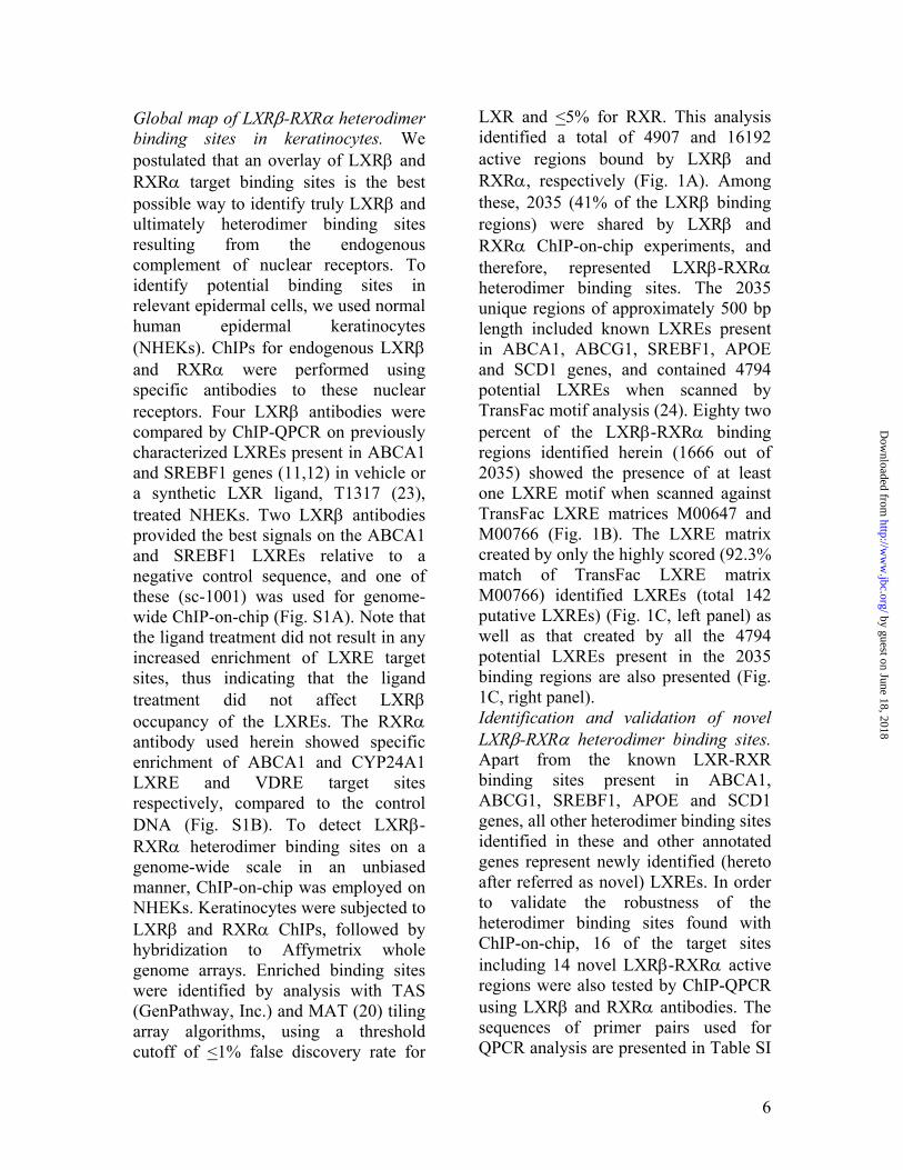
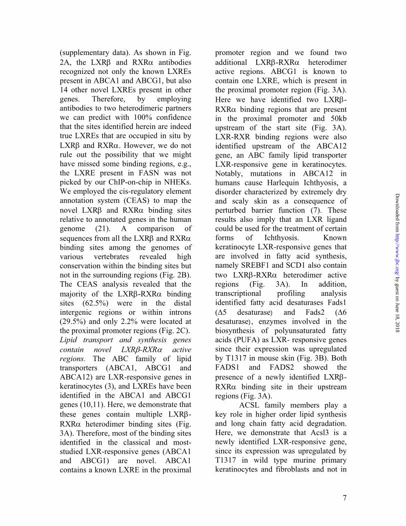
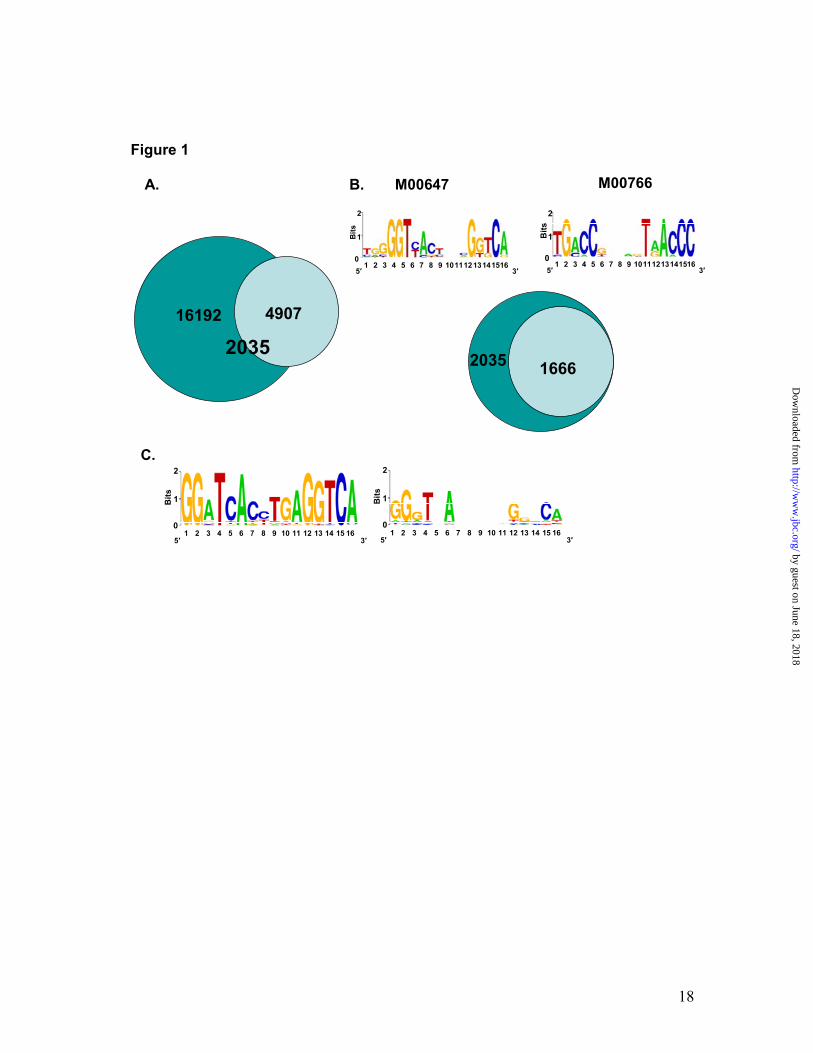
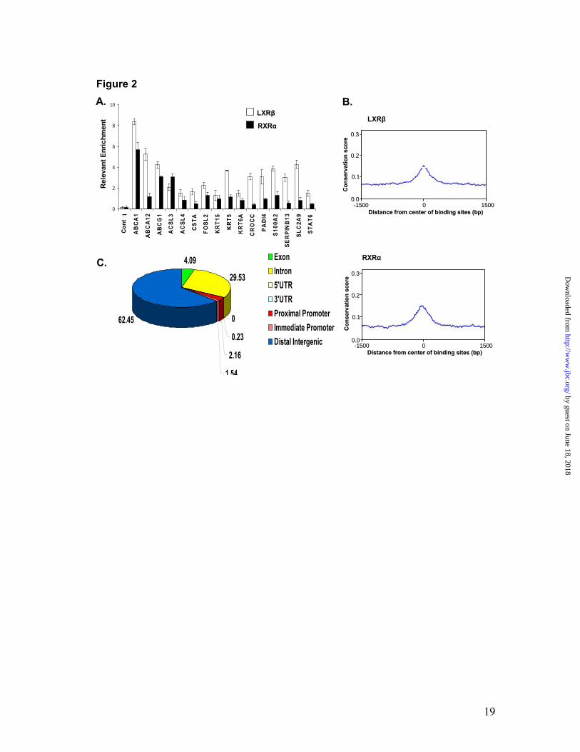
Global map of LXRβ-RXRα heterodimer binding sites in keratinocytes. We postulated that an overlay of LXRβ and RXRα target binding sites is the best possible way to identify truly LXRβ and ultimately heterodimer binding sites resulting from the endogenous complement of nuclear receptors. To identify potential binding sites in relevant epidermal cells, we used normal human epidermal keratinocytes (NHEKs). ChIPs for endogenous LXRβ and RXRα were performed using specific antibodies to these nuclear receptors. Four LXRβ antibodies were compared by ChIP-QPCR on previously characterized LXREs present in ABCA1 and SREBF1 genes (11,12) in vehicle or a synthetic LXR ligand, T1317 (23), treated NHEKs. Two LXRβ antibodies provided the best signals on the ABCA1 and SREBF1 LXREs relative to a negative control sequence, and one of these (sc-1001) was used for genome-wide ChIP-on-chip (Fig. S1A). Note that the ligand treatment did not result in any increased enrichment of LXRE target sites, thus indicating that the ligand treatment did not affect LXRβ occupancy of the LXREs. The RXRα antibody used herein showed specific enrichment of ABCA1 and CYP24A1 LXRE and VDRE target sites respectively, compared to the control DNA (Fig. S1B). To detect LXRβ-RXRα heterodimer binding sites on a genome-wide scale in an unbiased manner, ChIP-on-chip was employed on NHEKs. Keratinocytes were subjected to LXRβ and RXRα ChIPs, followed by hybridization to Affymetrix whole genome arrays. Enriched binding sites were identified by analysis with TAS (GenPathway, Inc.) and MAT (20) tiling array algorithms, using a threshold cutoff of <1% false discovery rate for
LXR and <5% for RXR. This analysis identified a total of 4907 and 16192 active regions bound by LXRβ and RXRα, respectively (Fig. 1A). Among these, 2035 (41% of the LXRβ binding regions) were shared by LXRβ and RXRα ChIP-on-chip experiments, and therefore, represented LXRβ-RXRα heterodimer binding sites. The 2035 unique regions of approximately 500 bp length included known LXREs present in ABCA1, ABCG1, SREBF1, APOE and SCD1 genes, and contained 4794 potential LXREs when scanned by TransFac motif analysis (24). Eighty two percent of the LXRβ-RXRα binding regions identified herein (1666 out of 2035) showed the presence of at least one LXRE motif when scanned against TransFac LXRE matrices M00647 and M00766 (Fig. 1B). The LXRE matrix created by only the highly scored (92.3% match of TransFac LXRE matrix M00766) identified LXREs (total 142 putative LXREs) (Fig. 1C, left panel) as well as that created by all the 4794 potential LXREs present in the 2035 binding regions are also presented (Fig. 1C, right panel). Identification and validation of novel LXRβ-RXRα heterodimer binding sites. Apart from the known LXR-RXR binding sites present in ABCA1, ABCG1, SREBF1, APOE and SCD1 genes, all other heterodimer binding sites identified in these and other annotated genes represent newly identified (hereto after referred as novel) LXREs. In order to validate the robustness of the heterodimer binding sites found with ChIP-on-chip, 16 of the target sites including 14 novel LXRβ-RXRα active regions were also tested by ChIP-QPCR using LXRβ and RXRα antibodies. The sequences of primer pairs used for QPCR analysis are presented in Table SI
by guest on June 18, 2018http://w
ww
.jbc.org/D
ownloaded from
7
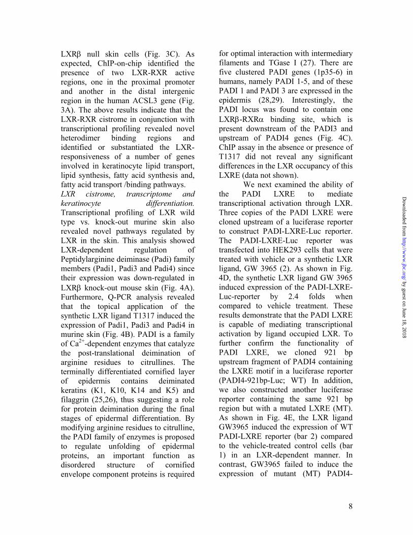
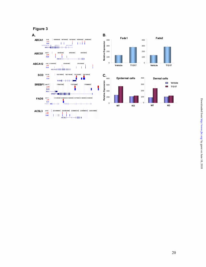
(supplementary data). As shown in Fig. 2A, the LXRβ and RXRα antibodies recognized not only the known LXREs present in ABCA1 and ABCG1, but also 14 other novel LXREs present in other genes. Therefore, by employing antibodies to two heterodimeric partners we can predict with 100% confidence that the sites identified herein are indeed true LXREs that are occupied in situ by LXRβ and RXRα. However, we do not rule out the possibility that we might have missed some binding regions, e.g., the LXRE present in FASN was not picked by our ChIP-on-chip in NHEKs. We employed the cis-regulatory element annotation system (CEAS) to map the novel LXRβ and RXRα binding sites relative to annotated genes in the human genome (21). A comparison of sequences from all the LXRβ and RXRα binding sites among the genomes of various vertebrates revealed high conservation within the binding sites but not in the surrounding regions (Fig. 2B). The CEAS analysis revealed that the majority of the LXRβ-RXRα binding sites (62.5%) were in the distal intergenic regions or within introns (29.5%) and only 2.2% were located at the proximal promoter regions (Fig. 2C). Lipid transport and synthesis genes contain novel LXRβ-RXRα active regions. The ABC family of lipid transporters (ABCA1, ABCG1 and ABCA12) are LXR-responsive genes in keratinocytes (3), and LXREs have been identified in the ABCA1 and ABCG1 genes (10,11). Here, we demonstrate that these genes contain multiple LXRβ-RXRα heterodimer binding sites (Fig. 3A). Therefore, most of the binding sites identified in the classical and most-studied LXR-responsive genes (ABCA1 and ABCG1) are novel. ABCA1 contains a known LXRE in the proximal
promoter region and we found two additional LXRβ-RXRα heterodimer active regions. ABCG1 is known to contain one LXRE, which is present in the proximal promoter region (Fig. 3A). Here we have identified two LXRβ-RXRα binding regions that are present in the proximal promoter and 50kb upstream of the start site (Fig. 3A). LXR-RXR binding regions were also identified upstream of the ABCA12 gene, an ABC family lipid transporter LXR-responsive gene in keratinocytes. Notably, mutations in ABCA12 in humans cause Harlequin Ichthyosis, a disorder characterized by extremely dry and scaly skin as a consequence of perturbed barrier function (7). These results also imply that an LXR ligand could be used for the treatment of certain forms of Ichthyosis. Known keratinocyte LXR-responsive genes that are involved in fatty acid synthesis, namely SREBF1 and SCD1 also contain two LXRβ-RXRα heterodimer active regions (Fig. 3A). In addition, transcriptional profiling analysis identified fatty acid desaturases Fads1 (Δ5 desaturase) and Fads2 (Δ6 desaturase), enzymes involved in the biosynthesis of polyunsaturated fatty acids (PUFA) as LXR- responsive genes since their expression was upregulated by T1317 in mouse skin (Fig. 3B). Both FADS1 and FADS2 showed the presence of a newly identified LXRβ-RXRα binding site in their upstream regions (Fig. 3A).
ACSL family members play a key role in higher order lipid synthesis and long chain fatty acid degradation. Here, we demonstrate that Acsl3 is a newly identified LXR-responsive gene, since its expression was upregulated by T1317 in wild type murine primary keratinocytes and fibroblasts and not in
by guest on June 18, 2018http://w
ww
.jbc.org/D
ownloaded from
8
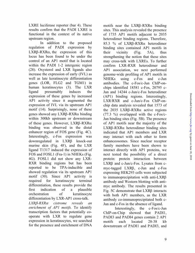
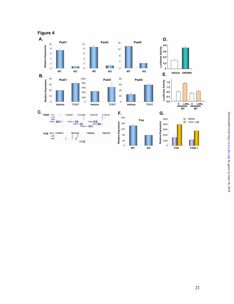
LXRβ null skin cells (Fig. 3C). As expected, ChIP-on-chip identified the presence of two LXR-RXR active regions, one in the proximal promoter and another in the distal intergenic region in the human ACSL3 gene (Fig. 3A). The above results indicate that the LXR-RXR cistrome in conjunction with transcriptional profiling revealed novel heterodimer binding regions and identified or substantiated the LXR-responsiveness of a number of genes involved in keratinocyte lipid transport, lipid synthesis, fatty acid synthesis and, fatty acid transport /binding pathways. LXR cistrome, transcriptome and keratinocyte differentiation. Transcriptional profiling of LXR wild type vs. knock-out murine skin also revealed novel pathways regulated by LXR in the skin. This analysis showed LXR-dependent regulation of Peptidylarginine deiminase (Padi) family members (Padi1, Padi3 and Padi4) since their expression was down-regulated in LXRβ knock-out mouse skin (Fig. 4A). Furthermore, Q-PCR analysis revealed that the topical application of the synthetic LXR ligand T1317 induced the expression of Padi1, Padi3 and Padi4 in murine skin (Fig. 4B). PADI is a family of Ca2+-dependent enzymes that catalyze the post-translational deimination of arginine residues to citrullines. The terminally differentiated cornified layer of epidermis contains deiminated keratins (K1, K10, K14 and K5) and filaggrin (25,26), thus suggesting a role for protein deimination during the final stages of epidermal differentiation. By modifying arginine residues to citrulline, the PADI family of enzymes is proposed to regulate unfolding of epidermal proteins, an important function as disordered structure of cornified envelope component proteins is required
for optimal interaction with intermediary filaments and TGase I (27). There are five clustered PADI genes (1p35-6) in humans, namely PADI 1-5, and of these PADI 1 and PADI 3 are expressed in the epidermis (28,29). Interestingly, the PADI locus was found to contain one LXRβ-RXRα binding site, which is present downstream of the PADI3 and upstream of PADI4 genes (Fig. 4C). ChIP assay in the absence or presence of T1317 did not reveal any significant differences in the LXR occupancy of this LXRE (data not shown).
We next examined the ability of the PADI LXRE to mediate transcriptional activation through LXR. Three copies of the PADI LXRE were cloned upstream of a luciferase reporter to construct PADI-LXRE-Luc reporter. The PADI-LXRE-Luc reporter was transfected into HEK293 cells that were treated with vehicle or a synthetic LXR ligand, GW 3965 (2). As shown in Fig. 4D, the synthetic LXR ligand GW 3965 induced expression of the PADI-LXRE-Luc-reporter by 2.4 folds when compared to vehicle treatment. These results demonstrate that the PADI LXRE is capable of mediating transcriptional activation by ligand occupied LXR. To further confirm the functionality of PADI LXRE, we cloned 921 bp upstream fragment of PADI4 containing the LXRE motif in a luciferase reporter (PADI4-921bp-Luc; WT) In addition, we also constructed another luciferase reporter containing the same 921 bp region but with a mutated LXRE (MT). As shown in Fig. 4E, the LXR ligand GW3965 induced the expression of WT PADI-LXRE reporter (bar 2) compared to the vehicle-treated control cells (bar 1) in an LXR-dependent manner. In contrast, GW3965 failed to induce the expression of mutant (MT) PADI4-
by guest on June 18, 2018http://w
ww
.jbc.org/D
ownloaded from
9
LXRE luciferase reporter (bar 4). These results confirm that the PADI LXRE is functional in the context of its native upstream region.
In addition to the proposed regulation of PADI expression by LXRβ-RXRα, the expression of this locus has been found to be under the control of an AP1 motif that is located within the PADI 1-2 intergenic region (28). Oxysterol and LXR ligands also increase the expression of early (IVL) as well as late keratinocyte differentiation genes (LOR, FLG2 and TGM1) in human keratinocytes (3). The LXR ligand presumably induces the expression of these genes by increased AP1 activity since it augmented the expression of IVL via its upstream AP1 motif (14). Surprisingly, none of these genes showed any LXRβ-RXRα binding within 300kb upstream or downstream of these genes. However, LXRβ-RXRα binding was observed in the distal enhancer region of FOS gene (Fig. 4C). Interestingly, c-Fos expression was downregulated in LXRβ knock-out murine skin (Fig. 4F), and the LXR ligand T1317 induced the expression of FOS and FOSL1 (Fra-1) in NHEKs (Fig. 4G). FOSL1 did not show any LXR-RXR binding regions but has been reported to be TPA-inducible and showed regulation via its upstream AP1 motif (30). Since AP1 activity is required for keratinocyte terminal differentiation, these results provide the first indication of a plausible orchestration of keratinocyte differentiation by LXR-AP1 cross-talk. LXRβ-RXRα cistrome reveals an enrichment of AP1 motifs. To identify transcription factors that potentially co-operate with LXR to regulate gene expression in keratinocytes, we searched for the presence and enrichment of DNA
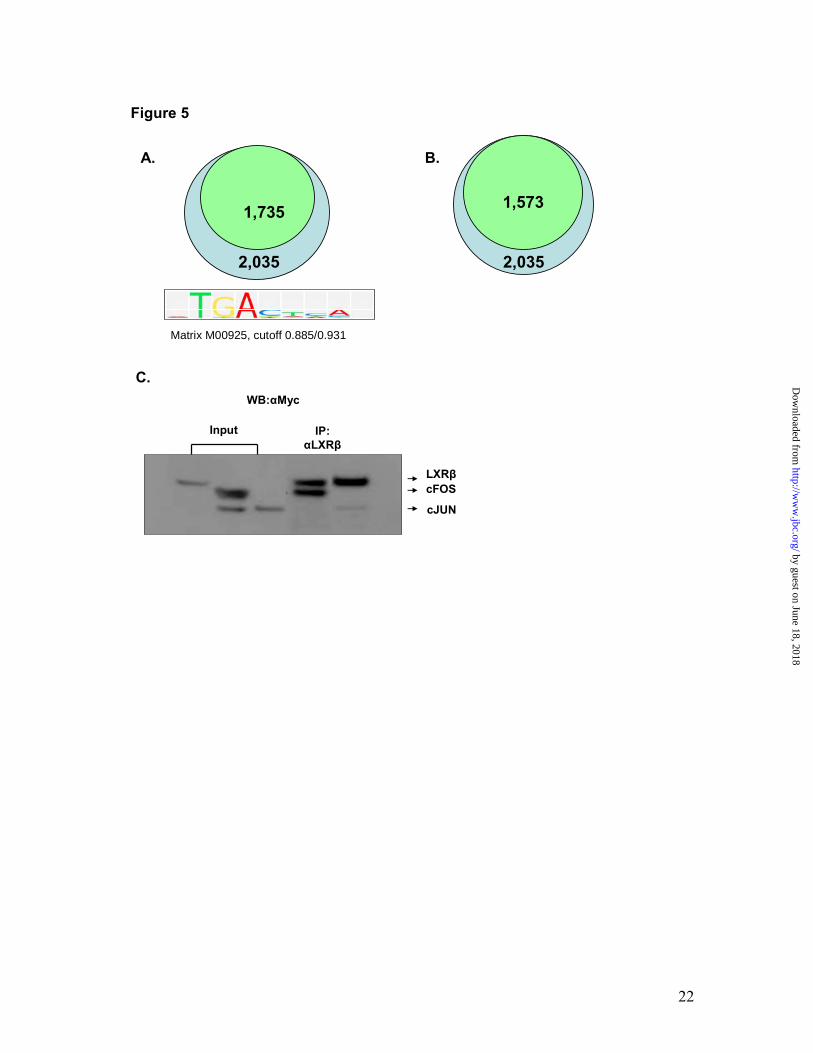
motifs near the LXRβ-RXRα binding sites. This analysis revealed the presence of 1735 AP1 motifs adjacent to 2035 heterodimer binding regions. Therefore, 85.3 % of LXRβ-RXRα heterodimer binding sites contained AP1 motifs in their vicinity (Fig. 5A), thus strengthening the notion that these sites may cross-talk with LXREs. To further confirm LXR-RXR heterodimer and AP1 association, we next performed genome-wide profiling of AP1 motifs in NHEKs using c-Fos and c-Jun antibodies. The c-Fos/c-Jun ChIP-on-chips identified 18581 c-Fos, 20795 c-Jun and 14244 c-Jun/c-Fos heterodimer (AP1) binding regions. Interestingly, LXR/RXR and c-Jun/c-Fos ChIP-on-chip data analysis revealed that 1573 of the 2035 LXRβ-RXRα active regions (77.3 %) overlapped with the c-Fos/c-Jun binding sites (Fig. 5B). The presence of AP1 motifs near the majority of the LXRβ-RXRα heterodimer binding sites indicated that AP1 members and LXR may interact with each other to form enhanceosomes. Since nuclear receptor family members have been shown to interact directly with AP1 proteins, we next tested the possibility of a direct protein: protein interaction between LXRβ and c-Jun/c-Fos. Lysates from c-myc-tagged LXRβ, c-Jun and c-Fos expressing HEK293 cells were subjected to immunoprecipitation with anti-LXRβ antibody and Western blotting with anti-myc antibody. The results presented in Fig. 5C demonstrate that LXRβ interacts with both AP1 members, as the LXR antibody co-immunoprecipitated both c-Jun and c-Fos in the absence of ligand. Interestingly, the c-Fos/c-Jun ChIP-on-Chip showed that PADI1, PADI3 and PADI4 genes contain 2 AP1 motifs each located 28-38 kb downstream of PADI1 and PADI3, and
by guest on June 18, 2018http://w
ww
.jbc.org/D
ownloaded from
10
approximately 10 kb upstream of the PADI4 start site (Table S2). Similarly, ABCA1, ABCG1, ABCA12, ABCA13, SCD1, FADS1, FADS2, ACSL3 and TGM6 showed multiple c-Fos/c-Jun binding regions ranging from 2-11 for each of these genes (Table S2). Taken together, these results indicate extensive cross-talk between LXR and AP1 for the regulation of a number of LXR-responsive genes in skin.
DISCUSSION
Epidermal homeostasis is critical for survival of an organism, and any changes in the skin barrier function through alterations in keratinocyte differentiation and/or lipid synthesis/transport may predispose individuals to cutaneous inflammation. To avoid severe consequences of epidermal barrier perturbations, and swiftly adjust epidermal homeostasis, the skin system appears to employ lipid sensing nuclear receptors, namely LXRs and PPARs. The natural ligands of these receptors present in skin regulate and fine-tune gene expression programs that modulate keratinocyte differentiation, lipid synthesis and transport, which are physiologically important processes essential for epidermal barrier formation and maintenance. A limited number of genes involved in keratinocyte differentiation (TGM1, IVL, LOR, FLG2) and lipid synthesis (SREBF1, SCD1) and transport (ABCA1, ABCG1 and ABCA12) have been identified as LXR-responsive genes, based upon their regulation by natural and/or synthetic LXR agonists in keratinocytes (3,6,31). However, the mechanism of their regulation and whether keratinocyte differentiation genes and ABCA12 are
direct LXR target genes is not clearly understood. To elucidate the mechanism of LXR action in skin, we employed an unbiased approach to identify LXRβ-RXRα heterodimer binding regions in human keratinocytes. As observed with PPARγ (18), most of the LXRβ-RXRα binding sites were located in the distal intergenic regions and introns, and very few sites were found in the proximal promoter regions (Fig. 2C). The distally located binding sites may interact with the basal transcription machinery by DNA looping, as suggested for estrogen and androgen receptors (15,17). The de novo motif search of the heterodimer binding regions against TransFac LXREs yielded DR4 as the predominant cis-acting element (Fig. 1C). As part of the LXR/RXR cistrome analysis, we discovered that 85% of the LXR-RXR heterodimer binding regions contained at least one AP1 motif (Fig. 5A). These results were confirmed by the identification of genome wide ChIP-on-chip of c-Fos/c-Jun (AP1) binding regions in NHEKs, which revealed that about 80% of the LXR-RXR heterodimer binding regions also contained AP1 binding sites (Fig. 5B). Our observation of specific enrichment of AP1 motifs in LXR-RXR binding regions suggests that these transcription factors may orchestrate the process of keratinocyte differentiation, as AP1 is required for the expression of a large number of keratinocyte differentiation genes. This point was further corroborated by the observation that the LXR-RXR cistrome analysis did not show the presence of LXR binding regions in classical keratinocyte differentiation genes (TGM1, IVL, LOR and FLG). These four major genes that facilitate keratinocyte differentiation are
by guest on June 18, 2018http://w
ww
.jbc.org/D
ownloaded from
11
AP1-responsive and the increased expression of IVL by oxysterol LXR ligands has been attributed to its AP1 motif (3,14,32-36). LXR ligands also increased AP1 activity and induced the expression of FOSL1 and FOS in NHEKs (Fig. 4F) (14). Therefore, LXR ligands could induce AP1-dependent expression of keratinocyte differentiation genes either by direct AP1-LXR cross-talk or indirectly by inducing AP1 activity via increased expression of Jun/Fos family members. The cistrome analysis showed the presence of an LXR-RXR motif upstream of the FOS gene, while the expression of both FOS and FOSL1 was increased by the synthetic LXR ligand T1317 in NHEKs (Fig. 4G), thus indicating that FOS and not FOSL1 may be a direct LXR target. Like other keratinocyte differentiation markers, FOSL1 is an AP1-responsive gene, therefore, LXR agonist may increase its expression by inducing AP1 activity and/or by a plausible mechanism involving long-range regulation by DNA looping and co-occupancy of distantly located LXR-RXR and AP1 motifs. Importantly, through both LXR knock-out and pharmacological studies using a specific synthetic ligand, we demonstrate for the first time that PADI family members (PADI1, PADI3 and PADI4) are LXR-responsive genes (Fig. 4A and B), and the expression of the PADI locus might well be under the control of an LXRE, which is located in the PADI3 and PADI4 intergenic region (Fig. 4C-E). Interestingly, PADI3 has been identified as an AP1-responsive gene, whose expression is controlled by a long range enhancer (AP1 site present at 86-kb upstream from the PADI3 promoter) by a mechanism involving chromatin looping (28). PADI1, PADI3
and PADI4 were also found to contain 2 AP1 motifs each (Table S2), thus pointing to a plausible AP1-LXR cross-talk for ligand-dependent regulation of this newly identified family of LXR-responsive genes. Further, TGM6, whose expression was regulated by LXR in keratinocytes and in mice (3), also contains 2 AP1 binding motifs (Table S2).
Inflammatory skin disorders like atopic dermatitis and psoriasis exhibit defects in keratinocyte terminal differentiation and epidermal barrier formation (37-39). Loss-of-function mutations in FLG strongly predispose individuals to atopic dermatitis (40). Similarly, psoriasis exhibits abnormal or psoriatic keratinocyte differentiation, which is characterized by decreased expression of LOR and FLG (41). Activation of LXR stimulates keratinocyte differentiation and lipid synthesis, thus leading to epidermal barrier formation (2-5). The novel mode of regulation of AP1-mediated gene expression by LXR assumes potential therapeutic significance since AP1 activity is reduced in psoriatic lesional skin, and in addition, deletion of its components JunB and c-Jun leads to psoriasis like plaques with inflammatory phenotype in double mutant mice (42). Based upon our results and previously reported pharmacology of LXR, we identify this nuclear receptor as a novel first-in-class target for psoriasis, a dermal inflammatory indication characterized by epidermal hyperproliferation, abnormal keratinocyte differentiation and cutaneous inflammation. LXR ligand inhibited epidermal proliferation in vivo, induced keratinocyte differentiation (Fig. 4A and B), and inhibited cutaneous
by guest on June 18, 2018http://w
ww
.jbc.org/D
ownloaded from
12
inflammation in murine models of atopic and irritant dermatitis (2-4).
Reduced epidermal neutral lipid synthesis (sphingolipids, cholesterol and fatty acids) leading to defective barrier repair is a hallmark of atopic dermatitis, a chronic inflammatory condition of the skin that afflicts approximately 15-20% of the children. Moreover, xerosis or dry skin because of reduced keratinocyte lipid synthesis is a major manifestation of chronological skin aging, which is recognized as a low-grade chronic cutaneous inflammatory condition. Therefore, enhancement of epidermal barrier function via increased keratinocyte differentiation and lipid production may result in amelioration of inflammation and contribute to the efficacy of LXR ligand in skin aging, psoriasis and atopic dermatitis. T1317 induced the expression of (a) enzymes involved in sphingolipid and ceramide biosynthetic pathways, (b) proteins required for fatty acid synthesis, (c) cholesterol/lipid transporters and (d) lipid binding proteins (3). We demonstrate that the genes involved in these processes contain multiple LXR-RXR heterodimer binding regions (Fig. 3A). Our observation that ABCA12 has two heterodimer binding regions with LXREs indicates it to be direct LXR target (Fig. 3A), and suggests that LXR ligands could be used for certain forms of Ichthyosis (keratinization disorders) and also for dryness and scalyness associated with skin aging and psoriasis. The studies presented herein demonstrating that ACSL3, FADS1 and
FADS2 are newly identified LXR-responsive genes (Figs. 3B and C) further strengthen the notion that LXR is a major regulator of epidermal lipid synthesis. Accordingly, LXR ligands improved epidermal barrier function in vivo and showed efficacy in models of contact and irritant dermatitis (2). Over the years nuclear receptors have proven to be ligands of choice for dermal inflammatory indications. Natural and synthetic ligands of glucocorticoid receptors, vitamin D receptor and retinoic acid receptor are currently in clinical practice for the treatment of psoriasis, dermatitis and photoaging. Glucocorticoids are extensively prescribed for the treatment of psoriasis and atopic dermatitis because of their potent anti-inflammatory properties. However, their prolonged use is associated with skin thinning, which probably results from their anti-AP1 activity since the expression of genes for a vast number of proteins that mediate keratinocyte differentiation is under the control of transcription factor AP1. Retinoid use is associated with skin irritation, scaling, erythema and burning sensation (43). Vitamin D ligands though used for the treatment of psoriasis have the potential for causing dermatitis (44). Therefore, we believe that LXR ligands because of their unique pharmacology may provide a new generation of safe therapeutic agents for the prevention and treatment of skin keratinization and inflammatory indications.
REFERENCES
by guest on June 18, 2018http://w
ww
.jbc.org/D
ownloaded from
13
1. Chawla, A., Repa, J. J., Evans, R. M., and Mangelsdorf, D. J. (2001) Science 294, 1866-1870
2. Fowler, A. J., Sheu, M. Y., Schmuth, M., Kao, J., Fluhr, J. W., Rhein, L., Collins, J. L., Willson, T. L., Mangelsdorf, D. J., Elias, P. M., and Feingold, K. R. (2003) J. Invest. Dermatol. 120, 246-255
3. Chang, K. C., Shen, Q., Oh, I. G., Jelinsky, S. A., Jenkins, S. F., Wang, W., Wang, Y., LaCava, M., Yudt, M. R., Thompson, C. C., Freedman, L. P., Chung, J. H., and Nagpal, S. (2008) Mol. Endocrinol. 22, 2407-2419
4. Kömüves, L. G., Schmuth, M., Fowler, A. J., Elias, P. M., Hanley, K., Man, M. Q., Moser, A. H., Lobaccaro, J. M., Williams, M. L., Mangelsdorf, D. J., and Feingold, K. R. (2002) J. Invest. Dermatol. 118, 25-34
5. Man, M. Q., Choi, E. H., Schmuth, M., Crumrine, D., Uchida, Y., Elias, P. M., Holleran, W. M., and Feingold, K. R. (2006) J. Invest. Dermatol. 126, 386-392
6. Jiang, Y. J., Lu, B., Kim, P., Elias, P. M., and Feingold, K. R. (2006) J. Lipid Res. 47, 2248-2258
7. Lefévre, C., Audebert, S., Jobard, F., Bouadjar, B., Lakhdar, H., Boughdene-Stambouli, O., Blanchet-Bardon, C., Heilig, R., Foglio, M., Weissenbach, J., Lathrop, M., Prud'homme, J. F., and Fischer, J. (2003) Hum. Mol. Genet. 12, 2369-2378
8. Zuo, Y., Zhuang, D. Z., Han, R., Isaac, G., Tobin, J. J., McKee, M., Welti, R., Brissette, J. L., Fitzgerald, M. L., and Freeman, M. W. (2008) J. Biol. Chem. 283, 36624-36635
9. Laffitte, B. A., Repa, J. J., Joseph, S. B., Wilpitz, D. C., Kast, H. R., Mangelsdorf, D. J., and Tontonoz, P. (2001) Proc. Natl. Acad. Sci. USA 98, 507-512
10. Kennedy, M. A., Venkateswaran, A., Tarr, P. T., Xenarios, I., Kudoh, J., Shimizu, N., and Edwards, P. A. (2001) J. Biol. Chem. 276, 39438-39447
11. Costet, P., Luo, Y., Wang, N., and Tall, A. R. (2000) J. Biol. Chem. 275, 28240-28245
12. Yoshikawa, T., Shimano, H., Amemiya-Kudo, M., Yahagi, N., Hasty, A. H., Matsuzaka, T., Okazaki, H., Tamura, Y., Iizuka, Y., Ohashi, K., Osuga, J., Harada, K., Gotoda, T., Kimura, S., Ishibashi, S., and Yamada, N. (2001) Mol. Cell. Biol. 21, 2991-3000
13. Joseph, S. B., Laffitte, B. A., Patel, P. H., Watson, M. A., Matsukuma, K. E., Walczak, R., Collins, J. L., Osborne, T. F., and Tontonoz, P. (2002) J. Biol. Chem. 277, 11019-11025
14. Schmuth, M., Elias, P. M., Hanley, K., Lau, P., Moser, A., Willson, T. M., Bikle, D. D., and Feingold, K. R. (2004) J. Invest. Dermatol. 123, 41-48
15. Carroll, J. S., Liu, X. S., Brodsky, A. S., Li, W., Meyer, C. A., Szary, A. J., Eeckhoute, J., Shao, W., Hestermann, E. V., Geistlinger, T. R., Fox, E. A., Silver, P. A., and Brown, M. (2005) Cell 122, 33-43
16. Carroll , J. S., Meyer, C. A., Song, J., Li, W., Geistlinger, T. R., Eeckhoute, J., Brodsky, A. S., Keeton, E. K., Fertuck, K. C., Hall, G. F., Wang, Q., Bekiranov, S., Sementchenko, V., Fox, E. A., Silver, P. A., Gingeras, T. R., Liu, X. S., and Brown, M. (2006) Nat. Genet. 38, 1289-1297
17. Massie, C. E., Adryan, B., Barbosa-Morais, N. L., Lynch, A. G., Tran, M. G., Neal, D. E., and Mills, I. G. (2007) EMBO Rep. 8, 871-878
by guest on June 18, 2018http://w
ww
.jbc.org/D
ownloaded from
14
18. Lefterova, M. I., Zhang, Y., Steger, D. J., Schupp, M., Schug, J., Cristancho, A., Feng, D., Zhuo, D., Stoeckert, C. J. J., Liu, X. S., and Lazar, M. A. (2008) Genes Dev. 22, 2941-2942
19. Zheng, Y., Du, X., Wang, W., Boucher, M., Parimoo, S., and Stenn, K. S. (2005) J. Invest. Dermatol. 124, 867-876
20. Johnson, W. E., Li, W., Meyer, C. A., Gottardo, R., Carroll, J. S., Brown, M., and Liu, X. S. (2006) Proc. Natl. Acad. Sci. USA 103, 12457-12462
21. Ji, X., Li, W., Song, J., Wei, L., and Liu, X. S. (2006) Nucleic Acids Res. 34, W551-W554
22. Sandelin, A., Alkema, W., Engström, P., Wasserman, W. W., and Lenhard, B. (2004) Nucleic Acids Res. 32, D91-D94
23. Schultz, J. R., Tu, H., Luk, A., Repa, J. J., Medina, J. C., Li, L., Scwendner, S., Wang, S., Thoolen, M., Mangelsdorf, D. J., Lusting, K., and Shan, B. (2000) Genes Dev. 14, 2831-2838
24. Matys, V., Fricke, E., Geffers, R., Gössling, E., Haubrock, M., Hehl, R., Hornischer, K., Karas, D., Kel, A. E., Kel-Margoulis, O. V., Kloos, D. U., Land, S., Lewicki-Potapov, B., Michael, H., Münch, R., Reuter, I., Rotert, S., Saxel, H., Scheer, M., Thiele, S., and Wingender, E. (2003) Nucleic Acids Res. 31, 374-378
25. Ohsawa, T., Maruyama, I., and Senshu, T. (1999) J. Dermatol. Sci. 19, 68-73 26. Ishida-Yamamoto, A., Senshu, T., Takahashi, H., Akiyama, K., Nomura, K., and
Iizuka, H. (2000) J. Invest. Dermatol. 114, 701-705 27. Tarcsa, E., Marekov, L. N., Mei, G., Melino, G., Lee, S. C., and Steinert, P. M.
(1996) J. Biol. Chem. 271, 30709-30716 28. Chavanas, S., Adoue, V., Méchin, M. C., Ying, S., Dong, S., Duplan, H.,
Charveron, M., Takahara, H., Serre, G., and Simon, M. (2008) PLoS One 3, e3408
29. Nakashima, K., Hagiwara, T., and Yamada, M. (2002) J. biol. Chem. 277, 49562-49568
30. Fleischmann, A., Hafezi, F., Elliott, C., Remé, C. E., Rüther, U., and Wagner, E. F. (2000) Genes Dev. 14, 2695-2700.
31. Jiang, Y. J., Lu, B., Kim, P., Paragh, G., Schmitz, G., Elias, P. M., and Feingold, K. R. (2007) J. Invest. Dermatol. 128, 104-109
32. Rossi, A., Jang, S. I., Ceci, R., Steinert, P. M., and Markova, N. G. (1998) J. Invest. Dermatol. 110, 34-40
33. Crish, J. F., and Eckert, R. L. (2008) J. Invest. Dermatol. 128, 530-541 34. Hanley, K., Ng, D. C., He, S. S., Lau, P., Min, K., Elias, P. M., Bikle, D. D.,
Mangelsdorf, D. J., Williams, M. L., and Feingold, K. R. (2000) J. Invest. Dermatol. 114, 545-553
35. Jang, S. I., and Steinert, P. M. (2002) J. Biol. Chem. 277, 42268-42279 36. Jang, S. I., Steinert, P. M., and Markova, N. G. (1996) J. Biol. Chem. 271, 24105-
24114 37. Elias, P. M. (2009) Clin. Med. Dermatol. 2, 1-3 38. de Guzman Strong, C., Conlan, S., Deming, C. B., Cheng, J., Sears, K. E., and
Segre, J. A. (2010) Hum. Mol. Genet. 19, 1453-1460 39. Nemoto-Hasebe, I., Akiyama, M., Nomura, T., Sandilands, A., McLean, W. H.,
and Shimizu, H. (2009) J. Invest. Dermatol. 129, 682-689
by guest on June 18, 2018http://w
ww
.jbc.org/D
ownloaded from
15
40. Palmer, C. N. A., Irvine, A. D., Terron-Kwiatkowski, A., Zhao, Y., Liao, H., Lee, S. P., Goudie, D. R., Sandilands, A., Campbell, L. E., Smith, F. J. D., O'Regan, G. M., Watson, R. M., Cecil, J. E., Bale, S. J., Compton, J. G., DiGiovanna, J. J., Fleckman, P., Lewis-Jones, S., Arseculeratne, G., Sergeant, A., Munro, C. S., Houate, B. E., McElreavey, K., Halkjaer, L. B., Bisgaard, H., Mukhopadhyay, S., and McLean, W. H. I. (2006) Nature Genetics 38, 441-446
41. DiSepio, D., Chandraratna, R. A., and Nagpal, S. (1999) Drug Discov. Today 4, 222-231
42. Zenz, R., Eferl, R., Kenner, L., Florin, L., Hummerich, L., Mehic, D., Scheuch, H., Angel, P., Tschachler, E., and Wagner, E. F. (2005) Nature 437, 369-375
43. Singh, M., and Griffiths, C. (2006) Dermatol. Ther. 19, 297-305 44. Li, M., Hener, P., Zhang, Z., Kato, S., Metzger, D., and Chambon, P. (2006) Proc.
Natl. Acad. Sci. USA 103, 11736-11741
FOOTNOTES We thank R. Bernotas for T1317, and Wei Wang for providing us with wt and LXRβ KO mouse keratinocytes. The abbreviations used are: LXR, liver X receptor; RXR, retinoid X receptor; ChIP, chromatin immunoprecipitation; NHEK, normal human epidermal keratinocytes; ER, estrogen receptor; AR, androgen receptor; PPAR, peroxisome proliferator activated receptor; WT, wild type; KO, knock-out; QPCR, quantitative polymerase chain reaction; TAS, tiling array analysis; MAT, microarray analysis tool.
FIGURE LEGENDS
Fig. 1. Genome-wide identification of LXR-RXR active regions in primary human keratinocytes. Active regions identified by Chip-on-Chip using LXRβ and RXRα antibodies and Affymetrix whole genome arrays. There are 4907 LXR and 16192 RXR binding sites detected by ChIP-chip experiments in NHEKs using receptor-specific antibodies. 2035 LXR-RXR overlapped binding sites were found (overlap means at least 1bp in common). Data was analyzed by MAT 2.09 with probes remapped to the hg18 Human Genome Assembly. The threshold cutoffs for binding sites were FDR < 1% for LXR and < 5% for RXR (A). Interrogation of LXR-RXR binding sites against TransFac reported LXREs. 1666 out of 2035 LXR-RXR binding sites contain at least one LXR binding element predicted by TransFac Match when analyzed against TransFac LXRE motif matrices M00647 and M00766 (B). SeqLogos of de novo generated LXR-RXR heterodimer binding motifs. The LXR binding site matrix generated by the top 142 LXREs with the highest scores is shown on the left, while the LXR binding site matrix generated from all 4794 LXREs is shown on the right side (C). Fig. 2. Analysis of LXR-RXR binding sites. Validation of LXRβ-RXRα ChIP-on-Chip predictions. NHEKs were subjected to ChIP-qPCR with LXRβ sc-1001 antibody (open bars) or RXRα sc-553 antibody (black bars). LXR and RXR binding was assayed at 16 LXR-RXR heterodimer-containing active regions from the ChIP-on-Chip experiment, as well as a gene-deficient region on chromosome 4 (Cont I) that served as a negative control (A). Sequence
by guest on June 18, 2018http://w
ww
.jbc.org/D
ownloaded from
16
conservation analysis. The LXRβ and RXRα ChIP active regions were aligned at their centers and uniformly expanded to 3 kb in each direction, and phastCons scores were retrieved (http://genome.ucsc.edu) and averaged at each position (B). Genome-wide mapping of LXR-RXR active regions. LXRβ-RXRα binding regions were mapped relative to their nearest RefSeq 3' genes using CEAS (21). Proximal promoter was defined as <1kb upstream from the transcription start site. When the LXR-RXR binding region is within a gene, it is further categorized as within the 5' UTR, 3' UTR, a coding exon, or an intron. Immediate downstream is defined as <1kb downstream from RefSeq 3' end. Enhancers are defined as more than 1kb from a RefSeq gene (C). Fig. 3. Identification of novel LXR-RXR heterodimer binding regions in human primary keratinocytes. LXR and RXR binding sites detected by ChIP-chip frequently overlap and cluster around target genes. All target genes are shown in their native chromosomal location according to 2006 Human Genome Assembly (hg18) in the UCSC Genome Browser (http://genome.ucsc.edu). Red and blue blocks represent regions of enriched LXR or RXR binding signal, respectively. Vertical lines within the genes represent exons, horizontal lines represent introns, and the arrowheads represent the direction of transcription (A). LXR-dependent regulation of newly identified responsive genes. The relative expression level of genes Fads 1 and Fads 2 was compared by real-time PCR in mouse skin after topical treatment with vehicle or T1317 (10 mM) (B). The relative expression level of Acsl3 was compared by Taqman real-time PCR in wild-type or LXRβ knock-out (KO) mouse primary epidermal keratinocytes and dermal fibroblasts treated with vehicle (purple bars) or T1317 (red bars) (D). Fig. 4. PADI family members are newly identified LXR-responsive genes in skin. The relative expression level of genes Padi1, Padi3 and Padi4 was compared in the skin tissue from wild type and LXRβ knock-out (KO) mouse (A) and in skin obtained from mice topically treated with vehicle or 10 mM T1317. Mean expression of these genes was determined by real-time QPCR, and the results were normalized to 18SRNA expression (B). Identification of a LXR-RXR binding site in PADI and FOS genes in human keratinocytes. LXR-RXR binding sites detected by ChIP-chip in FOS gene and the PADI gene locus are presented as LXR (red blocks) and RXR (blue blocks) active regions (C). The layout of the diagram is the same as described in Fig 3A. Analysis of the transcriptional activity of PADI LXRE. HEK293 cells transfected with the PADI-LXRE-Luc reporter show LXR synthetic ligand-dependent transcription (D). Mutational analysis of PADI LXRE. HEK293 cells were transfected with WT (wild type) or MT (mutant) PADI4-921bp-Luc in the presence (bars 2 and 4) or absence (C; bars 1 and 3) of synthetic LXR ligand GW3965 and LXRα expression vector (E). The relative expression level of Fos was compared in the mouse skin tissue from wild type and LXRβ knock-out (KO) mouse by Taqman real-time PCR (F). The relative expression level of FOS and FOSL1 was compared in NHEKs treated with vehicle (blue bars) or the synthetic LXR ligand, T1317 (gold bars) by Taqman real-time PCR (G). Fig. 5. LXR-AP1 association and interaction. Enrichment of AP1 motifs in LXRβ-RXRα active regions as determined by de novo analysis. 1735 out of 2035 LXR-RXR binding sites contained at least one AP-1 binding motif predicted by TransFac Match. The AP-1 motif matrix used (M00925) with cutoff 0.885/0.931 (matrix and core match %) is shown (A). Identification of genome-wide overlap between LXR/RXR and AP1 binding sites. 1573 out of 2035 LXR-RXR binding sites overlapped with at least one AP-
by guest on June 18, 2018http://w
ww
.jbc.org/D
ownloaded from
17
1 binding site detected by c-Fos/c-Jun ChIP-chip experiments. Overlap is defined by at least 1 bp in common (B). LXRβ directly interacts with c-Fos and c-Jun proteins. Co-immunoprecipitation was performed to examine the interaction between LXRβ and cFos/cJun. The lysate with co-expression of LXRβ and c-Fos/c-Jun was immunoprecipitated by anti-LXRβ antibody. Western blot using anti-Myc antibody showed co-existence of c-Jun and c-Fos in the same protein complex as LXRβ (C).
by guest on June 18, 2018http://w
ww
.jbc.org/D
ownloaded from
18
16192 4907
2035
Figure 1
A. M00647 M00766
16662035
B.
C.
2 3 4 5 6 7 8 9 10 1 5′
11 12 13 14 15 3′
16
2
1Bits
02 3 4 5 6 7 8 9 10 1
5′11 12 13 14 15
3′16
2
1Bits
0
2 3 4 5 6 7 8 9 10 1 5′
11 12 13 14 15 3′
16
2
1Bits
0 2 3 4 5 6 7 8 9 10 1 5′
11 12 13 14 15 3′
16
2
1Bits
0
by guest on June 18, 2018http://w
ww
.jbc.org/D
ownloaded from
19
--15001500 1500150000Distance from center of binding sites (Distance from center of binding sites (bpbp))
0.30.3
0.20.2
0.10.1
0.00.0C
onse
rvat
ion
scor
eC
onse
rvat
ion
scor
e
Figure 2
0
2
4
6
8
10
Unt
r4-II
AB
CA
1
AB
CA
12
AB
CG
1
AC
SL3
AC
SL4
CST
A
FOSL
2
KR
T15
KR
T5
KR
T6A
CR
OC
C
PAD
I4
S100
A2
SER
PIN
B13
SLC
2A9
STA
T6
Con
t
LXRβ
RXRα
Rel
evan
t Enr
ichm
ent
A.
--15001500 1500150000Distance from center of binding sites (Distance from center of binding sites (bpbp))
0.30.3
0.20.2
0.10.1
0.00.0
Con
serv
atio
n sc
ore
Con
serv
atio
n sc
ore
LXRβ
B.
4.09
29.53
0
0.23
2.16
1.54
62.45
ExonIntron5'UTR3'UTRProximal PromoterImmediate PromoterDistal Intergenic
C. RXRα
by guest on June 18, 2018http://w
ww
.jbc.org/D
ownloaded from
20
Figure 3
ABCA1
ABCG1
SCD
ABCA12
ACSL3
FADS
SREBF1
A. B.
C.
Vehicle
T1317
Dermal cells
WT KO0
100
200
300
400Epidermal cells
Rel
ativ
e Ex
pres
sion
0
200
400
600
800
WT KO
Fads1
Rela
tive
Exp
ress
ion
0
100
200
300
400
Vehicle T1317
Fads2
0
100
200
300
400
Vehicle T1317
by guest on June 18, 2018http://w
ww
.jbc.org/D
ownloaded from
21
Figure 4
FOS
PADIC. F. G.
T1317, 1 µM
A.R
elat
ive
Expr
essi
onPadi1
0
2
4
6
8
10
WT KO
Padi3
0
2
4
6
8
10
WT KO
Padi4
0
4
8
12
16
WT KO
B.Padi1
0
10
20
30
40
Vehicle T1317
Rel
ativ
e Ex
pres
sion Padi3
0
200
400
600
800
1000
Vehicle T1317
Padi4
0
20
40
60
80
Vehicle T1317
Fos
0
40
80
120
160
200R
elat
ive
Expr
essi
on
WT KO
Vehicle
0
1000
2000
3000
4000
5000
Rel
ativ
e Ex
pres
sion
FOS FOSL1
0
0.2
0.4
0.6
0.8
Luci
fera
se A
ctiv
ity
Vehicle GW3965
D.
C LXRα C LXRα0.0
0.4
0.8
1.2
1.6
WT MT
Luci
fera
se A
ctiv
ity
E.
by guest on June 18, 2018http://w
ww
.jbc.org/D
ownloaded from
22
2,035
1,735
Matrix M00925, cutoff 0.885/0.931
2,035
1,573
Input IP:αLXRβ
LXRβcFOS
cJUN
WB:αMyc
Figure 5
A. B.
C.
by guest on June 18, 2018http://w
ww
.jbc.org/D
ownloaded from
Freedman, Catherine C. Thompson and Sunil NagpalQi Shen, Yuchen Bai, Ken C. N. Chang, Yongjun Wang, Thomas P. Burris, Leonard P.
co-ordination of LXR and AP1 signaling in keratinocytesLiver X receptor-retinoid X receptor (LXR-RXR) heterodimer cistrome reveals
published online February 24, 2011J. Biol. Chem.
10.1074/jbc.M110.165704Access the most updated version of this article at doi:
Alerts:
When a correction for this article is posted•
When this article is cited•
to choose from all of JBC's e-mail alertsClick here
Supplemental material:
http://www.jbc.org/content/suppl/2011/02/24/M110.165704.DC1
by guest on June 18, 2018http://w
ww
.jbc.org/D
ownloaded from