lnvited review role of diffusible and transcription ... of diffusible and...types, which include ......
TRANSCRIPT

Histol Histopathol (2000) 15: 657-666
001 : 10.14670/HH-15.657
http://www.hh.um.es
Histology and Histopathology Cel/ular and Molecular Biology
lnvited Review
Role of diffusible and transcription factors in inner ear development: implications in regeneration L.M. Frago1, G. Camarero 1, S. CaA6n 1, C. PaAeda•, C. Sanz1, Y. Leon 1, F. Giraldez2 and l. Vare la-Nieto 1
'lnstitute of Biomedical Research "Alberto Sois", CSIC-UAM, Madrid, Spain and 21nstitute of Biology and Molecular Genetic (IBGM), University of Valladolid-CSIC, School of Medicine, Valladolid, Spain
Summary. Organogenesis involves a dynamic balance of the mechanisms regulating cell division, differentiation and death. The development of the chicken embryo inner ear offers a well-characterised model at the morphological level to study which signals are implicated in the modulation of cellular activation and commitment. The early developmental decisions that control the origin of the inner ear elements are just beginning to be identified by complementary in vivo and in vitro studies. Insulin-like growth factor-! (IGF-I) and nerve growth factor (NGF) are among the best characterised diffusible factors acting during inner ear development. Although the cellular actions of these factors are beginning to be understood, the signalling pathways triggered by them still remain largely unknown. In this context, viral vehicles can be used to deliver genes and then analyse their functional roles during inner ear development. A model is proposed where the actions of IGF-I and NGF contribute to the combinatoria! expression of Jun and Fos family members in particular domains of the otic vesicle. Sorne of these mechanisms may be also implicated in otic regeneration.
Key words: Apoptosis, Insulin-like growth factor-!, Nerve growth factor, RCAS retroviral vectors, Jun signalling pathways
1. The early development of the vertebrate inner ear
The adult inner ear in vertebrates is a complex, highly-differentiated structure responsible for audition, the perception of movement , and the sense of equilibrium. It contains more than a dozen different cell types, which include neurons , sensory hair cells, secretory cells, and supporting cells. These cell types are organised in functional regions (www.iurc.montp. inserm.fr/cric /audition). Different regions of the ear
Offprint requests to: Dr. Isabel Varela-Nieto, Instituto de Investigaciones Biomedicas 'Alberto Sois ' , CSIC-UAM, Arturo Duperier 4, 28029 Madrid, Spain. Fax: 34-91-5854587. e-mail: [email protected]
show an elaborated spatial organisation responsible for the different functions of the organ: the vestibular portion of the ear is responsible for the senses of motion and position while the auditory or cochlear region is responsible for the sense of hearing. In both portions, hair cells convert mechanical stimulation into electrical signals, which are then processed by the nervous system . Hair cells are innervated by sensory neurons that project towards specific nuclei in the central nervous system . The development of the chicken embryo inner ear offers a well-characterised model at the morphological level to study which signals are implicated in the modulation of cellular proliferation, differentiation and programmed cell death (Fekete, 1996) .
The vertebrate inner ear originates from the head ectoderm where the otic placode is formed. After it invaginates to forrn the otic pit that latter closes forrning the otic vesicle or otocyst (Fig. 1). This is a transient structure that undergoes a distinct period of intense cell proliferation ( stages 18-22 in the chicken, Hamburguer and Hamilton, 1951) that precedes the differentiation of the various cell types and compartments that will conform the adult inner ear (Bissonnette and Fekete, 1996). Neuroblasts for the cochleo-vestibular ganglion (CVG) migrate out from the medial wall of the otic vesicle. The CVG contains the afferent neurons that connect the sensory epithelium of the inner ear to the central nervous system (Hemond and Morest, 1991) . Later in development, the CVG is separated into two ganglia that innervate separately the cochlear and vestibular parts of the inner ear connecting them to the central nervous system .
There are severa! experimental approaches to study inner ear development in the chicken embryo. In vivo studies facilitate the analysis of the expression of target genes in whole mount preparations and also the study of cell proliferation and cell death patterns through development (reviewed by Torres and Giraldez, 1998). Embryonic day 2.5 (E2.5) otic vesicle s can be dissected and the explants cultured to analyse the role of extracellular factors and intracellular signals in a defined medium (León et al., 1998; Torres and Giraldez, 1998). Finally, viral vehicles can be used to deliver genes and

658
IGF-1 and NGF actions in the inner ear
then analyse the functional consequences of their overexpression (Hughes et al., 1987; Federspiel and Hughes, 1997), these genes being either wild type or dominant negative mutants. Therefore, ectopic expression of certain genes by means of retroviral vectors can provide a unique information on their functional roles during inner ear development (Federspiel and Hughes, 1997; Fekete et al., 1997). RCAS retroviral vectors facilitate the misexpresion of genes in ovo (Cepko, 1992 ; Fekete et al., 1997) and in cultured otic vesicles (Sanz et al., 1999) in order to study the consequences of manipulating the levels of selected extracellular factors or transducing proteins.
The basic molecules responsible for the normal ontogenesis of the inner ear have started to be unravelled. Diffusible factors form part of the environmental input that contributes to modulate the early development of the inner ear. IGF-1, retinoic acid and NGF actions have been reported to be fundamental for the correct deve lopment of the otic vesicle and CVG (León et al., 1998; Torres and Giraldez, 1998). Extrace ll ular factors initiate intracellular signalling processes that will induce long term cellular phenotypic responses. As will be discussed in detail below , IGF-1 stimulates the generation of lipidic second messengers (León et al., 1998), activates the Raf /mitogen -activated protein kinases (MAPKs) cascade (Sanz et al. , 1999) and increases c-fos, c-jun and PCNA levels leading to cell
growth and survival (León et al., 1998). On the contrary , NGF activates Jun N-terminal kinase (JNK) and stimulates the hydrolysis of spbingomyelin to generate ceramide, in a process that regulates apoptotic cell death in organotypic cultures of otic vesicles (Frago et al., 1998). Retinoic acid downregulates c-fos and proliferating ce ll nuclear antigen (PCNA) levels and induces hair cell differentiation (León et al., 1995a; Lee and Cotanche, 1996 ; Sanz et al., 1999) . Raf overexpression by means of retroviral RCAS vectors potentiates IGF-1 actions while it blocks both NGFinduced apoptosis and retinoic acid effects in the otic vesicle (Sanz et al., 1999).
2. Multiple roles of IGF-1 in inner ear development: antagonism with NGF actions
lnsulin and IGF-1 play an important role in the development, growth, and survival of normal cells. lnsu lin and IGF-1 interact in avían cells with at least two different receptors with tyrosine kinase act ivity, the type 1 IGF-1 and insul in receptors . In addition, a hybrid receptor composed of subunits from the insulin- and type 1 IGF-receptors has been identified. IGF -1 activity is further mod ulated by at least ten different IGF-binding proteins (Parrizas and LeRoith, 1997; Spagnoli and Rosenfeld, 1997). Insulin-like mo lecu les, their receptors, and binding proteins conform the IGF axis
OTIC PLACODE OTICCUP OTIC VESICLE LABYRINTH
El.5, stg 10 (36 h)
E2, stg 13 (50 h)
Neuroblasts delami nate E2-E3.5, stg 13-21
E3, stg 17 (58 h)
endolympbatic duct
semicircular ~l-- ...--;7 canals
Hair cells bom FA-E8, stg 24-33
Hair cells differentia te and mature E5-hatching
----utricle
saccule
cochi ea
E7, stg 30 (162 h)
Fig. 1. Schematic drawing of the early stages of chicken inner ear development. Stages have been set following the Hamburger and Hamilton criteria .

659
IGF-1 and NGF actions in the inner ear
(http: //www.lGF-society.org /). Hence , there is a great potential for complex interactions between insulin family factors to regulate normal cell growth and development.
The occurrence of IGF-1 in the otic vesicle and CVG
was explored by immunohistochemistry and by in situ hybridisation using a full-length chicken IGF-1 cDNA probe (Kajimoto and Rotwein, 1989). Fig. 2A shows that IGF-1 mRNA is homogeneously expressed in the CVG. However , in the otic vesicle epithelium IGF-1 transcripts and immunoreactivity were concentrated in the ventral, lateral and dorsal aspects , and were apparently excluded from the medial wall facing the neural tube (Fig. 2A,B and León et al., 1995b). The CVG displayed a strong IGF-1 immunoreactivity , without evidence of regional distribution . The occurrence of IGF-1 receptor in the otic vesicle was studied by in situ hybridisation using a chicken IGF-1 type 1 receptor probe (de la Rosa et al. , 1994). Fig. 2C shows the presence of type 1 receptors homogeneously distributed in the otic epithelium. Previous data on the presence of IGF-1 binding to specific receptors in the otic vesicle and CVG indicated that IGF-1 binding is related to the functional expression of type 1 IGF receptors (León et al. , 1995b) . The localisation of IGF-1 binding indicates that the ventromedial otic epith elium shows strong labelling , as does the adjacent developing CVG. Taken together, these data indicate that IGF-1 factor and receptor are abundantly expressed in the chicken inner ear at E3. The pattern of expression of IGF-1 binding proteins in the otocyst has not yet been reported. Our results suggest that the factor present in the dorsal areas of the otocyst may be sequester ed by a binding protein that prevents effective binding to the IGF-1 type 1 receptor.
Low doses of IGF-1 (1 nm) st imul ate proliferative growth in the otic vesicle and CVG (León et al. , 1995b , 1998). Organotypic cultures of stage 18 otic vesicles treated with IGF-1 increase in size and acquire the morphology that corresponds to stage 21-22 otic vesicles developed in vivo. Furthermore, the pattern of mitotic activity induced by IGF-1 is similar to that of serum and concentrated in the ventral and medial regions of the otic vesicle. IGF-1 treatment increases proliferative cell nuclear antigen (PCNA) levels in exp lanted otic vesicles (Frago et al. , 1998; León et al. , 1998) . Chicken insulin but not bovine or human recombinant insulin is as potent as IGF-1 in inducing otic vesicle growth (Varela-Nieto et al., 1991; León et al. , 1998) . Human IGF-11 showed about half the potency of IGF-1 in inducing DNAsynthesis. Data on the potency of chicken IGF-11 have not been reported. These results stre ss the importance of using homo]ogous species factors in in vitro studies on
Fig. 2. Expression of IGF-1 and IGF-1 type 1 receptor in the developing inner ear. The occurrence of IGF-1 and IGF-1 type 1 receptor mRNAs in the otic vesicle and CVG was analysed by in situ hybridisation using a full length chicken IGF-1 cDNA probe (A) or a chicken IGF-1 type 1 receptor probe (C) . Vibratome sec tions were obtained alter carrying out the procedure for in situ hybridisation with digoxigenin-labe lled RNA antisense probes in whole mount preparations. Control specimens were hybridised with sense probes (not shown) . IGF-1 protein levels (B) were analysed by immunohistochemistry using an anti -human IGF-1 antibody that crossreacts with chicken IGF-1 (León et al. , 1995b). Photomicrographs from 60 µm vibratome sections of stage 19 chick embryos are shown . A: anterior ; O: dorsal ; L: lateral. Bars: 1, 170µm: B, C, 145µm.

660
IGF-1 and NGF actions in the inner ear
the role of growth and diff erentiation factors during development. They also raise the question as to whethe r beside s IGF-1 there are other insulin-r elate d peptides invol ve d in modulating inn er ea r o rganogenes is . Therefore , otic vesicles growth can be modul ated by severa ! insulin-like molecul es. On the contrary, the effects of IGF-1 on cuJtured s ta ge 20 CYG are not reproduced by any other factor of the insulin fam ily. The mitogenic effect of IGF-I was dose-dependent and, like in the otic vesicle, it saturated at about 1 nM (León et al., 1995b). In addition, IGF-I , but not insulin , promotes neurite outgrowth in the CYG (León and Varela-Nieto, unpublished observation). In additi on, IGF-1 is a survival factor for the otic vesicle and CYG. IGF-1 blocks apoptosis induced by se rum depr ivat ion and by NGF (Frago et al., 1998).
NGF induc es apoptosis in specific areas of the otocyst epith el ium and in the CYG (Frago et al., 1998) through its low affinity p75 recep tor. NGF was initi ally characterised by its trophic role, including the prevention of cell death by apoptosis in spec ific populations of neuron s in the peripheral nervous syste m. NGF is the fir st identified memb er of a fa mil y of facto rs, ca lled neurotrophin s, which promote neuronal surviva l in both the central and the periph era l nervous sys tems. This neurotrophin family also includ es the brain-derived neurotrophic factor (BDNF), neurotrophin (NT) 3 and NT-4 /5. Ali memb ers of the neurotrophin family are known to bind two receptor s, p75NFR, which binds neurotrophin s with low affi nit y and one trk recepto r. Within the family of tyro sine kinase recepto r trks, three members have been desc rib ed: trkA that binds NGF, trkB that bind s BDNF and NT-4/5, and trkC that binds NT-3 (Chao, 1992). The role of neurotrophins in neuron al survival is mainly mediated by the activat ion of the trk receptor s (for a rev iew see Chao, 1992). Neurotrophins also bind the Jow aff init y rece ptor p75 that is a modulator of survi va l and death decisions. The p75 neurotrophin receptor is st ructu ra ll y related to tumour necrosis factor receptor and Fas family (Beutler and van Huff el, 1994) . The presence of a death dom ain moti f has been shown in p75 NGF receptor (Liepi nsh et al., 1997) which, upon neurotrophin bindin g, act ivates the sphingomyelin pathway to produ ce ceramide (Dobrowsky et al., 1994; Cassacia-Bonnefil et al., 1996). The presenc e of p75 NGF receptor has been exte nsive ly reported at different stages of inn er ear deve lopm ent in severa ! animal species includin g chicken (vo n Bartheld e t a l. , 1991; Sch ecterso n and Bothwell, 1994). T he patt ern of express ion of p75 recep tors within the otocyst epithelium is rest ri c ted a nd it ha s been associate d with presumptive sensory organ areas (Wu and Oh , 1996).
3. Cross-talk between IGF-1 and NGF signalling
3. 1 Lipid ic messengers : ceramide and ceram ide- 1-P
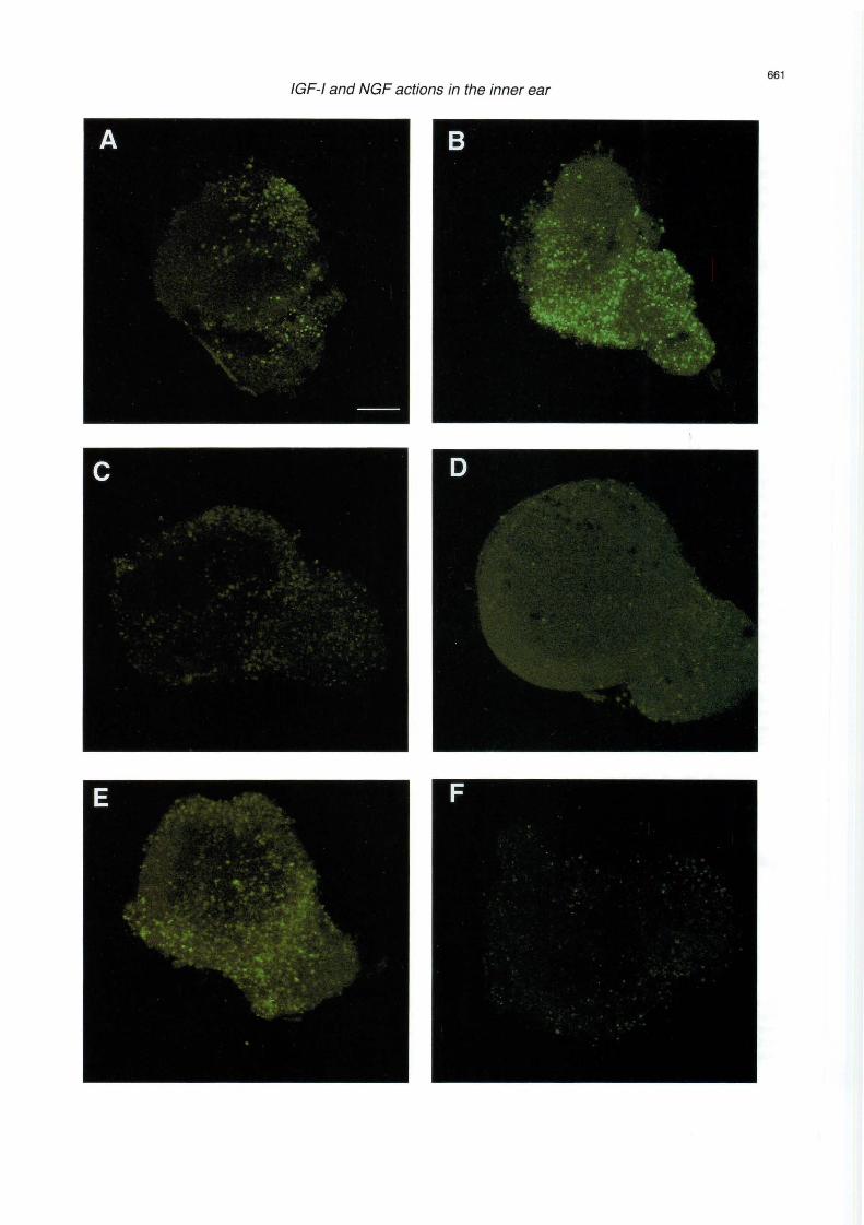
The turnover of lipid s at the plasma membrane of cells plays a critica! role in signa! transduction and ce llular ac tivation . The int erac tion of extracel lul ar ago nists with specific membrane receptors results in the activat ion of specific enzymes, phosph olipases, which generate intracellular second me sse ngers from lipid precursors. Sphingolipids have long been considered as st ru c tural unit s of the plasma me mb rane of cells. How ever, it has bee n recently s ho w n that these molecules have multipl e biological activiti es, and many have been desc rib ed as pote nt seco nd messengers and reg ul ators of cell activation (Kol es nick and Kronke , 1998). Ceramide can be conside red as the sphingo lipid core from which sp hingolipids that are more comp lex are derived. In add ition , the sphin gomye lin/ceramide cy cl e functions as a second messenger system. The physiolog ical signific ance of ceramide accumulation has been related to the induction of cell differentiation and pro gram med cell deat h. Severa! ex trac ellul ar ago nists indu ce the hydrolysis of sphi ngom ye lin to genera te cera mid e . Among them , NGF acts thro ugh its low aff init y p75 receptor (Dobrowsky et al., 1994). Shortchain cera mide analogues (Ci-cera mide) are useful tools to study the biological effec ts of natural ceramides, since the natural long-ch ain cera mid es are not permea nt to cells. Ceram ide can be converte d to ceramide-1-P by a Ca2+ -dependent kinase (Dressler and Kolesnick , 1990) . In turn, a phosphatase has been characterised that spec ifi cally hydro lyses cera mid e-1- P in the p las ma me mbr ane (Boudker and F ut e rm a n , 1993) . The conversion of cera mid e-1 -P in to ceram ide decreases DNA sy nthes is and promotes apo pto sis. These results suggest that ceramide-1-P may play an important role in ce ll ac tivatio n. Cz-ceramide ind uce s internucleoso mal DNA fragmentation which leads to pro gra mm ed cell death or apop tosis in the otic ves icle (Fig. 3 A,B). On the co ntrar y, cera mid e-1-p hosphate is a cy toprotecto r far otic vesic le exp lants that acts as a suppresse r of ce ll death upan se rum withdrawal (Fig. 3C). NGF indu ces sphin go myelin hydrol ysis and ceramide release in the otic ves icle in assoc iation wit h cell deat h in the ot ic ves icJ e and the CYG. NGF-induced apoptos is is app arent in spec ifi c areas and it contributes to the for m atio n of th e e ndol ymph at ic duct and to th e neuro ge nesis of the CYG (Fig . 3E). IG F-1 treatment effect ive ly blocks ceramide and NGF effec ts (F ig . 3D,F). F ur th e rmor e, IG F- 1 blo cks NGF-induced ceramide genera tion and co-operates with ceramide-1-P in cell surv ival (Fraga and Varela-Nieto unpub lish ed
Fig. 3. Distribution of apoptotic cells in the otocyst. Apoptotic cell death is revealed by in situ DNA-end labelling technique (TUNEL protocol) in cultures of otocysts from embryonic day 2.5. Optic sections of 2.5 µm are shown for the following conditions: otocysts were isolated and grown for 8 hours in serum-free medium (A) , 5 µM C2-ceramide (B), 25 µM Cer-1-P (C), 5 µM C2-ceramide plus 10 nM IGF-1 (D), 4 nM NGF (E) or 4 nM NGF plus 10 nM IGF-1 (F). Bar: 70 µm.
661 IGF-1 and NGF actions in the inner ear
662
IGF-1 and NGF actions in the inner ear
observations). These results indic ate that NGF and IGF-1 signalling pathways frame a network of intracellular signals that regulate early inner ear development via induction of regionally restricted areas of cell death and cell proliferation (Fig. 4).
3.2 A strict control of c-Raf kinase levels is essential far early inner ear development
c-Raf is a cytoplasmic serine / threonine protein kinase involv ed in signa! transduction from the plasma membrane to the nucleus (Mark and Rapp, 1984; Daum et al., 1994; Naumann et al., 1997). Increased Raf kinase activity is associated with an increase in the degree of phosphorylation of mitogen-activated protein kinase (MAPK) during the cellular response to various mitogenic agents including IGF-1 in a variety of cell types (Marshall, 1994). c-Raf is a memb er of a small family of proteins esse ntial for growth and development. Knockout and transgenic chimeric c-Raf-deficient mice show growth retardation (Naumann et al., 1997; Wojonowski et al., 1998). Our group has recently show n that during the early organogenesis of the inner ear there is a sustained expression of Raf kinase. This suggest s that Raf is required for the intense mitotic activity reported during this period (Sanz et al., 1999). c-Raf activity is increased in response to IGF -1 and the activation by IGF-1 of the c-Raf kinase pathway is a requirement to turn on cell proliferation in the otic vesicle. The role of c-Raf kinase was further explored by misexpressing both c-raf and a dominan! negative c-raf mutant (Raf-C4) cDNA by means of RCAS retro viral vectors. Overexpression of c-raf in E2.5 exp lants increases the proliferative response to low serum and IGF -1 and blocks differentiation induced by retinoic acid. The increase in c-Raf levels also prevents NGFdependent induction of programmed cell death and potentiates IGF-1 actions as a survival factor. Consistent with these results , the expression of a dominan! negative c-Raf mutant potentiates retinoic acid action and decreases the rate of cell proliferation (Sanz et al. , 1999).
NGF IGF -1
SMaseo--- ----r-,ffi ~,,, j
SURV IVAL
p75
j APOPTOSIS
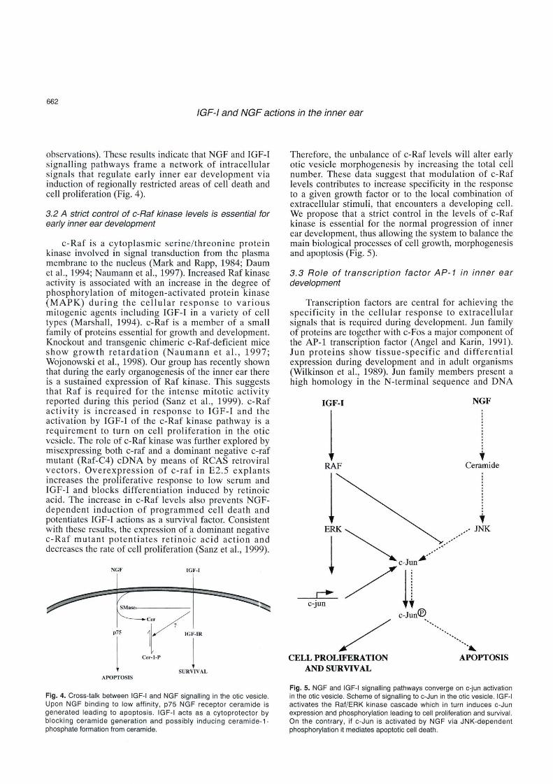
Fig. 4. Cross-talk between IGF-1 and NGF signalling in the otic vesicle. Upon NGF binding to low affinity , p75 NGF receptor ceramide is generated leading to apoptosis . IGF-1 acts as a cytoprotector by blocking ceramide generation and possibly inducing ceramide -1-phosphate formation from ceramide.
Therefor e, the unbalance of c-Raf levels will alter early otic vesicle morphogenesi s by increasing the total cell number. These data sugge st that modul ation of c-Raf level s contributes to increase spec ificit y in the response to a given growth factor or to the local combination of extracellular stimuli , that encounters a developing cell. We propose that a strict control in the levels of c-Raf kinase is essential for the normal progression of inner ear development , thus allowing the system to balance the main biological processes of cell growth, morpho genesis and apoptosis (Fig. 5) .
3. 3 Role of transcription factor AP-1 in inner ear development
Tran scr iption factors are central for achieving the specificity in the cellular response to extracellular signals that is required during development. Jun fami ly of proteins are together with c-Fos a majar component of the AP-1 transcription factor (Angel and Karin, 1991). Jun proteins show tissue-specific and differential expression during development and in adult organisms (Wilkinson et al., 1989). Jun fam ily member s present a high homology in the N-t ermina l sequence and DNA
IGF-1 NGF
1 .
• RAF Ceramide
1 ERK~ •••.• ...-JNK
l "--...,_,'."~---·/ ~/l:
c-jun ;
/ c-Jun~ •.......•.
/ ···· ... , CELL PROLIFERATION
AND SURVIV AL APOPTOSIS
Fig . 5. NGF and IGF-1 signalling pathways converge on c-jun activation in the otic vesicle. Scheme of signalling to c-Jun in the otic vesicle. IGF-1 activates the Raf/ERK kinase cascade which in turn induces c-Jun expression and phosphorylation leading to cell proliferation and suNival. On the contrary, if c-Jun is activated by NGF via JNK-dependent phosphorylation it mediales apoptotic cell death.

663 IGF-1 and NGF actions in the inner ear
binding domain. Jun proteins can form either homodimers or heterodimers with different proteins that include the Fo s-related proteins, ATF-2 or CREB . Oimerization takes place trhough leucine zipper domains and allows binding to ONA sequences, denominated TRE and CRE, that are present in the promoters and enhancer elements of target genes (for a review see Karin et al., 1997). c-jun , junB and juno differ in their binding properties to AP-1 and they display distinct and even opposite roles. Therefore , there are a large number of different combinations of dimers that allow for specific transcriptional responses by activating different sets of target genes depending on the physiological situation of the ce!!. c-jun knock out mice die in utero and cells lacking c-jun presented retarded cell growth in culture (Johnson et al., 1993) This and earlier reports pointed to c-jun as a masterpiece in the control of cell proliferation in response to multiple extracellular stimuli and during cellular transformation . Jun signalling has also been implicated in the regulation of cell differentiation. Finally, c-jun and juno are activated in response to apoptotic stimuli in a variety of cell types (Karin et al., 1997). Thus, the different AP-1 components can achieve diverse functions. Regarding the signalling mechanism that orchestrates Jun activation , Jun proteins can be activated by phosphorylation through two distinct pathways that have been extensively characterised in PC12 pheocromocytome cells and in mature oligodendrocytes (Karin et al., 1997). After growth factor binding to tyrosine kinase receptors the Raf /MAPKs cascade is turned on . The activation of c-jun by a subgroup of MAPKs , the extracellular signa! regulated kinases (ERKs), is associated with the stimulation of cell proliferation and differentiation. On the other hand, serum-deprivation, ceramide accumulation, or NGF binding to p75 receptors activates other members of the MAPK family; the JNKs that phosphorylate c-jun in the serines 63 and 73. This phosphorylation step activates c-jun distinctly and leads to an increase in apoptosis.
The early development of the inner ear provides a useful model to study the complexity and specificity of AP-1 signalling. c-Fos is expressed in the wall of the otic vesicle and in the CVG during those stages of early inner ear development exhibiting a high proliferation rate (León et al., 1995a). The expression pattern of c-fos indicates that it is more abundan! in the ventromedial epithelium of the otocyst and in the CVG. At embryonic day 2.5 the otic vesicle is formed by a morphologically homogeneous epithelium but it presents high mitotic activity in the ventromedial areas (Torres and Giraldez , 1998). The expression of c-Fos is induced by IGF-I and is required for otic vesicle proliferation (León et al., 1995b). In cultured otic vesicles, IGF-1 increases the levels of Jun proteins and antisense c-jun oligonucleotides partially block the cellular responses to IGF-1 (León et al. , 1998). Thus, there is an association between the simultaneous expression of Fos and Jun with the mitogenic effects of IGF-1. However , quiescent
otic vesicles maintained in culture exhibit a restricted expression of c-jun transcripts in the dorsal aspect that co-localises with an area where cells could be labelled by the TUNEL technique indicating apoptosis (Frago et al., 1998). These data suggest that Jun family proteins could play a dual role during early inner ear development. Indeed, whole mount in situ hybridisation analysis of jun expression indicates that c-jun and juno transcripts are expressed in the otic pit and otic vesicle as well as in the CVG with distinct expression patterns that co-localise with areas of high apoptosis. Furthermore, phosphorylation of c-Jun in the serine 63 residue occurs within the same area in the stages studied. The role of c-Jun phosphorylation during apoptosis was confirmed by studying the response to NGF in cultured otic vesicles and in otic vesicles overexpressing Raf. Upon NGF treatment of the explants, c-Jun phosphorylation was increased. When NGF responses were blocked, by co-treatment with IGF-I or by an increase in the intracellular Raf levels, c-Jun was not phosphorylated at the serine 63 (Fig. 5). These results describe an intermediate transcription step which links extracellular apoptotic signals to long term cellular re sponses during organogenesis (Sanz, Giraldez and Varela-Nieto, unpublished observations).
4. Molecular mechanlsms in organogenesis: implications in regeneration
The damage of mechanosensory cells in hearing organs causes hearing Ioss with a severity proportional to the number of hair cells missing. Hair cells of the adult inner ear are lost during ageing and also following acoustic trauma or treatment with certain drugs (www.nih.gov / nidcd; www.iurc.montp.inserm.fr / cric/audition). In the mature mammalian organ of Corti , the sensory cells are not replaced and the deficits are permanent. In contrast, new hair cells are produced to replace those that have been lost in the bird cochlea and in the vestibular sensory epithelia of birds, fish and mammals (for recent reviews see Cotanche , 1997; Staecker and Van de Water, 1998; Stone et al. , 1998). The regeneration of hair cells is preceded by renewed mitosis in the sensory epithelium. Although in the past ten years numerous studies have been directed to examine the events and regulatory mechanisms that control hair cell proliferation and differentiation , the factors that induce hair cell regeneration remain largely unknown.
In the otic vesicle and CVG, IGF-I modulates cell proliferation, differentiation and survival (this review and León et al., 1995b; Frago et al. , 1998). The importance of IGF-I in ear development is stressed by the report of a clinical case of a 15-year-old boy with severe prenatal and postnatal growth failure and sensorineural deafness who had a homozygous partial deletion of the Igf-i gene (Woods et al., 1996, 1997). Targeted disruption of the genes for IGF-I and type 1 IGF receptors has conclusively demonstrated an

664
IGF-1 and NGF actíons in the ínner ear
essential role for IGF-1 in prenatal growth and development. Postnatal mouse growth is also dependent on normal levels of these genes (Baker et al., 1993; Liu et al., 1993; Beck et al., 1995; Cheng et al., 1998; Gao et al., 1999), but these studies did not give specific details on the development and maturation of the inner ear. IGFI gene is transiently expressed during the maturation of the rat auditory system (Bondy, 1991) and, as discussed above, there are IGF-1 binding sites and IGF-1 in the avian otic vesicle and ganglion. These results, taken together with the high sensitivity to exogenous IGF-1 suggest that the proliferative period of ear development will be seriously affected. There might also be other factors stimulating growth in parallel with IGF-1 and partially rescuing the increase in cell number, but they may or may not mimic IGF-1 completely . In this context, the analysis of the details of the mutan! phenotype will be a substantial step forward in the knowledge of the functions of IGF-1 in the inner ear.
On the other hand, both avían and rat adult hair cells express IGF-I and insulin receptors (Lee and Cotanche, 1996; Saffer et al., 1996). IGF-1 receptors are expressed in high levels in inner ear epithelial cells after injury or deprivation of hair-cells (Jennische et al., 1987; Lee and Cotanche, 1996) and epithelial cell proliferation is inhibited by antibodies against IGF-1 (Swanson et al., 1990) . In addition, it has been reported that: i) IGF-1 and/or insulin promote cell growth in cultures of mature avian and rat utricular epithelial cells (Oesterle et al. , 1997; Zheng et al., 1997); ii) in vivo treatment of the vestibular sensory epithelium with IGF-I combined with other factors increases hair cell regeneration in the guinea pig and proliferation in the rat (Kopke et al., 1996; Kuntz and Oesterle, 1998); and iii) high doses of insulin have been shown to regulate avian CVG neuritogenesis and growth , acting as a cofactor for neurotrophins (Sokolowski, 1997).
Cell proliferation of hair cell progenitors is the early major event occurring during hair-cell regeneration after acoustic trauma or exposure to ototoxins. It has recently been reported that supporting cells and transducing haircells share a common progenitor (Fekete et al., 1998) These results suggest that an increased proliferation of supporting cells coupled to their transdifferentiation may lead to the formation of new hair cells. On the other hand, injury may induce inner ear epithelium to acquire immature properties typical of earlier developmental stages. Therefore, understanding the mechanisms regulating cell proliferation in the inner ear will help to understand hair cell regeneration and functional repair. In this context, IGF-1 is a good candidate to regulate proliferation during the regenerative response . In addition, manipulation of the signalling pathways that control apoptosis in the otic vesicle and CGV may contribute to cellular regeneration. This is the case of the JNK inhibitor CEP-1347 that promotes survival of cochlear neurons and attenuates hair cell loss following trauma (Pirvola et al., 1999).
In summary, the insulin family of growth factors are
potentially useful agents for the regeneration of inner ear cells and treatment of hearing impairments caused by ageing or ototoxic drugs, either alone or in combination with other growth factors.
Acknowledgements. C.S. and G.C. were supported by the Ministerio de Educación y Ciencia, S.C. by the Comunidad de Madrid; Y.L. held a CSIC research contrae! , L.M.F. and C.P. by Europharma and HRL, respectively. This work was supported by grants from the Dirección
General Investigación Ciencia y Tecnología (PM96-0075) and Europharma (Boehringer lngelheim lnc .) to l. V-N and !he Junta de Castilla y León to F.G. We thank Dr. Luis Alvarez far the critical reading of the manuscript and we also thank Antonio Fernández and Javier
Pérez far photographic work and illustrations, respectively.
References
Angel P. and Karin M. (1991). The role of Jun, Fos and the AP-1 complex in cell proliferation and transformation . Biochem. Biophys.
Acta 1072, 129-157. Baker J ., Liu J. , Robertson E.J. and Efstratiadis A. (1993). Role of
insulin-like growth factors in embryonic and postnatal growth. Cell
75, 73-82. Beck K.D., Powell-Braxton L., Widmer H.-R., Valverde J. and Hefti F.
(1995). lgf1 gene disruption results in reduced brain size, CNS
hypomyelination, and loss of hippocampal granule and striatal parvalbumin-containing neurons. Neuron 14, 717-730.
Beutler B. and van Huffel C. (1994). Unraveling function in !he TNF ligand and receptor familias. Science 264, 667-668.
Bissonnette J.P. and Fekete D.M. (1996). Standard atlas of the gross anatomy of the developing inner ear of the chicken . J. Comp. Neurol. 174, 1-11.
Bondy C.A. (1991). Transient IGF-1 gene expression during the maturation of functionally relatad central projection neurons . J. Neurosci. 11, 3442-3455.
Boudker O. and Futerman AH . (1993). Detection and characterization of Ceramide-1-P phosphatase activity in rat liver plasma membrana.
J. Biol. Chem. 268, 22150-22155. Cassaccia-Bonnefil P., Carter B.D., Dobrowsky R.T. and Chao M.V.
(1996) . Death of oligodendrocytes mediated by the interaction of
nerve growth factor with its receptor p75. Natura 383, 716-719. Cepko C. (1992). Transduction of genes using retrovirus vectors. In:
Curren! protocols in molecular biology. Ausubel F.M., Kingston R.E., Moore D.D., Seidman J.G., Smith J.A. and Struhl K. (eds). Greene
and Wiley-lnterscience. New York. pp 9.10.1-9.13.5. Chao M.V. (1992). Neurotrophin receptors: a window into neuronal
differentiation . Neuron 9, 583-593. Cheng C.M., Joncas G., Reinhardt R.R., Farrer R., Quarles R., Janssen
J., McDonald M.P., Crawley J.N. , Powell-Braxton L. and Bondy C.A. (1998). Biochemical and morphometric analyses show that myelination in the insulin-like growth factor 1 null brain is
proportionate to its neuronal composition. J. Neurosci. 18, 5673-5681.
Cotanche DA (1997). Hair cell regeneration in the avian cochlea. Ann.
Otol. Rhinol. Laryngol. 106, 9-15. Daum G., Eisenmann-Tappe l., Fries H.-W. , Troppma ir J. and Rapp
U.R. (1994). The ins and outs of Raf kinases. Trends Biochem. Sci. 19, 474-479.

665
IGF-1 and NGF actions in the inner ear
De la Rosa E.J., Bondy C.A., Hernández-Sánchez C., Wu X., Zhou J., Carranza A., Scavo L.M . and De Pablo F. (1994). lnsul in and lnsulin-like growth factor system components gene expression in the
chicken retina from early neurogenesis until late development and their effect on neuroepithelial cells. Eur. J. Neurosci. 6, 1801-181 O.
Dobrowsky R.T., Werner M.H., Castellino A.M., Chao M.V. and Hannun Y.A. (1994). Activation of the sphingomyelin cycle through the low
affinity neurotrophin receptor. Science 265, 1596-1599. Dressler K.A . and Kolesnick R.N. (1990) . Ceramide-1-P , a novel
phospholipid in human leukemia (HL-60) cells. J. Biol. Chem. 265, 14917-14921.
Federspiel M. and Hughes S. (1997). Retroviral gene delivery. Methods Cell Biol. 52, 179-214.
Fekete D.M. (1996). Cell late specification in the inner ear. Curr . Op. Neurol. 6, 533-541.
Fekete D.M., Homburger S., Waring M., Riedl A. and García L. (1997). lnvolvement of programmed cell death in morphogenesis of the vertebrate inner ear. Development 124, 2451-2461.
Fekete D.M., Muthukumar S. and Karagogeos D. (1998). Hair cells and supporting cells share a common progenitors in the avian inner ear.
J. Neurosci. 18, 7811-7821. Frago L.M., León Y., De la Rosa E., Gómez-Muñoz A. and Varela-Nieto
l. (1998). Nerve growth factor and caramida modulate cell death in
the early developing inner ear. J. Cell Sci. 111, 549-556. Gao W.Q., Shinsky N., Ingle G., Elias K. and Powell-Braxton L. (1999).
IGF-1 deficient mice show peripheral nerve conduction velocities and
decreased axonal diameters and respond to exogenous IGF-1 treatment. J. Neurosci. 39, 142-152.
Hamburger V. and Hamilton H.L. (1951). A series of normal stages in
the development of the chick embryo. J. Morphol. 88, 49-92. Hemond S.G. and Morest D.K. (1991). Ganglion formation from the otic
placode and the otic crest in the chick embryo : mitosis, migration, and the basal lamina. Anat. Embryol. 184, 1-13.
Hughes S.H., Greenhouse J.J ., Petropoulos C.J. and Sutrave P. (1987).
Adaptar plasmids simplify the insertion of foreign DNA into helperindependent retroviral vectors . J. Virol. 61, 3004-3012 .
Jennische E., Strottner A. and Hanson H.A. (1987). Dynamic changas in lnsulin -like growth factor I immunoreactivity correlata to repair
events in ratear alter freeze-thaw injury. Exp. Mol. Pathol. 47, 193-
201. Johnson R., van Lingen B., Papaioammou V. and Spiegelman B.
(1993). A null mutation in the c-jun locus causes embryonic lethality and retardad cell growth in culture. Genes Dev. 7, 1309-1317.
Kajimoto Y. and Rotwe in P. (1989). Structure and express ion of a
chicken insulin-like growth factor precursor . Mol. Endocr ino!. 3,
1907-1913. Karin M., Liu Z-G. and Zandi E. (1997). AP-1 function and regulation.
Curr. Op. Cell Biol. 9, 240-246. Kolesn ick R.N. and Kronke M. (1998). Regulation of ceramide
production and apoptosis. Annu. Rev. Physiol. 60, 643-665. Kopke R., García P., Gabaizadeh R., Feghali J., Liu W., Lefebvre P.P.
and Van de Water T.R . (1996). In vivo treatment with TGFa/ lGF-1/ retinoic acid mixture increases hair cell regeneration / repair in
guinea pig utricule. Assoc. Res. Otolaryngol. 789, 198. Kunz A.L. and Oesterle E.C. (1998). Tranforming growth factor alpha
with insulin stimulates cell proliferation in vivo in adult rat vestibular
sensory epithelium. J. Comp. Neurol. 399, 413-423. Lee K.H. and Cotanche D.A. (1996). Potential role of bFGF and retinoic
acid in the regeneration of chicken cochlear hair cells. Hear. Res.
94, 1-13.
León Y., Sánchez J., Minar C., Ariza-McNaughton L., Represa J. and Giraldez F. {1995a). Developmental regulation of Fos-protein during pro liferative growth of the otic vesicle and its relat ion to
differentiation induced by retinoic acid. Dev. Biol. 167, 75-86. León Y., Vázquez E., Sanz C., Vega J., Mato J.M., Giráldez F., Represa
J. and Varela-Nieto l. {1995b). lnsulin-like growth factor-! regulates cell proliferation in the developing inner ear , act ivating glycosyl
phosphatidylinositol hydrolysis and Fos expression . Endocrinology 136, 3494-3503 .
León Y., Sanz C., Giráldez F. and Varela-Nieto l. (1998). lnduction of
cell growth by insulin and insulin-like growth factor-1 is associated with jun expression in the otic vesicle . J. Comp. Neurol. 398, 323-332.
Liepinsh E., llag L.L., Otting G.and lbañez C.F. (1997). NMR structure of
!he death domain of the p75 neurotrophin receptor . EMBO J. 16, 4999-5005.
Liu J.P., Baker J., Perkins A.S., Robertson E.J . and Efstratiadis A.
(1993). Mica carrying null mutations of !he genes encoding insulinlike growth factor-1 {lgf-1) and type 1 EGF receptor {lgf1 r). Cell 75, 59-72.
Marshall C.J. (1984). MAP kinase kinase kinase , MAP kinase kinase and MAP kinase. Curr. Op. Genet. Dev. 4, 82-89.
Mark G.E. and Rapp U.R. (1984). Primary structure of v-raf : relatedness to the src family of oncogenes . Science 224, 285-289 .
Naumann U., Eisenmann-Tape l. and Rapp U.R. (1997). The role of Raf
kinases in development and growth of tumours . Recent Results Canear Res. 143, 237-244.
Oesterle E.C. , Tsue T.T. and Rubel E.W. (1997). lnduction of cell
proliferation in inner ear sensory epithelia by insulin-like growth factor-! and insulin. J. Comp. Neurol. 380, 262-274 .
Parrizas M. and LeRoith D. (1997). lnsulin-like growth factor-1 inhibition of apoptosis is associated with increased expression of the bcl-xL
gene product. Endocrinology 138, 1355-1358. Pirvola U., Xing-Qun L., Aarnisalo A.A ., Saarma M., Walton K.M. and
Ylikoski J. (1999). CEP-1347 {KT-7515) attenuates hair cell loss following traumas and prometes survival of cochlear neurons.
Abstrae! 895, ARO Meeting. http://www.aro.org /archives/ 1999/895. htlml.
Saffer L.D. , Gu R. and Corwin J.T . (1996). An RT-PCR ana lysis of
mRNA growth factor receptors in damaged and control sensory epithelia of rat utricles. Hear. Res. 94, 14-13.
Sanz C., León Y., Troppmair J., Rapp U.R. and Varela-Nieto l. (1999).
Strict regulation of c-Raf kinase levels is required for early organogenesis of the vertebrate inner. Oncogene 18, 429-437.
Schecterson L.C. and Bothwell M. (1994). Neurotrophin and neurotrophin receptor mRNA expression in developing inner ear. Hear. Res. 73, 92-1 OO.
Sokolowski B.H.A. (1997). Quantitative analysis of long-term survival and neuritogenesis in vitre: cochleovestibular ganglion of the chick embryo in BDNF, NT-3, NT-4/5, and insulin. Exp. Neurol. 145, 1-15.
Spagnoli A. and Rosenfeld R.G. {1997). lnsulin-like growth factor binding proteins. Curr. Op. Endocrino!. Diabetes 4, 1-9.
Staecker H. and Van de Water T.R. (1998). Factors controlling hair-cell
regeneration/repair in !he inner ear. Curr. Op. Neurol. 8, 480-487. Stone J.S., Oesterle E.C. and Rubel E.W. (1998). Recen! insights into
regeneration of auditory and vestibular hair cells. Curr. Op. Neurol. 11, 17-24.
Swanson G.J., Howard M. and Lewis J. (1990). Epithelial autonomy in

-·--..... ----. -..,-....
666
IGF-1 and NGF actíons in the ínner ear
the development of the inner ear of a bird embryo. Dev. Biol. 137, 243-257.
Torres M. and Giraldez F. (1998). The development of the vertebrate inner ear. Mech. Dev. 71, 5-21.
Treier M., Bohmann D. and Mlodzik M. (1995). Jun cooperates with the ETS domain protein pointed to induce photoreceptor R7 fate in the Drosophila eye. Cell 83, 753-760.
Varela-Nieto l. , Represa J ., Avila M.A., Miner C., Mato J .M. and Giraldez F. (1991). lnositol phospho-oligosaccharide stimulates cell proliferation in the early developing inner ear. Dev. Biol. 143, 432-435.
Von Bartheld C.S., Patterson S.L., Heuer J.G., Wheeler E.F., Bothwell M. and Rubel E.W. (1991) . Expression of nerve growth factor receptors in the developing inner ear of chick and rat. Development 113, 455-470.
Woods K.A., Camacho -Hubner C., Savage M.O. and Clark A.J.L. (1996). lntrauterine growth retardation and postnatal growth failure associated with deletion of the insulin-like growth factor I gene. New
England J. Med. 335, 1363-1367. Woods K.A., Camacho-Hubner C., Barter D., Clark A.J.L. and Savage
M.O. (1997) . lnsulin-like Growth Factor I gene deletion causing intrauterine growth retardation and severe short stature . Acta Paediatr. Suppl. 423, 39-45.
Wilkinson D., Bhatt S., Ryseck R. and Bravo R. (1989). Tissue-specific expression of c-jun and junB during organogenesis in the mouse. Development 106, 465-471.
Wojonowsk i L., Stancato L.F. , Zimmer A.M., Hahn H., Beck T.W., Larner A.C., Rapp U.R. and Zimmer A. (1998). Craf-1 protein kinase is essential for mouse development. Mech. Dev. 76, 141-149.
Wu D.K. and Oh S.H. (1996). Sensory organ generation in the chick inner ear . J. Neurosci. 16, 6454-6462.
Zheng J .L. , Helbig C. and Gao W .Q. (1997 ). lnduct ion of cel l proliferation by fibroblast and insulin-like growth factors in rat inner ear epithelial cell cultures. J. Neurosci. 17, 216-226.
Accepted December 13, 1999