localization - pnas.org fileproc. nati. acad. sci. usa vol. 87, pp. 9445-9448, december 1990...
TRANSCRIPT
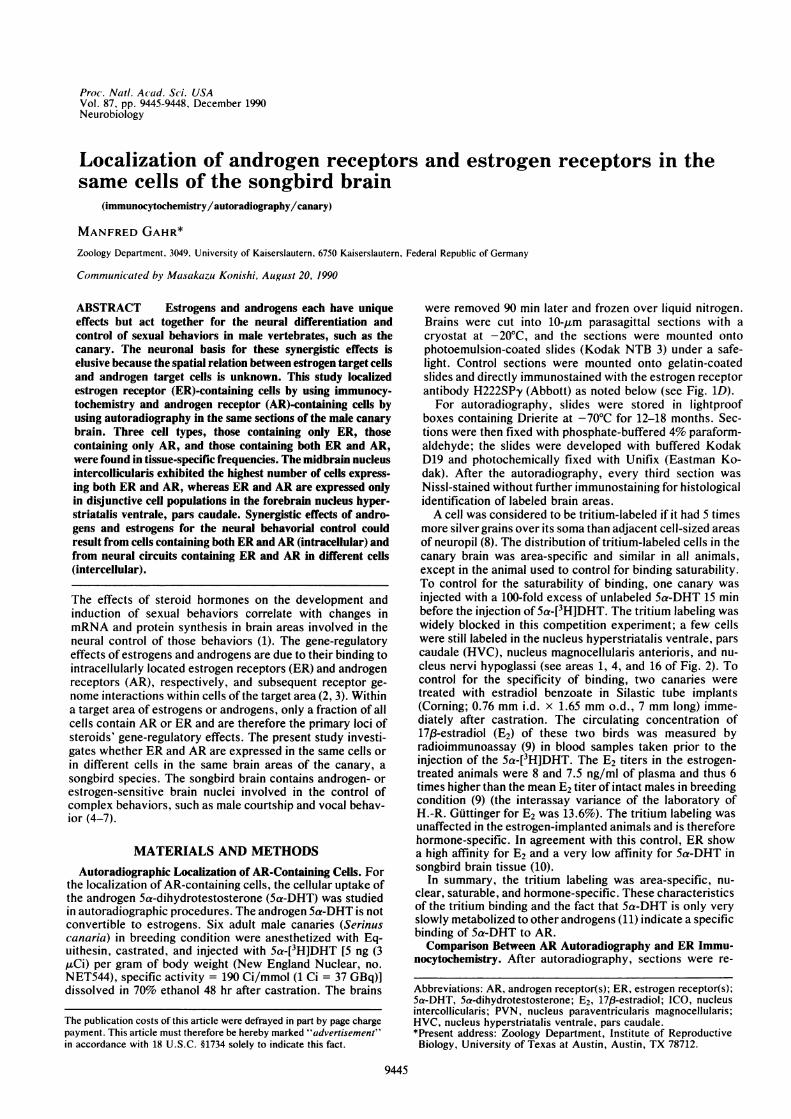
Proc. Nati. Ac ad. Sc i. USAVol. 87, pp. 9445-9448, December 1990Neurobiology
Localization of androgen receptors and estrogen receptors in thesame cells of the songbird brain
(immunocytochemistry/autoradiography/canary)
MANFRED GAHR*Zoology Department, 3049, University of Kaiserslautern, 6750 Kaiserslautern, Federal Republic of Germany
Communicated by Masakazu Konishi, August 20, 1990
ABSTRACT Estrogens and androgens each have uniqueeffects but act together for the neural differentiation andcontrol of sexual behaviors in male vertebrates, such as thecanary. The neuronal basis for these synergistic effects iselusive because the spatial relation between estrogen target cellsand androgen target cells is unknown. This study localizedestrogen receptor (ER)-containing cells by using immunocy-tochemistry and androgen receptor (AR)-containing cells byusing autoradiography in the same sections of the male canarybrain. Three cell types, those containing only ER, thosecontaining only AR, and those containing both ER and AR,were found in tissue-specific frequencies. The midbrain nucleusintercollicularis exhibited the highest number of cells express-ing both ER and AR, whereas ER and AR are expressed onlyin disjunctive cell populations in the forebrain nucleus hyper-striatalis ventrale, pars caudale. Synergistic effects of andro-gens and estrogens for the neural behavorial control couldresult from cells containing both ER and AR (intracellular) andfrom neural circuits containing ER and AR in different cells(intercellular).
The effects of steroid hormones on the development andinduction of sexual behaviors correlate with changes inmRNA and protein synthesis in brain areas involved in theneural control of those behaviors (1). The gene-regulatoryeffects of estrogens and androgens are due to their binding tointracellularly located estrogen receptors (ER) and androgenreceptors (AR), respectively, and subsequent receptor ge-nome interactions within cells of the target area (2, 3). Withina target area of estrogens or androgens, only a fraction of allcells contain AR or ER and are therefore the primary loci ofsteroids' gene-regulatory effects. The present study investi-gates whether ER and AR are expressed in the same cells orin different cells in the same brain areas of the canary, asongbird species. The songbird brain contains androgen- orestrogen-sensitive brain nuclei involved in the control ofcomplex behaviors, such as male courtship and vocal behav-ior (4-7).
MATERIALS AND METHODSAutoradiographic Localization of AR-Containing Cells. For
the localization of AR-containing cells, the cellular uptake ofthe androgen 5a-dihydrotestosterone (5a-DHT) was studiedin autoradiographic procedures. The androgen 5a-DHT is notconvertible to estrogens. Six adult male canaries (Serinuscanaria) in breeding condition were anesthetized with Eq-uithesin, castrated, and injected with Sa-[3HJDHT [5 ng (3,uCi) per gram of body weight (New England Nuclear, no.NET544), specific activity = 190 Ci/mmol (1 Ci = 37 GBq)]dissolved in 70% ethanol 48 hr after castration. The brains
were removed 90 min later and frozen over liquid nitrogen.Brains were cut into 10-gim parasagittal sections with acryostat at -20'C, and the sections were mounted ontophotoemulsion-coated slides (Kodak NTB 3) under a safe-light. Control sections were mounted onto gelatin-coatedslides and directly immunostained with the estrogen receptorantibody H222SP'y (Abbott) as noted below (see Fig. 1D).For autoradiography, slides were stored in lightproof
boxes containing Drierite at -70'C for 12-18 months. Sec-tions were then fixed with phosphate-buffered 4% paraform-aldehyde; the slides were developed with buffered KodakD19 and photochemically fixed with Unifix (Eastman Ko-dak). After the autoradiography, every third section wasNissl-stained without further immunostaining for histologicalidentification of labeled brain areas.A cell was considered to be tritium-labeled if it had 5 times
more silver grains over its soma than adjacent cell-sized areasof neuropil (8). The distribution of tritium-labeled cells in thecanary brain was area-specific and similar in all animals,except in the animal used to control for binding saturability.To control for the saturability of binding, one canary wasinjected with a 100-fold excess of unlabeled 5a-DHT 15 minbefore the injection of 5a-[3H]DHT. The tritium labeling waswidely blocked in this competition experiment; a few cellswere still labeled in the nucleus hyperstriatalis ventrale, parscaudale (HVC), nucleus magnocellularis anterioris, and nu-cleus nervi hypoglassi (see areas 1, 4, and 16 of Fig. 2). Tocontrol for the specificity of binding, two canaries weretreated with estradiol benzoate in Silastic tube implants(Corning; 0.76 mm i.d. x 1.65 mm o.d., 7 mm long) imme-diately after castration. The circulating concentration of17,8-estradiol (E2) of these two birds was measured byradioimmunoassay (9) in blood samples taken prior to theinjection of the 5a-[3H]DHT. The E2 titers in the estrogen-treated animals were 8 and 7.5 ng/ml of plasma and thus 6times higher than the mean E2 titer of intact males in breedingcondition (9) (the interassay variance of the laboratory ofH.-R. Guttinger for E2 was 13.6%). The tritium labeling wasunaffected in the estrogen-implanted animals and is thereforehormone-specific. In agreement with this control, ER showa high affinity for E2 and a very low affinity for 5a-DHT insongbird brain tissue (10).
In summary, the tritium labeling was area-specific, nu-clear, saturable, and hormone-specific. These characteristicsof the tritium binding and the fact that 5a-DHT is only veryslowly metabolized to other androgens (11) indicate a specificbinding of 5a-DHT to AR.Comparison Between AR Autoradiography and ER Immu-
nocytochemistry. After autoradiography, sections were re-
Abbreviations: AR, androgen receptor(s); ER, estrogen receptor(s);5a-DHT, 5a-dihydrotestosterone; E2, 17,8-estradiol; ICO, nucleusintercollicularis; PVN, nucleus paraventricularis magnocellularis;HVC, nucleus hyperstriatalis ventrale, pars caudale.*Present address: Zoology Department, Institute of ReproductiveBiology, University of Texas at Austin, Austin, TX 78712.
9445
The publication costs of this article were defrayed in part by page chargepayment. This article must therefore be hereby marked "advertisement"in accordance with 18 U.S.C. §1734 solely to indicate this fact.
Proc. Natl. Acad. Sci. USA 87 (1990)
acted with the monoclonal ER antibody H222Spy (Abbott) inindirect immunocytochemical procedures for the localizationof ER-containing cells as described elsewhere in detail (12).As a modification to this protocol, a double-bridge techniquewas used to enhance the staining (13). Briefly, sections weretreated with 0.1% Triton X-100 in 25 mM phosphate buffercontaining 0.9% NaCl (PBS) for 30 min. Sections were firstincubated with 2% goat serum in PBS (30 min), then in the ERantibody H222Spy (1 ,ug/ml in PBS) (Abbott), then in thesecond antibody (goat anti-rat IgG, 1:50 in PBS) (Sigma), thenin the third antibody (rat clonoPAP, 1:100 in PBS; Stern-berger-Meyer, Garretsville, MD), and then again with thesecond antibody followed by the third antibody. Incubationtimes were 1 hr at room temperature for each antibodyfollowed by a 30-min wash in PBS. After the last wash, thesections were incubated for 10 min in the chromagen con-taining 0.03% diaminobenzidine and 0.01% hydrogen perox-ide for visualization ofthe immunoproduct. Antibody-labeledcells had darkly stained cell nuclei (see Fig. 1). For analysisof the tritium labeling and the total cell number, immunos-tained sections were counterstained with methyl green.
In sections processed for both autoradiography and immu-nocytochemistry, antibody-labeled cells and tritium-labeledcells were counted under high power on a Zeiss microscope.Five sections (taken at 50-tum intervals) of each brain areathat contained labeled cells of each of three animals were
B
analyzed. In each area section, the percentage of a cell typewas calculated based on the total number of labeled cells inthis area (Fig. 3).To estimate the frequency of labeled cells relative to the
total cell number, all labeled cells were counted in 10 sectionsof the midbrain nucleus intercollicularis (ICO) as describedabove. In the areas defined by the distribution of labeledcells, all Nissl-stained cells with a visible nucleus werecounted under high power with an ocular grid on a Zeissmicroscope. The percentage of labeled cells among Nissl-stained cells was calculated for each section. The totalnumber of labeled cells of this area was estimated from thenumber of labeled cells per area-section and the intersectiondistances (50 ,um) for each animal. Given are the median,minimum, and maximum values of all animals.
RESULTSIn each animal, except the control that was injected withunlabeled 5a-DHT, the sections processed with both auto-radiography and immunocytochemistry exhibited three typesof labeled cells (Fig. 1 A-C): (i) cells labeled only with theantibody, (ii) cells labeled only with tritium, and (iii) cellsdouble-labeled with the antibody and tritium. The frequen-cies of these cell types in the canary brain were area-specific(Figs. 2 and 3). Among areas that were heavily labeled with
2* s X
-^.
3 2*; ;
. .. . . . . .: * ' .f
23 * .,
* / ..s ,
1
A
4
Gr10%Z4 I
0
a
I e
d
D 0 0
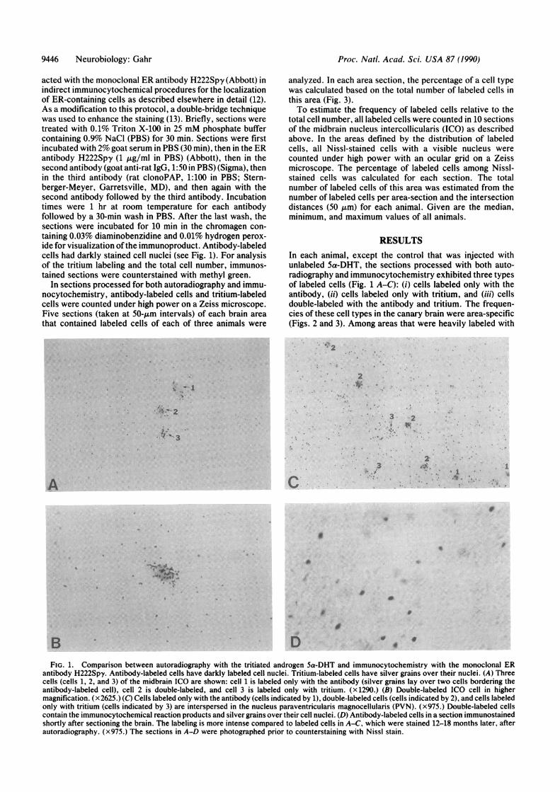
FIG. 1. Comparison between autoradiography with the tritiated androgen 5a-DHT and immunocytochemistry with the monoclonal ERantibody H222Spy. Antibody-labeled cells have darkly labeled cell nuclei. Tritium-labeled cells have silver grains over their nuclei. (A) Threecells (cells 1, 2, and 3) of the midbrain ICO are shown: cell 1 is labeled only with the antibody (silver grains lay over two cells bordering theantibody-labeled cell), cell 2 is double-labeled, and cell 3 is labeled only with tritium. (x1290.) (B) Double-labeled ICO cell in highermagnification. (x2625.) (C) Cells labeled only with the antibody (cells indicated by 1), double-labeled cells (cells indicated by 2), and cells labeledonly with tritium (cells indicated by 3) are interspersed in the nucleus paraventricularis magnocellularis (PVN). (x975.) Double-labeled cellscontain the immunocytochemical reaction products and silver grains over their cell nuclei. (D) Antibody-labeled cells in a section immunostainedshortly after sectioning the brain. The labeling is more intense compared to labeled cells in A-C, which were stained 12-18 months later, afterautoradiography. (x975.) The sections in A-D were photographed prior to counterstaining with Nissl stain.
9446 Neurobiology: Gahr
i
-0--I14.
..V" 3
sL;IP"
Proc. Natl. Acad. Sci. USA 87 (1990) 9447
0a)
-90
CDtBs
Cell TypesER-cellsAR-cells Ail
ER-AR-cells
ICO PVN HVC
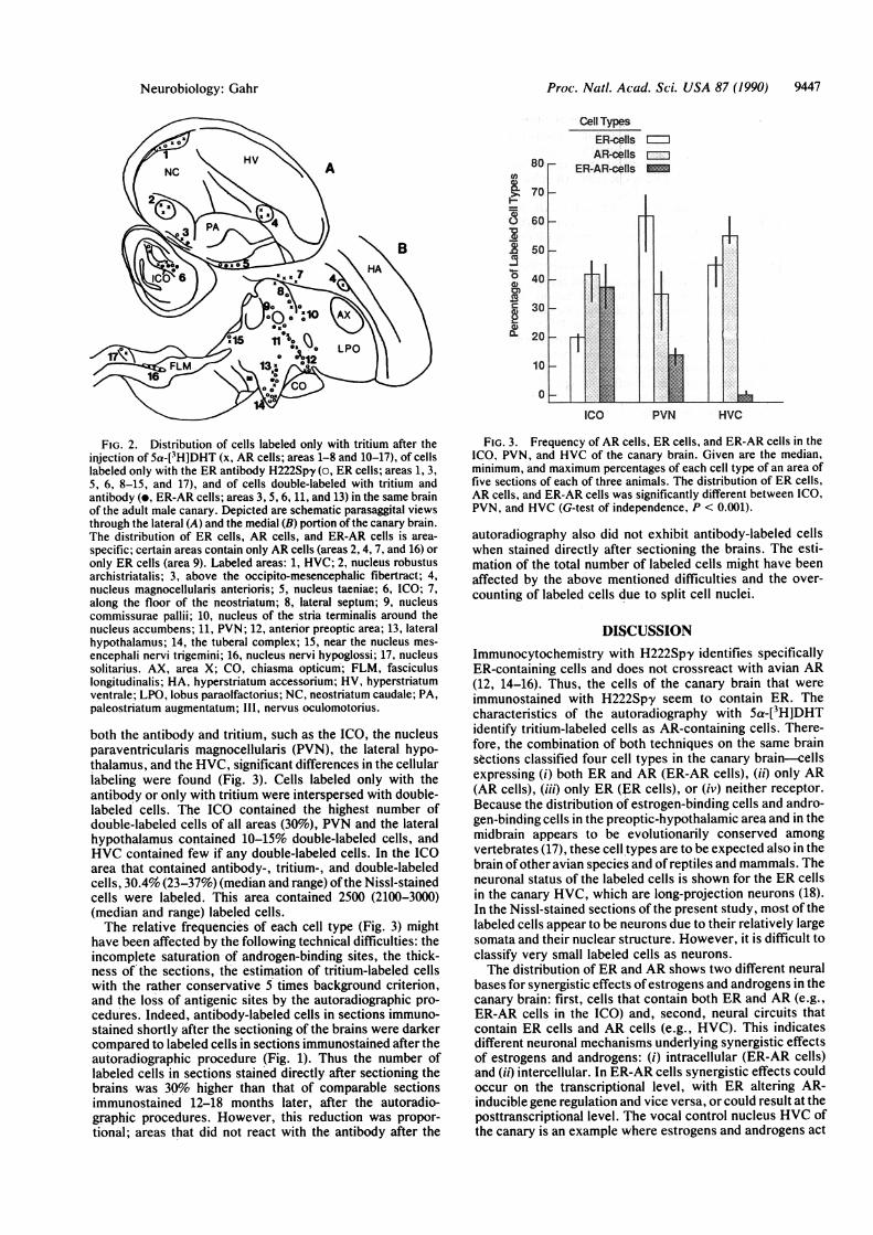
FIG. 2. Distribution of cells labeled only with tritium after theinjection of 5a-[3H]DHT (x, AR cells; areas 1-8 and 10-17), of cellslabeled only with the ER antibody H222Spy (o, ER cells; areas 1, 3,5, 6, 8-15, and 17), and of cells double-labeled with tritium andantibody (e, ER-AR cells; areas 3, 5, 6, 11, and 13) in the same brainof the adult male canary. Depicted are schematic parasaggital viewsthrough the lateral (A) and the medial (B) portion of the canary brain.The distribution of ER cells, AR cells, and ER-AR cells is area-
specific; certain areas contain only AR cells (areas 2, 4, 7, and 16) oronly ER cells (area 9). Labeled areas: 1, HVC; 2, nucleus robustusarchistriatalis; 3, above the occipito-mesencephalic fibertract; 4,nucleus magnocellularis anterioris; 5, nucleus taeniae; 6, ICO; 7,along the floor of the neostriatum; 8, lateral septum; 9, nucleuscommissurae pallii; 10, nucleus of the stria terminalis around thenucleus accumbens; 11, PVN; 12, anterior preoptic area; 13, lateralhypothalamus; 14, the tuberal complex; 15, near the nucleus mes-
encephali nervi trigemini; 16, nucleus nervi hypoglossi; 17, nucleussolitarius. AX, area X; CO, chiasma opticum; FLM, fasciculuslongitudinalis; HA, hyperstriatum accessorium; HV, hyperstriatumventrale; LPO, lobus paraolfactorius; NC, neostriatum caudale; PA,paleostriatum augmentatum; 111, nervus oculomotorius.
both the antibody and tritium, such as the ICO, the nucleusparaventricularis magnocellularis (PVN), the lateral hypo-thalamus, and the HVC, significant differences in the cellularlabeling were found (Fig. 3). Cells labeled only with theantibody or only with tritium were interspersed with double-labeled cells. The ICO contained the highest number ofdouble-labeled cells of all areas (30O), PVN and the lateralhypothalamus contained 10-15% double-labeled cells, andHVC contained few if any double-labeled cells. In the ICOarea that contained antibody-, tritium-, and double-labeledcells, 30.4% (23-37%) (median and range) ofthe Nissl-stainedcells were labeled. This area contained 2500 (2100-3000)(median and range) labeled cells.The relative frequencies of each cell type (Fig. 3) might
have been affected by the following technical difficulties: theincomplete saturation of androgen-binding sites, the thick-ness of the sections, the estimation of tritium-labeled cellswith the rather conservative 5 times background criterion,and the loss of antigenic sites by the autoradiographic pro-cedures. Indeed, antibody-labeled cells in sections immuno-stained shortly after the sectioning of the brains were darkercompared to labeled cells in sections immunostained after theautoradiographic procedure (Fig. 1). Thus the number oflabeled cells in sections stained directly after sectioning thebrains was 30% higher than that of comparable sectionsimmunostained 12-18 months later, after the autoradio-graphic procedures. However, this reduction was propor-tional; areas that did not react with the antibody after the
FIG. 3. Frequency of AR cells, ER cells, and ER-AR cells in theICO, PVN, and HVC of the canary brain. Given are the median,minimum, and maximum percentages of each cell type of an area offive sections of each of three animals. The distribution of ER cells,AR cells, and ER-AR cells was significantly different between ICO,PVN, and HVC (G-test of independence, P < 0.001).
autoradiography also did not exhibit antibody-labeled cellswhen stained directly after sectioning the brains. The esti-mation of the total number of labeled cells might have beenaffected by the above mentioned difficulties and the over-counting of labeled cells due to split cell nuclei.
DISCUSSIONImmunocytochemistry with H222Spy identifies specificallyER-containing cells and does not crossreact with avian AR(12, 14-16). Thus, the cells of the canary brain that wereimmunostained with H222Spy seem to contain ER. Thecharacteristics of the autoradiography with 5a-[3H]DHTidentify tritium-labeled cells as AR-containing cells. There-fore, the combination of both techniques on the same brainsections classified four cell types in the canary brain-cellsexpressing (i) both ER and AR (ER-AR cells), (ii) only AR(AR cells), (iii) only ER (ER cells), or (iv) neither receptor.Because the distribution of estrogen-binding cells and andro-gen-binding cells in the preoptic-hypothalamic area and in themidbrain appears to be evolutionarily conserved amongvertebrates (17), these cell types are to be expected also in thebrain of other avian species and of reptiles and mammals. Theneuronal status of the labeled cells is shown for the ER cellsin the canary HVC, which are long-projection neurons (18).In the Nissl-stained sections of the present study, most of thelabeled cells appear to be neurons due to their relatively largesomata and their nuclear structure. However, it is difficult toclassify very small labeled cells as neurons.The distribution of ER and AR shows two different neural
bases for synergistic effects of estrogens and androgens in thecanary brain: first, cells that contain both ER and AR (e.g.,ER-AR cells in the ICO) and, second, neural circuits thatcontain ER cells and AR cells (e.g., HVC). This indicatesdifferent neuronal mechanisms underlying synergistic effectsof estrogens and androgens: (i) intracellular (ER-AR cells)and (it) intercellular. In ER-AR cells synergistic effects couldoccur on the transcriptional level, with ER altering AR-inducible gene regulation and vice versa, or could result at theposttranscriptional level. The vocal control nucleus HVC ofthe canary is an example where estrogens and androgens act
Neurobiology: Gahr
Proc. Natl. Acad. Sci. USA 87 (1990)
synergistically but where ER and AR occur in different cells.Estrogens and androgens seem to act together for theseasonal neuronal differentiation and the singing control inthe HVC (6). These synergistic effects must result from director indirect mechanisms between ER cells, AR cells, and cellswithout receptors in such an area.
Intriguingly, the frequency of ER cells, AR cells, andER-AR cells differs among areas that contain both AR andER. This raises the question offactors limiting ER expressionin AR cells and vice versa. The expression of only onereceptor type per cell achieves hormone-specific cellularresponses in the case of the progesterone receptor and theglucocorticoid receptor (19). This explanation might notaccount for the lack ofAR in ER cells and vice versa, becauseER and AR appear to recognize different DNA sequences(20, 21). Instead of molecular constraints, the connectionalproperties of a brain nucleus could explain why certain cellsexpress only one receptor type in areas with both ER and AR.If ER and AR are expressed in different types of projectionneurons of a nucleus, then independent activation of thesecircuits through estrogens and androgens, respectively, ispossible; e.g., the canary HVC has two main projections, thenucleus area X and the nucleus robustus, and ER and AR arefound in different HVC cells. Because most of the ER cellsin HVC project to area X (18), AR -cells are either localinterneurons or project to the nucleus robustus. In such ascope, ER-AR cells relay androgen- and estrogen-sensitivecircuits, and such a cell could yield similar responses to eitherhormone.
I thank Drs. D. Crews, J. Bull, H. Zakon, and J. Lindzey forcomments on the manuscript; Dr. H.-R. Guttinger for financialsupport through a grant of the Deutsche Forschungsgemeinschaft(Gu 148/7-8); and the Deutsche Forschungsgemeinschaft for a fel-lowship (Nachwuchs-Qualifizierten-Forderung).
1. Pfaff, D. W. & Schwartz-Giblin, S. (1988) in The Physiology ofReproduction, eds. Knobil, E. & Neill, J. (Raven, New York),pp. 1487-1568.
2. McEwen, B. S. (1976) in Subcellular Mechanisms in Repro-ductive Neuroendocrinology, eds. Naftolin, F., Ryan, K. G. &Davies, 1. G. (Elsevier, Amsterdam), pp. 277-304.
3. Beato, M. (1989) Cell 56, 335-344.4. Gurney, M. E. & Konishi, M. (1980) Science 208, 1380-1383.5. Harding, C. F., Sheridan, F. & Walters, M. J. (1983) Horm.
Behav. 17, 111-133.6. DeVoogd, T. H. (1986) J. Neurobiol. 17, 177-201.7. Weichel, K., Heid, P. & Guttinger, H.-R. (1989) Ethology 80,
55-70.8. Morell, J. 1. & Pfaff, D. W. (1978) Am. Zool. 18, 447-460.9. Weichel, K., Schwager, G., Heid, P., Guttinger, H.-R. &
Pesch, A. (1986) Ethology 73, 281-284.10. Walters, M. J., McEwen, B. S. & Harding, C. F. (1988) Brain
Res. 459, 37-43.11. Lieberburg, I., MacLusky, N. J. & McEwen, B. S. (1977)
Endocrinology 100, 598-607.12. Gahr, M., Fligge, G. & Guttinger, H.-R. (1987) Brain Res 402,
173-177.13. Vacca, L. L., Abrahams, S. J. & Naftchi, N. E. (1980) J.
Histochem. Cytochem. 28, 297-307.14. Greene, G. L., Sobel, N. B., King, W. J. & Jensen, E. V. (1984)
J. Steroid Biochem. 20, 51-56.15. Cintra, A., Fuxe, K., Harfstrand, A., Agnati, L. F., Miller, L.,
Greene, G. L. & Gustafsson, J. A. (1986) Neurochem. Int. 8,587-595.
16. Gahr, M. & Konishi, M. (1988) Proc. Natl. Acad. Sci. USA 85,7380-7383.
17. Pfaff, D. W. (1980) Estrogens and Brain Function (Springer,New York).
18. Gahr, M. (1990) J. Comp. Neurol. 294, 30-36.19. Strahle, U., Boshart, M., Klock, G., Stewart, F. & Schutz, G.
(1989) Nature (London) 339, 629-630.20. Chang, C., Kokontis, J. & Liao, S. (1988) Science 240,324-326.21. Klock, G., Strahle, U. & Schutz, G. (1987) Nature (London)
329, 734-736.
9448 Neurobiology: Gahr