location: seatac airport conference center – central ... · location: seatac airport conference...
TRANSCRIPT
Version Officially Adopted on 11-18-2011
P.O. Box 42712 • Olympia, Washington 98504 • www.hta.hca.wa.gov • 360-923-2742 • FAX 360-923-2766 • TTY 360-923-2701
Health Technology Assessment - HTA
Health Technology Clinical Committee Date: September 16th, 2011 Time: 8:00 am – 3:30 pm Location: SeaTac Airport Conference Center – Central Auditorium Adopted: November 18th, 2011
HTCC MINUTES
Members Present: Dr. Carson Odegard; Dr. Richard Phillips; Dr. Craige Blackmore; Dr. Marie-Annette Brown; Dr. Kevin Walsh; Dr. Christopher Standaert; Dr. Michelle Simon; Dr. Joann Elmore; Dr. Michael Souter; Dr. Seth Schwartz and Dr. David McCulloch.
HTCC FORMAL ACTION 1. Call to Order: Dr. Blackmore, Chair, called the meeting to order. Sufficient members were present
to constitute a quorum.
2. June 17th, 2011 Meeting Minutes: Chair referred members to the draft minutes; motion to approve and second, and adopted by the committee.
Action: Eight committee members approved the June 17th, 2011 meeting minutes. Three committee members abstained from voting.
3. Applied Behavioral Analysis (ABA or ABA Therapy) based Behavioral Interventions for the Treatment of Autism Spectrum Disorder draft Findings & Decision: Chair referred members to the draft findings and decision and called for further discussion or objection. The ABA findings & decision was approved and adopted by the committee.
Action: Ten committee members approved the ABA Therapy findings & decision document. One committee members abstained from voting.
4. Positron Emission Tomography (PET) Scans for Lymphoma: The HTCC reviewed and considered the PET technology assessment report; information provided by the Administrator; state agencies; public members; and heard comments from the evidence reviewer, HTA program, an invited clinical expert, the public and agency medical directors. The committee considered all the evidence and gave greatest weight to the evidence it determined, based on objective factors, to be the most valid and reliable.
HTCC COMMITTEE COVERAGE DETERMINATION VOTE
Not
coveredCovered
Unconditionally
Covered Under
Certain Conditions
Positron Emission Tomography (PET) Scans for Lymphoma 0 1 10
Discussion: The Chair called for discussion on conditions related to PET due to the majority voting for coverage. The following conditions were discussed and approved by a majority:
Limitations of Coverage: Positron Emission Tomography (PET) scans for Lymphoma is a covered benefit when the following conditions are met:
1. One scan for initial treatment planning;
Version Officially Adopted on 11-18-2011
P.O. Box 42712 • Olympia, Washington 98504 • www.hta.hca.wa.gov • 360-923-2742 • TTY 360-923-2701 p. 2
Health Technology Assessment - HTA
2. Additional scans for restaging with clinical suspicion of disease progression or treatment failure subject to agency approval;
3. No coverage for routine surveillance
Action: The committee chair directed HTA staff to prepare a Findings and Decision document on PET reflective of the majority vote.
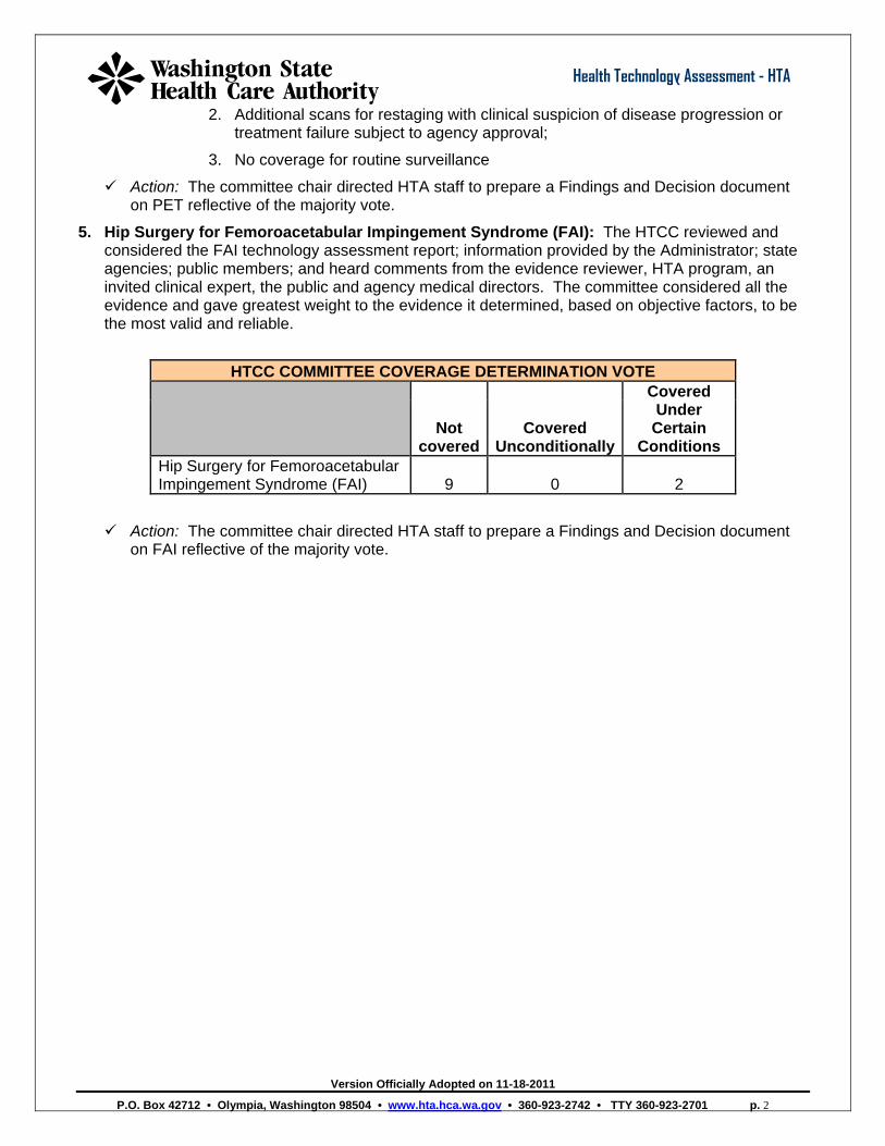
5. Hip Surgery for Femoroacetabular Impingement Syndrome (FAI): The HTCC reviewed and considered the FAI technology assessment report; information provided by the Administrator; state agencies; public members; and heard comments from the evidence reviewer, HTA program, an invited clinical expert, the public and agency medical directors. The committee considered all the evidence and gave greatest weight to the evidence it determined, based on objective factors, to be the most valid and reliable.
HTCC COMMITTEE COVERAGE DETERMINATION VOTE
Not
coveredCovered
Unconditionally
Covered Under
Certain Conditions
Hip Surgery for Femoroacetabular Impingement Syndrome (FAI) 9 0 2
Action: The committee chair directed HTA staff to prepare a Findings and Decision document on FAI reflective of the majority vote.

Version Officially Adopted on 11-18-2011
P.O. Box 42712 • Olympia, Washington 98504 • www.hta.hca.wa.gov • 360-923-2742 • TTY 360-923-2701 p. 3
Health Technology Assessment - HTA
SUMMARY OF HTCC MEETING TOPICS, PRESENTATION, AND DISCUSSION
Agenda Item: Welcome & Introductions The Health Technology Clinical Committee (HTCC) met on September 16th, 2011
Agenda Item: Meeting Open and HTA Program Update Dr. Craig Blackmore, HTCC Chair, opened the public meeting.
New committee member, Dr. David McCulloch, was introduced
Leah Hole-Curry, HTA Program Director, provided an overview of the agenda, meeting guide and purpose, room logistics and introductions.
Newly hired HTA Program Director, Josh Morse, was introduced. Josh Morse will start officially at HCA-HTA on October 1st, 2011.
Agenda Item: Previous Meeting Business June 17th, 2011 Meeting Minutes: Chair referred members to the draft minutes and called for a motion and discussion. Minutes were circulated prior to the meeting and posted.
Action: Eight committee members approved the June 17th, 2011 meeting minutes. Three committee members abstained from voting.
Applied Behavioral Analysis (ABA or ABA Therapy) based Behavioral Interventions for the Treatment of Autism Spectrum Disorder draft Findings & Decision: Chair referred members to the draft findings and decision and called for further discussion. The draft findings and decision document was circulated prior to the meeting and posted to the website for a two week comment period. Five public comments were received, included in the meeting materials, and were reviewed and discussed.
Action: Ten committee members approved the ABA Therapy findings & decision document. One committee members abstained from voting.
Agenda Item: HTA Program Review Leah Hole-Curry, HTA Program Director, provided the HTA context for the meeting and an
update on program activities including:
State purchasing context and budget reductions and reform efforts, medical technology is driver of increased medical costs and has quality gaps
HTA is designed to use reliable science and independent committee to get best information on what works, what is safe and what provides value
HTA outcomes include transparency; reports and articles reviewed; and coverage decisions made
Comparison with private industry and Medicare decisions completed
Program has received recent recognition from public media, clinical press, and various medical and health policy groups with either story highlights or invited presentations
Version Officially Adopted on 11-18-2011
P.O. Box 42712 • Olympia, Washington 98504 • www.hta.hca.wa.gov • 360-923-2742 • TTY 360-923-2701 p. 4
Health Technology Assessment - HTA
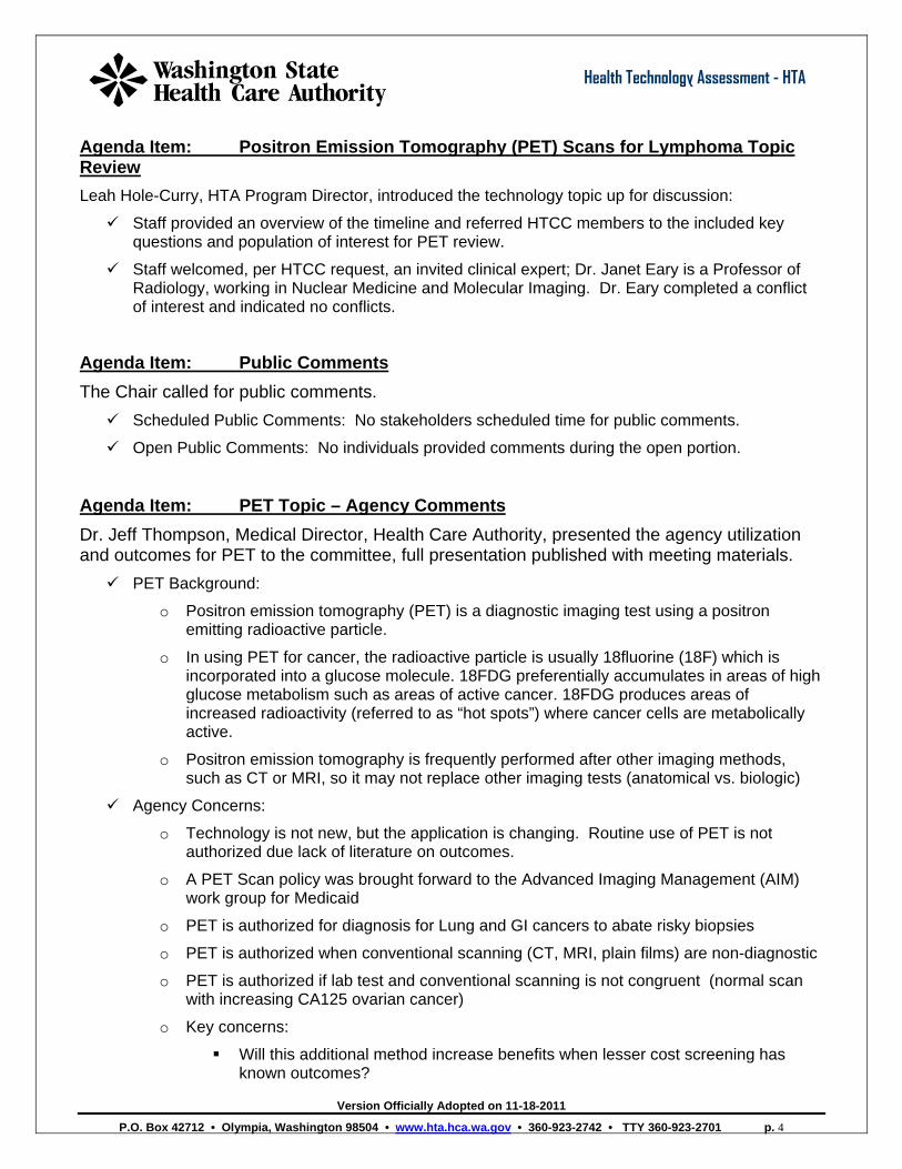
Agenda Item: Positron Emission Tomography (PET) Scans for Lymphoma Topic Review Leah Hole-Curry, HTA Program Director, introduced the technology topic up for discussion:
Staff provided an overview of the timeline and referred HTCC members to the included key questions and population of interest for PET review.
Staff welcomed, per HTCC request, an invited clinical expert; Dr. Janet Eary is a Professor of Radiology, working in Nuclear Medicine and Molecular Imaging. Dr. Eary completed a conflict of interest and indicated no conflicts.
Agenda Item: Public Comments The Chair called for public comments.
Scheduled Public Comments: No stakeholders scheduled time for public comments.
Open Public Comments: No individuals provided comments during the open portion.
Agenda Item: PET Topic – Agency Comments Dr. Jeff Thompson, Medical Director, Health Care Authority, presented the agency utilization and outcomes for PET to the committee, full presentation published with meeting materials.
PET Background:
o Positron emission tomography (PET) is a diagnostic imaging test using a positron emitting radioactive particle.
o In using PET for cancer, the radioactive particle is usually 18fluorine (18F) which is incorporated into a glucose molecule. 18FDG preferentially accumulates in areas of high glucose metabolism such as areas of active cancer. 18FDG produces areas of increased radioactivity (referred to as “hot spots”) where cancer cells are metabolically active.
o Positron emission tomography is frequently performed after other imaging methods, such as CT or MRI, so it may not replace other imaging tests (anatomical vs. biologic)
Agency Concerns:
o Technology is not new, but the application is changing. Routine use of PET is not authorized due lack of literature on outcomes.
o A PET Scan policy was brought forward to the Advanced Imaging Management (AIM) work group for Medicaid
o PET is authorized for diagnosis for Lung and GI cancers to abate risky biopsies
o PET is authorized when conventional scanning (CT, MRI, plain films) are non-diagnostic
o PET is authorized if lab test and conventional scanning is not congruent (normal scan with increasing CA125 ovarian cancer)
o Key concerns:
Will this additional method increase benefits when lesser cost screening has known outcomes?
Version Officially Adopted on 11-18-2011
P.O. Box 42712 • Olympia, Washington 98504 • www.hta.hca.wa.gov • 360-923-2742 • TTY 360-923-2701 p. 5
Health Technology Assessment - HTA
More expensive/additional test increases costs – what about outcomes?
Is the measure of a new test only SN/SP – what about PPV?
Is it appropriate to measure PET against CT scan – anatomic vs. biologic?
Are there better outcomes or reduced costs for the extra radiation dose?
Current State Agencies Policies:
o DSHS allows PET when: there is a Non-diagnostic conventional scan for diagnosis, biopsies, staging/restaging or surveillance
o UMP allows PET in lymphoma:
SURVEILLANCE OF ASYMPTOMATIC PATIENTS AFTER THERAPY FOR MALIGNANCY PET or PET/CT is considered not medically necessary for patients who have completed therapy twelve (12) or more months ago for lymphoma or six (6) or more months ago for all other malignancies unless the patient demonstrates signs, symptoms, laboratory or other objective findings suggestive of recurrence or spread of the original malignancy
SCREENING: PET or PET/CT IS NOT COVERED AS A SCREENING TEST (I.E., FOR EVALUATION OF PATIENTS WITHOUT SPECIFIC SIGNS AND SYMPTOMS OF DISEASE).
State Agencies Questions:
o Safety: Benefit vs. Harms issues?
Do less expensive diagnostics have less risk for radiation exposure? Does the identification of non-specific findings (false positives) lead to
unnecessary interventions? Is that a Red Flag for over use of PET? Mode was 1, the mean was 2, and the max per case 19 PET ( > 40 CT scans) in
5 year period
o Effectiveness:
Is the evidence of sensitivity, specificity, and reliability enough to make a benefit decision?
Can we define when an MRI/CT/Gallium scan vs. PET is needed in a diagnosis, staging/restaging, surveillance?
o Cost
Does routine PET lead to higher cost for unproven outcomes? What is the impact of differential activity in the community (multiple PET and CT
Scans per case)?
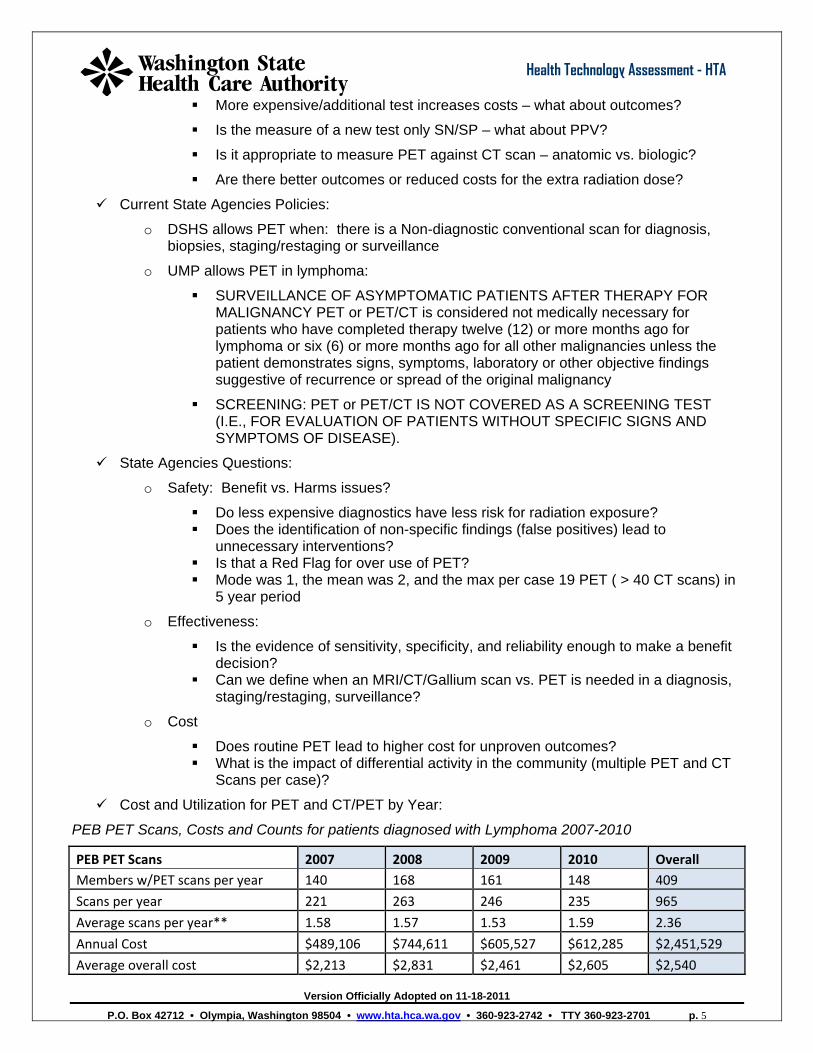
Cost and Utilization for PET and CT/PET by Year:
PEB PET Scans, Costs and Counts for patients diagnosed with Lymphoma 2007-2010
PEB PET Scans 2007 2008 2009 2010 Overall Members w/PET scans per year 140 168 161 148 409
Scans per year 221 263 246 235 965
Average scans per year** 1.58 1.57 1.53 1.59 2.36
Annual Cost $489,106 $744,611 $605,527 $612,285 $2,451,529
Average overall cost $2,213 $2,831 $2,461 $2,605 $2,540
Version Officially Adopted on 11-18-2011
Health Technology Assessment - HTA
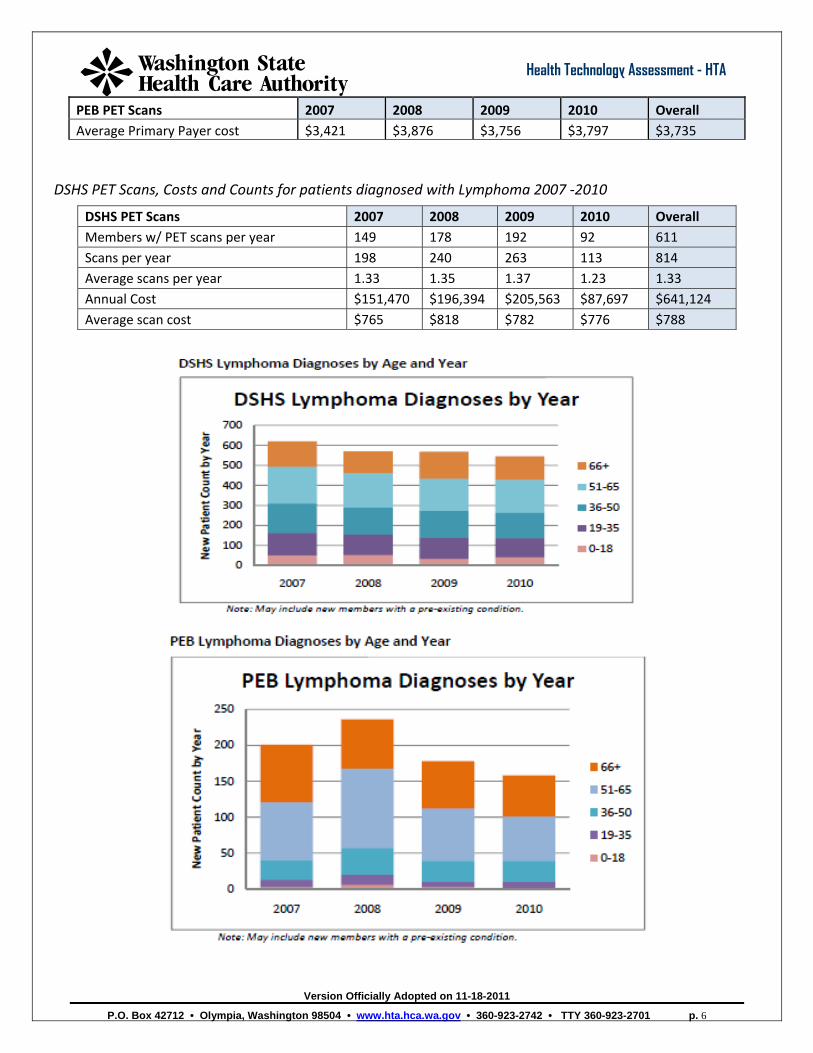
PEB PET Scans 2007 2008 2009 2010 Overall Average Primary Payer cost $3,421 $3,876 $3,756 $3,797 $3,735
DSHS PET Scans, Costs and Counts for patients diagnosed with Lymphoma 2007 ‐2010
DSHS PET Scans 2007 2008 2009 2010 Overall Members w/ PET scans per year 149 178 192 92 611 Scans per year 198 240 263 113 814 Average scans per year 1.33 1.35 1.37 1.23 1.33 Annual Cost $151,470 $196,394 $205,563 $87,697 $641,124 Average scan cost $765 $818 $782 $776 $788
P.O. Box 42712 • Olympia, Washington 98504 • www.hta.hca.wa.gov • 360-923-2742 • TTY 360-923-2701 p. 6
Version Officially Adopted on 11-18-2011
Health Technology Assessment - HTA
2007 2008 2009 2010
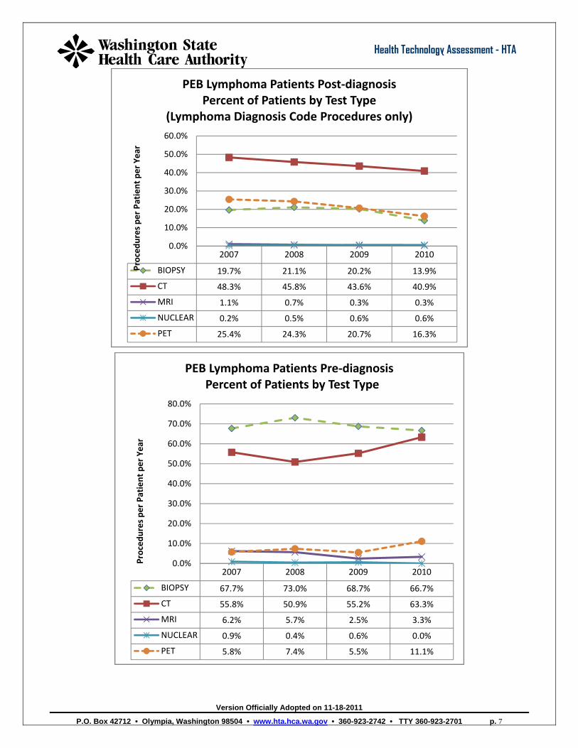
BIOPSY 19.7% 21.1% 20.2% 13.9%
0.0%
10.0%
20.0%
30.0%
40.0%
50.0%
60.0%Proced
ures per Patient per Year
PEB Lymphoma Patients Post‐diagnosis Percent of Patients by Test Type
(Lymphoma Diagnosis Code Procedures only)
CT 48.3% 45.8% 43.6% 40.9%
MRI 1.1% 0.7% 0.3% 0.3%
NUCLEAR 0.2% 0.5% 0.6% 0.6%
PET 25.4% 24.3% 20.7% 16.3%
P.O. Box 42712 • Olympia, Washington 98504 • www.hta.hca.wa.gov • 360-923-2742 • TTY 360-923-2701 p. 7
2007 2008 2009 2010
BIOPSY 67.7% 73.0% 68.7% 66.7%
CT 55.8% 50.9% 55.2% 63.3%
MRI 6.2% 5.7% 2.5% 3.3%
NUCLEAR 0.9% 0.4% 0.6% 0.0%
PET 5.8% 7.4% 5.5% 11.1%
0.0%
10.0%
20.0%
30.0%
40.0%
50.0%
60.0%
70.0%
80.0%
Proced
ures per Patient per Year
PEB Lymphoma Patients Pre‐diagnosisPercent of Patients by Test Type
Version Officially Adopted on 11-18-2011
Health Technology Assessment - HTA
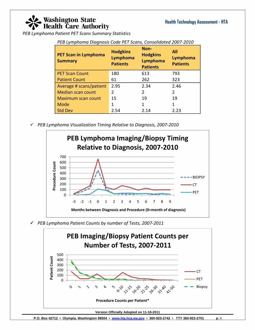
PEB Lymphoma Patient PET Scans Summary Statistics
PEB Lymphoma Diagnosis Code PET Scans, Consolidated 2007‐2010
PET Scan in Lymphoma Summary
Hodgkins Lymphoma Patients
Non‐Hodgkins Lymphoma Patients
All Lymphoma Patients
PET Scan Count 180 613 793 Patient Count 61 262 323 Average # scans/patient 2.95 2.34 2.46 Median scan count 2 2 2 Maximum scan count 15 19 19 Mode 1 1 1 Std Dev 2.54 2.14 2.23
PEB Lymphoma Visualization Timing Relative to Diagnosis, 2007‐2010
0100200300400500600700
‐3 ‐2 ‐1 0 1 2 3 4 5 6 7 8 9
Proced
ure Co
unt
PEB Lymphoma Imaging/Biopsy Timing Relative to Diagnosis, 2007‐2010
Months between Diagnosis and Procedure (0=month of diagnosis)
BIOPSY
CT
PET
PEB Lymphoma Patient Counts by number of Tests, 2007‐2011
P.O. Box 42712 • Olympia, Washington 98504 • www.hta.hca.wa.gov • 360-923-2742 • TTY 360-923-2701 p. 8
0100200300400500
Patien
t Cou
nt
Procedure Counts per Patient*
PEB Imaging/Biopsy Patient Counts per Number of Tests, 2007‐2011
CT
PET
Biopsy
Version Officially Adopted on 11-18-2011
P.O. Box 42712 • Olympia, Washington 98504 • www.hta.hca.wa.gov • 360-923-2742 • TTY 360-923-2701 p. 9
Health Technology Assessment - HTA
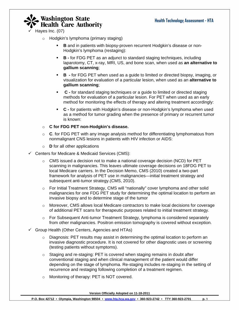
Hayes Inc. (07)
o Hodgkin’s lymphoma (primary staging)
B and in patients with biopsy-proven recurrent Hodgkin’s disease or non-Hodgkin’s lymphoma (restaging):
B - for FDG PET as an adjunct to standard staging techniques, including laparotomy, CT, x-ray, MRI, US, and bone scan, when used as an alternative to gallium scanning;
B - for FDG PET when used as a guide to limited or directed biopsy, imaging, or visualization for evaluation of a particular lesion, when used as an alternative to gallium scanning;
C - for standard staging techniques or a guide to limited or directed staging methods for evaluation of a particular lesion. For PET when used as an early method for monitoring the effects of therapy and altering treatment accordingly:
C - for patients with Hodgkin’s disease or non-Hodgkin’s lymphoma when used as a method for tumor grading when the presence of primary or recurrent tumor is known:
o C for FDG PET non-Hodgkin’s disease. o C. for FDG PET with any image analysis method for differentiating lymphomatous from
nonmalignant CNS lesions in patients with HIV infection or AIDS:
o D for all other applications
Centers for Medicare & Medicaid Services (CMS):
o CMS issued a decision not to make a national coverage decision (NCD) for PET scanning in malignancies. This leaves ultimate coverage decisions on 18FDG PET to local Medicare carriers. In the Decision Memo, CMS (2010) created a two‐part framework for analysis of PET use in malignancies—initial treatment strategy and subsequent anti‐tumor strategy.(CMS, 2010).
o For Initial Treatment Strategy, CMS will “nationally” cover lymphoma and other solid malignancies for one FDG PET study for determining the optimal location to perform an invasive biopsy and to determine stage of the tumor
o Moreover, CMS allows local Medicare contractors to make local decisions for coverage of additional PET scans for therapeutic purposes related to initial treatment strategy.
o For Subsequent Anti‐tumor Treatment Strategy, lymphoma is considered separately from other malignancies. Positron emission tomography is covered without exception.
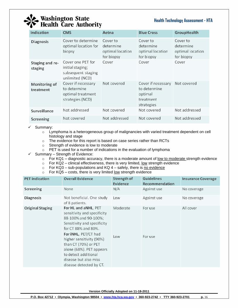
Group Health (Other Centers, Agencies and HTAs)
o Diagnosis: PET results may assist in determining the optimal location to perform an invasive diagnostic procedure. It is not covered for other diagnostic uses or screening (testing patients without symptoms).
o Staging and re‐staging: PET is covered when staging remains in doubt after conventional staging and when clinical management of the patient would differ depending on the stage of lymphoma. Re‐staging includes re‐staging in the setting of recurrence and restaging following completion of a treatment regimen.
o Monitoring of therapy: PET is NOT covered.
Version Officially Adopted on 11-18-2011
Health Technology Assessment - HTA
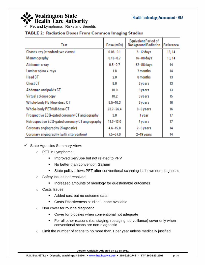
Pet and Lymphoma: Risks and Benefits
State Agencies Summary View:
o PET in Lymphoma:
Improved Sen/Spe but not related to PPV
No better than convention Gallium
State policy allows PET after conventional scanning is shown non-diagnostic
o Safety Issues not resolved
Increased amounts of radiology for questionable outcomes
o Costs Issues
Added cost but no outcome data
Costs Effectiveness studies – none available
o Non cover for routine diagnostic
Cover for biopsies when conventional not adequate
For all other reasons (i.e. staging, restaging, surveillance) cover only when conventional scans are non-diagnostic
o Limit the number of scans to no more than 1 per year unless medically justified
P.O. Box 42712 • Olympia, Washington 98504 • www.hta.hca.wa.gov • 360-923-2742 • TTY 360-923-2701 p. 10
Version Officially Adopted on 11-18-2011
P.O. Box 42712 • Olympia, Washington 98504 • www.hta.hca.wa.gov • 360-923-2742 • TTY 360-923-2701 p. 11
Health Technology Assessment - HTA
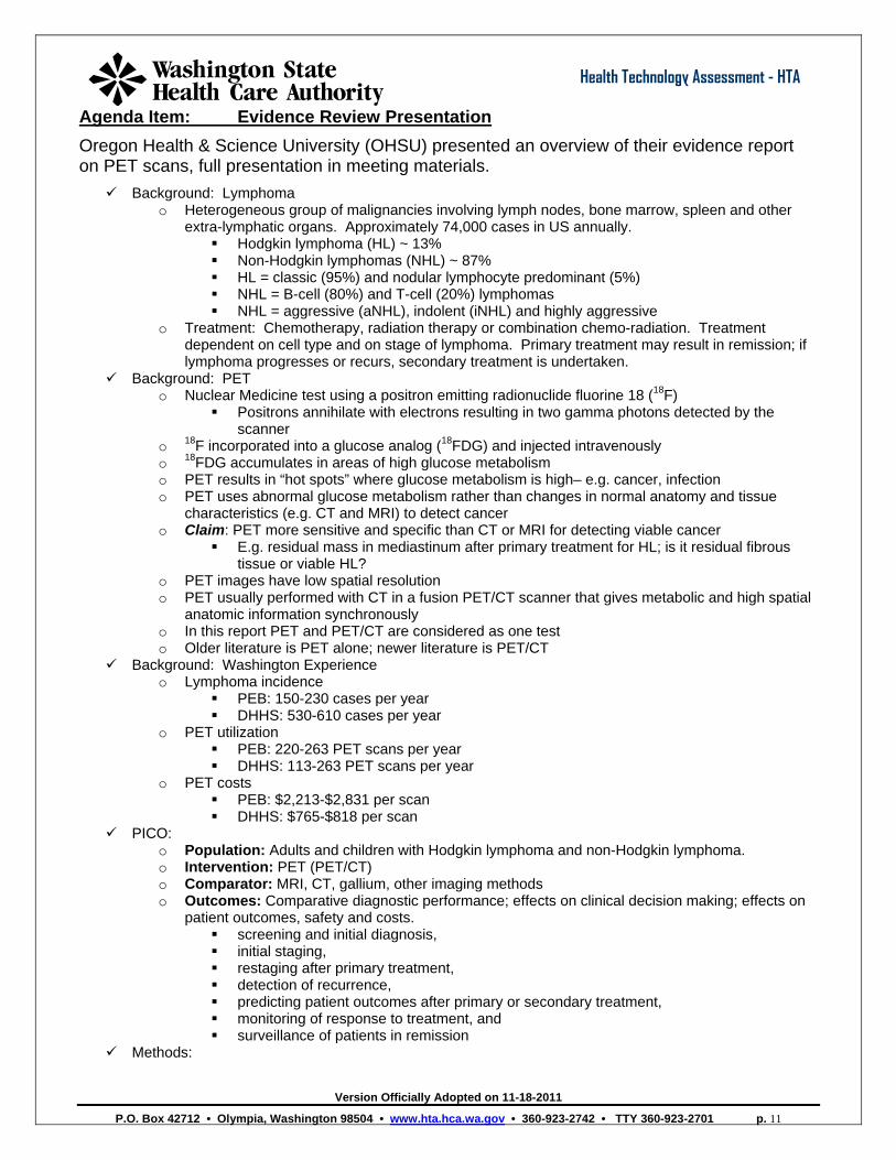
Agenda Item: Evidence Review Presentation Oregon Health & Science University (OHSU) presented an overview of their evidence report on PET scans, full presentation in meeting materials.
Background: Lymphoma o Heterogeneous group of malignancies involving lymph nodes, bone marrow, spleen and other
extra-lymphatic organs. Approximately 74,000 cases in US annually. Hodgkin lymphoma (HL) ~ 13% Non-Hodgkin lymphomas (NHL) ~ 87% HL = classic (95%) and nodular lymphocyte predominant (5%) NHL = B-cell (80%) and T-cell (20%) lymphomas NHL = aggressive (aNHL), indolent (iNHL) and highly aggressive
o Treatment: Chemotherapy, radiation therapy or combination chemo-radiation. Treatment dependent on cell type and on stage of lymphoma. Primary treatment may result in remission; if lymphoma progresses or recurs, secondary treatment is undertaken.
Background: PET o Nuclear Medicine test using a positron emitting radionuclide fluorine 18 (18F)
Positrons annihilate with electrons resulting in two gamma photons detected by the scanner
o 18F incorporated into a glucose analog (18FDG) and injected intravenously o 18FDG accumulates in areas of high glucose metabolism o PET results in “hot spots” where glucose metabolism is high– e.g. cancer, infection o PET uses abnormal glucose metabolism rather than changes in normal anatomy and tissue
characteristics (e.g. CT and MRI) to detect cancer o Claim: PET more sensitive and specific than CT or MRI for detecting viable cancer
E.g. residual mass in mediastinum after primary treatment for HL; is it residual fibrous tissue or viable HL?
o PET images have low spatial resolution o PET usually performed with CT in a fusion PET/CT scanner that gives metabolic and high spatial
anatomic information synchronously o In this report PET and PET/CT are considered as one test o Older literature is PET alone; newer literature is PET/CT
Background: Washington Experience o Lymphoma incidence
PEB: 150-230 cases per year DHHS: 530-610 cases per year
o PET utilization PEB: 220-263 PET scans per year DHHS: 113-263 PET scans per year
o PET costs PEB: $2,213-$2,831 per scan DHHS: $765-$818 per scan
PICO: o Population: Adults and children with Hodgkin lymphoma and non-Hodgkin lymphoma. o Intervention: PET (PET/CT) o Comparator: MRI, CT, gallium, other imaging methods o Outcomes: Comparative diagnostic performance; effects on clinical decision making; effects on
patient outcomes, safety and costs. screening and initial diagnosis, initial staging, restaging after primary treatment, detection of recurrence, predicting patient outcomes after primary or secondary treatment, monitoring of response to treatment, and surveillance of patients in remission
Methods:
Version Officially Adopted on 11-18-2011
Health Technology Assessment - HTA
o For the WA HTA program, MED core sources searched for SRs, MAs, TAs from 2000 to 2011. MEDLINE search for 2009-2011 included SRs, MAs, TAs and case reports. Search terms positron emission tomography, PET, lymphoma, Hodgkin disease.
o Search for relevant clinical practice guidelines using MED core sources and Guidelines.gov databases
o Quality of included systematic review and guidelines rated with standard MED instruments o State, private payers, and policy websites searched to identify insurance coverage policies
Search Results: o Core source search yielded 7 SRs and TAs, 3 cost or cost-effectiveness study designs and 6
clinical practice guidelines o MEDLINE search yielded 354 citations from which 18 observational studies were included in this
report Findings: Evidence presented by Lymphoma Type
o Hodgkin disease (HL) and aggressive non-Hodgkin disease (aNHL) are combined o Indolent non-Hodgkin disease (iNHL) is considered separately o Highly aggressive non-Hodgkin disease – no evidence identified
Findings: Overview o Primary evidence comes from case series
Case series considered to be lower strength of evidence than RCTs or cohort studies o SOE for most KQs is low to moderate even when SRs are of high quality o More evidence for diagnostic accuracy than for clinical effectiveness, safety, cost o More evidence for HL and aNHL than for iNHL
Accuracy of PET: Screening and Initial Diagnosis o No evidence on use of PET for screening or initial diagnosis o Diagnosis requires histology; PET cannot eliminate biopsy o No guidelines support PET for these indications
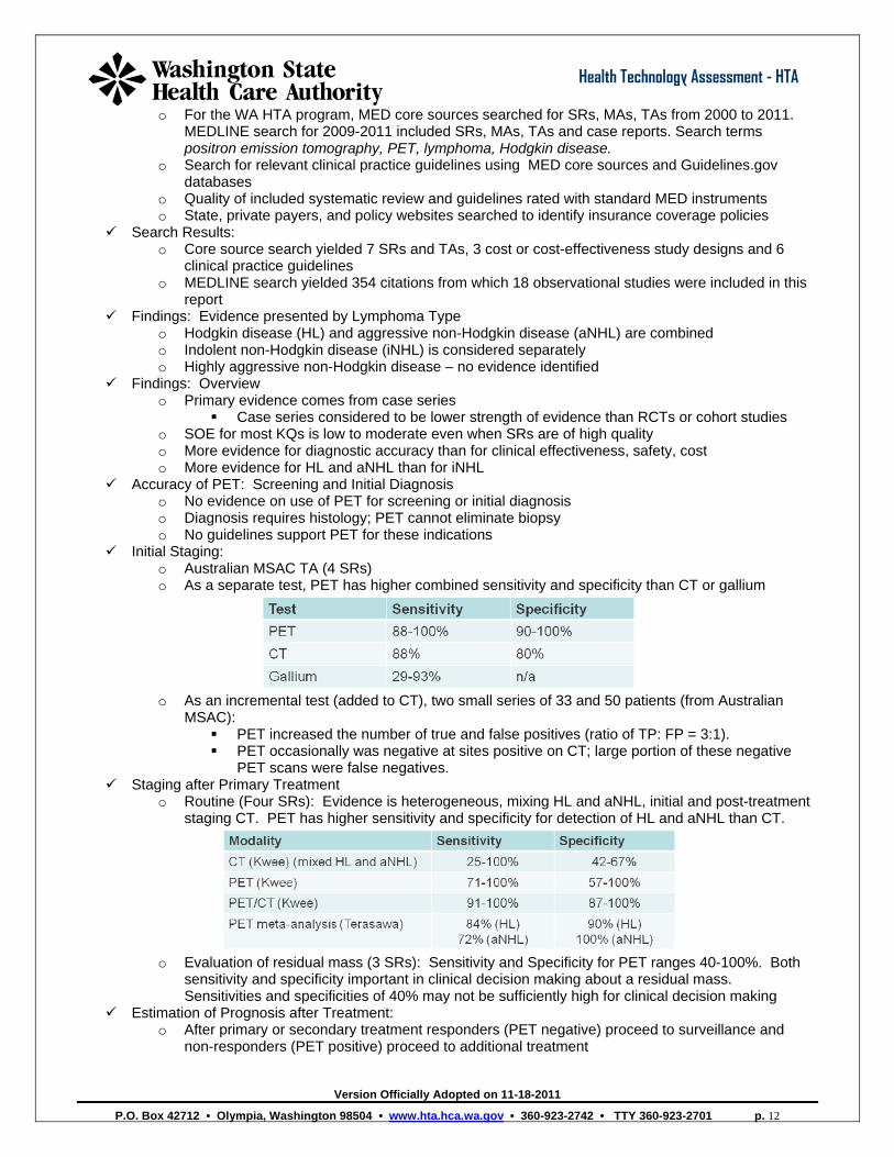
Initial Staging: o Australian MSAC TA (4 SRs) o As a separate test, PET has higher combined sensitivity and specificity than CT or gallium
o As an incremental test (added to CT), two small series of 33 and 50 patients (from Australian
MSAC): PET increased the number of true and false positives (ratio of TP: FP = 3:1). PET occasionally was negative at sites positive on CT; large portion of these negative
PET scans were false negatives. Staging after Primary Treatment
o Routine (Four SRs): Evidence is heterogeneous, mixing HL and aNHL, initial and post-treatment staging CT. PET has higher sensitivity and specificity for detection of HL and aNHL than CT.
o Evaluation of residual mass (3 SRs): Sensitivity and Specificity for PET ranges 40-100%. Both
sensitivity and specificity important in clinical decision making about a residual mass. Sensitivities and specificities of 40% may not be sufficiently high for clinical decision making
Estimation of Prognosis after Treatment: o After primary or secondary treatment responders (PET negative) proceed to surveillance and
non-responders (PET positive) proceed to additional treatment
P.O. Box 42712 • Olympia, Washington 98504 • www.hta.hca.wa.gov • 360-923-2742 • TTY 360-923-2701 p. 12
Version Officially Adopted on 11-18-2011
Health Technology Assessment - HTA
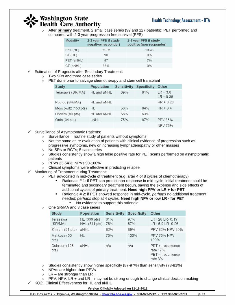
o After primary treatment, 2 small case series (99 and 127 patients): PET performed and compared with 2-3 year progression free survival (PFS)
Estimation of Prognosis after Secondary Treatment:
o Two SRs and three case series o PET done prior to salvage chemotherapy and stem cell transplant
Surveillance of Asymptomatic Patients:
o Surveillance = routine study of patients without symptoms o Not the same as re-evaluation of patients with clinical evidence of progression such as
progressive symptoms, new or increasing lymphadenopathy or other masses o No SRs or RCTs; 5 case series o Studies consistently show a high false positive rate for PET scans performed on asymptomatic
patients o PPVs 23-54%; NPVs 90-100% o Clinical symptoms were effective in predicting relapse
Monitoring of Treatment during Treatment: o PET advocated in mid-cycle of treatment (e.g. after 4 of 8 cycles of chemotherapy)
Rationale # 1: if PET can predict non-response in mid-cycle, initial treatment could be terminated and secondary treatment begun, saving the expense and side effects of additional cycles of primary treatment. Need high PPV or LR + for PET
Rationale # 2: if PET showed response in mid-cycle, perhaps no additional treatment needed; perhaps stop at 4 cycles. Need high NPV or low LR - for PET
No evidence to support this rationale o One SR/MA and 3 case series
o Studies consistently show higher specificity (87-97%) than sensitivity (78-81%) o NPVs are higher than PPVs o LR – are stronger than LR + o PPV, NPV, LR + and LR – may not be strong enough to change clinical decision making
KQ2: Clinical Effectiveness for HL and aNHL
P.O. Box 42712 • Olympia, Washington 98504 • www.hta.hca.wa.gov • 360-923-2742 • TTY 360-923-2701 p. 13
Version Officially Adopted on 11-18-2011
P.O. Box 42712 • Olympia, Washington 98504 • www.hta.hca.wa.gov • 360-923-2742 • TTY 360-923-2701 p. 14
Health Technology Assessment - HTA
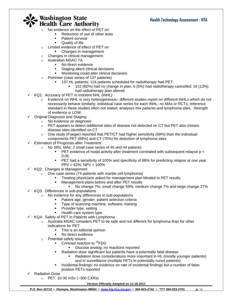
o No evidence on the effect of PET on Reduction of use of other tests Patient survival Quality of life
o Limited evidence of effect of PET on Changes in management
o Changes in clinical management: o Australian MSAC TA
No direct evidence Staging alters clinical decisions Monitoring could alter clinical decisions
o Pommier (case series of 137 patients) 137 HL patients; 124 patients scheduled for radiotherapy had PET:
102 (82%) had no change in plan; 6 (5%) had radiotherapy cancelled; 16 (13%) had radiotherapy plan altered
KQ1: Accuracy of PET in Indolent NHL (iNHL) o Evidence on iNHL is very heterogeneous– different studies report on different iNHLs which do not
necessarily behave similarly; individual case series for each iNHL; no MAs or RCTs; reference standard in these studies often not stated; analyses mix patients and lymphoma sites. Strength of evidence is LOW.
Original Diagnosis and Staging: o No evidence on diagnosis o PET appears to detect additional sites of disease not detected on CT but PET also misses
disease sites identified on CT o One study (Fueger) reported that PET/CT had higher sensitivity (99%) than the individual
components PET (68%) and CT (70%) for detection of lymphoma sites Estimation of Prognosis after Treatment:
o No SRs, MAs; 2 small case series of 45 and 44 patients PET evidence of nodal activity after treatment correlated with subsequent relapse p <
0.05 PET had a sensitivity of 100% and specificity of 88% for predicting relapse at one year.
PPV = 62%; NPV = 100% KQ2: Changes in Management
o One case series (74 patients with mantle cell lymphoma) Treating physicians asked for management plan blinded to PET results Management plans before and after PET results
No change 7%; small change 59%; medium change 7% and large change 27% KQ3: Differences in sub-populations
o No evidence for any differences in sub-populations Patient age, gender, patient selection criteria Type of scanning machine, software, training Provider type, setting Health care system type
KQ4: Safety of PET in Patients with Lymphoma o Australia MSAC considers PET to be safe and not different for lymphoma than for other
indications for PET This is an editorial opinion No direct evidence
o Potential safety issues: Contrast reaction to 18FDG
Glucose analog; no reactions reported Radiation dose significant but patients have a potentially fatal disease
Radiation dose considerations more important in HL (mostly younger patients) and in surveillance (multiple PETs in potentially cured patients)
Incidental findings: no evidence on rate of incidental findings but a number of false positive PETs reported
Radiation Dose o PET: 10-30 mSv (~300 CXRs)
Version Officially Adopted on 11-18-2011
Health Technology Assessment - HTA
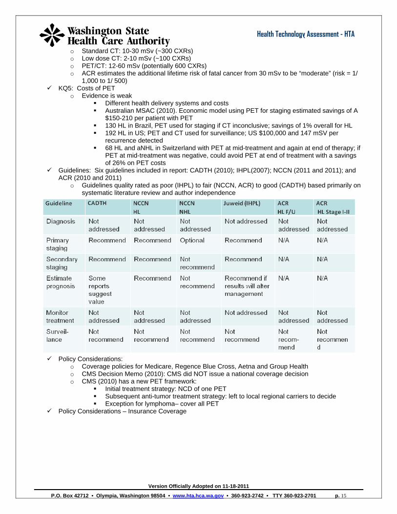
o Standard CT: 10-30 mSv (~300 CXRs) o Low dose CT: 2-10 mSv (~100 CXRs) o PET/CT: 12-60 mSv (potentially 600 CXRs) o ACR estimates the additional lifetime risk of fatal cancer from 30 mSv to be “moderate” (risk = 1/
1,000 to 1/ 500) KQ5: Costs of PET
o Evidence is weak Different health delivery systems and costs Australian MSAC (2010). Economic model using PET for staging estimated savings of A
$150-210 per patient with PET 130 HL in Brazil, PET used for staging if CT inconclusive; savings of 1% overall for HL 192 HL in US; PET and CT used for surveillance; US $100,000 and 147 mSV per
recurrence detected 68 HL and aNHL in Switzerland with PET at mid-treatment and again at end of therapy; if
PET at mid-treatment was negative, could avoid PET at end of treatment with a savings of 26% on PET costs
Guidelines: Six guidelines included in report: CADTH (2010); IHPL(2007); NCCN (2011 and 2011); and ACR (2010 and 2011)
o Guidelines quality rated as poor (IHPL) to fair (NCCN, ACR) to good (CADTH) based primarily on systematic literature review and author independence
Policy Considerations:
o Coverage policies for Medicare, Regence Blue Cross, Aetna and Group Health o CMS Decision Memo (2010): CMS did NOT issue a national coverage decision o CMS (2010) has a new PET framework:
Initial treatment strategy: NCD of one PET Subsequent anti-tumor treatment strategy: left to local regional carriers to decide Exception for lymphoma– cover all PET
P.O. Box 42712 • Olympia, Washington 98504 • www.hta.hca.wa.gov • 360-923-2742 • TTY 360-923-2701 p. 15
Policy Considerations – Insurance Coverage
Version Officially Adopted on 11-18-2011
P.O. Box 42712 • Olympia, Washington 98504 • www.hta.hca.wa.gov • 360-923-2742 • TTY 360-923-2701 p. 16
Health Technology Assessment - HTA
Summary:
o Lymphoma is a heterogeneous group of malignancies with varied treatment dependent on cell histology and stage
o The evidence for this report is based on case series rather than RCTs o Strength of evidence is low to moderate o PET is used for a number of indications in the evaluation of lymphoma
Summary – Strength of Evidence: o For KQ1 – diagnostic accuracy, there is a moderate amount of low to moderate strength evidence o For KQ2 – clinical effectiveness, there is very limited, low strength evidence o For KQ3 – sub-populations and KQ 4 – safety, there is no evidence o For KQ5 – costs, there is very limited low strength evidence